Reduced experimental autoimmune encephalomyelitis after intranasal and oral administration of...
184
Lactobacilli as antigen delivery system for mucosal tolerance induction in autoimmune disease
-
Upload
merckgroup -
Category
Documents
-
view
1 -
download
0
Transcript of Reduced experimental autoimmune encephalomyelitis after intranasal and oral administration of...

Lactobacilli as antigen delivery system for mucosal tolerance induction
in autoimmune disease

f:
ISBN 90-9013173-6 Printing: PrintPartners Ipskamp, Enschede
In de kosten van het drukken van dit proefschrift werd bijgedragen door: Yakult Nederland B.V. Stichting Vrienden MS Research
Omslag: Live lactobacilli

Lactobacilli as antigen delivery system for mucosal tolerance induction
in autoimmune disease
Lactobacillen als systeem voor produktie, transport en afgifte van antigeen voor de inductie van mucosale tolerantie in autoimmuunziekten
PROEFSCHRIFf
ter verkrijging van de graad van doctor aan de Erasmus Universiteit Rotterdam
op gezag van de Rector Magnificus Prof.dr. P.W.C. Akkermans M.A.
en volgens besluit van het College voor Promoties De openbare verdediging zal plaatsvinden op
woensdag 27 oktober 1999 om 11.45 uur
door
Catharina Beatrix Maria Maassen
geboren te Oss
PROMOTIECOMMISSIE
Promotor: Prof.dr. E.H.J.H.M. Claassen
Co-promotoren: Dr. J.D. Laman
Overige leden:
Dr. W.J.A. Boersma
Prof.dr. H.A. Drexhage Prof.dr. EG.A. v.d. Meche Prof.dr. A.D.M.E. Osterhaus
Het onderzoek van dit proefschrift werd verrricht op de afdeling Immunologische- en Infectieziekten van TNO-Preventie en Gezondheid te Leiden, in samenwerking met de afdeling Immunologie van de Erasmus Universiteit Rotterdam. Het onderzoek werd gefinancieerd door de Stichting Vrienden MS Research, project MS93-147.

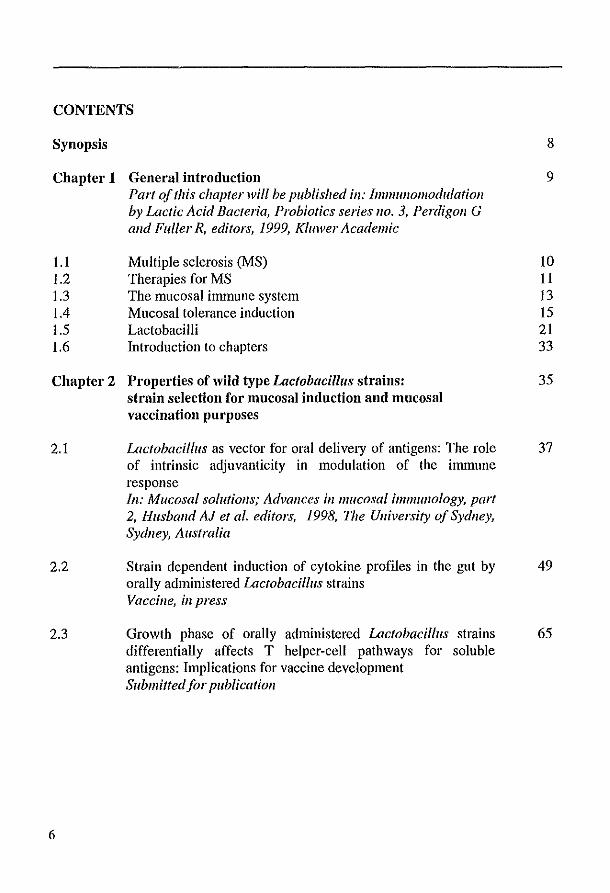
CONTENTS
Synopsis
Chapter 1 General introduction
1.1 1.2 1.3 1.4 1.5 1.6
Part of this chapter will be published ill: [mmullomodulatioll by Lactic Acid Bacteria, Probiotics series 110. 3, Perdigon G alld Fuller R, editors, 1999, Kil/lver Academic
Multiple sclerosis (MS) Therapies for MS The mucosal immune system Mucosal tolerance induction Lactobacilli Introduction to chapters
Chapter 2 Properties of wild type Lactobacillus strains: strain selection for mucosal induction and mucosal vaccination purposes
2.1
2.2
2.3
6
Lactobacillus as vector for oral delivery of antigens: The role of intrinsic adjuvanticity in modulation of the immune response [II: Mucosal solutions; Advances ill mucosal immunology, part 2, Husband AJ et al. editors, 1998, The Ulliversity of Sydney, Sydney, Australia
Strain dependent induction of cytokine profiles in the gut by orally administered Lactobacillus strains Vaccine, ill press
Growth phase of orally administered Lactobacillus strains differentially affects T helper-cell pathways for soluble antigens: Inaplications for vaccine development Submiffedfor publicatioll
8
9
10 II 13 15 21 33
35
37
49
65

Chapter 3 Genetic engineering of Lactobacillus strains
3.1
3.2
Instruments for oral disease-intervention strategies: recombinant Lactobacillus casei expressing tetanus toxin fragment C for vaccination or myelin proteins for oral tolerance induction in multiple sclerosis Vaccil/e 17: 2117-2128, 1999
A rapid and safe plasmid isolation method for efficient engineering of recombinant lactobacilli expressing immunogenic or tolerogenic epitopes for oral administration Jou/'llal of bmlllmological Methods 223: 131-136, 1999
79
81
99
Chapter 4 Mucosal tolerance induction with recombinant lactobacilli 107
4.1
4.2
Therapy with antibodies against CD40L (CD 154) and CD44-variant isoforms reduces experimental autoimmune encephalomyelitis induced by a proteolipid protein peptide Multiple Sclerosis 4: 147-153, 1998
Reduced experimental autoimmune encephalomyelitis after intranasal and oral administration of recombinant lactobacilli expressing myelin antigens Submitted for publicatiol/
Chapter 5 Summarizing discussion Part of this chapter will be published ill: Vaccil/e
References
Samenvatting voor niet-ingewijden
Dankwoord
Abbreviations
Curriculum vitae
Publications
109
123
143
160
178
180
182
183
184
7

SYllopsis
Synopsis Oral administration of autoantigens is a safe and convenient way to induce
peripheral T-cell tolerance in animal models of autoinnnune diseases such as multiple sclerosis (MS). However, oral administration of myelin has not been successful as treatment of MS patients yet. In order to increase the efficacy of oral tolerance induction, we use genetically modified lactobacilli which express myelin antigens. The lactobacilli serve as producers of the antigen and as vehicles to transpOli the antigen to the gut mucosa. The fact that those Gram-positive lactobacilli are generally regarded as safe (GRAS), makes them attractive candidates for this application.
It is known that lactobacilli may possess properties that influence the immune system, but such properties differ between Lactobacillus strains. Therefore Lactobacillus strain selection is very important. In order to select an appropriate Lactobacillus strain for the expression of myelin antigens, different wild type Lactobacillus strains were studied for several effects they have on the immune system after oral administration.
Of eight analyzed Lactobacillus strains, harvested in log phase, only three strains, L. reuteri, L. brevis and L. ferJllelltuJll appeared to be able to enhance the specific antibody response against a parenterally administered T-cell dependent antigen. When the same strains were tested for their ability to indnce cytokines in the gut villi upon oral administration, L. reuteri and L. brevis induced appearance of TNF-a, IL-lB and IL-2 producing cells. In contrast, L. casei and L. lIlurilles tended to increase the number of TGF-Il and IL-IO producing cells. These results suggest that L.casei and L. JIlurilles might be suitable strains for the induction of oral tolerance.
Determination of the specific IgG IIIgG2a ratio against a parenterally administered antigen, revealed a growth phase dependent skewing of T-cell pathways upon oral administration of some of the Lactobacillus strains. Feeding of stationary phase cultures of L. casei and L. JIlurilles induced higher IgG l/IgG2a ratios than after feeding of log phase cultures. Since IgG I production is a reflection of Th2 responses and IgG2a reflects Thl responses, these results imply that stationary phase cultures of those Lactobacillus strains skew more towards Th2, whereas log phase cultures direct the response towards Thl.
A series of vectors has been constmcted which, upon transformation to lactobacilli, direct the expression of antigens either intracellularly or secreted into the environment. Among others, guinea pig myelin basic protein (gpMBP), human MBP, MBPn.85 and PLP139. 151 were expressed by L. casei.
Nasal administration of MBPn.85 prior to disease induction with siginificarltly reduced EAE In cOlltrast,~6wo
! it is possible to reduce by mucosal of recombinant lactolJat,tni" expressing myelin antigens. Further studies are required to develop this approach for treatment of MS in humans.
8

General introduction
1.1 Multiple sclerosis (MS) 1.2 Therapies for MS 1.3 The mucosal immune system 1.4 Mucosal tolerance induction 1.5 Lactobacilli 1.6 Introduction to chapters
Part of this chapter will be published ill: Im11l1l1l011l0dulatioll by Lactic Acid Bacteria, Probiotics series 110. 3, 1999 Perdigoll G and Fuller R, editors, Killwer Academic

General introduction
1.1 Multiple sclerosis Multiple sclerosis (MS) is a chronic inflammatory disease of the central nervous system (eNS) that affects eNS myelin which isolates nerve axons and allows saltatory pulse conduction. It is the most important chronic disabling neurological disease in young adults, with a mean age of onset around 30 years. It has a prevalence of approximately I per 1000 in Western European countries and it affects women versus men at a ratio of 1.5 to I (Sadovnick and Ebers, 1993). Although the cause and pathogenesis of MS are unknown, it is generally believed that MS is an autoimmune disease in which environmental factors as well as genetic factors are involved. Studies with monozygotic twins reveal an MS concordance rate of 20-30%, ·whereas this is 3-5% for dizygotic twins (reviewed by Ebers and Sadovnick, 1994). This high concordance rate is a strong argument in favor of a genetic basis of MS, but can also be used as an argument for an environmental involvement in MS. The role of environmental factors in MS is supported by the presence of geographic gradients in the distribution of the disease. The general pattern is that MS prevalence rises with increasing distance from the equator. The nature of these environmental factors is unknown. Dietary factors, climate variation and infectious agents have all been postulated to be involved in MS, but there is no definitive proof for involvement in MS of any of these factors. Especially herpes viruses have gained interest the last few years (Dalgleish, 1997).
The notion that MS is a multifactorial disease is strengthened by the fact that the clinical course of MS is highly variable for each individual patient, and can be divided into four categories (Lublin and Reingold, 1996). The first category is relapsingremitting disease, in which discrete attacks are alternated with periods in which a patient returns to the pre-attack base line. In the relapsing-progressive form of MS, the patients do not return to base-line level in between attacks and disability increases in these patients. In chronic-progressive disease, the disease progresses without periods of stability. The uncommon fourth form is characterized by a progressive relapsing course of MS.
The most important pathological characteristics of MS are the presence of areas of demyelination (plaques) in the eNS white matter. In general, active demyelination is accompanied by inflammation. The inflammatory infiltrates are mainly composed of T lymphocytes, activated macrophages, microglia and some B cells. Proliferation of astrocytes accompanies the inflammatory changes (gliosis), eventually leading to scar formation. In inactive plaques, remyelination is often present at the lesion margin. The heterogeneity of lesions within and among different patients is large, as is the response to inununomodulatory treatments. These are additional arguments that the disease MS may be more heterogeneous than previously suspected (Lassmann et al., 1998).
10

Chapter 1
1.2 Therapies for MS NOll alltigell-specific therapies Rational development of immunotherapy for MS is hampered by limited insight into the effector cells and effector mechanisms involved in demyelination. Since an effective curative therapy for MS is not yet available, symptomatic treatment of the disease to improve the quality of life is still very important. Acute attacks in MS are usually treated with adrenocorticotropic hormone (ACTH) or the corticosteroids prednisone or methylprednisone, which have both immunosuppressive and anti-inflammatory properties. Based on the assumed inflammatory and immunologically mediated pathogenesis of MS, a large number of possible new therapies directed at immune intervention has been put forward (reviewed by Waubant et al., 1997). IFNB-la and IFNB-lb have been used in the clinic for treatment of relapsing-remitting patients for some time now. Although its mechanism of action ill vivo are unknown, ill vitro IFN-fl antagonizes IFN-y, inhibits T-cell proliferation, it can restore T suppressor cell function and it can reduce T-cell migration by inhibiting the activity ofT cell matrix metalloproteases (MMP)(reviewed by Yong et al., 1998). These properties could be responsible for the positive effects of IFN-fl on relapse rate, disease activity (as measured by serial gadolinium-enhanced magnetic resonance imaging (MRI) and disease progression (as measured by annual TI-weigthed MRI)(reviewed by Amason, 1999).
Treatment with Copolymer-l has also been approved for use in human. Copolymer-l is a mixture of synthetic peptides, composed of four amino acids (Johnson et al., 1995). There is evidence that Copolymer-l blocks or competes with binding of encephalitogenic peptides to MHC class II molecules, this way reducing the autoimmune response (Aharoni et al., 1999). Many more immunomodulatory compounds are used in MS treatment or are analysed in ongoing trials, but most of these therapies are not antigen-specific. Several non antigen-specific approaches are currently under investigation, in order to be used for application in human later, but also to reveal immunological mechanisms involved in autoimmune disease. Examples of such approaches are administration of cytokines or cytokine antagonists, blockade of migration of auto-reactive T-cells into the CNS and blocking lymphocyte co-stimulation by administration of anti-CD40 or anti-CD40 ligand monoclonal antibodies (Laman et al., 1998a; Gerritse et al., 1996). Such therapies may affect the entire immune system, both by suppression or by directing it into non-inflammatory immune responses. This could lead to unwanted side effects and a higher susceptibility for infectious diseases. Practically, this could mean that such treatments can not be given repeatedly or for a longer period.
Alltigell-specific therapies A major goal in the treatment of T-cell mediated autoimmune diseases is the
establishment of a therapy that selectively targets only pathogenic T cells, leaving the remainder of the T-cell repertoire intact. One of the reasons that such antigen-specific
11

General introduction
therapies are not easy to develop is the fact that the autoantigens in many autoimmune diseases and in MS in particular are as yet unknown. Nevertheless, extensive research in experimental autoimmune diseases in which the autoantigen is known, has provided information related to antigen-specific therapies. Some of the approaches for development of antigen-specific therapy are discussed below.
Intrathymic injection of auto-antigen The thymus plays a major role in development of self-tolerance. Intrathymic injection of neonatal BB rats with pancreatic islets prevented insulitis (Koevary and Blomberg, 1992). It was possible to prevent experimental autoimmune encephalomyelitis (EAE), an animal model for MS, by intrathymic injection of myelin basic protein (MBP) or the immunodominant peptide of MBP71 _90, without otherwise compromising the peripheral lymphocyte pool in adult Lewis rats (Khoury et al., 1993). This was also demonstrated by intrathymic injection of S-antigen in autoimmune uveoretinitis (Koevary and Caspi, 1997). However, intrathymic injection of acetylcholine receptor protein could not prevent experimental autoinunune myasthenia gravis, indicating that suppression of Th I function alone may not be sufficient for the prevention of antibody-mediated autoimmune disease (OhtsUlu et al., 1995). The mechanism of intrathymic injection of antigen for tolerance induction is unclear, but a role for CTLA4 has been postulated (Issazadeh et al., 1999). This approach is not likely to be used in human autoimmune diseases, but it may help to elucidate the role of the thymus in acquired systemic tolerance of autoreactive T-cells.
Systemic antigen immunotherapy It is well known that systemic injection of soluble deaggregated protein antigen specifically inhibits the induction of a subsequent T cell response to that antigen. Whether such suppression of the T cell response indeed is established and to what extent, is dependent on several parameters. The route of administration, the dose, the adjuvant and the nature of the antigen appear to be clUcial parameters. Intravenous and intraperitoneal administrations are more effective in tolerance induction than the subcutaneous route. In most experimental set-ups, high doses of antigen are required. The efficacy of tolerance induction is enhanced by the use of adjuvant such as alum or incomplete Freund's adjuvant (IFA), which are known to induce CD4+ T helper 2 (Th2) function. Pep tides or monomeric antigens are more potent tolerogens than aggregated proteins, which tend to be immunogenic. In addition, the immunodominance of an antigen or epitope, defined as the capacity to generate an immune-response, seems to correlate with its potential to induce hyporesponsiveness (Liblau et al., 1997). Intravenous or intraperitoneal injection of myelin protein or peptides can prevent EAE (reviewed by Liblau et al. 1997).
Other approaches to systemically target antigen-specific T cells are the use of peptide analogues (also known as altered peptide ligands (APL)) and soluble peptideMHC-complexes. Peptide analogues are capable of binding to the MHC molecule with
12
Chapter 1
the same affinity as the wild-type peptide but have decreased affinity for the T-cell receptor (TCR) through mutations in primaty TCR contact residues. Administration of peptide-MHC complexes is thought to act via occupation of the TCR of the targeted T cells. The T celIs receive only signal one, without co-stimulation, leading to anergy and/or deletion of the autoreactive T cells.
APL as well as peptide-MHC complexes of myelin antigens can prevent EAE (reviewed by Liblau et al., 1997)). Evidence for the treatment of established disease by systemic administration of antigen was obtained by Brocke et al. (1996), who used an APL of MBP'7.99 in a T cell line specific for MBPS7.99 induced EAE in (PLlJxSJLlJ)/F, mice. A major drawback of the use of APLs and peptide-MHC complexes in humans is that they require knowledge of the peptide epitopes recognized by the pathogenic T cells in individual patients.
Oral and nasal tolerance A promising approach to antigen-specific therapy in autoimmune diseases is the induction of peripheral T cell tolerance by oral or nasal administration of autoantigen, termed mucosal tolerance induction (definition see 1.4). Extensive research has been performed in animal autoimmune models, but also in humans, to induce tolerance by the oral and nasal route and to elucidate the mechanisms involved. Since the mucosal immune system has some unique features compared to the peripheral immune system, which play a role in the induction of mucosal tolerance, first some properties of the mucosal immune system will be discussed.
1.3 The mucosal immune system III/mune exclusion versus tolerance The mucosal surface area of the respiratory tract and the gastrointestinal tract are over a hundred times the surface area of the skin. Such a large area allows efficient uptake of oxygen and absorption of nutrients and excretion of waste products. As a consequence, a large area of the body is continuously exposed to many pathogens of the external milieu. To cope with both pathogens and innocuous substances, the mucosal immune system has generated two major arms of immune reactivity (reviewed by Brandtzaeg, 1995; 1998; Brandtzaeg et al., 1999). 1) Immune exclusion and elimination. Immune exclusion is a noninflammatory way of preventing colonization and penetration of harmful foreign material performed by the physical presence of indigenous bacteria leaving no niche for ingested pathogens, as well as by secretOlY IgA (and to some extent sIgM and IgG), which has a higher general crossreactivity with antigens than has IgG (Waldman et al., 1970, Shvartsman et al., 1977). Itlllnune elimination involves neutralization and removal of foreign material that has penetrated the epithelium by mainly proinflammatory, non-specific innate defense mechanisms like complement-activating IgG and IgM, and cytokines released from activated T cells and macrophages. 2) Oral tolerance. Since the mucosal epithelium has evolved for efficient transport of molecules, a continuous inflammatory response
13

General introduction
evoked against indigenous microflora, ingested food antigens or inhaled antigens is undesirable. Therefore, under physiological circumstances, penetrating soluble antigens and the indigenous microflora mainly induce hyporesponsiveness. Since the mucosae are natural sites of tolerance induction, they can be used to reinduce tolerance against antigens to which tolerance has been lost and has resulted in autoimmune disease.
RespiratO/y tract versus gastrointestinal tract The mucosae comprise a specific epithelial layer and underlying connective tissue that form the border between the internal and external milieu in the airway system, the gastrointestinal tract and other mucosal surfaces. It is a unique property of the intestinal immune system and some parts of the respiratory immune system that it is separated from the antigens by only one cell layer, and is not drained by lymphatic vessels (Neutra et al., 1996). When comparing the immune system of the upper respiratory tract and the gastro-intestinal tract, some differences are obvious. One difference is the presence of large numbers of intraepitheliallymphocytes (IEL) in the gut villi, which are mainly of the CD8+ phenotype, in contrast to the low numbers of IEL in the airways, which are mostly CD4+ T cells (Abreu-Martin and Targan, 1996). Another difference is the distribution of subepithelial and intraepithelial professional antigen presenting cells (APC). In the gut, most APC are located under the epithelial cell layer, whereas many APCs of the upper respiratory tract are located between the epithelial cells (Holt et al., 1990). How these differences may relate to mucosal tolerance induction is unclear. However, there is mounting evidence suggesting that the epithelium of the gastrointestinal tract, enterocytes, may act as antigen presenting cells (APC), preferentially stimulating CD8+ cells, probably IELs (Bland and Whiting, 1989; Yamamoto et al., 1998). It has been postulated that (human) enterocytes present antigen to CD8+ cells via CDld (Blumberg et al., 1991). However, for proper activation co-stimulatory molecules arc a prerequisite, but CD80 and CD86 costimulatory molecules have not been detected yet on enterocytes (Bloom et al., 1995). This lack of costimulation may promote the induction of tolerance to luminal antigens In contrast, due to scarcity of such CD8+ IEL and the limited amount of antigen that enters via the airways, the induction of peripheral tolerance probably will be mainly regulated by CD4+ Th2 cells stimulated in draining lymph nodes by migrated dendritic cells (Brandtzaeg, 1996).
1.4 Mucosal tolerance induction Definition The definition of mucosal tolerance induction in this thesis is: Any mechanism by which a potentially injurious immune response is prevented, suppressed or shifted to a noninjurious class of immune response by mucosal (e.g. oral and nasal) administration of antigen (Weiner HL, 10th Int. Congress Mucosal Immuno!., Amsterdam). In this thesis the injurious immune response is a Th I mediated inflammation of the central
14

Chapter 1
nervous system in EAE. Although the term oral administration strictly taken refers to administration of the antigen in the oral cavity, we use the term for intragastric intubation of the antigen in rodents, as is generally applied throughout the literature. On application of aerosolized antigen taken up via the nose, a small amount is thought to enter the gastro-intestinal tract, in contrast to administration of antigen in the nose by use of a pipet.
Mucosal tolerance induction in animal models Oral tolerance was first described in 1911 when Wells fed hen egg proteins to guinea pigs and found them resistant to anaphylaxis when challenged with the same antigens (Wells, 1911). It was shown that this is an antigen specific phenomenon by numerous investigators who demonstrated that animals fed proteins such as ovalbumin (OVA) do not respond as well to these antigens when subsequently immunized, but do respond normally to other antigens (reviewed by Strobel and Mowat, 1998). An example of peripheral T cell tolerance induction without concomitant induction of B cell tolerance was observed after oral administration keyhole limpet hemocyanin (KLH) in humans (Husby et 01., 1994). Although the T cell responses were downregulated, the oral administration of KLH primed the humoral response systemically as well as mucosally.
The T-cell hyporesponsiveness induced by autoantigen administered mucosally, could be beneficial in the treatment of autoimmune diseases. During the last decade, oral and nasal tolerance has been used successfully to treat autoimmune phenomena in animals (Table 1). Mucosal tolerance has been extensively studied, especially in Thl mediated experimental autoimmune disease such as EAE. In addition to T cell mediated autoimmune diseases, the antibody-mediated experimental autoimmune diseases myasthenia gravis and thyroiditis could be suppressed by previous nasal and/or oral administration of acetylcholine receptor (AChR) or thyroglobulin, respectively (Table 1). A wide range of experimental diseases (Table 1) could be prevented by oral administration of the autoantigen. A complicating factor is that treatment of humans occurs when the disease is already fully established. Therefore, treatment of ongoing disease in chronic autoimmune models is clinically more relevant. Miller et 01. (1993b) showed that orally MBP tolerized mice were not protected against adoptive transfer of EAE, indicating that suppression of active autoreactive T cells is much more difficult than prevention of induction. However, treatment of ongoing disease by oral administration of (auto)antigen has been demonstrated in several chronic autoimmune models such as EAE (Table 2)(al-Sabbagh et 01., 1996; Benson et 01., 1999; Brad et 01., 1991; Meyer et 01., 1996), collagen induced arthritis (CIA)(Khare et 01., 1995) and experimental autoimmune uveoretinitis (EAU)(Thurau et 01., 1997; Torseth and Gregerson, 1998).
Experimental autoimmune diseases have also been treated by nasal administration of (auto)antigen (Table 1). Nasal administration of high doses of AChR could ameliorate the manifestations of experimental autoimmune myasthenia gravis
15
General introductioll
(EAMG)(Shi et al., 1999). Aerosol MBP decreased the severity of subsequent relapses in animals with EAE recovered from the first attack (ai-Sabbagh et al., 1996). Nasal administration of residues 139-151 from proteolipid protein (PLP139.15I ) after onset of myelin induced EAE, reduced the number of relapses (Anderton and Wraith, 1998). Also nasal co-administration of MBP68.86 and MBP87.99 could reverse the disease in Lewis rats (Liu et al., 1998). Aerosol insulin could treat IDDM in NOD mice (Harrison et al., 1996).
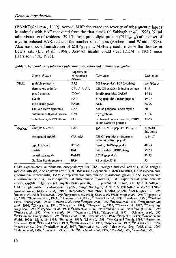
Table 1. Oral alld "asal tolerallce illduction ill experimelltal autoimmune models
Experimental Human disease autoimmune Toicrogen References
disease
ORAL mUltiple sclerosis EAE MBP (peptides), PLP (peptides) see Table 2
rheumatoid arthritis CIA,AIA, AA ClI, crr peptides, inducing-antigen 1·13
type I diabetes IDDM insulin (peptide), GAD65 14·18
uveitis EAU S·Ag (pep<ides), IRBP (peptide) 19·27
myasthenia gravis EAMG AChR 28,29
Guillain-Barre syndrome EAN bovine peripheral nerve myelin 30
autoimmune thyroid disease EAT thyroglobulin 31,32
inflammatory bowel disease EGC haptenized colonic proteins, WBS, 33·35 colitis extracted proteins
NASAL multiple sclerosis EAE gpMBP, MBP-peptides, PLPI39.]51 1,36-40, this thesis
rheumatoid arthritis CIA, AlA Cll, CII peptides or fragments, 1,41·47 inducing-antigen peptide
type I diabetes IDDM insulin, GAD65-peptides 48,49
uveitis BAU retinal extract, IRBP. S-Ag 50,51
myasthenia gravis EAMG AChR (peptides) 52·55
Guillain-Barre syndrome EUN P2 peptide 57·81 56
EAE: experimental autoimmune encephalomyelitis, CIA: collagen induced arthritis, AlA: antigeninduced arthritis, AA: adjuvant arthritis, IDDM: insulin-dependent diabetes mellitus, EAU: experimental autoimmune uveoritinitis, EAMG: experimental autoimmune myasthenia gravis, BAN: experimental autoimmune neuritis, EAT: experimental autoimmune thyroiditis, EGC: experimental granulomatous coli lis, (gp)MBP: (guinea pig) myelin basic prolein, PLP: proleolipid prolein, Cll: Iype II coilagen, GAD65: glutamate decarboxylase peptide, S·Ag: S·anligen, AChR: acely1choline receptor, TNBS: trinitrobenzene sulfonic acid, IRBP: interphotoreceptor retinol binding protein. lui-Sabbagh et al., 1996; 2Haque el al., 1996; lInada et al., 1997; 4Jorgensen et al., 1998; 5Khare et af., 1995; tiMyers el (11., 1998; 7Thompson el af., 1988; tyhompson et al., J993a; 9Thompson et al.,1993b; l6yhorbecke el al., 1999; IIYoshino, 1996b; 12Yoshino, 1996a; 13Zhang el al., 1990a; t4Bergerot et (II., 1994; 15Polanski el al., 1997; 16Ramiyu et al., 1997; Hvon Herrath MG et al., 1996; I~ang et al., 1991; 19Rizzo el al., 1994; 26yhurau el al .• 1991; 21Thumu el af., 1997; 2~orseth and Gregerson, 1998; 23Gregerson et al., 1993; 24Nussenbfatt e/ al., 1990; 25Silver e/ al., 1998; UiSingh et al., 1996; 27Yrabec et al .• 1992; 2S0kumum et al., 1994; 2"\Vang el (/1., 1993. 3{1Gaupp et af .• 1997, 31Guimaraes et al., 1995; 31Peterson and Braley-Mullen, 1995 ,3lEIson et al., 1996; 3-lNeurath et al., 1996; 3sTrop el al., 1999; 3tiAnderton and Wraith, 1998; l7Liu el al., 1998; l8Bai et lIl., 1997; 39Li et at., 1998; ~etzler and Wraith. 1993; 41Bayrak and Mitchison, 1998; 42Chu and Londei, 1999; 43Matsumoto et al., 1998; MMyers et al., 1997; 4sPrakken et al., 1997; 4tiStaines e/ al., 1996; 47Yerheijden et al., 1997; 48Harrison el af., 1996; 49Tian et al., 1996; SOoick et al., 1994; 5lL'l.liotou et al., 1997; 52Shi e/ al., 1998a; 53 1999; stKarachunski et al., 1997; 5sMa e/ ar, 1995; 5tizhu e/ al., 1998.
16
Chapter 1
Conversely, oral or nasal administration of antigen can lead to more severe clinical signs. Meyer et al. (1996) have shown that oral administration of a low dose of MBP enhanced the clinical signs of chronic EAE in B 1O.PL mice. As demonstrated in this thesis, nasal administration of two doses of gpMBP in Lewis rats enhanced the clinical signs of EAE in Lewis rats (Chapter 4.2). Nasal administration of MBP led to exacerbation of ongoing EAE in DA rats (Bai et al., 1998). Exacerbation was also found when rats with mild EAU were treated with S-antigen peptides, although mild disease could be treated with high doses of antigen. Oral administration of autoantigen can also lead to induction of disease as oral administration of OVA induced cytotoxic T cells in B6 mice and increased incidence of diabetes in chimeric RIP-OVA mice with high levels of CD8+ T cells specific for OVA infiltrates in Il-islets (Blanas et al., 1996). Terato et al. (1996) showed that oral administration of chicken collagen type II induced arthritis in DBN 1 mice.
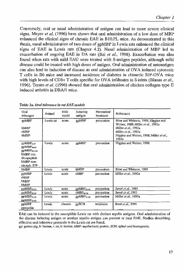
Table 2a. Oral tolerance ill rat EAE models
Oral Animal EAE Inducing Prevention! References
tolcrogen model antigen treatment
gpMBP Lewis rat acute gpMBP prevention Bitar and Whitacre, 1988; Higgins and Weiner, 1988; Miller el al., 1992a
hMBP Miller el af., 1992a rMBP Miller et af., 1992a bMBP Higgins and Weiner, 1988; Miller et al.,
1992a gpMBPl_37 Lewis acute gpMBP prevention Higgins and Weiner, 1988 gpMBP-U89 gpMBP9(}.17o b:MBP ene. decapeptide bMBPnoll-enceph. S79 hMBP Lewis acute hMBP prevention Bitar and Whitacre, 1988 gpMBP Lewis acute eMBP prevention Miller et ai., 1992a eMBP hMBP bMBP gpM BP6S.SS Lewis acute gpMBP6S-88 prevention Javed et al .• 1995 gpMBP6S.sg Lewis acute rMBP6S.~g prevention Javed et aI .. 1995 gpMBP21-40 Lewis acute gpMBPJ1 .90 prevention Miller et al .. 1993a gpMBP7I.90
gpMBP Lewis chronic gpSCH treatment Brod e/ at., 1991 gpmyclin
EAE can be induced in the susceptible Lewis rat with distinct myelin antigens. Oral administration of the disease inducing antigen or another myelin antigen can prevent or treat EAE. Studies describing effective oral tolerance protocols in the Lewis rat are listed. gp: guinea pig. h: human. r: mt. b: bovine. MBP: myelin basic protein, SCH: spinal cord homogenate.
17
General introduction
Apparently, there is a delicate balance between tolerance and immunity. The dosage, timing, frequency and route of application are important factors in the success of mucosal tolerance strategies for treatment of chronic autoimmune diseases. Many different EAE models have been used to demonstrate oral and nasal tolerance with many different antigens and protocols (Table 1 and 2) and these studies have been the basis for elucidation of mechanisms of mucosal tolerance. However, oral administration of (potential) autoantigens in humans has not been successful yet, as discussed below.
Table 2b. Oral tolerallce ill guinea pig and mOllse EAE models
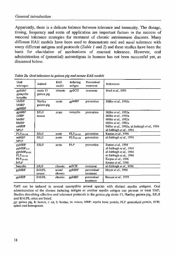
Oral Animal EAE Inducing Prevention! References tolerogen model antigen treatment
gpMBP strain 13 chronic gpSCH treatment Brode/al., 1991 gpmyelin guinea pig hmyelin hMBP Hartley acute gpMBP prevention Miller et al., 1992a bMBP guinea pig gpMBP gpMBP SJUJ acute mmyelin prevention Miller el al., 1992a ,MBP mouse Miller el al., 1992a hMBP Miller e/ al., 1992a bMBP Miller e/ al., 1992a mMBP Miller el al., 1992a; ai-Sabbagh el al., 1994 bPLP al-Sabbagh el al .• 1994 PLP139.151 SJUJ acute PLP139.151 prevention Kamus e/ ai., 1996 mMBP SJUJ acute PLP14(}'159 prevention ai-Sabbagh et al., 1994 bPLP gpMBP SJUJ acute PLP prevention Santos et a/., 1994 gpMBP'.1I ai-Sabbagh et af., 1994 gpMBPS9.101 aI-Sabbagh etat., 1994 PLP1.w.l~9 ai-Sabbagh et ai" 1994 PLPI39.151 Karpus et al., 1996 bPLP Karpus et at., 1996 bmyelin SJUJ chronic nlSCH treatment al-Sabbagh el al .• 1996 gpMBP BIO.PL acute! gpMBP prevention! Meyer et al., 1996
mouse chronic treatment gpMBP BIO.PL chronic gpMBP preventionJ Benson el al., 1999
treatment
EAE can be induced in several susceptible animal species with distinct myelin antigens. Oral administration of the disease inducing antigen or another myelin antigen can prevent or treat EAE, Studies describing effective oral tolerance protocols in the guinea pig strain 13, Hartley guinea pig, SJUJ and B 1O.PL mice are listed. gp: guinea pig, h: human, r: rat, b: bovine, m: mouse, MBP: myelin basic protein, PLP: proteolipid protein, SCH: spinal cord homogenate.
18

Chapter 1
Oral tolerance induction in hUlIlan trials From animal experiments it is known that dose, timing, frequency and route of administration are important factors influencing the efficacy of mucosal tolerance induction. Additional complications in the treatment of humans with chronic autoimmune disease by mucosal administration of (auto)antigen are the established disease and the fact that in most cases the autoantigen is unknown. Despite these difficulties, several clinical trials have been initiated or are currently planned (Table 3). These initial trials suggest that there is no systemic toxicity or exacerbation of disease associated with the oral administration of antigen. Although some positive effects have been observed with low doses of collagen in human arthritis (Barnett et al., 1998), consistent clinical efficacy has not yet been demonstrated. Results in humans, however, have mirrored several aspects of what has been observed in animal models. For instance, MS patients treated with oral bovine myelin contain MBP and PLP specific TGF-B secreting in peripheral blood, in contrast to untreated patients (Fukaura et al., 1996). The possible involvement of TGF-B in oral tolerance will be discussed in the next paragraph.
Table 3. Oral lolerance inductioll ill humall au(o;,mmme disease
Disease trial Multiple sclerosis
Rheumatoid arthritis (also juvenile rheumatoid arthritis)
Uveitis
Thyroid disease
Myasthenia gravis (planned)
Type I diabetes (planned in new-onset diabetes)
Oral antigen bovine myelin
type II collagen
S-Ag, retinal antigens
thyroglobulin
acetylcholine receptor
insulin
References Fukaura eta/., 1996; Hohol et ai., 1996; Weiner et al., 1993
Barnett et al., 1996; Barnett e/ al., 1998; Sieper et al., 1996; Trentham et al., 1993
Nussenblatt ela!., 1997
Lee et al., 1998
Drachman e/ af., 1996
Carel and Bougneres, 1996
Proteins were taken orally as treatment in several human autoimmune diseases in order to induce oral tolerance leading to reduced disease activity. Those human trials arc listed, as arc two planned trials. S-Ag: S-antigen, AChR: acetylcholine receptor
Mechanisllls of 11111 co sal tolerance Three main mechanisms that have been identified by which mucosal tolerance is mediated include active suppression, anergy and clonal deletion. These mechanisms appear to be determined primarily by the dose of administered antigen. Low doses of antigen favor the generation of regulatory cells that suppress the specific immune response in the target organ, whereas high doses of antigen induce an antigen-specific anergic state or deletion of peripheral T cells. Active suppression is the mechanism of peripheral T cell tolerance induction which is mediated by regulatory cells that suppress the specific immune response in the target organ. The tolerogenic state can be adoptively transferred by the regulatory T cells from mucosally tolerized animals to na'ive recipients. Several phenotypes of such regulatory T cells have been described.
19

General introduction
Initially, regulatory T cells of the CD8+ subset have been described to be involved in oral tolerance (Nussenblatt et al., 1990; Lider et al., 1989). Miller et al. (1992b) have demonstrated that the suppressive activity of such CD8+ cells may be due to induction of TGF-3. TGF-3 is generally a negative regulator of immune responses. Injection of TGF-3 has been shown to suppress EAE and other experimental autoimmune diseases (Kuruvilla et al., 1991; Racke el al., 1991). The appearance of TGF-3 in inflammatory infiltrates in the brain of animals recovering from EAE, indicates a down-regulatOlY role for TGF-3 in inflammatory processes (Khoury et al., 1992). Another group of regulatOlY T cells, are CD4+ Th2 cells (e.g. Tian el al., 1996; Chen el al., 1995), producing non-inflammatory cytokines such as JL-4, JL-IO and TGF-3. When in a Th I mediated autoimmune disease, the inflammatory Th I response shifts to a specific, non-inflammatory Th2 response, the mechanism of active suppression is further specified as immune deviation. However, LaFaille el al. (1997) have demonstrated that Th2 cells do have the potential to induce EAE in immunodeficient mice, implying that in the absence of a normal immune system Th2 cells can be equally pathogenic. The existence of a third T cell subset (Th3), potentially involved in induction/maintenance of mucosal tolerance, was proposed by Chen et al. (1994). These Th3 cells express CD4+ and produce TGF-3 mainly, sometimes with low co-expression of JL-4 or IFN-y.
T cell anergy is defined as a cellular state in which a T cell is alive but fails to display certain functional responses when optimally stimulated through both its antigen-receptor and other receptors that are normally required for T cell activation (Schwartz, 1996). Anergy is induced when no or no proper co-stimulation is present (Lafferty et al., 1983). In vilro, stimulation of anergic cells with antigen does not lead to proliferation and JL-2 production. Peripheral T cell tolerance can not be adoptively transferred to naIve animals with anergic antigen specific T cells.
Clonal deletion, peripheral deletion of antigen specific T-cells, has not yet been described in normal animals after mucosal administration of antigen. Deletion of specific T cells has only been shown after oral administration of very high doses of antigen in T cell receptor (TCR) transgenic mice (Chen et al., 1995a; Whitacre el al., 1996b). Circumstantial evidence for a role of clonal deletion as mechanism of mucosal tolerance has been provided by the observation that orally OVA tolerized lymphocytes die by apoptosis when cultured in vitro after an in vivo challenge with OVA (Garside et al., 1996).
All described mechanisms target T cells specific for the mucosally administered antigen. However, the regulatOlY effector T cells can also suppress T cell responses evoked by other antigens. By secretion of suppressive cytokines such as TGF-3 and JL-IO into the microenvironment, an ongoing response against a co-localized antigen may be down-regulated (Anderton and Wraith, 1998; Bayrak and Mitchison, 1998; reviewed by Weiner, 1997). This mechanism of active suppression is further specified as bystander suppression. Successful induction of this bystander suppression is of major importance for the therapeutic use of mucosal tolerance in the treatment of
20

Chapter 1
autoimmune disease, as it may obviate the need to identify and use specific autoantigens, and may circumvent complications of determinant/epitope spreading (reviewed by Vanderlugt et al., 1998).
Modulation 0/ mucosal tolerance When mucosal tolerance is mediated via active suppression, inducing Th2 or Th3 regulatory cells, any stimulus that favors Th I versus Th2ITh3 responses could potentially abrogate mucosal tolerance (reviewed by Weiner, 1997). For example, high doses of IFN-y abrogated oral tolerance (Zhang et al., 1990b). Conversely, administration of factors that favor Th2ITh3 over Thl may augment mucosal tolerance. Indeed it has been shown that factors such as oral IL-4 can enhance oral tolerance (Inobe et al., 1998). Surprisingly, also administration of IL-2 potentiated oral tolerance in the uveoretinitis model possibly by increasing TGF-Jl, IL-4 and IL-IO production in Peyer's patches (Rizzo et al., 1994). Peripheral T cell tolerance was also increased by oral administration of cholera toxin subunit B (CTB) and LPS, often used to enhance the immunogenicity of (mucosally) administered antigens for vaccination purposes (Arakawa et al., 1998; Bergerot et al., 1997; Gaupp et al., 1997; Khoury et al., 1990; Sun et al., 1996). Similar effects may be obtained with bacteria that can influence the immune response in a non-inflammatory fashion, an effect that could be obtained by oral administration of a specified Lactobacillus strain.
1.5 Lactobacilli Microorganisms as antigen delivel)' system/or vaccination purposes Oral vaccination against certain viruses or bacteria appears to be feasible when administered attenuated (Kaul and Ogra, 1998). However, there always will be a (small) risk of infection. Oral vaccination solely with pathogenic protein antigens is difficult to achieve, since oral administration of soluble antigen normally leads to hyporesponsiveness. Therefore a wide range of antigen delivery systems is being developed, which enable antigen delivery at the proper sites of antigen presentation and in addition may fulfill an adjuvant function. Transformed microorganisms expressing heterologous proteins of the target pathogen are used as such antigen delivery systems to evoke a protective immune response against the pathogen. The strains of microorganisms employed are usually genetically attenuated mutants of pathogenic bacteria (the carrier) transformed with a vector designed to obtain an adequate expression of antigens from another pathogen. Such carriers can then be used as local antigen producers at the gut mucosa, with the additional advantage of circumventing expensive and difficult antigen purification processes. For instance, Salmonella strains have been used as carrier for hepatitis B virus antigens (Fairweather et al., 1990). Other bacteria which are potentially useful as carriers include strains of E. coli, Mycobacteria, Vibrio cholera and Shigella (Butterton et al., 1997; Killeen et al., 1999; Noriega et al., 1996; Hale, 1990). The effectiveness of pathogenic bacteria as carrier probably is related to the pathogenicity of the bacterial
21

General introduction
strains. Most pathogenic bacteria have specific mechanisms to invade the human or animal body, normally evoking a strong immune response. This strong immune response against the carrier probably stimulates the immune response against the heterologous expressed antigen. However, due to the pathogenic character of most transformed microorganisms used for oral vaccination, their use as antigen carrier is limited. For this reason the use of a GRAS (generally regarded as safe) organism, such as Lactobacillus, as a safe antigen carrier for the delivery of foreign antigens in the gastrointestinal tract is to be preferred.
Microorganisms as antigen delivel}' system/or tolerance induction Using microorganisms as antigen delivery system is an approved method of vaccination in experimental animal models. We reasoned that the same technique could be used to establish the induction of oral tolerance for the treatment of autoimmune diseases. An ideal therapy consists of a continuous production and presence of the autoantigen at the site of tolerance induction, i.e. the gut associated lymphoid tissue (GALT). Oral administration and possibly subsequent colonization of microorganisms expressing autoantigens might realize the continuous presence of autoantigen at the gastro-intestinal tract. This approach provides several advantages over feeding purified autoantigens. Degradation of the autoantigens in the stomach and the rest of the gastrointestinal tract will be decreased and no bulk purification of human proteins is necessary, which makes this method also much cheaper. In addition, the use of recombinant lactobacilli increases safety of the method, because there is no risk of co-administering viruses or prions. When the transformed lactobacilli are able to colonize the gut, the antigens are released continuously in the gut, restricting the therapy to a single or a few administrations. Specified Lactobacillus strains could be suitable for this approach due to their GRAS-status and low intrinsic immunogenicity (Gerritse et al., 1990).
Lactobacillus Lactobacilli are Gram-positive rod-shaped lactic acid bacteria. Although there are some exceptions, most of the Lactobacillus strains are not pathogenic and lactobacilli are major constituents of the human and animal gut. Because of their harmless character they have the GRAS-status, and are frequently used in bio-processing and preservation of food and feed. Nowadays, lactobacilli are mainly known for their health stimulating properties. The importance of lactobacilli for human health was first recognized by Metchnikoff at the beginning of the century (Metchnikoff, 1908). He suggested that harmful effects of undesired bacteria could be overcome by establishing a new balance between intestinal bacteria, through ingestion of lactobacilli or fermented products made by these organisms. Especially during the last few decades, extensive research on the positive effects of Lactobacillus strains has been carried out, suggesting additional health and/or nutritional benefits for humans and animals by oral administration of lactobacilli. Such intrinsic properties include: anti-carcinogenic
22
Chapter 1
activity, control of intestinal infections, improvement of lactose metabolism, improvement of gnt barrier function, control of serum cholesterol levels and positive effects on allergy as well as on (experimental) autoimmune diseases (e.g. Matsuzaki et al., 1990; Mnrosaki et al., 1998; Perdigon et al., 1995; Salminen et al., 1996b; Shornikova et al., 1997). It should be noted that not all strains from the very large and diverse (e.g. in fermentation pathways) genus Lactobacillus exhibit the same health-stimulating properties and may vary in strength of the effect obtained (Klein et al., 1998). Some of the positive effects of lactobacilli are mediated by non-immune components such as modulation of vitamin production, enzymes, and antibiotics, but especially modulation of the immune system seems to play an important role (reviewed by Holzapfel et al., 1998; Femandes et al., 1987; Rao and Shah ani, 1987; Shahani et al., 1977).
bl1llll1ll0lllodlllatioll by lactobacilli One of the possible mechanisms for lactobacilli to influence allergy, autoimmunity, infection and carcinogenesis is by affecting cytokine expression in a specific or non-specific manner. Modification of the local cytokine profile in the gut probably is most effective. Therefore oral administration of lactobacilli could enhance protection and improve treatment of intestinal infections, food allergy and colon cancer by inducing or reducing levels of distinct cytokines. But apparently, oral lactobacilli can also influence non-mucosal disease sites, like autoimmune diseases affecting the joints or cancer of the pancreas and bladder. Figure 1 shows which cytokines positively influence certain disorders when their expression is altered by oral administration of lactobacilli.
Effect of lactobacilli on cytokine expression in infection It has been extensively reported that lactobacilli have probiotic effects (i.e. beneficial effects by improving the properties of the indigenous flora) which enhance protection against infections (Perdigon et al., 1995, partially reviewed by Salminen et al., 1996). These effects could be due to activation of innate immune defence effector functions (e.g. macrophages) andlor to support of the specific response against infectious agents by upregulation of IgA production. These two pathways are discussed below.
Many studies have shown that different strains of lactobacilli are able to activate macrophages ill vitro and ill vivo (Lehman et al., 1988; Nanno et al., 1989; Perdigon et al.,1986; Pool-Zobel et al.,1996; Tomita et al.,1993). Lactobacilli induce cytokines produced by macrophages, TNF-a, IL-l, IL-6, IL-12 andlor IFN-alB, in macrophage cell lines, human blood monocytes, spleen macrophages, Peyer's patch (PP) adherent cells or peritoneal macrophages (Table 4)(Kitazawa et al., 1992; 1994; Klebanoff et al., 1999; Marin et al., 1998; Murosaki et al., 1998; Popova et al., 1993; Shida et al., 1998; Tomita et al., 1993). Since IFN-y is the major macrophage-activating cytokine, induction of this cytokine by lactobacilli provides indirect evidence of macrophage-activating properties of lactobacilli (Laffineur et al., 1996; Murosaki et al., 1998; Shida et al., 1998; Solis-Pereyra et al., 1997). Intraperitoneal administration
23

Gelleral illtrodllctioll
ORAL TOLERANCE
ORAL LACTOBACILLI gut lumen
REDUCTION JgE
STIMULATION JgA
ACTIVATION MACROPHAGES
D ~() D D Prevention/treatment Protection/treatment Tumour Prevention/treatment autoimmune disease infection inhibition food allergy
Figure 1. Modulation of c)'tokines by oral administration of lactobacilli call affect disease. Oral administration of lactobacilli can modulate cytokine expression in the gastrointestinal tract and possibly elsewhere in the body. By inducing (lined arrows) and/or reducing (broken arrows) expression of one or more cytokines positive effects on infection and disease may be achieved, Enhancement of oral tolerance towards orally administered soluble (auto)antigens, even in the periphery, could limit autoimmune disease. Stimulation of IgA production and macrophage activation might improve protection against pathogens that enter via the gastrointestinal tract. The cytotoxic action of macrophages can inhibit tumour-growth. Reduction of IgE levels has positive effects on food allergy.
of L. casei cell wall, leading to macrophage activation, is now being used as a model for Kawasaki disease (cardiac arteritis)(Okitsu-Negishi et al., 1996; Tomita et al., 1993; Lehman et al., 1988). When lactobacilli are added to ill vitro cultures of mixed cell populations like splenocytes or human PBMC, upregulation or 110 effect on expression of IL-12, IL-l, IL-6, IFN-alB, IFNy and TNF-a was found (Laffineur et al., 1996; Matsushima et al., 1998; Miettinen et al., 1996,1998; Murosaki et al., 1998; Shida et al., 1998; Solis-Pereyra et al., 1997). This could reflect activation of the monocyte lineage, but may also be (partially) due to modulation of lymphocytes. The peptidoglycan layer of lactobacilli is able to activate macrophages to produce IL-l,
24

Chapter 1
IL-6 and TNF-a by binding to the CD14 receptor (e.g. Schrijver et al., 1999). However, there is some discrepancy between studies as to whether it is the peptidoglycan layer which is responsible for macrophage activation (de Ambrosini et al., 1996). III vivo administration of lactobacilli can also result in activation of macrophages. In different studies one or more of the cytokines IL-12, IL-I, TNF-a and IL-6 were upregulated by peritoneal macrophages after oral, intramuscular or intraperitoneal administration of the lactobacilli (Table 5)(Murosaki et al., 1998; Okitsu-Negishi et al., 1996; Popova et al., 1993; Saito et al., 1987). Although almost all Lactobacillus strains could activate macrophages, the extent of cytokine induction differed between strains. TNF-a was also induced in the gut after oral administration of different Lactobacillus strains (Maassen et al., 1999b). Significantly increased numbers of TNF-a producing cells in the gut villi and in the submucosa were demonstrated by immunohistochemistry after feeding BALB/c mice with L. reuteri (Chapter 2.2).
Lactobacilli can affect infection by macrophage activation (immune elimination), but also by stimulating IgA production (immune exclusion). It has been shown previously that lactobacilli are able to upregulate IgA production, locally as well as systemically (Perdigon et al., 1991; Link-Amster et al., 1994; Kaila et al., 1995; Majamaa et al., 1995). This is probably linked to upregulation of one or more cytokines. For instance, TGF-Il is necessary for the isotype switch to IgA (reviewed by Brandtzaeg, 1995). Terminal differentiation of B cells into plasma cells in the secretory tissues involves cytokines such as IL-5, IL-6 and IL-IO and possibly IFN-y. These cytokines can all be produced by mucosal T-cells (Nilsen et al., 1995). Little is known about the cytokine profile or other microenvironmental requirements for the enhancement of J chain production by mucosal B cells, a necessary component of secretory IgA and IgM. Probably IL-2, IL-5 and maybe IL-6 are involved in its upregulation, whereas IL-4 may have an opposing effect (Brandtzaeg, 1994). In a T-cell line the expression of IL-2 and IL-5 was enhanced by L. bulgaricus (Marin et aI., 1998), possibly correlating with the upregulation of J-chain expression by plasma cells (Table 4). In other cultures no effect or an increased expression of IFN-y, IL-2, JL-4, IL-6 and IL-IO was measured (Table 4). It is noteworthy that JL-4 levels remained either unaffected or were down-regulated. This is consistent with the results found ill vivo when, after intraperitoneal or oral application of lactobacilli, JL-4 levels were down-regulated (Table 5)(Kato et al., 1998; Maassen et al., 1999b; Matsuzaki et al., 1998; Murosaki et al., 1998b). In conclusion, lactobacilli may reduce infection by modulation cytokine expression.
In tumour growth Lactobacilli show anti-tumour effects not only in rodents, but also in humans. In particular, intralesional injection of lactobacilli effectively inhibits tumour growth (lung carcinoma)(Masuno et al., 1991), but also oral administration of lactobacilli can prevent tumour growth (colon/bladder cancer)(Aso et aI., 1995). JL-l and TNF-a
25

General introduction
Table 4a LactobaciJIus strains affect cylokille expression in vitro
(Possible) effect of Lactobacillus strains Technique used Ref. lactobacilli
L. acidophilus DDS-J
anti-tumour L acidophilus NRRL 6934 culture! ELISA 1 L acidophillls NRRL B4527 L. acidopliillls NRRL 0734
immunomodulation L. buigariclls Lr 78
culturc/ ELISA 2 L. buigariclIs NCK 231
prevention IgE-L. casel Shirota culture! ELISA 3
mediated allergy influence on vaginal physiology and host L. crispatlls culture! ELISA 4 defense induction of
L. case; (cell wall) culture! immunoradiometry (lNF-a)
5 cnrdioangitis cuiture! ELISA (IL-l S, INF-y)
resistance against L. bulgaricus thymocyte proliferation! 6 bacterial infections (lysozyme lysate) imlllll 00-fluorescence
physiologically L gasseri
culture! 50% plaque reduction! 7
functional food neutralization. rt-peR physiologically
L. acidophilus (4 strains) culture! neutralization 8 functional foods prevention! treatment
L. plolltanlm L-137 culture! ELISA 9 of food allergy prevention IgB- L. case; Shirota culture! ELISA 3 mediated allergy L. joJmsollii JCM 0212
induction of pulpit is L. case; (peptidoglycan) rt·PCR JO
immunomodulation L. hlilgaricus culture! immunoradiometry II
immunomodulation L. heivelicus 5089 (medium) culture! ELISA 12
L. paracasei ssp paracasei E506 L partlcasei ssp paracasei E510 L. acidophillis E507 culture! ELISA.
immunomodulation L pltllltanim E98 Northern blot 13,14 L. rlwl1l11osliS E509 L rhamllosus GG E522 L buiJUlriclls E585
immunomodulation L buigariclis Lr 78 culture! ELISA 2' L. blligariclls NCK 231
Tables 4a and 4b show an overview of studies on the in vitro effects of Lactobacillus strains on cytokine expression. From top to bottom the analyzed cytokine producing cells are divided into macrophages, mixed cell populations and T-cells. All experiments were performed with material from mice or humans. The experiments using human material are indicated in the column with cytokine producing cells. Northern blotting and rt-PeR were used to determine mRNA levels of cytokine genes. The cytokines analysed are indicated by I (inducJion), I (inhibition) or - (no effect). References including remarks: IRangavajhyala et al.. 1997 (L acidophilus DDS-1 far best inducer); 2Marin ct aI., 1998; lShida et al.. 1998; 4K}ebanoff et al.. 1999; sTomita et al.. 1993; 6Popova et al., 1993; 7Kitazawa et al.. 1994; 8Kitazawa et aI., 1992; 9Murosaki et aI., 1998; IOMatsushima ct al., 1998; IISolis-Pereyra cl al.. 1997;
26

Chapter 1
Table 4b. Lactobacillus strains affect cyfokille expression in vitro
Lactobacillus strains Cytokine producing
Cytokines analyzed Ref. cells analyzed
\" \" \" \" ~ ~ ~ \" ~ \" \" Z N ~ Q ~ '" v. 0,
'" Q 8. -< 0
'" L. addophilus DDS-J I I L acidophillls NRRL 6934 macrophage cell line I I
1 L. acidophifus NRRL 84527 (RAW 264.7) 1 1 L. acidonhilus NRRL0734 1 1 L. blt/garlclls Lr 78 macrophage ceHline 1 I
2 L. bu/gariclts NCK 231 (RAW 264.7) 1 I
L. easel Shirota macrophage cells J774.1 1 3
L. crispatffs macrophage cell line
1 1 4 (THP-I)
L. easel (cell wall) human blood monocytes 1 I 5
L. buigaricils human blood monocytes 1 1 6 (lysozyme lysate)
L. gasseri spleen macrophages 1 - 7 PP adherent cells 1 . L. acidopliilus (4 strains) peritoneal macrophages 1 8
L. plantan/III L-137 peritoneal macrophages 1
9 splenocytes 1 1
L. casei Shirola splcnocytes
1 1 I 1 3 L. jo/msonii JCM 0212 . . .
L. easel (peptidoglycan) dental pulp cells 1 10
L. bulgaricus humanPBMC 1 1 1 II
L. heiveticfls 5089 (medium) humanPBMC I I 12
L. paracasei ssp parac.E506 1 L. paracasei ssp parac.E510 . L. acidophilffS E507 1
13, L. pialltamm E98 humanPBMC 1 1 14 L. rhamllosus E509 1 1 I I 1 1
L. rhamllosus GG E522 1 1 1 I 1 I L. bulgariclfs E585 . 1 1 1 1 L. bulgariclfs Lr 78 T-heJper cell line 1 I
2' L. bulgariclfs NCK 231 (EIA.IL·2) 1 1
12L.'lffineur et aI., 1996 (in 2 out of 4 strains); 13Miettinen et aI., 1998; 14Mietlinen et aI., 1996 (fixed bacteria: no cytokine induction, IL-18 induced by L ralmmosus and L. bulgan'clfs, other strains not tested) '" only with co-stimulation by phorboI12-myristate-13-acetate.
27

Gelleral illtroductioll
28
Table Sa. Lactobacillus straills affect cylokille expressioll in vivo
(Possible) effect of Lactobacillus strains Route Technique used Ref.
lactobacilli
resistance bacterial L. bulgariclls thymocyte proliferation! I
infections (Ivsozvrne lysate) po immunofluorescence
anti·infcclion L. casei Y1T9018 im JH thymidine incorporation 2
culture! ELISA (TNF~"), induction of
L. casei (cell wall) ip proliferation of B3B I cells
3 cardioangitis (lL-6), Northern blo. (lL-lB,
TNF~")
prevention! treatnient L. plamamlli L~137 ip culturel ELISA 4 of food allergy L. casel L. acidophillfs
probiotic L. hel~'eticlls POy culture! ELISA 5 L. gasseri L. reuteri
reduction CIA L. case; Shiro/a po y culture! ELISA 6
prevention JDDM L. easel DO flV culture! ELISA 7 treatment NIDDM L. casel DOny culturel ELISA 8 inhibition of IgE
L. case; SIJirofa po flV culture! ELISA 9 production
anti-tumour L. easei Shirota ipl culture! ELISA, rt-PCR 10, II
L. rellteri L. brevis L. gasseri
immunomodulation L. murilles inununohistochemistry 12 L. plalllamlll NelB
POy
L. plal/lamlll 14917 L. easei L. (enllentll11/
immunomodulation L. brevis ssp. coagulalls po y
2'-5' A synthetase 13 ! DO nv
treatment neoplastic L. bulgaricfls po ELISA 14
disease (lysozyme lysate) food processing L. bulJ!llriclIs in ELISA 15 anti-Trichinella
L. easei ip v ELISA 16 spiralis infection prevention! treatment L. plalllarum L-137 ip ELISA 4 of food allergy
I jH thymidine incorpomtion 10,
anti-tumour L. casei Y/T9018 iI (lL-I, IL-2), ELISA (IFN~y), cvtoslasis (TNF-a)
17
Tables 5a and 5b show an overview of studies on the ill vivo effects of Lactobacillus strains on cytokine expression. From top to bottom the analyzed cytokine producing cells are divided into macrophages, mixed cell populations, T-cells and soluble components. All experiments were performed in mice or humans. TIle experiments in humans are indicated in the column with cytokine producing cells. The route of administration is indicated as follows; orally (po), intramuscular (im), intraperitoneally (ip), intrapleural (ipl), intralesional in the lung of tumourbearing L. casei primed mice (i1). Where known it was indicated whether viable (v) or non-viable (nv) lactobacilli were used. Experiments which demonstrated that lactobacilli had no effect on cytokine expression are not included in this table. Experiments in which mixed bacterial cultures

Chapter 1
Table Sb. Lactobacillus strains affect cylokille expression in vivo
Lactobacillus stmins Cylokine producing Cytokines analyzed Ref. cells analyzed
I;' I;' I;' I;' ~ ~ ~ I;' ~ ~ I;' I;' ~ ~ Q t;; '0 '" ::; '" Q "- -<
'" L. blligariclfs
peritonealmacrophages 1 I (lysozyme Iysale)
L. case; YfF9018 peritoneal mncrophages 1 2
L. casei (cell wall) peritoneal macrophu.l!;es I 1 1 3
L. pial//amlll 1.-137 peritoneal macrophages I
4 splenocvtes - 1 L ellsei I - 1 L. acidophillis I I 1 L helve/iells peritoneal leukocytes - - I I 5 L. gasser; - I I L. refller; - I I L. case; Shiro/a splenocytes I 6 L case; spleno~ytes I 1 1 7 L. case; spleno~ytes I I 8
L. casei Shiro/a splenocytes 1 1 1 I I I I 9
f.,. case; Shirola thoracic exudated cells 1 1 1 1 1 10, II
L. rellfer; - 1 I 1 - -L. brevis - 1 1 - -L gasser; - I - - -L. IIlflrilies gut villi
1 - - - 12 L p/(mlantm NelB - - 1 - - - -L plallfarum 14917 - - 1 - - -L. case; - 1 - - -L. Ierll/elllum - - -L. brevis ssp. coaglliolls (v) humanPBMC 1
13 (ny) -L. bllfgariclfs serum 1 14 (Ivsozyme lysate) L. bu/f;:ariclls senlln 1 1 15 L. casei serum 1 16
L. plal/tamlll L-J37 serum 1 4
/" casei YlT9018 peritoneal exudate 1 1 1 I 10, 17
were used and where the effect of lactobacilli could not be positively identified were also excluded. 3H~thymidine incorporation in cells dependent for their growth on the cytokine of interest, was used as a measure for specific cytokine production of the effector cells. Northern blotting and rt-PCR were used to determine mRNA levels of cytokine genes. TIle cytokines analysed are indicated by 1 (induction), I (inhibition) or - (no effect). References including remarks: IPopova et aI., 1993; 2Saito et al., 1987 (only with infection); 30kitsu-Negishi et a!., 1996 (depictcd results obtained from experiments done in BALB/c, in C3H/HcJ mice no cytokine induction); "Murosaki et aL, 1998; s-rejada-Simon et aI., 1999 (no effects detected in sF,lenocytes and Peyer's patches); 6Kalo et a!.. 1998 (only after collagcn immunization); 7Matsuzaki et at, 1997a; Matsuzaki et aI., 1997b; 9Matsuzaki et aI., 1998; IOMalsuzaki, 1998; IIMatsumki et al., 1996; 12Maassen et al., 1999b; 13Kishi ct al., 1996; 14Davidkova et al., 1992; ISPcreyra et ai., 1991; 16Bautista-Garfias et al.. 1999; 17Matsuzaki et al .• 1990.
29

Gel/eral illfroductiol/
secreted by macrophages exhibit cytostatic and cytocidal effects on several tumour cell lines il/ vitro (Onozaki et al .• 1985; Urban et al., 1986). It is thought that the tumour-suppressive activity of lactobacilli is dependent on the activation of macrophages producing IL-l and TNF-cx (Table 4 and 5)(Davidkova et al., 1992; Matsuzaki et al., 1996; Matsuzaki, 1998; Rangavajhyala et al., 1997). IFN-V not only activates macrophages but also natural killer cells, which can nonspecifically kill tumour cells. Therefore, induction of IFN-V by lactobacilli could positively affect anti-tumour activity.
In autoimmunity Many of the autoimmune diseases are chronic inflammatory disorders mediated by Thl cytokines, such as IFN-V and TNF-a (Liblau et al., 1995; Powrie and Coffman, 1993). As discussed in section 1.4, oral administration of the autoantigen can prevent/treat experimental autoimmune diseases. Immune deviation towards Th2fTh3 can be the mechanism of oral tolerance. Because large amounts of autoantigen are necessary to skew the response towards the non-inflammatory side, a local environment more permissive for this type of response is desirable. Treatment with Lactobacillus strains that stimulate production of TGF-Jl, IL-IO and IL-4 locally in the gut could possibly enhance tolerance induction against autoantigens. Unfortunately, a Lactobacillus strain with these inducing properties has not been identified yet. Only L.casei tended to enhance expression of both IL-IO and TGF-Jl in the gut villi upon oral administration in mice, but these increases were not significant (Maassen et al., 1999b). Down-regulation of the Thl pathway can also be beneficial. Several reports show that Lactobacillus strains can positively affect experimental autoimmune diseases, such as arthritis and diabetes. In these studies at least IFN-V was down regulated after oral administration of lactobacilli (Kato et al., 1998; Matsuzaki et al., 1997a; Matsuzaki et al., 1997b).
In food allergy Food allergy is thought to be caused by production of 19B against dietary antigens in atopic individuals, due to inappropriate generation and activation of Th2 cells. The Th2-type cytokine IL-4 not only induces switching of B cells to 19B-producing cells, but also inhibits the production of the Thl-type cytokine IFN-V (Gascan et al., 1991; Peleman et al., 1989; Pene et al., 1988). Conversely, IFN-V inhibits the proliferation of Th2 cells and suppresses the switching of B cells. JL-12 is known to stimulate Th I cells to produce IFN-V, resulting in inhibition of Th2-type of immune responses, but IL-12 is also capable of preventing Th2 responses independently of IFN-V (Kiniwa et ai., 1992). Lactobacilli that induce IL-12 and/or IFN-V could therefore help to prevent or treat 19B-mediated food allergy. From Table 4 and 5, it is clear that some strains are able to induce IL-12 or IFN-V, and sometimes both. When L. casei Shirota was administered orally, the Th2 response against OVA, which was injected i.p, was skewed towards a Th1 response, also reducing the OVA specific 19B response (Shida et al., 1998). Bven intraperitoneal administration of L. plal/tam/ll L-137 reduced
30

Chapter 1
casein specific 19GI and 19E titers in casein fed mice (Murosaki et al., 1998). This indicates that lactobacilli can have a role in treatment of food allergy (Shida et al., 1998).
Concluding remarks Clearly, selected Lactobacillus strains are able to modulate immune responses by affecting cytokine expression ill vitro and ill vivo, thus having the potential to suppress infection, autoimmunity, cancer and allergy. The most prominent feature of lactobacilli is that most strains can induce TNF-a, probably correlating with activation of macrophages. Although for most tested cytokines either no induction or enhanced expression was found, especially the data for cytokines IL-6 and IFN-y were conflicting. This is probably due to the use of different mouse strains (Okitsu-Negishi et al., 1996), routes of administration, doses, bacterial viability and effector cells in individual studies. For instance, L. casei Shirota showed opposite effects in different experimental set-ups (Kato et al., 1998; Matsuzaki et al., 1996; 1998; Matsuzaki, 1998). On the other hand, various Lactobacillus strains within the same experiment differentially affected cytokine expression, indicating that the obtained effect also depends on the Lactobacillus strain used (Maassen et al., 1999b; Miettinen et al., 1996; 1998; Shida et al., 1998; Tejada-Simon et al., 1999).
The levels of induction/reduction of cytokines vary greatly and are difficult to compare due to different techniques used. Furthermore, it is difficult to estimate the impact of up- or down-regulation of a particular cytokine ill vivo. This hampers accurate investigations into application of lactobacilli for prevention or treatment of disease. Since many pathogens enter the body via the gastrointestinal tract, it is likely that the local cytokine profile at those sites is decisive for the kind of response. Some reports show that Lactobacillus strain induced cytokine expression is directly correlated with disease inhibition. Further study is needed to determine the effects of lactobacilli at the effector sites, including a complete as possible local cytokine profile. The cytokine profile induced by a particular Lactobacillus strain after oral administration may be crucial to decide which strains can be used as a probiotic, but also for other applications, such as oral vaccination with recombinant Lactobacillus strains and recombinant Lactobacillus strains as therapeutics in autoimmune disease.
Lactobacillus straill selectioll Whether oral administration of recombinant lactobacilli expressing a pathogenic antigen or autoantigen results in the desired immune response does not only depend on the level of expression, location of expression (intracellularly, surface anchored or secreted)(discussed below), dose of lactobacilli and frequency of administration, but also the Lactobacillus strain used can be of major importance (Maassen et al., 1999a; Pouwels et al., 1996). The large genetic and physiologic diversity of this genus in many properties allows selection of the most appropriate strain for oral vaccination purposes as well as for oral tolerance induction. As discussed in the preceding sections Lactobacillus strains can influence the immune response by modulation of cytokine
31

Gelleral illtrodllctioll
expression. In addition, Lactobacilllls strains may differ in many more properties such as colonization/persistence, immunogenicity and intrinsic adjuvanticity, here defined as the property to enhance the humoral response against a parenterally administered antigen. Althongh probably all Lactobacilllls strains can maintain a stable population in germ-free animals or can colonize the human gut when applied at very young age before the gut flora is established, only some strains are able to persist for a longer time when administered to man or animal with a normal gut flora. Concerning intrinsic immunogenicity, it has been shown that lactobacilli can be found in the Peyer's patches (Claassen et al., 1995), suggesting presentation to the mucosal immune system, but so far, none of the tested strains did evoke a high humoral immune response against itself after oral administration (Gerritse et al., 1990). In order to select the best suitable strains and that way enhance the effectiveness of the Lactobacilllls antigen delivery system for the use in vaccination and tolerization, several of the above mentioned properties have been investigated in this thesis.
Vector desigll The expression vectors available for lactobacilli can direct the heterologous
protein to the microenvironment (secretion), to the surface of the lactobacilli (surfaceanchored expression) or retain the heterologous protein in the cytoplasm (intracellular expression). The location of the heterologous antigen may influence for example adherence, access to enterocytes or M-cells and preferential uptake by different APC. As suggested in the preceding sections, Lactobacilllls strains can be used as delivery system in two distinct applications. They can be used as delivery system of pathogenic antigens in oral vaccination as well as delivery system of antoantigens in order to induce oral tolerance in autoimmune diseases. The success of these approaches depends on the appropriate presentation of the (auto)antigen at the mucosal immune system, leading to mucosal/systemic immune response or to systemic T cell tolerance, dependent on the application. Vector design may help to direct the inunune response towards the desired immune response. However, it is unknown in what wayan orally administered Lactobacilllls strain has to present the antigens to the immune system. There are several methods of antigen presentation possible, dependent on the design of the vector. 1) The antigen is secreted by the lactobacilli in the gut. 2) The antigen is expressed intracellularly and is released from the cytoplasm of the lactobacilli upon death and cytolysis. Both methods lead to soluble antigen in the gut, which normally induces non-responsiveness towards the antigen. 3) The antigen is expressed intracellularly and presented to the GALT after processing of the lactobacilli by antigen presenting cells. 4) The antigen is expressed on the surface of the lactobacilli and is presented to the GALT as a particulate antigen. Theoretically, this seems the best option for the induction of a good immune response against a pathogenic antigen. Nevertheless, the most efficient route of antigen presentation for both applications in combination with an appropriate Lactobacilllls strain has to be revealed by ill vivo experiments.
32

Chapter 1
1.6 Introduction to chapters The aim of the study was to develop an antigen-specific therapy for EAE based on oral tolerance induction by recombinant lactobacilli. Before testing this approach in animal models, a suitable Lactobacillus strain had to be chosen and transformed to express myelin antigens.
Chapter 2. Properties of wild type Lactobacillus strains: sirain selection for mucosal tolerance induction and mucosal vaccination plllposes Lactobacilli are attractive candidates for mucosal vaccination as well as for mucosal tolerance induction purposes. The large Lactobacillus genus and the diverse effects of its different species and strains on the immune system, necessitates careful selection of an appropriate strain for either of both purposes. In order to make a more directed Lactobacillus strain selection, several properties of wild type Lactobacillus strains which may have implications for induction of tolerance or immunity were investigated. Chapter 2.1: In this chapter the potential of recombinant lactobacilli in tolerance induction by oral administration of recombinant lactobacilli was shown. Just mixing autoantigen with a particular Lactobacillus strain did not affect the EAE disease course. However, oral administration of the wild type Lactobacillus strain alone had an adverse effect on EAE.
Chapter 2.2: The local cytokine environment in the gut plays a role in the effectiveness of inducing oral tolerance or immunity. Therefore the effect of orally administered wild type Lactobacillus strains on cytokine profiles in the gut villi was determined by immunohistochemistry. Some Lactobacillus strains were able to induce cytokines such as IL-2, IL-Ill and TNF-a. In the same chapter the enhancement of the specific humoral response against a parenterally administered T-cell dependent antigen by orally administered Lactobacillus strains (adjuvanticity), was investigated in Th2-biased BALB/c mice. The Lactobacillus strains that induced the abovementioned cytokines also showed adjuvant activity.
Chapter 2.3: Four of the Lactobacillus strains analyzed in chapter 2.2 were analyzed in a similar experimental set-up for their adjuvant activity in Thl-biased SJLlJ mice. In this mouse strain no adjuvant activity could be detected, but further analysis of the antibody responses evoked in the SJLlJ mice revealed a growth phase dependent effect on the IgG lIIgG2a ratio. This indicates a growth phase dependent skewing of T cell pathways by orally administered Lactobacillus strains.
Chapter 3. Genetic engineering of Lactobacillus strains Based upon the results described in chapter 2, a choice was made which Lactobacillus strain to transform with myelin antigens for the use in mucosal tolerance induction experiments in EAE.
Chapter 3.1: This chapter describes the development of vectors allowing expression of heterologous antigens by lactobacilli. It was shown that dependent on the vector, the heterologous proteins are indeed directed to the surface of the
33
General introduction
bacterium, secreted into the environment or retained intracellulariy. Parenteral immunization of recombinant lactobacilli evoked specific antibody responses against the expressed antigens, confirming expression of the antigen by the lactobacilli.
Chapter 3.2: Because most of the Lactobacillus strains did not readily accept plasmid ligation mixtures for transformation, intact plasmids needed to be purified from L. casei, in order to transform other Lactobacillus strains. In this chapter an easy and rapid method is described to isolate plasmids from lysis-resistant lactobacilli.
Chapter 4. Mucosaltolerallce illduction with recombinantiactobacilli In the last experimental part of this thesis we combined the results obtained in the previous chapters to induce mucosal tolerance with recombinant lactobacilli. We selected Lactobacillus casei based on its effects on the immune system (intrinsic adjuvanticity, intrinsic immunogenicity, induction of cytokine profile)(chapter 2). A panel of recombinant L. casei expressing myelin antigens was generated and analyzed (chapter 3.1). A selection of recombinants was tested for its ability to reduce EAE (chapter 4.2).
Chapter 4.1: Before the effectiveness of recombinant lactobacilli expressing myelin antigens in preventing EAE by mucosal administration could be investigated, a dose response analysis was performed with PLP!39.151 for the induction of EAE. This was done in order to select a dose of PLP!39.151 that induced medium disease severity, permitting modulation.
Chapter 4.2: In this chapter we provide 'proof of principle' that it is possible to reduce EAE by mucosal administration of recombinant lactobacilli expressing myelin antigens. It was demonstrated that nasal administration of Lactobacillus extracts containing myelin antigens could reduce the EAE disease burden when administered prior to disease induction. In addition, it was demonstrated that live recombinant lactobacilli expressing myelin antigens were able to reduce the EAE disease burden when administered orally prior to disease induction.
The nasal tolerance experiments in the Lewis rats were performed in an EAE model not previously used for nasal tolerance induction. In addition, we demonstrated that nasal administration of purified MBP could enhance the disease burden in this EAEmodel.
Chapter 5. Summarizing discussioll The discussion stalis with the rationale behind the Lactobacillus strain selection for mucosal tolerance experiments in EAE. This is followed by a discussion about intranasal versus oral tolerance induction. In the subsequent part is discussed what might happen after oral administration of recombinant lactobacilli. Finally, approaches for future development of this approach for clinical use are discussed.
34

Properties of wild type Lactobacillus strains: strain selection tolerance
induction and
2.1
2.2
2.3
Lactobacillus as vector for oral intrinsic adjuvanticity in modulation
Strain dependent indnction of administered Lactobacillus
antigens: The role of nmlune response
profiles in the gut by orally
Growth phase of orall y affects T helper-cell vaccine UvVvlUJ~ll"
Lactobacillus strains for soluble antigens:


Chapter 2.1
Lactobacillus as a vector for oral delivery of antigens: The role of intrinsic adjuvanticity in modulation
of the immune response
C.B.M. Maassen, K. Gerritse, R.J. Leer, D. Heemskerk, W.J.A. Boersma and E. Claassen
In: Mucosal solutions; Advances in mucosal immunology, part 2, Husband AJ ef al. editors, 1998, The University of Sydney, Sydney, Australia

Immune modulation by lactobacilli
Summary Oral deliveJY of antigens under specific conditions induces systemic illlmunotolerance. For this plllfJose non-pathogenic genetically engineered lactobacilli can be used as deliveJY vehicles. The intrinsic adjuvant activity of LactobaciIlus strains could playa role in tolerization versus induction of immune responsiveness. To assess whether a soluble antigen orally delivered by a particulate vehicle can suppress the systemic immune response, we orally administered j3-galactosidase expressed in Lactobacillus plantarum 80, a strain with relatively low adjuvanticity. This was followed by an i.p. immunization with j3-galactosidase. The pre-treatment led to a suppression of the anti-galactosidase antibody response, indicating that oral antigen deliveJy by a Lactobacillus strain with low adjuvant properties can have down modulatory effects.
To assess whether ill a mouse autoimmune model lactobacilli can promote tolerance induction, we orally co-administered Lactobacillus casei, a strain with a relatively high adjuvant activity, with an encephalitogenic PLP peptide. After this pre-treatment of SJL mice, experimental autoimmune encephalomyelitis (EAE) was induced with the same peptide. This treatment did not result in tolerance, but instead enhanced this autoimmune disease.
77,is study indicates that the intrinsic adjuvant activity of Lactobacillus strains affects the immune response in a stimulating or inhibiting manner, ill conjunction with the dose and character of the antigen This has implications for vaccine development for infectious diseases and therapy of autoimmune disease.
38
Chapter 2.1
Introduction Recent developments have brought clinical application of the modulation of the immune system within reach. Application is for example the enhancement of the immune response for vaccines for infectious diseases, while response suppression or tolerance induction against autoantigens may be the goal in therapy of autoimmune diseases. Systemic immune responses can be modulated via the gut associated lymphoid tissues (GALT) through oral administration of immunogenic or tolerogenic proteins. Immune stimulation by oral administration of antigen is safe and simple. Oral vaccines are advantageous in many aspects over vaccines parenterally administered. The gut mucosa forms a natural site for suppression or downregulation of the immune response against a.o. food antigens. This property may be taken advantage of for the treatment of autoimmune disease. It has been shown in rats and mice that it is possible to prevent experimental autoimmune encephalomyelitis (EAE), a model for multiple sclerosis, by oral administration of myelin proteins identical to those used for disease induction (Hafler and Weiner, 1995; Whitacre et ai., 1996a). Clinical trials suggest that MS patients could benefit from oral administration of autoantigen (Weiner et ai., 1993).
The complexity of the antigen may be a major factor determining tolerance induction versus enhancement of the immune response. Soluble proteins are more likely to induce tolerance, whereas particulate antigens, like antigens expressed on the cell surface of bacteria tend to stimulate immune responses (Mondino et ai., 1996). But the modulatory effects of oral antigen administration also depend on other factors such as size, dose, immunization regimen of the antigen, the use of adjuvants and use of live or dead bacteria.
An important obstacle in mucosal immune modulation is the difficulty in establishing effective local dosages of the antigen or tolerogen in the gut as a result of the risk of proteolytic degradation of the antigen. To prevent this degradation of antigens in the stomach and to improve the efficiency of oral immunization, specific protective carrier systems can be used. Inert particles but also transformed bacteria expressing antigens or tolerogens can fulfill this purpose. Additional advantages of the latter are the local production of the antigens at the gut mucosa and the intrinsic adjuvanticity. However, many bacteria are potential pathogens and are therefore not suitable for use in humans. An attractive alternative is the use of Lactobacillus strains, which are gram positive commensals of the human gut and which are generally regarded as safe (GRAS status). In previous studies we have shown that orally administered lactobacilli can be very efficient antigen carriers as reflected by enhanced antibody responses against the hapten TNP conjugated to the lactobacilli (Gerritse et ai., 1990; 1991). Furthermore, gene transfer and expression systems for efficient production of foreign antigens by a variety of Lactobacillus species have been developed recently (Posno et ai., 1991b; Pouwels et al., 1992). In addition, it has been shown that oral administration of lactobacilli can have immunostimulating
39
Immulle modulatioll by lactobacilli
effects in humans (Isolauri et al., 1995). Several dairy companies use Lactobacillus strains in a new generation of health foods, because of the putative health-stimulating properties resulting from their intrinsic adjuvanticity. It should be emphasized that the Lactobacillus genus consists of a wide variety of species that among others share the property of the production of lactic acid, but have a wide variety in fermentation pathways, growth characteristics and cell wall composition. Consequently, they differ in their intrinsic adjuvant properties, although the mechanism of adjuvanticity is not known.
In this study we investigated whether oral administration of two distinct ulctobacillus strains can be used to positively or negatively modulate the systemic immune response. In the first case we used a Lactobacillus strain with low intrinsic adjuvanticity as a vehicle for the antigen B-galactosidase. B-galactosidase was administered orally either as an expression product of Lactobacillus plalltarum 80 (intracellular) or dissolved in PBS. Antibody responses were measured after intraperitoneal immunization with B-galactosidase.
In addition we investigated whether a Lactobacillus strain with high intrinsic adjuvanticity, L. casei, could promote systemic tolerance induction. The effect of oral administration of L. casei with and without co-administration of PLP139.151 prior to EAE disease induction with the same peptide was studied. We discuss implications of our data for selection of appropriate Lactobacillus strains for vaccine versus tolerance induction approaches.
Materials and methods COllstructioll of Lactobacillus plantarum 80 producillg E. colij3-galactosidase For the intracellular expression of B-galactosidase by Lactobacillus plalltarum 80 (LP80), LP80 was transformed with pCBH72. To construct pCBH72, the promoter of the xylR gene in pLPCR2 (Carlsson and Bratthall, 1985; Forrest, 1988) was replaced by a fragment comprising the promoter of the conjugated bile acid hydrolase gene of Lacto8bacillus plalltarum 80 (LP80)(Christaens et al., 1992) followed by insertion of the E. coli lacZ gene, encoding for B-galactosidase into the multiple cloning site.
LP80 was cultured and transformed by electroporation as described previously (Christaens et al., 1992). LP80 transformants were selected on solid MRS medium supplemented with 10 flg!ml erythromycin. B-galactosidase production was confirmed in the lysate of LP80 transformants by standard gel electrophoresis, western blotting and an enzymatic assay. B-galactosidase producing and non B-galactosidase producing LP were designated LP80+ and LP80-, respectively. The B-galactosidase expression levels in LP80+ were approximately 1-2% of total protein contents.
Oral admillistratioll ofLP80+ alld immullizatiolllVithj3-galactosidase To assess whether oral administration of B-galactosidase expressed intracellulary in Lactobacillus plallfarum 80 (LP80+) induces suppression of a systemic antibody
40

Chapter 2.1
response, six groups of three female BALB/c mice (10-12 weeks of age) received different doses of live LPSO+ (50, 100, 500 or 1000 flg dry weight) by intragastric intubation. As controls, wild type Lactobacillus plantarulII 80 (1000 flg dry weight LPSO-) or PBS only were administered by intragastric intubation. For a continuous delivery of antigen during a period of ten days, lactobacilli were administered (500 fll) five times with a two day interval. At days 3 and 31 after the last oral administration all mice were immunized i.p. with S flg B-galactosidase in 200 fll PBS. This amount is the lowest dose of antigen sufficient to mount an anti-B-galactosidase semm Ig plateau level response after an i.p. booster injection in low dose B-galactosidase primed mice (data not shown). Semm anti-B-galactosidase responses were determined in a direct ELISA (see below).
Adoptive transfer of spleen cells To assess whether, in an alternative experimental seUing, the results which were obtained by oral administration of LPSO+ were an intrinsic property of the lymphoid cells of the systemic immune system, an adoptive transfer to naive animals was performed. Spleen cells from mice pre-treated as stated above were injected into naive irradiated mice. Mice were sacrificed by CO, euthanasia and the spleens were removed. Erythrocytes were removed by standard ammoniumchloride treatment. Cell suspensions of individual spleens (500fll) were injected i.v. into naive 5 Gy irradiated recipient BALB/c mice (10-12 weeks old). Two days after cell transfer the mice were immunized with S flg B-galactosidase in 200fli PBS i.p. The sera were screened for anti-B-galactosidase antibody reactivity in a direct ELISA.
ELISA PVC microtiter plates were coated with B-galactosidase in PBS (5 flg/mi, 50 fll/well) overnight at 4 C. Subsequently free sites were blocked for 30 min at 25 C with gelatin (0.5 mg gelatinlmi PBS, 50 fll/well) to prevent non-specific antibody binding. After washing with PBS-G (0.1 mg gelatin/mi PBS), the plates were incubated for 60 min at 25 C with dilutions of immune sera. The plates were washed with PBS-G and incubated with alkaline-phosphatase labelled goat anti-mouse total IgM + IgG antibodies (KPL, Inc., Gaithersburg) for 60 min at 25 C. The plates were washed with PBS-G and incubated with para-nitrophenyl phosphate (I mg/mi 10 mM dietanolamine,1 mM MgCI" pH 9.S) as enzyme substrate. After incubation for 30 min at 25 C the absorption was determined at 405 nm.
Peptide synthesis, purification and characterization The synthesis of the EAE-inducing PLP-peptide was carried out with 9-fluororenylmethoxycarbonyl (f-moc) protected amino acids, using routine solid phase synthesis method with an automated Milligen 9050 Continuous Flow Synthesizer (Millipore Co, Bedford, MA, USA). The PLP-peptide is a 13-mer amino
41

Immune modulation by lactobacilli
acid sequence analogous to residue numbers 139-151 of the rat PLP sequence: NH2.His-Cys-Leu-Gly-Lys-Trp-Leu-Gly-His-Pro-Asp-Lys-Phe-COOH. Side-chains were protected as follows: asparagine: 2,4,6-trimethoxybenzyl; histidine and cysteine: trityl; lysine: tertiar-butoxycarbonyl. Peptide cleavage and side-chain deprotection of the completed peptide were performed by trifluoracid treatment (Van Denderen et al., 1987). Peptide purification was performed according to Shively (1986). Amino acid composition of the synthetic peptide was confirmed by HPLC analysis according to the method of Janssen et al. (1986).
Oral administration of PLPJ39.151 and Lactobacillus casei ATCC393 To assess whether oral administration of wild type Lactobacillus casei together with PLP139•151 has an effect on EAE, female SJUJ mice (12-15 weeks old, 8 mice per group) were pre-treated by oral administration of PBS only (group A), PLP peptide (group B and C), Lactobacillus casei (group D) or Lactobacillus casei and PLP139. 151
(group E and F). For a continuous delivery of antigens during a period of ten days, administration of suspensions or solutions (500 III per administration) was repeated five times at two-day intervals. PLP-peptide was administered in two amounts: 100 Ilg (group B and E) and 500 Ilg (group C and F). Lactobacillus casei suspensions, administered to mice of group D, E and F, contained 5* 10' CFU per mouse per administration. EAE induction was performed three days after the last oral administration. The mice were followed until day 43.
EAE induction after oral pre-treatment EAE induction was performed by s.c. injection of 150 Ilg PLP139-151 emulsified in CFA (100 Ill/injection site) at two sites in the abdominal flanks. The emulsion contained per 100 Ill: 25 Ilg Mycobacterium tuberculosis (H37RA, Difco) in 50 III CFA and 75 Ilg peptide in 50 III PBS. In addition each mouse was injected i.v. on day o and 2, with 200 III of a Bordetella pertussis suspension containing 2* I 010 bacteria in PBS. The severity of EAE clinical signs was evaluated each day according to the following criteria: Disability scale (DAS) grade 0 = no clinical signs, grade 1 = tail paralysis, grade 2 = mild paraparesis and ataxia of the hind legs, grade 3 = severe paraparesis or ataxia of the hind legs, grade 4 = moribund, grade 5 = death due to EAE (Gerritse et al., 1996).
Results Oral administration of a Lactobacillus strain with low adjuvant activity producing j3-galactosidase. To investigate the immunomodulating properties of lactobacilli overproducing an intracellularly expressed heterologous antigen, we fed BALB/c mice up to Img LP80 which either produce B-galactosidase or not. After the first i.p. immunization of B-galactosidase in pre-treated mice no specific anti-B-galactosidase Ig-total serum
42

Chapter 2.1
responses were observed. After the second immunization, anti-fl-galactosidase seIUm antibody responses were measured with a maximum at day 8 (Figure 1). A reduction in the anti-fl-galactosidase seIUm responses was observed after the second immunization with fl-galactosidase in all groups of mice orally pre-treated with fl-galactosidase containing lactobacilli (Figure 1). The anti-fl-galactosidase antibody responses of the animals which received 50, 100, 500 and 1000 Ilg transformed LP80+ orally, were reduced with 64%, 51 %, 27% and 39% respectively as compared to the anti-fl-galactosidase response measured in control animals which received identical doses of non-transformed LP80. In conclusion, lower doses of fl-galactosidase administered orally by a Lactobacillus strain appeared to have better downmodulating effects than higher doses.
1.4 ,-------------------------,
1.2
1.0
0.8
0.6
0.4
,-·-·-. __ 1000"(V) --- . ./ '-. /' " .--"-"-"-"--.. PBS (V~ / /' ............
/ /' ", /' ./ _ _ _ 500+(iil).
/' /' -- .................. --..... " . / /~........... .. ............... >, / / /.- ;.::::........... 1000 i'(tV.} .....
1/ / ........ . 1/ ,/ ...... . /: / .. .// -- -- __ -- - - - -lOOr(II)
--1/ . 50+ (I)
.1;/ ~/ 0.2 /;/ ~
0.0 fL.L __ .L.-__ -'-__ -'-__ --'-__ --"'-__ -'--_---'
o 2 4 6 8 10 12 14
Time after second fi·galactosidase injection (days)
Figure 1. Allti-jJ-galactosidase alltibody resp01lses after oral administration of LP80, a strain with low adjuvant activity. The anti 8-galactosidase serum antibody responses on day 0,5,8 and 14 after the second 8-galactosidase immunization were measured in direct ELISA. The results are expressed as the average absorbance at 405 11m of serum dilution 1/800 of three mice per group. The animals of the four test groups received 50, 100, 500 and 1000 JIg Lactobacillus plantarum expressing fi·galactosidase (LP80+) orally, prior to two Lp. fi-galactosidase immunizations spaced 30 days apart. The animals of the control groups received wild type Lactobacillus piallfarum (LP80-) or PBS only, prior to two i.p. fi-galactosidase immunizations.
43

IlIllllllne modlliation by lactobacilli
Post-transfer anti-j3-galactosidase serum antibody responses To confirm whether the reduction of the anti-B-galactosidase antibody response in the groups of mice pre-treated with LP80+ was due to the intrinsic property of the lymphoid cells, spleen cells of these mice were transfered to naive mice. Mice which received spleen cells of donor mice pre-treated by oral administration of LP80+ showed a decreased antibody response after a subsequent injection with B-galactosidase in three out of four test groups, as compared to mice which received spleen cells from mice pre-treated with PBS. The lowest anti-B-galactosidase responses were found in sera from mice, which received the spleen cells of donor mice orally pre-treated with a low dose of Il-galactosidase producing LP80+ (lOOl1g) (Figllre 2). The highest anti-B-galactosidase responses were observed in sera from the mice which received spleen cells of donor mice pre-treated with 500l1g LP80+. These donor mice showed the lowest reduction (27%) in anti-Il-galactosidase antibody responses after the second immunization with B-galactosidase before transfer. In contrast to the other three groups, the anti-Il-galactosidase responses of this group of
PBS
o 50 100 150
Relative titer (percent)
Figure 2. Relative allti-jJ-galactosidase titers after adoptive trallsfer of spleen cells of mice orally pre-treated wit" LP80'. Mice received spleen cells of mice pre-treated orally with LP80+, LP80' or PBS followed by two i.p. inununizations of fi-galactosidase. TIle anti fi-galactosidase serum antibody responses on day 8 after post-transfer B-galactosidase injection were measured in direct ELISA. The average titer of the control recipient animals which received the spleen cells of the PBS pre-treated donor animals was set at 100%. The results are expressed as the average of relative titers. Standard deviations are indicated.
44

Chapter 2.1
recipient mice were not reduced as compared to the responses of both control groups of mice. These data partially confirm that the reduction of the immune response in mice pre-treated orally with lactobacilli expressing B-galactosidase is due to an intrinsic property of the lymphoid spleen cells.
Role of high adjuvallticity o/Lactobacillus casei ill EAE inductioll To investigate whether oral administration of a Lactobacillus strain with high intrinsic adjuvant activity can promote tolerance induction of orally administered antigen, we orally co-administered wild type Lactobacillus casei and PLP139-1SIo prior to EAE induction with the same encephalitogenic PLP peptide. Oral administration of Lactobacillus casei only (group D) resulted in enhanced disease, as compared to the cOurse of disease in animals which were not pre-treated with Lactobacillus casei (A, Band C) (Figure 3). Animals which were pre-treated with PBS only (group A) followed by EAE induction developed a moderate form of EAE, with the highest average DAS score (1.38) at day 31. The peak levels of the DAS scores of animals of groups B (1.63) and C (1.75), which were pre-treated by oral administration of different doses of 100 and 500 Ilg PLP-peptide respectively prior to EAE induction, were reached on day 34 and 32 respectively. During the entire course of the disease, the average DAS scores of the animals of groups B (100 Ilg PLP139-1sIl and C (500 Ilg PLP139-151) were slightly higher than the animals of group A (PBS) (Figure 3). The oral administration of PLP139-151 simultaneously with Lactobacillus casei (groups E [100 IlgJ) and F [500 IlgJ) resulted in enhancement of EAE, comparable to administration of Lactobacillus casei only (E and F vs D) (Figure 3). Peak levels of average DAS scores of the animals of group E (3.14) and F (3.38) were slightly higher compared to group D (2.71) which received Lactobacillus case; only.
Discussion We showed that oral administration of B-galactosidase produced by a Lactobacillus strain, with low adjuvant properties, may induce a state of peripheral immune suppression, reflected by a reduced anti-B-galactosidase antibody response after parenteral immunization with B-galactosidase. This finding was confirmed in an adoptive transfer of the spleen cells to naive recipient mice. In contrast, the oral administration of a Lactobacillus strain with high adjuvanticity, together with an EAE inducing peptide, results in stimulation of the system immune system as is reflected by EAE disease enhancement. These findings demonstrate that dependent on their intrinsic adjuvant properties, oral administration of different Lactobacillus strains can have either immune stimulating or immune inhibiting effects.
The oral pre-treatment of BALBlc mice with the intact protein B-galactosidase expressed within Lactobacillus pialltart/III 80, prior to i.p. immunization with B-galactosidase, induced a reduction in anti-B-galactosidase antibody responses. The observation that small amounts of orally administered antigen were more efficient in
45

Immune modulation by lactobacilli
Group A PBS
,
o 10 15 20 25 30 35 40 45 Time (days)
GroupB lOOJ1g PLP m.ISt
o 10 IS 20 25 30 35 40 45 Time (days)
Group C 500pg PLP n?1S1
o 10 IS 20 2S 30 35 40 45 Time (days)
Group D L. case;
o 10 15 20 2S 30 35 40 45 Time (days)
GroupE lOOpg PLP m.B! + 1... casei ,
* O~~WLLi ________ i
o
Group F
,
o
10 IS W 25 W ~ ~ ~
Time (days)
500pg PLP m.m + L. easel
10 IS 20 2S 30 3S 40 4S Time (days)
Figure 3. Role of high adjllVQlllicity o/Lactobacillus casei ill EAE i1lduction Mice were orally pre-treated with L. case; (group D), L. easel together with PLP139-ISI (group E and F), PLP139-1S1 only (group Band C) or PBS only (group A) prior to EAE induction with PLPI39_1S1' Oral administrations \verc performed on day 0, 2. 4, 6 and 8 (arrows). EAE was induced on day 10 (asterisk). TIle severity of clinical signs was evaluated every day according to the criteria as described in Materials and methods. The clinical signs are expressed as average DAS units per group per day.
46

Chapter 2.1
inducing non-responsiveness as compared to large amounts of orally administered antigen suggests that low zone tolerance for B-galactosidase exists (Hafler and Weiner, 1995). In the present experiments only a state of incomplete low zone tolerance was observed. This might be explained by differences in antigen processing of intact lactobacilli with cytoplasmic B-galactosidase and free soluble B-galactosidase derived from lactobacilli degraded in the gastro-intestinal tract. It is noteworthy that live lactobacilli were used, and therefore B-galactosidase could also have been produced in the gut. The fact that the antigen probably was present in both particulate and soluble form, might have influenced immunological (un)-responsiveness.
Rigorously controlled conditions are required for ill vivo modulation. To be able to evaluate up- and down modulatory effects of Lactobacillus strains on EAE disease course, a dose of the encephalitogenic peptide needs to be chosen, which induces a medium level of disease. To determine this dose, the intensity of disease was evaluated in a dose-response experiment with PLP139.151 (data not shown). Oral pre-treatment of SJL mice with Lactobacillus casei (group D) prior to EAE induction resulted in a considerable enhancement of disease (Figure 3) as compared to the course of disease of animals which were orally pre-treated with PBS (group A). We expected that oral pre-treatment with soluble PL,l'139.151 peptide alone would induce systemic immune tolerance in SJL mice. However, we did not find any reduction of the disease after oral pre-treatment with PLP139. 151 (group B and C). There even seems to be a slight enhancement of the disease. In the experiment with B-galactosidase described here, it has been shown that, among other factors the dose is of importance for the outcome. In the EAE experiment the dose of antigen but also the treatment regimen could be responsible for the absence of down modulatory effects of PLP139. 151 . When Lactobacillus casei was co-administered with PLP139.151 (group E and F) prior to EAE induction, a marked enhancement of disease (Figure 3) was observed as compared to animals which received the PLP peptide only (group Band C). The severity of clinical signs was similarly enhanced when Lactobacillus casei were orally administered with or without peptide (group D vs E and F). Therefore the enhancement of disease is most likely due to the high adjuvant properties of this Lactobacillus strain. .
The adjuvant properties of lactobacilli may influence whether the immune system is activated or tolerized, upon oral delivery of a particular Lactobacillus strain expressing antigen. This suggests that dependent on the aim of the specific application an appropriate Lactobacillus strain needs to be chosen with respect to its adjuvant properties. The results implicate that for tolerance induction a Lactobacillus strain with no or low intrinsic adjuvant activity is the best choice for antigen delivery. In contrast, for immune stimulating approaches, like vaccines for infectious diseases, Lactobacillus strains with high intrinsic adjuvanticity are better candidates. It is clear that some Lactobacillus strains can exert non-specific immunostimulatory effects (adjuvanticity) (Bloksma et al., 1979). This seems mainly due to wall components of
47
[mmulle modulation by lactobacilli
the bacteria (Henderson et al., 1996). However, lactobacilli do not produce LPS and the mechanism (component) of adjuvant activity of lactobacilli is still unknown.
Because immune-stimulating effects are easier to achieve with particulate antigens, we have constructed vectors for lactobacilli which are able to anchor (viral) antigens to the cell-surface of the lactobacilli. Probably, for the best immune stimulating results these vectors need to be transformed into Lactobacillus strains with high adjuvanticity. For vaccine applications vectors expressing virus epitopes on the cell-surface of the Lactobacillus casei have been made. In contrast, soluble antigens presented at the gut mucosa normally induce tolerance. We are currently developing Lactobacillus constructs which are designed to secrete the antigen, which could be applied to treat autoimmune disease. These vectors are being transformed into Lactobacillus strains with low adjuvanticity. The tolerizing properties of oral administration of low adjuving Lactobacilllls strains excreting myelin proteins on EAE will be investigated. For further optimalisation of the immunizationltolerization protocols the expression levels of antigens expressed by the lactobacilli need to be controlled and standardized.
In conclusion, the results of this study indicate that lactobacilli are promising candidates for the delivery of immune stimulating signals for vaccine applications as well as for the induction of peripheral tolerance against putative autoantigens in human autoimmune diseases, depending on strain characteristics and expression levels of the (auto)antigens.
48
Chapter 2.2
Strain dependent induction of cytokine profiles in the gut by orally administered Lactobacillus strains
C.B.M. Maassen, J. c.P.A. van Holten, Francis Balk, M.I. Heijne den Bak-Glashouwer, R.I. Leer, J.D. Laman,
W.I.A. Boersma and E. Claassen
Vaccine, in press

Cytokine induction by oral administration of lactobacilli
Summary Different Lactobacillus strains are frequently used in consumer food products. In addition, recombinant lactobacilli which contain novel expression vectors can/lOW be used in immunotherapeutic applications such as oral vaccination strategies and in T-cell tolerance induction approaches for autoimmune disease. Both for food and clinical applications of lactobacilli, proper selection of wild type strains is cl'llcial.
For that plllpose, eight different common Lactobacillus strains were analysed with respect to mucosal induction of pro- and anti-infiamlllatOlY cytokines, IgA producing plasma cells in the gut, as well as systemic antibody responses against a parenterally administered antigen. Immunohistochemical analysis of cytokine producing cells in the gut villi showed /10 significant induction of the cytokines IL- I a, IFN- Y, IL-4 or IL-10 after oral administration of wild type Lactobacillus strains. In contrast, oral administration of L. reuteri and L. brevis induced expression of the pro-infiammatOlyfTh1 cytokines TNF-a, IL-2 and/or IL-1jJ. Oral administration of these two strains and L. fermentum also significantly enhanced the IgG response against parenterally administered haptenated chicken gamma globulin (TNP-CGG). The five other strains did not show this adjllvanticity. L. reuteri induced relatively high levels of IgG2a compared to L. murines, a /lOn-adjllving Lactobacillus strain.
These findings imply that different Lactobacillus strains indllce distinct mllcosal cytokine profiles and possess differential intrinsic adjllvanticity. This sllggests that rational Lactobacillus strain selection provides a strategy to infillence cytokine expression and thereby influence immllne responses.
50

Chapter 2.2
Introduction The capability of the mucosal immune system to act in both an immunogenic as well as in a tolerogenic manner protects our body against infection as well as inadvertent inflammation. The specific functions of mucosa-lined organs, e.g. the exchange of gasses (lung) and uptake of nutrients (gut), are essential and therefore the occurrence of destructive inflammatory immune responses at those sites is undesirable. Therefore, normally T-cell tolerance is induced against ingested food antigens. Nevertheless, the mucosal immune system is responsible for over 50% of the daily production of immunoglobulins (Mestecky and McGhee, 1987), reflecting continuous protective inmmne reactions. However, experimental induction of immune responses has proven difficult, as the precise nature of the parameters which determine the induction of such responses at the mucosa are poorly defined (Mestecky et al., 1997). It is likely that local cytokine profiles may favour either Th I type immunogenic responses or Th2 type tolerogenic responses (Gonnella et al., 1998).
Vaccination against infectious diseases versus peripheral T-cell tolerance induction in autoimmune therapy upon oral antigen administration is thought to be at least partly dependent on particulate versus soluble nature of the antigen, respectively (Benson et al., 1999; Fairweather et al., 1990; Melamed et al., 1996). It has previously been shown that lactobacilli with surface-linked haptens can induce an antibody response when administered orally (Gerritse et al., 1990). For oral vaccine applications recombinant lactobacilli have now been constructed which express heterologous protein on the surface of the bacteria, which serve as a particulate antigen for induction of immune responses and B-cell memory (Maassen et al., 1999a). Orally fed soluble autoantigens can induce peripheral T cell tolerance and thereby prevent the induction of EAE (experimental autoimmune encephalomyelitis), an animal model for multiple sclerosis (Higgins and Weiner, 1988; Whitacre et al., 1991). In order to induce peripheral T-cell tolerance with the use of genetically engineered lactobacilli, recombinant lactobacilli have been designed to produce and locally secrete soluble autoantigens in medium or ill vivo (Maassen et al., 1999a).
Lactobacilli are frequently used in dairy products because of their health promoting effects such as the non-specific enhancement of the immune response (adjuvanticity), control of intestinal infections, control of serum cholesterol levels and anti-carcinogenic activity (Bloksma et al., 1979). Oral administration of these diverse species of Gram-positive lactic acid bacteria with the generally regarded as safe (GRAS) status is economical and simple (Pouwels et al., 1996). Since the bacterial properties required for the different applications are clearly distinct, strain selection is very important for wild type as well as recombinant lactobacilli. Therefore, in this study two properties of Lactobacillus strains were investigated in order to more rationally select appropriate Lactobacillus strains for use in oral vaccination and oral tolerance induction. First, the capability of Lactobacillus strains to induce specific cytokines at the mucosa by oral administration was determined in BALB/c mice,
51

Cytokille illdllction by oral admillistratioll of lactobacilli
which are Th2-cell biased (Sun et al., 1997; Nishimura et al., 1997). Mice were immunized i.p. with Chikungunya vhus and fed eight different Lactobacilllls strains for four days. Actual ill vivo cytokine production was demonstrated immunohistochemically in gut tissue sections. That oral administration of an antigen can indeed lead to increased cytokine production in the gut has previously been shown with the model antigen ovalbumin (OVA) in an OVA T-cell receptor transgenic mouse (Gonnella et al., 1998). Here we show that oral lactobacilli can influence local cytokine production after parenteral immunisation with a pathogen (Chikungunya virus) in BALB/c mice. Second, in order to assess whether orally administered Lactobacilllls strains were able to non-specifically enhance the humoral response (adjuvanticity) and whether adjuvanticity correlated with the induced cytokine profiles, systemic antibody responses of mice immunised intraperitoneally with trinitrophenyl conjugated to chicken gamma globulin (TNP-CGG) were analysed.
Materials and methods Bacteria Eight Lactobacilllls strains were used in this study. Lactobacilllls rellferi MLl, L. brevis MLl2, L. gasseri ML21 and L. mllrilles CNRZ were originally isolated from mouse. Strain L. casei ATCC 393 was isolated from cheese and strain L. plalltarllm ATCC 14917 was isolated from sauerkraut (Pouwels et al., 1996). The strains L. fermellfllm 104R and L. plalltamm NCIB 8826 were isolated from pig and human saliva, respectively (Pouwels et al., 1996).
Allimals alld alltigells Female BALB/c mice were purc\1ased from Charles River Laboratories (Sulzfeld, Germany). These mice were kept under fiitertop hoods. The mice were 6-10 weeks old at the start of the experiments. Experiments were performed according to regulations in the Dutch law on animal experimentation. BALB/c mice are Th2-biased, favoring antibody responses (Sun et al., 1997; Nishimura et al., 1997).
Trinitrophenyl (TNP) was conjugated to chicken gamma globulin (CGG)(Sigma Chemical, La Jolla, CAl according to the method described by Claassen and Van Rooijen (1984). TNP-CGG was used, as a soluble protein antigen, because the dose used without adjuvant will give a suboptimal immune response. Chikungunya virus was cultured and inactivated by UV according to the method described by Nakao and Hotta (1973). This pathogen was used since it is an ARBO (arthropod borne, insect vector) virus causing disease in man and animal, which is being widely used as a model for other ARBO vhuses (Greiser-Wilke et al., 1989).
For the detection of cytokines the following antibodies were used. Mouse antibodies against human lL-l ex and lL-113 were obtained from the Instituto Ricerche Immunobiologiche Siena, Italy. The lL-2 specific rat MAb, S4B6, was a kind gift of
52

Chapter 2.2
Dr. T. Mosmann, Department of Medical Microbiology and Immunology, University of Alberta, Canada (Mosmann et al., 1986). The rat MAb llBll, directed to IL-4, was a kind gift of Dr. W.E. Paul, Laboratory of Immunology, National Institute of Allergy and Infectious Diseases, NIH, Bethesda, MD (Ohara and Paul, 1985). SXC-I-biotin, a rat MAb against IL-IO, was obtained form Pharmingen (San Diego, CA). The murine Mab DB-I directed to IFN-y and rabbit selUm against TNF-a were both obtained from Dr. P.H. van del' Meide from the Biomedical Primate Research Centre (BPRC), Rijswijk, The Netherlands. All antibodies were crossreactive with their murine counterparts, but not with eachother. Goat anti Mouse-IgA-HRP (horseradish peroxidase) was obtained from KPL (Gaithersburg, MD). StreptavidinHRP was obtained from Life Technologies (Gaithersburg, MD) and Swine anti Rabbit-Ig-HRP from Dako AlS (GlostIUP, Denmark).
Oral administration of Lactobacillus strains All strains were cultured in MRS broth (Difco, Detroit, MI) at 37°C until the OD69Q was 1.1 (end log phase). The cells were harvested by centrifugation at 4000g for 20 min at 4°C, washed twice with PBS and once with 0.2M NaHCO,. Before use, the cells were resuspended in 0.2M NaHC03 buffer (pH 8.4) to compensate for the acidic gastric environment. Each mouse received approximately 1010 CFU lactobacilli via the oral route. Administration was performed intragastrically.
Adjuvant activity of orally administered Lactobacillus strains Eight different Lactobacillus strains were orally administered to groups of 3 mice for 4 consecutive days. On the first day the mice were also immunized intraperitoneally (i.p.) with 25J.1g TNP-CGG in PBS. Oral administration of lactobacilli was repeated on days 21, 22, 23 and 24. The booster immunisation with TNP-CGG was performed on day 21. SelUm was collected on day 7 and 14 after prime and booster immunizations. Specific antibodies against CGG and TNP were detected by ELISA.
ELISA Polyvinyl chloride (PVC) microliter plates (Titertek, Flow Laboratories, Irvine, Scotland) were coated with CGG (5J.1g1mI, 50J.lllwell) or TNP-BSA (5J.1g1mI, 50J.ll/well) overnight at 4°C. To block non-specific antibody binding the plates were incubated for 1 hour with 0.2% gelatine in PBS (50J.lllwell) at room temperature. Subsequently the plates were incubated for 1 hour at 25°C with dilutions of TNP-CGG induced anti-sera and preimmune sera to correct for background reactivity. After washing, the plates were incubated with either alkaline phosphatase-labelled goat-anti mouse IgG or anti-IgM (H+L)(KPL). For the detection of CGG specific IgG 1 or IgG2a antibodies, rabbit anti-mouse IgG 1 or rabbit anti-mouse IgG2a antibodies (ICN Immunobiologicals, Costa Mesa, CA) were used respectively, followed by 1 hour incubation with alkaline phosphatase-labelled swine anti-rabbit Ig
53

Cytokine indllction by oral administration of lactobacilli
antibodies (Dako NS). After addition of the substrate paranitrophenyl phosphate, the absorbance was read at 405nm. An IgG I monoclonal antibody directed against CGG was used as reference for detection of IgG I CGG specific antibodies on each ELISAplate. A polyclonal mouse serum containing high levels of IgG2a antibodies specific for CGG was used as reference in the ELISA's to detect IgG2a specific antibodies against CGG. Sera of mice immunized with TNP-CGG in specol (water-in-oil adjuvant) served as reference serum for relative concentrations of IgG specific antibodies against TNP and CGG.
Relative concentrations of IgG I, IgG2a and total IgG were calculated after subtraction of the absorbance of preimmune sera at the corresponding dilutions. The linear part of the reference curve was used to perform linear regression. Only those measurements of the test sera falling within the same absorbance range as reference samples used for regression (with comparable slope), were used for the calculation of the relative concentrations of IgG I, IgG2a and IgG in arbitrary units (a.u.).
Immllnohistochemical detection of cytokine prodllcing cells in gilt villi indllced by lactobacilli Eight different Lactobacilllls strains were orally administered to groups of 3 mice on days 0, 2, 4 and 6, in order to obtain a high continuous level of administered lactobacilli in the gastrointestinal tract during the experiment. On day 0 the mice were also immunized i.p. with 25J.1g UV-inactivated Chikungunya vitus in PBS. One day after the last feeding the mice were euthanized. The first 10 cm of the small intestine containing the most Peyer's patches were taken out and rinsed in PBS. Swiss rolls were made by rolling up the small intestine around a match stick, whereby the terminal end is on the center of the role and the "stomach" end is on the outside. Subsequently, these swiss roles were frozen in liquid nitrogen.
Mouse cytokines were detected as described before (Herzenberg et al., 1997). Briefly, frozen sections (-20°C) of 8 J.lm were thaw-mounted on silane coated glass slides and kept overnight under high humidity at RT. The next day, slides were air-dried for 1 hour and fixed at room temperature in acetone containing 0.02% H20 2•
After air-drying for at least 10 minutes, sections were incubated with pre-determined optimal dilutions of primary reagents overnight at 4°C, in humidified atmosphere. Primary reagents were diluted in PBS containing 0.1% BSA. Incubations with secondary reagents diluted in PBS containing 1 % BSA were done during 1 h at room temperature under high humidity. Between the incubation steps slides were washed with PBS. Biotinylated antibodies followed bij streptavidin-HRP were used for detection of IL-Ia, IL-1P, IL-2, ILA, IL-10 and IFN-y. TNF-a was detected with swine anti rabbit-Ig-HRP conjugate. IgA containing plasmacells were directly detected with goat anti mouse-IgA-HRP. Incubation with AEC (3-amino-9-ethylcarbazole) substrate for 10 minutes was used to reveal HRP activity (bright red precipitate) (Jeurissen et al., 1997). After washing, slides were counterstained with
54

Chapter 2.2
hematoxylin and embedded with glycerol/gelatin. Primary antibody/reagent omission control stainings were pelformed for the different reagents used.
Quantitation was performed by counting cytokine positive cells by light microscopy. Comparable areas of gut villi were scored for positive cells by two independent observers blinded to treatment. The average number of positive cells per five villi per mouse was determined. Significance of differences between all groups were determined by a single factor anova. Only the significant differences of the Lactobacillus fed groups as compared to the NaHC03 fed group i.p. immunized with virus are indicated.
Statistical alia lysis A single factor ANOV A was used to analyse the data. Since per group all mice were kept in one cage, it is allowed to use the t -test to calculate the least significant differences. When p<O.05 the difference was interpreted as significant. This approach was used to compare the total IgG antibody responses, the IgG lIIgG2a ratios and the number of cytokine positive cells.
Results Adjuvallt activity of orally admillistered Lactobacillus straills In order to examine whether there is a difference between distinct Lactobacillus strains in their ability to influence the humoral immune response when administered orally to BALB/c mice, which are Th2-cell biased (Sun et al., 1997; Nishimura et al., 1997), eight different strains were tested. Seven days after booster immunisation TNP and CGG specific IgG titers were determined in sera of mice treated according to the immunisation protocol. No differences could be detected between the levels of IgM antibodies against CGG and TNP 7 days after prime and booster immunization. Differences in specific IgG titer were interpreted as adjuvant effects of Lactobacillus strains (Figure 1). Although most of the tested strains did not show any effect as compared to the control group immunized with TNP-CGG which received NaHC03 orally, L. reuteri significantly enhanced the specific CGG antibody response (p<O.Ol). L. reuteri appeared to be significantly different from all groups, except from L. brevis (significance not indicated in Figure 1 B). When the same sera were tested for the presence of TNP specific antibodies, again L. reuteri (p<O.OI), but also L. brevis (p<O.OI) and L. fermellfum (p<O.05) showed significant enhancement of the humoral response as compared to the TNP-CGG immunized group of mice which orally received NaHC03 only (Figure 1A). This indicates that some orally administered Lactobacillus strains, but not others, can non-specifically enhance the humoral response against a parenterally immunized antigen (adjuvanticity).
55

Cytokine induction by oral administration 0/ lactobacilli
NaHCO,
L JIIllrilieS
L. case;
L. gassed
L. plallt. 14917
L. plallt. NeIll
L. jenllellfllm
L. brevis
L. rellted
A B
* **
** **
o 10 20 30
Anti-lNP (hapten) response in arbitrary units
~o 10 W m ~ ~ @ W
Anti·CGG (carrier) response in arbitrary units
Figure 1. Adjuvant activity of orally admillistered Lactobacillus straius. The effect of orally administered Lactobacillus strains on the antibody response against the thymus dependent antigen 1NP-CGG administered intraperitoneally was analyzed by ELISA. The mice received a Lactobacillus strain or NaHC03 only orally on 4 consecutive days. On the first day the mice were also immunized i.p. with 1NP-CGG in PBS. From day 21 on this protocol was repeated. The left panel shows the mean IgG antibody response ± SEM against lNP per group in arbitrary units 7 days after the second immunization. 111e right panel shows the JgG antibody response ± SEM against the carrier protein CGG. Only significant differences compared to the NaHCOJ group are indicated by asteriks. No antibody responses were detected in mice orally fed NaHCOJ and inununized with PBS (data not shown). *: p<O.05, ** : p<O.O I, plafll.: planfamm
IgOIIIg02a ratio after oral administration o/Lactobacillus strains It was demonstrated that the quantity of the antibody response evoked against the parenterally immunized antigen CGG-TNP could be enhanced by some of the orally administered Lactobacillus strains. Since it is generally accepted that the IgG 1 response reflects activity of Th2 CD4+ T-cells and IgG2a is a reflection of Thl-activity (see Martin and Lew, 1998; O'Garra, 1998 and references therein), the difference in the quality of the immune response was determined by analysing those antibody isotypes. The IgG I and IgG2a responses against CGG were detected after oral administration of 4 Lactobacillus strains. The antibody responses were expressed as IgGI/IgG2a ratios (Figure 2). Only the antibody response against CGG was detected since the IgG2a responses against TNP were in most cases too low to be calculated. Oral administration of L. reuteri and L. plallfal'll/ll NCIB induced relatively high IgG2a levels against CGG when compared to the control group which was fed buffer only, although the differences did not reach significance. The reduced IgG l/IgG2a ratio was significant compared to oral administration of L. /IIurilles,
56

Chapter 2.2
which slightly increased the IgG IIIgG2a ratio. The IgGl/IgG2a ratio induced by L. 1Il1irilles was also significantly higher than after parenteral immunization of CGG-TNP in specol, a water-in-oil adjuvant. The ratio detected after oral administration of L. casei was comparable to the ratio of the control group fed buffer and the group fed L. IIlllrilles.
NaHCO,
NaHCOyadjuvant
L. mllrines *
L, casei
L. plan/arum NCIB
L. reuteri
o 3 6 9 12 15
IgGlflgG2a ratio of response against CGG (a.u.)
Figure 2. /gGl//gG2a ratio of aflli·CGG resp01lse after oral administration o/Lactobacillus strains. The effect of 4 orally administered Lactobacillus strains on the IgGlfIgG2a ratio of the response against coa was analyzed by ELISA. The mice received a Lactobacillus strain or NaHCOJ only orally on 4 consecutive days. On the first day the mice were also immunized i.p. with TNP~CGG in PBS. A control group was fed NaHCO, and inununized i.p. with TNP-CGG in specol (NaHCOYadjuvant). From day 21 on this protocol was repeated. Seven days after the second immunization the IgG 1 and IgG2a responses against COO were calculated in arbitrary units with the use of reference curves. The mean IgG lIIgG2a ratio per group ±SEM is shown. *: p<O.05 compared to the groups fed L. plan/arum NC1B, L. rellteri and the group immunized with TNP-CGG in adjuvant.
Immllnohistochemical detection of IgA and cytokines ill gut villi To investigate whether orally administered Lactobacillus strains are able to induce cytokines and IgA at the mucosa, BALB/c mice were immunized i.p. with Chikungunya virus and fed wild type lactobacilli for four days. Seven days after administration of the inactivated pathogen the animals were sacrificed and frozen gut tissue sections were analysed for IgA and cytokine production. In Figure 3A the production of IgA in this primary immune response is shown in a complete crosssection of the duodenum including a Peyer's patch (lower right corner). In al1 animals we could find massive amounts of IgA in the B (plasma) cells located central1y in the villi (Figure 3B). Hardly any antibody producing B-cells were seen in the Peyer's patches. As Figure 3B shows, the lamina propria lymphocytes were the ones exclusively involved in antibody production. No IgA containing cells were seen in the
57

Cytokille induction by oral administration of lactobacilli
Figure 3. ImlllllllohistoclIemicai detectioll of IgA alld cylokine-producillg cells ill gut villi A) IgA producing cells in the duodenum of a mouse imrnunised with Chikungunya virus. B) Lamina propria plasma cells stained for IgA of a mouse fed with L. reuler; and immunised with
Chikungunya virus. C) Absence of IL.2 producing cells in gut villi stained for IL-2 of a mouse inununised with PBS and
red NaRCO,. D) IL-2 producing cells in gut villi of a mouse immunised with Chikungunya virus and fed NaHCOJ•
E) 1NF-o: producing cells in the lamina propria of a mouse fed L. reuter; and immuniscd with Chikungunya virus.
F) Localisation ofTNF-cx positive cells in submucosa of mouse described in E. Some of the positively stained cells are indicated by arrows. For color figures see the original paper.
58

Chapter 2.2
outer rim of the villi. Figure 3B also gives a clear visual indication of the maximum number of cells per villus (8-10) that can be observed for both IgA production as well as for cytokine production by T-cells (9-12, see also Figure 3E).
The background number of positive cells was different for all cytokines, as shown in a quantitative way in Figure 4. Figure 3C demonstrates that for IL-2 no cytokine producing cells are found when the animals are given NaHC03 orally and PBS intraperitoneally (no virus). It should be noted that sections on the same glass slide included positive tissue controls (van den Eertwegh et al., 1991) for IL-2. Brown (not red) rims circumfering the villi were found in all samples stained for IL-2 with AEC (for revelation of horseradish peroxidase activity), even in the absence of IL-2 positive cells, representing non-specific staining.
After intraperitoneal immunisation with Chikungunya virus, but without oral pretreatment with lactobacilli, IL-2 positive cells could be demonstrated in the lamina propria of the villi (identical to the localisation pattern of IgA) as indicated by arrows (Figure 3D). As is clear from Figure 3D the cells were not always homogeneously distributed over the tissue when relatively small numbers (0.2-2 on average per villus) are found.
When antigen was given in conjuction with oral lactobacilli, the number of cytokine producing cells increased in a strain-dependent manner (Figure 4 alld 5). Most cytokine producing cells (±10 per villus) were found for TNF-cx in L. reuteri treated animals as shown in Figure 3E.
In the submucosa, where Brunner's glands are located, TNF-cx was also found in the endothelial cells after oral administration of L. reuteri (Figure 3F). This localisation pattern was not observed for any of the other cytokines studied.
Quantitative analysis of cytokine producillg cells ill gut villi induced by lactobacilli In order to investigate whether Lactobacillus strains are able to differentially induce cytokines at the gut mucosa after oral administration in Chikungunya-immunized mice, the frequency of cytokine producing cells was determined. The levels of IgG and IgM antibodies against Chikungunya virus in sera obtained at sacrifice, were too low to be correlated to the found cytokine profiles. The cytokines IL-lcx, IL-IB, IL-2, IL-4, IL-IO, lPN-y and TNF-cx were immunohistochemically analysed by counting the number of cytokine producing cells in gut villi. The background level of positive cells differed for each cytokine. Of the cytokines tested, only ILA was hardly detectable in all treatment groups, despite the fact that positive control tissue, (TNP-ficoll reactive spleen) contained numerous IL-4 producing cells (van den Eertwegh et al., 1991; 1993). The rest of the tested cytokines can be roughly divided into two groups. Some cytokines are not induced by oral administration of any of the tested Lactobacillus strains when compared to the control group which wasimmunized i.p. with virus and orally received NaHC03 only (group NaHC03)
(Figure 4). Other cytokines are induced by one or more Lactobacillus strains (Figure 5). Cytokines that were not induced are IL-Icx, IL-4, lPN-V and IL-IO. A low level of
59

Cytokine induction by oral administration of lactobacilli
no virus NaHCO,
L. JIlurines L. case; L. gassed L. pIal/I. 14917 L. pIal/I. NelB L. fermelllltlll
L. brevis L. rellteri
no virus NaHCO,
L. /1IlIrines L. case; L. gasseri L. pIal/I. 14917 L. plmll. NelB L. jenllellfllm
L. brevis L. rellteri
no virus NaHCO,
L. J1lurines L. casei L. gassed L. pIal/I. 14917 L. pIal/I. NelB L jel1l1elltlllll
L. brevis L. reuteri
IL-lo:
o 3 6 9 12 15
IFN-v
o 3 6 9 12 15
IL-1O
o 3 6 9 12 15 Number of positive cells per five villi
18
18
18
Figure 4. Quantitative analysis of background cytokille produci1lg cells. ill gilt villi. A quantitation on the immunohistochemically stained sections from mice immunized with Chikungunya virus and fed different Lactobacillus species was performed. Animals in the control groups were fed NaHCO) only and were immunized with TNP~CGG (NaHC03 group) or PBS (no antigen), Sections of the duodenum were stained for the cytokincs IL.-Ia, IFN-y and IL.-IO. The mean number of positive cells per five villi per group ± SEM is shown.
60

no virus NaHCO,
L. lIIurines L. case; L. gasseri L. plant. 14917 L. plaw. NCiB L. jemlelltum
L. brevis L. reltteri
no virus NaHCO,
L. lII11rines L. case; L. gasseri L. plow. 14917 L. plant. NC1B L. fennell/11m
L. brevis L. rellled
no virus NaHC03
L. 11lUrilleS
L. casei L. gassed L. plant. 14917 L. plant. NCIB L.jermenfltlll
L. brevis L. reuted
Chapter 2.2
IL-ll3
o 3 6 9 12 15 18
TNF-a
..
~ .. .. 0 10 20 30 40 50 60 70 80
H IL-2
r=~ .. .. , 0 3 6 9 12 15 18
Number of positive cells per five villi Figure 5. Quantitative allalysis of cylokille.producillg cells ill gilt villi induced by lactobacilli. A quantitation on the immunohistochemically stained sections from mice immunized with Chikungunya virus and fed different Lactobacillus species was performed. Animals in the control groups were fed NaHCO, only and were immunized with TNP-CGG (NaHCO, group) or PBS (no anligen). Seclions of the duodenum were stained for the cytokines IL-16. lNF-a and fL..2. The mean number of positive cells per five villi per group ± SEM is shown. Only significant differences compared to the NaHCOJ fed group are indicated by asterisks. *: p<O.05, **: p<O.Ol
61

Cytokine induction by oral administration of lactobacilli
IFN-y and IL-Ia positive cells could be detected in all mice, but there was no significant difference with the NaHCOJ fed group (Figure 4). For IL-IO we observed that i.p. immunisation with virus only (group NaHCOJ) increased the frequency of IL-IO producing cells above background (no virus), although this difference did not reach significance (Figure 4). No further enhancement by oral administration of lactobacilli was observed. Only L. casei tended to further increase IL-IO producing cells as compared to the virus immunized group which received NaHCOJ orally (group NaHCOJ), but this increase was not significantly different either. The group of cytokines that can be induced by oral administration of wild type Lactobacillus strains consists of IL-IB, IL-2 and TNF-a (Figure 5). An enhancement of TNF-a levels in the gut villi could be detected after oral administration of all of the tested Lactobacillus strains, except for L. fermelltum, when compared to the group fed NaHCOJ only (group NaHCOJ)(Figure 5). L. wiled and L. brevis both showed an approximately IS-fold increase in the number of TNF-a positive cells. Like TNF-a, IL-2 is already induced in the gut by parenteral immunisation with Chikungunya virus alone (compare group 'no vitus' with group 'NaHC03)(see Figure 3D), but is significantly further increased by oral administration of L. reuteri and L. brevis (Figure 5). L. reuted is also able to induce IL-IB (Figure 5). Clearly, there are Lactobacillus strains which are able to induce an inflammatory cytokine profile (L.reuteri, L. brevis).
Discussion This study shows that orally administered wild type Lactobacillus strains can induce differential cytokine profiles' in the gut of Th2-biased BALB/c mice. The Lactobacillus strains L. reuteri and L. brevis induce several of the proinflanunatory andlor Thl cytokines IL-IB, IL-2 and TNF-a but not anti-inflammatory Th2 cytokines such as IL-IO and IL-4 (Figure 4). These same Lactobacillus strains were able to significantly enhance the systemic antibody response against a parenterally immunized antigen. In the current dogma, Th I cells stimulate IgG2a antibodies production regulated by IFN-y, whereas IgG I antibodies are induced under control of the Th2 cytokine IL-4. Analysis of the antibody isotypes induced after oral administration of L. reuteri revealed that high IgG 1 as well as high IgG2a levels could be detected. However, after oral administration of L. reuteri the IgG IIIgG2a ratio of antibodies directed against CGG were (significantly) lower than the IgG IIIgG2a ratios after oral administration of buffer or non-adjuving Lactobacillus strains. These results indicate that L. reuteri and probably also L. brevis are inducers of the cellular as well as humoral responses. Most of the tested Lactobacillus strains did not affect the systemic humoral immune response and showed antibody levels similar to the control group that orally received NaHCOJ only. A possible explanation is that these strains act as natural gut commensals of the BALB/c mice and do not disturb the existing immune status. This is in accordance with the observation that lactobacilli are not or hardly immunogenic (Maassen et al., 1999c). Possibly, the Lactobacillus
62

Chapter 2.2
strains that induce IL-2 and high levels of IL-IB and TNF-a and enhance the humoral immune response do evoke an immune response against themselves, leading to local inflammation. It should also be kept in mind that mucosal cytokine induction may not be directly linked to systemic antibody secretion. That there can be a relation between mucosal cytokine production and systemic antibody responses has been demonstrated by e.g. Marinaro et al. (1999) and Shi et al. (1998b).
Most cytokines are differentially regulated, which may result for instance in the presence of the Th1 cytokine IL-2 without the presence of IFN-y, which is also a Th1 cytokine (van den Eertwegh et al., 1992). In addition, due to different kinetics of expression of the cytokines, analysis at another timepoint may show induction of other cytokines, such as IFN-y. Also IL-1a and IL-1B appear to be under separate transcriptional control, which may lead to expression of one form of IL-l without the other form (Dinarello, 1998).
Most Lactobacillus strains induce TNF-a, although there is a strain dependent difference in the number of TNF-a containing cells which is induced. This has also been seen after parenteral injection of cell wall components of L. casei, where L. casei components led to coronary arteritis due to activation of macrophages, which produced mainly IL-l and TNF-a (Lehman et ai., 1988; Okitsu-Negishi et ai., 1996; Tomita et al., 1993). III vitro, an increased production of IL-1 and TNF-a or IFN-y by macrophages exposed to various strains of Lactobacillus could be demonstrated, as well as enhanced phagocytosis (Perdigon et ai., 1986; Rangavajhyala et al., 1997; Kitazawa et al., 1994). From these studies, it seems that components in the cell wall of lactobacilli are responsible for this activation. That this is not necessary due to the peptidoglycan, a major cell wall component of Gram-positive bacteria, was demonstrated by de Ambrosini et al. (1996) who showed that after oral administration of peptidoglycan of four different Gram-positive bacteria only the L. casei cell wall preparation could activate macrophages. Although no double staining was performed to determine which cells produce the detected cytokine, due to results obtained in other studies it is likely to assume that the induced TNF-a and IL-1B are produced by macrophages. Whether also IL-lO was produced by macrophages can not be excluded. TNF-a containing cells were located in the villi like all other cytokine producing cells. In addition to the TNF-a positive cells in the villi, TNF-a was also found to be produced by cells in the submucosa after oral administration of L. reuteri, but not after administration of other Lactobacillus strains. Also in humans, with appendicitis, TNF-a can be found in the submucosa (Wang et al., 1996).
Although inflammatoryrrh1 cytokine inducing Lactobacillus strains and non-cytokine inducing strains were identified, our panel did not contain a Th2 cytokine inducing strain despite the fact that a Th2-biased mouse strain was used. Only L. casei tended to induce IL-lO and TGF-B. Although TGF-B was clearly found to be high for L. casei and L. Jill/rilles, the TGF-B data in general were not always clear to interpret and are therefore not shown. IL-IO and TGFE have
63

Cytokine induction by oral administration of lactobacilli
immunosuppressive effects on Thl cells and are thought to be involved in oral tolerance (Miller et al., 1992b). An IL-lOrrGF-J3 cytokine profile in the lamina propria can be found in ovalbumin (OVA) T cell receptor transgenic mice after oral administration of OVA, leading to T-cell tolerance towards OVA (Gonnella et al., 1998). From our panel of Lactobacillus strains, no Lactobacillus strain emerged, inducing a local cytokine environment permissive for oral tolerance induction.
As mentioned above, orally administered lactobacilli can activate macrophages. The known tumor-suppressive activity of lactobacilli is thought to be due to the cytotoxic action of TNF-cx and IL-l produced by macrophages (Kato et al., 1994; Nanno et al., 1989; Pool-Zobel et al., 1996; Matsuzaki et al., 1990; 1996; Perdigon et al., 1995). It seems that most of the tested Lactobacillus strains do have this anti-tumor property, but not all. Also many orally administered Lactobacillus strains, like L. casei, L. reuferi, L. bulgaricus and L. acit/ophilus seem able to protect against infections (Alak et al., 1997; De Simone et al., 1988; Kabir et al., 1997; Perdigon ef al., 1990; 1991; Wagner et al., 1997). Intrinsic properties of different Lactobacillus strains can playa role in the effectiveness of the treatment. Therefore, the choice of the Lactobacillus strain is of major importance. Properties that can playa role are the capability to induce cytokines in the gut and stimulate antibody responses (adjuvanticity), but also adhesion and colonization properties of the strains. Even the growth phase of the strain can be of importance, since we have shown that log phase versus stationary phase cultures differentially influence the IgG IIIgG2a ratio of systemic antibody responses against protein antigen dependent on the growth phase (Maassen et al., 1999c).
Lactobacilli can also be used for oral vaccination and immunotherapeutic purposes in autoimmune diseases. Therefore, recombinant lactobacilli were developed which express bacterial or viral antigens on the cell surface (Maassen et al., 1999a). These immunogenic particles can be used as oral vaccines. In contrast, the soluble autoantigen secreted by other recombinant lactobacilli probably will induce peripheral T-cell tolerance and protect against experimental autoimmune diseases like experimental autoimmune encephalomyelitis (EAE), an animal model for multiple sclerosis (Maassen et al., 1999a). For oral tolerance induction with a recombinant Lactobacillus strain secreting autoantigens, a wild type strain without adjuvanticity but able to induce Th2 cytokines might be optimal. Such a strain did not emerge from the current analyzed panel. On the other hand, the Lactobacillus strains L. rellferi and L. brevis are good candidates for the expression of immunogenic antigens for oral vaccination purposes since they may stimulate innate, cellular and humoral immune responses.
Acknowledgements The authors thank Marjan van Meurs for excellent immunohistochemical assistance. This work was financially supported by grant number MS93-147 from The Netherlands Foundation for the support of Multiple Sclerosis Research.
64

Chapter 2.3
Growth phase of orally administered Lactobacillus strains differentially affects T helper-cell pathways for soluble
antigens: Implications for vaccine development
C.B.M. Maassen, W.J.A. Boersma, J.c.p.A. van Holten, E. Claassen and J.D. Laman
Submitted for publication

Growth phase dependent skewing of IgG11IgG2a ratio
Summary Lactobacillus strains with probiotic activity are major constituents of nwnerous common food products. Due to their 'generally regarded as safe' -status, Lactobacillus strains can also be genetically engineered for lise ill oral immunotherapeutic applications, such as vaccination and T lymphocyte tolerance induction in autoimmune disease.
1n the current study we demonstrate that the growth phase of orally administered individual Lactobacillus strains can differentially affect systemic activity of T helper cell type 1 (Thl) and T helper 2 (Th2) pathways supporting antigenspecific antibody formation (lgG2a versus IgG1 subclass, respectively). Mice were orally fed different wild type Lactobacillus strains in log phase or stationOl), phase and immunized intraperitoneally with a T-cell dependent protein antigen. Sera were evaluated for the ratio of antigen-specific IgG I and IgG2a antibodies. Stationary Lactobacillus murines and Lactobacillus casei cultures, but not two other Lactobacillus strains, evoked significantly higher IgG111gG2a ratios than log phase cultures, implying increased activity of the Th2-patlnvay. Despite normal variation in antibody responses against TNP-CGG among individual mice, a high correlation was found between the IgG1 and IgG2a responses of mice within experimental groups. This differential activation of Th-pathways is likely due to growth phase-dependent differences in bacterial cell composition.
As growth phase-dependent skewing ofT-cell pathways may inadvertently affect allergic and (auto)-immune responses, the current findings strongly caution against unidimensional views on the oral administration of individual Lactobacillus strains for probiotic or immunotherapeutic pwposes, but also suggest additional possibilities for immune modulation.
66

Chapter 2.3
Introduction The genus Lactobacilllls belongs to the family of Lactobacteriaceae, also known as lactic acid bacteria, a group of microorganisms that have been used for centuries in bio-processing and preservation of food and feed. Some fermented dairy products containing Lactobacillus species are believed to have certain health promoting properties for humans. This assumption was originally based upon Metchnikoff's theory (Metchnikoff, 1908) that harmful effects of undesired bacteria can be overcome by establishing a new balance between intestinal bacteria, through ingestion of lactobacilli or fermented products made by these organisms. Research carried out during the last few decades has resulted in additional claims of health andlor nutritional benefits for humans and animals associated with the consumption of fermented milk products. Such claims include; control of intestinal infections, control of serum cholesterol levels, enhancement of immune responses (adjuvanticity), and anti-carcinogenic activity (Perdigon et ai., 1995; Pouwels et ai., 1996).
Because lactobacilli are commensals of the gut and are generally regarded as safe (GRAS-status)(Maassen et ai., 1999a), lactobacilli are good candidates for use as production and delivery hosts of heterologous proteins in food but also health care products. Genetically engineered lactobacilli expressing bacterial or viral proteins on the cell surface can potentially be used as oral vaccines (Maassen et aI., 1999a). On the other hand, Lactobacillus recombinants secreting tolerogenic autoantigens can be used to induce systemic peripheral T-cell tolerance by oral administration for treatment of autoimmune disease (Maassen et al., 1999a).
Due to the diversity of species within the Lactobacillus genus not each Lactobacillus strain possesses (all) of the above mentioned health stimulating properties. For instance, it has been shown that Lactobacillus strains differ in their ability to enhance the humoral immune response (adjuvant activity) and that they induce distinct cytokine profiles in the gut after oral administration (Maassen et ai., 1999b). This implies that strain selection is a crucial issue for the use of wild type Lactobacillus strains in food products, but also when recombinant lactobacilli are to be used to promote different types of immune responses such as vaccination, T-cell tolerance induction and treatment of allergy.
Most inunune responses are centrally regulated by the activity of CD4-positive T helper (Th) cells. In mice and humans, two functionally polarized responses of Th cells can be distinguished mediated by Thl versus Th2 cells (reviewed by Romagnani, 1997). The distinction depends on the profile of cytokines which these Th cell subsets secrete. Thl cells produce proinflammatory cytokines like interleukin 2 (IL-2), IL-12, tumor necrosis factor alfa (TNF-a) and interferon gamma (IFN-y). Th I cells promote cellular immune responses including macrophage activation, delayed type hypersensitivity (DTH) and cytotoxicity. Thl cells positively contribute to the humoral (antibody) response to a limited extent by supporting the production of the IgG2a antibody subclass, but they inhibit production of several other subclasses like IgG 1. Although the Th I pathway is crucial to protection against many pathogens, it
67

Growth phase depelldellt skewillg of IgGlllgG2a ratio
may also be the predominating type of response in chronic inflammatory autoimmune diseases like multiple sclerosis and insulin-dependent diabetes mellitus (Romagnani, 1997; Laman et 01., 1998b; O'Garra, 1998).
Th2 cells produce !LA, !L-S and !L-I0, which are generally regarded to be anti-inflammatory. These cytokines are involved in stimulating strong antibody production by B cells (e.g. IgG I, IgM and IgE), stimulation of eosinophils and mast cells, as well as deactivation of macrophages. Dominant Th2 responses may contribute to diseases such as atopic allergy. In animal studies, the IgG llIgG2a ratio is an accepted and convenient marker for differential Th2-Thl reactivity (see O'Garra, 1998; Martin and Lew, 1998 and references therein).
A multitude of approaches to skew T helper cell pathways to treat human diseases is under intense investigation (Weiner, 1997). When oral biotherapy with wild type Lactobacillus strains or genetically engineered recombinants (Maassen et 01., 1999a) is considered, the way these strains impinge on T helper pathways is an important factor determining successful application. Therefore, the aim of the present study was to establish whether orally administered individual Lactobacillus strains can differentially affect T-helper cell pathways as reflected in IgG 1 (Th2) versus IgG2a (Th 1) antibody responses against systemically administered exogenous protein antigen, and whether such T-helper cell skewing is dependent on the growth phase of the lactobacilli.
Materials and Methods Allimais alld Lactobacillus straills Female SJL mice (Erasmus University Rotterdam, The Netherlands) were kept under filtertop hoods in a DII facility with free access to mouse chow and acidified water (pH 2.8). Experiments were performed according to regulations in the Dutch law on animal experimentation.
The Lactobacillus strains L. reuteri MLl and L. murilles CNRZ were originally isolated from mouse. The strains L. casei ATCC 393 and L. pialltarum NCIB 8826 were originally isolated respectively from cheese and human saliva.
Cuituring of Lactobacillus strains All Lactobacillus strains were cultured overnight, in 5 ml MRS broth followed by 15 mI MRS broth (Difco, Detroit, MI), before inoculation in 500ml MRS broth (1:50). The cultures were grown to late log phase (6 hours) or stationary phase (16 hours). The cells were harvested, washed twice with PBS and once with 0.2M NaHC03• This extensive washing was performed to ensure no secreted Lactobacillus products were present in the suspension. Before use the cells were resuspended in 0.2M NaHC03•
From this suspension a sample was taken for plating on MRS broth plates for the calculation of the number of colony forming units (CPU) as well as to rule out contamination.
63

Chapter 2.3
Oral administration of Lactobacillus strains and immunisation schedule To investigate the effect of orally administered lactobacilli on the humoral immune response, mice orally received wild type lactobacilli, prepared as described above, on four consecutive days. Each animal received approximately 10 '0 CFU per intragastric administration. On the first day the mice were also immunized intraperitoneally with 25 ;tg of the hapten trinitrophenyl (TNP) conjugated to the thymus dependent antigen chicken gamma globulin (CGG) (Sigma Chemical, La Jolla, CA). One control group was immunized with TNP-CGG in a water-in-oil adjuvant, specol (Bokhout et al., 1981), which resembles Freund's incomplete adjuvant. From day 49 onwards oral administration of lactobacilli as well as intraperitoneal immunisation with TNP-CGG was repeated as above. This day was chosen as titers had declined to below 50% of maximal values. Blood samples were taken before the start of the experiment and every 7 days.
Semi-quantitative ELISA Serum titers of CGG and TNP specific antibodies after immunisation with TNP-CGG were determined by ELISA. Polyvinyl chloride (PVC) microtiter plates (Titertek, Flow Laboratories, Irvine, Scotland) were coated with CGG (5;tg/ml, 50;tl!well) or TNP-BSA (5;tg/ml, 50;tllwell) overnight at 4°C. Non-specific antibody binding was blocked by incubation with 0.2% gelatin in PBS (50;tllwell) for I hour at 25°C. Subsequently the plates were incubated for I hour at 25°C with dilutions of TNP-CGG induced antisera and preimmune sera to correct for background reactivity. For the detection of IgG antibodies specific for CGG or TNP, alkaline phosphatase-labelled goat anti-mouse IgG (KPL, Gaithersburg, MD) was used. For the detection of CGG or TNP specific IgG 1 or IgG2a antibodies, rabbit anti-mouse IgG 1 or rabbit anti-mouse IgG2a antibodies (ICN lmmunobiologicals, Costa Mesa, CA) were used respectively, followed by 1 hour incubation with alkaline phosphatase-labelled swine anti-rabbit 19 antibodies (Dako AlS, Glostrup, Denmark). After addition of the substrate paranitrophenyl phosphate, the absorbance was read at 405nm. An IgG 1 monoclonal antibody directed against CGG was used as reference for detection of IgG and IgG 1 CGG specific antibodies on each ELISA-plate. A polyclonalmouse serum containing high levels of IgG2a antibodies specific for CGG was used as reference in the ELISA's to detect IgG2a specific antibodies against CGG. The reference used in all TNP-specific ELISA's was a polyclonal mouse serum with high levels of IgG 1 and IgG2a TNP-specific antibodies.
Relative concentrations of IgG I, IgG2a and total IgG were calculated after subtraction of the absorbance of preimmune sera at the corresponding dilutions. The linear part of the reference curve was used to perform linear regression. Only those measurements of the test sera falling within the same absorbance range as reference samples used for regression (with comparable slope), were used for the calculation of the relative concentrations of IgG 1, IgG2a and IgG in arbitrary units (a.u.).
To detect antibody responses against Lactobacillus murines after oral
69

Growth phase depelldellt skewillg of IgGlllgG2a ratio
administration, PVC plates were coated with fragmented L. murilles (50ng/mI, 50llllwell). The fragments were obtained by sonification of cells harvested after overnight growth in MRS broth. The following incubation steps and development were performed as described for total IgG. SenUll obtained after intraperitoneal immunisation of mice with L. casei in adjuvant (Difco's complete adjuvant, Difco) was used as reference.
Statistical alia lysis A single factor ANOV A was used to analyse the data. Because the groups of mice were too large to be held in one cage, it was calculated whether there were differences between cages holding mice of the same group. Since this was not the case, the t-alfa for student's t-test could be used to calculate the least significant differences instead of the F-alfa of the F-test. When p<O.05 the difference was interpreted as significant. This approach was used to compare the total IgG antibody responses and the IgGI/IgG2a responses of the groups on one day (Figure I and Figure 2). When the IgGI/IgG2a ratio's were compared over time (7, 14 and 21 days after the second immunisation), linear regression was performed on the data of each individual animal. The same procedure as used for the single data points was applied to the regression lines (Figure 5). Differences between the intercepts indicate different IgG l/IgG2a ratio's. Distinct slopes indicate difference in change of the ratio's over time. Linear regression was performed to calculate the correlation lines in Figure 4.
Results and Discussion Four wild type Lactobacillus straills do IIOt ellhallce systemic alltibody respollses In order to investigate whether different Lactobacillus strains have different intrinsic adjuvanticity (defined as the capability to non-specifically enhance the antibody response), four wild type Lactobacillus strains were orally administered to SJLlJ mice (n=5). SJLlJ mice are biased towards the Th I pathway and are susceptible to induction of experimental autoimmune diseases such as EAE (experimental autoimmune encephalomyelitis), a model for multiple sclerosis. The bacteria were grown to log phase (6h) or stationary phase (l6h) to investigate whether the intrinsic adjuvanticity is growth phase dependent. The mice received one of the wild type Lactobacillus strains, L. plal/tarum NelS, L. reuteri, L. /IIurilles or L. casei in NaHC03 orally on four consecutive days. The control groups received NaHCO, only. On the first day, the mice were also immunized i.p. with TNP-CGG in PBS. One control group was immunized with TNP-CGG in adjuvant. The immunisation with TNP-CGG as well as the oral administration of lactobacilli was repeated from day 49 onwards. Total IgG antibody responses against CGG were determined in sera collected 14 days after the second immunisation (Figure 1). A reference curve was used to calculate the arbitrary units (a.u.). None of the tested Lactobacillus strains exhibited intrinsic adjuvanticity in SJL mice, as no significant differences between groups fed Lactobacilllls strains and the control group immunized with TNP-CGG in PBS and fed with NaHCO, only were
70

Chapter 2.3
seen. These data are consistent with results obtained with log phase Lactobacillus strains in the Th2-biased BALB/c mouse strain, except for L. reuteri (Maassen et a/., 1999b). This strain did enhance the antibody response against CGG in BALB/c mice. In addition, no significant differences between log and stationary phase lactobacilli fed groups were present in SJUJ mice, indicating that in this Thl-biased mouse strain, the growth phase of these Lactobacillus species does not affect adjuvant activity. As expected, the control group immunized with TNP-CGG in adjuvant showed a significant and strong increase in CGG-specific antibody levels when compared to all other groups (p<O.OI). Total IgG responses against TNP were lower but comparable to the anti-CGG responses.
Adjuvant Oral
NaHCOJ
+ NaHCO,
L. pial/I. NClB
L. rellteri
L.lllurines
L. caset
Growth phase
log stationary
log stationary
log stationary
log stationary
o 5 10 15 TotallgG response against CGG (a.u.)
Figure 1. Four wild type Lactobacillus strains do lIot enha1lce systemic alltibody respo1lses
20
Total IgG antibody responses against eGG were determined in sera of mice immunized Lp. with lNP-CGG in PBS (n::;5). One control group was irnl!lunized with 1NP-CGG in adjuvant. TIle mice received wild Iype lactobacilli in NaHCO) orally on four consecutive days. The lactobacilli were grown to log phase (white bars) or stationary phase (black bars), The control groups received NaHCOJ buffer only (hatched bars). The immunisation with TNP-CGG as well as the oral administration procedure was repeated from day 49 onwards. Results (± SEM) obtained with sera collected 14 days after this second immunisation are shown. A reference curve was used to calculate the arbitrary units.
Two wild type Lactobacillus straills skew the IgGlllgG2a ratio depelldellf all growth phase Although no significant differences occurred in the total IgG response of mice fed log phase or stationary phase cultures of the same Lactobacillus strain, the antibody response might be qualitatively different. It is generally accepted that the IgG I response reflects activity ofTh2 CD4+ T-cells, where IgG2a results from Thl-activity
71

Growth phase dependent skewing of IgG J/IgG2a ratio
(see O'Garra, 1998; Martin and Lew, 1998 and references therein). Therefore the IgGI and IgG2a responses against CGG were calculated for sera obtained 14 days after the second immunisation and were expressed as the IgG l/IgG2a ratio (Figure 2). Oral administration of a stationmy phase culture of L. casei or L. murines lead to a significantly higher IgG 1/IgG2a ratio than log phase bacteria of the same strain (p<O.Ol). Although L. pianta/'llm NelB induced a similar pattern of IgGlIIgG2a ratio's, the difference did not reach significance. L. reuteri did not show a difference in IgG l/IgG2a ratio between log and stationmy phase cultures. Because of the differences found between Lactobacilllls strains, it can be concluded that this isotype skewing is growth phase- as well as bacterial strain-dependent. Although the anti-TNP IgG2a responses in many cases were too low to calculate the IgG lIIgG2a ratio, the ratio's that were calculated showed a similar trend as the ratio's calculated for anti-CGG (data not shown).
Adjuvant Oral Growth phase
NaHCO,l
+ NaHCO, ~
tog tl ] n.s. L. plalil. NelB
stationary r-tog ] stationary
n.s.
h-, log ::::1p<O.OI stationary
h log ::::1p<O.OI stationary
I ,
L. reuteri
L. mllrilles
L. case;
0 5 10 15 20 25
IgG l/IgG2a ratio of response against CGG (a.u.)
Figure 2. Two wild type Lactobacillus strains skew the IgGlIIgG2a ratio dependellt 011 growth phase Mice (n::::5) were immuniscd Lp. with TNP-CGG in PBS and fed with wild type Laclobacillus strains in log (white bars) or stationary phase (black bars), The control groups orally received NaHCOJ only (hatched bars) and were immunised i.p. with TNP-CGG in PBS or adjuvant. The IgGI and IgG2a responses were calculated in arbitrary units with the use of reference curves. TIle ratio between IgGl and IgG2a antibodies specific for CGG was calculated for sera obtained 14 days after the second immunisation with TNP~CGG (±SEM). Significance of differences observed between groups of mice fed log or stationary phase cultures of the same Lactobacillus strain is indicated.
72

Chapter 2.3
No systemic alltibody respollse agaillst L. murines after oral admillistratioll From the previous experiments it was clear that some Lactobacillus strains are able to skew the Thl and Th2 response dependent on their growth phase. In addition to L. casei, also L. murilles showed a growth phase-dependent difference in IgG lIIgG2a ratio. L. murilles was selected for further investigation. One of the rationales to use lactobacilli for oral vaccination or other therapeutical purposes is the fact that they are commensals of the human gut (perdigon et al., 1986; Pouwels et al., 1993) and are generally regarded as safe (GRAS-status). In addition, they should be non-immunogenic reflected in an inability to evoke antibody responses against themselves. To evaluate this in the current experimental setting, a similar experimental design as in Figures 1 and 2 was used. Total IgG and IgM responses of SJLlJ mice (n=8) against L. murilles were determined by ELISA in pooled sera obtained 21 days after the second intraperitoneal immunisation with TNP-CGG and oral administration of L. /Ilurilles. Figure 3 shows that, as expected, the treatment protocol did not induce IgG responses against L. murilles itself, nor IgM (data not shown). In contrast, when separate mice were immunized intrapelitoneally with L. Illllrilles in adjuvant as a positive control group, high IgG responses against L. murilles itself occurred. These data confirm that oral administration of L. murilles does not induce antibody responses against the bacteria, and imply that growth phase-dependent skewing of the TNP-CGG specific IgGIlIgG2a ratio is not related to an antibody response against the bacteria.
pre~immune serum
positive control
NaHC03
L. mllrilles
L. llIurilies stationary
o 0.5 1.5
Total IgG response against L. murilles (OD,o,)
2
Figure 3. No systemic alltibody response against L. murines after oral administration TIle total IgG antibody response against L. 11I11rilles was determined after oral administration of the lactobacilli to mice (11::::8) in sera obtained 21 days after the second immunization with 1NP-CGG. The anti-L. mllrilles antibody response is indicated in absorbance at 405 om of a 1: 100 dilution of the pooled sera. Pooled pre-immune serum was used as negative control serum and serum obtained from mice immunized i.p. with L casei in adjuvant was used as reference serum.
73

Growth phase dependent skewillg of IgG11IgG2a ratio
L. murines of differellt growth phases illdllce distillct bllt cOllstallt IgG 1I1gG2a ratio's The skewing of IgGlIIgG2a ratio's as depicted in Figllres I and 2 was further confirmed in the experiment described above (Figllre 3). The total IgG responses against eGG and TNP were similar to the data presented in Figllre I, although the total IgG response against eGG after oral administration of L. /lllIrilleS in stationary phase to mice (n=8) was more pronounced (data not shown). From the previous experiments (Figllres 1 and 2) it was apparent that the IgG I/IgG2a ratio is independent of the level of the total IgG response. This raised the question whether the IgG IIIgG2a ratio is also independent of the level of the IgG I and IgG2a response per animal, i.e. whether the ratio is constant. Therefore, in Figllre 4 the IgG I response of individual animals is depicted against the IgG2a response of the same animal, with each circle representing an individual animal. Because comparable observations were made for time points 7, 14 and 21 days, only data are shown from gronps which received L. /lllIrilleS in log or stationary phase 21 days after the second immunization.
5 ,------------------------------"
•
• L tuurines
log phase .
o L-______________ ~ ____________ ~
o 1 2 IgG2a response againsl eGG (a.u.)
Figure 4. L. murines of different growlh phases induce distillct but COllstallt /gGl//gG2a ratio's Mice (n=8) were fed L. lIIurilles in stationary or log phase. Sera obtained 21 days after the second immunisation with 1NP-CGG were tested for IgG 1 and IgG2a antibody responses against CGG. JgG I and JgG2a responses are indicated in arbitrary units which were calculated with the use of reference curves. TIle IgG2a response of two animals in the group fed log phase L. 111llrifles was too low to be calculated. These mice were excluded from the analysis to prevent undue further emphasis on the existing clear difference between the groups. Each circle represents an individual animal. Mice fed log phase L. 11Iurilles arc indicated by 0 and mice fed stationary phase L. 11I11rilleS are indicated by •. The correlation lines were calculated by linear regression. The variance is 0.67 for the group fed log phase L. Ilillrilles and 0.85 for the group fed stationary phase cultures. R= 0.43 for the group fed log phase cultures and R= 2.43 for the group fed stationary phase cultures.
74

Chapter 2.3
Two conclusions can be drawn from this figure: First, there is a strong correlation between the IgG 1 and IgG2a response of each individual animal within an experimental group (variance = 0.67 for the group fed log phase cultures and variance = 0.85 for the group fed stationary phase cultures), despite considerable differences in IgG 1 and IgG2a responses between the animals. This strong correlation indicates that the IgG l/IgG2a ratio of individual animals within an experimental group is highly comparable. This was demonstrated not only for the groups fed L. murilles but also for the control groups fed buffer only. Second, the different slopes of the correlation lines of the groups fed log phase (R=0.43) or stationary phase (R=2.43) L. murilles indicate that the IgG IIIgG2a ratio's between those groups are different (see Figure 2 and Figure 5), confirming that the growth phase of orally administered lactobacilli differentially affects T-cell pathways.
IgG subclass skewillg by statiOi/lIIY phase L. murines decreases but persists over time In order to investigate whether the antigen specific IgGlIlgG2a ratio's persist over time, the ratio between IgG I and IgG2a antibodies specific for CGG was calculated for sera obtained at 7,14 and 21 days after the second intraperitoneal immunisation of mice (n=8) with TNP-CGG. Figure 5A shows the mean IgG I/IgG2a ratio of responses against CGG in mice fed L. murilles in log phase and Figure 5B L. lIlurilles in stationaty phase. When the ratio's were compared over time, the group fed L. murilles in log phase showed a significantly lower IgG IIIgG2a ratio than the stationary phase fed group (intercept difference with p<0.05). This is consistent with the data presented in Figure 2. Furthermore, the IgGIIIgG2a ratio decreased significantly with time when L. murilles in stationary phase were administered (R= -1.04), in contrast to a comparable IgGlIlgG2a ratio over time when L. IIlllrilles in log phase was given (R= -0.07) (slope difference with p<0.05). This change in IgG I/IgG2a ratio over time was not seen in the control groups (R=0.06 for the buffer fed group and R=0.02 for the group immunized with TNP-CGG in adjuvant).
Perspectives This study shows that orally administered individual Lactobacillus strains are able to differentially affect CD4-positive T helper-cell pathways supporting systemic antigenspecific antibody production. Stationary phase cultures skewed T-cell responses to the Th2 pathway as reflected by increased production of the IgG 1 subclass. This skewing was evident even while the Thl-biased SJLlJ mouse strain was used. The most likely explanation for these findings is the occurrence of growth phase-dependent differences in bacterial cell wall composition, for which there is ample prior evidence, including proinfiammatOlY compounds (Davies et al., 1991; Derre et al., 1999; Graumann et al., 1999; Henderson et al., 1996; Hu and Coates, 1999). Another not mutually exclusive explanation is that log phase versus stationary lactobacilli differentially affect dendritic cell populations (DCI versus DC2), which govern directional activation and .polarization ofT-cell subsets during antigen presentation (Rissoan et al., 1999).
75

Growth phase depelldent skewillg of IgGIIIgG2a ratio
A 6
5
4
3
~ g 2
8 .~ ~o 0
~ Ii}<
Log phase L. IllUrilieS
T T T
7 14 21
B g r---------------------------------'5 6
2
o 7 14
Stationary phase L. murilles
21
Time after second immunisation (days)
Figure 5. IgG subclass skewing by statiollary phase L lJIurilles decreases but persists over time TIle ratio of IgGI and IgG2a antibodies specific for eGa was calculated for sera obtained 7, 14 and 21 days after the second inununisation. (A) IgGl/IgG2a ratio of response against CGG in mice fed L. IIIltril/es in log phase (±SEM). (B) IgG llIgG2a ratio of response against eGG in mice fed L. l1Iurilles in stationary phase (±SEM). TIle IgGllIgG2a ratio decreased significantly (p<O.OS) over time when L. I1lflr;neS in stationary phase was fed (R::; -1.04), in contrast to a similar IgGllIgG2n ratio over time when L mllrilles in log phase was fed (R= -0.07).
76

Chapter 2.3
Growth phase dependent skewing of T helper cell pathways has important consequences for industrial application of Lactobacillus strains, as skewing may affect desired probiotic activities of food products. With respect to inununotherapeutic applications, including genetically engineered lactobacilli, our data caution that oral administration may inadvertently promote autoimmune or allergic reactions. However, this property of some Lactobacillus strains may also offer promising opportunities for immune modulation. Oral vaccination strategies (Clements, 1997; Robinson et ai., 1997) may exploit skewing properties to enhance T helper cell pathways dependent on the desired protective response, for example induction of cell mediated immunity versus antibody production, or preferential induction of specific functional IgG subclasses (e.g. differential complement fixation by IgGI versus IgG2a). Oral Lactobacillus administration strategies for peripheral T cell tolerance induction as a treatment of autoimmune disease (Maassen et ai., 1999a; Weiner, 1997) may benefit from skewing towards Th2 responses, whereas reduction of Th2 activity is required in treatment of allergy.
The current findings strongly emphasize the need for rational Lactobacillus strain selection and detailed evaluation prior to application in food or health care products, and imply that the bacterial growth phase is a crucial parameter allowing additional manipulation of immune responses by oral administration of lactobacilli.
Acknowledgements We thank dr. J. de Bree for help with statistical analysis.
77


Genetic engineering of Lactobacillus strains
3.1 Instruments for oral dlseas;e-Intet' expressing tetanus toxin fragment C for vaccin; for oral tolerance induction in multiple sclerosis
3.2 A rapid and safe plasmid isolation method for efttcie;nt recombinant lactobacilli expressing immunogenic epitopes for oral administration


Chapter 3.1
Instruments for oral disease-intervention strategies: recombinant Lactobacillus casei expressing tetanus toxin
fragment C for vaccination or myelin proteins for oral tolerance induction in multiple sclerosis
CB.M. Maassen, J.D. Laman, M.I. Heijne den Bak-Glashouwel~ F.I. Tielen, J.CPA van Holten, L. Hoogteijling, C Antonissen,
R.I. Leer, P.B. Pauwels, WJ.A. Boersma and D.M. Shaw
Vaccine 17: 2117-2128, 1999

Recombinant lactobacilli as antigen carrier
Summary Lactobacillus strains possess properties that make them attractive candidates as vehicles for oral administration of therapeutics. In this report we describe the constl'llction and analysis of recombinant Lactobacillus casei applicable in oral vaccination against an infectious disease (tetanus) and in oral tolerance induction for intervention in an autoimmune disease, multiple sclerosis.
Recombinant Lactobacillus casei which express slllface-ancJlOred teta/llIs toxin jragmel/t C (1TFC) were generated. Quantitative analysis by flow cytomefly demonstrated a high level of cell wall-bound expression of 1TFC and immllllOgenicity was demonstrated by parel/teral immunisation with whole cell extracts of the recombinants.
A series of expression vectors was constructed to secrete human myelin basic protein (hMBP) or IIMBP as a fusion protein with fJ-glucuronidase from Escherichia coli. These heterologous products produced by L. casei were detected in the growth medium and parenteral immunisation with this medium evoked antibodies against hMBP, confirming that secretion indeed had occurred.
Based on the different localisation of the heterologous proteins, lactobacilli expressing slllface-anchored 1TFC are ideally suited for the induction of antibody responses, whereas lactobacilli that secrete myelin proteins can be used for the induction of peripheral T-cell tolerance. In conc/usion, the specific technology described here al/ows the construction of a wide array of safe live recombinant lactobacilli which //lay prove to be useful in oral intervention strategies for the prevention of infectious diseases or treatment of alltoimmune diseases.
82

Chapter 3.1
Introduction Among the acquired disorders of the central nervous system (CNS) are infectious and autoimmune diseases. For the prevention of infectious diseases, oral vaccines possess a variety of advantages over parenterally administered vaccines (Czerkinksy and Holmgren, 1995). Similarly, as an intervention in autoimmune diseases, autoantigens can be administered orally to restore peripheral T-cell tolerance. Active vaccination as opposed to the induction of T-cell tolerance following oral administration of antigens is considered to be crucially dependent on the particulate or soluble nature of the antigen delivered to the intestinal tract (Brandtzaeg, 1996; Gebert et al., 1996). Therefore, in the current study, we have developed various recombinant Lactobacillus strains which differ in the form of expression of the antigen, facilitating either surfaceanchored or secreted heterologous antigen expression.
It has long been recognized that feeding of soluble proteins can result in a state of immunological non-responsiveness termed oral tolerance (Strobel et al., 1983; Mowat, 1985). This natural feature of the gut mucosa can be exploited to generate tolerance towards autoantigens. We have previously shown that oral tolerance to B-galactosidase can be induced with L. pialltarulIl transformed to express B-galactosidase (Maassen et al., 1998a). In this context, it has been reported that oral administration of MBP can result in inhibition of experimental autoimmune encephalomyelitis (EAE) in Lewis rats, a model for multiple sclerosis (MS)(Higgins and Weiner, 1988; Bitar and Whitacre, 1988).
The mucosal surfaces of the intestinal tract are designed for the uptake of nutrients. Immune responses towards dietary antigens are unwanted and therefore suppressed and as a result it is very difficult to evoke systemic immune responses following delivery of antigens to the mucosal surfaces. Enhanced immunogenicity following oral administration is achieved by presenting antigen to the immune system in association with inert particles, bacteria (Challacombe and Tomasi, 1980) or live viral vectors (reviewed for vaccinia by Moss (1996». Due to the many advantages of oral vaccination, the 'Children's Vaccine Initiative' (CVI) of the World Health Organization (WHO) supports the development of a new generation of vaccines that can be administered orally.
Lactobacillus species are members of the diverse group of Gram-positive lactic acid bacteria that are generally recognized as safe (GRAS). These bacteria are potentially food grade, a status corroborating their particular suitability for oral therapeutical applications. Presentation of antigens on the surface of lactobacilli is attractive for vaccine design, especially because the peptidoglycan layer of some strains appears to exhibit natural immuno-adjuvanticity (perdigon et al., 1991; Boersma et al., 1994; Link-Amster et al., 1994; Pouwels et al., 1996). The chemical coupling of the hapten trinitrophenyl (TNP) to the surface of lactobacilli has resulted in the induction of significant TNP-specific antibody responses following oral immunization (Gerritse et al., 1990). In contrast to immunogenic applications of the strains expressing the antigen at the cell surface, recombinant lactobacilli that secrete
83

Recombinant lactobacilli as antigen carrier
(auto)antigens locally in the gastrointestinal tract may also be used for the induction of peripheral T-cell tolerance following oral administration.
Here we describe the construction of recombinant L. casei expressing tetanus toxin fragment C (TTFC) on the cell surface. Tetanus is an infectious disease affecting the CNS of which over 400.000 children die worldwide each year. Infection with Clostridium tetani results in the production of tetanus toxin, which enters the CNS via the blood, leading to muscle cramping and eventually to death. TTFC is a non-toxic but immunogenic 47kDa fragment of the tetanus holotoxin heavy chain which is capable of inducing protective antibody responses, and hence TTFC is a rational choice for inclusion in a subunit (Fairweather et ai., 1990; Robinson et ai., 1997). In this study we examine the expression of TTFC by recombinant L. casei using both immunochemistry and flow cytometry analysis. The immunogenicity of the transformed cells was evaluated following parenteral immunisation.
For oral tolerance induction, L. easei recombinants expressing myelin antigens were constructed. Multiple sclerosis is a T cell mediated autoimmune disease of the CNS that affects the myelin sheath, which insulates the nerve axons. EAE (experimental autoimmune encephalomyelitis), a model for MS, can be induced in several animal species by imunization with myelin antigens such as MBP. Two vectors were created directing secretion of the encephalitogenic/tolerogenic compound hMBP or hMBP fused to B-glucuronidase. In this study we report the analysis of the MBP recombinants and the immunogenicity of the hMBP secreted by transformed L.casei following immunisation of mice with concentrated growth medium.
Consequently, based on this generic technology a wide array of recombinant lactobacilli can be designed for application in oral intervention strategies for the prevention and treatment of infectious as well as autoimmune diseases.
Materials and methods Bacterial strains and vector constmction The E. coli strain DHSa (Hanahan, 1983) was used as host strain for the construction of shuttle vectors. L. casei (ATCC 393) was cultured in MRS broth (Difco Laboratories, Detroit, MI). General molecular cloning, PCR techniques and transformation of Lactobacillus casei were carried out essentially as described elsewhere (Sambrook et al., 1989; Posno et al., 1991a).
A series of TTFC expression vectors was constructed, based upon the constitutive Idh (L-( + )-Iactate dehydrogenase gene) promoter of L. easei and regulatable amy (a-amylase gene) promoter of L. amylovoms, as described previously (Pouwels et ai., 1996). The TTFC gene, kindly provided by Dr. A. Mercenier (Transgene, Strasbollrg, France), containing a BamHI site at its 5' end was extended by PCR at the 3' end using designed primers in order to obtain XllOI and NcoI sites. A BamHI-NcoI DNA fragment was cloned into the multiple cloning site (MCS) of pTUAT (Figure]). The pTUT/pTUAT vector series were developed for convenient analysis of DNA sequences and to simplify construction of the desired DNA fragment
84

A B9'
Noll NoH
""'S
Tldh
pTUAT
Anchor
Tcbh
Xh,' Nh"
B
orl-
8g1l NoH Noll
""'S
Tldh
pTUT
Chapter 3.1
Figure 1. pTUTlpTUAT vectors /0 simplify clolling DNA fragments illto Lactobacillrn expression vectors TIle pTUAT (Figure IA) and pTUT (Figure IB) vector series are based on the pOEM-3 vector (promega, Madison, WI). In brief, the multiple cloning site (MCS) of pOEM-3 was replaced by sequences encompassing a fragment flanked by NotI sites containing the ldll gene transcription terminator sequences, the uMA gene encoding J3-glucuronidase and the transcription terminator sequence of the L. plalltarum cbh gene. In case of pTUAT the B-glucuronidase open reading frame (ort) sequence was fused in frame with that of a sequence encoding the sequences responsible for the anchoring of prtP of L casei ATCC 393.111C MeS, which is direct in front of the uidA gene sequence, contains several restriction sites including Bamffi and Nco!.
into the pLP400lS00 series of LactobacilluslE.coli shuttle vectors (Pouwels et al., 1996). Subsequently, the BaIllHI/Nhe! fragment containing TTFC-lIidA was exchanged with the BaIllHI/Nile! fragment of the expression-secretion vectors of the pLP400lS00 series. In the pLP40l-TTFC/U expression vector TTFC is fused to the marker gene uidA, which encodes B-glucuronidase. This expression vector is under the transcriptional control of the promoter and secretion signal sequence of the regulatable amylase gene of L. alllylovol'lls (aIllY). The fusion TTFC-uidA open reading frame is in frame with a sequence coding for the cell wall anchor sequence of the Pl'tP gene of L.casei in order to expose the synthesized product at the cell surface of the bacterium. The pLPSOl expression vector resembles the pLP40l expression vector, however expression is under the transcriptional control of the constitutive ldll promoter and translocation is facilitated by fusion of the orf to the secretion signal sequences of the prtP gene of L. casei. By digestion with XhoI the uidA gene sequencewas removed resulting in expression vectors pLP40l-TTFC and pLPS01-TTFC (Figure 2).
85
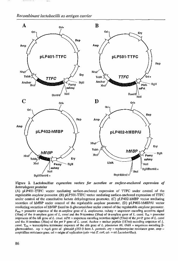
Recombinant lactobacilli as allfigell carrier
A
Amp
c Orl
Amp
Orl+
Orl
pLP401-TTFC
TTFC
Orl+
pLP402-hMBP
lOlol Pamy ssAmy
Noll 8g1lVBamHI+
Ery
6g111
Ery
6g111
B Ery
pLP501-TTFC
Nhe
D Orl+
pLP402-hM BP/U
lOlol
UldA
lOlol BspHVNcol +
Rep
6gl11
Rep
Ery
/-=--6gl11
Not!
BgllUBamHI +
Figure 2. Lactobacillus expression vectors for secretioll or slIr!ace-allcllOred expression of heterologous pro/eills (A) pLP401-TfFC vector mediating surface-anchored expression of TTFC under control of the regulatable amylase promoter. (B) pLP501-TIFC vector mediating surface-anchored expression of TfFC under control of the constitutive lactate dehydrogenase promoter. (C) pLP402-hMBP vector mediating secretion of hMBP under control of the regulatable amylase promoter. (D) pLP402-hMBP/u vector mediating secretion of hMBP fused to 6-g1ucuronidase under control of the regulatable amylase promoter. Pamy = promoter sequence of the a-amylase gene of L amy/ovonts. ssAmy = sequences encoding secretion signal (36aa) of Ihe a-amylase gene of L casei and the N-tenninus (26aa) of a-amylase gene of L casei. P1dh = promoter sequences of the Idh gene of L casei. ssPrt = sequences encoding secretion signal (33aa) of the prt P gene of L. easei and the N-tenninus (36aa) of the prt P gene of L. easei. Anchor = anchor peptide (U7aa) encoding sequence of L. easei. Tan = Imnscription tenninator sequence of Ihe ebh gene of L plantart/Ill 80. VidA = sequences encoding 15-glucuronidase. rep = repA gene of plasmid p353-2 from L pelltosis. el)' = crythromycine resistance gene. amp = ampicilline resistance gene. on = origin of replication (ori+ =ori E. coli, ori- = ori Lactobacillus).
86

Chapter 3.1
A series of expression vectors was constructed to secrete human MBP or hMBP fused to uidA under transcriptional control of the amy promoter, in a similar fashion as used for the TIFC vectors. The 513 bp DNA coding for human MBP (ATCC clone M13577) was extended at its 3' end with a BspHI site and at the 5' end with a BglIT site using PCR. The BglII - BspHI MBP DNA fragment was cloned in pTUT (Figure 1), following digestion of pTUT with Ball/HI and NcoL Subsequently, the BglWNheI fragment containing the insert was exchanged with the BamHI/NheI fragment of pLP402 creating pLP402-hMBP/u (Figure 2D). Digestion with X/wI removed the uidA gene, resulting in the vector containing only hMBP (Figure 2C). The pLP4001500 vectors are stable in E. coli due to the presence of the Idll terminator (Tldh)(pouwels et al., 1996). After removal of the Tldh terminator by NotI digestion, ligation of the vectors juxtaposed the heterologous protein encoding sequences in frame with the first few codons of the translation initiation region present downstream of the promoter sequences, allowing heterologous gene expression in Lactobacillus species (Figure 2).
Expression analysis of heterologous gene products For expression analyses, recombinant L. casei containing the vectors pLP401 and pLP50 I expressing TIFC (designated pLP40 1-TTFC and pLP50 1-TTFC) or containing the vector pLP402 expressing hMBP or hMBP fused to B-glucuronidase (designated pLP402-hMBP and pLP402-hMBP/U) were routinely prepared from glycerol stocks by two consecutive overnight (o/n) cultures without aeration of a 1:50 dilution in MRS medium containing 5 flglml elythromycin.
Transformants with expression vectors containing the constitutive Idh promoter were subsequently grown in antibiotic selective MRS medium or Lactobacillus carrying medium (LCM)(Efthymiou and Hansen, (962) containing 1% glucose (w/v) at 37"C, using a I :50 dilution of the last o/n culture. In order to induce expression of the heterologous proteins in transformants containing plasmids under the transcriptional control of the regulatable a-amy promoter, cells were grown in mannitol (1% w/v) supplemented LCM or modified MRS medium (ruMRS) (1% w/v proteose peptone, 0.5% w/v yeast extract, 0.2% w/v meat extract, 0.1 % v/v Tween SO, 37mM sodium acetate, O.S ruM magnesium sulfate, 0.24 ruM manganese sulfate, S.S ruM diammonium citrate in O.1M potassium phosphate buffer [pH 7.0)). Cells were harvested at defined time points, pelleted by centrifugation and suspended in phosphate buffered saline (PBS). Cell extracts were obtained from the bacteria by sonicating the cells four times on a 30 sec on/30 sec off cycle using an MSE Soniprep 150 sonicator at 10 amplitude microns (Sanyo Gallenkamp PLC, Leicester, UK), to release both cytoplasmic and cell wall-bound proteins. After centrifugation the soluble fraction was used for immunoblot analysis.
To detect heterologous proteins secreted into the medium, the culture medium was collected after spinning down the cells and filtered through a 0.2 flm filter to remove the remainder of cells. After addition of 0.2mglml trypsin inhibitor (Sigma
87

l(ecombillant lactobacilli as antigen carrier
Chemical, La Jolla, CA) the culture medium was freeze-dried and concentrated approximately 200-fold in PBS. Dilutions of these concentrated media are used for immunoblot analysis. Proteins in cell extracts or culture medium were separated by SDS-polyacrylamide gel electrophoresis (SDS-PAGE)(7.5, 10 or 14% acrylamide, 400 mM Tris pH 8.8) and lun in a 25 mM Tris, 192 mM glycine buffer (pH 8.3) at 200V for 45 minutes. Protein was transferred electrophoretically onto nitrocellulose using a Bio-Rad Miniprotean II blotting unit (Bio-Rad Laboratories, Hercules, CA). Immunoblots were incubated with optimally diluted rabbit anti-TTFC (CalbiochemNovabiochem International, San Diego, CA), rabbit anti-hMBP or rabbit anti-pglucuronidase antiserum. Antibodies against hMBP and p-glucuronidase were induced by immunization of New Zealand White rabbits with human MBP and Bglucuronidase in complete adjuvant containing Mycobacterium tuberculosis H37Ra (Difco Laboratories). The second incubation step was performed with swine antirabbit Ig-HRP (Dako NS, Glostrup, Denmark). The immunoblots were incubated with ECL detection reagents (Amersham Life Science Ltd, Buckinghamshire, UK). A light sensitive film (Kodak X-omat) was exposed to the blots for a variety of time periods before development. In all immunoblots, recombinant TTFC antigen (List Biological Laboratories, Campbell, CA), bovine MBP (purified according to Deibler et al., 1972) or B-glucuronidase (Sigma Chemical) were used as references for the immunoreagents. Culture medium of various recombinant L. casei strains expressing fusion proteins with B-glucuronidase were used as controls for secretion or lack of secretion of heterologous proteins into the culture medium in immunoblot experiments. pLP402-HNU, which secretes the 111-120 peptide of hemagglutinin of influenza (HA 111-120) fused to p-glucuronidase, was used as a positive control for secretion (Hackett et al., 1985). Surface-expressed TTFC fused to B-glucuronidase (pLP40 1-TTFCIU) and intracellularly expressed TTFC fusion protein (pLP503-TTFCIU) are examples of heterologous proteins that are not directed to the medium and were utilized as negative controls. The amount of soluble heterologous hMBP-B-glucuronidase was calculated by comparison of several dilutions of extracts of transformants with a reference of purified B-glucuronidase in immllnoblot analysis making use of Bio-ID V6.32 software (Vilber Lourmat, Marne la Vallee, France).
FlolV cytomelly analysis At defined time points bacteria were prepared for analysis by FACScan. Cells were washed twice with FACS fluid (FF), Facsflow™ (Becton-Dickinson, San Jose, CA) with 1 % BSA and 0.1 % sodium azide. Cells were resuspended in 200fll FF containing optimal dilutions of antibodies specific for TTFC, MBP or p-glucuronidase and incubated for one hour at 4°C. Cells were again washed twice in FF. Bound antibody was detected by incubation with fluorescein isothiocyanate-conjugated (FITC) labeled goat anti-rabbit immunoglobulin G (Jackson Immunoresearch Laboratories, West Grove, PA) at a dilution of 1: 1500 for lh at 4°C. Cells were then washed twice prior to analysis on a flow cytometer (FACScan™, Becton-Dickinson). CellQuest® software
88

Chapter 3.1
was used for analysis of the data. A gate was set for events representing lactobacilli as detennined by cytograms of forward and side scatter to enable exclusion of debris. Control strains were wild type L. casei or recombinant strains expressing an irrelevant protein harvested in the same growth phase. For control stainings normal rabbit selUm was used or the antigen-specific primary antibody was omitted. For each sample, data was collected for 10,000 or 20,000 gated events. TIle fluorescence obtained from bacterial cell suspensions was represented by fluorescence histograms and mean channel intensities were calculated.
For quantification of surface expression, L. casei pLP401-TrFC cells were analysed by examining TrFC antibody binding at the point of antibody saturation. This precise determination of fluorescence levels at the point of saturation of binding sites was achieved empirically following the generation of a titration curve for the TrFC-specific polyclonal antibody.
The conversion of measured fluorescence intensities into values representing the number of TrFC molecules accessible for binding on the surface (ABC) was undertaken using the Quantum25® FITC Kit calibration beads (Flow Cytometry Standards, Leiden, The Netherlands). To enable quantitative estimates of the numbers of antibodies bound pel' cell, peak channel data generated from the calibration beads was regressed against the defined values of molecules of equivalent soluble fluorochrome (MESF) possessed by each bead population. Because of the broader distribution of channel intensities for stained cells, extrapolation of the MESF value for pLP40 1-TTFC vectors was then undertaken using the mean channel fluorescence intensity obtained at antibody saturation of the lactobacilli presenting TrFC on the cell surface. Following corrections for fluorochrome/protein (F/P) ratios in the anti-rabbit FITC conjugate (second) step an estimation of numbers of surface molecules available for binding on the smface of the recombinant lactobacilli was made.
Parellferal illllllunisationwith L. casei recombinants In order to demonstrate the immunogenicity of the expressed heterologous proteins, mice were immunized parenterally with cell extracts or concentrated growth medium of the L. casei recombinants. Cell extracts of the recombinants pLP401-TTFC and pLP501-TTFC were obtained as described above. Three BALB/c mice per group were immunized s.c. with an emulsion of 112.5/11 total cell extract and 137.5/11 Difco's complete adjuvant with M. tuberculosis H37Ra (Difco Laboratories). Concentrated culture medium from the recombinants pLP402-hMBP and pLPhMBP/u was used for immunisation. Three SJUJ mice per group were immunized s.c. with 250/11 concentrated culture medium emulsified in Difco's complete adjuvant or i.p. in incomplete adjuvant (Difco Laboratories). The immunisation procedure was repeated 28 days after the fn'st immunisation. SelUm was collected for analysis in ELISA every seven days.
89

Recombillallt lactobacilli as alltigell carrier
TTFC alld MBP specific ELISA To test serum for the presence of anti-TTFC or anti-MBP antibodies, Nunc immuno plates (Life Technologies, Gaithersburg,MD) were coated with tetanus toxoid (RIVM, Bilthoven, The Netherlands)(5J.lglml, 50J.lllwell) in 0.05M carbonate-bicarbonate buffer (pH 9.6) or guinea pig MBP (5J.lglml, 50J.ll/well) in PBS overnight at 4°C. Non-specific binding was blocked by incubation with 3% BSA in PBS (150J.ll/well) for I hour at 37°C. Subsequently the plates were incubated for 1 hour at 37°C with dilutions of sera of mice immunized with cell extracts or culture medium of L. casei recombinants and preimmunization sera to correct for background reactivity. For the detection of IgG antibodies specific for TTFC or MBP, alkaline phosphatase-labelled goat anti mouse IgG (V) (KPL, Gaithersburg, MD) was used (lh, 37°C). After addition of the substrate 4-nitrophenylphosphate, the absorbance at 405nm was read at several time points. Endpoint titer was defined as the highest dilution which gives an absorbance above twice the absorbance with normal mouse serum.
Results TTFC-e>.pressillg recombillallt L. casei
Construction of TTFC-expressing L. casei transformants To enable the constl1lction of TTFC-expressing L. casei vectors, the 1329bp DNA coding for TTFC was cloned into the Lactobacilllls expression vectors pLP401 and pLP501 (Fig lire 2). The pLP401 vectors drive expression via the regulatable ((-amy promoter and include the secretion signal and N-terminus of the amylase gene of L. amylovol'lls. The pLP501 vectors contain the promoter sequence of the Idll gene of L. casei and include the secretion signal and N-terminus of the prtP gene of L. casei. Both the pLP401 and the pLP501 vectors contain the anchor sequence of the prtP gene of L. casei, resulting in expression of the TTFC-anchor protein at the cell surface. Candidate vectors were consltucted in E. coli DHSa hosts and after removal of the Idll terminator introduced in L. casei by eiectroporation.
Immunoblot analysis of TTFC-expressing L. casei transformants Expression of TTFC was demonstrated by immunoblot analysis using a rabbit polyclonal TTFC-specific antiserum (Figllre 3A). Cell extracts were prepared of the L. casei transformant carrying pLP401-TTFC approximately 8 hours (log phase) after inoculation in LCM supplemented with mannitol at OD690= 0.6. Expression of TTFC in the pLP501-TTFC strain, in which expression is driven by Pldh, is not particularly time dependent (data not shown). This confirmed the constitutive expression by pLP50 1-TTFC during growth, whereas in transformants containing pLP40 1-TTFC, TTFC synthesis was induced by derepression of the ((-amy promoter following replacement of glucose in the growth medium by mannitol. Expression was demonstrated to be optimal at the end of the log phase (OD690=0.6). Figllre 3A demonstrates that the amount of TTFC product present in pLP50 1-TTFC (falle 2) is higher than in pLP401-TTFC (falle 4). The TTFC products of both vectors are larger
90

Chapter 3.1
than the purified TTFC (falle 5) due to additional elements such as the anchor sequence. The apparent molecular weight of these TTFC products on immunoblot is approximately 7S kDa. In extracts of either of the transformants types a variety of breakdown products was observed. This in contrast with pLPS03-TTFC transformants which retain the TTFC product intracellularly. The breakdown products of this recombinant are less abundant (Figure 3A, lalle 6). This difference in breakdown products between anchored/secreted products and intracellular products holds hue for all pLP400/S00 vectors constructed so far. L. casei transformed with the pLP401lpLPSOl vectors with irrelevant sequences demonstrated no corresponding heterologous product expression (Figure 3A, lalle 1 alld 3).
L. casei producillg MBP alld MBP fusioll products For oral tolerance induction Lactobacillus transformants containing expression vectors with which secretion of heterologous proteins can be achieved were constructed. DNA encoding human MBP was cloned into the expression vector pLP402 in frame with or without the sequence of p-glucuronidase (Figure 2CID). These pLP402 plasmids drive the expression via the regulatable a-amy promoter and contain the secretion signal and N-terminus of the amylase gene of L. amylovol'l/s.
Expression of both fusion products was demonstrated in extracts by imrnunoblot analysis using antiserum against hMBP (Figure 3B, lalle 3 alld Figure 3C, lalle 3). HMBP fused to fi-glucuronidase was also detected after incubation of the immunoblots with anti-p-glucuronidase serum (Figure 3D, lalle 1). Semi-quantitative analysis yielded a calculated amount of 1- I.S% MBP-p-glucuronidase fusion protein as a percentage of total protein.
Secretioll of MBP alit! MBP fusioll producfS illfo the medium Culture medium used for the propagation of the recombinant lactobacilli was analysed in order to detect whether the cells secreted MBP or MBP fusion proteins. Using immunoblot analysis of cell extracts, the time point of optimal expression was determined to be 12 hours after initiation of growth in mannitol-supplemented mMRS medium (OD690=1.1). The fusion protein hMBP fused to p-glucuronidase was detectable in SO-fold concentrated medium (=7S0/11 medium) in low concentration using the p-glucuronidase specific anti-serum (Figure 3C, lalle 3). The breakdown products, which are abundantly present, are specific for fi-glucuronidase. This indicates that the product is, at least partially, secreted. The fusion product of hMBP with fi-glucuronidase could not be detected in the media when anti-hMBP serum is used (data not shown). Other secreted fusion proteins could be detected in the medium, such as the influenza peptide fused to fi-glucuronidase of L. casei pLP402-HAIU (Figure 3D, lalle 4). That this product is probably intact can be concluded from the fact that the MW of this product is higher than secreted fi-glucuronidase from the L. casei pLP402/u recombinant (Figure 3D, lalle 8). During the secretion process the signal sequence is cleaved off.. Therefore, the secreted
91

Recombillallt lactobacilli as antigell carrier
A 102-
78-
49.5-
B
28.5
20
D
102
78
2 3 4 5 6 anti-TTFC
- c 28.5
"-~_~...J .... e 20 2 3
anti-hMBP
anti-B-glucuronidase
2 3 4 anti-hMBP
8 9
.... d
~h· :::oil j I
..... k
Figure 3. Itmmmobiot a1lalysis of Lactobacillus casei recombillatlls
.... g
5
(A) E'pressioll of TTFC by recombillallts ca/'l)'illg pLP401-TTFC (lalle 4) or pLP501-TFFC (lalle 2). When the culture reached an optical density of 0.6 at 690 nm after derepression of the a-amy promoter, cell extracts were prepared and analysed by immunoblot. Nitrocellulose membranes were stained with serum specific for TfFC. Purified TfFC was used as positive control (lane 5) and cell extracts of wild type L. case; carrying an pLP501 vector with an irrelevant insert (lane 1) Of a pLP401 vector with an irrelevant insert (lane 3) were used as negative controls. pLP503-TfFC, which retains the TfFC product
92

Chapter 3.1
products are smaller than the heterologous proteins detected in the cell extracts, as can be seen by comparison of the cell extract (Figure 3D, lane 7) with medium of L. casei pLP402/u (Figure 3D, lane 8). In concentrated culture medium from two control recombinant L. casei strains which expressed irrelevant antigens fused to B-glucuronidase either intracellular (pLP503-TI'FCIU) (Figure 3D, lane 5) or anchored to the bacterial surface (pLP401-TIFCIU) (Figure 3D, lane 6), no heterologous proteins were detected using antiselUm specific for p-glucuronidase, as expected. This indicated that the positive signals obtained with the medium samples of the recombinants are unlikely to result from leakage or lysis of cells.
Although the large hMBP-B-glucuronidase protein is not efficiently secreted, the much smaller hMBP product from the pLP402-hMBP transformant was secreted well. This product was clearly demonstrated in 25 fold-concentrated culture medium on a immunoblot incubated with anti-hMBP selUm (Figure 3C, lane 2). All medium samples have the property that they lUn in a wide pattern (Fig 3D, lane 3, 4 and 8), but they show vety wide and diffuse bands in the region of the smaller proteins (Figure 3C, lane 2). As a negative control pLP402/u, which secretes only B-glucuronidase, did not show a specific reactivity, as expected (Figure 3C, lane 4).
Flow cytollletric analysis and selection Flow cytometry analysis of surface association of TI'FC
Presentation of the TI'FC antigen on the surface of L. casei transfOlmed with plasmids pLP401-TIFC or pLP50I-TI'FC was demonstrated by FACScan analysis of recombinant lactobacilli. TI'FC gene expression by L. casei was analysed using a polyclonal rabbit TI'FC-specific antiselUm in the period of 4 to 9 h following the onset of
intraccllularly. is shown in lane 6. (B) Expression of IIMBP fused to fl-glucutollidase by recombinallls can)'illg pLP402-hMBP/U. At an optical density of 1.1 at 690 11m, cell extracts were prepared and analysed by immunoblot. Nitrocellulose membrane was stained with serum specific for hMBP. Purified bovine MBP was used as positive control antigen (lane 1) and a cell extract of L. case; expressing n·glucuronidase (pLP402·U)(lane 2) was used as a negative control. (C) E.'pression of hMBP in cell extracts (llld ill culture medium. Cell extracts (lane 3) and culture medium (lane 2) of L. case; recombinants pLP402-hMBP were analysed by immunoblot for the presence of heterologous hMBP. Nitrocellulose membrane was stained with serum specific for hMBP. Purified bovine MBP was used as positive control antigen (lane 1) and a cell extract (lane 5) and concentrated culture medium (lane 4) of L. easei with pLP402-U were used as negative controls. (D) Detectioll of secreted fi-g/Ucllfollidase fusion products. At an optical density of 1.1 at 690 nm, cell extracts and culture medium were prepared for analysis by immunoblot. Nitrocellulose membrane was stained with serum specific for B-glucuronidase. Lane I showed hMBP-B-glucuronidase in a cell extract from pLP402-hMBPIU. Lane 2 is kept empty, to be sure that the secreted product in medium from pLP402-hMBP/U in lane 3 can not be due to overflow. Lane 4 and lane 8 show the secreted products in medium from pLP402-HNU and pLP402-U respectively. Lane 5 and 6 showed the concentrated media of pLP503-TfFC (intracellular TfFC) and pLP401.TfFC (anchored TfFC) respectively. In lane 7, pLP402·U extract was analysed. Purified B-glucuronidase was used as positive control antigen in lane 9. a= heterologous anchored TTFC, b= intracellular TIFC, c= purified TfFC, d= hM:BP-B-glucuronidase, C=: purified MBP, f= non-secreted hMBP, g= secreted hMBP, h =secreted HA-B.glucuronidase, i= non-secreted B-glucuronidase, j= secreted 3-glucuronidase, k= purified 3-glucuronidase. Sizes of the low range marker are indicated in kDa.
93

Recombinant lactobacilli as antigen carrier
growth. Data obtained 7 h after inoculation are presented (Figure 4A). Mean flnorescence levels observed with pLPS01-TIFC after staining with non-immune rabbit selUm were used as control (Figure 4A). A higher level of surface-anchored exposure of TIFC under control of the ldh promoter (pLPSO 1-TIFC) in comparison to that of the surface-anchored exposure of TIFC under control of the a-cuny promoter (pLP401-TYFC)(pouwels and Leer, 1993) was indicated by the distinct mean channel fluoresence intensities of the two recombinants at an equivalent time point. Similar results were obtained at other time points (results not shown).
Quantitation of the number of TIFC molecules present on the cell slllface To estimate the mean number of TIFC molecules present on the sUiface of the cell, mean channel intensities obtained following staining of L. casei pLP40 1-TYFC and L. casei pLPS01-TIFC with anti-TIFC were used for calculation. As shown in Table I, after 7 hours of growth approximately 3.9x10³ and 1.4x10³ TIFC molecules were found on the surface of the pLPSO 1-TIFC and pLP401-TIFC recombinant L. casei, respectively.
Table 1. Quantitafioll of the Ilwnber of7TFC molecules expressed Oil the cell slIJiace of recomb;'Wlll L easei.
Cells
pLP401-TfFC
pLP501-TIFC
Mean channel fluorescence
21
55
MESF
4282
12170
FIP ratio
3.1
3.1
Surface-molecules / per cell
1.4*10'
3.9*103
Analysis of secreted hMBP and hMBP fusion products In order to determine whether it is possible to detect epitope-specific sequences associated with the cell wall of heterologous proteins which are directed to be secreted, transformants secreting MBP and MBP fusion proteins were analysed by flow cytometry, at time points of optimal expression as determined by immunoblot analysis (data not shown). The cells were incubated with specific antiselUm against p-glucuronidase or against MBP. Wild type L. casei as well as L. casei transformed the vector pLP402-U, which secreted p-glucuronidase, were used as controls. As shown in Figure 4B and 4C, it was possible to detect myelin proteins fused to fl-glucuronidase associated with the cell surface by flow cytometry. Significant levels of hMBP-p-glucuronidase (mean channel fluorescence = 13.S channels) could be detected using anti-p-glucuronidase selUm as compared to the wild type L. casei (mean channel fluorescence ~ 2.S). Using anti-MBP serum, the hMBP fusion protein (Figure 4C) and hMBP (Figure 4D) only could also be detected, although the level of surface expression of hMBP only was very low (mean channel fluorescence = 8.1
94

Chapter 3.1
versus 5.1 of pLP402-U). The increase in fluorescence intensity of pLP402-MBPIU compared to the control recombinant pLP402-U, which only secretes fl-glucuronidase (mean fluorescence = 15.5 respectively 7.7 channels) (Figllre 4C) was slightly less than the increase in fluorescence intensity of the control strains and the recombinant pLP402-MBPIU detected with anti-fl-glucuronidase (Figllre 4B).
A B
J
c D
Figure 4. Flow cylomelry allalys;s of Lactobacillus casei recombinants
~U'~02 h'JIlP:\J (¥I(,fi-;''-'o!'.;=;j~Hl
(A) Sill/ace-association of 1TFC. TIFC was detected on the cell surface of Lactobacillus case; recombinants with pLP401·TfFC (a·amy promoter) or pLP501·TfFC (ldh promoter) by incubation of cells with rabbit antibodies specific fOf TIFC and stained with FITC labelled goat anti-rabbit serum. Recombinant pLP501-TfFC incubated with normal rabbit serum (NRS) was used as negative control. (B) SlIIface-association of MBP·/J-gillcllrollidase /lsillg antiserum agaillst fJ-gluwrollidase. Lactobacillus casei recombinants secreting MBP-p-glucuronidase (pLP402-MBPIU) showed surface-association when the cells were incubated with rabbit antibodies against P-glucllronidase and stained with FITC labelled goat anti-rabbit serum. Wild type Lactobacillus casei (wO incubated with serum specific for B-glucuronidase and pLP402-MBPIU incubated with normal rabbit serum (NRS) were used as negative controls. (C) Smface-association of hMBP-j3-glucurDllidase using hMBP specific antiserum. pLP402-MBPIU recombinants were incubated with rabbit antibodies directed against MBP and stained with FITC labelled goat anti-rabbit serum. Recombinant L. easei secreting p-glucuronidase only (pLP402-U) incubated with serum specific for hMBP and pLP402·MBP/u incubated with normal rabbit serum (NRS) were used as negative controls. (D) Swface-association of hMBP using hMBP specific antiserum. pLP402-hMBP recombinants were incubated with rabbit antibodies directed against hMBP and stained with FITC labelled goat anti-rabbit serum. Recombinant L. casei secreting l3-glucuronidase only pLP402-U) incubated with serum specific for hMBP and pLP402·hMBP incubated with normal rabbit serum (NRS) were used as negative controls.
95

Recolllbillallf lactobacilli as alltigen carrier
ImlllllllOgellicity of heterologous proteills expressed by L. casei To determine whether the heterologous TTFC proteins expressed by L. casei recombinants pLP401-TTFC and pLP501-TTFC were immunogenic, BALB/c mice were immunized with total cell extracts of these recombinants. Immunogenicity of TTFC construct pLP50 I was demonstrated in a tetanus toxoid specific ELISA within 14 days after the second immunization with an IgG endpoint titer of 103
.2 (LoglO) for
the rooled mice sera. The TTFC construct pLP401-TTFC showed an endpoint titer of 10'· (LoglO) 21 days after the second immunization.
SJLlJ mice were immunized with cell-free culture medium of L. casei recombinants pLP402-hMBP and pLP402-hMBP/u to demonstrate immunogenicity as well as secretion of the heterologous proteins into the medium. Immunogenicity of secreted MBP was shown by IgG endpoint titers of 1023 (LoglO) for pLP402-hMBP and 1029 (LoglO) for pLP402-hMBP/u after 21 days with negligible values at 14 days. None of the selected control recombinants (i.e. L. casei pLP402/u) induced MBP or TTFC antibodies, as expected.
Discussion In this study we describe the construction and characterisation of Lactobacillus expression/delivery vectors as potential vaccines or therapeutics for diseases of the central nervous system. Essentially, similar vectors are designed for use of two distinct applications; live oral vaccines to actively immunise versus live oral therapeutics to restore peripheral T cell tolerance in autoimmune disease. The distinct effects are facilitated through the precise location of the antigen; surface-anchored antigenexpression for the vaccine application versus antigen secretion into the local environment for tolerance induction (Brandtzaeg, 1996; Gebert et al., 1996).
Two plasmids were constructed to enable surface-anchored expression of TTFC. The expression is under transcriptional control of the constitutive Idh promoter (pLP501-TTFC) or under the control of the a-amy promoter (pLP401-TTFC), which is repressed in the presence of glucose. For application in vaccine strategies, the number of molecules of the antigen present on the cell-surface is considered important for immunogenicity. Flow cytometry has previously been used to determine the composition of bacterial cultures, in which each bacterial species has its own characteristic 'signature' (Pinder et al., 1990). In addition, heterologous proteins have also been demonstrated on the surface of other recombinant Gram-positive bacteria such as Staphylococcus xylosus (Samuelson et al., 1995). Flow cytometry, to our best knowledge, has not been used before to detect heterologous proteins associated with the Lactobacillus surface. We have shown that this technology allows an estimation of the relative number of molecules expressed on the cell wall by individual cells. In the utilisation of a polyclonal antibody we assumed that each TTFC molecule is bound by only one antibody molecule, enabling the relative measurements between recombinant lactobacilli. With this strategy it was demonstrated that for L. casei pLP50 1-TTFC, under control of the constitutive Idh promoter, the number of TTFC molecules per
96

Chapter 3.1
cell, present on the Lactobacilllls cell wall is three times higher than observed for L. casei pLP40 1-TIFC which expresses TIFC under the control of the regulatable a-amy promoter. The difference in expression level can be explained by the fact that the promoter sequences used have different transcription activities (pouwels et at., 1996). The different secretion signals (proteinase versus amylase) inherent to the vectors may also lead to higher translocation efficiencies in the pLP501-TIFC recombinant and may result in elevated levels of TIFC on the cell wall accessible for binding by antibody (Pouwels and Leer, 1993; Hols et at., 1997). The difference in level of expression/surface association may also account for the higher levels of TIFC specific antibodies found after parenteral immunisation with total cell extracts of L. casei pLP501-TIFC as compared to pLP401-TIFC. Although both recombinant strains appear to be immunogenic, the results argue for the use in vaccine applications of recombinant strains utilising the constitutive promoter. The breakdown products present in extracts obtained from either the pLP401 or pLP501 recombinants, which are not as abundantly present in extracts obtained from pLP503 recombinants maintaining the heterologous proteins intracellular, may well be due to activity of proteases present at the surface of the bacterium or in the medium.
Vectors for the use in oral tolerance studies preferably should secrete the tolerogen (Brandtzaeg, 1996; Gebert et at., 1996). Therefore, two vectors that secrete hMBP (pLP402-hMBP) or hMBP fused to Il-glucuronidase were constlUcted and analysed. Although secretion of both products was demonstrated in immunoblot analysis using protein samples prepared from the culture medium, there was a clear difference in the levels of secretion. The quantity of hMBP-Il-glucuronidase fusion protein actually present in the culture medium was very low, and the product apparently was digested into two main products. However, this fusion product was detectable by flow cytometry using either anti-MBP or anti-Il-glucuronidase antibodies. This suggests that although the protein is translocated over the membrane, it did not undergo signal peptide cleavage or was degraded by extracellular proteases. When cleavage does not occur within the encephalitogenic epitopes, the secreted protein could still maintain its tolerogenicity. Association of the mature form of translocated heterologous proteins with the cell wall has been described for Grampositive bacteria (Suanders et at., 1987; Simonen and Paiva, 1993; Wells et at., 1993). The smaller hMBP protein without Il-glucuronidase is much more efficiently secreted than hMBP fused to Il-glucuronidase. Following analysis of this recombinant by flow cytometry, we only found a very low surface-association of hMBP. Apparently, this product is indeed quite efficiently secreted into its environment. We have shown that protein secretion into the medium from recombinants transformed with vectors using the same secretion signals (pLP402-hMBP, pLP402-hMBPIU, pLP402-HAlU, pLP402-U) does not occur at similar levels. These differences probably arise from biochemical properties of the protein itself (e.g. size, hydrophobicity, conformation). In addition, varying sensitivities of each protein for proteases may affect the amount of protein found in the medium. Introduction of pLP402-MBPIU in alternative
97

Recombinant iactobacilli as antigen carrier
Lactobacillus strains may result in an increase of the level of secretion of this particular protein.
Immunisation of mice with concentrated culture medium of the hMBP recombinants evoked MBP-specific antibodies, from which it could be concluded that the heterologous proteins were immunogenic and secreted into the medium. However, the antibody levels induced by medium containing the hMBP-B-glucuronidase fusion protein were higher than the levels induced by medium containing hMBP.
Host strain selection is a factor that may impact significantly on the capacity to successfully induce antibodies or restore peripheral T-cell tolerance by oral administration. Recently, Lactobacillus strains have been identified that persist for extended periods in the intestinal tract of mice (D.M. Shaw, unpublished) as well as in humans in comparison to the prototype L. casei, and this property may improve efficacy of oral vaccination and of tolerance induction considerably. We have previously shown that induction of oral tolerance can be induced with L. piantal'll/ll transformed to express p-galactosidase (Maassen et ai., 1998a) and mucosal antibody responses can be induced with orally administered trinitrophenyl-Lacfobacillus acidophilus conjugates (Gerritse et ai., 1991).
In conclusion, the generic technology described here allows the construction of a multitude of safe recombinant lactobacilli which may prove advantageous in oral intervention strategies for the prevention and treatment of infectious and autoimmune diseases of the central nervous system. Currently, recombinant lactobacilli expressing TIFC or viral antigens are tested in animal models for their ability to induce specific antibodies after oral administration. Whether orally administered recombinant lactobacilli secreting myelin antigens are capable of inducing peripheral T-cell tolerance is currently investigated in mouse and rat BAE-models for multiple sclerosis.
Acknowledgements The authors thank Bric Claassen for critically reading the manuscript and Lizet Groenewegen and Chantal Smittenaar for their expert technical assistance. This work was financially supported by contract number B104-CT96-0542 of BC-Biotech and by grant number MS93-147 from The Netherlands Foundation for the support of Multiple Sclerosis Research.
98
Chapter 3.2
A rapid and safe plasmid isolation method for efficient engineering of recombinant lactobacilli expressing immunogenic or tolerogenic epitopes
for oral administration
C.B.M. Maassen
Journal of Immunological Methods 223: 131-136, 1999

Rapid alld safe plasmid isolationmethodfor lactobacilli
Summary Recombillallt lactobacilli are beillg developed which call be used as expression alld delivelY vectors of heterologous antigens in oral vaccillatioll alld other therapeutic applicatiolls. Because most Lactobacillus strains do not accept ligation mixtures, sufficielltly pure plasmid DNA lIeeds to be isolated from L. casei to trallsform other Lactobacillus strains. The isolation of plasmid DNA from Gram-positive lactobacilli is complicated by the resilience of the peptidoglycan layer. Here a rapid, safe and efficient method is described that combilles ellzymatic breakdown of the cell wall alld purification of the plasmid by commercially available DNA-billding columlls. For the lysis-resistallt Lactobacillus casei straill, this method yields high levels of pure plasmid DNA that call be used for common molecular techniques, such as digestion alld transformation, with high efficiellcy.
100

Chapter 3.2
Introduction Lactobacilli are lactic acid bacteria, which are founs as conunensals of the human gut. Lactobacilli are increasingly being used in food products. In addition, pharmaceutical applications are being developed, such as genetically engineered lac'tobacilli as oral vaccines and for oral T-cell tolerance induction for the treatment of autoimmune diseases such as multiple sclerosis (MS) (pouwels et al., 1996; Maassen et al., 1999a). Currently, in our laboratory, recombinant lactobacilli which express viral and microbial heterologous antigens on the cell surface are orally administered for the induction of antibodies in mice. Lactobacilli which secrete autoantigens such as myelin basic protein (MBP), are administered orally for the induction of peripheral T-cell tolerance in a mouse model for MS (experimental autoimmune encephalomyelitis, EAE).
Because most Lactobacillus strains do not accept ligation mixtures, the DNA required to transform these strains needs to be isolated from Lactobacillus casei ATCC 393, which can be transformed with ligated DNA. For efficient transformation of Lactobacillus strains, highly pure DNA is required. Current isolation techniques used to obtain large quantities of plasmid DNA consist of a cell lysis method followed by alkaline lysis, phenol-extraction and cesium chloride gradient centrifugation. The major obstacle in these isolation procedures is the efficient lysis of the Gram-positive cell wall. This can be achieved by mechanical or enzymatic procedures (Klaenhammer and Sutherland, 1980; Posno et al., 1991a; Frere, 1994; Reinkemeier et al., 1996). Lactobacillus plasmid DNA obtained after alkaline lysis and phenol-extraction contains contaminating chromosomal DNA and cell wall debris. Therefore additional cesium chloride equilibrium density gradient centrifugation is necessaty to obtain plasmid DNA of high purity suitable for electroporation (Sambrook et al., 1989). The procedure is time-consuming (2-3 days) and requires the use of toxic chemicals.
As a consequence, there is clearly a need for a more rapid, high yield isolation method to obtain plasmid DNA from lactobacilli, which does not require the use of hazardous agents. The aim of this study was to develop a simple method that yields large amounts of pure plasmid DNA from several different Lactobacillus strains, including lysis-resistant strains such as L. casei. Furthermore, this method should be more rapid than methods such as cesium chloride gradient centrifugation and has the advantage that no hazardous agents such as phenol and ethidium bromide need to be used. This method would simplify the development of recombinant lactobacilli, which can be used in various therapeutic applications.
Materials and methods Plasmid isolation Recombinant Lactobacillus strains were cultured overnight in 500 ml MRS (Difco Laboratories, Detroit, MI). Lysis of the lactobacilli was performed essentially as described by Posno et al. (199Ia). The cells were harvested by centrifugation at 4000g for 20 min. The cells were washed twice in 25 ml 20 mM Tris/HCI buffer pH 8. The
101

Rapid and safe plasmid isolation method for lactobacilli
pellet was suspended in 20 ml Tris/HCI (20 mM/pH 8) with 5 mg/ml hen egg white lysozyme (Boehringer Mannheim GmbH, Mannheim, Germany) and 20 ml of 24% polyethylene glycol 20,000 (F1uka Chemie AG, Buchs, Switzerland). After 2 h of incubation at 37"C, the cells were centrifuged and washed again with TrislHCI (20 mM/pH 8). After discarding the supernatant the cells were taken up in the solution buffer E1 of the commercial kit (Jetstar plasmid maxi kit, Genomed GmbH, Bad Oeynhausen, Germany). The manufacturer's protocol, comprising alkaline lysis and subsequent neutralization with potassium acetate, was followed. After precipitation of the proteins present in this solution, the resulting DNA solution was applied to the provided column. After washing, the DNA was eluted with the buffer provided. Finally, the DNA was concentrated by alcohol precipitation and dissolved in 200 III TE buffer (10 mM TrislHCI pH 8, 1 mM EDT A). Digestion of the isolated DNA and analysis by agarose gel electrophoresis were carried out essentially as described by Sambrook et al. (1989).
Electroporation of Lactobacillus strains Electroporation of L. casei was conducted as described by Posno et al. (1988), slightly adjusted from the method described by Chassy and Flickinger (1987). Electroporation of L. plantarum 256 was essentially performed as described for L. plallfa/'/(m 80 by Josson et al. (1989). Briefly, an overnight culture of L. plantarum 256 was diluted (1/50) in MRS broth and incubated anaerobically at 37°C. The cells were harvested in the mid-log phase, washed twice with "MilliQ" water, and washed once in 30% (w/v) PEG-IOOO. The cells were resuspended in 30% PEG-IOOO in 1/50 of the original culture volume. Plasmid DNA (lllg) was mixed with 100111 of the cell suspension in a disposable electroporation cuvette with an interelectrode distance of 0.2 cm. A single pulse of 8500 V!em2 was delivered (lOOn parallel resistor and 25 IlF capacitance settings, Gene Pulser and Pulsecontroller from Bio-Rad). Following the pulse, the cell suspension was diluted with 900 III MRS broth and incubated for 2 h at 37°C. Transformants were selected by plating the cells on MRS agar plates containing erythromycin. Transformants were visible after approximately 48 h.
bmmlllobiot analysis of Lactobacillus transformants Immunoblots were performed in order to investigate the expression of heterologous proteins by L. casei and L. plantarum transformants. To induce expression, cells were grown in modified MRS medium (mMRS) (1 % w/v proteose peptone, 0.5% w/v yeast extract, 0.2% w/v meat extract, 0.1 % v/v Tween 80, 3.7mM sodium acetate, 0.8mM magnesium sulfate, 0.24mM manganese sulfate, 8.8mM diammonium citrate in potassium phosphate buffer (pH 7.0) containing 1 % w/v mannitol) at 37°C, using a 1/50 dilution of an overnight culture. Cells were harvested in exponential phase, pelleted by centrifugation and suspended in phosphate buffered saline (PBS). Cell extracts were obtained from the bacteria by sonicating the cells four times on a 30 sec on/3D sec off cycle using an MSE Soniprep 150 sonicator at 10 amplitude microns
102

Chapter 3.2
(Sanyo Gallenkamp PLC, Leicester, UK), to release both cytoplasmic and cell wall-bound proteins. After centrifugation the soluble fraction was used for immunoblot analysis. Proteins in cell extracts (30llg) were separated by SDS-polyacrylamide gel electrophoresis (SDS-PAGE), (7.5% acrylamide for the UreB transformants and II % acrylamide for the MBP/u transformants, 400 ruM Tris pH 8.8) and run in a 25 ruM Tris, 192 ruM glycine buffer (pH 8.3) at 200V for 45 min. Protein was transferred electrophoretically onto nitrocellulose using a Bio-Rad Miniprotean II blotting unit (Bio-Rad Laboratories, Hercules, CA). Immunoblots blots were incubated with polyclonal rabbit anti-H. pylori (Simoons-Smit et al., 1996) or rabbit anti-MBP serum. The second incubation step was performed with swine anti-rabbit Ig-HRP (Dako AlS, Glostrup, Denmark). The immunoblots were incubated with ECL detection reagents (Amersham Life Science Ltd, Buckinghamshire, UK). A light sensitive film (Kodak X-omat) was exposed to the blots for 1 min before development.
Results and Discussion Isolation 0/ plasmid DNA/rom L. casei recombinants Two recombinant Lactobacillus casei strains were used to demonstrate the advantages of this novel plasmid isolation method. The first L. casei recombinant contained a vector (pLP402-MBP/U)(Pouwels et al., 1996) with an insert coding for myelin basic protein (MBP), which is a central nervous system protein thought to be involved in the pathogenesis of multiple sclerosis. The heterologous MBP protein is fused to the marker protein l3-glucuronidase. Because this fusion protein will be secreted, these transformants can be used for oral tolerance induction in an animal model for multiple sclerosis (EAE). The second Lactobacillus casei recombinant contained the vector pLP401-UreB (pouwels et ai., 1996) with an insert encoding UreB, a subunit of the urease enzyme of Helicobacter pylori. The UreB-product is expressed on the cell surface, because the vector also contains an anchor sequence. UreB transformants have proposed for use in oral vaccination experiments and plasmid DNA from both recombinants were isolated with the present novel isolation procedure. The ratio of A260 over A280 of the purified DNA was approximately 1.8, indicating that the DNA was indeed highly purified. The yield obtained from a 500 ml culture was approximately 200 Ilg of plasmid DNA. This is similar to the amounts of DNA usually obtained with the classic method using alkaline lysis and cesium chloride DNA banding.
Restriction analysis o/isolated plasmid DNA Two vectors isolated from the highly lysis-resistant Lactobacillus casei were digested with two restriction enzymes at the same time. The vector pLP402-MBP/u was digested with the restriction enzymes Bamffi and PVIII for 2 h at 37"C. The vector pLP401-UreB was digested with two other enzymes, PstI and NheI. Both constructs were fully digested, confirming the high purity of the plasmids (Figure I).
103

Rapid and safe plasmid isolationmetllOdfor lactobacilli
2 3
IOKb 8 6 , 4
3 2.5
2
LS
Figure I. Restriction analysis of plasmids isolated from L. casei TIle vector encoding the urease subunit B of H. pylori was digested with PstI and NlleI (lane 1). The vector encoding myelin basic protein fused to 6-g1ucuronidase was digested with BamID and PvuI (Jane 2). For each restriction enzyme only one site was present. Lane 3, molecular weight DNA ladder.
Transformation of isolated plasmid DNA to other Lactobacillus strains To assess whether the plasmids isolated by this method were sufficiently pure for use in transformation, the plasmids pLP402-MBPfU and pLP401-UreB were transformed to L. casei and L. plantal'llm 256 by electroporation. Transformation efficiencies of 106_107 transformants per flg plasmid DNA were obtained for L. casei and 105_106
transformants per flg DNA for L. plantaruIII. These efficiencies are equal to those obtained with plasmid DNA purified by CsCI gradient centrifugation. This suggests that the plasmid DNA is highly pure and that limited breakdown of the DNA occurs.
Expression of heterologous proteins by L. casei and L. plantarum In order to demonstrate that the L. casei and L. plantal'llm transformants are able to express heterologous proteins, extracts of these transformants were analysed by immunoblotting. After electroporation of the isolated plasmid DNA, the transformants were grown under conditions that permit expression of the heterologous proteins to occur. After 8 h of culture the cells were harvested and extracts were made. The soluble fraction of the extracts was analysed for expression by inununoblotting. Figure 2A shows the expression of UreB in L. casei containing pLP401-UreB (lane 3) as compared to wild type L. casei (lane 2) which, as expected, does not show any expression. UreB can also be demonstrated in L. plantarum pLP401-UreB extracts (lane 4). Wild type L. plallfarulIl is negative for UreB (lane 5). As a positive control recombinant UreB (66kDa) isolated from E. coli was used (lane 1). UreB from L. caseilL. plantart/III is larger (82kDa) due to the presence of signal peptide and anchor sequences. Expression of MBP fused to B-glucuronidase is demonstrated in Figure 2B. Lane 3 shows the expression by L. casei pLP402-MBPfU and lane 4 shows the expression in L. plantarulIl pLP402-MBPfU. Again the wild type strains were used as negative controls (lane 2 = L. casei, lane 5 = L. plantal'llll/ 256). The size difference between the heterologous MBP, which runs at 85kDa (lane 3 and 4), and purified guinea pig MBP (21kDa)(lane 1) is mainly due to the fusion of the heterologous MBP with B-glucuronidase. The additional bands present in the lanes with heterologous proteins result from cross-reaction of the polyclonal rahbit sera with Lactobacillus
104

Chapter 3.2
antigens when those bands are also present in the corresponding wild type lane. All bands of smaller size, which are not due to such cross-reactivity, are breakdown products of the heterologous proteins. These results clearly demonstrated that Lactobacillus strains transformed with plasmids isolated using the present method are able to express the heterologous proteins that are encoded on the plasmids.
A
2
Figure 2.
UreB Lac/obacillus UreB E. coli
B
(A) IJlJmlmoblot analysis o/Lactobacillus Irallsjormallts expressing UreB
MBPa-glucuronidase
Lane I, positive control of recombinant UreB isolated from E. coli. Lane 2, wild type L. casei. Lane 3, L. case; containing pLP401-UreB. Lane 4, L. planta1'll1ll containing pLP401-UreB. Lane 5, wild type L. 'plallfarum. TIle immunoblot was incubated with anti-H. pylori serum. (B) 1I1111l111l0blot analysis of Lactobacillus trallsjonllallts expressi1lg MBP Lane I, purified MBP. Lane 2, wild type L. casei. Lane 3, L. casei containing pLP402.MBPfU. Lane 4, L. plan/omit! containing pLP402-MBPIU, Lane 5, wild type L. plal/farum. The immunoblot was incubated with anti-MBP semm.
Plasmid isolation from Lactobacillus plantarum 256 In order to investigate whether this method is more generally applicable to various Lactobacillus strains, plasmid DNA from L. plalltarulIl 256 transformed with the MBP containing plasmid was re-isolated as described above. Plasmid DNA was also isolated from L. plantartlm 256 transformed with a plasmid encoding tetanus toxin fragment C (pLP40 1-TTFC), which was isolated using a classic method followed by cesium chloride gradient centrifugation (posno et at., 1988). Figure 3 shows that, from this Lactobacillus strain also, plasmid DNA could be isolated using this method. Breakdown of plasmid DNA, as reflected by the DNA smear in Figure 3, can probably be diminished when the incubation time with lysozyme is shortened, because the stringent lysis protocol for L. casei was also used for L. plantarum.
105

Rapid al/d safe plasmid isolatiollmethodfor lactobacilli
2 3
Conclusion
lOKI;> &
6 l 4
3 25
Figure 3. Plasmids isolated/rom L. plalltarum 256 TIle vectors pLP402-MBP/u (lane I) and pLP401-TfFC (lane 2) were isolated from L. plantarum 256 with the method described above and analysed on a 1% agarose gel. Lane 3, molecular weight DNA ladder.
In conclusion, this new method for the isolation of plasmid DNA from lysis-resistant lactobacilli has major advantages over the classic method that requires phenolextraction and cesium chloride gradient centrifugation. This new method takes approximately five hours to perform instead of approximately two days, does not employ highly toxic reagents, is considerably cheaper and is easy to perform. This method provides high yields of pure DNA, which can be used for restriction analysis, transformation and other molecular techniques. This plasmid isolation technique is also applicable to other Gram-positive bacteria, and will improve the efficient engineering of biotechnologically and therapeutically useful lactobacilli recombinants for oral administration.
Acknowledgements The author thanks Dr. Patricia Conway for kindly providing the Lactobacillus plal/tarum 256 strain and Dr. Ben Appelmelk for kindly providing the antibody against H. pylori. Drs. Rob Leer, Mike Shaw, Jon Laman and Wim Boersma are acknowledged for suggestions and critical reading of the manuscript. This work was financially supported by grant number MS93-147 from The Netherlands Foundation for the support of Multiple Sclerosis Research.
106

4.1
4.2
Mucosal tolerance induction with
and oral antigens
recombinant lacto
om'ltHilis induced


Chapter 4.1
Therapy with antibodies against CD40L (CD154) and CD44-variant isoforms reduces experimental autoimmune encephalomyelitis induced by a proteolipid protein peptide
I.D. Laman, C.B.M. Maassen, M.M. Schellekens, L. Visser, M. Kap, E. de long, M. van Puijenbroek, M.l.B. van Stipdonk,
M. van Meurs, C. Schwarzler and U. Giinthert
Multiple Sclerosis 4: 147-153,1998

Antibodies against CDi54 and CD44 block EAE
Summary Interactions between /l1O/IOIIl/clear cells are required for the formation of injlammatOlY infiltrates in the CNS and the activation of cellular effector functions provoking demyelination in MS. Membrane-expressed costimulatOlY molecules are cl'/lcia/ to such interactions. We therefore investigated whether two costimulatOl)' molecules, CD40L (CDi54, expressed on activated CD4-positive Tcells) and selected CD44-variant isoforms (expressed on activated CD4-positive T cells), are targets for immunotherapy in MS. TI,e model of experimellla/ autoimmune encephalomyelitis (EAE) induced in SJL-mice by immunization with a peptide derived from the proteolipid protein (PLP139-151) was optimized to address these questions. A previous obse/vation that anti-CD40L (CDI54) monoclonal antibodies can effectively prevent EAE in this model was confirmed, and extended by demonstrating that CD40 is expressed by cells of the monocytic lineage infiltrating the spinal cord. In vivo treatment with antibody against the standard isoform of CD44 (CD44s or CD44H) did not affect disease burden In contrast, combined treatment with antibodies against the isoforms CD44v6, v7 and viO, which are thought to be involved in injlammatOl)' processes, reduced the disease burden considerably. In addition, CD44vlO-eJ.pressing cells were detected in the spinal cord. These data support the idea that CD40-CD40L interactions form a target for immunotherapy of MS, and indicate that cells expressing CD44v6, v7 andlor vIO-containing isoforms have such potential as well.
110

Chapter 4.1
Introduction Temporary and cumulative neurological dysfunction of multiple sclerosis (MS) patients results from chronic autoinunune inflammatory events within the central nervous system (CNS), leading to edema, demyelination of nerve axons, and axonal loss. Perivascular and periventricular inflammatory infiltrates of mononuclear cells containing mostly macrophages and CD4+ T-cells most probably affect these three processes, but the effector mechanisms involved have been elusive thus far. The formation of such infiltrates and activation of cellular effector mechanisms requires intercellular communication mediated by soluble mediators such as cytokines, as well as costimulatory (accessory) molecules on the cell surface.
Immunotherapy of MS with antibodies directed against costimulatory molecules such as CD40L, CD80/CD86 and CD44-isoforms, may affect one ore more of the following events: (i) Peripheral activation of autoreactive T-cells; (ii) Adhesion, blood-brain barrier transmigration, and migration of mononuclear cells within the CNS; (iii) Peripheral or local (CNS) cellular activation events, leading to further (co)-stimulation of T-cells, and/or induction of cytokines, chemokines and proteolytic enzymes. In the current study, the co-stimulatOlY CD40L and CD44-isoform molecules were evaluated as candidate targets for therapy, in a mouse EAE model.
We have previously shown that CD40-CD40L interactions are involved in EAE and MS, and that this interaction is indeed a target for therapy of MS (Gerritse et al., 1996). CD40 is expressed on antigen presenting cells, such as dendritic cells, macrophages, microglia, endothelial cells and B cells. Interaction with CD40L expressed by activated, antigen-experienced CD4+ T-cells can trigger a wide array of effector functions of both the antigen presenting cell and the T cell itself (Laman et al., 1996). In human MS autopsy brain, CD40 is expressed on the surface of monocytic cells within mononuclear infiltrates of the CNS. Functionally, we have demonstrated that development of EAE in SJL mice immunized with the proteolipid protein (PLP) peptide residues 139-151 can be prevented by treatment with anti-CD40L monoclonal antibody (Gerritse et al., 1996). Finally, we have demonstrated that CD40 and CD40L are expressed in the CNS of marmoset monkeys with demyelinating EAE in relation to expression of pro-inflammatory and anti-inflanunatory cytokines (Laman et al., 1998c). This novel model is useful for the evaluation of human-specific immunotherapeutics.
In view of the proposed roles of differential CD44-isoform expression in tumor metastasis and chronic (autoinunune) inflammation, we hypothesized that CD44 variant isoforms are also involved in the development of MS and EAE (Rosenberg et al., 1995; GUnthert, 1996).
CD44 (formerly also called Pgp-I, gp90Hennes and H-CAM) has been implicated to act in processes such as pattern formation in embryogenesis, formation of the CNS, hematopoiesis, lymphocyte homing and activation, tumor dissemination and inflammatory reactions (Kaayk et al., 1997). CD44 is an 80-250 kDa type I (extracellular N-terminus) transmembrane glycoprotein, encoded by only one gene.
III

Alltibodies agaillst CD154 alld CD44 block EAE
Due to the complex RNA splicing mechanism of CD44 and post-translational modifications, about a thousand CD44-isoforms can potentially be generated. In practice, about one hundred different functional isoforms have been identified to date. The combination of structural features of CD44-isoforms allows high versatility in a wide array of cellular adhesion and activation events under physiological as well as pathological conditions. The CD44H (hematopoietic) or CD44s (standard) isoform is expressed widely, but no clear functional defects are apparent in CD44s knockout mice. CD44-variant isoforms, however, are only expressed on haematopoietic precursor cells, transiently during activation, and on memory cells. The pro-inflammatory cytokines TNF-alpha and IFN-gamma upregulate CD44 expression. Multiple ligands exist for CD44s, including hyaluronate, fibronectin, osteopontin, and collagen types I and IV, which are all components of the extracellular matrix (Lesley et al., 1993). By virtue of glycosylation and heparan sulfate side chains, CD44v3 isoforms can also capture and concentrate cytokines as well as other soluble components, and present these to neighboring cells. The functions of CD44 and its variants still remain unknown, and putative ligands for these molecules await identification.
The aim of the current study was to provide further evidence for the involvement of CD40L in EAE, and to establish whether CD44-variant isoforms could also form a target for therapy in MS. The model of EAE induced in the SJL-mouse by immunization with PLP139-151 peptide was further optimized to address these questions.
Materials and methods Allimals Female SJL-mice were bred and maintained under SPF-conditions with free access to 0.9 MRAD-irradiated pelle ted food (Hope Farms, Woerden, The Netherlands) and acidified water (pH 2.2) at the animal facility of the Department of Immunology, Erasmus University Rotterdam. This mouse strain is derived from the original SJLlJ (Jackson) line, and has a low microbiological burden as assessed by routine screening. This strain is characterized by low IgE levels in na'ive mice and animals display markedly more docile behavior than the original line. All experiments were performed with animals of 8-12 weeks old, at TNO-PG in Leiden, according to regulations in the Dutch law on animal expedmentation.
EAE-indllctioll EAE was induced by immunization with PLP139- 151 peptide and additional treatment with Bordetella pertllssis to affect blood-brain barrier integrity. This is a classic and widely used EAE-model in mice. Animals develop paralytic disease 10-11 days after disease induction and recover spontaneously around day 20 (Kono et al., 1988; Sobel et al., 1990; Gerritse et al., 1996). Animals also show weight loss up to 25% concurrent with paralytic disease. On day 0, animals were injected subcutaneously with 50-100 micrograms (or other doses in the ill vivo titration experiments) of peptide
112

Chapter 4.1
(sequence HSLGKWLGHPDKF), divided over the four flanks, with 50 microliter per site. The peptide in PBS was mixed at I: I in Difco complete adjuvant (Difco 3113-60-5, with Mycobacterium tuberculosis H37RA). At day I and 3, animals received intravenous injections with 10'0 heat-killed Bordetella pertussis bacteria from the vaccine preparation used for vaccination of humans (National Institute of Public Health and Environmental Protection, BiIthoven, The Netherlands). Clinical disease was monitored daily from day 7 onward by weighing the mice, and by grading symptoms of paralysis using an internationally accepted clinical scoring scale, ranging from 0 to 5 (0 = no disease, 0.5 = tail paresis or partial paralysis, I = complete tail paralysis, 2 = paraparesis: limb weakness and tail paralysis, 2.5 = partial limb paralysis, 3 = complete hind- or front limb paralysis, 3.5 = paraplegia, 4 = quadriplegia, moribund,S = death due to EAE).
Treatment lVith antibodies against costimulatory lIlolecules To intelfere with development of EAE, animals were treated intraperitoneally with protein A-purified monoclonal antibody preparations from the day of immunization onward. For CD40L, this treatment has previously been shown to effectively prevent EAE, even when treatment was started shortly before onset of clinical signs (Gerritse et al., 1996). For CD44-variant isoforms such experiments have not been reported yet. In the current study, anti-CD40L treatment was performed to confirm previous findings, and to extend these by analysis of spinal cord mononuclear cell infiltrates for expression of CD40. In addition, anti-CD40L treatment served as an internal treatment control for the anti-CD44 isoform experiments. Animals were treated by i.p. injection of 250 microgram of antibody against CD40L and CD44s on day 0, 2 and 4, or on day 0, 2, 4 and 6 with a combination of antibodies against CD44 v6, v7 and vl0. This protocol was based on previous experiments where anti-CD40L successfully prevented EAE (Gerritse et al., 1996). The rat IM-7 antibody (IgG2b) directed against the mouse CD44s isoform was used, as well as mouse antibodies against isoforms v6 (LN 6.1), v7 (LN 7.1) and vlO (LN 10.1). These antibodies are further referred to as anti-v6, anti-v7 and anti-vlO. CD40 was detected by means of FGK-45 (Rolink et al., 1996). As the blocking antibody against CD40L is raised in hamster, hamster immunoglobulin (Hig) was used as a negative control antibody lacking blocking activity (Noelle et al., 1992).
[mlllunohistochelllisfly To evaluate expression of CD40, CD40L and CD44-variant isoforms in the CNS of animals used in the experiments above, immunohistochemistry was pelformed on frozen sections from brain and spinal cord of mice, using biotinylated preparations of the antibodies mentioned above, according to standard methods with previously described modifications (Gerritse et al., 1996; Laman et at., 1998c).
113

Alltibodies agaillst CD154 alld CD44 block EAE
Results In vivo titratioll of PLP/39.151 Animal models for EAE and other autoimmune diseases are notoriously sensitive to minute changes in a wide array of variables, including the origin, age, sex, hormonal and microbiological status of the animals, as well as the dose and composition of the disease-inducing antigen, and of the adjuvant used. In addition, for modulation experiments in such animal models, it is a prerequisite that the dose of antigen and the level of clinical disease are carefully balanced to be able to detect both diseasereducing and disease-aggravating effects of treatment. In other words, the lowest dose of antigen inducing a 100% incidence of disease should be used, and not a higher dose inducing plateau-levels of disease, obscuring modulatory effects of experimental treatment. For that reason, the EAE PLP139. 151 peptide was titrated ill vivo in SJL-mice to accurately determine the optimal dose allowing modulation by treatment with antibodies or by oral feeding of Lactobacillus strains genetically engineered to express myelin antigens for mucosal T cell tolerance induction (Maassen et al., 1999a). Table 1 shows two independent ill vivo titration experiments performed. Both the disease incidence and the level of disease of individual mice need to be considered. As is clear from comparing the upper and lower panel of the Table, peptide doses of over 25 microgram induce a 100% disease incidence. The low dose of 3.125 microgram of peptide is ineffective in inducing disease. The 6.25 microgram dose does induce disease, but only in a single animal out of three, with a delayed onset
Table 1. In vivo titration of 'he e1lcephalitogenic PLPIJ9.151 peptide
Peptide dose (~g) Incidence Mean day of onset ±SDa Mean max, score ±SDb
25 3/4 11.3±1.5 2.7±O.3 50 4/4 1O.5±0.6 3.3±O.3 75 4/4 1O.5±0.6 3.4±O.3 100 4/4 1O.5±1.0 2.6±1.l
0 0/4 3.13 0/3 6.25 1/3 16c I' 12.5 3/4 13.0±1.7 2.2±1.0 25 3/3 11.0±0.0 3.3±O.3 50 212 11.5±O.7 3.3±O.4 100 4/4 11.3±1.0 3.4±1.2' 200 3/4 11.0±0.0 3.0±O.0 400 4/4 IO.S±0.5 3.3±O.5 SOO 3/3 11.3±0.6 3/3±0.3
Two independent experiments using partially overlapping dose ranges, are shown in the upper and lower part of the table. amean day of disease onset of animals with clinical signs, bmean maximal disease score of animals with clinical signs. Cdata of a single individual animal with clinical signs. uone animal in this group died ofEAE (clinical score 5).
114

Cilapter4.1
and a lower disease score than that of animals receiveing higher doses. 12.5 microgram induces disease in three out of four animals, of which two have disease scores comparaple to animals treated with higher peptide doses. Doses of 25 microgram and higher lead to a 100% incidence. Interestingly, the higher doses of peptide (even up to 800 microgram per mouse) did not lead to death from EAE. Only one animal in the 100 microgram dose group died of EAE (clinical score of 5: lower panel). This is clearly a desirable aspect of a model for MS, in which death due to acute inflammation of the CNS is not typical at all. On the basis of these results, a dosage of 50-100 microgram peptide was considered to be optimal for use in subsequent experiments, resulting in a 100% incidence of disease.
Prevention of EAE by antibodies against CD40L alld CD44-variant isoforms To assess whether the standard form of CD44 (CD44s) is involved in development of EAE, animals were treated with a specific antibody (IM-7). Control animals were either treated with an irrelevant antibody (disease control group) or with anti-CD40L (blocking of disease control group). Although the irrelevant antibody did not interfere with disease as expected (Figure 1, top panel) and anti-CD40L antibody blocked disease effectively (middle panel), the monoclonal antibody against CD44s did not affect disease burden (lower panel)(Gerritse et al., 1996).This indicates that either treatment was not effective in the regimen used, or that the standard isofonn CD44s is not involved in development of EAE. As it has been claimed that specific isoforms of CD44 are involved in (autoimmune) inflammatory disorders such as colitis and arthritis, we next investigated whether CD44v6, v7 and vlO have a role in development of EAE in this model. Therefore, animals were treated with a combination of three different monoclonal antibodies against the CD44 isoforms v6, v7, and vlO on days 0, 2, 4 and 6 after EAE-induction. As can be seen in Figure 2, animals treated with the irrelevant antibody developed disease and recovered spontaneously, following normal disease kinetics (top panel). According to expectation, anti-CD40L antibody again effectively blocked development of EAE in this administration regimen (middle panel). Interestingly, administration of antibodies against v6, v7 and vlO considerably reduced EAE disease burden (lower panel). Importantly, one out of four animals was totally protected, and a second one only displayed minimal clinical symptoms for a period of three days versus eight days in the controls. The other two animals developed disease at a regular level, consistent with the all-or-none character of the model as was found in the in vivo titration experiments described above.
ImmunohistocilemistlY To assess whether CD40, CD40L and CD44-variant isoforms are expressed within mononuclear cell infiltrates within the CNS, brain and spinal cord tissue of the animals from the experiment was evaluated in situ by immunohistochemistry. As animals were sacrificed after resolution of the disease, it should be kept in mind that
115

Antibodies against CD 154 alld CD44 block EAE
5
4
-;r 3 e w 2 ill
o
5
4 ';i'
3 e
..
7 8
control antibody
T r LI ...... vV"
;I; 9 10 11 12 13 14 15 16 17181920
time (days)
antl·CD40L antibody
120
100
80
60
40
20
120
100
80
w 2 .. 60 ill
40
0 20 7 8 9 10 11 12 13 14 15 16 17181920
time (days)
antl·CD44s antibody
5 120
4 100 ';i' e 3 80
w 2 60 « w 40
0 20 7 6 9 10 11 12 13 14 15 16 17181920
time (days)
Figure 1. Treahllellt with allli-CD44s alltibody does 1IO( pre~'ellt EAE
~ '" -.-
J: " "'~ '4) ~
;: ill C
.". J: Ol -.-
.<: " "';: '4) t! ;: J3
<II
C
l? '" ~ '~
'0:; 't! ;: J3
<II ~ ~
Four SJL mice per group were treated with PLP139-151 peptide to induce EAE. On days 0, 2 and 4 after immunization they received intrapedtoneal injections with irrelevant control antibody (top panel: disease control group), anti·CD40L antibody (middle panel: disease blockade control group), or anti·CD44s antibody IM-7 (lower panel). Graphs show averages of four mice, with the line indicating the weight of the animals as a percentage of the start weight, and the bars represent clinical signs of disease, scored on a scale from 0 to 5 (see Materials and methods section).
116

Chapter 4.1
control antibody
5 120
4 100 ~ -;r 01
e 3· 80 ~ .~ w 2 60 .~ ~ « w ~
40 C 0 20
7 8 9 10 11 12 13 1<1 15 16 17181920
time (days)
antl·CD40L antibody
5 120 ..A. .A. '"'
~ 4 100 ,:-
-;r ~ .c 01
e 3 80 ~.~ UJ 2 .. 60 '0:; t! « ;= ~ w
40 ~
0 20 7 8 9 10 II 12 13 1<1 15 16 17 18 19 20
time (days)
anti·CD44 VS, v7, v10 antibodies
5 120
4 100 ,:-..." .<:
'" " 3 80 ~ .-e .<: " "';= w 2 60 'iii 1:: ~ := ~
40 C
0 20 7 8 9 10 II 12 13 14 15 16 17181920
time (days)
Figure 2. Treahllellt witll auti-CD44 v6, v7 alUl vlO reduces EAE disease burdell Four SJL mice per group were treated with PLPI39-ISI peptide to induce EAE. On days 0, 2, 4 and 6 after immunization they received intraperitoneal injections with irrelevant control antibody (top panel: disease control group), anti-CD40L antibody (middle panel: disease blockade control group), or anti-CD44 v6, v7 and vI 0 antibody (lower panel). Graphs show averages of four mice, and are organized as described in the legend to Figure 1.
117

Alltibodies agaillst CD154 alld CD44 block EAE
only late-stage infiltrates were analysed. In general, the infiltrates within the CNS of animals treated with anti-CD44-variant antibodies or anti-CD40L antibodies were markedly reduced in number and size compared to animals treated with irrelevant antibody (data not shown), indicating that antibody treatment directly or indirectly interfered with the local inflammatory process. CD40L-positive cells were only occasionally found within the infiltrates (not shown), consistent with findings in human MS brain (Gerritse et al., 1996). However, we demonstrated for the first time that CD40 is clearly and abundantly expressed in mononuclear cell infiltrates of the mouse spinal cord (Figllre 3a). Similar to the situation in human MS autopsy brain and marmoset EAE brain, CD40 was abundantly expressed by cells with intracellular
Figure 3. Expression of CD40 alld CD44 isofonlls ill tlte !Jpillai cord Control antibody-treated animals from the experiment shown in Figure 3 were sacrificed after resolution of disease and frozen spinal cord tissue analyzed by immunohistochemistry for expression of CD40, CD40L and CD44 isofonns using the respective specific monoclonal antibodies (see Materials and methods). (A) Abundant expression of CD40 by cells present within perivascular mononuclear cell infiltrates in the spinal cord. Star indicates vessel. Bar indicates 15 micrometer. (B) CD40 is expressed on the surface of cells (examples indicated by arrowheads) by cells in the perivascular mononuclear cell infiltrates. Some of the CD40~positive cells belong to the monocytic lineage as indicated by the activity of intracellular acid phosphatase, a lysosomal enzyme (examples indicated by stars). Bar indicates 20 micrometer. (C) Abundant expression ofCD44s by mononuclear cells within the spinal cord of an animal treated with control antibody, as detected by antibody JM~7. Bar indicates 50 micrometer. (D) Expression of the CD44vlO isoform (red) within the spinal cord of an SJL~mouse treated with control antibody (examples indicated by arrowheads). Bar indicates 10 micrometer.
lIS

Chapter 4.1
acid phosphatase activity, characteristic for cells of the monocytic lineage (Figure 3b)(Laman et al., 1998c). Single CD40-positive cells were also present (again similar to human MS and marmoset EAE), which might represent B cells or other antigen presenting cells lacking detectable acid phosphatase activity. Furthermore, cells with clear acid phosphatase activity but lacking CD40 expression were found too, indicating that local conditions do not necessarily induce CD40 expression of cells of the monocytic lineage. CD44s was clearly present within mononuclear cell infiltrates as can be seen in Figure 3e. In addition, expression of CD44vlO, but not v6 or v6, by cells within the infiltrates could be demonstrated in the CNS of animals treated with irrelevant antibody after resolution of the disease (see Figure 3d), indicating that this isoform is indeed expressed in the target organ.
Discussion This study confirms previous findings that anti-CD40L treatment can prevent EAE induced by PLPIJ9-151 in SJL-mice, and extends this concept by showing that CD40 is expressed abundantly in the spinal cord by infiltrating mononuclear cells. In addition, it is shown that combined treatment with antibodies against the CD44 variant isoforms v6, v7 and vlO reduces EAE disease burden, and that CD44s and CD44vlO are expressed by inflammatory cells in the spinal cord.
Although CD40-CD40L interactions have initially been described with respect to physiological T-B cell communication, it has now become clear that these interactions are also involved in B cell-driven autoimmune disease, and in autoimmune diseases driven by T cells and macrophages such as EAE in SJL-mice (Durie et al., 1994; Gerritse et al., 1996; Laman et al., 1996; Mohan et al., 1995). In addition, CD40-CD40L interactions are relevant for the chronic inflammatory process underlying atherosclerosis (Laman et al., 1997). We have previously proposed that the CD40-CD40L interaction is a target for immunotherapy of MS (Gerritse et al., 1996). Further support for this idea is provided by data in the current study showing that CD40 is expressed in the spinal cord of SJL-mice, mostly on the membrane of infiltrating cells of the monocytic lineage characterized by intracellular acid phosphatase activity. This is fully concordant with what was described previously for human MS autopsy brain and marmoset monkeys (Callithrix jaec!llIs) with demyelinating EAE, a recently developed model for MS allowing evaluation of human-specific reagents (Gerritse et al., 1996; Laman et al., 1998c). The local expression of CD40 strongly suggests that HLA class II-restricted antigen presentation is ongoing, and that effector functions of macrophages expressing CD40 can also be triggered by CD40L-expressing activated CD4+ T cells. III vitro, such T cell/macrophage interactions can lead to production of nitric oxide, pro-inflammatory cytokines (including interleukins 1, 8, 12 and TNF-alpha), and matrix metalloproteinases (MMP-9: gelatinase B), which are all compounds thought to be involved in the immunopathogenesis of MS as well as other chronic inflammatory diseases (Laman et al., 1997).
119

Alltibodies agaillst CD154 alld CD44 block EAE
In view of the role of CD40-CD40L interactions in MSIEAE, and data on the involvement of CD44-variant isoforms in inflammation, we hypothesized that specific variant isoforms of CD44 might be involved in development of MS and EAE as well. Interestingly, it has been shown previously that CD40-CD40L interactions can activate CD44s expression ill vitro, thereby functionally linking these receptor-coreceptor systems (Guo et 01., 1996). CD44-isoforms could be involved in the process of leukocyte extravasation (DeGrendele et 01., 1997) and trans-blood-brain barrier migration, as well as cellular interactions within perivascular and periventricular infiltrates, triggering effector functions that evoke MS and EAE-pathology (Wittig et 01., 1997). The concept that CD44-directed therapy may have promise for treatment of MS is supported by studies describing successful CD44-directed immunotherapy of experimental tumor formation, colitis and the inflammatory autoimmune disease arthritis (Mikecz et 01.,1995; Verdrengh et 01.,1995; Brennan et 01., 1997; Croft et 01., 1997).
Our ill vivo treatment experiments with an antibody against CD44s (IM-7: see Figure 1) might argue against an involvement of the CD44s molecule, despite its expression on cells within infiltrates of the CNS. However, it cannot be excluded at this stage that factors inherent to the current experimental setting have influenced the findings, such as high and ubiquitous expression of CD44s on diverse cell types, limiting the amount of antibody available for blocking migration or effector functions of mononuclear cells effecting clinical disease. In addition, antibody characteristics such as affinity and functional capabilities of the Fc-portion of this IgG2b molecule may limit its blocking efficacy. In contrast, combined treatment with antibodies against v6, v7 and vlO effectively prevented development of EAE in half of the animals (Figure 2). CD44 isoforms (by staining with IM-7 antibody) and CD44v1O separately (by staining with a specific antibody) were found to be expressed in the spinal cord of animals treated with the control antibody (Figure 3). Clearly, it needs to be addressed whether treatment with antibodies against v6, v7 and v 10 can prevent disease individually. Also, further evaluation of in situ expression of the isoforms in relation to disease stage is required.
What activities of CD44 might specific antibodies be blocking to prevent EAE? In comparison to other chronic inflammatory diseases, the insight into the roles of CD44-variant isoforms in EAE and MS is limited as yet. It has been described that CD44 is expressed within the CNS of SJL-mice with EAE by CD4+ T-cells of the activated memory/effector cell phenotype known to effect disease (Zeine and Owens, 1992). Possibly, CD44-expression by activated T-cells supports passage over the blood-brain barrier, but it is also conceivable that CD44 mediates activation of cellular effector functions within the mononuclear cell infiltrates in the CNS (Merrill et 01., 1992). CD44 also participates in binding ofT cells to astrocytes (Haegel et 01.,1993).
Current experimentation is focusing on expression of CD44-isoforms in human MS and marmoset EAE brain, and on the role of selected isoforms in EAE in SJL-mice. To this end, both treatment with single isoform-specific antibodies and
120

Chapter 4.1
isoform knockout mice are being used. In due course, such experiments should provide further insight into the mechanisms of CD44-involvement in inflammation of the CNS, as well as into the feasibility of CD44-isoform directed immunotherapy of MS.
Acknowledgements The authors thank Jan Andersson and Manfred Kopf of the Basel Institute for Immunology for critical reading of the manuscript and Jan Andersson for the gift of anti-CD40 antibody FGK-45. This work was supported by grant 94-171 MS from the Dutch Foundation for the Support of MS-Research and grant 940-70-007 from NWO-New Dmg Research Foundation. The Basel Institute for Immunology was founded and is supported by F. Hoffmann-La Roche, Basel.
121


Chapter 4.2
Reduced experimental autoimmune encephalomyelitis after intranasal and oral administration of recombinant
lactobacilli expressing myelin antigens
C.B.M. Maassen, i.D. Laman, i.C.P.A. van Holten, L. Hoogteijling, L. Groenewegen, L. Visser, M.M. Schellekens,
W.J.A. Boersma and E. Claassen
Submitted for publication

Reduced EAE after mucosal administration of recombinant lactobacilli
Summary Oral administration of autoclIltigens is potentially a safe and convenient way to induce peripheral T cell tolerance in autoimmune diseases like multiple sclerosis. To increase the efficacy of oral tolerance induction and obviate the need for purification of human myelin proteins, we use genetically modified lactobacilli that locally secrete the antigell. A panel of recombinant Lactobacillus strains was constructed which secrete myelin proteins and peptides, including human and guinea pig MBP and PLP139•151• In this study we examined whether these Lactobacillus recombinants are able to induce oral and nasal tolerance in an animal model for multiple sclerosis, experimellfal autoimmune encephalomyelitis (EAE). Lewis rats and SJUJ mice received soluble cell extracts of Lactobacillus transformants intranasally three times prior to induction of EAE. For the induction of oral tolerance, rats and mice were fed live transformed lactobacilli for 20 days. Ten days after the first oral administration EAE was induced. Intranasal administration of extracts containing gpMBP or MBP72-85 significantly inhibited EAE in Lewis rats. Extracts of control tram/ormants did not reduce EAE. Live lactobacilli expressing guinea pig MBP72-85 fused to }J-gluc were also able to significantly reduce disease burden when administered orally, in collfrast to total guinea pig MBP. Also in SJUJ mice Lactobacillus constructs tended to reduce disease after oral or nasal administration. In conclusion, these experiments provide proof of principle that lactobacilli expressing myelin antigens reduce EAE disease burden after mucosal administration. Optimization of myelin expression levels and administration regimens will further increase efficacy of peripheral T-cell tolerance induction in rodents.
124

Chapler 4.2
Introductiou Multiple sclerosis (MS) is a chronic inflammatory disease of the central nervous system (CNS) that leads to destlUction of CNS myelin. It is widely assumed that MS is an autoimmune disease mediated by CD4+ T -cells of the T helper 1 subset. The current therapies available for MS are non-specific, may have toxic side effects or can not be used in long term treatment. Therefore, antigen-specific therapies to silence or delete autoreactive T-cells are preferred. In animal models for MS (EAE, experimental autoimmune encephalomyelitis), induction of peripheral T cell tolerance can be achieved by injection of large doses of soluble myelin antigens intravenously or intrathymically (Goss el al., 1994; Khoury el al., 1993; reviewed by Liblau el al., 1997). Also systemic administration of altered peptide ligands (APL) or MHC-IIpeptide complexes can suppress EAE in an antigen-specific manner (reviewed by Liblau el al., 1997). For use in humans a disadvantage is that some of these therapies need to be tailored to the HLA haplotype of individual patients. During disease T cell responses against more epitopes of the myelin will develop (epitope spreading), which further complicates these approaches.
A promising antigen-specific therapy is the induction of peripheral T-cell tolerance by mucosal administration of autoantigen (mucosal tolerance). Three main mechanisms have been described; anergy, deletion and active suppression. When anergy (non-responsiveness of autoreactive T-cells) (Karpus el al., 1996; Whitacre el al., 1991) or deletion (Chen el al., 1995a) are the mechanisms of peripheral T-cell tolerance induction, the autoantigen needs to be known. This can be circumvented when active bystander suppression is induced (e.g. ai-Sabbagh el al., 1994; Anderton and Wraith, 1998; Higgins and Weiner, 1988; Miller el al., 1991). A local Th2rrh3 (e.g. Chen el al., 1994) environment or a suppressive CD8+ T-cell subset (Lider el al., 1989; Chen et al., 1995b) can suppress the inflammatory autoimmune response. Mucosal tolerance by oral and nasal administration has been achieved with several different myelin antigens in distinct EAE models (e.g. Anderton and Wraith, 1998; Bitar and Whitacre, 1988; Higgins and Weiner, 1988; Li el al., 1998).
In the Lewis rat, EAE can be induced by immunisation with guinea pig spinal cord homogenate (gpSCH), myelin basic protein (MBP) or proteolipid protein (PLP) emulsified in complete Freund's adjuvant (CFA). The dominant encephalitogenic epitope of MBP in the Lewis rat is MBPn.85 (Hashim, 1977; Mannie el al., 1985). EAE induced with this peptide is mediated by CD4+ T lymphocytes. A second MBP epitope that is encephalitogenic for Lewis rats has been localized to residues 86-98 (Offner el al., 1989; Sun el al., 1992). However, this peptide is only weakly encephalitogenic requiring higher doses to elicit clinical signs (Gould and Swanborg, 1993). This secondmy encephalitogenic peptide of MBP in the Lewis rat is the major encephalitogenic MBP peptide in SJUJ mice, but 100% disease incidence is hard to achieve, even with high peptide doses (Kono el al., 1988; Sakai et al., 1988). EAE in SJUJ mice can be induced by immunization with SCH (e.g. ai-Sabbagh el al., 1996), MBP (e.g. Fritz el al., 1983) or PLP139. 151 (e.g. Laman el al., 1998a).
125

Reduced EAE after mucosal administration of recombinant lactobacilli
In all these models oral tolerance induction leading to reduction of the disease hurden was demonstrated. Of increasing interest is the induction of mucosal tolerance by nasal application of autoantigen. Several groups have now reported that nasal administration of autoantigens or peptides derived from self as well as foreign antigens can induce peripheral T cell tolerance, thereby protecting animals from the induction of autoimmune disease (Dick et ai., 1994; Li et al., 1998; Liu et al., 1998; Metzler and Wraith, 1993; Staines et al., 1996; Tian et al., 1996). Nasal tolerance induction has not been reported yet in MBP"_'5 EAE in Lewis rats, the rat model used in this study. Some studies have shown that the nasal route is more effective than the oral route, requiring significantly lower antigen doses (Ma et al., 1995; Metzler and Wraith, 1993). The mechanisms by which nasal administration of soluble antigens suppress autoimmunity are poorly understood. A number of studies has suggested that nasal administration of soluble antigens can lead to peripheral T-cell tolerance by processes of active suppression, such as immune deviation (i.e. shift from Th 1 to Th2 response) or induction of regulatOlY TGF-B producing Th3 cells (aI-Sabbagh et al., 1996a; Bai et al., 1997; Li et ai., 1998; Ma et ai., 1996; Tian et ai., 1996). Li et al. showed that, like in oral tolerance, dose-dependent mechanisms are involved in nasal tolerance induction. Nasal administration of 5 doses of only 6J-lg gpMBP led to complete prevention of EAE in Lewis rats, probably due to an active suppression mechanism. In contrast, nasal tolerance induction by administration of 600J-lg gpMBP probably induced anergy (Li et ai., 1998).
Mucosal administration of autoantigens is velY effective in animal models in preventing autoimmune disease, but in humans chronic disease often has been established. In some experimental autoimmune models it is possible to treat ongoing disease (Anderton and Wraith, 1998; Benson et ai., 1999). The efficacy of these treatments is crucially dependent on dosing and scheduling (Meyer et ai., 1996). For several autoimmune diseases in humans therapy by oral administration of antigen has been attempted. Although some trials were promising, no clear positive results were obtained (e.g. Barnett et al., 1998; Weiner et ai., 1993; Nussenblatt et al., 1997). For MS, one of the problems is the fact that the autoantigen is still unknown, a problem that does not exist in animal models. Another major problem is the purity and the amount of antigen that needs to be obtained. In the human MS trial, bovine myelin was used. From animal studies it is known that heterogeneous antigen mixtures such as myelin are less effective in inducing oral tolerance than single protein antigens such as MBP (Benson et ai., 1999). In addition, very high doses are required in oral tolerance induction, partially due to breakdown of antigen in the stomach. In order to overcome a number of these problems, we use genetically modified lactobacilli that secrete the antigen locally in the gut. The use of these recombinant lactobacilli is probably safer than purified myelin, because there is no risk of administering viruses or prions co-isolated with myelin. A panel of recombinant Lactobacillus strains was constructed which secrete myelin proteins and pep tides, including human and guinea pig MBP and PLP139_151 (Maassen et ai., 1999a). cDNAs of a number of
126

Chapter 4.2
encephalitogenic myelin proteins and pep tides were cloned, because encephalitogenic antigens have been demonstrated to be efficient tolerogens (e.g. Liu et al., 1998; Karpus et al., 1996).
Lactobacilli are Gram-positive lactic acid bacteria which are frequently used in dairy products because of their health promoting effects such as the non-specific enhancement of the immune response (adjuvanticity), control of intestinal infections, control of serum cholesterol levels and anti-carcinogenic activity (Bloksma et al., 1979). Oral or nasal administration of these diverse species of lactic acid bacteria with the generally regarded as safe (GRAS) status is cost-effective and simple (Pouwels et al., 1996). Since individual Lactobacillus strains are clearly distinct, strain selection is very important. We have chosen L. casei for recombinant autoantigen expression, because this strain possibly favors tolerance induction in the gut by inducing TGF-fl and IL-IO expression (Maassen et al., 1999b), while the bacterium itself is not immunogenic and does not enhance the humoral immune response to exogenous protein antigen in a non-specific manner (Maassen et al., 1999c).
Our data show that Lactobacillus recombinants can prevent EAE by oral and nasal administration, and that in at least one model, nasal administration enhances EAE.
Materials and methods Animals Female Lewis rats of approximately 175 gram were obtained from Charles River/ The Broekman Institute, Someren, The Netherlands. Female SILII mice (8-12 weeks) were obtained from the Erasmus University Rotterdam, The Netherlands. All animals were kept under filtertop hoods in a DII facility with free access to pelleted food and acidified water (pH 2.8). Experiments were performed according to regulations in the Dutch law on animal experimentation.
Recombinant lactobacilli For the induction of tolerance by oral or nasal administration of recombinant lactobacilli in SILII mice or Lewis rats, a combination of the following vectors was used; pLP402-gpMBP, pLP402-hMBP, pLP402-MBP86 (pLP402-peptide), pLP402-gpMBP/u, pLP402-hMBP/u, pLP402-MBP72/u, pLP403-MBP72/u, pLP402-PLP 139/u, pLP402/u and pLP402-Halu (pLP402-peptide2/u). All pLP402 vectors secrete heterologous protein. The pLP403 vector retains the heterologous protein intracellularly. The basic E. coli/Lactobacillus shuttle vectors and the general consltuction method have been described by Maassen et al. (I 999a). All pLP400/u vectors express a fusion protein with the marker enzyme fl-glucuronidase (fl-gluc) from E. coli. Vectors were transformed to L. case; (ATCC 393). gpMBP = guinea pig myelin basic protein, hMBP = human MBP, MBP86 = MBP86_98
(VHFFKNIVTPRTP), MBP72 = MBPn _85 (QKSQRSQDENPV), PLP139 = proteolipid protein 139-151 (HCLGKWLGHPDKF), Ha = hemagglutinin peptide 111-120.
127
Reduced EAE after mucosal administration of recombinant lactobacilli
Culturing of recombinant lactobacilli For the oral administration of recombinant lactobacilli, the cells were prepared as described below. One liter (rat experiments) or 500 ml (mouse experiments) of mMRS containing 1 % mannitol and erythromycin (Maassen et al., 1999a) was inoculated at 1 :200 with a stationary phase culture of the recombinant Lactobacillus strains and cultured without aeration at 37"C till an OD69o of 1.0 was reached. For all recombinants used in this study the highest level of heterologous gene expression was approximately at the optical density of 1.0. The cells were harvested and washed twice with PBS and once with 0.2M NaHCO). The cells were resuspended in NaHCO) to a volume of 12 ml (rat) or 3 mI (mouse). A small volume was plated out to calculate the number of colony forming units (CFU) orally administered.
For the induction of tolerance by nasal administration, extracts of recombinant lactobacilli were used. The cells were grown as described above, harvested and washed with PBS. The extracts were made by sonicating the cells in PBS as described before (Maassen et al., 1999a). The soluble fraction was used for intranasal administration.
Intranasal tolerance induction Rats received either 80rll of Lactobacillus extracts (~320Ilg total protein), synthetic MBP""5 (100 Ilg or 200 Ilg), purified guinea pig MBP (Deibler et al., 1972)(lOOllg or 200llg) or PBS divided over two nostrils. Nasal administration took place at day -15, -10 and -5. At day 0, EAE was induced (Prakken et al., 1997).
In order to determine the dose dependent effect of nasal administration of PLP139. 151 in PLP139. 151 induced EAE, groups of 3 mice received 50llg, lOOllg or 200llg PLP 139. 151 in PBS 10 and 5 days prior to induction of EAE. To study the effect of nasal administration of Lactobacillus extracts on PLPJJ9. J51 induced EAE and spinal cord homogenate (mSCH) induced EAE, groups of 3 to 6 mice received either 20111 of Lactobacillus extracts or PBS, divided over two nostrils. The mice received 10 nasal applications from day -10 till day -1 (PLP139. 151 induced EAE and SCH induced EAE), or at day -10 and -5 (PLP139. J51 induced EAE). At day 0, EAE was induced.
Oral tolerance induction Rats or mice were fed one strain of recombinant lactobacilli or NaHCO) buffer daily for 20 days (from day -10 till day 9). The cells were prepared as described above. The rats received approximately 2.1 0" lactobacilli in 2 ml with a gastric syringe, while the mice received approximately 5.1010 lactobacilli in 500111. At day 0, EAE was induced.
EAE induction in Lewis rats EAE was induced by subcutaneous immunisation in the hind footpads with in total 70llg MBP""5 emulsified in Difco's incomplete adjuvant with 4 mg/ml Mycobacterium tuberculosis H37Ra (Difco, Detroit, MI). Clinical disease was monitored daily from day 6 onward by weighing the rats, and by grading symptoms of
128

Chapler 4.2
paralysis using an internationally accepted clinical scoring scale ranging from 0 (no signs) till 5 (death)(see Laman el al., 1998a).
EAE illdliCtiOIl ill SJL mice Two mouse EAE models were used in this study. In the first model EAE was induced by subcutaneous immunisation with 50/1g synthetic PLP139-15 " in the other model the induction of EAE was performed with 1.8 mg spinal cord homogenate prepared from BALB/c mice (mSCH). On day 0, 200/11 of an emulsion of 9 parts homogenate in NaHCOJ with 11 parts Difco's complete adjuvant containing 1 mg/mI M. Iliberclilosis H37Ra (Difco) was divided over two flanks. At day 1 and 3, animals received intravenous injections with 1010 heat-killed Bordetella perlllssis bacteria (National Institute of Public Health and Environmental Protection, Bilthoven, The Netherlands). Weight and symptoms of paralysis were monitored from day 6 onward as described above.
ELISA Setum was collected every 7 days and tested in ELISA for the presence of antibodies against myelin proteins and peptides as a measure of T cell and B cell stimulation and tolerance induction. Plates were coated with 5/1g/ml antigen in PBS (50/1l1well) overnight at 4°C. Non-specific antibody binding was blocked by incubation with 0.2% gelatin in PBS (50/1l/well) for I hour at 25°C. Subsequently the plates were incubated with dilutions of setum of treated animals and preimmune sera to correct for background reactivity for one hour at room temperature. For the detection of 19G antibodies specific for the diverse myelin antigens in rat, alkaline phosphatase-labelled goat anti-rat IgG (Sigma Chemical, La Jolla, CAl was used. For the detection of IgG antibodies specific for PLP139- 15 " gpMBP, hMBP, MBP86-98 or mSCH in mouse sera, alkaline phosphatase-labelled goat anti-mouse IgG (KPL, Gaithersburg, MD) was used. For the detection of PLPIJ9-151 specific IgG 1 or IgG2a antibodies, rabbit anti-mouse IgG 1 or rabbit anti-mouse 19G2a antibodies (lCN lmmunobiologicals, Costa Mesa, CAl were used respectively, followed by I hour incubation with alkaline phosphatase-labelled swine anti-rabbit Ig antibodies (Dako AlS, Glost11lp, Denmark). At several timepoints after addition of the substrate paranitrophenyl phosphate, the absorbance was read at 405nm.
Statistics In the EAE experiments, the disease burden of each animal was calculated in percentages of the mean disease burden of the control group which received intranasally PBS or orally NaHCOJ only within the same experiment. The disease burden per treatment over several experiments was determined by calculating the mean disease burden of all animals that had received the same treatment in percentages. In order to determine whether differences reached significance, statistical analysis was performed using a single factor ANOVA, followed by calculating the least significant difference.
129

Reduced EAE after mucosal administration of recombinant lactobacilli
Results Exacerbation versus inhibition of EAE in Lewis rats by intranasal administration of MBP protein or peptide To investigate whether it was possible to induce tolerance in Lewis rats by intranasal administration of myelin antigen, guinea pig MBP (gpMBP) and the immunodominant synthetic peptide MBP72-S5 were administered intranasally before induction of EAE. The protein and the peptide were administered in two doses of 100[lg or 200[lg at 15, 10 and 5 days before induction of EAE with MBPn-8s. The disease burden of each animal was expressed as percentage of the mean disease burden of the control group which intranasally received PBS only within the same experiment. The disease burden per treatment over 3 experiments was determined by calculating the mean disease burden of all animals which had received the same treatment (Figure 1). Intranasal administration of the MBP peptide 72-85 partially ameliorated EAE in a dose dependent manner. When 100[lg peptide was administered per application, the disease burden was reduced with 56%. A significant reduction of EAE with 85% compared to the control group was found when 200fig MBPn-8s was given intranasally (p<O.05). In contrast, when the whole MBP protein was administered intranasally, an enhancement of EAE was found. The increase of the disease burden was comparable for both doses applied (100[lg =171%, 200[lg =155%), and this was significant for the 100[lg dose (p<O.05).
PBS 11=13
l(X)lg ~mP72-85
* 200,Jg MBP72-8S n=9
l(X)lg gpMBP n=9
2(X)lg gpMBP
o 50 100 150 200 250 dise..1Se burden (mean % ± SEM)
Figure 1: Exacerbation versus inhibition oj EAE ill Lewis rals by intranasal administration of MBP prolei" or peptide At day -15. -10 and -5 Lewis rals inlranasally received 100~g or 200~g MBP peplide 72-85 (black bars) or gpMBP protein (gray bars). At day 0 EAE was induced with MBPn_ss . TIle disease burden of each animal was expressed as a percentage of the mean disease burden of the control group which received intranasally PBS only within the same experiment. The disease burden per treatment over 3 experiments was determined by calculating the mean disease burden of all animals which had received the same treatment. n= number of animals per treatment * = p<O.05 compared to control group which nasally received PBS (white bar)
130

Chapter 4.2
Illtranasal tolerallce illduction ill Lewis rats by Lactobacillus extracts colltailling myelin alltigells Prior to intranasal application of recombinant lactobacilli extracts containing myelin antigens for tolerance induction, expression of myelin antigens by the recombinants was analyzed. The constructs pLP402-MBP72/u and pLP402-gpMBP/u secrete MBPn _85 peptide and gpMBP protein fused to B-gluc, respectively. The construct pLP402/u, which secretes B-gluc only, was used as a negative control. Expression of these heterologous proteins by L. casei was confirmed by immunoblotting with antiB-glucuronidase antibody (Maassen et 01., 1999a; results not shown). The heterologous protein gpMBP fused to B-gluc was also detected with anti-gpMBP antibody (Maassen et 01., 1999a). The fusion protein MBP72-sslB-gluc was also demonstrated with antiMBP72-85 antibody (results not shown). Soluble fractions of recombinant lactobacilli were administered intranasally 3 times, at day -15, -10 and -5 prior to EAE induction with MBPn _85 on day O. The mean disease burden was calculated as a percentage of the mean disease burden of the control group which received PBS intranasally (Figure 2). Intranasal application of Lactobacillus extracts of the control recombinant pLP402/u did not affect the EAE course, as expected. Intranasal pretreatment with Lactobacillus extracts of pLP402-gpMBP/u reduced the mean disease burden
heterologous antigen expressed by lactobacilli
no extract
l3~glucurollidase
MBP72-851B-glue
gpMBPIB-glue
o 25
n=8
n=4
n=8
* 11=8
'0 75 100 125
disease burden (mean % ± SEM)
Figure 2: Reduction of EAE after intranasal admiuistration of Lactobacillus extracts cOlltaining myelin antigells ill Lewis rats At day -15, -to and -5 Lewis rals intranasally received 80,..1 Lactobacillus extract containing approximately 320jlg protein, TIle Lactobacillus contained expressed heterologous myelin antigen fused to B-glue (black bars) or B-gluc only (white bar). At day 0 EAE was induced with MBP12-85. The disease burden was expressed as a percentage of the mean disease burden of the control group which intranasally received PBS only. The results of 2 experiments are shown. n=number of animals per treatmcnt * = p<O.05 compared to both control groups (white bars), **= p<O.Ol compared to both control groups (white bars)
131

Reduced EAE after mllcosal administration of recombinant lactobacilli
significantly when compared to either of both control groups (pBS and pLP402fu) (both p<0.05). When Lactobacilllls extracts containing MBPn.85 were applied intranasally, EAE was even further ameliorated. The mean disease burden was reduced with almost 80% (p<O.OI) of the PBS treated group (Figllre 2).
Oral tolerance induction in Lewis rats by recombinant lactobacilli expressing MBP72-85 intracelllliarly or extracellularly Three recombinant Lactobacilllls strains were used to test whether oral administration of live lactobacilli expressing myelin antigens could prevent EAE. In addition to the strains used in the intranasal tolerance experiments pLP402-MBP72fu (secretion of MBPn.85 fused to B-gluc) and the control strain pLP402fu (secretion of B-gluc), the strains with pLP402-gpMBP, which secretes guinea pig MBP protein and pLP403-MBP72fu, which intracellularly retains the MBPn.85 peptide fused to B-gluc, were used. Approximately 2.10" cells were orally administered per animal daily from day -10 to day 10. At day 0 rats were immunized with MBPn.S5 to induce EAE. When lactobacilli expressing B-gluc only (pLP402fu) was administered orally, only 3 out of 5 rats developed EAE, but no significant difference was found in day of onset, mean maximum score or mean disease burden, when compared to the control group who had received buffer (NaHCOJ) orally (Figllre 3). Oral administration of pLP402-gpMBP had no effect on EAE, but when lactobacilli expressing the MBP peptide 72-85
heterologous antigen localization by lactobacilli
nO lactobacilli n=5
secreted IJ-glucuronidase fl=5
secreted gpMBP fl::::5
secreted MBP72-85/Ll-g!uc n::::4
intracellular MBP72-851fi-gluc
0 15 100
disease burden (mean % ± SEM)
Figure 3: Oral lolerallce iuduction ill Lewis rats by recombi1lant lactobacilli expressing MBP71.S5 illtracellularly or extracelllliarly Groups of 5 rats received oraBy 2.1011 live recombinant lactobacilli expressing myelin antigens for 20 days (day ~10 till 10). The vectors used were designed for intracellular expression or secretion of the heterologous antigen. EAE was induced with MBP12-85 at day O. TIle disease burden was expressed as a percentage of the mean disease burden of the control group which received orally NaHCO) only (white bar).*=p<O.05 compared to the control group which received orally NaHCO), **=p<O.Ol compared to the control group which received orally NaHCO) only and p<O.05 compared to the group fed gpMBP.
132

Chapter 4.2
intracellular or secreted (pLP402-MBP72/u and pLP403-MBP72/u), EAE was significantly inhibited (Figure 3). The mean disease burden of animals treated with pLP402-MBP72/n was significantly reduced to 35% compared to the NaHC03 buffer control group (p<0.05). A further reduction was achieved by oral administration of pLP403-MBP72/u. The mean disease burden was only 16% of the buffer control group (p<O.O I). This reduction was also significant compared to the group orally treated with pLP402-gpMBP (p<0.05) (Figure 3).
Intranasal administration of Lactobacillus extracts containing MBP inhibits EAE in SJUJ mice In order to determine whether EAE could be inhibited by intranasal administration of Lactobacillus extracts containing myelin antigens in SJLlJ mice, two EAE models were used. EAE was induced with PLP139_ISI or with SCH isolated from BALB/c mice. To optimize the model for modulation experiments a dose response analysis with 0-8001lg PLPIJ9-IS1 was performed previously (Laman et al., 1998a). 50llg of peptide induced an EAE of medium severity (mean maximum score 3.3) with 100% incidence, which could effectively be modulated by immunotherapy with antibodies against CD40L and CD44. Anderton and Wraith (I998) already demonstrated that it was possible to prevent PLPI39.ISI induced EAE by nasal administration of IOOllg PLP139•IS1 . In this study, three different doses were tested for their ability to prevent EAE. 50llg, IOOllg or 200llg PLP139-151 were administered intranasally at day -10 and -5 prior to EAE induction. A clear reduction in mean disease burden was observed,
PBS
200ttg PLP139.151
o sO 100 150
disease burden (mean % ± SEM)
Figure 4: Iutrallosallo{erallce induction with PLPIJ9.151 ill 8JUJ mice Groups of 3 mice received different doses (0, 50~lg, lOOllg or 200!lg in PBS) of PLPI39.1.'S1 inlranasally at days -10 and -5. At day 0, EAE was induced with 50,..g PLPI39.ISI' TIle disease burden was expressed as percentage of the mean disease burden of the control group which received no PLPm.151 intranasally.
133

Reduced EAE after mucosal administration of recombinant lactobacilli
which was dose dependent (Figure 4). Due to the small groups (n=3) these differences did not reach significance. Fifteen days after EAE induction, relatively high PLP139. 151 specific IgG2a levels were detected in the control group which had received PBS intranasally. The groups which showed reduced EAE after intranasal application of PLP139. 151 (50f\g or 200f\g), had relatively high levels of IgG I PLP139. 151 specific antibodies. The group treated with 100f\g PLPI39-I51 had diminished levels of PLP139. 151 specific antibodies. No correlation could be determined between the antibody levels and disease course.
Mice intranasally received Lactobacillus extracts containing PLPI39I.151 fused to l3-gluc (pLP402-PLP139/u) or PLP139.151 only (pLP402-PLPI39) daily from day -10 to day 10 prior to induction of EAE with PLP139. 151 or at day -10 and day -5 prior to EAE induction. Lactobacillus extracts containing a hemagglutinin peptide fused to B-gluc (pLP402-peptide2/u) were used as negative control. None of these intranasal treatments had any effect on the disease course (data not shown).
In contrast, when mSCH was used to induce EAE, intranasal administration of Lactobacillus extracts reduced the disease burden (Figure 5). The Lactobacillus strain secreting MBP86." was considered as a negative control, because this peptide could not be detected by innnunobiotting. The recombinant is further referred to as pLP402-peptide. Lactobacillus extracts containing pLP402-peptide, pLP402-hMBP
heterologous antigen expressed by lactobacilli
no extract
control peptide
huMBP
gpMBP
o
11=6
n=3
n=4
n=4
50 100 150
disease burden (mean % ± SEM)
Figure 5. Intranasal administration of Lactobacillus extracts containing t.fBP inhibits EAE ill SJUJ mice Groups of mice received intranasally Lactobacillus extracts containing heterologous myelin antigens at day -15, ·10 and ·5. AI day 0, EAE was induced with msch. The disease burden was expressed as a percentage of the mean disease burden of the control group which did not receive Lactobacillus extracts. n= number of animals per group.
134

Chapter 4.2
(human MBP) or pLP402-gpMBP (guinea pig MBP) were administered daily 10 days before EAE induction. No effect was found with pLP402-peptide treatment. Extracts with human MBP partially inhibited EAE (34% reduction of the mean disease burden) and extracts with guinea pig MBP showed further reduction of EAE (58% reduction), but both effects did not reach significance (Figure 5).
No correlation was found between the reduction in EAE disease course and the antibody response, since no specific antibody responses could be detected against mSCH, PLP139•151 or MBP in any of the tested mouse sera as was also demonstrated by ai-Sabbagh et al. (1996b).
Oral administration of recombinant lactobacilli expressing myelin antigens inhibits EAE in SJUJ mice In order to determine whether oral administration of live lactobacilli expressing myelin antigens can prevent EAE in SJLlJ mice, six recombinant Lactobacillus strains were tested. Three Lactobacillus strains with and three strains without Il-gluc were used. The strains without Il-gluc pLP402-hMBP (secreting hMBP) and pLP402-gpMBP (secreting gpMBP) were tested for their EAE reducing capacity,
heterologous antigen secreted by lactobacilli
no lactobacilli
control peptidet======::J---------< huMBP
gpMBP
control peptide/G·gluc 1==========]--------< huMBPI3·gluc
PLPI39-l5I1G-gluc
o 50 100 150
disease burden (mean % ± SEM) 200
Figure 6: Oral admi1listration o/recombinant lactobacilli expressillg myeli" a1ltigens i"hibUs EAE ill SJUJmice Groups of 5 mice were fed recombinant lactobacilli containing secretory vectors for the expression of myelin antigens. Approximately 5.1010 live lactobacilli were administered for 20 days, from day -lO till day 10. At day 0, EAE was induced with mSCH. Lactobacilli expressing myelin antigens fused to B-gluc and without B-gluc were tested for their effect on EAE. Lactobacilli expressing an irrelevant peptide fused to Il-gluc and lactobacilli expressing a control peptide were considered as controls for heterologous fusion proteins and non-fused heterologous proteins respectively. The disease burden was expressed as a percentage of the mean disease burden of the control group which did 110t receive lactobacilli.
135

Reduced EAE after mucosal administration of recombinant lactobacilli
whereas pLP402-peptide was used as negative control. The Il-gluc containing Lactobacillus strains are pLP402-hMBP/u (secreting hMBP fused to Il-gluc) and pLP402-PLP139/u (secreting PLP139_151 fused to Il-gluc) and the control pLP402-peptide2/u (secreting control peptide fused to Il-gluc). Approximately 5.1010
live lactobacilli per animal were orally administered daily from day -10 to day 10. At day 0, EAE was induced with spinal cord homogenate. As expected, lactobacilli expressing the irrelevant peptide-Il-gluc fusion protein had no effect on EAE course (Figure 6). Although none of the tested recombinant lactobacilli had a significant effect on EAE, strong reductions in the mean disease burden were found after oral administration of pLP402-gpMBP (70% reduction), pLP402-PLP139/u (75% reduction) and pLP402-hMBP/u (66% reduction)(Figure 6). Lactobacillus recombinants expressing human MBP without Il-gluc slightly enhanced EAE (20% increase of disease burden). Small reductions in disease burden were determined with the control recombinants pLP402-peptide (29% reduction) and pLP402-peptide2/u (4% )(Figure 6).
Similar to intranasal administration of recombinant lactobacilli, no correlation was found between disease reduction and antibody responses. Except for one mouse, orally untreated, which showed a low response against mSCH, no specific antibody responses occurred against any of tested myelin antigens, as was also demonstrated by aI-Sabbagh et al. (1996b).
Antibody responses after il/tral/asal tolerance il/duction in rats In all rat experiments the animals were immunized with MBPn _85 to induce EAE. In none of the animals an anti-MBP72-85 antibody response could be detected, despite the fact that T-cell priming was effective as evidenced by a 100% incidence in control groups. In mucosally untreated animals which were immunized with MBPn _8S, no MBP specific antibodies could be detected. This gave us the opportunity to investigate whether intranasal application of gpMBP induced gpMBP specific systemic antibodies. Only in animals with an enhanced disease burden after intranasal treatment with gpMBP, IgG antibodies specific for gpMBP could be detected in selUm 14 days after EAE induction. No gpMBP-specific antibody responses could be detected in any of the rats which mucosally received lactobacilli expressing MBP or MBP peptide.
Discussion General In this study we have demonstrated nasal as well as oral tolerance in a rat EAE model induced by MBP72-85, which was not previously used for mucosal tolerance induction. In the same model we also showed enhancement of disease after nasal administration of purified gpMBP. Further, we showed that nasal as well as oral administration of recombinant lactobacilli expressing myelin antigens could effectively reduce EAE.
136

Chapter 4.2
Mucosal administration oj low al/tigen doses can prime iI/stead oj tolerize The MBP72-85 induced rat EAE model was not previously used for mucosal tolerance experiments. In another rat EAE model induced with gpMBP complete prevention of EAE was demonstrated with 5 nasal doses of either 61-lg or 120l-lg of purified gpMBP (Li et al., 1998). Five nasal administrations of 120l-lg of the immunodominant epitope MBP68-86 were slightly less effective in preventing EAE (Liu et al., 1998). Here we show that comparably high doses of MBP72-85 peptide reduced MBPn _B5 induced EAE to the same degree as was demonstrated in gpMBP induced EAE. In contrast, EAE induced by MBP72-B5 was enhanced by nasal administration of gpMBP, although it has been demonstrated that it is possible to induce oral tolerance with intact protein (MBP and PLP) in peptide (PLPI40-159) induced EAE (ai-Sabbagh et ai., 1994). It is known that the dose and administration regimen is clUcial for the induction of mucosal tolerance. Oral administration of low doses can enhance disease, as was demonstrated by Meyer et al. (1996). Also feeding of very low doses of OVA appears to prime rather than tolerize the immune response, resulting in enhanced DTH responses (Lamont et ai., 1989). Based on molarity, an approximately 15 times lower number of the MBP72-85 epitope was present in nasally administered gpMBP, compared to synthetic MBPn _85 peptide. In our experiments the amount of nasally administered gpMBP equalled 6,71-lg and 13,31-lg MBPn _85 peptide, indicating that much lower doses were administered than the lOOl-lg synthetic MBPn _B5 peptide that not even completely prevented EAE induction after nasal administration. However, this dose related explanation is not consistent with the findings that low doses (5 times 61-lg gpMBP) can prevent disease in a different EAE model (induced with gpMBP) in the Lewis rat (Li et al., 1998). Possibly, the fact that gpMBP harbours more Tcell and Bcell epitopes has influenced the tolerizing properties of gpMBP. Also other myelin components which were retained in the purified MBP fraction could have affected the immune response. Benson et al. (1999) have demonstrated that heterogeneous antigen preparation such as myelin are less effective in inducing tolerance than single antigens (e.g. MBP).
MBP72.85 does I/ot cOl/tain a B-cel/ epitopejor the Lewis rat No peptide specific antibody response could be detected after subcutaneous immunization of Lewis rats with MBPn _B5 (containing a T-cell epitope). Gould and Swanborg (1993) have demonstrated strong IgG antibody responses in Lewis rats against MBP67-B5 (YGSLPQKSQRSQDENPV). These apparent contradictory results may be explained by the additional residues of the MBP67_B5 peptide that could result in a B-cell epitope. Hashim et al. (1986) showed that the MBP peptide SQRSQDEN, which residues are also present in our MBP peptide 72-85, was able to induce weak antibody responses in Lewis rats. In accordance with the results of Gould and Swanborg (1993), elongation of the peptide by three or six naturally found residues at its N-terminal end, strongly increased the immunogenicity of the peptide (Hashim et ai., 1986). Also elongation at the C-terminus with the residues VHF increased
[37

Reduced EAE after /Ilucosal administration of recombinant lactobacilli
antibody levels (Fritz el al., 1979). These results indicate that the peptide used in our EAE model contains only part of the residues of two distinct B-cell epitopes, therefore hardly evoking an antibody response. Consequently, the MBP specific antibody response detected after nasal administration of whole gpMBP and subcutaneous immunisation with MBPn _B5 is probably only due to the nasally applied MBP.
Presentation of recombinant myelin anligens to the mucosal immune syslem According to the general dogma, mucosal administration of soluble antigens leads to systemic T-cell tolerance whereas particulate antigens can induce local and systemic humoral and cellular responses (e.g. Benson et al., 1999; Fairweather el al., 1990). Therefore, we expected that oral administration of lactobacilli secreting soluble antigens would be more effective in reducing EAE than lactobacilli that retain the antigen intracellularly. Contrary to this expectation, lactobacilli that expressed MBP72-85 fused to (I-gluc intracellular appeared to reduce EAE f1ll1her than lactobacilli secreting the peptide-fusion protein. Interpretation of these findings is hampered by the general lack of insights into behaviour of Lactobacillus strains in the gut, as well as cellular uptake and processing of lactobacilli and their intracellular or secreted antigens.
When antigens are presented by APCs without or with low levels of costimulatory molecules, anergy of CD4+ T cells can be induced. Increasing evidence is emerging that also dendritic cells can act as tolerogenic APCs in the mucosal immune system priming for Th2 responses (Viney et al., 1998; Kalinski et at., 1997). It has been proposed that such dendritic cells can sustain the induction of T-cell tolerance to soluble proteins or non-invasive micro-organisms, such as lactobacilli. Active immunity is generated only when the DCs are confronted with potentially harmful antigens in the context of appropriate secretion of cytokines or other inflammatory signals.
gpMBP is less effective Ihan MBPn .85 in reducing MBP72-85 induced EAE In this study, nasal administration of gpMBP enhanced MBPn _85 induced EAE. However, nasal administration of gpMBP (fused to (I-gluc) expressed by lactobacilli did reduce EAE significantly, but less effectively than when lactobacilli expressing MBPn _85 fused to (I-gluc were used. Comparable results were obtained by oral administration of lactobacilli expressing gpMBP or MBP72-85, although in those experiments gpMBP was not fused to (I-gluc. In contrast to the experiments performed with purified gpMBP and synthetic peptide, equimolar amounts of recombinant gpMBP and MBP72-85 both fused to (I-gluc were administered. This was deduced from immunoblots that showed approximately equal amounts of (I-gluc per unit weight of total Lactobacillus protein, indicating equal numbers of molecules of heterologous antigen. This was true for all secretory vectors and not for vectors which retain the heterologous protein intracellularly. Therefore, the difference in inhibition
138
Chapter 4.2
of EAE can not be explained by a difference in dose. Conformational differences between recombinant MBPn-85 and gpMBP both fused to Il-gluc, partially due to fusion to Il-gluc, could render the immunodominant epitope less accessible in the gpMBP recombinant than in the MBPn-85 recombinant. Differences in susceptibility to proteases can also playa role.
Beneficial effects of lactobacilli on tolerance illductioll Although it was demonstrated that PLP139_15J induced EAE can be prevented by nasal administration of the same peptide, we were unable to prevent disease by nasal or oral application of Lactobacillus casei secreting PLP139_151 fused to Il-gluc (data not shown). The applied nasal dose of recombinant PLP139-151 (±I/-lg) was much lower than that used to prevent EAE with synthetic peptide (IOO/-lg, Anderton and Wraith, 1998). However, in the other nasal experiments also lower doses of recombinant antigen were able to prevent EAE than those necessary to prevent EAE with synthetic or purified antigen. For instance, in rats significant reduction of EAE was demonstrated with Lactobacillus extracts containing approximately l/-lg MBP"_85 or lO/-lg gpMBP. This indicates that the presence of Lactobacillus antigens had a positive effect on disease reduction, even though administration of lactobacilli itself did not have any effect on the disease course (Figure 2, lactobacilli expressing Il-gluc). In a previous study, we demonstrated that wild type administration of L. casei did not influence EAE disease course (Maassen et al., 1998b). Also in SJUJ mice, nasal administration of low dosages of hMBP and gpMBP (both approximately 3/-lg) partially prevented EAE, although the reduction was not significant.
Also in the oral tolerance experiments lactobacilli may have additional beneficial effects in the reduction of EAE. Effective oral tolerance can be achieved by oral administration of multiple doses of 1 mg gpMBP (5 times, Miller et al., 1992a) up to four doses of 5 mg gpMBP in gpMBP induced EAE in Lewis rats (Whitacre et al., 1991). When peptide containing the MBP residues 72-85 was used for peripheral T-cell tolerance induction, 4 oral administrations of 1.25 mg peptide were necessary to prevent EAE induced with the same peptide (Javed et al., 1995). In this study Lewis rats which were fed lactobacilli containing approximately 375/-lg gpMBP or 25/-lg MBP"_'5 for 20 days showed significantly reduced signs of EAE. Even though the cumulative amount of recombinant gpMBP might be more than necessary to prevent EAE by oral administration with purified gpMBP in a 20-day administration regimen, this recombinant Lactobacillus strain was not able to suppress EAE by this administration regimen. The cumulative amount of MBP"_85 peptide was still lO-fold lower than the one used to suppress MBP68-88 induced EAE with MBP68_88• This implies that the presence of lactobacilli has additional beneficial effects. The results in the SJUJ mice are more difficult to interprete. The orally administered doses of recombinant antigens in lactobacilli containing secretory vectors in SJUJ mice were approximately 6/-lg of the peptides and 95/-lg of the MBP proteins, administered for 20 days. The cumulative amount of MBP is similar to the cumulative amount used to
139

Reduced EAE after mucosal administration of recombinant lactobacilli
(partially) prevent myelin induced EAE (ai-Sabbagh et al., 1994; Miller et al., I 992a). Therefore it can not be concluded that in this case lactobacilli have an additional beneficial effect. As there are no published reports on oral tolerance induction in myelin or SCH induced EAE by MBP or PLP peptides, no definite conclusions about the contribution of lactobacilli can be drawn.
The higher efficacy in tolerance induction by oral administration of lactobacilli retaining MBPn _85 fused to !I-gluc intracellularly, could simply be the result of the higher expression level of this heterologous protein (approximately 3 times higher than secretory MBPn_85 fused to !I-gluc). There is currently no obvious explanation for the different effects found with lactobacilli expressing hMBP fused to !I-gluc and lactobacilli expressing hMBP without !I-gluc. Conceivably, it could be that different conformation renders the recombinant hMBP more susceptible to proteases, possibly affecting an important epitope.
Comparison of the results obtained by nasal and oral tolerance induction suggests that the efficacy of the nasal route is higher than the oral route, requiring lower doses of antigen, which is in accordance with the current view derived from studies with soluble antigens (Ma et al., 1995; Metzler and Wraith, 1993). It has to be kept in mind that the form in which the antigens administered was different for nasal versus oral administration (soluble extracts versus particulate live bacteria).
Concluding remarks This study has provided 'proof of principle' that EAE can be reduced by nasal as well as oral administration of recombinant lactobacilli expressing myelin antigens. Tolerance induction may be improved by optimizing expression, dose and administration regimen. The importance of the dose in mucosal tolerance induction was demonstrated by the enhancement of EAE disease burden by nasal administration of gpMBP in Lewis rats.
140


Summarizing discussion
5.1 Introduction 5.2 Lactobacillus strain selection 5.3 Intranasal versus oral tolerance 5.4 Orally administered lactobacilli 5.5 Future directions 5.6 Concluding remarks
Part of this chapter will be published in: Vaccine

Summarizing discllssion
5.1 Introduction Multiple sclerosis is a T-cell dependent autoimmune disease causing chronic inflammation of the central nervous system. The cause of MS and the autoantigen(s) involved are still unknown. Consequently, the currently available therapies for this severe disease are all non-antigen-specific. Antigen-specific therapy was effective in the experimental animal model EAE by oral administration of myelin antigens inducing peripheral T-cell tolerance (e.g. ai-Sabbagh et al., 1994; Benson et al., 1999). However, oral administration of myelin failed as a treatment of MS patients (Fukaura et al., 1996). The purity, the heterogeneity of myelin proteins and degradation of antigen in the stomach could be among the causes of this failure. To circumvent largescale purification of myelin antigens and to avoid infection with prions and viruses, recombinant proteins produced by microorganisms could be a solution. This way also homologous (human) protein antigens can be used. In the human trials, bovine myelin was given orally since it was practically impossible and considered unethical to administer purified human brain antigens. Additional advantages for local delivery of myelin antigens are provided by using non-pathogenic bacteria as a delivery system, such as Lactobacillus strains. Oral administration of these recombinant Gram-positive bacteria circumvents antigenic degradation in the stomach. The large genus Lactobacillus provides the opportunity to select a Lactobacillus strain with properties that may increase peripheral T-cell tolerance induction.
These studies provide 'proof of principle' that mucosal administration of recombinant Lactobacillus strains expressing myelin antigens can reduce EAE. Further studies could potentially develop this approach for treatment of MS in man.
5.2 Lactobacillus strain selection Gel/eral Lactobacillus properties The genus Lactobacillus consists of many species and strains (Klein et al., 1998). Several of these strains are used as probiotics, which are defined as 'mono- or mixed cultures of live microorganisms which, when applied to animal or man, beneficially affect the host by improving the properties of the indigenous microflora' (Havenaar et al., 1992). A main probiotic effect is the enhancement of immune responses against enteric pathogens, preventing diarrhoea (reviewed by Salminen et al., 1996). The extent to which Lactobacillus strains have probiotic activity and the mechanisms of probiotic activity are clearly different between individual Lactobacillus strains. Lactobacillus strains transformed to express antigens from pathogens or autoantigens for use as oral vaccine or therapeutic in autoimmune disease respectively, do not necessarily have to possess probiotic activities. Examples of criteria for Lactobacillus strain selection for use as recombinant oral vaccines or recombinant mucosal therapeutics are listed in Table 1. All mentioned properties are strain dependent and may influence the efficacy of vaccination or tolerance induction. However, the impact of each individual property is difficult to predict and requires empirical evaluation.
144

Chapter 5
Adherence and persistence of lactobacilli Adhesion of lactobacilli to the mucosa enhances the persistence of the strains in the gastro-intestinal tract (and possibly aids in colonization) but as a result may also facilitate uptake by the body. Longer persistence also increases the opportunity to locally produce and secrete recombinant heterologous antigen, which is important for both tolerization and vaccination purposes. Persistence is Lactobacillus strain and host dependent. Shaw et al. have demonstrated that the persistence of different Lactobacillus strains in BALB/C mice varies between 3 and 30 days (personal communication). In mice, niche colonization of the gut by orally administered lactobacilli is unlikely to happen because of competition with the existing microflora. A promising approach to increase persistence is cloning the gene encoding the S-Iayer protein from L. acidophilus into the Lactobacillus strain chosen for a certain application (Pouwels et al., 1998), since this protein is thought to be involved in adherence to the mucosa (Coconnier et al., 1992; Schneitz etal., 1993).
Table 1: Cn"en'a!or Lactobacillus straiu selection
General properties: Non-adhering ~ ~ Adhering Non-persisting .. ~ Persisting <II .. Colonizing Low intrinsic immullogenicity ..... I----1"'~ High intrinsic inununogenicity
Properties affecting the immune system: Induction of cytokines in gut (proinflammatory ... <III----1 .. ~ non-inflammatory) No adjuvanticity <Ill '" High adjuvanticity Growth phase dependent skewing ofT-cell pathways (Till <II<III----l .. 1> Th2) Proinflammatory properties of cell wall components (low <II .. high)
Properties in relation to heterologous antigen expression: Suitable for transformation (yes/no) Expression level (low <II '" high) Efficacy translocation system (low <II .. high)
Intrinsic immunogenicity of lactobacilli We have indications that the intrinsic immunogenicity (i.e. ability to elicit an adaptive immune response against Lactobacillus antigens) after oral administration of the Lactobacillus strains is positively related to the intrinsic adjuvanticity of the Lactobacillus strain. Orally administered Lactobacillus strains that enhance the antibody response against a parenterally immunized antigen, evoke an antibody response against themselves, in contrast to non-adjuving Lactobacillus strains (unpublished observation and in chapter 2.3). This is consistent with data that an immunogenic Lactobacillus strain induces higher antibody responses against the expressed heterologous antigen than a non-inmmnogenic Lactobacillus strain (Shaw et
145

Summarizing discussion
al., personal communication). Exactly how the immunogenicity of Lactobacillus strains is related to persistence is currently unknown.
The composition of the cell wall is strain- and growth phase-dependent (Logardt and Neujahr, 1975; Wicken et al., 1982; Knox and Wicken, 1978). Probably, the differences in cell wall composition are related to the persistence and intrinsic immunogenicity of individual Lactobacillus strains (Knox and Wicken, 1978). Since it is known that peptidoglycan, a major cell wall component, can influence cytokine expression ill vitro (reviewed by Henderson et al., 1996), it is assumed that the differential effects of various Lactobacillus strains on the immune system are at least partially due to differences in cell wall composition.
Lactobacillus properties affectillg the immulle system Intrinsic adjuvanticity and cytokine inducing properties of lactobacilli
Several of the effects which lactobacilli may have on the immune system as mentioned in Table 1 were investigated in this thesis for a proper selection of Lactobacillus strains. From the species L. pialltarlllll, two strains were tested for cytokine profiles in the gut and adjuvanticity after oral administration. No differences were detected between those two strains. Adjuvanticity, here defined as enhancement of the specific humoral response against a parenterally administered antigen, appeared to be dependent both on the Lactobacillus strain as well as on the mouse strain. Analysis of the antibody subclasses of the induced antibodies revealed that the strain with the strongest adjuvanticity, L. rellteri, induced relatively high antigen specific IgG2a levels when compared to specific IgGI (chapter 2.2). In mice, IgG2a specifically mediates opsonization and complement activation. IgG2a reflects activation of Th I cells which can activate macrophages and may provide help to cytotoxic T-cells. Direct and indirect evidence of activation of Thl cells is provided by a strong induction of TNF-a, IL-JB and IL-2 positive cells in the gut villi of mice fed L. rellte"; and immunized parenterally with Chikungunya vims. Since IL-2 is produced by proliferating Thl cells, IL-2 induction may be considered as direct evidence of activation of Thl cells. TNF-a can be produced by Thl cells, but it is more likely that this cytokine is produced by macrophages as is IL-Ill (Arai et al., 1990). The induction of IL-lll and probably TNF-a indicates that macrophages are activated. Transformation of this particular Lactobacillus strain with viral antigens containing B-cell and T-cell epitopes might induce protective immunity by activating humoral as well as cell-mediated immunity upon oral administration. Multiple administrations of large numbers of these bacteria theoretically may have adverse effects due to the inflammatory response they can evoke, but we did not find indications of damage to the mucosa after oral administration of L. reuteri or L. brevis (cf Bloksma et al., 1979).
146

Chapter 5
Growth phase dependent effects of lactobacilli in BALB/c mice As was demonstrated in the Th 1 biased SILII mouse strain, the growth phase of the individual Lactobacillus strains can differentially affect the IgGlllgG2a ratio (chapter 2.3). Stationary phase cultures of L. casei and L. /IIurilles induced high ratios of IgG l/IgG2a. The tendency to induce Th2 cytokines after oral administration of log phase cultures in BALB/c mice (chapter 2.2) may be further increased by oral administration of stationary phase cultures of recombinant L. casei or L. IIll/rilles. It appeared that after oral administration of stationary phase cultures of L. casei, the total IgG response specific for the i.p. administered antigen was significantly increased (p<0.05) compared to the control group and the group fed log phase cultures (unpublished observation). However, oral administration of stationary phase cultures of L. casei only slightly increased the IgG IIIgG2a ratio (unpublished observation). The increase of the IgG l/IgG2a ratio in BALB/c mice probably was not as pronounced as in SILII mice due to the relatively high IgG IIIgG2a ratio of the control group that did not receive lactobacilli and of the group fed log phase cultures in this Th2 biased mouse strain. For vaccination purposes L. casei and probably also L. /IIurilles can therefore best be used in stationary phase.
Intrinsic adjuvanticity of the Lactobacillus strain: An advantage or disadvantage for the induction of peripheral T-cell tolerance?
The advantage of a Lactobacillus strain with high adjuvant activity for the use in vaccination is clear. Whether adjuvanticity is an advantage or disadvantage in oral tolerance induction is not that obvious. Therefore it is necessary to point out that we want to achieve peripheral T-cell tolerance in a Thl mediated autoimmune disease by mucosal administration of autoantigen. Oral co-administration of the systemic adjuvant LPS with MBP enhanced T-cell tolerance in EAE (Khoury et al., 1990). Oral administration of MBP or insulin conjugated to cholera toxin subunit B (CTB) was also found to enhance T-cell tolerance in EAE and diabetes respectively (Bergerot et al., 1997; Sun et al., 1996). This latter findings present an apparent paradox, because CTB is also used to increase the efficacy of mucosal vaccines (e.g. Dertzbaugh and Elson, 1993). The resulting responses of CTB conjugates in tolerance induction as well as in vaccination are closely related to the induction of Th2-associated cytokines and antibody isotypes (IgG I)(reviewed by Williams et al., 1999).
The occurrence of T-cell tolerance without B-cell tolerance is a comparable paradox. The antibody responses to soluble antigens are generally more difficult to suppress than T-cell responses (B-cell versus T-cell tolerance)(Husby et al., 1994; Mowat et al., 1982). However, it remains controversial how local mucosal immune responses to orally administered soluble antigens are generated. Some reports suggest that suppression of systemic immunity is accompanied by local secretory IgA production (Challacombe and Tomasi, 1980; Husby et al., 1994), but others have reported that intestinal IgA antibody production is reduced by oral administration of soluble antigen (Kelly and Whitacre, 1996). A possible explanation for this apparent
147

Summarizing discussion
contradiction is the fact that different mechanisms of T-cell tolerance can be induced. It can be reasoned that when the mechanism of oral tolerance induction is based on active suppression involving TGF-Il and/or immune deviation towards Th2, a local IgA response may be increased, since TGF-Il and the Th2 cytokine IL-5 are involved in isotype switching to IgA as well as in IgA production. Alternatively, TGF-Il is known to inhibit Th I as well as Th2 responses, suggesting reduction of antibody responses in addition to suppression of cell-mediated immunity (Higgins and Weiner, 1988; Lider et al., 1989). Induction of T-cell anergy as the main mechanism of oral tolerance induction may lead to inactivation of antigen specific B-cell responses as well. Consistent with this hypothesis are the fmdings of Kelly and Whitacre (1996), who found reduced numbers of IgA antibody forming cells in Peyer's patches in experiments where an anergy inducing protocol in EAE was used. Also IgG antibody responses were reduced in tolerized animals in which anergy is the main mechanism of peripheral T cell tolerance (Inada et al., 1997; Melamed and Friedman, 1994; Mowat et al., 1996). When T-cell tolerance is due to immune deviation towards a Th2 response, in addition to a possible increase in IgA, a reduction of the specific IgG2a response against the orally administered antigen can be observed as well as an increase in the antigen specific IgG I response. This is supported by findings of Bai et al. (1997), Staines et af. (1996) and Tian et al. (1996), who demonstrated increased levels of IgG 1 and/or reduced levels of IgG2a after mucosal administration of autoantigen. Active suppression was the main mechanism of tolerance in these experimental models of autoimmunity (Bai et al., 1997; Staines et al., 1996; Tian et al., 1996). In contrast, Ke and Kapp (1996) reported suppression of both IgGI and IgG2a responses, and this suppression could be adoptively transferred with CD8+ T cells.
All these data together imply that adjuvanticity can be advantageous in mucosal tolerance induction dependent on the mechanism of tolerance induction. When immune deviation towards Th2 is preferred as the mechanism of mucosal tolerance, a Lactobacillus strain with high adjuvanticity could be advantageous, with the restriction that IgG2a production (in mice) will not be enhanced.
Orally administered lactobacilli affect EAE disease course Some of the properties of lactobacilli affecting the immune system appeared to be growth phase dependent. Different growth phases may also account for the different results obtained when L. casei was orally administered before induction of EAE. In chapter 2.1 enhancement of EAE was shown after oral administration of stationary phase cultures of L. casei. In experiments using log phase cultures of L. casei (Maassen et al., 1998b) or end-log-phase cultures of L. casei transformed with an irrelevant antigen, no effect on EAE could be demonstrated (chapter 4.2). The enhancement of EAE by orally administered L. casei was surprising and not in accordance with the results obtained with respect to the growth phase dependent effects on IgG 1 and IgG2a. However, we and others have demonstrated that oral
148

Chapter 5
administration of L. casei can induce proinflammatOlY cytokines such as TNF-a, IL-12 and IFN-V (Maassen et al., 1999b; Matsuzaki et al., 1998; Tejada-Simon et al., 1999). These seemingly contradictory results could also be due to the number of CFUs administered. Approximately 10'0 (or 5.10'0 in the case of control recombinant) lactobacilli were orally administered of L. casei log phase cultures, whereas only 108
cells of stationary phase cultures were orally administered (chapter 2.1). Low doses of stationary phase cultures of L. casei may have been presented by professional APCs such as dendritic cells with high levels of co-stimulatory molecules such as B7-1, leading to activation of T cells. High doses of log phase cultures may have led to presentation of Lactobacillus antigens by na'ive B cells which outnumber professional APCs (Brandtzaeg et al., 1999). Moreover, there are indications that presentation by na'ive and memory B cells can result in down-regulation of the T-cell response by engagement of B7 with CTLA-4, transiently expressed on activated T-cells (Alegre et al., 1998; Samoilova et al., 1998).
Noteworthy are the effects of orally administered log phase cultures of L. /IIurines, L. reuteri and L. plantarulll NClB on EAE disease course in SILII mice. L. plantarllln did not affect EAE, L. reuteri increased the severity of EAE and L. lIIurines significantly reduced EAE (Maassen et al., 1998b). Possibly EAE can be further reduced by oral administration of L. /Ill/rines, since stationary phase cultures of this Lactobacillus strain induce relatively more IgGI, reflecting activation of Th2 cells, without enhancing the overall humoral response. We do not have results in SILII mice which support or oppose these findings. However, the results in BALB/c mice are consistent with the observations in EAE. Log phase L. reuteri enhanced the antibody response by inducing relatively high levels of IgG2a and induced inflammatory cytokines in the gut villi, properties that could be related to enhanced EAE disease course. The tendency to induce TGF-Il and the induction of relatively high levels of IgG I, could be cause of the disease limiting effect of L. murines.
Selection of a Lactobacillus strain for oral tolerance induction in EAE The strain of choice for use in mucosal tolerance induction in EAE, based on the results obtained in chapter 2, (without taking into account the effects of heterologous antigen expression), would be L. /IIurines in stationalY phase. This choice is based upon the reduction of EAE, the skewing towards Th2 when stationmy phase cultures were used, no (strong) induction of inflammatory cytokines and the tendency to induce TGF-Il by wild type L. lIIurines. The second choice would be log phase L. casei, mainly for the same reasons as L. lIIurines. Log phase culture is preferred since it lacks the enhancing effects of stationmy phase cultures of L. casei on EAE. Another advantage of L. casei is the existing experience in industrial production.
Lactobacillus and vector properties in relation to heterologous antigen expression Not only the intrinsic characteristics of Lactobacillus strains influence the efficacy of tolerance induction, also properties in relation to transformation may playa role. The
149

Summarizing discussion
ability to genetically transform the Lactobacillus strain is of utmost importance for the use of recombinant lactobacilli as oral vaccine or therapeutic. Despite considerable effort, we were not able to transform our Lactobacillus strain of choice, L. murines. Gram-positive bacteria in general are difficult to transform due to their peptidoglycan layer, in contrast to the easy to transform Gram-negative bacteria, such as E. coli. The difference in cell wall composition of Lactobacillus strains requires specific electroporation settings for each individual strain, and no settings are yet known for L. l1lurilles.
A series of expression vectors was developed which are segregationally stable in many Lactobacillus strains (Pouwels et al., 1996 and chapter 3.1). These plasmids allow expression of the antigen of choice at three defined cellular locations under control of a constitutive or regulatable promoter. The antigen can be expressed either in the intracellular compartment, anchored to the cell wall, or secreted into the environment. A continuous presence of soluble antigen locally at the gut mucosa was thought to be the most efficient way to induce tolerance, because oral administration of purified soluble antigens, in contrast to most particulate antigens, induced hyporesponsiveness in previous studies (e.g. Benson et al., 1999; Fairweather et al., 1990; Melamed et al., 1996). In order to achieve high levels of soluble protein, vectors that secrete myelin antigens were chosen for oral tolerance induction purposes. Attempts to clone PLP!39.151 and human MBP behind the constitutive Idh promoter in a secretion vector failed. Although the vector without heterologous antigen was stable, insertion of cDNA of the myelin antigens caused segregational instability in E. coli. cDNA from PLPJ39.151 and other myelin antigens were successfully cloned in secretion vectors containing the regulatable alllY promoter. In addition, cDNA from MBPn.85 was cloned in an expression vector under control of the regulatable amy promoter which retained the antigen intracellularly.
The expression level of an intracellularly expressed antigen is much higher than the same antigen expressed to be secreted (both under control of the alllY promoter). In alii)' controlled vectors resulting in secretion of the heterologous antigen, the expression levels of antigen per colony forming unit increased during growth, with an optimum at the end of log phase and a decrease in stationary phase. This has implications for the choice of growth phase of recombinant lactobacilli for use in oral tolerance induction. Based on the highest expression level we have chosen to use L. casei recombinants grown tiII end-log-phase for oral tolerance experiments. In contrast to mid-log phase cultures, no enhancing effects on EAE disease course were found after oral administration of end-log phase cultures of L. casei expressing an irrelevant antigen (chapter 4.2).
The expression level of heterologous protein is not solely dependent on the expression vector. It appeared that the expression levels varied between Lactobacillus strains when genetically transformed with the same vector. Strain dependent differences were also found when the amount of secreted heterologous protein was analyzed. This could be due to differences in the efficacy of the translocation systems.
150
Chapter 5
In addition to the continuous translocation of antigen, an inefficient translocation system could be the cause of the presence of heterologous antigen at the cell surface of L. casei recombinants containing a secretory expression vector (chapter 3.1).
5.3 Intranasal versus oral tolerance Three mechanisms have been described for the induction of oral tolerance in autoimmune disease; active suppression, anergy and deletion of auto-reactive T-cells. In general, active suppression mechanisms are induced with (multiple) low doses of antigen (see introduction, section 1.4). Anergy and deletion were repOlted to result from high (single) dose administrations (Chen et al., 1995a; Whitacre et al., 1991). These dose dependent mechanisms have also been described for nasal tolerance induction (Li et al., 1998). The doses necessary to induce the different mechanisms via the nasal route are much lower than via the oral route. Approximately ten fold higher doses are necessary to induce tolerance by oral administration (Ma et al., 1995; Metzler and Wraith, 1993). The reason of this more efficient tolerance induction via the nasal route is not known. Orally administered soluble antigens may be degraded in the acidic environment of the stomach, decreasing the local antigenic dose at the gut mucosa. But also a more rapid uptake of antigen, uptake by other cells or drainage to functionally different lymph nodes after nasal administration may be responsible for this difference. Wolvers et al. (1999) have demonstrated that superficial cervical lymph nodes, the nose-draining lymph nodes, are necessary for nasal tolerance induction. Removal of these lymph nodes abrogates nasal tolerance induction. Tolerance can be restored by transplantation of the superficial cervical lymph nodes but not by replacing them with peripheral lymph nodes, indicating that intrinsic properties of the draining cervical lymph nodes are important in nasal tolerance induction. Removal of Peyer's patches does not abolish oral tolerance induction (Enders et al., 1986). Whether oral tolerance can be induced in animals without lymph nodes is interesting in order to elucidate the function of these lymph nodes in oral tolerance (Fu et al., 1997).
Dosing appears to be important in nasal as well as in oral tolerance induction. The low zone and high zone tolerance as demonstrated after a second s.c. or i.p. immunization with antigen do not occur after mucosal administration. Low zone and high zone tolerance have been extensively investigated for humoral responses against various parenterally administered antigens with low and high doses respectively. A broad dose range between low and high induces humoral immunity. After mucosal administration of soluble antigen enhancement of the response occurs at the very low doses (Figure l)(Lamont et al., 1989). It has been demonstrated that (very) low doses of antigen can enhance EAE severity (Bai et al., 1998; Meyer et al., 1996). Low doses are associated with the induction of active suppression mechanisms and very high dosages can lead to anergy and possibly deletion. However, it is important to consider that all of these tolerance mechanisms may coexist and are not mutually exclusive. Which mechanism predominates depends on the frequency of antigen administration
151

Summarizing discussion
t .1 'C
" " ~ E E
~ .. . ~
I
stimulation active :suppresslon
doses of m ucosally adm Inlstered soluble autoantlge n ~
Figure 1. Dose dependellt effects of antigen Olllllucosal tolerallce induction Mucosal administration of soluble antigens induces peripheral T cell tolerance, in particular of the Thl subset. In this figure the dose dependent effects of mucosally administered soluble antigen on a TIll mediated autoimmune disease are depicted. TIle horizontal line in the middle of the graph indicates the mean disease severity in animals which did not receive 3utoantigen rnucosally. Mucosal administration of very low doses of autoantigen can enhance disease severity (left part). Higher doses of autoantigen probably suppress disease via active suppression (middle part), Very high doses of autoantigen suppress disease via the induction of anergy or deletion (right part), The three mechanisms of tolerance induction are dose dependent but not mutually exclusive as indicated by the arrows,
in addition to the dose. The dose range determining the mechanism of T-cell tolerance is different per peptide, protein or antigenic mixtures administered, and in addition dependent on the mucosal route, host and state of disease (prevention or treatment).
When mucosal routes are used to apply therapeutics in order to restore tolerance towards autoantigens or allergens, it is important that no adverse effects occuI'. From Figllre 1 it can be deduced that very low doses need to be avoided, but it is hard to predict what minimal dose will be effective. Taking into account that tolerance induction occurs after intranasal administration at far lower doses compared to oral administration, it can be argued that the risk of stimulating the immune response is higher via the oral route (chapter 4.2). Especially when antigen availability is a limiting factor, nasal application can be preferred. Antigen availability is no longer an issne when recombinant lactobacilli are used. In our experiments the soluble fraction of Lactobacillus extracts, without cell wall components, was used, because the effect of induction of intranasal tolerance is unknown. It appeared that the presence of
152
Chapter 5
soluble Lactobacilills components enhanced tolerance induction, because much lower doses of recombinant antigen were effective as compared to the amount of synthetic peptide used to decrease EAE. Although the EAE reducing effects can be achieved by intranasal administration of lower doses than for equal reduction after oral administration, the oral route may be preferable. Since it was reported that maintenance of oral tolerance requires continuous antigenic presence, daily oral intake of recombinant lactobacilli could be a suitable strategy. It is not likely that the dose administered will be too high to induce oral tolerance. However, when bystander suppression is desired as the mechanism of T cell tolerance, theoretically the administered dose can be too high, resulting in the induction of anergy. Lactobacilli do not persist in the nose or in the normally sterile lungs, and negative effects of multiple nasal administrations of large numbers of live lactobacilli cannot be IUled out.
It has been demonstrated that the nasal route also can be used for vaccination purposes. Nasal application of live lactobacilli expressing an immunogenic bacterial antigen intracellularly induced antigen specific IgG and IgA responses (Shaw et al., manuscript in preparation). The efficacy of this route for vaccination could be due to the particulate nature of the lactobacilli, according to the current dogma that the soluble versus particulate nature of the antigens is at least partially responsible for the induction of T cell tolerance versus immunity respectively.
5.4 Orally administered lactobacilli Persistellce After oral administration most protein antigens are proteolytically digested in the stomach and the rest of the gastro-intestinal tract. Cytoplasmic presence in lactobacilli protects the antigen against this degradation. From all known probiotic lactic acid bacteria Lactobacilills strains were chosen because of their live passage of the acidic environment of the stomach, in contrast to Lac/OCOCCIIS species. Live (attenuated) bacteria are more efficient inducers of immunity than dead bacteria (Kaul and Ogra, 1998). This live passage could therefore be an advantage for the use of recombinant lactobacilli as oral vaccines. In addition, this live passage allows persistence and subsequent ill vivo expression of the heterologous antigen. In order to determine the importance of persistence/colonization in oral tolerance induction, experiments could be performed in germ-free mice. A disadvantage is that those mice have small Peyer's patches with quiescent B cell follicles through the lack of interaction with the indigenous microflora (Cebra et al., 1998). Alternatively, deliberately colonized adult mice with a simple mixture of bacterial strains (gnotobiotic mice) including the wild type form of the chosen Lactobacilills strain as the only Lactobacilills strain introduced, can be used (Cebra, 1999). By treating those mice with huge amounts of recombinant lactobacilli, a competition will take place, possibly resulting in colonisation by the recombinant strain. Another method is to remove the present microflora physically or by treatment with antibiotics before the start of treatment with recombinant lactobacilli. This latter method could also be applied in humans.
153

Summarizing discussion
In vivo expression 0/ heterologous proteins A non-persisting Lactobacillus strain was used in the mucosal tolerance experiments described in chapter 4.2. It is known that L. casei passes the gastrointestinal tract in two to three days. The dividing rate of the used recombinant lactobacilli under optimal conditions in vitro is around 2.5 hours in log phase. Since the growth conditions ill vivo are not optimal (e.g. limited availability of nutrients), it is unlikely that the recombinant lactobacilli will multiply in the gastro-intestinal tract. There are two related arguments to assume that no de novo synthesis of heterologous protein under control of the regulatable amy promoter will occur when the lactobacilli do not multiply ill vivo. First, under control of the amy promoter, the highest rate of heterologous antigen synthesis is observed in log phase cultures. In stationary phase the level of heterologous protein expression is decreased. Second, the regulatable amy promoter is de-repressed in the presence of a carbon source other than glucose, with the prerequisite that the Lactobacillus strain is able to ferment the particnlar sugar. In vitro, the stationary phase is entered when the carbon source is exhausted, resulting in repression of the alllY promoter. When the Lactobacillus strain manages to multiply ill vivo, glucose will be the most probable carbon source. As a result, the amy promoter will remain repressed and no de IlOVO synthesis will take place. However, in case of a colonized recombinant Lactobacillus strain, feeding sugar that results in derepression of the amy promoter could provide an in vivo regulatable system of myelin protein expression. Since the constitutive ldh promoter is not dependent on the carbon source, synthesis of heterologous proteins under control of this promoter is more likely to happen in vivo.
Mechanisms a/tolerance induction Even when orally administered recombinant lactobacilli are able to secrete heterologous proteins, it will be only trace amounts, because we assume that de novo synthesis of heterologous antigen will not occur ill vivo. The same is hue for the surface associated heterologous proteins from a secretory vector that could be detected by flow cytometry (chapter 3.1). The proteins associated with the cell wall at the moment of administration probably will be (partially) degraded. This implies that in the gastro-intestinal tract Lactobacillus strains secreting heterologous antigen are comparable to Lactobacillus strains expressing the heterologous antigen intracellularly. Both Lactobacillus strains expressing the heterologous MBPn .85
peptide fused to B-glucuronidase, intracellularly or secreted, were able to reduce EAE. How this MBP peptide was presented to the immune system is not known. Two possible ways of antigen uptake and presentation can be distinguished. First, the antigen is released from the cytoplasm of the lactobacilli upon death and cytolysis in the gut lumen. This results in soluble antigen, which can be taken np by epithelial cells of the gut villi and probably presented to CD8+ intraepithelial lymphocytes (JEL), leading to T-cell tolerance induction (Yamamoto et al., 1998). Although lactobacilli are quite resistant to proteolytic digestion (bacteria in log phase probably are more
154

Chapter 5
susceptible to enzymatic lysis than stationary phase bacteria (Logardt and Neujahr, 1975)) eventually also these bacteria will be lysed. Another pathway is the uptake of lactobacilli via the Peyer's patches, which has been demonstrated by Claassen et al. (1995). When the second mechanism is the main mechanism of antigen presentation, the question remains why particulate stluctures containing autoantigen were able to induce T-cell tolerance. Possibly adjuvanticity and cytokine induction playa role.
In the dome of the Peyer's patch the lactobacilli probably will be taken up by APC. Studies have revealed that conventional APC, abundantly present throughout the intestinal wall, PPs and MLNs are likely to contribute to the induction of tolerance (Mowat and Viney, 1997). APCs isolated from the lamina propria of normal mice, preferentially induce tolerance when transferred to naIve animals after stimulation with antigen ill vitro (Harper e/ al., 1996). Although the nature of these APCs is unknown, recent studies implicate a role for dendritic cells in tolerance induction. Administration of the growth factor Flt3 ligand to mice expands the number of DCs in the intestine and other lymphoid organs. This increase in number of DCs enhanced the susceptibility to tolerance induction by feeding OVA (Viney et al., 1998). These findings are in contrast with the common assumption that DCs have a constitutive ability to activate T cells. It seems that the phenotype of DC is tissue dependent. The DC reclUited to the intestine by Flt3 ligand express only low levels of B7-1 or CD40 costimulatory molecules, supporting the view that intestinal DC may normally be in a resting state without the ability to prime T cells. It has been proposed that DCs are important regulators of the mucosal immune response (Mowat and Viney, 1997). The induction of T-cell tolerance to soluble proteins or non-invasive microorganisms is supported by DC. However, when confronted by potentially harmful antigens accompanied by appropriate cytokines or other inflammatory signals, active immunity can be stimulated. When the low immunogenicity and non-invasive character of the Lactobacillus strains used is considered, induction of anergy via DC expressing low levels of costimulatory molecules could be a possible mechanism for oral tolerance induction. When presentation of antigen by DC leads to the priming of Th2 cells, active suppression mechanisms could be underlying T-cell tolerance induction. The increase of IL-IO and TGF-J3 in gut villi after oral administration of L. casei suggests that active suppression occurs as a tolerance induction mechanism (chapter 2.2). Whether this is the case can not be deduced from our data, although the reduction of EAE by oral administration of MBPn -85 in a spinal cord homogenate induced EAE could be due to bystander suppression (chapter 4.2). When oral administration of MBPn _85 induced regulatory cells that secreted the appropriate cytokines, the inflammatory response against various, anatomically co-localized myelin antigens present in the spinal cord homogenate could be suppressed. This bystander suppression allows oral treatment with antigens which are present at the site of inflammation without the need to identify the initiating autoantigen(s)(reviewed by Weiner, 1997).
ISS

Summarizing discllssion
5.5 Future directions In this thesis it has been demonstrated that lactobacilli can be genetically engineered to express myelin antigens, and that mucosal administration of some of these recombinant lactobacilli reduced the severity of the T-cell mediated experimental autoimmune disease EAE. In order to improve this method of mucosal tolerance induction several directions can be followed. Approaches that could lead to enhanced T-cell tolerance induction are discussed below.
Genetic engineering Since it does not seem important that the tolerogenic myelin antigen is secreted by the lactobacilli, the myelin antigens can be expressed in vectors that express the antigen intracellularly, leading to increased expression levels. By choosing a vector containing the constitutive ldh promoter, the expression levels are further increased. Constructing vectors with even stronger promoters than Ielh, such as the promoter of the S-Iayer protein, would lead to an additional increase in expression level (Savijoki et al., 1997). In case of heterologous peptide expression, using a vector containing repeated cDNA sequences of the peptide increases the number of epitopes per microbe. Another method to achieve this, is the use of superoxide dismutase (SOD). This protein is expressed as a tetramer, allowing 4 pep tides to be expressed in one molecule.
Design 0/ a possible/lltllre experiment The design of an experiment to improve tolerance induction by recombinant lactobacilli could be as follows. L. rellteri and L. IIll/rines containing vectors for the intracellular expression of MBP"'85 and a control peptide of hemagglutinin (Ha) both fused to B-glucuronidase are used. These Lactobacilllls strains are of special interest because they have the most extreme properties with respect to modulation of the induction of cytokines in the gut, EAE severity and IgG l/lgG2a ratio. The expression is under control of the Idh promoter, in order to express as much antigen as possible. The maximum dose that can be handled practically is approximately 2.1011 CFU per oral administration. Ha is used as a control antigen, because it is an (immunogenic) peptide. Parenteral challenge with the same peptide will reveal whether B cell activation occurs. In order to be able to detect the effects on antibody levels, the rat EAE model induced with gpMBP will be used. After determining the persistence of L. mllrines and L. rellteri, two oral administration regimens will be used to determine the importance of the maintenance of antigen. Animals will be fed antigen from day -2 till day 2, or from day -6 throughout the experiment. When EAE can be prevented by recombinant lactobacilli expressing MBP"'85 fused to B-gluc, adoptive transfer experiments to determine whether active suppression mechanisms underlie the induction of tolerance can be designed.
Therapy in established EAE In humans recombinant lactobacilli expressing myelin antigens must function as a therapeutic in established disease. Since we demonstrated prevention of disease in
156

Chapter 5
animal models, it remains to be shown that oral administration of recombinant lactobacilli can have beneficial effects in a chronic disease model. Possibly this can be achieved by simple administration of recombinant lactobacilli after onset of EAE. However, it is known that it is difficult to tolerize an ongoing vigorous Thl response, therefore co-administration of for instance anti-ILl2 (Matth et al., 1996) or lactococci expressing TGF-B could enhance the efficacy of tolerance induction. Alternatively, suppression the total immune response prior to mucosal treatment with recombinant lactobacilli to induce tolerance may increase the efficacy as well. The immune system can be suppressed by for instance corticosteroids or blocking the interaction of T-cells, B-cells and APC by injection of antagonist anti-CD40 or anti-CD40L monoclonal antibodies (Gerritse et aI., 1996; Laman et al., 1998a).
Potential application ill humalls In this thesis we demonstrated proof of principle that recombinant lactobacilli expressing myelin antigens can partially prevent EAE. Before we can apply this approach to humans with MS, we have to make sure that it is possible to treat chronic EAE in animals. It also has to be elucidated whether active bystander suppression is the main mechanism of T-cell tolerance induction. If not, this would be an obstacle for use in the heterogeneous human MS population. When it appears to be possible to treat chronic disease mediated by regulatory T-cells, several approaches can be followed to use recombinant lactobacilli in MS patients.
When Lactobacillus strains can be identified which are commensals in a large part of the (European/American) population, such strains can be characterized for properties such as intrinsic inununogenicity, adjuvanticity and transformation suitability after isolation. Factors responsible for adhesion and persistence can be important but may be altered by in vitro culturing (Pouwels et al., 1998). Alternatively, Lactobacillus strains can be isolated from individual MS patients. A Lactobacillus strain against which no antibodies can be detected in the patient and which can be transformed could be a useful strain for that particular patient.
The efficacy of peripheral T-cell tolerance induction by oral administration of recombinant lactobacilli may be enhanced when the microflora is removed physically or by treatment with antibiotics. This should be studied in animal models first.
In addition, no harmful effects are reported so far after oral intake of lactobacilli. When a study reveals that (daily intake) of industrial Lactobacillus strains do not negatively affect the MS course, such a strain could also be used after extensive in vitro evaluation.
Recombinant lactobacilli can only be used in humans when the vectors are food grade. Food grade vectors are characterized by the absence of an antibiotic resistance marker and by the presence of a nutritional selection system, which allow bacteria containing such vector to be distinguished from the indigenous microflora ill vitro. Such food grade vectors are currently under development. III vitro, lactobacilli containing vectors with an antibiotic resistance gene will loose the vector after a
157

SUlllmarizing discussion
number of cycles when cultured without selection pressure. Since in vivo no selection pressure is present for lactobacilli containing a food grade vector, the lactobacilli probably will discard the vector after a number of cycles. Therefore it is also very unlikely that bacterial strains of the host will inherit the vectors.
5.6 Concluding remarks The aim of this thesis was to prevent EAE by oral administration of recombinant lactobacilli expressing myelin antigens. However, since the genus Lactobacillus is very large and diverse in genetic and biochemical properties, we first studied some of the effects lactobacilli can have on the immune system. The large diversity within the Lactobacillus genus opens opportunities to increase the desired effects by choosing an appropriate strain. However, the demonstrated strain and growth phase dependent differences cautions that not all Lactobacillus strains have beneficial effects on human health.
Based on the effects of Lactobacillus strains and the analysis of the recombinant lactobacilli, we have chosen L. Nisei grown till end-log phase for mucosal tolerance experiments. We succeeded in reducing EAE disease burden by nasal administration of soluble recombinant Lactobacillus extracts and by oral administration of live recombinant lactobacilli. These results are 'proof of principle' that it is possible to reduce EAE by mucosal administration of recombinant lactobacilli. Oral administration of recombinant lactobacilli expressing myelin antigen can therefore be a promising antigen specific approach for the treatment of multiple sclerosis.
158


References
References
Abreu-Martin MT ~,"d Targnn SR. Regulation of immune responses of the intestinal mucosa, Crit. Rev, 111111111110/, (1996) 16:277-309
Aharoni R, Teitelbaum D, Arnon Rand Sela M, Copolymer 1 acts against the immunodominant epitope 82-100 of myelin basic protein by T cell receptor antagonism in addition to major histocompatibility complex blocking, PI'OC, Nat/, Acad, Sci, USA (1999) 96:634-639
aI-Sabbagh A, Miller A, Santos LM and \Veiner HL. Antigen-driven tissue-specific suppression fonowing oral tolerance: orally administered myelin basic protein suppresses proteolipid proteininduced experimental autoimmune encephalomyelitis in the SJL mouse. Eur. j, !mfllwlOl. (1994) 24:2104-2109
ai-Sabbagh A, Nelson PA, Akselband Y, Sobel RA and Weiner HL. Antigen-driven peripheral immune tolerance: suppression of experimental autoimmmune encephalomyelitis and collageninduced arthritis by aerosol administration of myelin basic protein or type II collagen. Cell. Imllllllwl. (1996) 171:111-119
al.Sabbagh AM, Goad EP, \Veiner HL and Nelson PA. Decreased CNS inflammation and absence of clinical exacerbation of disease after six months oral administration of bovine myelin in diseased SJUJ mice with chronic relapsing experimental autoimmune encephalomyelitis. J. Neurosci. Res. (1996) 45:424-429
Alak JI, 'Volf B'V, Mdurvwa EG, Pimentel·Smith GE and Adeyemo O. Effect of Lactobacillus reuleri on intestinal resistance to Ctyplospol'idium parvu1II infection in a murine model of acquired immunodeficiency syndrome, j, Infect, Dis, (1997) 175:218-221
Alegre ML, Shiels H, Thompson CB and Gajewski TF. Expression and function of CTLA·4 in 1111 and Th2 cells, j, Immtlllo/, (1998) 161:3347-3356
Anderton SM and Wraith DC. Hierarchy in the ability of T cell epitopes to induce peripheral tolerance to antigens from myelin, EliI', j, Immtlllo/, (1998) 28:1251-1261
Ami KI, Lee F, Miyajima A, Miyatake S, Arai N and Yokota T. Cytokines: coordinators of immune and inflammatory responses. Anllf/. Rev. Biochem. (1990) 59:783·836:783-836
Arakawa T, Yu J, Chong DK, Hough J, Engen PC and Langridge \VH. A plant-based cholera toxin B subunit-insulin fusion protein protects against the development of autoimmune diabetes. Nat. Biotec/mo/, (1998) 16:934-938
Arnason BG. Immunologic therapy of multiple sclerosis. Amm. Re~l. Med. (1999) 50:291-302 Aso Y, Akaza H, Kotake T, Tsukamoto T, Imai K and Naito S. Preventive effect of a Lactobacillus
case; preparation on the recurrence of superficial bladder cancer in a double-blind trial. The BLP Study Group, EliI', Uro/, (1995) 27: 104-109
Bai XF, Shi FD, Xiao BG, Li HL, van de .. Meide PH and Link H. Nasal administration of myelin basic protein prevents relapsing experimental autoimmune encephalomyelitis in DA rats by activating regulatory cells expressing IL-4 and TGF-beta mRNA. J. Neuroimmw/OI. (1997) 80:65-75
Bai XF, Li HL, Shi FD, Liu JQ, Xiao BG, van der Meide PH and Link H. Complexities of applying nasal tolerance induction as a therapy for ongoing relapsing experimental autoimmune encephalomyelitis (EAE) in DA rats, Clill, Exp, Immt"'o/, (1998) 111:205-210
Balk SP, Ebert EC, Blumenthal RL, McDermott FV, Wucherpfennig K\V, Landau SB and Blumberg RS. Oligoclonal expansion and CD I recognition by human intestinal intraepithelial lymphocytes, Sciellce (1991) 253:1411-1415
Barnett ML, Combitchi D and Trentham DE. A pilot trial of oral type II collagen in the treatment of juvenile rheumatoid arthritis, Arthritis Rhellm, (1996) 39:623-628
Kremer JM, E'V, Clegg DO, Furst D, \Veisman M, Fletcher MJ, . and Trentham DE. Treatment of rheumatoid
le- t
160

References
Bautista-Gaffias eR, Ixta 0, Orduua M,. Martinez F, Aguilar n and Cortes A. Enhancement of resistance in mice treated with Lactobacillus casei: effect on Trichinella spiralis infection. Vet. Parasito!. (1999) 80:251-260
Bayrak Sand Mitchison NA. Bystander suppression of murine collagen-induced arthritis by long-term nasal administration of a self type II collagen peptide, Clill. E:\p. ImmwlOl, (1998) 113:92-95
Benson JM, Stuckman SS, Cox KL, Wardrop IUd, Gienapp IE, Cross AH, Trotter JL and \Vhitacre ce. Oral administration of myelin basic protein is superior to myelin in suppressing established relapsing experimental autoimmune encephalomyelitis, 1. ImmwlOl. (1999) 162:6247-6254
Bergerot I, Fabien N, MagDer V and Thivalet C. Oral administration of human insulin to NOD mice generates CD4+ T cells that suppress adoptive transfer of diabetes. J. Allfoimmun. (1994) 7:655-663
Bergerot I, Ploix C, Petersen J, Moulin V, Rask C, Fabien N, Lindblad M, Mayer A, Czerkinsky C, Holmgren J and Thivolet C. A cholera toxoid-insulin conjugate as an oral vaccine against spontaneous autoimmune diabetes. Proc. Nat!. Acad. Sci. USA (1997) 94:4610-4614
Bitar DM and \Vhitacre Cc. Suppression of experimental autoimmune encephalomyelitis by the oral administration of myelin basic protein. Cell. Immunol. (1988) 112:364-370
Bhmas E, Carbone FR, Allison J, Miller JF and Heath \VR. Induction of autoimmune diabetes by oral administration of autoantigen. Sciellce (1996) 274: 1707-1709
Bland P'V and 'Vhiting CV. Antigen processing by isolated rat intestinal villus enterocytes. {II/IIIIIIIO!ogy (1989) 68:497-502
Bloksnta N, de Heel' E, van Dijk H and 'Villers JM. Adjuvanticity of lactobacilli. 1. Differential effects of viable and killed bacteria. Clill. E.,p. {/IIIIHIIIO!. (1979) 37:367-375
Bloom S, Sinutlons D and Jewell DP. Adhesion molecules intercellular adhesion molecule-l (lCAM-I), ICAM-3 and B7 are not expressed by epithelium in normal or inflamed colon. Clifi. Exp. III/IIl/lIlol. (1995) 101:157-163
Blumberg RS, Terhorst C, Bleicher P, McDermott FV, Allan CH, Landau SE, Trier' JS and Balk SP. Expression of a nonpolymorphic MHC class I-like molecule, CDID, by human intestinal epithelial cells. J. {/II/IIW/O!. (1991) 147:2518-2524
Boersma 'VJA, Zegers N, Van den Bogaerdt A, Leer RJ, Bergmans A, Pouwels PH, Posno M, and Claassen E. Development of safe oral vaccines based on Lac/obacillus as a vector with adjuvant activity. In: Proc. {CHEM 2nd (nt. Congr. Biotech .. UK, Brighton (1994) 43-46
Bokhout BA, Van Gaalen C and Van del' Heijden J. A selected water-in-oil emulsion: Composition and usefulness as an immunological adjuvant. Vet. Imm/mol.lmmwlOpathol. (1981) 2:491
Brandtzaeg P. Distribution and characteristics of mucosal immunoglobulin-producing cells. In: Handbook of Mucosal Imm/molog)'. Edited by Ogra PL, Mestecky J, Lamm M, Strobe W, McGhee JR and Bienenstock J. Orlando, Florida, Academic press (1994) 251-262
Brandtzacg P. Molecular and cellular aspects of the secretory immunoglobulin system. APMIS (1995) 103:1-19
Bl'3ndtzaeg P. History of oral tolerance and mucosal immunity. Ann. NY Acad. Sci. (1996) 778: 1-27 Brandtzaeg P. Development and basic mechanisms of human gut immunity. Null'. Rev. (1998)
56:S5-SI8 Brandtzaeg P, Bllckkevold ES, Farstad IN, Jahnsen FL, Johansen FE, Nilsen EM and Yamanaka T.
Regional specialization in the mucosal immune system: what happens in the microcompartments? ftIl/I111110{. Today (1999) 20: 141-151
Brennan FR, Mikecz K, GIant TI, Jobanputra P, Pinder S, Bavington C, Morrison P and Nuki G. CD44 expression by leucocytes in rheumatoid arthritis and modulation by specific antibody: implications for lymphocyte adhesion to endothelial cells and synoviocytes in vitro. Scafld. J. {I/ll/lllno!. (1997) 45:213-220
Brocke S, GijbeJs K, AllegreUa M, Ferber I, Piercy C, Blankenstein T, Marlin R, Vtz U, Karin N and MitcheH D. Treatment of experimental encephalomyelitis with a peptide analogue of myelin basic protein. Nalllre (1996) 379:343-346
Brod SA, ai-Sabbagh A, Sobel RA, Hailer DA and 'Veiller HL. Suppression of experimental
161

Referellces
autoimmune encephalomyelitis by oral administration of myelin antigens: IV. Suppression of chronic relapsing disease in the Lewis rat and strain 13 guinea pig. Ann. Nellrol. (1991) 29:615-622
Dutterton JR, Ryan ET, Acheson D'V and Calderwood Sn. Coexpression of the B subunit of Shiga toxin 1 and EaeA from enterohemorrhagic Escherichia coli in Vibrio cllOlerae vaccine strains. Illfect. Immlln. (1997) 65:2127-2135
Carel Je, Dougneres P and Vardi P. Suppression of diabetes in nonobese diabetic mice by oral administration of porcine insulin. J. Endocrino/, invest. (1994) 17:573-580
Carlsson P and DraUhall D. Secretory and serum antibodies against Streptococcus tactis, Streptococcus rltermophilus, and lActobacillus bltfgariCIJs in relation to ingestion of fermented milk products, Acta Odontol. Scand. (1985) 43: 147-153
Cebra JJ, Periwal SB, Lee G, Lee F and Shroff KE. Development and maintenance of the gut~associated lymphoid tissue (GALT): the roles of enteric bacteria and viruses, Dev, Immww/, (1998) 6:13-18
Cebra JJ, Influences of microbiota on intestinal immune system development. Am. j, Clin, NutI'. (1999) 69:SI046-SI051
Challacombe SJ and Tomasi TBJ. Systemic tolerance and secretory immunity after oral immunization. J. Exp. Med. (1980) 152:1459-1472
Chassy BM and Flickinger JL. Transformation of Lactobacillus casei by eiectroporation. FEMS Microbiol. Lett. (1987) 44: 173-177
Chen Y, Kuchroo VK, Inobe J, Hatler DA and \Veiner HL. Regulatory T cell clones induced by oral tolerance: suppression of autoimmune encephalomyelitis. Science (1994) 265: 1237~ 1240
Chen Y, Inobe J, Marks R, Gonnella P, Kuchroo VK and \Veinel' HL. Peripheral deletion of antigenreactive T cells in oral tolerance. Nature (l995a) 376: 177-180
Chen Y, Inobe J and Weiner HL. Induction of oral tolerance to myelin basic protein in CD8-depleted mice: both CD4+ and CD8+ cells mediate active suppression. J. 1111111111101. (l995b) 155:910-916
Christiaens H, Leer RJ, Pouwels PH and Versfraete W. Cloning and expression of a conjugated bile acid hydrolase gene from Lactobacillus plafltarum by using a direct plate assay. Appl. Environ. Microbial. (1992) 58:3792-3798
Chu CQ and Londei M. Differential activities ofimmunogenic collagen type II peptides in the induction of nasal tolerance to collagen-induced arthritis. j. Autoifmmlll. (1999) 12:35-42
Claassen E and Van Rooijen N. TNP-enzyme conjugates for the detection of anti-TNP antibody producing cells in vivo. J. ImIllUlIOI. Meth. (1984) 75:181-188
Claassen I, Osterhaus A, Boersma WJA, Schellekens MM and Claassen E. Fluorescent labelling of virus, bacteria and iscoms: in vivo systemic and mucosal localisation patterns. Adv. E:\1'. Med. Bioi. (1995) 37IB:1485-1489
Clements JD. Surface warfare against pathogens using mucosal vaccines. Nat. Bioteclmol. (1997) 15:622-623
Coconniel' MH, Klaenhammel' TR, Kerneis S, Bernet MF and Servin AL. Protein-mediated adhesion of Lactobacillus acidophilus BG2F04 on human enterocyte and mucus-secreting cell lines in culture. Appl. E/iviroll. Microbiol. (1992) 58:2034-2039
Croft DR, DaIl P, Davies D, Jackson DG, McIntyre P and Kramer LM. Complex CD44 splicing combinations in synovial fibroblasts from arthritic joints. Eul'. j. 111111111110/. (1997) 27: 1680-1684
Czerkinsky C and Holmgren J. TIle mucosal immune system and prospects for anti-infectious and anti~inflalUmatory vaccines. The Immullologist (1995) 3:97-103
Dalgleish AG. Viruses and multiple sclerosis. Acta Nellrol. Scalld. SlIppl. (1997) 169:8-15 Davidkova G, Popova P, Guencheva G, Bogdanov A, Pacelli E, Auteri A and Mincheva V.
Endogenous production of tumor necrosis factor in normal mice orally treated with Deodan, a preparation from wctobacillus bu/gariclls "LB51 1l
• Int, j. Im11lwwpharmaco/. (1992) 14: 1355-1362 Davies RL. Outer membrane protein profiles of Yersinia ruckeri. Vet. Microbiol. (1991) 26: 125-140 de Ambrosini VM, Gonzalez S, Perdigon G, de Ruiz Holgado AP and Oliver G. Chemical
composition of the cell wall of lactic acid bacteria and related species. Chem. Pha1111, Bull.(Tokyo) (1996) 44:2263-2267
162

References
De Simone C, Tzantzoglou S, Baldinelli L, Di Fabio S, DianchiMSalvadori H, Jirillo E and Vesely R. Enhancement of host resistance against Salmonella typhimuriulll infection by a diet supplemented with yogurt. ImmwlOphamtacol, Immullotoxicol. (1988) 10:399-415
DeGrendelc He, Estess P and Siegelman MH. Requirement for CD44 in activated T cell extravasation into an inflammatory site, Sciellce (1997) 278:672-675
Deibler GE, Martenson RE and Kies M\V. Large scale preparation of myelin basic protein from central nervous tissue of several mammalian species. Prep. Biochell/. (1972) 2: 139- 165
Derre I, Rapoport G and Msadek T. CtsR, a novel regulator of stress and heat shock response, controls c1p and molecular chaperone gene expression in gram-positive bacteria. Mol. MicroMol. (1999) 31:117-131
Dertzbaugh MT and Elson CO. Comparative effectiveness of the cholera toxin B subunit and alkaline phosphatase as carriers for oral vaccines. Illfect. Immull. (1993) 61 :48-55
Dick AD, Cheng YF, Liversidge J and Forrester JV. Intranasal administration of retinal antigens suppresses retinal antigen-induced experimental autoimmune uveorelinitis. bmmmology (1994) 82:625-631
Dinarello CA. Interleukin-I, intcrleukin-I receptors and interleukin-l receptor antagonist. Int. Rev. [111111111101, (1998) 16:457-499
Drachman DB, Okumura S, Adams RN and Mcintosh KR. Oral tolerance in myasthenia gravis. Anfl. NY Acad, Sci, (1996) 778:258-272
Durie FH, Foy TM, Masters SR, Laman JD and Noelle RJ. TIle role of CD40 in the regulation of humoral and cell-mediated immunity. Im1llltlwl. Today (1994) 15:406-411
Ebers GC and Sadovnick AD. Association studies in multiple sclerosis. J. Neuroimmullol. (1994) 53:117-122
Efthymiou C and Hansen PA. Antigenic analysis of Lactobacillus acidophilus. J. Infect. Dis. (1962) 110:258-267
Elson CO, Beagley K\V, Sharmanov AT, Fujihashi K, Kiyono H, Tennyson GS, Cong Y, Black CA, Ridwan B\V and McGhee JR. Hapten-induced model of murine inflammatory bowel disease: mucosa immune responses and protection by tolerance. 1.111I1lI1l1101. (1996) 157:2174-2185
Enders G, Gottwald T and Brendel \V. Induction of oral tolerance in rats without Peyer's patches. [lIIl11tJllology (1986) 58:311-314
Fairweather NF. Chatfield SN. Makoff AJ. Strugnell RA. Bester.T. Maskell DJ and Dongan G, Oral vaccination of mice against tetanus by use of a live attenuated Salmonella carrier. Infect. 11111111111.
(1990) 58: 1323-1326 Fernandes CF, Shahani KM and Amer MA, TIlcrapeutic role of dietary lactobacilli and lactobacillic
fermented dairy products, FEMS Microbio/, Rev. (1987) 46:343-356 Forrest BD. Identification of an intestinal immune response using peripheral blood lymphocytes. Lancet
(1988) 1:81-83 Frere J. Simple method for extracting plasmid DNA from lactic acid bacteria. Lett. Appl. Microbiol.
(1994) 18:227-229 Fritz RB, Chou FC, Chou CH and Kibler RF. TIle immune response of Lewis rats to peptide 68-88 of
guinea pig myelin basic protein. II. B cell determinants. J. ImmtUlol. (1979) 123: 1544-1547 Fritz RB, Chou CH and McFarlin DE. Induction of experimental allergic encephalomyelitis in PUJ
and (SJUJ x PUJ)FI mice by myelin basic protein and its peptides: localization of a second encephalitogenic determinant. J. Immullol. (1983) 130: 191-194
Fu YX, Huang G, Matsumoto M, Molina H and Chaplin DD. Independent signals regulate development of primary and secondary follicle structure in spleen and mesenteric lymph node. Proc. Nail, Acad, Sci, USA (1997) 94:5739-5743
Fukuura H, Kent SC, Pietrusewicz MJ, Khoury SJ, Weiner HL and Hallet· DA. Induction of circulating myelin basic protein and proteolipid protein-specific transforming growth factor-beta I-secreting Th3 T cells by oral administration of myelin in multiple sclerosis patients. J. Clill, [llvesl, (1996) 98:70-77
Garside P, Steel M, Worthey EA, Kewin PJ, Howie SE, Harrison DJ, Bishop D and Mowat AM.
163

References
Lymphocytes from orally lolerized mice display enhanced susceptibility to death by apoptosis when cultured in the absence of antigen in vitro. Alii. 1. Palhol. (1996) 149: 1971-1979
Gascan H, Gauchat JF, Roncarolo MG, Yssel H, Spits H and de Vries JE. Human B cell clones can be induced to proliferate and to switch to IgE and IgG4 synthesis by interleukin 4 and a signal provided by activated CD4+ T cell clones. 1. E.'p. Med. (1991) 173:747-750
Gaupp S, Hartung HP, Toyka K and Jung S. Modulation of experimental autoimmune neuritis in Lewis rats by oral application of myelin antigens, J. Neuroimmullol. (1997) 79: 129-137
Gebert A, RothkoUcr HJ and Pabst R. M cells in Peyer's patches of the intestine. Int. Rev, Cytol. (1996) 167:91-159
Gerritse K, Posoo M, Schellekens MM, Boersma \VJ and Claassen E. Oral administration of TNP-Lactobacillus conjugates in mice: a model for evaluation of mucosal and systemic immune responses and memory formation elicited by transformed lactobacilli. Res. Microbial. (1990) 141 :955-962
Gerritse K, Posno M, Fasbender MJ, Schellekens MM, BOeI'snm 'VJA and Claassen E. Mucosal immune responses and systemic immunological memory after oral administration of trinitrophenyl Lactobacillus conjugates in mice. In: Lymphatic Tissues and in vivo Immune Response. Edited by Imhof, B., Berrih-Akhin, S., Ezine, S. New York, Marcel Dekker Inc. (1991) 84:497-504
Gerritse K, Laman JD, Noelle RJ, Aruffo A, Ledbetter JA, Boersma 'VJ and Claassen E. CD40-CD40 ligand interactions in experimental allergic encephalomyelitis and multiple sclerosis. Proc. NaIl. Acad. Sci. USA (1996) 93:2499-2504
Gonnella PA, Chen Y, Inobe J, Konmgata Y, Quartulli M and Weiner HL. In situ immune response in gut-associated lymphoid tissue (GALT) following oral antigen in TCR-transgenic mice. J. 11/11/111/101. (1998) 160:4708-4718
Goss JA, Nakafusa Y, Roland CR, Hickey 'VF and Flye M'V. Immunological tolerance to a defined myelin basic protein antigen administered intrathymically. J. Immwwl. (1994) 153:3890-3898
Gould KE and Swanborg RH. T and B cell responses to myelin basic protein and encephalitogenic epitopes. 1. Nellroilllllllll/Ol. (1993) 46: 193-198
Graumann PL and Marahiel MA. Cold shock proteins CspB and CspC are major stationary-phaseinduced proteins in Bacil/lls slIbtilis. Arch. Microbiol. (1999) 171: 135-138
Gregerson DS, Obritsch 'VF and Donoso LA. Oral tolerance in experimental autoimmune uveoretinitis. Distinct mechanisms of resistance are induced by low dose vs high dose feeding protocols. J. 1111111111101. (1993) 151:5751-5761
Greiser·'Vilke I, f\.foenning V, Kaadcll OR and Figueiredo LT . Most alphaviruses share a conserved epitopic region on their nucleocapsid protein. 1. Gelt. Virol. (1989) 70:743-748
Guimaraes VC, Quilltans J, Fisfalcn ME, Straus FH, 'Vilhelm K, Medeiros-Neto A and DeGroot LJ. Suppression of development of experimental autoimmune thyroiditis by oral administration of thyroglobulin. EI/docrillology (1995) 136:3353-3359
Gilnthe!'t U. CD44 in malignant disorders. ClIrr. Top. Microbiol. 1111111111/01. (1996) 213:271-285 Guo Y, 'Vu Y, Shin de S, Sy MS, Aruffo A and Liu Y. Identification of a costimulatory molecule
rapidly induced by CD40L as CD44H. 1. Exp. Med. (1996) 184:955-961 Hackett CJ, Hurwitz JL, Dietzschold n and Gerhard 'V. A synthetic decapeptide of influenza virus
hemagglutinin elicits helper T cells with the same fine recognition specificities as occur in response to whole virus. 1. 1111111111/01. (1985) 135: 1391-1394
Haegel H, Tolg C, Hofmann M and Ccrcdig R. Activated mouse astrocytes and T cells express similar CD44 variants. Role ofCD44 in astrocyterr cell binding. 1. Cell Bioi. (1993) 122:1067-1077
Hallcr DA and Weinel' HL. Antigen-specific immunosuppression: oral tolerance for the treatment of autoimmune disease. Cllelll. 1111111"'/01. (1995) 60: 126-49: 126-149
Hale TL. Hybrid vaccines using Escherichia coli as an antigen carrier. Res. Microbial. (1990) 141:913-919
Hanahan D. Studies on transformation of Escherichia coli with plasmids. J. Mol. BioI. (1983) 166:557-580
Haque MA, Yoshino S, Inada S, Nomaguchi H, Tokunaga 0 and Kohashi O. Suppression of adjuvant
164

References
arthritis in rats by induction of oral tolerance to mycobacterial 65·kDa heat shock protein. EliI'. 1. fllllllllllol. (1996) 26:2650·2656
Harper JUvI, Cochrane Land \Villiams NA. TIle role of small intestinal antigen-presenting cells in the induction ofT-cell reactivity to soluble protein antigens: association between aberrant presentation in the lamina propria and oral tolerance. /mmlill%gy (1996) 89:449-456
Harrison Le, DempseYMCollier M, Kramer DR and Takahashi K, Aerosol insulin induces regulatory CD8 gamma delta T cells that prevent murine insulin-dependent diabetes. j, E),1" Med. (1996) 184:2167·2174
Hashim GA, Experimental allergic encephalomyelitis in Lewis rats: chemical synthesis of diseaseinducing determinant. Scielice (1977) 196: 1219·1221
Hashim GA, Day ED, Fredane L, Intintola P and Carvalho E. Biological activity of region 65-102 of the myelin basic protein. J. Nel/rosci. Res. (1986) 16:467-478
Havenaar R, Ten Brink B, and Huis in 't Veld JHJ. Probiotics, The Scientific Basis. Edited by Fuller R, London, Chapmann & Hall (1992) 209·224
Henderson B, Poole Sand \Vilson M. Bacterial modulins: a novel class of virulence factors which cause host tissue pathology by inducing cytokine synthesis. Microbiol. Rev. (1996) 60:316·341
Herzenberg LA, \Veir DM, and Blackwell C. \Veir's Handbook of Experimental Immunology. Blackwell Science (1997)
Higgins PJ and Weinel' HL. Suppression of experimental autoimmune encephalomyelitis by oral administration of myelin basic protein and its fragments. J. Immwwl. (1988) 140:440-445
Hohol MJ, Khoury SJ, Cook SL, Ora v EJ, Haller DA and \Veiner HL. Three-year open protocol continuation study of oral tolerization with myelin antigens in multiple sclerosis and design of a phase III pivotal trial. AIIII. NY Acad. Sci. (1996) 778:243·250
Hols P, Sios P, Dutot P, Reymund J, Chabot P, Delplace B, Delcour J and Merccnier A. Efficient secretion of the model antigen M6-gp41E in Lactobacillus plal/tarum NCIMB 8826. Microbiology (1997) 143:2733·2741
HoJt PG, Schon-Hegrad MA and McMenamin PG. Dendritic cells in the respiratory tract. lilt. Rev. 1111111111101. (1990) 6: 139·149
Holzapfel WH, Haberer P, Suel J, Schillinger U and Huis in 't Veld JHJ. Overview of gut flora and probiotics.lllt. J. Food Microbial. (1998) 41:85·101
Hu Y and Coates AR. Transcription of the stationary-phase-associated IMPX gene of Mycobacterium tuberculosis is inversely related to synthesis of the 16- kilodaJtou protein. J. Bacteriol. (1999) 181:1380·1387
Husby S, Mestecky J, Moldoveanu Z, Holland S and Elson CO. Oral tolerance in humans. T cell but not B cell tolerance after antigen feeding. J. 11Il/lIllllol. (1994) 152:4663·4670
luada S, Yoshino S, Haque MA,-Ogata Y and Kohashi O. Clonal anergy is a potent mechanism of oral tolerance in the suppression of acute antigen-induced arthritis in rats by oral administration of the inducing antigen. Cell. 1111111111101. (1997) 175:67·75
Inobe J, Slavin AJ, Komagata Y, Chen Y, Liu L and Weiner HL. IL-4 is a differentiation factor for transforming growth factor-beta secreting Th3 cells and oral administration of IL-4 enhances oral tolerance in experimental allergic encephalomyelitis. Eur. J. lmmwwl. (1998) 28:2780-2790
Isolauri E, Joensuu J, Suomalaineu H, Luomala M and Vesikari T. Improved immunogenicity of oral D x RRV reassortant rotavirus vaccine by Lactobacillus case; GG. Vaccine (1995) 13:310-312
Issazadeh S, Zhang M, Sayegh MH and Khoury 8J. Acquired thymic tolerance: role of CTLA4 in the initiation and maintenance of tolerance in a clinically relevant autoimmune disease model. 1. fllIIlIllllol. (1999) 162:761·765
Janssen P8L, Van Nispen J\V, Melgers PATA, Van den Bogaart HWM, Hamelinck RLAE and Goverde BC. HPLC analysis of phenylthiocarbarnyl (PTC) aminoacids I. Evaluation and optimization of the procedure. Chrolllatographia (1986) 22:351·357
Javed NH, Gienapp IE, Cox KL and \Vhitacre CC. Exquisite peptide specificity of oral tolerance in experimental autoimmune encephalomyelitis. J. fll/IlIllllol. (1995) 155: 1599·1605
Jeurissen SHM, Claassen E, and Boyd RJ. Immunohistochemistry in the analysis of lymphoid organs
165

References
and their function, In: Immullology Methods Manual, Edited by Lefkovits I, Academic Press Ltd. (1997) 2235-2245
Johnson KP, Brooks DR, Cohen JA, Ford ee, Goldstein J, Lisak RP, Myers L\V, Panitch HS, Rose J\V and Schiffer RB. Copolymer 1 reduces relapse rate and improves disability in relapsing~
remitting multiple sclerosis: results of a phase III multicenter, double-blind placebo-controlled trial. The Copolymer I Multiple Sclerosis Study Group, Nellrology (1995) 45:1268-1276
Jorgensen C, Gedon E, Jaquet C and Sany J. Gastric administration of recombinant 65 kDa heat shock protein delays the severity of type II collagen induced arthritis in mice. J. Rheumalo!, (1998) 25:763-767
Josson K, Scheil'linck T, Michiels F, PlaHeeuw C, Stansscns P, Joos H, Dhaese P, Zabeau M and Mahillan J. Characterization of a gram-positive broad-host-range plasmid isolated from Lactobacillus hilgardii, Plasmid (1989) 21 :9-20
Kaayk P, Pals ST, Morsink F, Dosch DA "nd Troost D. Differential expression of CD44 splice variants in the normal human central nervous system. j. Neuroimmllflol. (1997) 73:70-76
Kabir AM, Aiba Y, Takagi A, Kamiya S, Miwa T and Koga Y. Prevention of Helicobacter pylori infection by lactobacilli in a gnotobiotic murine model. Gill (1997) 41:49-55
Kaila M, Isolauri E, Saxelin M, Arvilommi Hand Vcsikari T. Viable versus inactivated Lactobacillus strain GO in acute rotavirus diarrhoea. Arch. Dis. Child (1995) 72:51-53
Kalinski P, Hilkens CM, Snijders A, Snijdewint FG and Kapsenberg ML. IL-12-deficient dendritic cells, generated in the presence of prostaglandin 82, promote type 2 cytokine production in maturing human naive T helper cells, j, /mmllllol, (1997) 159:28-35
Karachunsld PI, Ostlie NS, OkHa DK and Conti-Fine DM. Prevention of experimental myasthenia gravis by nasal administration of synthetic acetylcholine receptor T epitope sequences. ]. Clin. Invest. (1997) 100:3027-3035
Karpus \VJ, Kennedy KJ, Smith \YS and Miller SD. Inhibition of relapsing experimental autoimmune encephalomyelitis in SJL mice by feeding the immunodominant PLP139-151 peptide. ]. Nelll'Osci. Res, (1996) 45:410-423
Kato I, Endo K and Yokokura T. Effects of oral administration of Lactobacillus casei on antitumor responses induced by tumor resection in mice. lilt. j. ImmwlOpharmacol. (1994) 16:29-36
Kato I, Endo-Tanaka K and Yokokul'a T. Suppressive effects of the oral administration of Lactobacilllls casei on type II collagen-induced arthritis in DBAII mice, Life Sci, (1998) 63:635-644
Kaul D and Ogra PL. Mucosal responses to parenteral and mucosal vaccines. Dev. Bioi. Sland. (1998) 95:141-146
Ke Y and Kapp JA. Oral antigen inhibits prillung of CD8+ CTL, CD4+ T cells, and antibody responses while activating CD8+ suppressor T cells. j. lnwlltllol. (1996) 156:916-921
KeJly KA and Whitacre CC. Oral tolerance in EAE: reversal of tolerance by T helper cell cytokines. ]. Nel/mill/lll1l11ol, (1996) 66:77-84
Khare SD, Krco CJ, Griffiths MM, Luthra HS and David CS. Oral administration of an immunodominant human collagen peptide modulates coHagen-induced arthritis. 1. ImmwlOl. (1995) 155:3653-3659
Khoury SJ, Lider 0, ai-Sabbagh A and \Veiner HL. Suppression of experimental autoimmune encephalomyelitis by oral administration of myelin basic protein. III. Synergistic effect of lipopolysaccharide, Cell, /t1l/1II11101, (1990) 131 :302-310
Khoury SJ, Hancock \Y\V and \Yeinel' HL. Oral tolerance to myelin basic protein and natural recovery from experimental autoimmune encephalomyelitis are associated with downregulation of inflammatory cytokines and differential upregulation of transforming growth factor beta, interleukin 4, and prostaglandin E expression in the brain, j, E.,p, Med, (1992) 176: 1355-1364
Khoury SJ, Sayegh MH, Hancock 'V'Y, Gallon L, Carpenter CD and \Veiner HL. Acquired tolerance to experimental autoimmune encephalomyelitis by intrathymic injection of myelin basic protein or its major encephalitogenic peptide, j, E"I', Med, (1993) 178:559-566
Killeen K, Spriggs D and Mekalanos J. Bacterial mucosal vaccines: Vibrio cllolerne as a live attenuated vaccine/vector paradigm. Curl'. Top. Microbiol. Immwwl. (J999) 236:237-254
166

References
Kiniwa M, Gately M, Gubler V, ChizzonHe R, Fargeas C and Delespesse G. Recombinant interleukin-12 suppresses the synthesis of immunoglobulin E by interleukin-4 stimulated human lymphocytes, j, Clill, Illvest, (1992) 90:262-266
Kishi A, Uno K, Matsubara Y, OJwda C and Kishida T. Effect of the oral administration of Lactobacillus brevis subsf', coagulans on interferon-alpha producing capacity in humans. J. Am. Col/. Nlltr, (1996) 15:408-412
KUazawa H, Matsumura K, Itoh T and Yamaguchi T. Interferon induction in murine peritoneal macrophage by stimulation with Lactobacillus acidop"ilus. Microbial. ImmwlOf, (1992) 36:311-315
KUazawa H, Tomioka Y, Matsumura K, Aso H, Mizugaki M, Itoh T and Yamaguchi T. Expression of mRNA encoding IFN alpha in macrophages stimulated with Lactobacillus gassed. FEMS Microbio/, Lett, (1994) 120:315-321
Klaenhammer TR and Sutherland SM. Detection of plasmid deoxyribonucleic acid in an isolate of Lactobacilllls acidophillls, App/, EIIViI'OIl, Microbial, (1980) 39:671-674
Klebanoff SJ, \VaHs DH, Mehlin C and Headley CM. Lactobacilli and vaginal host defense: activation of the human immunodeficiency virus type I long terminal repeat, cytokine production, and NF-kappaB. j, Illfect, Dis, (1999) 179:653-660
Klein G, Pack A, Bonaparte C and Reuter G. Taxonomy and physiology of probiolic lactic acid bacteria, lilt, j, Food Microbiol, (1998) 41:103-125
Knox K\V and \Vicken AJ. Effect of growth conditions on the antigenic components of Streptococcus IIIlItalls and lactobacilli. Adv, E"I', Med, Bioi, (1978) 107:629-637
Koevary SB and Blomberg M. Prevention of diabetes in BBlWor rats by il1trathymic islet injection. J. elill, III vest, (1992) 89:512-516
Koevary SB and Caspi RR. Prevention of experimental autoimmune uveoretinitis by intrathymic S~antjgen injection. OClll. lmmwlOl. lnflamm. (1997) 5: 165~ 172
Kono DH, Urban JL, Horvath SJ, Ando DG, Saavedra RA and Hood L. Two minor determinants of myelin basic protein induce experimental allergic encephalomyelitis in SJUJ mice. J. Etp. Med. (1988) 168:213-227
KlIrllviBa AP, Shah R, Hochwald GM, Liggitt HD, Palladino MA and Thorbecke GJ. Protective effect of transforming growth factor beta I on experimental autoimmune diseases in mice. Proc. Natl. Acad, Sci, USA (1991) 88:2918-2921
Lafaille JJ, KeeI'e FV, HSlI AL, Baron JL, Haas \V, Raine CS and Tonegawa S. Myelin basic protein~ specific T helper 2 (TII2) cells cause experimental autoimmune encephalomyelitis in immunodeficient hosts rather than protect them from the disease, j, £,,1', Med, (1997) 186:307-312
Lafferty KJ, Prowse SJ, Simeonovic CJ and \Val'l'en HS. Immunobiology of tissue transplantation: a return to the passenger leukocyte concept. Amm. Rei'. bmmmol. (1983) 1: 143-/73
LaffineuI' E, Genetet Nand Leonil J. Immunomodulatory activity of beta~casein permeate medium fermented by lactic acid bacteria, j, Daily Sci, (1996) 79:2112-2120
Laliotou B, Liversidge J, Forrester JV and Dick AD. Interphotoreceptor retinoid binding protein is a potent tolerogen in Lewis rat: suppression of experimental autoimmune uveoretinitis is retinal antigen specific, Br, j, Ophthallllol, (1997) 81:61-67
Laman JD, Claassen E and Noelle RJ, Functions of CD40 and its ligand, gp39 (CD40L), Crit, Rei'. 111111111110/, (1996) 16:59-108
Laman JD, de Smet BJ, Schoneveld A and van Mellrs M. CD40·CD40L interactions in atherosclerosis. lmmwlOl. Today (1997) 18:272~277
Laman JD, Maassen CBM, Schellekens MM, Visser L, Kap M, de Jong E, van PuijenbI'oek M, van Stipdonk MJ, van Meul's M, Schwarzler C and GUntheI't U. 111erapy with antibodies against CD40L (CD154) and CD44~variant isoforms reduces experimental autoimmune encephalomyelitis induced by a proteolipid protein peptide, Mllit. Scler, (l998a) 4: 147-153
Laman JD, Thompson EJ and Kappos L. Balancing the Thlffh2 concept in multiple sclerosis. 111111111/101, Today (1998b) 19:489-490
Laman JD, van MellI'S M, Schellekens MM, de Boer M, Melchers B, Massacesi L, Lassmann H, Claassen E and Hart BA. Expression of accessory molecules and cytokines in acute EAE in
167

References
marmoset monkeys (Calft'thrixjacchus), J. Neuroim11lwlOl. (1998c) 86:30-45 Lamont AG, Mowat AM and Parrott DM. Priming of systemic and local delayed-type hypersensitivity
responses by feeding low doses of ovalbumin to mice. IIIIIIIullology (1989) 66:595-599 Lassmann H, Raine CS, Antel J and Prineas J'V, Immunopathology of multiple sclerosis: rcport on an
international meeting held at the Institute of Neurology of the University of Vienna. J. Nell/'oillllllllllol. (1998) 86:213-217
Lee S, Scherberg N and DeGroot LJ. Induction of oral tolerance in human autoimmune thyroid disease. ThYl'oid (1998) 8:229-234
Lehman TJ, 'Varren R, Gietl D, Mahnovski V and Prescott M . Variable expression of Lac/obacillus casei cell wall-induced coronary arteritis: an animal model of Kawasaki's disease in selected inbred mouse strains, Clin, Immuno!, hmwmopatilOJ, (1988) 48:108~118
Lesley J, Hyman R and Kincade P\V, CD44 and its interaction with extracellular matrix, Adv, ImmwlOl, (1993) 54:271-335
Li HL, Liu JQ, Bai XF, van der Meide PH and Link H, Dose-dependent mechanisms relate to nasal tolerance induction and protection against experimental autoimmune encephalomyelitis in Lewis rats, IIIIlIIlIlIology (1998) 94:431-437
Liblau RS, Singe .. SM and McDevitt HO. TIll and Th2 CD4+ T cells in the pathogenesis of organspecific autoimmune diseases, ImmwlOl, Today (1995) 16:34-38
Liblau R, Tisch R, Bercovici N and McDevitt HO, Systemic antigen in the treatment of T-cell-mediated autoimmune diseases, hwmt1lo/, Today (1997) 18:599-604
Lider 0, Santos LM, Lee CS, Higgins PJ and \Veiner HL. Suppression of experimental autoimmune encephalomyelitis by oral administration of myelin basic protein, II, Suppression of disease and in vitro immune responses is mediated by antigen-specific CD8+ T lymphocytes, j, ImmwlOl, (1989) 142:748-752
Link-Amster H, Rochat F, Saud an KY, Mignot 0 and Aeschlimann JM, Modulation of a specific humoral immune response and changes in intestinal flora mediated through fermented milk intake, FEMS 1111111111101. Med. Microbiol. (1994) 10:55-63
Liu JQ, Bai XF, SIIi FD, Xiao BG, Li HL, Levi M, Mustafa M, Walwen Band Lin1{ H, Inhibition of experimental autoimmune encephalomyelitis in Lewis rats by nasal administration of encephalitogenic MBP peptides: synergistic effects of MBP 68-86 and 87-99. [lit. 1111111/11101. (1998) 10: 1139-1148
Logardt 1M and Neujahr HY, Lysis of modified walls from Lactobacillus !ermefltllm, j, Bacterial, (1975) 124:73-77
Lublin FD and Reingold SC, Defining the clinical course of multiple sclerosis: results of an international survey, National Multiple Sclerosis Society (USA) Advisory Committee on Clinical Trials of New Agents in Multiple Sclerosis. Neurology (1996) 46:907-911
Ma CG, Zhang GX, Xiao BG, Link J, Olsson T and Link H, Suppression of experimental autoimmune myasthenia gravis by nasal administration of acetylcholine receptor, j, Neuro;mmwzoJ, (1995) 58:51-60
Ma CG, Zhang GX, Xiao DG, \Vang ZY, Link J, Olsson T and Link H, Mucosal tolerance to experimental autoimmune myasthenia gravis is associated with down-regulation of AChR-specific IFN-gamma-expressing Thl-like cells and up-regUlation of TOF-beta mRNA in mononuclear cells, AIIII. NY Acad. Sci. (1996) 778:273-287
Maassen CBM, Gerritse K, Leer RJ, Heemskerk D, Boersma \VJA, and Claassen E. Lactobacillus as a vector for oral delivery of antigens: TIle role of intrinsic adjuvanticity in modulation of the immune response, In: Mucosal solutiolls,' Advances ill mucosal immunology, Edited by Husband Al, Sydney, Australia, TIle University of Sydney (1998a) 2:235-247
Maassen CBM, van Holten JCPA, Balk F, Heijne den Bak·Glashouwer MJ, Leer RJ, Laman JD, Boersma WJA and Claassen E. Orally administered Lactobacillus strains differentially affect the direction and efficacy of the immune respollse. Vet. Q. (l998b) 20 Suppl 3:S81-S83
Maassen CBM, Laman JD, den Hak-Glashouwer MJ, Tielen FJ, van Holten-Neelen JCPA, Hoogteijling L, Antollissen C, Leer RJ, Pouwels PH, Boersma \VJA and Shaw DM, Instruments
168

References
for oral disease-intervention strategies: recombinant Lactobacillus case; expressing tetanus toxin fragment C for vaccination or myelin proteins for oral tolerance induction in multiple sclerosis. Vaccine (1999a) 17:2117-2128
Maassen CBM, van Holten·Neelen JCPA, Balk F, Heijne den Bak·Glashouwer MJ, Leer RJ, Laman JD, Boersma WJA and Claassen E. Strain dependent induction of cytokine profiles in the gut by orally administered Lactobacillus strains. Vaccine, ill press (1999b)
Maassen CBM, Boersma WJA, van Holteu·Neelen JCPA, Claassen E and Laman JD. Growth phase of orally administered Lactobacillus strains differentially affects T helper-cell pathways for soluble antigens: lmplicalions for vaccine development. submitted for publicatioll (l999c)
Majamaa Hand Isolauri E. Probiotics: a novel approach in the management of food allergy. J. Allergy Clill. 1"'"1111101. (1997) 99: 179-185
Mannie MD, Paterson PY, Prichard DC and Flouret G. Induction of experimental allergic encephalomyelitis in Lewis rats with purified synthetic peptides: delineation of antigenic determinants for encephalitogenicity, in vitro activation of cellular transfer, and proliferation of lymphocytes. Proc. Nail. Acad. Sci. USA (1985) 82:5515-5519
Marin ML, Tejada-Simon MV, Lee JH, Murtha J, Ustul10l Z an~ Pestka JJ. Stimulation of cytokine production in clonal macrophage and T-cell models by Streptococcus thermopltillls: comparison with Bijidobacteriulll sp. and Lactobacillus bulgariclls. J. Food Prot. (1998) 61 :859-864
Marinaro M, Boyaka PN, Jackson RJ, Finkelman FD, Kiyono H, Jirillo E and McGhee JR. Use of intranasal IL-12 to target predominantly TIll responses to nasal and 1112 responses to oral vaccines given with cholera toxin. J.I"'"l1l11ol. (1999) 162:114-121
Marth T, Strober \V and Kelsall BL. High dose oral tolerance in ovalbumin TCR-transgenic mice: systemic neutralization of IL-12 augments TOP-beta secretion and T cell apoptosis. J. Imf1lwlOl. (1996) 157:2348-2357
Martin RM and Lew AM, Is Ig02a a good 1111 marker in mice? IlI/mUltol. Today (1998) 19:49 MaSUllO T, Kishimoto S, Ogura T, Honma T, Niitani H, Fukuolm M and Ogawa N. A comparative
trial of LC9018 plus doxorubicin and doxorubicin alone for the treatment of malignant pleural effusion secondary to lung cancer. Callcel' (1991) 68:1495-1500
Matsumoto T, Amctani A, Hachimura S, Iwaya A, Taguchi Y, Fujita K, Shigehisa T and Kaminogawa S. Intranasal administration of denatured type II coHagen and its fragments can delay the onset of collagen-induced arthritis. CUll. Immunol. 111IllIul/opatltol. (1998) 88:70-79
Matsushima K, Ohbayashi E, Takeuchi H, Hosoya S, Abiko Y and Yamazaki M. Stimulation of interleukin-6 production in human dental pulp cells by peptidoglycans from Lactobacillus case;. J. Elldod. (1998) 24:252-255
Matsuzaki T, Shimizu Y and Yokokura T. Augmentation of antirnetastatic effect on Lewis lung carcinoma (3LL) in C57BU6 mice by priming with Lactobacillus casei. Med. Microbiol. ImmwlOl. (Berl.)(1990) 179:161-168
Matsuzaki T, Hashimoto Sand Yokokura T. Effects on antitumor activity and cytokine production in the thoracic cavity by intrapleural administration of Lactobacillus casei in tumor~bearing mice. Med. Microbiol.ltlllllllllol. (Bel'l.)(1996) 185:157-161
Matsuzaki T, Nagata Y, Kado S, Uchida K, Kato I, Hashimoto Sand Yokokura T. Prevention of onset in an insulin-dependent diabetes mellitus model, NOD mice, by oral feeding of Lactobacillus casei. APMIS (1997a) 105:643-649
Mafsuzaki T, Yamazaki R, Hashimoto Sand Yokokura T. Antidiabetic effects of an oral administration of Lactobacillus casei in a non~insulin-dependent diabetes mellitus (NJDDM) model using KK-Ay mice. Elldocr. J. (1997b) 44:357-365
Matsuzaki T. Immunomodulation by treatment with Lactobacillus casei strain Shirota. lilt. J. Food Microbiol. (1998) 41: 133-140
Matsuzaki T, Yamazaki R, Hashimoto Sand Yokokura T. The effect of oral feeding of Lactobacillus case; strain Shirota on immunoglobulin E production in mice. J. DailY Sci. (1998) 81 :48~53
Melamed D and Friedman A. In vivo tolerization of Th I lymphocytes following a single feeding with ovalbumin: anergy in the absence of suppression. Eltr, J. ImmwlOl, (1994) 24: 1974-1981
169

References
Melamed D, Fishman-Lovell J, Uni Z, 'Veiner HL and Friedman A. Peripheral tolerance of Th2 lymphocytes induced by continuous fceding of ovalbumin. bu. Inl1l1lmol. (1996) 8:717-724
Merrill JE, Kono DH, Clayton J, Ando DG, Hinton DR and Hofman FM. Inflammatory leukocytes and cytokines in the peptide-induced disease of experimental allergic encephalomyelitis in SJL and BIO.PL mice. Proc. Nail. Acad. Sci. USA (1992) 89:574-578
Mestecky J and McGhee JR. Immunoglobulin A (lgA): molecular and cellular interactions involved in IgA biosynthesis and immune response, Adv. ImmwlOl. (1987) 40:153-245
Mestecky J, Michalek SM, Moldoveanu Z and Russell MW. Routes of immunization and antigen delivery systems for optimal mucosal immune responses in humans. Behring.lnsi. Mitt. (1997) 33·43
Melchnikoff E. 111e prolongation of life. G.P.Putnam's & Sons, New York (1908) Metzler D and \Yraith DC. Inhibition of experimental autoimmune encephalomyelitis by inhalation but
not oral administration of the encephalitogenic peptide: influence of MHC binding affinity. 1111. 1111111111101. (1993) 5:1159-1165
Meyer AL, Denson JM, Gienapp IE, Cox KL and ·Whitacre CC. Suppression of murine chronic relapsing experimental autoimmune encephalomyelitis by the oral administration of myelin basic protein. J. blllllllllol. (1996) 157:4230-4238
Miettinen M, Vuopio-Varkila J and Varkila K. Production of human tumor necrosis factor alpha, interleukin-6, and interleukin-lO is induced by lactic acid bacteria. Infect. 1111111101. (1996) 64:5403-5405
MieUinen M, Matikainen S, Vuopio-Varkila J, Pirhonen J, Varkila K, Kurimoto M and Julkunen I. Lactobacilli and streptococci induce interleukin-12 (IL-12), IL-18, and gamma interferon production in human peripheral blood mononuclear cells. Ilifeci. 11111111111. (1998) 66:6058-6062
Mikecz K, Brennan FR, Kim JH and GIant IT. Anti-CD44 treatment abrogates tissue oedema and leukocyte infiltration in murine arthritis. Nal. Med. (1995) 1:558-563
Miller A, Lider ° and \Veiner HL. Antigen-driven bystander suppression after oral administration of antigens. 1. Exp. Med. (1991) 174:791-798
Miller A, Lider 0, ai-Sabbagh A and \Yeiner HL. Suppression of experimental autoimmune encephalomyelitis by oral administration of myelin basic protein. V. Hierarchy of suppression by myelin basic protein from different species. 1. NellroimmwlOl. (1992a) 39:243-250
Miller A, Lider 0, Roberts AD, Sporn MB and \Veiner HL. Suppressor T cells generated by oral tolerization to myelin basic protein suppress both in vitro and in vivo immune responses by the release of transforming growth factor beta after antigen-specific triggering. Proc. NaIl. Acad. Sci. USA (1992b) 89:421-425
Miller A, ai-Sabbagh A, Santos LM, Das MP and \Veiner HL. Epitopes of myelin basic protein that trigger TGF-beta release after oral tolerization are distinct from encephalitogenic epitopes and mediate epitope-driven bystander suppression. J. Immll1lOl. (1993a) 151 :7307-7315
Miller A, Zhang ZJ, Sobel RA, ai-Sabbagh A and \Yeiner HL. Suppression of experimental autoimmune encephalomyelitis by oral administration of myelin basic protein. VI. Suppression of adoptively transferred disease and differential effects of oral vs. intravenous tolerization. J. Nelll'OillllllllllOl. (1993b) 46:73-82
Mohan C, Shi Y, Laman JD and Datta SK. Interaction between CD40 and its ligand gp39 in the development of murine lupus nephritis. J. 111111111/101. (1995) 154: 1470-1480
Mondino A, Khoruts A and Jenkins MK. TIle anatomy of T-cell activation and tolerance. Proc. Nail. Acad. Sci. USA (1996) 93:2245-2252
Mosmann TR, Cherwinski H, Bond MW, Giedlin l\fA and Coffman RL. Two types of murine helper T cell clone. I. Definition according to profiles of Iymphokine activities and secreted proteins. J. 1111111/11/01. (1986) 136:2348-2357
Moss B. Genetically engineered poxviruses for recombinant gene expression, vaccination, and safety. Proc. Natl. Acad. Sci. USA (1996) 93:11341-11348
Mowat AM:, Strobel S, Drummond HE and Ferguson A. Immunological responses to fed protein antigens in mice. I. Reversal of oral tolerance to ovalbumin by cyclophosphamide. Iml111tfl%gy (1982) 45:105-113
170
References
Mowat AM. The role of antigen recognition and suppressor cells in mice with oral tolerance to ovalbumin. {1111111/1I010gy (1985) 56:253·260
Mowat AM:, Steel M, 'Vorfhey EA, Kewin PJ and Garside P. Inactivation of 1111 and 1112 cells by feeding ovalbumin. AIIII. NY Acad. Sci. (1996) 778:122·132
Mowat AM and Viney JL. The anatomical basis of intestinal immunity. Immul1ol. Rev. (1997) 156:145·166
Murosaki S, Yamamoto V, Ito K, Inokuchi T, Kusaka H, Ikeda Hand Yoshikai Y. Heat~killed
Lactobacillus plallta11lm L~ 137 suppresses naturally fed antigen-specific IgE production by stimulation of IL· 12 production in mice. J. Allergy Clill. {lIIl11wwl. (1998) 102:57·64
Myers LK, Brand DD, Ye XJ, Cremer MA, Rosloniec EF, Bodo M, MyHyharju J, Helaakoski T, No]{clainen M, Pihlajaniemi T, Kivirikko K, Yang eL, Ala·Kokko L, Prockop DJ, Notbohm H, Fiefzek P, Stuart JM and Kang AH, Characterization of recombinant type II collagen: arthritogenicity and tolerogenicity in DBAII mice, Im11/wlO!ogy (1998) 95:631-639
Myers LK, Seyer J1\1, Stuart JM and Kang AH, Suppression of murine collagen-induced arthritis by nasal administration of collagen. {IIIIIIIIIIOlogy (1997) 90: 161·164
Nakao E and HoUa S. Immunogenicity of purified, inactivated chikungunya virus in monkeys. Bul/. World Health Orgall. (1973) 48:559·562
Nanno M, Shimizu-Takeda T, Mike A, Ohwaki M, Togashi Y, Suzuki R, Kumagai K and Mutai M. Increased production of cytotoxic macrophage progenitors by Lactobacillus casei in mice. J. Lellkoc. Bioi. (1989) 46:89·95
Neurath MF, Fuss I, Kelsall BL, Presky DH, \VaegeH \V and Strobel' \V. Experimental granulomatous colitis in mice is abrogated by induction of TGP-beta-mediated oral tolerance. J. E.\p. Med. (1996) 183:2605·2616
Neutl'a MR, Pringault E and Kl'aehenbuhl JP. Antigen sampling across epithelial barriers and induction of mucosal immune responses. AllflU. Rev. ImmllllOl. (1996) 14:275-300
Nilsen EM, Lundin KE, Krajci P, Scott H, Sollid LM and Bl'andtzaeg P. Gluten specific, HLA-DQ restricted T cells from coeliac mucosa produce cytokines with 1111 or ThO profile dominated by interferon gamma. G/II (1995) 37:766·776
Nishimul'a T, Santa K, Yahata T, Sato N, Oltta A, Ohmi Y, Sato T, Hozumi K and Hahu S. Involvement of IL·4·producing Vbeta8.2+ CD4+ CD62L· CD45RB· T cells in non·MHC gene-controlled predisposition toward skewing into T helper type-2 immunity in BALB/c mice. J. {111111/11101. (1997) 158:5698·5706
Noelle RJ, Roy M, Shepherd DM, Stamenkovic I, Ledbetter JA and Aruffo A. A 39·kDa protein on activated helper T cells binds CD40 and transduces the signal for cognate activation of B cells. Proc. Natl. Acad. Sci. USA (1992) 89:6550·6554
Noriega FR, Losonsky G, \Vung JY, Formal SB and Levine MM. Purther characterization of delta aroA delta virG Shigella flexlieri 2a strain CVD 1203 as a mucosal Shigella vaccine and as a live-vector vaccine for delivering antigens of enterotoxigenic Escherichia coli. Infect. Immul/. (1996) 64:23·27
Nussenblatt RB, Caspi RR, Mahdi R, Chan ee, Roberge F, Lider 0 and \Veiner HL. Inhibition of S-antigen induced experimental autoimmune uveoretinitis by oral induction of tolerance with S·antigen. J. {1II111111101. (1990) 144: 1689· 1695
Nussenblatt RB, Gery I, \Veiner HL, Ferris FL, Shiloach J, Remaley N, Perry C, Caspi RR, Haller DA, Foster CS and Whitcup SM. Treatment of uveitis by oral administration of retinal antigens: results of a phase UII randomized masked trial. Alii. J. Ophthallllol. (1997) 123:583·592
O'Gal'ra A. Cytokines induce the development of functionally heterogeneous T helper cell subsets. hlllll/Illit. (1998) 8:275·283
Offner H, Hashim GA, Celnik B, Galang A, Li XB, Bul'IlS FR, Shen N, Heber-Katz E and Vandenbark AA. T cell determinants of myelin basic protein include a unique encephalitogenic I·E·restricted epitope for Lewis rats. J. E,p. Med. (1989) 170:355·367
Ohal'a J and Paul \VE. Production of a monoclonal antibody to and molecular characterization of B-cell stimulatory factor· I. Natllre (1985) 315:333·336
171

References
Ohtsuru I, Matsuo H, Fukudome T, Suenaga A, Tsujihata M and Nagataki S. 'Split tolerance' induction by intrathymic injection of acetylcholine receptor in a rat model of autoimmune myasthenia gravis; implications for the design of specific immunotherapies. Clill. Exp. ImmwlOl. (1995) 102:462-467
OkitsuwNegishi S, Nakano I, Suzuki K, Hashira S, Abe T nnd Yoshino K. TIle induction of cardioangilis by Lactobacillus easei cell wall in mice. 1. The cytokine production from murine macrophages by Lactobacilffls easei cell wall extract. elill. ImmwlOl. lmmwlOpatllOl, (1996) 78:30·40
Okulllura S, Mcintosh K and Drachman DB. Oral administration of acetylcholine receptor: effects on experimental myasthenia gravis. Ann. Neural. (1994) 36:704-713
Onozaki K, Matsushima K, Aggarwal DB and Oppenheim JJ. Human interleukin 1 is a cytocidal factor for several tumor celllincs. J. 11111111/1101. (1985) 135:3962-3968
Pene J, Rousset F, Briere F, Chretien I, Bonnefoy JY, Spits H, Yokota T, Arai N, Arai K and Uanchereau J. 19B production by normal human lymphocytes is induced by interleukin 4 and suppressed by interferons gamma and alpha and prostaglandin E2. Proc. Natl. Acad. Sci. USA (1988) 85:6880-6884
Perdigon G, de Macias ME, Alvarez S, Oliver G and Pesce de Ruiz Holgado AA. Effect of perorally administered lactobaciUi on macrophage activation in mice. Infect. Immufl. (1986) 53:404-410
Perdigoll G, Nader de Macias M.E., Alvarez S, Oliver G and Pesce de Ruiz Holgado A.A. Prevention of gastrointestinal infection using immunobiologicaJ methods with milk fermented with Lactobacilllls casei and Lactobacilll/s acidophill/". J. Dail)' Res. (1990) 57:255-264
Perdigon G, Alvarez S and Pesce de Ruiz Holgado A.A. Immunoadjuvant activity of oral Lactobacillus casei: influence of dose on the secretory immune response and protective capacity in intestinal infections. J. Dail)' Res. (1991) 58:485-496
Perdigon G, Alvarez S, Rachid M, Aguero G and Gobbato N. Immune system stimulation by probiotics. J. Dail)' Sci. (1995) 78:1597-1606
Pereyra US, Faleoff R, Faleoff E and Lemonrncr D. Interferon induction by Lactobacillus blligariclls and Streptococcus thermophUus in mice. Ellr. Cytokbie Netw. (1991) 2:299~303
Peterson KE and DraleYMMullen H. Suppression of murine experimental autoimmune thyroiditis by oral administration of porcine thyroglobulin. Cell. 1111111111101. (1995) 166: 123-130
Pinder AC, Purdy P\V, Poulter SA and Clark DC. Validation of flow cytometry for rapid enumeration of bacterial concentrations in pure cultures. J. AppJ. Bacteriol. (1990) 69:92~ 100
Polanski M, Melican NS, Zhang J and \Veincl' HL. Oral administration of the immunodominant B-chain of insulin reduces diabetes in a co~transfer model of diabetes in the NOD mouse and is associated with a switch from 1111 to 1112 cytokines. J. Autoimmwi. (1997) 1O:339~346
Pool-Zobel DL, Neudecker C, Domizlaff I, Jl S, Schillinger V, Rumney C, Moretti M, Vilarini I, Scassellafi-Sforzolirn R and Rowland I. Lactobacillus~ and bifidobacterium"mediated antigenotoxicity in the colon of rats. Nutr. Cancer (1996) 26:365-380
Popova P, Guencheva G, Davidkova G, Dogdanov A, Pacelli E, Opalchenova G, Kutzal'Ova T and Koychev C. Stimulating effect of DEODAN (an oral preparation from Lactobaciffus bulgariclls "LB51 ") on monocytes/macrophages and host resistance to experimental infections. Int. J. 1l/IlIlIlllophal7l1acol. (1993) 15:25-37
Posno M, Leer RJ, van Rijn JMM, Lokman DC, and Pouwels PH. Transformation of Lactobacillus plantarum by plasmid~DNA. In: Genetics and biotechnology of bacilli. Edited by Ganesan AT and Hoeh lA. New York, Academic Press Inc. (1988) 397-401
Posno M, Heuvelmans PT, van GM, Lokman DC, Leer RJ and Pouwels PH. Complementation of the inability of Lactobacillus strains to utilize D-xylose with D~xylose catabolism-encoding genes of LactobaciUlis pentos/IS. Appl. Ellviron. Microbial. (199Ia) 57:2764-2766
Posno M, Leer RJ, van Luijk N, van Giezen MJF, Heuvelmans PTHl\1, Lokman DC and Pouwels PH. Incompatibility of Lactobacillus vectors with replicons derived from small cryptic Lactobacillus plasmids and segregational instability of the introduced vectors. Appl. Ellviroll. Microbiol. (I991b) 57:1822-1828
172

References
Pouwels PH, Leer RJ and Posno M. Genetic modification of Lactobacillus: A new approach towards strain improvement. Symposium Acles du Colloqlle Lactic (1992) 91: 133-148
Pouwels PH and Leer RJ. Genetics of lactobacilli: plasm.ids and gene expression. Amollie Van LeellwenilOek (1993) 64:85-107
Pouwels PH, Leer RJ and Boersma 'VJA, The potential of Lactobacillus as a carrier for oral immunization: development and preliminary characterization of vector systems for targeted delivery of antigens. J. Bioree/mol. (1996) 44: 183-192
Powrie F and Coffman RL. Cytokine regulation of T-cell function: potential for therapeutic intervention. 1111111/11/01. Today (1993) 14:270-274
Prakken BJ, van del' Zee R, Anderton SM, van Kootcn PJS, Kuis \V and van Eden W. Peptideinduced nasal tolerance for a mycobacterial heat shock protein 60 T cell epitope in rats suppresses both adjuvant arthritis and nonmicrobially induced experimental arthritis. Proc. Nail. Acad. Sci. USA (1997) 94:3284-3289
Racke MK, Dhib-Jalbut S, Cannella D, Albert PS, Raine CS and McFarlin DE. Prevention and treatmcnt of chronic relapsing experimental allergic encephalomyelitis by transforming growth factor-beta 1. J. 11111111/1101. (1991) 146:3012-3017
Ramiya VK, Shang XZ, \Yasserfall CH and Maclaren NK. Effect of oral and intravenous insulin and glutamic acid decarboxylase in NOD mice. Autoimmunity (1997) 26: 139-151
Rangavajhyala N, Shah ani KM, Sridevi G and Srikumaran S. Nonlipopolysaccharide component(s) of lActobacillus acidophilus stimulate(s) the production of interleukin-l alpha and tumor necrosis factor-alpha by murine macrophages. Mar. Cancer (1997) 28: 130-134
Rao DR and Shahani KM. Vitamin content of cultured dairy products. Cult. Dairy Pl'Od. J. (1987) 22:6-10
Relnkemeiel' M, Rocken \Y and Leitzmann C. A rapid mechanical lysing procedure for routine analysis of plasmids from lactobacilli, isolated from sourdoughs. lilt. J. Food Microbiol. (1996) 29:93-104
Rissoan MC, Soumelis V, Kadowaki N, Grouard G, Briere F, de 'Vaal MR and Liu YJ. Reciprocal control ofT helper cell and dendritic cell differentiation. Sciellce (1999) 283: 1183-1186
Rizzo LV, Miller-Rivc.·o NE, Chan CC, \Viggert H, Nussenblatt RB and Caspi RR. Interlcukin-2 treatment potentiates induction of oral tolerance in a murine model of autoimmunity. J. CIi". Invest. (1994) 94:1668-1672
Robinson K, Chamberlain LM, Schofield KM, 'VeIls JM and Le Page R'V. Oral vaccination of mice against tetanus with recombinant LactococCtlS laclis. Nat. Bioteclmol. (1997) 15:653-657
Rolink A, Melchers F and Andersson J. TIle SCID but not the RAG-2 gene product is required for S mu-S epsilon heavy chain class switching. Immunity (1996) 5:319-330
Romaguani S. The Thlffh2 paradigm.lllllllfllIOI. Today (1997) 18:263-266 Roscnberg WM, Prince C, Kaklamanis L, Fox SB, Jackson DG, Simmons DL, Chapman RW,
Trowell JM, Jewell DP and Bell JI. Increased expression of CD44v6 and CD44v3 in ulcerative colitis but not colonic Crohn's disease. Lallcer (1995) 345: 1205-1209
Sadovnick AD and Ebers GC. Epidemiology of multiple sclerosis: a critical overview. Cafl. J. New'ol. Sci. (1993) 20:17-29
Saito H, Tontioka Hand Nagashima K. Protective and therapeutic efficacy of lActobacillus case; against experimental murine infections due to Mycobacterilllnjo:rtuituflI complex. J. Cell. Microbiol. (1987) 133:2843-2851
Sakai K, Zamvil SS, Mitchell DJ, Lim M, Rothbard JD and Steinman L. Characterization of a major encephalitogenic T cell epitope in SJUJ mice with synthetic oligopeptides of myelin basic protein. J. Nelll'Oillllllllllol. (1988) 19:21-32
Salminen S, Isolam,j E and Salminen E. Clinical uses of probiotics for stabilizing the gut mucosal barrier: successful strains and future challenges. All/Ollie Van Leeuwenhoek (1996) 70:347-358
Sambrook J, Fritsch E, and Maniatis T. Molecular cloning: A laboratory manual. Cold Spring Harbor, NY, Cold Spring Harbor University Press (1989)
173

References
Samoilova EB, Horlon JL, Zhang H, Khoury 8J, \Veiner HL and Chen Y. C1LA~4 is required for the induction of high dose oral tolerance. lilt. IlIIlIIlIIwl. (1998) 10:491-498
Samuelson P, Hansson M, Ahlborg N, Andreoni C, Gotz F, Bachi T, Nguyen TN, Binz H, Vhlen M and Stahl S. Cell surface display of recombinant proteins on Staphylococcus carnosus, J. Bacterial. (1995) 177:1470-1476
Santos LM, aI.Sabbagh A, Londono A and Weiner HL. Oral tolerance to myelin basic protein induces regulatory TOP-beta- secreting T cells in Peyer's patches of SJL mice. Cell. ImmwlOl. (1994) 157:439-447
Saunders C\V, Schmidt EJ, Mallonee RL and Guyer MS. Secretion of human serum albumin from Bacillllssllbtilis. J. Bacteriol. (1987) 169:2917-2925
Savijoki K, Kahala M and Pulva A. High level heterologous protein production in wC/OCOCC/IS and Lactobacillus using a new secretion system based on the Lactobacillus brevis S~layer signals. Gene (1997) 186:255-262
SchneHz C, Nuotio Land Lounatma K. Adhesion of LaC/ObllCillus llcidophilus to avian intestinal epithelial cells mediated by the crystalline bacterial cell surface layer (S~ layer). J. Appf. Bacteriof. (1993) 74:290-294
Schrijver lA, Melief MJ, Euldel'ink F, Hazenbel'g MP and Laman .TD. Bacterial peptidoglycan polysaccharides in sterile human spleen induce proinflammatory cytokine production by human blood cells. J. Illfect. Dis. (1999) 179:1459-1468
Schwartz RH. Models of T cell anergy: is there a common molecular mechanism? J. E>.p. Med. (1996) 184:1-8
Scott D, Liblau R, Degermann S, Marconi LA, Ogata L, Caton AJ, McDevitt HO and Lo D. A role for non~MHC genetic polymorphism in susceptibility to spontaneous autoimmunity. Immunity (1994) 1:73-83
Shahani KM, Vakil JR and Kilara A. Natural antibiotic activity of L. acidophilus and bllfgariclls. II. isolation of acidophil in from L. acidophillls. ClIlt. Dail)' Prod. J. (1977) 12:8-11
Shi FD, Dai XF, Li HL, Huang YM, van del' Meide PH and Link H. Nasal tolerance in experimental autoimmune myasthenia gravis (EAMG): induction of protective tolerance in primed animals. Clill. Exp. Immllllol. (l998a) 111:506-512
Shi FD, Li H, 'Yang H, Dai X, van del' Mejde PH, Link Hand Ljunggren HG. Mechanisms of nasal tolerance induction in experimental autoimmune myasthenia gravis: identification of regulatory cells. J.lllllllllllOl. (1999) 162:5757-5763
Shi HN, Ingui CJ, Dodge I and Nagler-Anderson C. A helminth-induced mucosal 1112 response alters nonresponsiveness to oral administration of a soluble antigen. 1. Im11l111101. (1998b) 160:2449-2455
Shida K, Makino K, Morishita A, Takamizawa K, Hachimul'a S, Ametani A, Sato T, Kumagai Y, Hahu Sand Kaminogawa S. Lactobacillus casei inhibits antigen-induced IgE secretion through regulation of cytokine production in murine splenocyte cultures. Int. Arch. Allergy ImmllllOf. (1998) 115:278-287
Shively .TE. Reversed~phase HPLC isolation and microsequencc analysis. In: Methods of Protein Microcharacterization. The Humana Press Inc. (1986) 41-87
Shornikova A V, Casas lA, Isolauri E, Mykkanen Hand Vesikal"i T. Lactobacillus reuteri as a therapeutic agent in acute diarrhea in young children. J. Pediatr. Gastroellterol. NutI'. (1997) 24:399-404
Shv3rfsm3n YS, Agranovskaya EN and Zykov MP. Formation of secretory and circulating antibodies after immunization with live and inactivated influenza virus vaccines. 1. Ill/ecl. Dis. (1977) 135:697-705
Sieper J, Kary S, Sorensen H, Alten R, Eggens U, Huge \-v, Hiepe F, Kuhne A, Listing J, Ulbrich N, Braun J, Zink A and Mitchison NA. Oral type II collagen treatment in early rheumatoid arthritis. A double-blind, placebo~controlled, randomized trial. Arthritis Rheum. (1996) 39:41-51
Silver PD, Rizzo LV, Sun B, Chan CC, \Viggert B, NussellbJatt RD and Caspi RR. Heterologous epitopes of IRBP protect against autoimmune uveitis induced by the autologous epitope. J. NeuroimlllfillOl. (1998) 88: 128-136
174

References
Simonen M and Paiva I. Protein secretion in Bacillus species. Microbial. Rev. (1993) 57: 109-137 Simoons-Smit ill, Appelmelk BJ, Verboom T, Negrini R, Penner JL, Aspinall GO, Moran AP, Fei
SF, Shi DS, Rudnica 'V, Savio A and de Graafr J. Typing of Helicobacter pylori with monoclonal antibodies against Lewis antigens in lipopolysaccharide, 1. Clirl. Microbiol. (1996) 34:2196-2200
Singh VK, KaJra HK, Yamaki K and Shinahara T. Suppression of experimental autoimmune uveitis in rats by the oral administration of the uveitopathogenic S-antigen fragment or a cross-reactive homologous peptide. Cell. IlIIlIIullol. (1992) 139:81-90
Singh VK, Anand R, Sharma K and Agarwal SS. Suppression of experimental autoimmune uveitis in Lewis rats by oral administration of recombinant Escherichia coli expressing retinal S~antigen. Cell. 11II1II1IIwl. (1996) 172:158-162
Sobel RA, Tuohy VK, Lu ZJ, Laursen RA and Lees MD. Acute experimental allergic encephalomyelitis in SJUJ mice induced by a synthetic peptide of myelin proteolipid protein. J. Nellropatlwl. Exp. Nelll·ol. (1990) 49:468-479
Solis-Pereyra B, Aattouri Nand LenlOnnier D. Role of food in the stimulation of cytokine production. Alii. J. Clill. NutI'. (1997) 66:521 S-525S
Staines NA, Harper N, 'Yard FJ, Malmstrom V, Holmdahl R and Bansal S. Mucosal tolerance and suppression of collagen-induced arthritis (CIA) induced by nasal inhalalion of synthetic peptide 184-198 of bovine type II coJlagen (CII) expressing a dominant T ceJl epitope. Clill. Exp. Imf1iwwl. (1996) 103:368-375
Strobel S and Mowat AM. Immune responses to dietary antigens: oral tolerance.lmmullol. Today (1998) 19:173-181
Strobel S, Mowat AM, Drummond HE, Pickering MG and Ferguson A. Immunological responses to fed protein antigens in mice. II. Oral tolerance for CMI is due to activation of cyclophosphamidesensitive cells by gut-processed alltigen.Immullology (1983) 49:451-456
Sun B, Rizzo LV, Sun SH, Chan ee, 'Viggert B, \Vilder RL and Caspi RR. Genetic susceptibility to experimental autoimmune uveitis involves more than a predisposition to generate a T helper-I-like or aT helper-2- like response. J. 1111111111101. (1997) 159: 1004-1011
Sun D, Gold DP, Smith L, Brostoff Sand Coleclough C. Characterization of rat encephalitogenic T cells bearing non-V beta 8 T cell receptors. Eft,.. J. ImmllllOl. (1992) 22:591-594
Sun JB, Rask C, Olsson '1', Holmgren J and Czel'kinsky C. Treatment of experimental autoimmune encephalomyelitis by feeding myelin basic protein conjugated to cholera toxin B subunit. Proc. Natl.Acad. Sci. USA (1996) 93:7196-7201
Tejada·Simon lVIV, UstunoJ Z and Pestka JJ. Ex vivo effects of lactobacilli, streptococci, and bifidobacteria ingestion on cytokine and nitric oxide production in a murine model. J. Food Prot. (1999) 62: 162-169
Terato K, Ye XJ, Miyahara H, Cremer MA and Griffiths MM. Induction by chronic autoimmune arthritis in DRAII mice by oral administration of type II collagen and Escherichia coli lipopolysaccharide. Br. J. Rhelllllatol. (1996) 35:828-838
Thompson HS, Henderson D, Spencer JM, Hobbs SM, Peppard JV and Staines NA. Tolerogenic activity of polymerized type II collagen in preventing collagen-induced arthritis in rats. Clill. Exp. IlIIlIItlllol. (1988) 72:20-25
Thompson HS, Harper N, Bevan DJ and Staines NA. Suppression of collagen induced arthritis by oral administration of type II collagen: changes in immune and arthritic responses mediated by active peripheral suppression. Autoimllllmity (1993a) 16: 189-199
Thompson SJ, Thompson HS, Harper N, Day 1\1J, Coad AJ, Elson CJ and Staines NA. Prevention of pristane-induced arthritis by the oral administration of type II collagen. Immuliology (1993b) 79:152-157
Thorbecke GJ, Schwarcz R, Leu J, Huang C and Sinlllons 'VJ. Modulation by cytokines of induction of oral tolerance to type II collagen. Arthritis Rhelllll. (1999) 42: II 0-118
Thurau SR, Chan ec, Nussenblatt RB and Caspi RR. Oral tolerance in a murine model of relapsing experimental autoimmune uveoretinitis (BAU): induction of protective tolerance in primed animals. Clill. £.'1'. 1111111111101. (1997) 109:370-376
175

References
Tian J, Atkinson MA, Clare-Salzler M, Herschenfeld A, Forsthuber T, Lehmann PV and Kaufman DL. Nasal administration of glutamate decarboxylase (GAD6S) peptides induces Th2 responses and prevents murine insulin-dependent diabetes. J. E.,p. Med. (1996) 183:1561-1567
Tomita S, Myones BL and Shulman ST. In vitro correlates of the L. casci animal model of Kawasaki disease. J. Rheumatol. (1993) 20:362-367
Torseth J\V and Gregerson DS. Oral tolerance in experimental autoimmune uveoretinitis: feeding after disease induction is less protective than prefeeding. CUll. Immwwl. ImmwzopatllOl, (1998) 88:297-304
Trentham DE, Dynesius-Trentham RA, Orav EJ, Combitchi D, Lorenzo C, Sewell KL, Haller DA and \Veincr HL. Effects of oral administration of type II collagen on rheumatoid arthritis, Science (1993) 261:1727-1730
Trop S, Samsonov D, Gotsman I, Alper R, Diment J and lIan Y. Liver-associated lymphocytes expressing NKL 1 are essential for oral immune tolerance induction in a murine model. Hepatology (1999) 29:746-755
Urban JL, Shepard HM, Rothstein JL, Sugarman BJ and Schreiber H. Tumor necrosis factor: a potent effector molecule for tumor cell killing by activated macrophages. Proc. Natl. Acad, Sci. USA (1986) 83:5233-5237
van den Eertwegh AJ, Fasbender MJ, Schellekens MM, van Oudellarell A., Boersma \VJA and Claassen E. In vivo kinetics and characterization of IFN-gamma-producing cells during a thymusindependent immune response. J. Immullol. (1991) 147:439-446
van den Eertwegh AJ, Boersma WJA and Claassen E. Immunological functions and in vivo cell-cell interactions ofT cells in the spleen. Crit. Rev. Immtmol. (1992) 11 :337-380
van den Eertwegh AJ, Noelle RJ, Roy M, Shepherd DM, Muffo A, Ledbetter JA, Boersma WJA and Claassen E. III vivo CD40-gp39 interactions are essential for thymus-dependent humoral immunity. I. III vivo expression of CD40 ligand, cytokines, and antibody production delineates sites of cognate T-B cell interactions. J. Exp. Med. (1993) 178:1555-1565
van Dcnderen J, Hermans A, Meeuwsen T, Troelstra e, Zegers N, Boersma WJA, Grosveld G and van Ewijk \V. Antibody recognition of the tumor-specific bcr-abl joining region in chronic myeloid leukemia. J. E,p. Med. (1989) 169:87-98
Vandedugt CL, Begolka WS, Neville KL, Katz-Levy Y, Howard LM, Eagar TN, Bluestone JA and Miller SD. The functional significance of epitope spreading and its regulation by co-stimulatory molecules. fllI/lIllliol. Rev. (1998) 164:63-72:63-72
Verdrengh M, Holmdahl R and Tarkowski A. Administration of antibodies to hyaluronanreceptor (CD44) delays the start and ameliorates the severity of collagen II arthritis. Scafld. J. ImmwlOl. (1995) 42:353-358
Verheijden GF, Rijnders A\Y, Bos E, Coenen-de Roo CJ, van Staveren eJ, Miltenburg AM, Meijerink JH, Elewaut D, de Keyser F., Veys E and Boots AM. Human cartilage glycoprotein-39 as a candidate autoantigen in rheumatoid arthritis. Arthritis Rheum. (1997) 40:1115-1125
Viney JL, Mowat Al\1, O'Malley JM, \VilIiamson E and Fanger NA. Expanding dendritic cells ill vivo enhances the induction of oral tolerance. J. 1111111/(1101. (1998) 160:5815-5825
von Herrath MG, Dyrberg T and Oldstone MD. Oral insulin treatment suppresses virus-induced antigen-specific destruction of beta cells and prevents autoimmune diabetes in transgenic mice. J. Clill. {livest. (1996) 98: 1324-1331
Vrabec TR, Gregerson DS, Dua HS and Donoso LA. Inhibition of experimental autoimmune uveoretinitis by oral administration of S-antigen and synthetic peptides. Autoimmunity (1992) 12:175-/84
Wagner RD, Pierson e, 'Varner T, Dohnalek M, Farmer J, Roberts L, Hilty M and Balish E. Biotherapeutic effects of probiotic bacteria on candidiasis in immunodeficient mice. hljecl. il1l111lUl.
(1997) 65:4165-4172 Waldman RH, Mach JP, Stella MM and Rowe DS, Secretory IgA in human serum. J. bmmlflol. (1970)
105:43-47
176

References
'Yang Y, Reeo DJ and Puri P. Is a histologically normal appendix following emergency appendectomy always normal? Lallcet (1996) 347:1076-1079
'Yang ZY, Qiao J and Link H, Suppression of experimental autoimmune myasthenia gravis by oral administration of acetylcholine receptor. j, NeuroimllllJlloi. (1993) 44:209·214
\Vaubant EL, Oksenberg JR and Goodkin DE. Pathophysiology of multiple sclerosis lesions. Science & Medicille (1997) :32-41
'Vciner HL, Mackin GA, Matsui M, Orav EJ, Khoury SJ, Dawson DM and Hatler DA. Doubleblind pilot lrial of oral tolerization with myelin antigens in multiple sclerosis. Science (1993) 259:1321-1324
\Veiner HL. Oral tolerance: immune mechanisms and treatment of autoimmune diseases. Immunol. Today (1997) 18:335-343
'Vens HG. Studies of the chemistry of anaphylaxis. III. Experiments with isolated proteins, especially those of hen's egg. l./lI/ect. Dis. (1911) 8:147-171
\Vells JM, Wilson P\V, Norton PM and Le PRo A model system for the investigation of heterologous protein secretion pathways in Laclococcus lactis. Appl. Environ. Microbiol. (1993) 59:3954-3959
\Vhitacre CC, Gienapp IE, Orosz CG and Bitar OM. Oral tolerance in experimental autoimmune encephalomyelitis. Ill. Evidence for clonal anergy. l. 1111111111101. (1991) 147:2155-2163
Whitacre CC, Gienapp IE, Meyer A, Cox KL and Javed N. Oral tolerance in experimental autoimmune encephalomyelitis. Ann. NY Acad. Sci. (1996a) 778:217-227
\Yhitacre ce, Gienapp IE, Meyer A, Cox KL and Javed N. Treatment of autoimmune disease by oral tolerance to autoantigens. Clin. ImmltflOl. Imlll/mopa/hol. (l996b) 80:S31-S39
\Vicken AJ, Evans .TD, Campbell LK and Knox K\V. Teichoic acids from chemostat-grown cultures of Streptococclls mil/ailS and Lactobacillus plan/ammo Infect. h1l111llfl. (1982) 38: 1-7
\Villiams NA, Hirst TR and Nashal' TO. Immune modulation by the cholera-like enterotoxins: from adjuvant to therapeutic. fllllwlIlol. Today (1999) 20:95-101
\Vittig D, Seiter S, Foger N, Schwarzler C, Gtillthert U and Zoller M. Functional activity of murine CD44 variant isoforms in allergic and delayed type hypersensitivity. Imf1lfmol. Lett. (1997) 57:217-223
Yamamoto M, Fujihashi K, Kawabata K, McGhee JR and Kiyono H. A mucosal intcanet: intestinal epithelial cells down-regulate intraepithelial, but not peripheral, T lymphocytes. J. /tmlllmol. (1998) 160:2188-2196
Yong V\V, Chabot S, Stuve 0 and Williams G. Interferon beta in the treatment of multiple sclerosis: mechanisms of action. Neurology (1998) 51 :682-689
Yoshino S. Prevention of antigen-induced arthritis in rats by oral administration of the inducing antigen. Seal/d. l. Clill. Lab. III vest. (l996a) 56: 1I1-1I5
Yoshino S. Suppression of adjuvant arthritis in rats by oral administration of type n collagen in combination with type I interferon. J. Pharm. Pharmacol. (l996b) 48:702-705
Zeille R and Owens T. Direct demonstration of the infiltration of murine central nervous system by Pgp-IICD44high CD45RB(low) CD4+ T cells that induce expenmental allergic encephalomyelitis. l. Nellroillllllllllol. (1992) 40:57-69
Zhang ZJ, Davidson L, Eisenbarth G and \Yeiner HL. Suppression of diabetes in 110nobese diabetic mice by oral administration of porcine insulin. Proc. Natl. Acad. Sci. USA (1991) 88: 10252-10256
Zhang ZY, Lee CS, Lider 0 and \Veiller HL. Suppression of adjuvant arthritis in Lewis rats by oral administration of type II collagen. l. IlIllllfll/Ol. (l990a) 145:2489-2493
Zhang ZY and l\fichael JG. Orally inducible immune unresponsiveness is abrogated by IFN-gamma treatment. l.IIIIIIlIIl/ol. (l990b) 144:4163-4165
Zhu J, Dellg GM, Levi M, \Yahren B, Diab A, van del' Meide PH and Link H. Prevention of experimental autoimmune neuritis by nasal administration of P2 protein peptide 57-81. J. Nellropathol. Exp. Nellrol. (1998) 57:291-301
177

Samenvattillg
Samenvatting voor niet-ingewijden
Multiple sclerose (MS) is een chronische aandoening van het centrale zenuwstelsel. In het centrale zenuwstelsel van MS patienten zijn meerdere ontstekingshaarden aanwezig en wordt de myelineschede rondom de zenuwbanen afgebroken. Het wordt aangenomen dat MS een autoimmuunziekte is. Dat betekent dat het immuunsysteem lichaamseigen eiwitten (autoantigenen) aanvalt, in dit geval myeline eiwitten. Normaliter bestaat er tolerantie voor lichaamseigen eiwitten, maar blijkbaar is die tolerantie niet meer aanwezig in MS patienten. Het is daarom van belang om opnieuw tolerantie te verkrijgen voor de myeline eiwitten. Hiertoe kunnen wij gebtuik maken van het immuunsysteem in het maag-darm kanaal (een onderdeel van het mucosale immuunsysteem)(mucosa = slijmvlies). Tegen voedseleiwitten wordt normaliter geen sterke immuunrespons opgewekt, omdat dat tot een continue ontsteking zou leiden en tot beschadiging van de darmwand. Het mucosale innnuunsysteem is dus een natuurlijke plaats voor tolerantie-inductie. Het autoantigeen aanbieden aan het mucosale immuunsysteem, dus via de orale route (mond), maar ook via de nasale route (neus) zou tot tolerantie kunnen leiden voor het autoantigeen. Orale en nasale toediening van autoantigenen remt ziekteverschijnselen in diennodellen voor verschillende autoimmuunziekten. Ook in het diermodel voor MS, experimentele autoimmuun encefalomyelitis (EAE) kan orale en nasale toediening van myeline eiwitten de ziekteverschijnselen voorkomen.
Om de efficientie van de inductie van orale tolerantie te verhogen maken wij gebtuik van genetisch gemodificeerde lactobacillen die het autoantigeen kunnen uitscheiden in de darm. Lactobacillen zijn Gram-positieve bacterien die van nature voorkomen in de menselijke darm. Omdat ze in het algemeen als veilige microorganismen worden beschouwd (GRAS-status), zijn ze uitermate geschikt als aanbieder van het autoantigeen.
Het is bekend dat melkzuurbacterien zoals lactobacillen gezondheidsstimulerende eigenschappen kunnen bezitten. Dit komt onder meer doordat verschillende Lactobacillus stammen in staat zijn om het immuunsysteem te moduleren. Dit geldt echter niet voor aile stammen. Om de inductie van tolerantie te stimuleren en niet tegen te werken, is het van belang om de meest geschikte Lactobacillus stam te kiezen. Daarom hebben wij van verschillende Lactobacillus stammen diverse immuunsysteem modulerende eigenschappen na orale toediening bestudeerd. Ten eerste is onderzocht of verschillende Lactobacillus stammen intrinsieke adjuvant activiteit bezitten, dat is de mogelijkheid om de antilichaam respons tegen een ge'injecteerd eiwit te verhogen. Van de acht onderzochte stammen, bleken L. reuteri, L. brevis en L. jerll1elltlllll intrinsieke adjuvant activiteit te bezitten. Cytokinen, uitgescheiden door cellen van het inmmunsysteem, worden beschouwd als belangrijke regulatoren van de immuunrespons. Cytokine profielen in de darmen werden bestudeerd na orale toediening van lactobacillen. Twee van de Lactobacillus stammen die een adjuverende activiteit vertoonden, bleken cytokinen te induceren die
178

Samellvaffillg
betrokken zijn bij ontstekingsreacties (Thl). Twee andere Lactobacillus stammen, L.casei en L. murilles leken juist onstekings-remmende cytokinen te induceren, TOF-Il en !L-lO (Th2). Deze laatste twee stammen zouden mogelijk geschikt zijn voor de inductie van orale tolerantie nadat ze getransformeerd zijn vectoren die coderen voor myeline eiwitten.
Niet aileen wctobacillus stam afhankelijke effecten op het immuunsysteem werden gevonden, ook de groeifase waarin de lactobacillen zich bevinden bij de orale toediening bleek van invloed te kunnen zijn op het immuunsysteem. Het antilichaamisotype subklasse Ig02a een reflectie is van een ontstekings-inducerende Thl respons en IgO I een reflectie is van een Th2 respons die mogelijk betrokken is bij T-cel tolerantie inductie. Wij hebben gevonden dat de [gO l/Ig02a ratio verhoogd is wanneer stationaire culturen van L. casei of L. murilles oraal werden toegediend. De ratio was verlaagd wanneer log fase culturen oraal aan nmizen werden toegediend. Dit impliceert dat, afhankelijk van de groeifase, lactobacillen de Th IfTh2 balans differentieel kunnen beinvloeden. Op grond van deze gegevens hebben wij gekozen voor L. casei als drager van de myeline antigenen voor de inductie van orale tolerantie.
Extracten van getransformeerde L. casei die myeline basisch eiwit (MBP)(een myeline eiwit) of een klein stukje hiervan (MBP peptide MBPn.85 ) tot expressie brengen, werden intranasaal toegediend aan Lewis ratten, voordat EAE werd geinduceerd. Beide L. casei transformanten remden de ziekte significant. Ook orale toediening van dezelfde recombinanten, maar dan levend, verminderde de ziekteverschijnselen significant. Ook orale en nasale toediening van lactobacillen die myeline antigenen tot expressie brengen, aan SJL muizen lijkt tot vermindering van EAE te leiden.
Uit dit onderzoek is dus gebleken dat het mogelijk is om met oraal en nasaal toegediende recombinante lactobacillen die myeline antigenen tot expressie brengen, EAE te remmen. Verder onderzoek is nodig naar mogelijkheden voor deze vorm van therapie voor MS.
179

Dankwoord
Dankwoord
Eindelijk is het dan zover, het proefschrift is goedgekeurd. Maar voor het naar de dmkker kan moet eerst nog het meest gelezen onderdeel geschreven worden, het dankwoord. Ik wil beginnen met iedereen te bedanken die direct of indirect heeft bijgedragen aan de tot stand korning van dit boekje, op de werkvloer, erboven, in de sporthal, op het terras, aan het thuisfront of waar dan ook.
Wim, Jon en Eric, jullie hebben rnij alledrie na ongeveer een jaartje begeleiden verlaten, maar nooit in de steek gelaten. Al geeft het natnurlijk weI te denken als al je begeleiders er vandoor gaan. Wim, het moet je ego toch weI gestreeld hebben dat de afdeling op zijn kop stond toen je vertelde dat je naar Lelystad vertrok. Er hebben zelfs tranen gevloeid. Een betel' bewijs dat wij je zouden rnissen was er niet. Bedankt dat je ook vanuit Lelystad altijd enthousiast en betrokken bent gebleven bij mijn promotieonderzoek. Jon, daar was ik dan ineens. Jij kreeg mij en mijn project met bacterien en moleculaire biologie toegeschoven. Gelukkig heb je je dit onderwerp snel eigen gemaakt. Het feit dat jij vaak je yinger op de zere plek legde en ALTUD gelijk had, maakte mij soms wanhopig. Acllteraf ben ik je erg dankbaar voor alles wat ik van jou geleerd heb en kan ik lachen over het feit dat je zelfs begeleidende briefjes corrigeerde. Bedankt voor je stimulerende begeleiding en correcties, tot in de trein naar je vakantiebestemming toe. Eric, jouw nimmer aflatende enthousiasme en de manier waarop jij alles aanelkaarwisttebreien hebben altijd motiverend gewerkt. Zelfs in jouw positie wist je toch altijd weI wat tijd voor mij vrij te maken, met name in het laatste jaar. Bedankt voor het zijn van een 'Iaag drempelige' promotor. Conny, het is maar goed datjouw tolerantie niveau za hoog is. Zoals je zelf altijd zei, zat je niet voor niets op het tolerantie-project. Ik heb heel veel van je gevraagd en altijd zander mopperen gekregen. Het weekendwerk is afgelopen, zodat je straks ook in de weekenden heerlijk van je kindje kan genieten. Het was fijn met je te werken. Linsy en Lizet, ik yond het leuk om jullie te begeleiden. Bedankt voor al het verzette werk.
Op de MS-AIO-kamer zijn heel wat biertjes gedronken, chips veroberd, doosjes tissues doorgedraaid om tranen te drogen, felle discussies gevoerd en hebben wij krom gelegen van het lachen. Arianne, jouw geschater deed altijd goed en het maakte je ook nog gemakkelijk vindbaar. Stip, de beklimming was zwaar, maar ik heb de top gehaald. Jeffrey, heerlijk he, om als glazenwasser alles te kunnen zeggen. Anette, zo gezand eten als jij zou ik nooit kunnen (nou zag die fles met bruine inhoud er ook niet erg aantrekkelijk uit). Marc, de roeitocht over de Leidse grachten, de kuif van Suske (inclusief het achterlaten van rnijn haar in Luciennes kapsalon) en de Duvels in de zan zal ik nooit vergeten. Kees, pas op, dit proefschrift staat vol met Kitty-curves. Jeroen,
180

DanklVoord
jij en verhalen ...... Michele, ik hoop dat je snel weer nieuwe collega AIO's zult krijgen.
Toen ik zo'n 5 jaar geleden aan dit project begon, was het lactobacillenclubje erg klein. Inmiddels is het lab uit zijn voegen gegroeid en prive roddelen is er dan ook niet meer bij (helaas Marie-Joan). Lacto's (en andere CI-ers) bedankt voor de goede sfeer en het voor elkaar klaar staan. Chantal, jonw vraag: Kitty, is die muis echt dood?, staat in mijn geheugen gegrift. Mike, thanks for your comments, fmitful discussions and chats. If you stay here, you have to get used to the three kisses. Zeister, voorheen Rijswijkse, lacto's bedankt voor de eerste stappen in het lacto-onderzoek. Naast het beroep dat ik altijd op iedereen kon doen wat betreft werk, wil ik aile (ex) I&I-ers ook bedanken voor de gezellige, vaak spontane borrels, de uitbundige verkleedpat1ijen tijdens de retraites, van pimat tot oermens, het stelen van mijn fiets en het ketenen aan Frank (was best leuk, he Frank), ijskoude nachten in de Ardennen en meer van dit soort activiteiten. EAE scoorders, bedallkt. Malika, bedankt voor jouw hulp bij het maken van de foto voar de omslag. Zonder hulp van Hans en Thijs had het lac to-lab nooit kunnell draaiell. Ik hoop dat jullie niet gek zijn geworden van mij. Jaap, bedankt voor de mooie posters. Gerrit en Kees, bedankt voor het verzorgen van al die muizen en ratten. Het is vast een stuk mstiger in DII, zo zonder mij en mijn diet·en.
Lieve Marie-Joan, ik had nooit gedacht zo'n vriendin over te houden aan mijn promotietijd. Samen, min of meer opgesloten in het CI of DrI, heb jij mij wegwijs gemaakt in het lactobacillenwerk. Maar wij hebben ook heel wat afgeroddeld, gelachen en gehuild en zijn wij elkaars deelgenoot geweest van wat er dageJijks gebeurde inclusief trouwen en zwangerschappen. Bedankt.
Lieve pap, mam en Helma. Altijd zijn jullie er geweest om met mij mee te leven, te steullen, op te beuren en met mij in spanning te zitten. Al lijkt dit voorlopig de laatste fase, er komt vast weI weer iets nieuws. Bedankt voar jullie liefde en vertrouwen.
Lieve Koen, het was weI erg bijzonder om met onze dochter Lysanne spelend op de grond, tegelijk aan de afronding van onze proefschriften te werken. Promoveren was voor ons beiden erg belallgrijk, maar wat jij, en nu ook Lysanne voor mij betekenen is simpelweg niet te verwoorden.
181

Abbreviatiolls
Abbreviations
AA adjuvant arthritis IL interleukin AChR acetylcholine receptor IRBP interphotoreceptor retinol binding AlA antigen~induced arthritis protein amy a-amylase gene i.p. intraperitoneal APC antigen presenting cell i. v. intravenous APL altered peptide ligand kDa kilodalton a.lI. arbitrary unit KLH keyhole limpet haemocyanin Il-gluc Il-glucuronidase Idh L-( + )-Iactate dehydrogenase gene CII type II collagen LP80 Lactobacillus plalltarum 80 CFA complete Freund's adjuvant Mab monoclonal antibody CFU colony forming units MBP myelin basic protein CGG chicken gamma globulin MCS multiple cloning site CIA collagen induced arthritis MHC major histocompatibility complex CNS central nervous system MLN mesenteric lymph nodes CTB cholera toxin subunit B MRI magnetic resonance imaging DAS disability scale MS multiple sclerosis DC dendritic cell mSCH mouse spinal cord homogenate DTH delayed type hypersensitivity NIDDM non-insulin-dependent diabetes EAE experimental autoimmune mellitus
encephalomyelitis NRS normal rabbit serum EAMG experimental autoimmune OD optical density
myasthenia gravis orf open reading frame EAN experimental autoimmune neuritis ori origin of replication EAT experimental autoimmune thyroiditis OVA ovalbumin EAU experimental autoimmune PAGE polyacrylamide gel electrophoresis
uveoritinitis PBS phosphate buffered saline EGC experimental granulomatous colitis PLP proteolipid protein ELISA enzyme-linked immunosorbent assay PP Peyer's patches FITC fluorescein isothiocyanate- PVC polyvinyl chloride
conjugated rMBP rat myelin basic protein GAD65 glutamate decarboxylase peptide rt-PCR reverse transcriptase polymerase GALT gut associated lymphoid tissue chain reaction GRAS generally regarded as safe S-Ag S-antigen gpMBP guinea pig myelin basic protein SDS sodium dodecyl sulphate Hig hamster immunoglobulin TCR T cell receptor HLA human leukocyte antigen TGF transforming growth factor hMBP human myelin basic protein T helper (cell) HRP horseradish peroxidase trinitrobenzene sulfonic acid IDDM insulin-dependent diabetes mellit IEL intraepitheliallymphocyte IFA incomplete Freund's adjuvant IFN interferon
182

Curriculum vitae
Curriculum vitae
Kitty Maassen werd op II juni 1968 geboren te Oss. In 1986 behaalde zij het VWO-B diploma aan het Liemers College te Zevenaar. In datzelfde jaar begon zij met de studie Biologie aan de Universiteit Utrecht. Als hoofdvakken werden stages gelopen bij de vakgroep Moleculaire Microbiologie (Dr. M. Kleerebezem) en de vakgroep Medische Microbiologie (Drs. J,J. Cornelissen en T.A.M. Oosterlaken). In augustus 1992 behaalde zij het docteraal examen. Als vrijwilligster heeft zij enkele maanden gewerkt bij de vakgroep Farmacognosie aan de Universiteit Utrecht (Dr. K. Beukelman). Van september 1993 tot en met oktober 1994 heef! zij als assistent in opleiding gewerkt bij de vakgroep Toxicologie aan de Rijksuniversiteit Leiden (Dr. l.H. Meerman). Van november 1994 tot en met oktober 1998 was zij werkzaam als onderzoeker in opleiding bij de afdeling Immunologie van de Erasmus Universiteit Rotterdam. In die periode werd, onder begeleiding van Drs. J.D. Laman en W,J.A. Boersma en Prof.dr. E. Claassen, het in dit proefschrift beschreven onderzoek uitgevoerd bij de divisie Immunologische- en Infectieziekten van TNO Preventie en Gezondheid te Leiden.
183
Publications
Publications
Maassen CBM, Laman JD, Boersma \VJA and Claassen E. Recombinant lactobacilli as mucosal vaccines or as therapy in autoimmune disease. review for Vaccine, manllscript iii preparation
Maassen CBM, Laman JD, van HoHen-Neelen JCPA, Groenewegen L, Hoogteijling L, Visser L, Schellekens MM and Claassen E. Reduced experimental autoimmune encephalomyelitis after intranasal and oral administration of recombinant lactobacilli expressing myelin antigens. slIbmilledjor pllblicatioll (1999)
Maassen CBM, Boersma WJA, van HoUen-Neelen JCPA, Claassen E and Laman JD, Growth phase of orally administered Lactobacillus strains differentially affects T helper-cell pathways for soluble antigens: Implications for vaccine development. submittedforpublicatioll (1999)
Maassen CBM, Laman JD, Boersma \VJA and Claassen E. Modulation of cytokine expression by lactobacilli, and its possible therapeutic use. In: Immunomodulation by Lactic Acid Bacteria, Probiotics series no. 3. Perdigon G and Fuller R, editors, Kluwer Academic. in press (1999)
Maassen CBM, van Holten-Neelen JCPA, Balk F, Heijne den Bak-Glashouwer MJ, Leer Rj, Laman JD, Boersma WJA and Claassen E. Strain dependent induction of cytokine profiles in the gut by orally administered Lactobacillus strains. Vaccine, in press (1999)
Maassen CBM, Laman JD, den Bak-Glashouwer MJ, Tielen FJ, van Holten-Neelen JCPA, Hoogteijling L, Antonissen C, Leer RJ, Pouwels PH, Boersma WJA and Shaw DM. Instruments for oral disease-intervention strategies: recombinant Lactobacillus case; expressing tetanus toxin fragment C for vaccination or myelin proteins for oral tolerance induction in multiple sclerosis. Vaccine (1999) 17:2117-2128
Maassen CBM. A rapid and safe plasmid isolation method for efficient engineering of recombinant lactobacilli expressing immunogenic or tolerogenic epitopes for oral administration. J. hwmmol. Methods (1999) 223:131-136
Laman JD, Maassen CBM, Schellekens MM, Visser L, Kap M, de Jong E, van Puijenbroek M, van Stipdonk MJ, van Meurs M, Schw3l'zlel' C and GUnthert U. Therapy with antibodies against CD40L (CD154) and CD44-variant isoforms reduces experimental autoinunune encephalomyelitis induced by a proteolipid protein peptide. MIIII. Scler. (1998) 4:147-153
Maassen Cn:~f, Gerritse K, Leer RJ, Heernskerk D, Boersma WJA and Claassen E. Lactobacillus as a vector for oral delivery of antigens: The role of intrinsic adjuvanticity in modulation of the immune response. In: Mucosal solutions: Advances in mucosal immunology. Husband AI et al., editors, TIle University of Sydney, Sydney, Australia (1998) 2: 235-247
Maassen CBM, van Holten JCPA, Balk F, Heijne den Bak-Glashouwer MJ, Leel' RJ, Laman JD, Boersma WJA and Claassen E. Orally administered Lactobacillus strains differentially affect the direction and efficacy of the immune response. Vet. Q. (1998) 20 Suppl. 3:S81-S83
van Gijssel HE, Maassen CBM, Mulder GJ and Meermall JH. p53 protein expression by hepatocarcinogcns in the rat liver and its potential role in mitoinhibition of normal hepatocytes as a
._."m "' """" m.~, ~'I"""'-'"""" "~,, "'&" "OJ Corneliss ,se, I ; Ir~. M..
Hont"I·SS5
U1fI0
TAM d 1 f A l-idMtylllc m za n OVI ote 0
'c 1m " 'gy 9. ) :6 ,68 . -
184