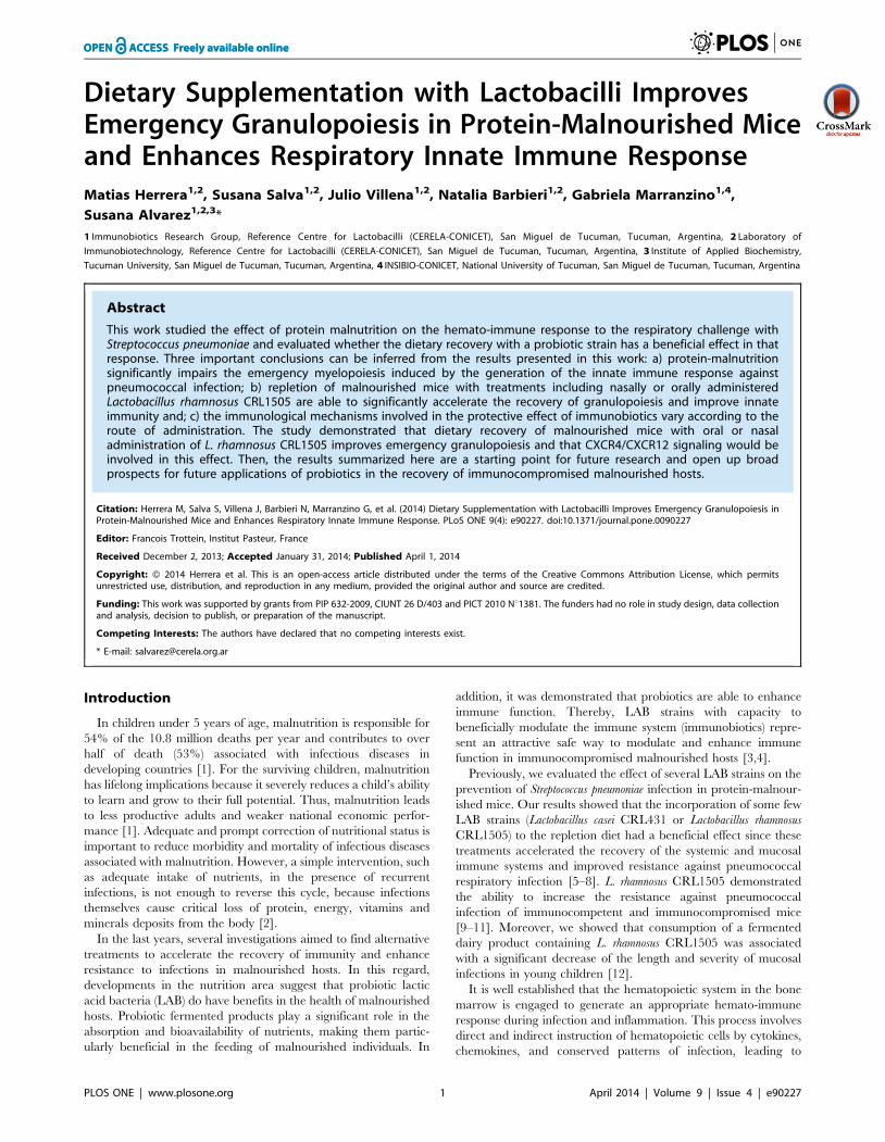
Dietary Supplementation with Lactobacilli Improves Emergency Granulopoiesis in Protein-Malnourished...
13
Dietary Supplementation with Lactobacilli Improves Emergency Granulopoiesis in Protein-Malnourished Mice and Enhances Respiratory Innate Immune Response Matias Herrera 1,2 , Susana Salva 1,2 , Julio Villena 1,2 , Natalia Barbieri 1,2 , Gabriela Marranzino 1,4 , Susana Alvarez 1,2,3 * 1 Immunobiotics Research Group, Reference Centre for Lactobacilli (CERELA-CONICET), San Miguel de Tucuman, Tucuman, Argentina, 2 Laboratory of Immunobiotechnology, Reference Centre for Lactobacilli (CERELA-CONICET), San Miguel de Tucuman, Tucuman, Argentina, 3 Institute of Applied Biochemistry, Tucuman University, San Miguel de Tucuman, Tucuman, Argentina, 4 INSIBIO-CONICET, National University of Tucuman, San Miguel de Tucuman, Tucuman, Argentina Abstract This work studied the effect of protein malnutrition on the hemato-immune response to the respiratory challenge with Streptococcus pneumoniae and evaluated whether the dietary recovery with a probiotic strain has a beneficial effect in that response. Three important conclusions can be inferred from the results presented in this work: a) protein-malnutrition significantly impairs the emergency myelopoiesis induced by the generation of the innate immune response against pneumococcal infection; b) repletion of malnourished mice with treatments including nasally or orally administered Lactobacillus rhamnosus CRL1505 are able to significantly accelerate the recovery of granulopoiesis and improve innate immunity and; c) the immunological mechanisms involved in the protective effect of immunobiotics vary according to the route of administration. The study demonstrated that dietary recovery of malnourished mice with oral or nasal administration of L. rhamnosus CRL1505 improves emergency granulopoiesis and that CXCR4/CXCR12 signaling would be involved in this effect. Then, the results summarized here are a starting point for future research and open up broad prospects for future applications of probiotics in the recovery of immunocompromised malnourished hosts. Citation: Herrera M, Salva S, Villena J, Barbieri N, Marranzino G, et al. (2014) Dietary Supplementation with Lactobacilli Improves Emergency Granulopoiesis in Protein-Malnourished Mice and Enhances Respiratory Innate Immune Response. PLoS ONE 9(4): e90227. doi:10.1371/journal.pone.0090227 Editor: Francois Trottein, Institut Pasteur, France Received December 2, 2013; Accepted January 31, 2014; Published April 1, 2014 Copyright: ß 2014 Herrera et al. This is an open-access article distributed under the terms of the Creative Commons Attribution License, which permits unrestricted use, distribution, and reproduction in any medium, provided the original author and source are credited. Funding: This work was supported by grants from PIP 632-2009, CIUNT 26 D/403 and PICT 2010 Nu1381. The funders had no role in study design, data collection and analysis, decision to publish, or preparation of the manuscript. Competing Interests: The authors have declared that no competing interests exist. * E-mail: [email protected] Introduction In children under 5 years of age, malnutrition is responsible for 54% of the 10.8 million deaths per year and contributes to over half of death (53%) associated with infectious diseases in developing countries [1]. For the surviving children, malnutrition has lifelong implications because it severely reduces a child’s ability to learn and grow to their full potential. Thus, malnutrition leads to less productive adults and weaker national economic perfor- mance [1]. Adequate and prompt correction of nutritional status is important to reduce morbidity and mortality of infectious diseases associated with malnutrition. However, a simple intervention, such as adequate intake of nutrients, in the presence of recurrent infections, is not enough to reverse this cycle, because infections themselves cause critical loss of protein, energy, vitamins and minerals deposits from the body [2]. In the last years, several investigations aimed to find alternative treatments to accelerate the recovery of immunity and enhance resistance to infections in malnourished hosts. In this regard, developments in the nutrition area suggest that probiotic lactic acid bacteria (LAB) do have benefits in the health of malnourished hosts. Probiotic fermented products play a significant role in the absorption and bioavailability of nutrients, making them partic- ularly beneficial in the feeding of malnourished individuals. In addition, it was demonstrated that probiotics are able to enhance immune function. Thereby, LAB strains with capacity to beneficially modulate the immune system (immunobiotics) repre- sent an attractive safe way to modulate and enhance immune function in immunocompromised malnourished hosts [3,4]. Previously, we evaluated the effect of several LAB strains on the prevention of Streptococcus pneumoniae infection in protein-malnour- ished mice. Our results showed that the incorporation of some few LAB strains (Lactobacillus casei CRL431 or Lactobacillus rhamnosus CRL1505) to the repletion diet had a beneficial effect since these treatments accelerated the recovery of the systemic and mucosal immune systems and improved resistance against pneumococcal respiratory infection [5–8]. L. rhamnosus CRL1505 demonstrated the ability to increase the resistance against pneumococcal infection of immunocompetent and immunocompromised mice [9–11]. Moreover, we showed that consumption of a fermented dairy product containing L. rhamnosus CRL1505 was associated with a significant decrease of the length and severity of mucosal infections in young children [12]. It is well established that the hematopoietic system in the bone marrow is engaged to generate an appropriate hemato-immune response during infection and inflammation. This process involves direct and indirect instruction of hematopoietic cells by cytokines, chemokines, and conserved patterns of infection, leading to PLOS ONE | www.plosone.org 1 April 2014 | Volume 9 | Issue 4 | e90227
-
Upload
independent -
Category
Documents
-
view
1 -
download
0
Transcript of Dietary Supplementation with Lactobacilli Improves Emergency Granulopoiesis in Protein-Malnourished...
Dietary Supplementation with Lactobacilli ImprovesEmergency Granulopoiesis in Protein-Malnourished Miceand Enhances Respiratory Innate Immune ResponseMatias Herrera1,2, Susana Salva1,2, Julio Villena1,2, Natalia Barbieri1,2, Gabriela Marranzino1,4,
Susana Alvarez1,2,3*
1 Immunobiotics Research Group, Reference Centre for Lactobacilli (CERELA-CONICET), San Miguel de Tucuman, Tucuman, Argentina, 2 Laboratory of
Immunobiotechnology, Reference Centre for Lactobacilli (CERELA-CONICET), San Miguel de Tucuman, Tucuman, Argentina, 3 Institute of Applied Biochemistry,
Tucuman University, San Miguel de Tucuman, Tucuman, Argentina, 4 INSIBIO-CONICET, National University of Tucuman, San Miguel de Tucuman, Tucuman, Argentina
Abstract
This work studied the effect of protein malnutrition on the hemato-immune response to the respiratory challenge withStreptococcus pneumoniae and evaluated whether the dietary recovery with a probiotic strain has a beneficial effect in thatresponse. Three important conclusions can be inferred from the results presented in this work: a) protein-malnutritionsignificantly impairs the emergency myelopoiesis induced by the generation of the innate immune response againstpneumococcal infection; b) repletion of malnourished mice with treatments including nasally or orally administeredLactobacillus rhamnosus CRL1505 are able to significantly accelerate the recovery of granulopoiesis and improve innateimmunity and; c) the immunological mechanisms involved in the protective effect of immunobiotics vary according to theroute of administration. The study demonstrated that dietary recovery of malnourished mice with oral or nasaladministration of L. rhamnosus CRL1505 improves emergency granulopoiesis and that CXCR4/CXCR12 signaling would beinvolved in this effect. Then, the results summarized here are a starting point for future research and open up broadprospects for future applications of probiotics in the recovery of immunocompromised malnourished hosts.
Citation: Herrera M, Salva S, Villena J, Barbieri N, Marranzino G, et al. (2014) Dietary Supplementation with Lactobacilli Improves Emergency Granulopoiesis inProtein-Malnourished Mice and Enhances Respiratory Innate Immune Response. PLoS ONE 9(4): e90227. doi:10.1371/journal.pone.0090227
Editor: Francois Trottein, Institut Pasteur, France
Received December 2, 2013; Accepted January 31, 2014; Published April 1, 2014
Copyright: � 2014 Herrera et al. This is an open-access article distributed under the terms of the Creative Commons Attribution License, which permitsunrestricted use, distribution, and reproduction in any medium, provided the original author and source are credited.
Funding: This work was supported by grants from PIP 632-2009, CIUNT 26 D/403 and PICT 2010 Nu1381. The funders had no role in study design, data collectionand analysis, decision to publish, or preparation of the manuscript.
Competing Interests: The authors have declared that no competing interests exist.
* E-mail: [email protected]
Introduction
In children under 5 years of age, malnutrition is responsible for
54% of the 10.8 million deaths per year and contributes to over
half of death (53%) associated with infectious diseases in
developing countries [1]. For the surviving children, malnutrition
has lifelong implications because it severely reduces a child’s ability
to learn and grow to their full potential. Thus, malnutrition leads
to less productive adults and weaker national economic perfor-
mance [1]. Adequate and prompt correction of nutritional status is
important to reduce morbidity and mortality of infectious diseases
associated with malnutrition. However, a simple intervention, such
as adequate intake of nutrients, in the presence of recurrent
infections, is not enough to reverse this cycle, because infections
themselves cause critical loss of protein, energy, vitamins and
minerals deposits from the body [2].
In the last years, several investigations aimed to find alternative
treatments to accelerate the recovery of immunity and enhance
resistance to infections in malnourished hosts. In this regard,
developments in the nutrition area suggest that probiotic lactic
acid bacteria (LAB) do have benefits in the health of malnourished
hosts. Probiotic fermented products play a significant role in the
absorption and bioavailability of nutrients, making them partic-
ularly beneficial in the feeding of malnourished individuals. In
addition, it was demonstrated that probiotics are able to enhance
immune function. Thereby, LAB strains with capacity to
beneficially modulate the immune system (immunobiotics) repre-
sent an attractive safe way to modulate and enhance immune
function in immunocompromised malnourished hosts [3,4].
Previously, we evaluated the effect of several LAB strains on the
prevention of Streptococcus pneumoniae infection in protein-malnour-
ished mice. Our results showed that the incorporation of some few
LAB strains (Lactobacillus casei CRL431 or Lactobacillus rhamnosus
CRL1505) to the repletion diet had a beneficial effect since these
treatments accelerated the recovery of the systemic and mucosal
immune systems and improved resistance against pneumococcal
respiratory infection [5–8]. L. rhamnosus CRL1505 demonstrated
the ability to increase the resistance against pneumococcal
infection of immunocompetent and immunocompromised mice
[9–11]. Moreover, we showed that consumption of a fermented
dairy product containing L. rhamnosus CRL1505 was associated
with a significant decrease of the length and severity of mucosal
infections in young children [12].
It is well established that the hematopoietic system in the bone
marrow is engaged to generate an appropriate hemato-immune
response during infection and inflammation. This process involves
direct and indirect instruction of hematopoietic cells by cytokines,
chemokines, and conserved patterns of infection, leading to
PLOS ONE | www.plosone.org 1 April 2014 | Volume 9 | Issue 4 | e90227

adapted hematopoietic generation and directed migration of cells
in need [13,14]. We performed studies to analyze the extent of the
damage induced by malnutrition on B cell development in the
spleen and bone marrow and its impact in B cell-mediated
immunity in the respiratory tract [11,15]; and we observed that
the number of B220+ cells (the whole B cell compartment) was
reduced in the bone marrow of malnourished mice [15].
Moreover, protein deprivation induced a significant reduction of
pro-B/pre-B (B220intermIgMneg) and immature B cell (B220inter-
mIgM+) suggesting that nutritional deprivation leads to the
alteration of B-cell development [15]. More recently, we
demonstrated that these alterations in B cell development
significantly impair the generation of the systemic and respiratory
B cell-mediated immunity, conducting to a reduced capacity of
malnourished mice to mount an appropriate humoral immune
response [11]. We also showed that the addition of L. rhamnosus
CRL1505 to repletion treatments was able to induce a recovery of
B cells and to normalize the numbers of bone marrow immature
B220 cells [15], splenic immature B cells and lung mature B
lymphocytes [11]. In addition, malnourished mice treated with L.
rhamnosus CRL1505 were able to establish an improved humoral
immune response against the respiratory pneumococcal infection
[11].
Previous work indicates that probiotic supplementation is able
to improve myelopoiesis in malnourished hosts [15]. However, the
capacity of immunobiotic strains to influence myeloid cells
development in malnourished hosts and the impact of such effect
on innate immunity have not been evaluated in depth. Therefore,
in the current study we aimed to: a) evaluate the effect of
malnutrition on myeloid cells development in bone marrow and its
systemic effect when emergency myelopoiesis is required during
the generation of the innate immune response against a respiratory
pathogen; b) study the effect of L. rhamnosus CRL1505 on
emergency myelopoiesis and respiratory innate immune response
against S. pneumoniae in protein-malnourished mice, in order to
gain insight in the knowledge of the mechanism involved in the
immunomodulatory effect of the CRL1505 strain. Moreover,
considering that nasally administered antigens can induce
respiratory and systemic immune responses superior to those
obtained using oral stimulation [4], we also aimed to c)
comparatively study the effect of orally and nasally administered
L. rhamnosus CRL1505 in order to establish which is the most
effective treatment to improve protection against pneumococcal
infection.
Results
Protein-malnutrition significantly impairs respiratory andsystemic innate immune responses againstpneumococcal infection
Malnourished mice were obtained after 21 days of feeding a
protein free-diet. Then, mice were replete for 7 days with a
balanced conventional diet (BD) or BD supplemented with orally
or nasally administered L. rhamnosus CRL1505 (BD+LrO and BD+LrN respectively). Malnourished (MNC) and well-nourished
(WNC) mice were used as controls.
We first studied the number and activity of respiratory
phagocytes in both WNC and MNC mice. No differences were
observed in number of bronchoalveolar lavages (BAL) leukocytes,
neutrophils and macrophages when the two groups were
compared (Figure S1). However, as we have previously reported
[5], MNC mice showed significantly lower levels of BAL NBT+
cells than WNC mice. S. pneumoniae infection induced the
recruitment of neutrophils and macrophages into the alveoli,
resulting in an increase in BAL leukocyte counts on day 2 post-
infection (Figure 1). MNC mice showed an impaired recruitment
of neutrophils and macrophages since significantly lower numbers
of leukocytes, CD45+Gr1-AF+ macrophages and CD45+Gr1+
neutrophils were found in BAL when compared with WNC mice
(Figure 1). In addition to the quantitative alterations of BAL
phagocytes, we also observed that malnutrition significantly
impaired phagocytic activity of BAL cells (Figure 1).
A decreased number of leukocytes and neutrophils as well as
reduced peroxidase scores were detected in MNC mice than in
WNC group (Figure S2; Figure 2). We confirmed the impairment
of blood neutrophils induced by malnutrition by evaluating the
numbers of blood Gr1+, Gr1high and Gr1low cells. Significantly
lower levels of Gr1+ and Gr1high cells were detected in MNC mice
while the numbers of blood Gr1low cells were not different from
WNC mice (Figure 2). S. pneumoniae infection increased blood
leukocytes and neutrophils counts, peroxidase scores and blood
Gr1+ and Gr1high numbers in both experimental groups; however,
all these parameters were significantly lower in MNC than in the
WNC group (Figure 2). Gr1low cells were also increased by the
pneumococcal infection; however, the levels of these cells were
equal in both groups (Figure 2). In addition, CD34+ blood cells
were significantly decreased in MNC (Figure 2). After the
challenge with pneumococci, blood CD34+ cells were significantly
increased in both experimental groups in relation to basal levels.
However, MNC mice showed significantly higher numbers of
blood CD34+ cells than WMC mice (Figure 2).
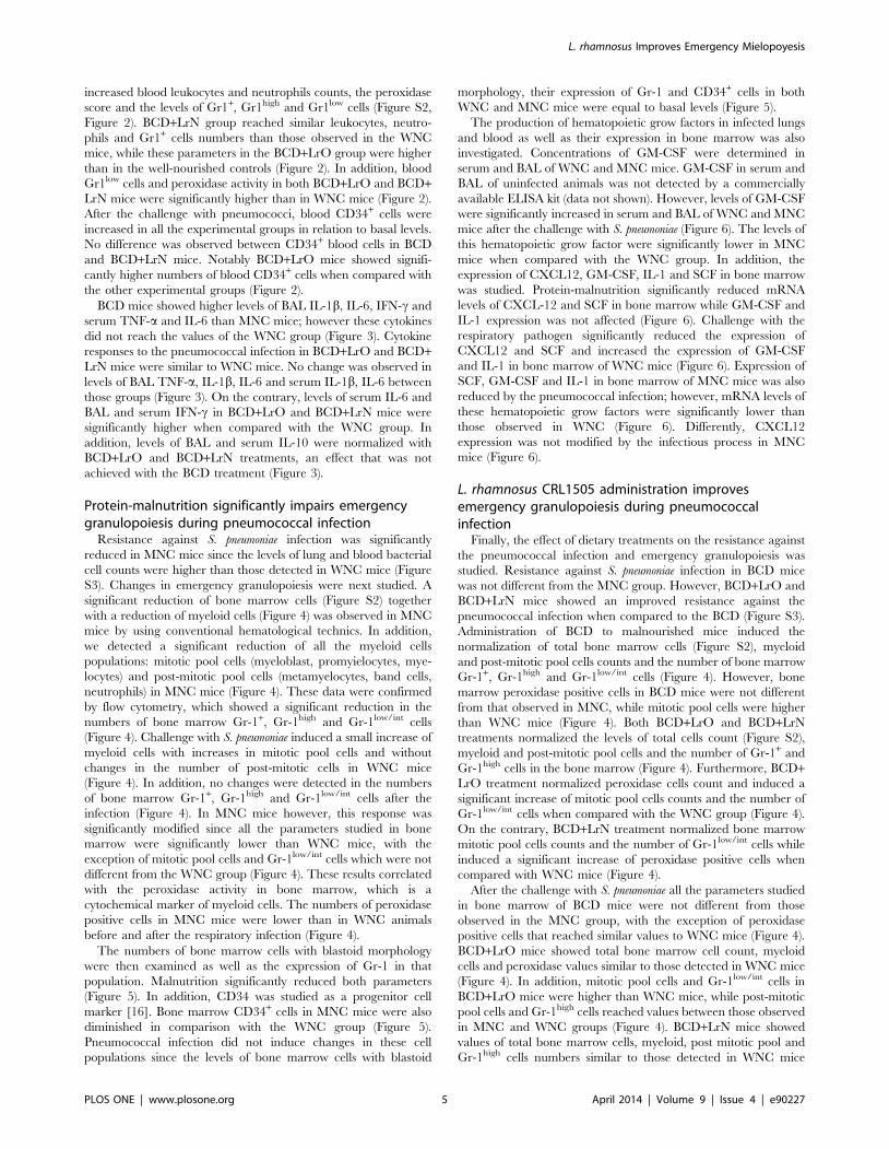
The levels of TNF-a, IL-1b, IL-6, IFN-c and IL-10 were
studied in serum and BAL samples before the challenge with S.
pneumoniae. No significant difference on BAL and serum TNF-aand IL-1b was found between WNC and MNC mice (Figure 3).
On the contrary, levels of BAL and serum IL-6, IFN-c and IL-10
were significantly reduced in MNC mice when compered with the
WNC group. Challenge with the respiratory pathogen induced an
early increase (12 hours post-infection) of TNF-a, IL-1b and IL-6
levels and a late increase of IFN-c (48 hours post-infection) in BAL
and serum, in both experimental groups. However MNC mice
showed significantly lower levels of serum and BAL TNF-a, IL-1b,
IL-6 and IFN-c than WNC mice (Figure 3). Infection also
increased the levels of the immunoregulatory cytokine IL-10 in
serum and BAL of WNC mice; however, MNC mice were not able
to increase the production of this cytokine in response to pathogen
(Figure 3).
L. rhamnosus CRL1505 administration enhancesrespiratory and systemic innate immune responses inmalnourished mice
We next evaluated the effect of different repletion treatments on
blood and BAL cells affected by malnutrition. No significant
differences were observed in the number of BAL leukocytes,
macrophages and neutrophils between BCD, BCD+LrO and
BCD+LrN mice and controls (MNC and WNC mice) (Figure S1).
In addition, BCD, BCD+LrO and BCD+LrN mice showed
normal values of BAL NBT+ cells (Figure S1). After the challenge
with S. pneumoniae, treatment of malnourished mice with BCD
increased the number of BAL leukocytes and CD45+Gr1-AF+
macrophages; however, the treatment was not able to normalize
these parameters. Moreover, numbers of BAL CD45+Gr1+
neutrophils and percentages of NBT+ from BCD mice were not
different from the MNC group (Figure 1). Treatment with BCD+LrO significantly increased the number of BAL neutrophils and
macrophages, as well as phagocytes’ activity in response to the
infection. In fact BCD+LrO mice showed values of BAL
leukocytes and CD45+Gr1-AF+ macrophages that were similar
L. rhamnosus Improves Emergency Mielopoyesis
PLOS ONE | www.plosone.org 2 April 2014 | Volume 9 | Issue 4 | e90227
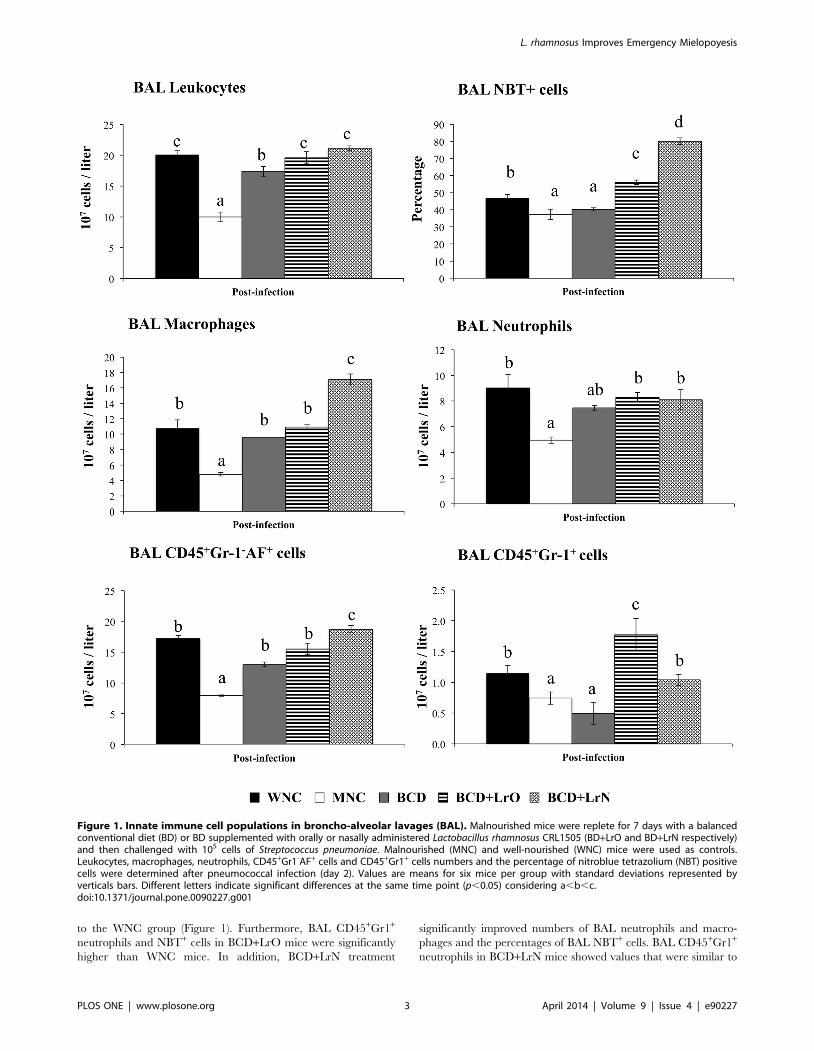
to the WNC group (Figure 1). Furthermore, BAL CD45+Gr1+
neutrophils and NBT+ cells in BCD+LrO mice were significantly
higher than WNC mice. In addition, BCD+LrN treatment
significantly improved numbers of BAL neutrophils and macro-
phages and the percentages of BAL NBT+ cells. BAL CD45+Gr1+
neutrophils in BCD+LrN mice showed values that were similar to
Figure 1. Innate immune cell populations in broncho-alveolar lavages (BAL). Malnourished mice were replete for 7 days with a balancedconventional diet (BD) or BD supplemented with orally or nasally administered Lactobacillus rhamnosus CRL1505 (BD+LrO and BD+LrN respectively)and then challenged with 105 cells of Streptococcus pneumoniae. Malnourished (MNC) and well-nourished (WNC) mice were used as controls.Leukocytes, macrophages, neutrophils, CD45+Gr1-AF+ cells and CD45+Gr1+ cells numbers and the percentage of nitroblue tetrazolium (NBT) positivecells were determined after pneumococcal infection (day 2). Values are means for six mice per group with standard deviations represented byverticals bars. Different letters indicate significant differences at the same time point (p,0.05) considering a,b,c.doi:10.1371/journal.pone.0090227.g001
L. rhamnosus Improves Emergency Mielopoyesis
PLOS ONE | www.plosone.org 3 April 2014 | Volume 9 | Issue 4 | e90227
the WNC group (Figure 1). However, values of BAL
CD45+Gr1-AF+ macrophages and NBT+ cells in BCD+LrN mice
were significantly higher than those observed in the WNC group
(Figure 1).
Treatment with BCD slightly increased blood neutrophils
numbers, peroxidase scores and Gr1+ and Gr1high cells; however,
the values did not reach the levels of WNC mice (Figure 2). On the
contrary, repletion with both BCD+LrO and BCD+LrN signifi-
cantly increased blood leukocytes (Figure S2), neutrophils, Gr1+
and Gr1high cells numbers reaching similar values than those
observed in WNC mice (Figure 2). Additionally, blood peroxidase
activity and Gr1low cells in BCD+LrO and BCD+LrN mice were
significantly higher than in WNC mice (Figure 2). After S.
pneumoniae challenge, malnourished mice treated with BCD
showed significantly higher levels of neutrophils and Gr1high cells
than MNC mice; however blood peroxidase activity and Gr1low
cells were not different from malnourished controls (Figure 2).
Treatment with both BCD+LrO and BCD+LrN significantly
Figure 2. Innate immune cell populations in blood. Malnourished mice were replete for 7 days with a balanced conventional diet (BD) or BDsupplemented with orally or nasally administered Lactobacillus rhamnosus CRL1505 (BD+LrO and BD+LrN respectively) and then challenged with 105
cells of Streptococcus pneumoniae. Malnourished (MNC) and well-nourished (WNC) mice were used as controls. Blood neutrophils, CD34+ cells, Gr1+
cells, Gr1high cells and Gr1low/int cells numbers and the score of blood neutrophils myeloperoxidase were determined before (basal) and afterpneumococcal infection (day 2). Values are means for six mice per group with standard deviations represented by verticals bars. Different lettersindicate significant differences at the same time point (p,0.05) considering a,b,c.doi:10.1371/journal.pone.0090227.g002
Figure 3. Cytokines in broncho-alveolar lavages (BAL) and blood. Malnourished mice were replete for 7 days with a balanced conventionaldiet (BD) or BD supplemented with orally or nasally administered Lactobacillus rhamnosus CRL1505 (BD+LrO and BD+LrN respectively) and thenchallenged with 105 cells of Streptococcus pneumoniae. Malnourished (MNC) and well-nourished (WNC) mice were used as controls. Levels of BAL andserum TNF-a, IL-1b, IL-6, IFN-c and IL-10 were determined before (basal) and after pneumococcal infection (hours 12 and 48). Values are means for sixmice per group with standard deviations represented by verticals bars. Different letters indicate significant differences at the same time point(p,0.05) considering a,b,c.doi:10.1371/journal.pone.0090227.g003
L. rhamnosus Improves Emergency Mielopoyesis
PLOS ONE | www.plosone.org 4 April 2014 | Volume 9 | Issue 4 | e90227
increased blood leukocytes and neutrophils counts, the peroxidase
score and the levels of Gr1+, Gr1high and Gr1low cells (Figure S2,
Figure 2). BCD+LrN group reached similar leukocytes, neutro-
phils and Gr1+ cells numbers than those observed in the WNC
mice, while these parameters in the BCD+LrO group were higher
than in the well-nourished controls (Figure 2). In addition, blood
Gr1low cells and peroxidase activity in both BCD+LrO and BCD+LrN mice were significantly higher than in WNC mice (Figure 2).
After the challenge with pneumococci, blood CD34+ cells were
increased in all the experimental groups in relation to basal levels.
No difference was observed between CD34+ blood cells in BCD
and BCD+LrN mice. Notably BCD+LrO mice showed signifi-
cantly higher numbers of blood CD34+ cells when compared with
the other experimental groups (Figure 2).
BCD mice showed higher levels of BAL IL-1b, IL-6, IFN-c and
serum TNF-a and IL-6 than MNC mice; however these cytokines
did not reach the values of the WNC group (Figure 3). Cytokine
responses to the pneumococcal infection in BCD+LrO and BCD+LrN mice were similar to WNC mice. No change was observed in
levels of BAL TNF-a, IL-1b, IL-6 and serum IL-1b, IL-6 between
those groups (Figure 3). On the contrary, levels of serum IL-6 and
BAL and serum IFN-c in BCD+LrO and BCD+LrN mice were
significantly higher when compared with the WNC group. In
addition, levels of BAL and serum IL-10 were normalized with
BCD+LrO and BCD+LrN treatments, an effect that was not
achieved with the BCD treatment (Figure 3).
Protein-malnutrition significantly impairs emergencygranulopoiesis during pneumococcal infection
Resistance against S. pneumoniae infection was significantly
reduced in MNC mice since the levels of lung and blood bacterial
cell counts were higher than those detected in WNC mice (Figure
S3). Changes in emergency granulopoiesis were next studied. A
significant reduction of bone marrow cells (Figure S2) together
with a reduction of myeloid cells (Figure 4) was observed in MNC
mice by using conventional hematological technics. In addition,
we detected a significant reduction of all the myeloid cells
populations: mitotic pool cells (myeloblast, promyielocytes, mye-
locytes) and post-mitotic pool cells (metamyelocytes, band cells,
neutrophils) in MNC mice (Figure 4). These data were confirmed
by flow cytometry, which showed a significant reduction in the
numbers of bone marrow Gr-1+, Gr-1high and Gr-1low/int cells
(Figure 4). Challenge with S. pneumoniae induced a small increase of
myeloid cells with increases in mitotic pool cells and without
changes in the number of post-mitotic cells in WNC mice
(Figure 4). In addition, no changes were detected in the numbers
of bone marrow Gr-1+, Gr-1high and Gr-1low/int cells after the
infection (Figure 4). In MNC mice however, this response was
significantly modified since all the parameters studied in bone
marrow were significantly lower than WNC mice, with the
exception of mitotic pool cells and Gr-1low/int cells which were not
different from the WNC group (Figure 4). These results correlated
with the peroxidase activity in bone marrow, which is a
cytochemical marker of myeloid cells. The numbers of peroxidase
positive cells in MNC mice were lower than in WNC animals
before and after the respiratory infection (Figure 4).
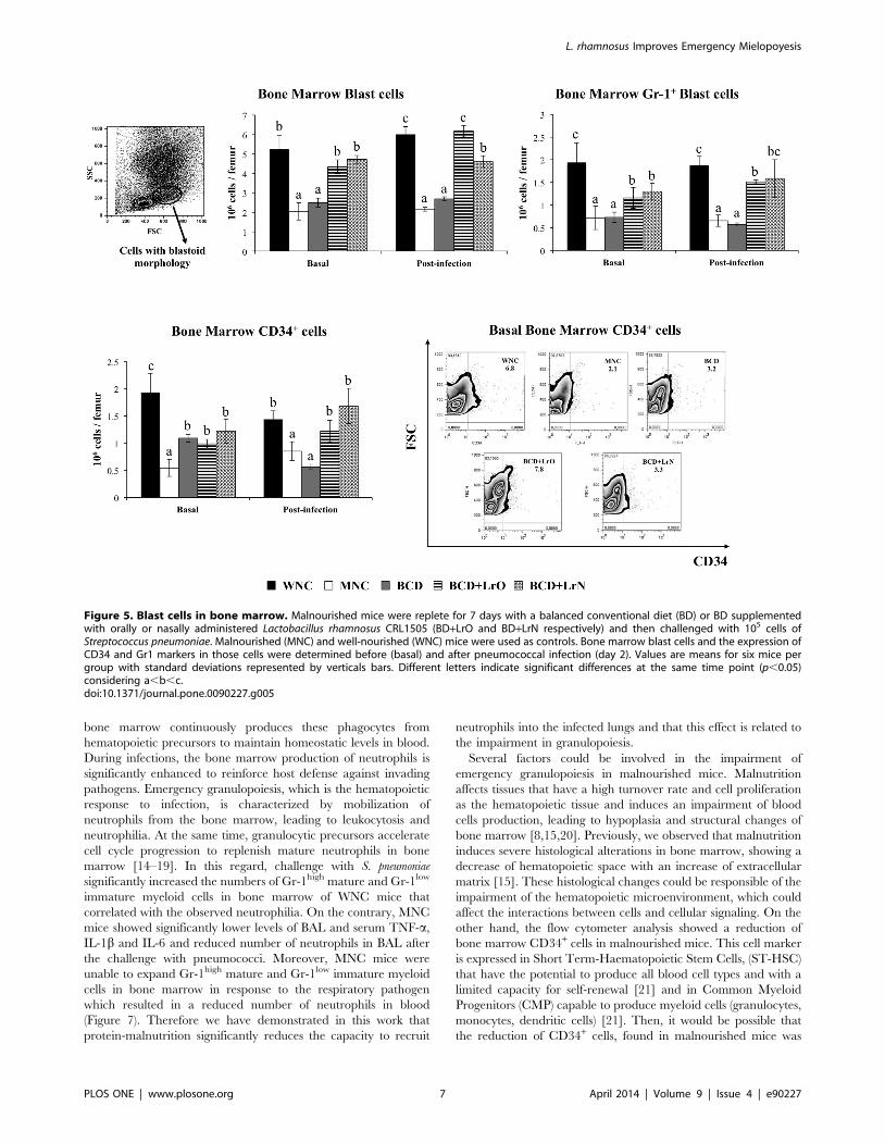
The numbers of bone marrow cells with blastoid morphology
were then examined as well as the expression of Gr-1 in that
population. Malnutrition significantly reduced both parameters
(Figure 5). In addition, CD34 was studied as a progenitor cell
marker [16]. Bone marrow CD34+ cells in MNC mice were also
diminished in comparison with the WNC group (Figure 5).
Pneumococcal infection did not induce changes in these cell
populations since the levels of bone marrow cells with blastoid
morphology, their expression of Gr-1 and CD34+ cells in both
WNC and MNC mice were equal to basal levels (Figure 5).
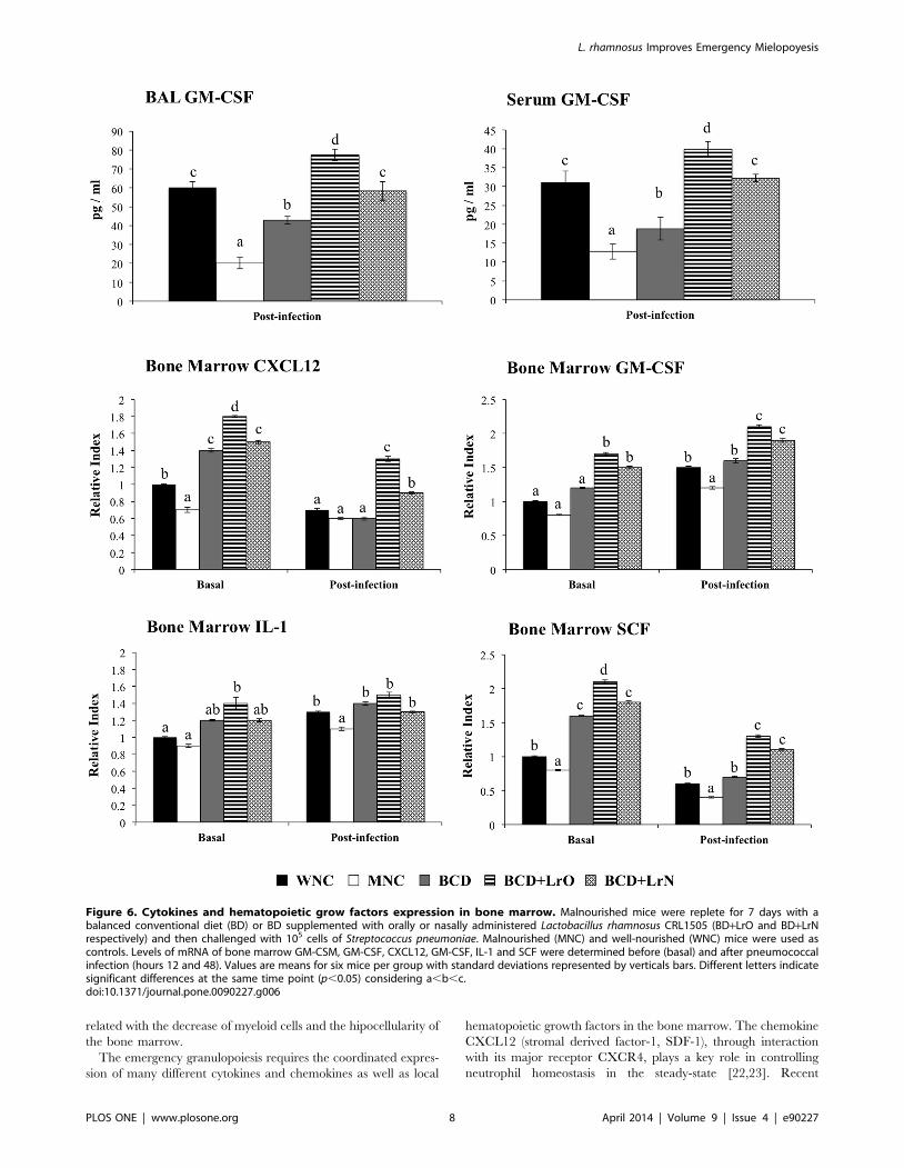
The production of hematopoietic grow factors in infected lungs
and blood as well as their expression in bone marrow was also
investigated. Concentrations of GM-CSF were determined in
serum and BAL of WNC and MNC mice. GM-CSF in serum and
BAL of uninfected animals was not detected by a commercially
available ELISA kit (data not shown). However, levels of GM-CSF
were significantly increased in serum and BAL of WNC and MNC
mice after the challenge with S. pneumoniae (Figure 6). The levels of
this hematopoietic grow factor were significantly lower in MNC
mice when compared with the WNC group. In addition, the
expression of CXCL12, GM-CSF, IL-1 and SCF in bone marrow
was studied. Protein-malnutrition significantly reduced mRNA
levels of CXCL-12 and SCF in bone marrow while GM-CSF and
IL-1 expression was not affected (Figure 6). Challenge with the
respiratory pathogen significantly reduced the expression of
CXCL12 and SCF and increased the expression of GM-CSF
and IL-1 in bone marrow of WNC mice (Figure 6). Expression of
SCF, GM-CSF and IL-1 in bone marrow of MNC mice was also
reduced by the pneumococcal infection; however, mRNA levels of
these hematopoietic grow factors were significantly lower than
those observed in WNC (Figure 6). Differently, CXCL12
expression was not modified by the infectious process in MNC
mice (Figure 6).
L. rhamnosus CRL1505 administration improvesemergency granulopoiesis during pneumococcalinfection
Finally, the effect of dietary treatments on the resistance against
the pneumococcal infection and emergency granulopoiesis was
studied. Resistance against S. pneumoniae infection in BCD mice
was not different from the MNC group. However, BCD+LrO and
BCD+LrN mice showed an improved resistance against the
pneumococcal infection when compared to the BCD (Figure S3).
Administration of BCD to malnourished mice induced the
normalization of total bone marrow cells (Figure S2), myeloid
and post-mitotic pool cells counts and the number of bone marrow
Gr-1+, Gr-1high and Gr-1low/int cells (Figure 4). However, bone
marrow peroxidase positive cells in BCD mice were not different
from that observed in MNC, while mitotic pool cells were higher
than WNC mice (Figure 4). Both BCD+LrO and BCD+LrN
treatments normalized the levels of total cells count (Figure S2),
myeloid and post-mitotic pool cells and the number of Gr-1+ and
Gr-1high cells in the bone marrow (Figure 4). Furthermore, BCD+LrO treatment normalized peroxidase cells count and induced a
significant increase of mitotic pool cells counts and the number of
Gr-1low/int cells when compared with the WNC group (Figure 4).
On the contrary, BCD+LrN treatment normalized bone marrow
mitotic pool cells counts and the number of Gr-1low/int cells while
induced a significant increase of peroxidase positive cells when
compared with WNC mice (Figure 4).
After the challenge with S. pneumoniae all the parameters studied
in bone marrow of BCD mice were not different from those
observed in the MNC group, with the exception of peroxidase
positive cells that reached similar values to WNC mice (Figure 4).
BCD+LrO mice showed total bone marrow cell count, myeloid
cells and peroxidase values similar to those detected in WNC mice
(Figure 4). In addition, mitotic pool cells and Gr-1low/int cells in
BCD+LrO mice were higher than WNC mice, while post-mitotic
pool cells and Gr-1high cells reached values between those observed
in MNC and WNC groups (Figure 4). BCD+LrN mice showed
values of total bone marrow cells, myeloid, post mitotic pool and
Gr-1high cells numbers similar to those detected in WNC mice
L. rhamnosus Improves Emergency Mielopoyesis
PLOS ONE | www.plosone.org 5 April 2014 | Volume 9 | Issue 4 | e90227
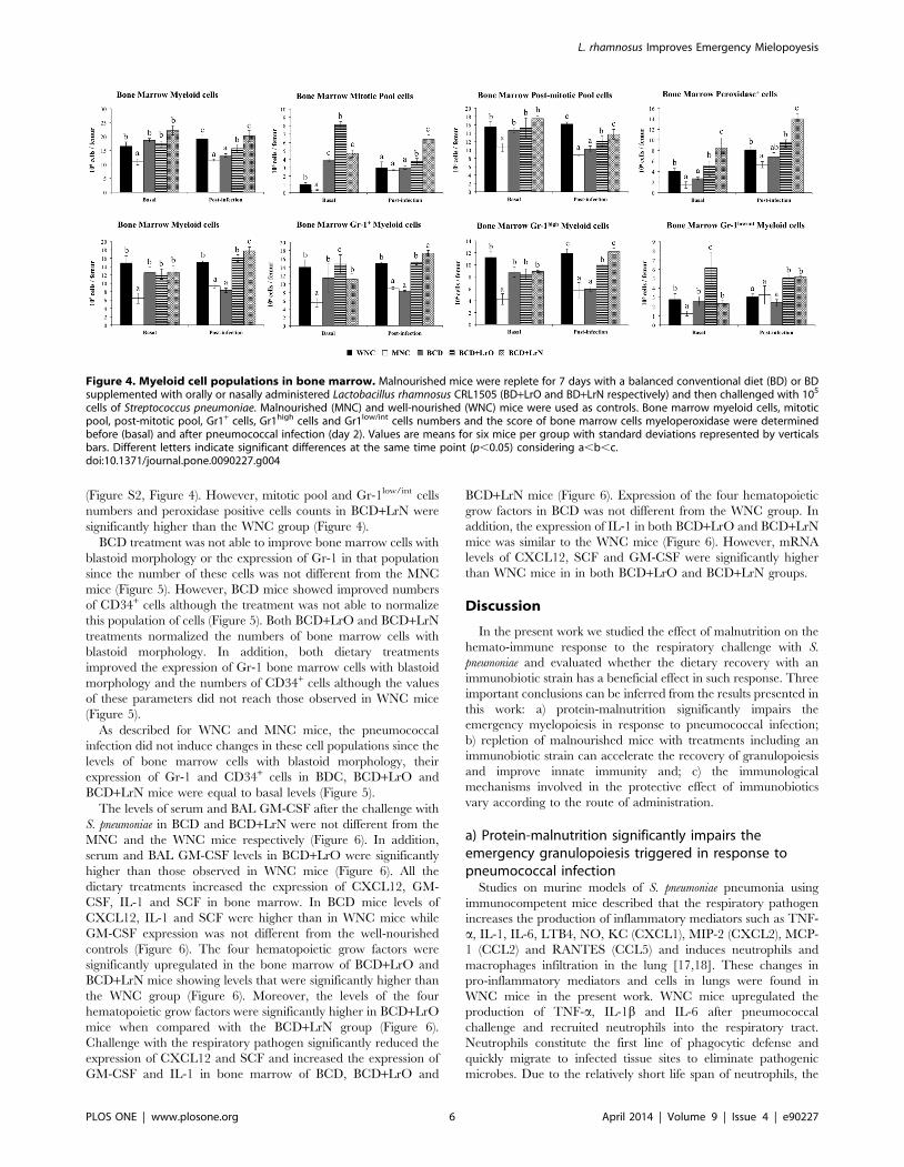
(Figure S2, Figure 4). However, mitotic pool and Gr-1low/int cells
numbers and peroxidase positive cells counts in BCD+LrN were
significantly higher than the WNC group (Figure 4).
BCD treatment was not able to improve bone marrow cells with
blastoid morphology or the expression of Gr-1 in that population
since the number of these cells was not different from the MNC
mice (Figure 5). However, BCD mice showed improved numbers
of CD34+ cells although the treatment was not able to normalize
this population of cells (Figure 5). Both BCD+LrO and BCD+LrN
treatments normalized the numbers of bone marrow cells with
blastoid morphology. In addition, both dietary treatments
improved the expression of Gr-1 bone marrow cells with blastoid
morphology and the numbers of CD34+ cells although the values
of these parameters did not reach those observed in WNC mice
(Figure 5).
As described for WNC and MNC mice, the pneumococcal
infection did not induce changes in these cell populations since the
levels of bone marrow cells with blastoid morphology, their
expression of Gr-1 and CD34+ cells in BDC, BCD+LrO and
BCD+LrN mice were equal to basal levels (Figure 5).
The levels of serum and BAL GM-CSF after the challenge with
S. pneumoniae in BCD and BCD+LrN were not different from the
MNC and the WNC mice respectively (Figure 6). In addition,
serum and BAL GM-CSF levels in BCD+LrO were significantly
higher than those observed in WNC mice (Figure 6). All the
dietary treatments increased the expression of CXCL12, GM-
CSF, IL-1 and SCF in bone marrow. In BCD mice levels of
CXCL12, IL-1 and SCF were higher than in WNC mice while
GM-CSF expression was not different from the well-nourished
controls (Figure 6). The four hematopoietic grow factors were
significantly upregulated in the bone marrow of BCD+LrO and
BCD+LrN mice showing levels that were significantly higher than
the WNC group (Figure 6). Moreover, the levels of the four
hematopoietic grow factors were significantly higher in BCD+LrO
mice when compared with the BCD+LrN group (Figure 6).
Challenge with the respiratory pathogen significantly reduced the
expression of CXCL12 and SCF and increased the expression of
GM-CSF and IL-1 in bone marrow of BCD, BCD+LrO and
BCD+LrN mice (Figure 6). Expression of the four hematopoietic
grow factors in BCD was not different from the WNC group. In
addition, the expression of IL-1 in both BCD+LrO and BCD+LrN
mice was similar to the WNC mice (Figure 6). However, mRNA
levels of CXCL12, SCF and GM-CSF were significantly higher
than WNC mice in in both BCD+LrO and BCD+LrN groups.
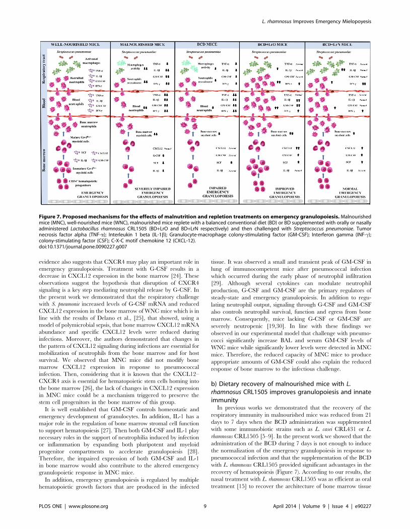
Discussion
In the present work we studied the effect of malnutrition on the
hemato-immune response to the respiratory challenge with S.
pneumoniae and evaluated whether the dietary recovery with an
immunobiotic strain has a beneficial effect in such response. Three
important conclusions can be inferred from the results presented in
this work: a) protein-malnutrition significantly impairs the
emergency myelopoiesis in response to pneumococcal infection;
b) repletion of malnourished mice with treatments including an
immunobiotic strain can accelerate the recovery of granulopoiesis
and improve innate immunity and; c) the immunological
mechanisms involved in the protective effect of immunobiotics
vary according to the route of administration.
a) Protein-malnutrition significantly impairs theemergency granulopoiesis triggered in response topneumococcal infection
Studies on murine models of S. pneumoniae pneumonia using
immunocompetent mice described that the respiratory pathogen
increases the production of inflammatory mediators such as TNF-
a, IL-1, IL-6, LTB4, NO, KC (CXCL1), MIP-2 (CXCL2), MCP-
1 (CCL2) and RANTES (CCL5) and induces neutrophils and
macrophages infiltration in the lung [17,18]. These changes in
pro-inflammatory mediators and cells in lungs were found in
WNC mice in the present work. WNC mice upregulated the
production of TNF-a, IL-1b and IL-6 after pneumococcal
challenge and recruited neutrophils into the respiratory tract.
Neutrophils constitute the first line of phagocytic defense and
quickly migrate to infected tissue sites to eliminate pathogenic
microbes. Due to the relatively short life span of neutrophils, the
Figure 4. Myeloid cell populations in bone marrow. Malnourished mice were replete for 7 days with a balanced conventional diet (BD) or BDsupplemented with orally or nasally administered Lactobacillus rhamnosus CRL1505 (BD+LrO and BD+LrN respectively) and then challenged with 105
cells of Streptococcus pneumoniae. Malnourished (MNC) and well-nourished (WNC) mice were used as controls. Bone marrow myeloid cells, mitoticpool, post-mitotic pool, Gr1+ cells, Gr1high cells and Gr1low/int cells numbers and the score of bone marrow cells myeloperoxidase were determinedbefore (basal) and after pneumococcal infection (day 2). Values are means for six mice per group with standard deviations represented by verticalsbars. Different letters indicate significant differences at the same time point (p,0.05) considering a,b,c.doi:10.1371/journal.pone.0090227.g004
L. rhamnosus Improves Emergency Mielopoyesis
PLOS ONE | www.plosone.org 6 April 2014 | Volume 9 | Issue 4 | e90227
bone marrow continuously produces these phagocytes from
hematopoietic precursors to maintain homeostatic levels in blood.
During infections, the bone marrow production of neutrophils is
significantly enhanced to reinforce host defense against invading
pathogens. Emergency granulopoiesis, which is the hematopoietic
response to infection, is characterized by mobilization of
neutrophils from the bone marrow, leading to leukocytosis and
neutrophilia. At the same time, granulocytic precursors accelerate
cell cycle progression to replenish mature neutrophils in bone
marrow [14–19]. In this regard, challenge with S. pneumoniae
significantly increased the numbers of Gr-1high mature and Gr-1low
immature myeloid cells in bone marrow of WNC mice that
correlated with the observed neutrophilia. On the contrary, MNC
mice showed significantly lower levels of BAL and serum TNF-a,
IL-1b and IL-6 and reduced number of neutrophils in BAL after
the challenge with pneumococci. Moreover, MNC mice were
unable to expand Gr-1high mature and Gr-1low immature myeloid
cells in bone marrow in response to the respiratory pathogen
which resulted in a reduced number of neutrophils in blood
(Figure 7). Therefore we have demonstrated in this work that
protein-malnutrition significantly reduces the capacity to recruit
neutrophils into the infected lungs and that this effect is related to
the impairment in granulopoiesis.
Several factors could be involved in the impairment of
emergency granulopoiesis in malnourished mice. Malnutrition
affects tissues that have a high turnover rate and cell proliferation
as the hematopoietic tissue and induces an impairment of blood
cells production, leading to hypoplasia and structural changes of
bone marrow [8,15,20]. Previously, we observed that malnutrition
induces severe histological alterations in bone marrow, showing a
decrease of hematopoietic space with an increase of extracellular
matrix [15]. These histological changes could be responsible of the
impairment of the hematopoietic microenvironment, which could
affect the interactions between cells and cellular signaling. On the
other hand, the flow cytometer analysis showed a reduction of
bone marrow CD34+ cells in malnourished mice. This cell marker
is expressed in Short Term-Haematopoietic Stem Cells, (ST-HSC)
that have the potential to produce all blood cell types and with a
limited capacity for self-renewal [21] and in Common Myeloid
Progenitors (CMP) capable to produce myeloid cells (granulocytes,
monocytes, dendritic cells) [21]. Then, it would be possible that
the reduction of CD34+ cells, found in malnourished mice was
Figure 5. Blast cells in bone marrow. Malnourished mice were replete for 7 days with a balanced conventional diet (BD) or BD supplementedwith orally or nasally administered Lactobacillus rhamnosus CRL1505 (BD+LrO and BD+LrN respectively) and then challenged with 105 cells ofStreptococcus pneumoniae. Malnourished (MNC) and well-nourished (WNC) mice were used as controls. Bone marrow blast cells and the expression ofCD34 and Gr1 markers in those cells were determined before (basal) and after pneumococcal infection (day 2). Values are means for six mice pergroup with standard deviations represented by verticals bars. Different letters indicate significant differences at the same time point (p,0.05)considering a,b,c.doi:10.1371/journal.pone.0090227.g005
L. rhamnosus Improves Emergency Mielopoyesis
PLOS ONE | www.plosone.org 7 April 2014 | Volume 9 | Issue 4 | e90227
related with the decrease of myeloid cells and the hipocellularity of
the bone marrow.
The emergency granulopoiesis requires the coordinated expres-
sion of many different cytokines and chemokines as well as local
hematopoietic growth factors in the bone marrow. The chemokine
CXCL12 (stromal derived factor-1, SDF-1), through interaction
with its major receptor CXCR4, plays a key role in controlling
neutrophil homeostasis in the steady-state [22,23]. Recent
Figure 6. Cytokines and hematopoietic grow factors expression in bone marrow. Malnourished mice were replete for 7 days with abalanced conventional diet (BD) or BD supplemented with orally or nasally administered Lactobacillus rhamnosus CRL1505 (BD+LrO and BD+LrNrespectively) and then challenged with 105 cells of Streptococcus pneumoniae. Malnourished (MNC) and well-nourished (WNC) mice were used ascontrols. Levels of mRNA of bone marrow GM-CSM, GM-CSF, CXCL12, GM-CSF, IL-1 and SCF were determined before (basal) and after pneumococcalinfection (hours 12 and 48). Values are means for six mice per group with standard deviations represented by verticals bars. Different letters indicatesignificant differences at the same time point (p,0.05) considering a,b,c.doi:10.1371/journal.pone.0090227.g006
L. rhamnosus Improves Emergency Mielopoyesis
PLOS ONE | www.plosone.org 8 April 2014 | Volume 9 | Issue 4 | e90227
evidence also suggests that CXCR4 may play an important role in
emergency granulopoiesis. Treatment with G-CSF results in a
decrease in CXCL12 expression in the bone marrow [24]. These
observations suggest the hypothesis that disruption of CXCR4
signaling is a key step mediating neutrophil release by G-CSF. In
the present work we demonstrated that the respiratory challenge
with S. pneumonie increased levels of G-CSF mRNA and reduced
CXCL12 expression in the bone marrow of WNC mice which is in
line with the results of Delano et al., [25], that showed, using a
model of polymicrobial sepsis, that bone marrow CXCL12 mRNA
abundance and specific CXCL12 levels were reduced during
infections. Moreover, the authors demonstrated that changes in
the pattern of CXCL12 signaling during infections are essential for
mobilization of neutrophils from the bone marrow and for host
survival. We observed that MNC mice did not modify bone
marrow CXCL12 expression in response to pneumococcal
infection. Then, considering that it is known that the CXCL12–
CXCR4 axis is essential for hematopoietic stem cells homing into
the bone marrow [26], the lack of changes in CXCL12 expression
in MNC mice could be a mechanism triggered to preserve the
stem cell progenitors in the bone marrow of this group.
It is well established that GM-CSF controls homeostatic and
emergency development of granulocytes. In addition, IL-1 has a
major role in the regulation of bone marrow stromal cell function
to support hematopoiesis [27]. Then both GM-CSF and IL-1 play
necessary roles in the support of neutrophilia induced by infection
or inflammation by expanding both pluripotent and myeloid
progenitor compartments to accelerate granulopoiesis [28].
Therefore, the impaired expression of both GM-CSF and IL-1
in bone marrow would also contribute to the altered emergency
granulopoietic response in MNC mice.
In addition, emergency granulopoiesis is regulated by multiple
hematopoietic growth factors that are produced in the infected
tissue. It was observed a small and transient peak of GM-CSF in
lung of immunocompetent mice after pneumococcal infection
which occurred during the early phase of neutrophil infiltration
[29]. Although several cytokines can modulate neutrophil
production, G-CSF and GM-CSF are the primary regulators of
steady-state and emergency granulopoiesis. In addition to regu-
lating neutrophil output, signaling through G-CSF and GM-CSF
also controls neutrophil survival, function and egress from bone
marrow. Consequently, mice lacking G-CSF or GM-CSF are
severely neutropenic [19,30]. In line with these findings we
observed in our experimental model that challenge with pneumo-
cocci significantly increase BAL and serum GM-CSF levels of
WNC mice while significantly lower levels were detected in MNC
mice. Therefore, the reduced capacity of MNC mice to produce
appropriate amounts of GM-CSF could also explain the reduced
response of bone marrow to the infectious challenge.
b) Dietary recovery of malnourished mice with L.rhamnosus CRL1505 improves granulopoiesis and innateimmunity
In previous works we demonstrated that the recovery of the
respiratory immunity in malnourished mice was reduced from 21
days to 7 days when the BCD administration was supplemented
with some immunobiotic strains such as L. casei CRL431 or L.
rhamnosus CRL1505 [5–9]. In the present work we showed that the
administration of the BCD during 7 days is not enough to induce
the normalization of the emergency granulopoiesis in response to
pneumococcal infection and that the supplementation of the BCD
with L. rhamnosus CRL1505 provided significant advantages in the
recovery of hematopoiesis (Figure 7). According to our results, the
nasal treatment with L. rhamnosus CRL1505 was as efficient as oral
treatment [15] to recover the architecture of bone marrow tissue
Figure 7. Proposed mechanisms for the effects of malnutrition and repletion treatments on emergency granulopoiesis. Malnourishedmice (MNC), well-nourished mice (WNC), malnourished mice replete with a balanced conventional diet (BD) or BD supplemented with orally or nasallyadministered Lactobacillus rhamnosus CRL1505 (BD+LrO and BD+LrN respectively) and then challenged with Streptococcus pneumoniae. Tumornecrosis factor alpha (TNF-a); Interleukin 1 beta (IL-1b); Granulocyte-macrophage colony-stimulating factor (GM-CSF); Interferon gamma (INF-c);colony-stimulating factor (CSF); C-X-C motif chemokine 12 (CXCL-12).doi:10.1371/journal.pone.0090227.g007
L. rhamnosus Improves Emergency Mielopoyesis
PLOS ONE | www.plosone.org 9 April 2014 | Volume 9 | Issue 4 | e90227
altered by the malnutrition (Figure S4). In this regard, we observed
a significant recovery of the bone marrow cellularity and sub-
endosteal epithelium. Considering that hematopoiesis is the result
of the interactions between progenitors stem cells and the
microenvironment that comprised the osteoblastic and vascular
niches [31–33], it is possible that the recovery observed in bone
marrow is important in the reestablishment of the hematopoiesis in
BCD+LrO y BCD+LrN groups. In addition, L. rhamnosus
CRL1505 administration was able to induce an increase in bone
marrow proliferating cells (mitotic pool cells), Gr-1high mature
myeloid cells and neutrophils. Although the underlying mecha-
nisms were not elucidated, there is evidence that, during
colonization of gut mucosa by commensal bacteria or probiotics,
peptidoglycan is constantly turned over and either excreted or
translocated across the intestinal mucosa into the circulation.
Furthermore, peptidoglycan can accumulate in the bone marrow
since it was reported that this molecule could be detected in the
neutrophil fraction [34]. Then, these data showed a mechanism
for systemic immunomodulation by the microbiota and provide a
direct example of a probable mechanism for a probiotic activity of
the microbiota, demonstrating that translocated microbial prod-
ucts benefit the host by enhancing systemic innate immune
function.
On the other hand, it was described that some LAB can
influence interleukins levels in blood [35], which agree with our
results demonstrating the capacity of LAB to normalize levels of
TNF-a, IL-1b, IL-4, IL-6 and IL-10 in malnourished mice [4].
Taking into account that new evidence indicates that hematopoi-
etic progenitors respond to cytokine signaling directly and that
IFN-a/b, IFN-c, and TNF-a directly regulate hematopoietic
progenitors function [36], it is probably that the changes in
interleukins levels induced by LAB could influence on the
normalization of hematopoiesis. Moreover, in this work we
demonstrated for the first time that dietary supplementation with
probiotics can modulate the production of the hematopoietic
growth factor GM-CSF in infected lungs and its expression in the
bone marrow. In response to infectious stimuli, bone marrow B
lymphocytes and mature granulocytes are mobilized to the
peripheral circulation through a CXCR4/CXCL12-associated
mechanism. We have demonstrated before that the treatment with
L. rhamnosus CRL1505 accelerates the recovery of B lymphopoiesis
[15]. Here, we found a recovery of the mielopoyesis in BCD+LrO
and BCD+LrN groups. Then, the signaling axis CXCR4/
CXCL12 would be important in the recovery of hematopoiesis
induced by L. rhamnosus CRL1505. Further studies of the influence
of immunobiotics in the CXCR4/CXCL12 signaling in bone
marrow are an interesting topic for future investigations.
c) The immunological mechanisms involved in theprotective effect of immunobiotics would vary accordingto the route of administration
The results presented here are in line with our previous
publications in demonstrating that nasal priming with immuno-
biotics is more efficient than oral administration to improve
resistance against respiratory pathogens, since lower pnuemococ-
cal cell counts were found in lungs and blood of BCD+LrN mice
when compared to the BCD+LrO group. In addition, comparative
studies using BCD+LrO and BCD+LrN showed that both
treatments were efficient for improving the recovery of bone
marrow myelopoiesis (Figure 7). However, oral administration of
L. rhamnosus CRL1505 was more effective than nasal priming to
improve emergency granulopoiesis in malnourished mice. Then,
additional mechanism(s) should explain the higher efficiency of the
nasal priming to protect against pneumococcal infection.
It was described that different immunization routes (intranasal
and oral) can induce generalized immune responses, although the
relative representation of dominant antibody isotypes may vary.
Nevertheless, nasal immunization appears to induce secretory IgA
immunity in a broader range of mucosal tissues than oral
vaccination [37–40]. In this context, it is probable that the oral
administration of L. rhamnosus CRL1505 could stimulate local and
systemic immunity, while the nasal priming could induce systemic
immunity and stimulate especially the respiratory mucosal tissue,
which could be an advantage in the protection against respiratory
pathogens. Therefore, the comparative study between oral and
nasal treatments addressed in this work, gives important informa-
tion for the selection of the appropriate route for immunobiotics
administration. Moreover, the results obtained here are critical for
the future application of LAB as mucosal adjuvants, which could
permit the use of low immunogenic antigens when administered
by mucosal routes.
Conclusions
There are some reviews about the effect of immunobiotics on
innate immunity in immunocompetent and immunocompromised
hosts, and about the mechanisms involved in their action at
intestinal and respiratory levels [4,12,41,42]. However, there are
very few reports about the influence of the probiotic administra-
tion on hematopoiesis. Our laboratory has made some important
findings in this sense and although there is still a long way to go in
the study of the interactions between immunobiotic and hemato-
poiesis, the results obtained so far, demonstrated that is possible to
reverse the alterations of myeloid and lymphoid progenitors in
malnourished mice by using a dietary supplementation with an
appropriate immunobiotic LAB [8,11,15]. In the present study we
demonstrated that dietary recovery of malnourished mice with
oral or nasal administration of L. rhamnosus CRL1505 improves
emergency granulopoiesis and that CXCR4/CXCR12 signaling
would be involved in this effect. Then, the results summarized here
are a starting point for future research and open up broad
prospects for future applications of immunobiotics.
Materials and Methods
Lactic acid bacteriumLactobacillus rhamnosus CRL1505 was obtained from the
CERELA culture collection (Chacabuco 145, San Miguel de
Tucuman, Argentina). The culture was kept freeze-dried and then
rehydrated using the following medium: peptone 15.0 g; tryptone
10.0 g; meat extract 5.0 g; distilled water 1l, pH 7. It was cultured
for 8 h at 37uC (final log phase) in Man-Rogosa-Sharpe broth
(MRS, Oxoid). The bacteria were harvested through centrifuga-
tion at 3000 g for 10 min and washed three times with sterile
0.01 mol/l phosphate buffer saline (PBS), pH 7.2.
Animals and feeding proceduresThe experimental protocols were approved by the Ethical
Committee of Animal Care from the Reference Centre for
Lactobacilli (CERELA-CONICET, Tucuman, Argentina), under
the protocol number LI-2013-02.
Male 3-week-old Swiss albino mice were obtained from the
closed colony kept at CERELA. They were housed in plastic cages
at 25uC. The assays were performed in six mice per group for each
time point (in three independent experiments). Weaned mice were
fed with a protein-free diet (PFD) for 21 days, and the animals that
weighed 45–50% less than well-nourished mice were selected for
the experiments [5].
L. rhamnosus Improves Emergency Mielopoyesis
PLOS ONE | www.plosone.org 10 April 2014 | Volume 9 | Issue 4 | e90227
Malnourished mice were separated in three groups for repletion
treatment: i) balanced conventional diet (BCD) for 7 consecutive
days (BCD group); ii) BCD for 7 days with oral L. rhamnosus
CRL1505 supplementation (108 CFU/mouse/day) during last 5
days of the treatment (BCD+LrO group); iii) BCD for 7 days with
nasal L. rhamnosus CRL1505 supplementation (108 CFU/mouse/
day) during last 2 days of the treatment (BCD+LrN group). The
administration of 108 cells of L. rhamnosus CRL1505 was previously
selected as the optimal dose able to improve protection against S.
pneumoniae and S. typhimurium in immunocompetent mice [9]. The
malnourished control (MNC) group received only PFD and well-
nourished control (WNC) mice consumed BD ad-libitum.
Pneumococcal infectionThe experimental animal model of pneumococcal respiratory
infection was used as previously described [5,6]. Streptococcus
pneumoniae serotype 14 (ANLIS, Argentina) was obtained from the
respiratory tract of a patient from the Children’s Hospital,
Tucuman, Argentina [5,6]. Briefly, the different experimental
groups of mice were nasally challenged with S. pneumoniae
(105 CFU/ml in PBS) at the end of each treatment (day 8th).
WNC and MNC (without repletion treatment) groups were
infected equally. Animals were sacrificed at day 0 (before
challenge) and at different days after infection.
Bacterial cell counts in lung homogenates and bloodMice were sacrificed on days 2 after challenge with S. pneumoniae
and their lungs were excised, weighed and homogenized in 5 ml of
sterile 0.1% peptone water [5,6]. Homogenates were diluted
appropriately, plated in duplicate on blood agar and incubated
18 h at 37uC. The results were expressed as log of CFU/g lung.
Progression of bacterial growth to the bloodstream was monitored
by sampling blood obtained through cardiac puncture and plating
on blood agar. Results were reported as CFU/ml.
Cytokine concentrations in bronchoalveolar lavage (BAL)and serum
Tumour necrosis factor (TNF)-a, interleukin (IL)-1b, IL-6, GM-
CSF, IL-10 and interferon (IFN)-c concentrations in serum and
BAL samples were measured with commercially available enzyme-
linked immunosorbent assay kits following the manufacturer’s
recommendations (R&D Systems, MN, USA).
Total and differential number of leukocytes in blood andBAL
Blood and BAL samples were obtained as described above on 0
and 2 days after infection. The total number of leukocytes and
differential cell counts were performed as described previously [5–
43].
Flow cytometry in BALBAL cells were preincubated with antimouse CD32/CD16
monoclonal antibody (Fc block) and stained with the following
antibodies from BD PharMingen: APC-Cy7 anti-mouse CD45;
phycoerythrin anti-mouse Gr-1. In addition, alveolar macrophag-
es population was detected by their positive autofluorescence in
FL1 as described by [44]. In all cases, cells were then acquired on
a BD FACSCaliburTM flow cytometer (BD Biosciences) and data
were analyzed with FlowJo software (TreeStar). The number of
cells in each population was determined by multiplying the
percentages of subsets within a series of marker negative or positive
gates by the total cell number determined for each tissue.
Phagocytic cell activationNitroblue tetrazolium (NBT) test. The phagocytic bacte-
ricidal activity (oxidative burst) of macrophages and neutrophils
was measured using the NBT reduction test (catalogue no. 840-W,
Sigma-Aldrich Co.) in the pellet of BAL. NBT was added to each
sample with (positive control) or without addition of the bacterial
extract; then samples were incubated at 37uC for 20 min. In
presence of oxidative metabolites, NBT (yellow) is reduced to
formazan, which forms a blue precipitate. Smears were prepared;
after staining, samples were examined under a light microscope for
blue precipitates. A hundred cells were counted and NBT positive
(+) cells were determined.
Washburn test. The measurement of myeloperoxidase
activity of blood neutrophils was performed as described
previously [5]. Blood cells were graded as negative or weakly,
moderately, or strongly positive and were used to calculate the
score.
Cells in the bone marrowThe studies in bone marrow were performed at the end of
repletion period (day 0) and day 2 after challenge. The total and
differential cell counts of bone marrow samples were obtained as
described previously [8]. The results were expressed as 106 cells/
femur. The measurement of myeloperoxidase activity of bone
marrow myeloid cells was performed as described above and we
reported as the number of peroxidase positive cells (106 cells/
femur).
Flow cytometry in blood and bone marrowBlood and bone marrow cells were preincubated with anti-
mouse CD32/CD16 monoclonal antibody (Fc Block) and the
expression of Gr-1 and CD34 was examined by flow cytometry.
Fluorescein isothiocyanate (FITC) rat anti-mouse CD34 antibody
(RAM34 monoclonal antibody; BD Pharmingen) and phycoery-
thrin (PE) rat anti-mouse Gr-1 antibody (RB6-8C5 monoclonal
antibody; BD Pharmingen) were used. We also studied the Light
scatter characteristics of bone marrow cells. Bone marrow was
gated into myeloid population gate and the area of blastoid
morphology cells [45]. Data were acquired on a BD FACScalibur
cytometer and analyzed using Flow Jo (Tree Star) software as
described above.
Quantitative expression analysis by real-time PCRWe performed two-step real-time quantitative PCR to charac-
terize the expression of CXCL12, GM-CSF, IL-1b and SCF
mRNAs in bone marrow. Total RNA was isolated from each
sample using TRIzol reagent (Invitrogen). All cDNAs were
synthesized using a Quantitect reverse transcription (RT) kit
(Qiagen, Tokyo, Japan) according to the manufacturer’s recom-
mendations. Real-time quantitative PCR was carried out using a
7300 real-time PCR system (Applied Biosystems, Warrington,
United Kingdom) and the Platinum SYBR green qPCR SuperMix
uracil-DNA glycosylase (UDG) with 6-carboxyl-X-rhodamine
(ROX) (Invitrogen). The following primers were used: CXCL12(sense: 59-GTC CTC TTG CTG TCC AGC TC-39; antisense: 59-
TAA TTT CGG GTC AAT GCA CA-39); GM-CSF (sense: 59-
CAT CAA AGA AGC CCT GAA CC-39; antisense: 59-TGC
ATT CAA AGG GGA TAT CAG-39); IL-1b sense: 59-GAC
CTT CCA GGA TGA GGA CA-39; antisense: 59-AGG CCA
CAG GTA TTT TGT CG-39); SCF (sense: 59-CGG GAA TCC
TGT GAC TGA TAA-39; antisense: 59-GGC CTC TTC GGA
GAT TCT TT-39). The PCR cycling conditions were 2 min at
50uC, followed by 2 min at 95uC, and then 40 cycles of 15 s at
L. rhamnosus Improves Emergency Mielopoyesis
PLOS ONE | www.plosone.org 11 April 2014 | Volume 9 | Issue 4 | e90227
95uC, 30 s at 60uC, and 30 s at 72uC. The reaction mixtures
contained 5 ul of sample cDNA and 15 ul of master mix, which
included the sense and antisense primers. Expression of b-actin
was used to normalize cDNA levels for differences in total cDNA
levels in the samples.
Statistical analysisExperiments were performed in triplicate and results were
expressed as mean values and standard deviations. A two-way
analysis of variance (ANOVA) test was used to evaluate the main
effects and the interactions between treatments (InfoStat, 2006).
Comparisons between mean values were carried out using one-
way analysis of variance and Fisher’s least-significant-difference
test. For these analyses, P values of ,0.05 were considered
significant.
Supporting Information
Figure S1 Innate immune cell populations in broncho-alveolar lavages (BAL). Malnourished mice were replete for 7
days with a balanced conventional diet (BD) or BD supplemented
with orally or nasally administered Lactobacillus rhamnosus CRL1505
(BD+LrO and BD+LrN respectively). Malnourished (MNC) and
well-nourished (WNC) mice were used as controls. Leukocytes and
macrophages numbers and the percentage of nitroblue tetrazolium
(NBT) positive cells were determined. Values are means for six
mice per group with standard deviations represented by verticals
bars. Different letters indicate significant differences at the same
time point (p,0.05) considering a,b,c.
(TIFF)
Figure S2 Leucocytes counts in blood and bone marrow.Malnourished mice were replete for 7 days with a balanced
conventional diet (BD) or BD supplemented with orally or nasally
administered Lactobacillus rhamnosus CRL1505 (BD+LrO and BD+LrN respectively) and then challenged with 105 cells of Streptococcus
pneumoniae. Malnourished (MNC) and well-nourished (WNC) mice
were used as controls. Blood and bone marrow total leucocytes
numbers were determined before (basal) and after pneumococcal
infection (day 2). Values are means for six mice per group with
standard deviations represented by verticals bars. Different letters
indicate significant differences at the same time point (p,0.05)
considering a,b,c.
(TIFF)
Figure S3 Resistance to pneumococcal infection. Mal-
nourished mice were replete for 7 days with a balanced
conventional diet (BD) or BD supplemented with orally or nasally
administered Lactobacillus rhamnosus CRL1505 (BD+LrO and BD+LrN respectively) and then challenged with 105 cells of Streptococcus
pneumoniae. Malnourished (MNC) and well-nourished (WNC) mice
were used as controls. Bacterial cell counts in lung (log CFU/g of
lung) and blood (log CFU/mL) after challenge. Values are means
for six mice per group with standard deviations represented by
verticals bars. Different letters indicate significant differences at the
same time point (p,0.05) considering a,b,c,d.
(TIFF)
Figure S4 Bone marrow histological examination. Mal-
nourished mice were replete for 7 days with a balanced
conventional diet (BD) supplemented with orally or nasally
administered Lactobacillus rhamnosus CRL1505 (BD+LrO and
BD+LrN respectively). The femur was removed; the BM fixed in
paraformaldehyde, decalcified in formic acid and sodium citrate,
and stained with Hematoxylin and eosin. Light micrographs,
original magnification x400.
(TIFF)
Author Contributions
Conceived and designed the experiments: SS JV SA. Performed the
experiments: MH SS NB GM. Analyzed the data: MH SS JV SA. Wrote
the paper: JV SA.
References
1. Benguigui Y, Stein F (2006) Integrated management of childhood illness: an
emphasis on the management of infectious diseases. Semin Pediatr Infect Dis. 2:
80–98.
2. Rodriguez L, Cervantes E, Ortiz R (2011) Malnutrition and gastrointestinal and
respiratory infections in children: A public health problem. J Neurosci Rev 31:
8373–8380.
3. Alvarez S, Villena J, Salva S (2009) Humoral immunity against respiratory
pathogens: can lactic acid bacteria improve it? In: Global Research Network,
editors. Research Advances in Infection and Immunity. India. pp. 1–19.
4. Villena J, Oliveira ML, Ferreira PC, Salva S, Alvarez S (2011) Lactic acid
bacteria in the prevention of pneumococcal respiratory infection: future
opportunities and challenges. Int Immunopharmacol 11: 1633–1645.
5. Villena J, Racedo S, Aguero G, Bru E, Medina M, et al (2005) Lactobacillus casei
improves resistance to pneumococcal respiratory infection in malnourished
mice. J Nutr 135: 1462–1469.
6. Villena J, Racedo S, Aguero G, Alvarez S (2006) Yoghurt accelerates the
recovery of defense mechanisms against Streptococcus pneumoniae in protein-
malnourished mice. Brit J Nutr 95: 591–602.
7. Aguero G, Villena J, Racedo S, Haro C, Alvarez S (2006) Beneficial
immunomodulatory activity of L. casei in malnourished mice pneumonia: effect
on inflammation and coagulation. Nutr 22: 810–819.
8. Salva S, Villena J, Racedo S, Alvarez S, Aguero G (2008) Lactobacillus casei
addition to a repletion diet induced early normalization of cytokine profiles
during a pneumococcal infection in malnourished mice. Food Agricul Immunol
19: 195–211.
9. Salva S, Villena J, Alvarez S (2010) Immunomodulatory activity of Lactobacillus
rhamnosus strains isolated from goat milk: impact on intestinal and respiratory
infections. Int J Food Microbiol 141: 82–89.
10. Salva S, Nunez M, Villena J, Ramon A, Font G, et al (2011) Development of a
fermented goats’ milk containing Lactobacillus rhamnosus: in vivo study of health
benefits. J Sci Food Agricul 91: 2355–2323.
11. Barbieri N, Villena J, Herrera M, Salva S, Alvarez S (2013) Nasally administered
Lactobacillus rhamnosus accelerate the recovery of humoral immunity in B
lymphocyte-deficient malnourished mice. J Nutr 143(2):227–35.
12. Villena J, Salva S, Nunez M, Corzo J, Tolaba R, et al (2012) Probiotics for
everyone! The novel immunobiotic Lactobacillus rhamnosus CRL1505 and the
beginning of Social Probiotic Programs in Argentina. Int J Biotechnol Wellness
Indust 1: 189–198.
13. Kaushansky K (2006) Hematopoietic growth factors, signaling and the chronic
myeloproliferative disorders. Cytokine Growth Factor Rev 17:423–430.
14. Metcalf D (2008) Hematopoietic cytokines. Blood 111:485–491.
15. Salva S, Merino MC, Aguero G, Gruppi A, Alvarez S (2012) Dietary
supplementation with probiotics improves hematopoiesis in malnourished mice.
PLoS One 7(2):e31171.
16. Okuno Y, Iwasaki H, Huettner CS, Radomska HS, Gonzalez DA, et al (2002)
Differential regulation of the human and murine CD34 genes in hematopoietic
stem cells. Proc Nat Acad Sci U S A 99: 6246–6251.
17. Wang E, Ouellet N, Simard M, Fillion I, Bergeron Y, et al (2001) Pulmonary
and systemic host response to Streptococcus pneumoniae and Klebsiella pneumoniae
bacteremia in normal and immunosuppressed mice. Infection and Immunity.
69: 5294–5304.
18. Fillion I, Ouellet N, Simard M, Bergeron Y, Sato S, et al (2001) Role of
chemokines and formyl peptides in pneumococcal pneumonia-induced mono-
cyte/macrophage recruitment. J Immunol 166: 7353–7361.
19. Shi C, Pamer EG (2011) Monocyte recruitment during infection and
inflammation. Nat Rev Immunol 11: 762–774.
20. Vituri CL, Alvarez-Silva M, Trentin AG, Borelli P (2000) Alterations in proteins
of bone marrow extracellular matrix in undernourished mice. Braz J Med Biol
Res 33:889–895.
21. Rosenbauer F, Tenen DG (2007) Transcription factors in myeloid development:
balancing differentiation with transformation. Nat Rev Immunol 7(2): 105–117.
22. Link DC (2005) Neutrophil homeostasis: a new role for stromal cell-derived
factor-1. Immunol Res 32: 169–178.
23. Martin C, Burdon PC, Bridger G, Gutierrez- Ramos JC, et al (2003)
Chemokines acting via CXCR2 and CXCR4 control the release of neutrophils
from the bone marrow and their return following senescence. Immuny 19: 583–
593.
L. rhamnosus Improves Emergency Mielopoyesis
PLOS ONE | www.plosone.org 12 April 2014 | Volume 9 | Issue 4 | e90227
24. Kim HK, De La Luz Sierra M, Williams CK, Gulino AV, Tosato G (2006) G-
CSF down-regulation of CXCR4 expression identified as a mechanism formobili- zation of myeloid cells. Blood 108: 812–820.
25. Delano MJ, Kelly-Scumpia KM, Thayer TC, Winfield RD, Scumpia PO, et al
(2011). Neutrophil Mobilization from the Bone Marrow during PolymicrobialSepsis Is Dependent on CXCL12 Signaling. J Immunol 187: 911–918.
26. Nie L, Xiang R, Zhou W, Lu B, Cheng D, et al (2008) Attenuation of acute lunginflammation induced by cigarette smoke in CXCR3 knockout mice. Respir Res
9: 82.
27. Cain DW, Snowden PB, Sempowski GD, Kelsoe G (2011) Inflammation triggersemergency granulopoiesis through a density-dependent feedback mechanism.
PLoS One. 6(5):e19957.28. Ueda Y, Cain DW, Kuraoka M, Kondo M, Kelsoe G (2009) IL-1R type I-
dependent hemopoietic stem cell proliferation is necessary for inflammatorygranulopoiesis and reactive neutrophilia. J Immunol 182: 6477–6484.
29. Dominis-Kramaric M, Bosnar M, Kelneric Z, Glojnaric I, Cuzic S, et al (2011)
Comparison of pulmonary inflammatory and antioxidant responses to intranasallive and heat-killed Streptococcus pneumoniae in mice. Inflammation 34(5): 471–486.
30. Lieschke GJ, Grail D, Hodgson G, Metcalf D, Stanley E, et al (1994) Micelacking granulocyte colony-stimulating factor have chronic neutropenia,
granulocyte and macrophage progenitor cell deficiency, and impaired neutrophil
mobilization. Blood 84: 1737–1746.31. Granick JL, Simon SI, Borjesson DL (2012) Hematopoietic stem and progenitor
cells as effectors in innate immunity. Bone Marrow Res 165107.32. Zhang Z, Cotta CV, Stephan RP, de Guzman CG, Klug CA (2003) Enforced
expression of EBF in hematopoietic stem cells restricts lymphopoiesis to the Bcell lineage. EMBO J 22: 4759–4769.
33. Zhu J, Emerson SG (2004) A new bone to pick: osteoblasts and the
haematopoietic stem-cell niche. Bioessays 26: 595–599.34. Clarke TB, Davis KM, Lysenko ES, Zhou AY, Yu Y, et al (2010) Recognition of
peptidoglycan from the microbiota by Nod1 enhances systemic innate immunity.Nat Med 16: 228–231.
35. Vinderola G, Perdigon G, Duarte J, Farnworth E, Matar C (2006) Effects of the
oral administration of the exopolysaccharide produced by Lactobacillus kefirano-
faciens on the gut mucosal immunity. Cytokine 36: 254–260.
36. Baldridge MT, King KY, Margaret A, Goodell MA (2011) Inflammatory signals
regulate hematopoietic stem cells. Trends Immunol 32: 57–65.
37. Czerkinsky C, Anjuere F, McGhee JR, George-Chandy A, Holmgren J, et al
(1999) Mucosal immunity and tolerance: relevance to vaccine development.
Immunol Rev 170: 197–222.
38. Czerkinsky C, Holmgren J (2012) Mucosal delivery routes for optimal
immunization: targeting immunity to the right tissues. Curr Top Microbiol
Immunol 354: 1–18.
39. Pavota V, Rochereaub N, Geninb C, Verriera B, Paul S (2012) New insights in
mucosal vaccine development. Vaccine 30: 142–154.
40. Mestecky J, Raska M, Novak J, Alexander RC, Moldoveanu Z (2010) Antibody
mediated protection and the mucosal immune system of the genital tract:
relevance to vaccine design. J Repr Immunol 85:8–85.
41. Lebeer S, Vanderleyden J, De Keersmaecker SCJ (2008) Genes and Molecules
of Lactobacilli Supporting Probiotic Action. Microbiol. Mol Biol Rev 72: 728–
764.
42. Lebeer S, Vanderleyden J, De Keersmaecker SCJ (2010) Host interactions of
probiotic bacterial surface molecules: comparison with commensals and
pathogens. Nat Rev Microbiol. 8: 171–184.
43. Racedo S, Villena J, Medina M, Aguero G, Rodrıguez V, et al (2006)
Lactobacillus casei administration reduces lung injuries in a Streptococcus pneumoniae
infection in mice. Microbes Infect 8: 2359–2366.
44. Kirby AC, Coles MC, Kaye PM (2009) Alveolar macrophages transport
pathogens to lung draining lymph nodes. J Immunol 183: 1983–1989.
45. Yankelevich M, Goodell MA, Kaplan J (2008) Efficacy of delayed administration
of post-chemotherapy granulocyte colony-stimulating factor: evidence from
murine studies of bone marrow cell kinetics. Exp Hematol 36: 9–16.
L. rhamnosus Improves Emergency Mielopoyesis
PLOS ONE | www.plosone.org 13 April 2014 | Volume 9 | Issue 4 | e90227