Expression of a Heterologous Manganese Superoxide Dismutase Gene in Intestinal Lactobacilli Provides...
10
10.1128/AEM.70.8.4702-4710.2004. 2004, 70(8):4702. DOI: Appl. Environ. Microbiol. Libby, Todd R. Klaenhammer and Hosni M. Hassan Jose M. Bruno-Bárcena, Jason M. Andrus, Stephen L. Hydrogen Peroxide Toxicity Lactobacilli Provides Protection against Superoxide Dismutase Gene in Intestinal Expression of a Heterologous Manganese http://aem.asm.org/content/70/8/4702 Updated information and services can be found at: These include: REFERENCES http://aem.asm.org/content/70/8/4702#ref-list-1 at: This article cites 54 articles, 27 of which can be accessed free CONTENT ALERTS more» articles cite this article), Receive: RSS Feeds, eTOCs, free email alerts (when new http://journals.asm.org/site/misc/reprints.xhtml Information about commercial reprint orders: http://journals.asm.org/site/subscriptions/ To subscribe to to another ASM Journal go to: on July 30, 2014 by guest http://aem.asm.org/ Downloaded from on July 30, 2014 by guest http://aem.asm.org/ Downloaded from
Transcript of Expression of a Heterologous Manganese Superoxide Dismutase Gene in Intestinal Lactobacilli Provides...
10.1128/AEM.70.8.4702-4710.2004.
2004, 70(8):4702. DOI:Appl. Environ. Microbiol. Libby, Todd R. Klaenhammer and Hosni M. HassanJose M. Bruno-Bárcena, Jason M. Andrus, Stephen L. Hydrogen Peroxide ToxicityLactobacilli Provides Protection againstSuperoxide Dismutase Gene in Intestinal Expression of a Heterologous Manganese
http://aem.asm.org/content/70/8/4702Updated information and services can be found at:
These include:
REFERENCEShttp://aem.asm.org/content/70/8/4702#ref-list-1at:
This article cites 54 articles, 27 of which can be accessed free
CONTENT ALERTS more»articles cite this article),
Receive: RSS Feeds, eTOCs, free email alerts (when new
http://journals.asm.org/site/misc/reprints.xhtmlInformation about commercial reprint orders: http://journals.asm.org/site/subscriptions/To subscribe to to another ASM Journal go to:
on July 30, 2014 by guesthttp://aem
.asm.org/
Dow
nloaded from
on July 30, 2014 by guesthttp://aem
.asm.org/
Dow
nloaded from

APPLIED AND ENVIRONMENTAL MICROBIOLOGY, Aug. 2004, p. 4702–4710 Vol. 70, No. 80099-2240/04/$08.00�0 DOI: 10.1128/AEM.70.8.4702–4710.2004Copyright © 2004, American Society for Microbiology. All Rights Reserved.
Expression of a Heterologous Manganese Superoxide Dismutase Genein Intestinal Lactobacilli Provides Protection against
Hydrogen Peroxide ToxicityJose M. Bruno-Barcena,1 Jason M. Andrus,1† Stephen L. Libby,1‡
Todd R. Klaenhammer,1,2,3 and Hosni M. Hassan1,2,3*Departments of Microbiology1 and Food Science2 and the Southeast Dairy Foods Research Center,3
North Carolina State University, Raleigh, North Carolina 27695-7615
Received 28 January 2004/Accepted 8 April 2004
In living organisms, exposure to oxygen provokes oxidative stress. A widespread mechanism for protectionagainst oxidative stress is provided by the antioxidant enzymes: superoxide dismutases (SODs) and hydroper-oxidases. Generally, these enzymes are not present in Lactobacillus spp. In this study, we examined the potentialadvantages of providing a heterologous SOD to some of the intestinal lactobacilli. Thus, the gene encoding themanganese-containing SOD (sodA) was cloned from Streptococcus thermophilus AO54 and expressed in fourintestinal lactobacilli. A 1.2-kb PCR product containing the sodA gene was cloned into the shuttle vectorpTRK563, to yield pSodA, which was functionally expressed and complemented an Escherichia coli straindeficient in Mn and FeSODs. The plasmid, pSodA, was subsequently introduced and expressed in Lactobacillusgasseri NCK334, Lactobacillus johnsonii NCK89, Lactobacillus acidophilus NCK56, and Lactobacillus reuteriNCK932. Molecular and biochemical analyses confirmed the presence of the gene (sodA) and the expression ofan active gene product (MnSOD) in these strains of lactobacilli. The specific activities of MnSOD were 6.7, 3.8,5.8, and 60.7 U/mg of protein for L. gasseri, L. johnsonii, L. acidophilus, and L. reuteri, respectively. The ex-pression of S. thermophilus MnSOD in L. gasseri and L. acidophilus provided protection against hydrogenperoxide stress. The data show that MnSOD protects cells against hydrogen peroxide by removing O2
·� andpreventing the redox cycling of iron. To our best knowledge, this is the first report of a sodA from S. thermo-philus being expressed in other lactic acid bacteria.
Lactic acid bacteria (LAB) constitute a commercially impor-tant group of microorganisms extensively utilized in the pro-duction of fermented foods. The importance of LAB in humanhealth is becoming more popular, since they are considered asbeneficial microorganisms and are used as dietary adjuncts.Studies in the area of probiotics have shown that severalstrains of LAB can help alleviate gastrointestinal disorders(11, 48, 53). Furthermore, LAB may be used as a vehicle forthe delivery of therapeutic agents (pharmaceuticals or nu-traceuticals) into the intestinal tract of humans and animals(31).
LAB are typically regarded as aerotolerant anaerobes (5);they can grow in the presence of oxygen (13, 19, 29, 34, 54) andgenerate partially reduced reactive oxygen species (ROS).ROS include the superoxide radical (O2
·�), hydrogen peroxide(H2O2), and the hydroxyl radical (HO˙). ROS have been dem-onstrated to be both cytotoxic (25, 26) and mutagenic (16, 42).To offset the harmful effects of ROS, cells have evolved pro-tective mechanisms that utilize antioxidant enzymes such assuperoxide dismutases (SODs) and hydroperoxidases, whichscavenge superoxide radicals and hydrogen peroxide, respec-
tively, and thus prevent the formation of HO˙ via the Fentonchemistry (17).
Superoxide dismutases (SODs) (EC 1.15.1.1) are metalloen-zymes that catalyze the conversion of the superoxide anion intohydrogen peroxide and dioxygen (38). There are three forms ofthe enzyme that are distinguished by their metal center: man-ganese, copper-zinc, or iron (25). These enzymes are foundacross a broad range of organisms, which can use one, two, orall three enzymes to meet their antioxidant needs (25). Forexample, Escherichia coli possesses all three isoforms (7, 32,60). In most Streptococcus and Lactococcus spp., elimination ofROS conform to this general antioxidant defense system sincethey both possess MnSOD (46, 52). Previously, our group hasidentified, characterized, and cloned the gene (sodA) encodingthe manganese-containing SOD from Streptococcus thermophi-lus AO54 (1, 12). Unlike most sodA genes, S. thermophilus sodAis expressed under both anaerobic and aerobic conditions andis not induced by the redox cycling compound, paraquat (12).This antioxidant enzyme was shown to be essential for thegrowth of S. thermophilus under aerobic conditions (1). How-ever, most lactobacilli lack this general defense system (SODs).Lactobacillus plantarum developed an alternative nonenzy-matic defense system that involves the accumulation of highintracellular concentrations of manganese ions, which can sca-venge O2
·� (2).The lack of endogenous SODs (49) and catalase may ac-
count for the high sensitivity of most species of Lactobacillus tooxidative stress. In this study, we report the expression ofthe sodA from S. thermophilus in four lactobacilli: L. gasseri
* Corresponding author. Mailing address: Department of Microbi-ology, North Carolina State University, Box 7615, Raleigh, NC 27695-7615. Phone: (919) 515-7081. Fax: (919) 515-7867. E-mail: [email protected].
† Present address: Department of Genetics, University of NorthCarolina—Chapel Hill, Chapel Hill, NC 27599-7264.
‡ Present address: University of Washington School of Medicine,Seattle, WA 98195-7110.
4702
on July 30, 2014 by guesthttp://aem
.asm.org/
Dow
nloaded from

NCK334, L. johnsonii NCK89, L. acidophilus NCK56, andL. reuteri NCK932. Under the conditions used in this study, theenzyme provided a clear protection against the inhibitory effectof H2O2 on the growth of L. gasseri and L. acidophilus.
MATERIALS AND METHODS
Bacterial strains and media. The bacterial strains, phage, and plasmids usedin this study are listed in Table 1. E. coli strains were grown either at 37°C inLuria-Bertani medium or M-9 minimal medium (51) supplemented with theappropriate antibiotics. The antibiotics used were chloramphenicol (20 �g/ml),kanamycin (50 �g/ml), tetracycline (20 �g/ml), and erythromycin (200 �g/ml). S.thermophilus AO54 (40) was grown at 42°C in M17 (55) medium supplementedwith 0.5% glucose (M17G). Lactobacillus spp. were grown at 37°C in MRS (14)or APT broth (15). Solid media for plating were prepared by adding 1.5% agarto the appropriate liquid media. When required, erythromycin (2 or 0.5 �g/ml)was added to LAB cultures. For anaerobic growth, cells were grown in a Coyanaerobic chamber (Coy Laboratory Products, Ann Arbor, Mich.).
Sources of chemicals and enzymes. Cytochrome c3�, xanthine, xanthine oxi-dase, riboflavin, nitroblue tetrazolium, lysozyme, proteinase K, phenol, chloro-form, 2, 2�-dipyridyl (DIP), methyl viologen (paraquat), and all antibiotics used
were purchased from Sigma (St. Louis, Mo.). All other chemicals as well asbacteriological media were purchased from Fisher Scientific (Pittsburgh, Pa.).Restriction enzymes, DNA ligase, Taq polymerase, and Klenow fragment werepurchased from Promega (Madison, Wis.), New England BioLabs (Beverly,Mass.), Qiagen (Valencia, Calif.), or Roche (Indianapolis, Ind.). All radiochemi-cals were purchased from ICN (Irvine, Calif.).
P1 phage transduction. P1 phage transduction was carried out as previouslydescribed (41).
DNA isolation and manipulation. Isolation of plasmid DNA from E. coli wasperformed by using the QIAGEN Mini Spin isolation kit (Qiagen) as per thesupplier’s recommendation. Chromosomal DNA isolation was carried out aspreviously described (4). Restriction enzymes, T4 ligase, and other DNA-mod-ifying enzymes were used as per the instructions of the respective suppliers. DNAisolation from agarose gels was carried out with either GeneCleanII (Bio101, LaJolla, Calif.) or a Qiagen QiaQuick isolation kit, as per the respective supplier’srecommendations.
Southern blot hybridization and DNA probes. DNA probes were made byrandom hexamer nucleotide labeling (4). Unincorporated nucleotides were re-moved by using a ProbeQuant G-50 Micro Column (Amersham PharmaciaBiotech, Inc., Piscataway, N.J.). Prior to use, probes were denatured by heatingat 95°C for 10 min and quickly chilled on ice for 5 min. Southern blot analysis was
TABLE 1. Bacterial strains, plasmids, and phage used in this studya
Strain, plasmid, or phage Relevant characteristic(s) Source and/or reference
StrainsEscherichia coli
DH5� rec cloning strain StratageneQC774 F� �lacU169 (sodA-lacZ)49 Lac� RpsL� (sodB-Kan)1-�2 Cmr Kmr 56BL21 (DE3)pLysS Protein expression strain NovagenMC1061 �� F� �(araA-leu)7697 araD139 �(codB-lac)3��lac74 galK16 galE15 mcrA0
relA1 rpsL150 spoT1 mcrB9999 hsdR2 (Strr)Stratagene (10)
MCKO As MC1061, but (sodA-lacZ)49 Lac� (sodB-Kan)1-�2 Cmr Kmr This study
Streptococcus thermophilus AO54 Wild-type industrial strain 40
Lactobacillus gasseriNCK 334 Type strain, human isolate ATCC 33323 T. Klaenhammer (collection
stock of NC)NC1500 NCK334 harboring pTRK563 This studyNC1501 NCK334 harboring pSodA This study
Lactobacillus johnsoniiNCK 89 Derivate of NCK88 VPI 11088 ATCC 11506 Lac� laf 1 str-6 rif-7 (pPM4) (pPM27) 43NC1510 NCK89 harboring pTRK563 This studyNC1511 NCK89 harboring pSodA This study
Lactobacillus acidophilusNCK 56 Human intestinal isolate, NCFM Rhodia, Madison, Wis. T. Klaenhammer (collection
stock of NC)NC1520 NCFM harboring pTRK563 This studyNC1521 NCFM harboring pSodA This study
Lactobacillus reuteriNCK 932 Type strain, human intestinal isolate DSM20016, ATCC 23272 T. Klaenhammer (collection
stock of NC)NC1530 NCK932 harboring pTRK563 This studyNC1531 NCK932 harboring pSodA This study
PlasmidspET16b NovagenpGEM-T Easy PromegapTRK563 Emr; �cat derivative of pGK12 with lacZ from pBluescript II KS(�) 50pSodX-1 1.2-kb sodA PCR amplicon from S. thermophilus cloned into pGEM-T Easy This studypSodA 1.2-kb sodA fragment from pSODX-1 cloned into pTRK563 This studypETSodA 0.63-kb sodA PCR amplicon from S. thermophilus cloned into pET16b This study
PhageP1 Collection stock of NC
a NC and NCK, culture collection at North Carolina State University, Raleigh; ATCC, American Type Culture Collection; VPI, Virginia Polytechnic Institute; DSM,Deutsche Sammlung von Mikroorganismen.
VOL. 70, 2004 sodA EXPRESSION IN INTESTINAL LACTOBACILLI 4703
on July 30, 2014 by guesthttp://aem
.asm.org/
Dow
nloaded from
carried out according to Sambrook et al. (51). The blot was then probed with aDNA probe labeled with either 32P or digoxigenin (DIG) (Roche) and detectedby using the respective protocols (51).
PCR. Standard PCR was carried out with QIAGEN Taq polymerase (QIA-GEN) as per the manufacturer’s instructions. Primers STSODF (5�-GAGAGGACAGATTCAAGATG-3�) and STSODR (5�-GTTTTGGCGGCTCC-3�) (In-tegrated DNA Technologies, Coralville, Iowa) were used to amplify a 1.2-kbDNA fragment containing the structural gene of sodA from S. thermophilus andflanking sequences, in particular the upstream region of the gene, to include anypromoter elements. Primers KOF (5�-GGAATTCCCTTCCTTACGCTTACGATGTTTGG-3�) and KOR (5�-GGAATTCCCTCAGCAACTTTATTC-3�) (Inte-grated DNA Technologies) were used to amplify an approximately 500-bp DNAfragment consisting of the internal region of the S. thermophilus sodA, which wasused as the probe in the Southern hybridization analysis. In both instances,chromosomal DNA isolated from S. thermophilus served as the template. AllPCRs were carried out with a Perkin-Elmer Biosystems GeneAmp 2400 PCRsystem (Boston, Mass.).
Bacterial transformations. E. coli strains were transformed via electroporationusing a Bio-Rad Gene Pulser (Bio-Rad, Richmond, Calif.) according to themanufacturer’s instructions. The strains of Lactobacillus were transformed aspreviously described (58).
Preparation of dialyzed CFEs. Cultures of S. thermophilus, E. coli, and Lac-tobacillus spp. containing pSodA were grown aerobically with shaking at 200 rpmat 37°C. The cultures were harvested in the exponential growth phase (opticaldensity at 600 nm [OD600] of 0.2 to 0.4) by centrifugation at 5,000 g for 30 minat 4°C. The cells were washed twice in equal volumes of 0.05 M phosphate buffercontaining 10�4 M EDTA (pH 7.8) (phosphate-EDTA buffer), pelleted by cen-trifugation, and resuspended in the same buffer at 1/40 the original volume. Thecells were disrupted by bead beating the cell suspension with a MiniBeadbeater-8(Biospec Products, Bartlesville, Okla.) with six 1-min intervals for a total of 6min. Overheating was prevented by placing the tubes on ice for 3 min betweentreatments. Cellular debris was removed by centrifugation at 14,000 g at 4°Cfor 30 min. The supernatants (cell-free extracts [CFEs]) were dialyzed at 4°C for24 h against four changes of the phosphate-EDTA buffer.
Biochemical assays. Dialyzed cell extracts were assayed for protein concen-tration by the Lowry method (36), using bovine serum albumin as the standard.The proteins were separated by electrophoresis on a 10% nondenaturing poly-acrylamide gel and stained for SOD activity using nitroblue tetrazolium (6).Specific activity of SOD in the cell extracts was assayed by using the cytochromec3� method (38).
Cloning of S. thermophilus sodA in pET16b and its expression in E. coli. ThepET16b bacterial expression vector, based on the T7 promoter-driven system andcontaining a His tag, was purchased from Novagen (Madison, Wis.). Promoter-less sodA was amplified by PCR from S. thermophilus chromosomal DNA, usingprimers containing the NdeI and BamHI restriction sites. The primers start-SODF 5�-GGACCTTTCATATGGCTATTATCC-3� and stopSODR 5�-TCAAGACTGAGGATCCTTCTAGAC-3� were used. The PCR product was digestedand directionally cloned into an NdeI-BamHI site of pET16b. The plasmidcontaining the insert (pETsodA) was identified and used for transformation ofcompetent BL21 (DE3)pLysS cells (Novagen).
Affinity purification of S. thermophilus MnSOD from E. coli harboringpETSodA. E. coli BL21 (DE3)pLysS/pETsodA was grown at 37°C at 200 rpm inLB medium supplemented with 1 mM MnCl2. Isopropyl--D-thiogalactopyrano-side (IPTG) (5 mM) was added at an OD600 of 1.0, and growth was continued foranother 3 h to allow for the maximum expression of the His-tagged fusionprotein (His-tag SodA). The fusion protein was further purified by publishedprocedures (Qiagen) using His-tagged Ni-nitrilotriacetic acid resin for chroma-tography. The purified SodA protein was lyophilized and stored at �80°C.
Preparation of antibodies against S. thermophilus MnSOD and Western blothybridization. Antiserum containing polyclonal antibodies against the purifiedSodA protein (anti-SodA) was prepared by standard rabbit immunization pro-tocols (Scantibodies, Inc., Ramona, Calif.). For the detection of SodA in cellextracts, samples were subjected to electrophoresis on 4 to 15% gradient SDS-polyacrylamide minigels before electrotransfer to nitrocellulose membranes orwere dot blotted onto Hybond-ECL nitrocellulose membranes (Amersham Bio-science, Piscatway, N.J.). Membranes were blocked with 5% (wt/vol) nonfat milkin TBS-T (Tris-buffered saline containing 0.1% [vol/vol] Tween 20) for 2 h atroom temperature and subsequently incubated for another 2 h with the specificprimary antibodies (anti-SodA) diluted 1:5,000. The membranes were washedthree times with TBS-T and incubated for 2 h at room temperature with thesecondary antibodies (goat anti-rabbit immunoglobulin G [IgG]) conjugated withhorseradish peroxidase (Sigma) diluted 1:10,000. Signal detection was realized
with SuperSignal reagents (Pierce, Rockford, Ill.) followed by exposure to X-rayfilm.
RTQ-PCR. One-step real-time quantitative PCR (RTQ-PCR) was used for thedetection and quantification of 16S rRNA and sodA mRNA in S. thermophillusand all species of Lactobacillus used in this study. The ribosomal DNA (rDNA)primers used included RTLact16SF (5�-GTAGGGAATCTTCCACAATG-3�),RTLact16SR (5�-TAGTTAGCCGTGACTTTCTG-3�), and sodDNA primersKOF (as above) and RTSodAR (5�-GCAACTTACGTGGCGAATG-3�) (Inte-grated DNA Technologies). The 16S rRNA primers amplified 157-bp productswith a melting temperature (Tm) ranging from 83 to 86°C as a function of thespecies analyzed. The sod primers amplified a 232-bp product with a Tm of 84°Cconsisting of the internal region of the S. thermophilus sodA.
Total RNA from cultures in the log phase of growth were extracted with theRNeasy Mini kit and checked for quality by electrophoresis on 1.2% agarose gel.DNase treatment and SYBR Green I RTQ-PCR were carried out as describedby the manufacturer (Qiagen). In brief, RTQ-PCR amplification mixtures (20 �l)contained a 50-ng template of total mRNA, 2 SYBR Green I Master Mixbuffer (10 �l) (Qiagen), and 200 nM forward and reverse primers. Reactionswere run on an iCycler iQ Real-Time PCR detection system (Bio-Rad). Thecycling conditions comprised 30 min of reverse transcriptase reaction at 50°C, 15min of polymerase activation at 95°C, and 40 cycles at 95°C for 15 s and 50°C for30 s, followed by 31 cycles to obtain the melting curve. Each assay included (induplicate) a standard curve of four serial dilution points of sodA DNA (rangingfrom 50 ng to 50 pg), a no-template control, and a template control withoutreverse transcriptase added. PCR efficiencies were obtained by using SequenceDetection iCycler iQ optical system software (version 3.0 a) (Bio-Rad), and thevalues always were above 95%. The median coefficient of variation (based oncalculated quantities) of duplicated samples was lower than 6%. Results wereexpressed and presented as the sodA/16S mRNA ratio and as the absolute copynumber of mRNA of each molecule.
Effect of hydrogen peroxide on growth. The maximum specific growth rate(�max) per hour of each strain was determined in media containing differentconcentrations of hydrogen peroxide by monitoring changes in OD600 as afunction of time.
Changes in OD600 as function of time. Overnight (16 h) cultures (harboringeither pTRK563 or pSodA) growing in APT medium containing erythromycin (2�g/ml) were used to prepare cells in the exponential phase (OD600 � 0.2 to 0.4).Cells from the overnight cultures or from the exponential-phase cultures wereharvested via centrifugation, washed in sterile medium, and resuspended in APTmedium containing erythromycin (0.5 �g/ml) and used as the inocula. Standard-ized inocula were used to inoculate fresh APT (200 �l) containing erythromycin(0.5 �g/ml) plus different concentrations of hydrogen peroxide, placed in 100-well microtiter plates, to an initial OD600 of ca. 0.1. The cultures in the microtiterplates were incubated at 37°C with continuous shaking, and growth was auto-matically monitored by measuring the changes in OD600 as a function of timeusing a Bioscreen-C microbiological analyzer (Labsystems, Frankfurt, Germany).For each culture, the �max per hour was calculated from the slope by fitting to alinear regression of the exponential-growth-phase data with a correlation coef-ficients, r2, of 0.99. Each point represents the mean of five independent assays.
Effect of DIP on the toxicity of H2O2. The effect of different concentrations ofDIP on the �max (per hour) of L. gasseri in the presence of 0, 1, and 2.5 M H2O2
was tested by using a Bioscreen-C microbiological analyzer, as outlined above.
RESULTS
In order to assess the expression of sodA from genetic con-structs, an E. coli host deficient in sodA and sodB was required.We transferred the sodA sodB mutation from E. coli QC774(56) into MC1061 (RecA�) via P1 phage transduction. Thenew strain was named MCKO. Transductants [MCKO (�sodAsodB)] were verified for the lack of sodA sodB products (i.e.,MnSOD and FeSOD, respectively), where no SOD activity wasdetected. The levels of SOD specific activity for MC1061 (wildtype) and MCKO(pSodA) were 24.2 � 1.87 and 42.5 � 2.25U/mg of protein, respectively (mean � standard deviation forresults from three independent experiments).
Construction of pSodA. The cloning strategy of pSodA isshown in Fig. 1. A 1.2-kb fragment containing the sodA from S.thermophilus AO54 (1) was amplified with primers STSODF
4704 BRUNO-BARCENA ET AL. APPL. ENVIRON. MICROBIOL.
on July 30, 2014 by guesthttp://aem
.asm.org/
Dow
nloaded from

and STSODR and cloned into the PCR cloning vector pGEMT-Easy (Promega), creating the construct pSodX-1. This con-struct was digested with EcoRI to reveal the expected 1.2-kbfragment encoding the sodA. The isolated sodA fragment wasthen cloned into the pTRK563 shuttle vector (50). The new-ly formed plasmid, pSodA, was transformed into the E. coliMCKO strain. Positive transformants were recovered fromM-9 selective minimal medium containing 10�6 M paraquat.To confirm that the sodA was correctly expressed in the Sod-deficient strain of E. coli, SOD activity gel (not shown) andassays for SOD in the cell extracts were used. The specificactivity of S. thermophilus MnSOD in cell extracts from E. coliMCKO containing pSodA was 42.5 � 2.25 U/mg of protein.The presence of pSodA was confirmed by plasmid extractionand digestion, Southern blot hybridization (data not shown),and sequencing. These data indicated that the 1.2-kb fragmentcontaining the MnSOD gene from S. thermophilus AO54 wasfunctionally expressed in E. coli MCKO.
Transformation of four Lactobacillus species with pSodAand activity of MnSOD in the transformants. The plasmidspSodA and pTRK563 vector (i.e., without sodA) were trans-formed into four Lactobacillus spp. via electroporation. The
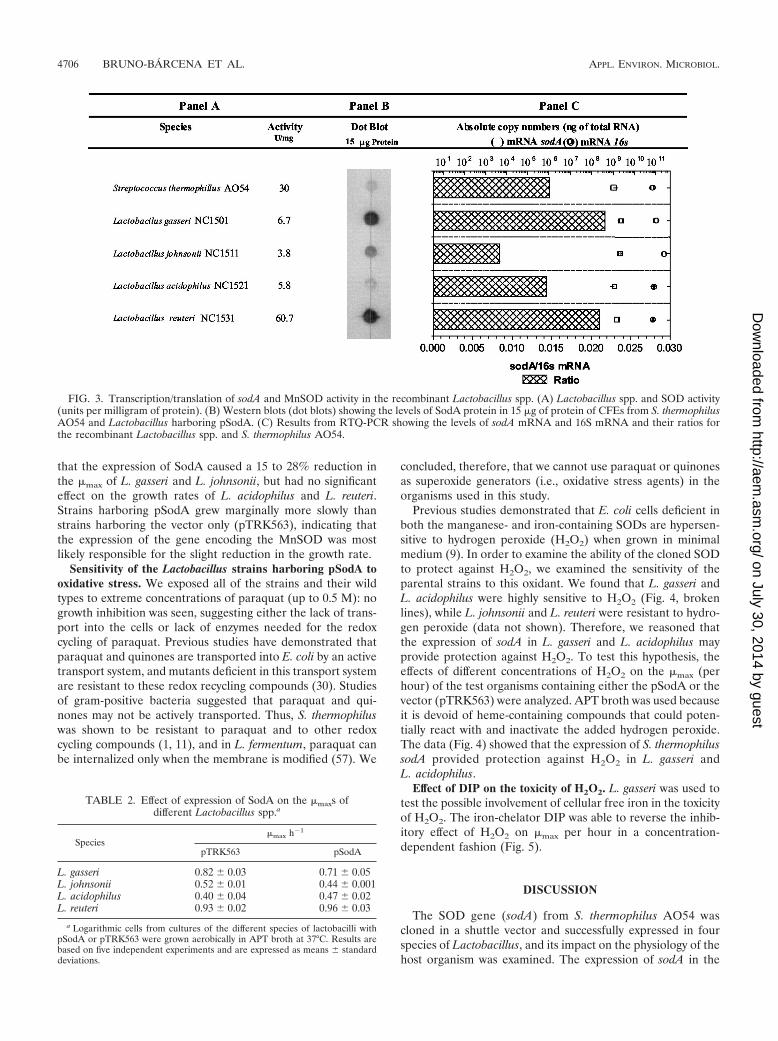
presence of the pSodA plasmid was verified by PCR. Only thestrains containing pSodA amplified the expected 1.2-kb frag-ment representing the sodA of S. thermophilus AO54 (Fig.2), and nondenaturing polyacrylamide gel electrophoresis(PAGE) SOD activity gels of the positive clones revealed thepresence of an active MnSOD that migrated to the same po-sition as that of S. thermophilus AO54 (data not shown). Thespecific activities of MnSOD in CFEs prepared from the trans-formants are presented in Fig. 3A. These results indicate thatthe sodA from S. thermophilus is functionally expressed in thefour species of Lactobacillus tested.
CFEs from the transformants (up to a maximum of 100 �gof total protein) were subjected to electrophoresis on 4 to 15%gradient sodium dodecyl sulfate (SDS)-polyacrylamide mini-gels, electrotransferred to nitrocellulose membranes, and sub-jected to Western analysis with SodA antibodies. No cross-reacting material was detected in the controls (i.e., cellstransformed with pTRK563) even after extended exposuretimes (�16 h), whereas a single band was detected when CFEsfrom cells transformed with pSodA were used. Because thesedata indicated absence of cross-reacting material or degradedSodA protein, the samples were subjected to dot blotting toavoid electrotransfer of artifacts from SDS-PAGE to the ni-trocellulose membranes. The results revealed the presence ofdifferent levels of SodA protein that closely correlated with thelevel of sodA transcripts from the different strains (Fig. 3B andC), while the specific activity of MnSOD in the transformantsdid not correlate with the concentration of SodA protein ob-served (Fig. 3A and B).
Effect of S. thermophilus sodA product on the �max (perhour) of Lactobacillus spp. The effect of sodA expression on the�max of the different strains was examined to determine itsimpact on the host strains. Standardized inocula from station-ary- and exponential-phase cultures of Lactobacillus harboringeither pTRK563 or pSodA were grown aerobically in APTbroth. The effects of the expression of SodA on �max per hourin the different strains are shown in Table 2. The data showed
FIG. 1. Construction of pSodA plasmid. pSodX-1 containing thesodA gene and flanking sequence from S. thermophilus in pGEM-TEasy cloning vector (Promega) was digested with EcoRI. The 1.2-kbfragment was gel purified and ligated into the EcoRI-digested shuttlevector, pTRK563. The new plasmid construct was termed pSodA.
FIG. 2. PCR confirmation of sodA in the transformants. Lactoba-cillus spp. were grown at 37°C in MRS medium supplemented with 2%glucose. S. thermophilus AO54 was grown at 42°C in M17 mediumsupplemented with 0.5% glucose. Primers STSODF and STSODR andcells from presumptive transformants or S. thermophilus were used forPCRs. Lanes: 1, 1-kb ladder; 2, pSodA; 3, negative control; 4, L. gasseriNC1501; 5, L. johnsonii NC1511; 6, L. acidophilus NC1521; 7, L. reuteriNC1531; 8, S. thermophilus AO54.
VOL. 70, 2004 sodA EXPRESSION IN INTESTINAL LACTOBACILLI 4705
on July 30, 2014 by guesthttp://aem
.asm.org/
Dow
nloaded from
that the expression of SodA caused a 15 to 28% reduction inthe �max of L. gasseri and L. johnsonii, but had no significanteffect on the growth rates of L. acidophilus and L. reuteri.Strains harboring pSodA grew marginally more slowly thanstrains harboring the vector only (pTRK563), indicating thatthe expression of the gene encoding the MnSOD was mostlikely responsible for the slight reduction in the growth rate.
Sensitivity of the Lactobacillus strains harboring pSodA tooxidative stress. We exposed all of the strains and their wildtypes to extreme concentrations of paraquat (up to 0.5 M): nogrowth inhibition was seen, suggesting either the lack of trans-port into the cells or lack of enzymes needed for the redoxcycling of paraquat. Previous studies have demonstrated thatparaquat and quinones are transported into E. coli by an activetransport system, and mutants deficient in this transport systemare resistant to these redox recycling compounds (30). Studiesof gram-positive bacteria suggested that paraquat and qui-nones may not be actively transported. Thus, S. thermophiluswas shown to be resistant to paraquat and to other redoxcycling compounds (1, 11), and in L. fermentum, paraquat canbe internalized only when the membrane is modified (57). We
concluded, therefore, that we cannot use paraquat or quinonesas superoxide generators (i.e., oxidative stress agents) in theorganisms used in this study.
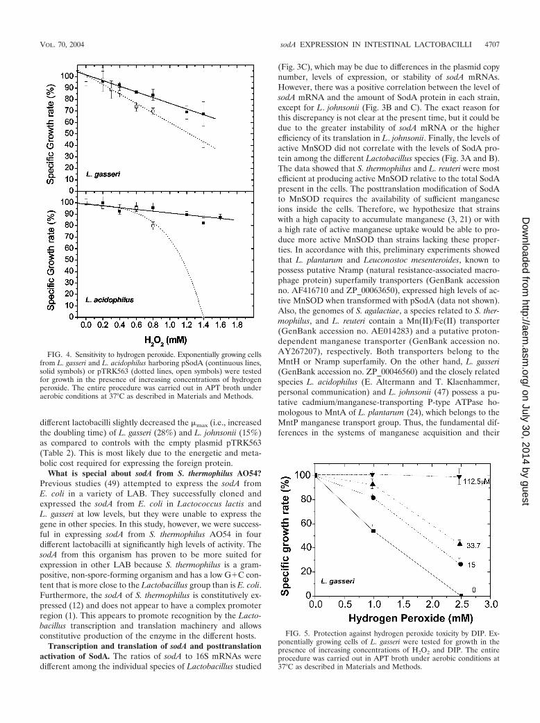
Previous studies demonstrated that E. coli cells deficient inboth the manganese- and iron-containing SODs are hypersen-sitive to hydrogen peroxide (H2O2) when grown in minimalmedium (9). In order to examine the ability of the cloned SODto protect against H2O2, we examined the sensitivity of theparental strains to this oxidant. We found that L. gasseri andL. acidophilus were highly sensitive to H2O2 (Fig. 4, brokenlines), while L. johnsonii and L. reuteri were resistant to hydro-gen peroxide (data not shown). Therefore, we reasoned thatthe expression of sodA in L. gasseri and L. acidophilus mayprovide protection against H2O2. To test this hypothesis, theeffects of different concentrations of H2O2 on the �max (perhour) of the test organisms containing either the pSodA or thevector (pTRK563) were analyzed. APT broth was used becauseit is devoid of heme-containing compounds that could poten-tially react with and inactivate the added hydrogen peroxide.The data (Fig. 4) showed that the expression of S. thermophilussodA provided protection against H2O2 in L. gasseri andL. acidophilus.
Effect of DIP on the toxicity of H2O2. L. gasseri was used totest the possible involvement of cellular free iron in the toxicityof H2O2. The iron-chelator DIP was able to reverse the inhib-itory effect of H2O2 on �max per hour in a concentration-dependent fashion (Fig. 5).
DISCUSSION
The SOD gene (sodA) from S. thermophilus AO54 wascloned in a shuttle vector and successfully expressed in fourspecies of Lactobacillus, and its impact on the physiology of thehost organism was examined. The expression of sodA in the
FIG. 3. Transcription/translation of sodA and MnSOD activity in the recombinant Lactobacillus spp. (A) Lactobacillus spp. and SOD activity(units per milligram of protein). (B) Western blots (dot blots) showing the levels of SodA protein in 15 �g of protein of CFEs from S. thermophilusAO54 and Lactobacillus harboring pSodA. (C) Results from RTQ-PCR showing the levels of sodA mRNA and 16S mRNA and their ratios forthe recombinant Lactobacillus spp. and S. thermophilus AO54.
TABLE 2. Effect of expression of SodA on the �maxs ofdifferent Lactobacillus spp.a
Species�max h�1
pTRK563 pSodA
L. gasseri 0.82 � 0.03 0.71 � 0.05L. johnsonii 0.52 � 0.01 0.44 � 0.001L. acidophilus 0.40 � 0.04 0.47 � 0.02L. reuteri 0.93 � 0.02 0.96 � 0.03
a Logarithmic cells from cultures of the different species of lactobacilli withpSodA or pTRK563 were grown aerobically in APT broth at 37°C. Results arebased on five independent experiments and are expressed as means � standarddeviations.
4706 BRUNO-BARCENA ET AL. APPL. ENVIRON. MICROBIOL.
on July 30, 2014 by guesthttp://aem
.asm.org/
Dow
nloaded from
different lactobacilli slightly decreased the �max (i.e., increasedthe doubling time) of L. gasseri (28%) and L. johnsonii (15%)as compared to controls with the empty plasmid pTRK563(Table 2). This is most likely due to the energetic and meta-bolic cost required for expressing the foreign protein.
What is special about sodA from S. thermophilus AO54?Previous studies (49) attempted to express the sodA fromE. coli in a variety of LAB. They successfully cloned andexpressed the sodA from E. coli in Lactococcus lactis andL. gasseri at low levels, but they were unable to express thegene in other species. In this study, however, we were success-ful in expressing sodA from S. thermophilus AO54 in fourdifferent lactobacilli at significantly high levels of activity. ThesodA from this organism has proven to be more suited forexpression in other LAB because S. thermophilus is a gram-positive, non-spore-forming organism and has a low G�C con-tent that is more close to the Lactobacillus group than is E. coli.Furthermore, the sodA of S. thermophilus is constitutively ex-pressed (12) and does not appear to have a complex promoterregion (1). This appears to promote recognition by the Lacto-bacillus transcription and translation machinery and allowsconstitutive production of the enzyme in the different hosts.
Transcription and translation of sodA and posttranslationactivation of SodA. The ratios of sodA to 16S mRNAs weredifferent among the individual species of Lactobacillus studied
(Fig. 3C), which may be due to differences in the plasmid copynumber, levels of expression, or stability of sodA mRNAs.However, there was a positive correlation between the level ofsodA mRNA and the amount of SodA protein in each strain,except for L. johnsonii (Fig. 3B and C). The exact reason forthis discrepancy is not clear at the present time, but it could bedue to the greater instability of sodA mRNA or the higherefficiency of its translation in L. johnsonii. Finally, the levels ofactive MnSOD did not correlate with the levels of SodA pro-tein among the different Lactobacillus species (Fig. 3A and B).The data showed that S. thermophilus and L. reuteri were mostefficient at producing active MnSOD relative to the total SodApresent in the cells. The posttranslation modification of SodAto MnSOD requires the availability of sufficient manganeseions inside the cells. Therefore, we hypothesize that strainswith a high capacity to accumulate manganese (3, 21) or witha high rate of active manganese uptake would be able to pro-duce more active MnSOD than strains lacking these proper-ties. In accordance with this, preliminary experiments showedthat L. plantarum and Leuconostoc mesenteroides, known topossess putative Nramp (natural resistance-associated macro-phage protein) superfamily transporters (GenBank accessionno. AF416710 and ZP_00063650), expressed high levels of ac-tive MnSOD when transformed with pSodA (data not shown).Also, the genomes of S. agalactiae, a species related to S. ther-mophilus, and L. reuteri contain a Mn(II)/Fe(II) transporter(GenBank accession no. AE014283) and a putative proton-dependent manganese transporter (GenBank accession no.AY267207), respectively. Both transporters belong to theMntH or Nramp superfamily. On the other hand, L. gasseri(GenBank accession no. ZP_00046560) and the closely relatedspecies L. acidophilus (E. Altermann and T. Klaenhammer,personal communication) and L. johnsonii (47) possess a pu-tative cadmium/manganese-transporting P-type ATPase ho-mologous to MntA of L. plantarum (24), which belongs to theMntP manganese transport group. Thus, the fundamental dif-ferences in the systems of manganese acquisition and their
FIG. 4. Sensitivity to hydrogen peroxide. Exponentially growing cellsfrom L. gasseri and L. acidophilus harboring pSodA (continuous lines,solid symbols) or pTRK563 (dotted lines, open symbols) were testedfor growth in the presence of increasing concentrations of hydrogenperoxide. The entire procedure was carried out in APT broth underaerobic conditions at 37°C as described in Materials and Methods.
FIG. 5. Protection against hydrogen peroxide toxicity by DIP. Ex-ponentially growing cells of L. gasseri were tested for growth in thepresence of increasing concentrations of H2O2 and DIP. The entireprocedure was carried out in APT broth under aerobic conditions at37°C as described in Materials and Methods.
VOL. 70, 2004 sodA EXPRESSION IN INTESTINAL LACTOBACILLI 4707
on July 30, 2014 by guesthttp://aem
.asm.org/
Dow
nloaded from

possible growth phase and/or gene regulation may explain thedifference in the values of active MnSOD observed (Fig. 3).
MnSOD and protection against hydrogen peroxide. Data inFig. 4 showed that the expression of S. thermophilus MnSOD inL. gasseri and L. acidophilus provided protection against H2O2.Figure 6 explains how SOD could provide such protection eventhough H2O2 is not a substrate for this enzyme. It is known thatthe deleterious effects of H2O2 on cell growth and survival arelargely dependent on the availability of “free” soluble iron[Fe(II)]. Free iron could be released from labile iron-sulfur[4Fe-4S]2� clusters by the action of superoxide radicals (Fig. 6,reaction 1) (18, 33) or could come from other intracellularsources. Ferrous iron reacts with H2O2 to catalyze the produc-tion of the highly reactive hydroxyl radical (HO˙) via the Fen-ton chemistry (17) (Fig. 6, reaction 2). The involvement ofFe(II) in the toxicity of H2O2 in L. gasseri and L. acidophilus issupported by the protective effect of DIP (Fig. 5). The contin-uous generation of HO˙ requires a continuous supply ofFe(II), which can be provided by reaction 1 or via the reduc-tion of Fe(III) by O2
·� (reaction 3, Fig. 6). The sum of reactions2 and 3 is also known as the Haber-Weiss reaction (22, 37).The reduction of Fe(III) could also be accomplished by otherintracellular reductants: e.g., glutathione, thioredoxin, flavoen-zymes, NAD(P)H-dependent enzymes, etc. (23, 28, 45). Figure6 shows that the cells have redundant mechanisms for protec-tion against the toxicity of H2O2. Thus, the protection againstH2O2 and against the generation of HO˙ can be accomplishedby (i) elimination of O2
·� by SODs to inhibit reactions 1 and 3;(ii) elimination of H2O2 by hydroperoxidases to inhibit reac-tion 2; (iii) chelation of free iron by iron-chelator or by specificDNA-binding proteins (e.g., Dpr and Dps in gram-positive andgram-negative organisms, respectively) (27, 59) to inhibit re-action 2; (iv) use of specific [Fe-S]cluster protectors—e.g.,
YggX in Salmonella enterica (20) and the Shetna protein(FeSII) in Azotobacter vinelandii (35) to inhibit reaction 1; or(v) the repair of damaged DNA and other macromolecules (8).Data in Fig. 4 clearly showed that SOD provided significantprotection against H2O2 in L. gasseri and L. acidophilus bypresumably interfering with reactions 1 and 3 (Fig. 6). Previousstudies have shown that E. coli cells deficient in sodA and sodBare more sensitive to hydrogen peroxide (9). Also, recent stud-ies have shown that the endogenous SOD levels control theiron-dependent HO˙ formation in E. coli cells exposed tohydrogen peroxide (39). Our data and those of others clearlydemonstrate the benefits of expressing SODs in order for thecells to withstand the challenges of oxidative stress. Since mostof the Lactobacillus spp. lack endogenous SODs and hydroper-oxidases, it is tenable that strains expressing a cloned SODwould be more resistant to hydrogen peroxide and oxidativestress.
Conclusion. This research has shown the expression of the S.thermophilus sodA into different species of Lactobacillus, dem-onstrating that SOD can increase tolerance to oxidative stressin L. acidophilus and L. gasseri. The physiological advantagesof the cloned SOD in L. reuteri and L. johnsonii remain to bedetermined, but further work is needed to define the appro-priate test parameters. Nevertheless, strains of Lactobacillusproducing heterologous MnSOD may offer some benefits tothe intestinal tract of the host if provided as probiotics. Oraladministration of probiotics has clear effects on the numbersand activities of intestinal and fecal bacteria (11, 44, 53). It maybe possible to use these SOD-rich species in biotherapy (i.e., asvehicles for the delivery of MnSOD in the gastrointestinaltracts of humans and farm animals). Delivery of MnSOD-richcells may provide a substantial amount of this antioxidantenzyme in vivo to the gastrointestinal tract for the treatment of
FIG. 6. A schematic presentation showing how SODs, hydroperoxidases, iron chelators, iron-binding proteins, protectors of [Fe-S] clusters,and/or DNA and cellular repair mechanisms could protect against H2O2 toxicity. Reaction 1 shows the oxidation of labile iron-sulfur clusters byO2
·�, reaction 2 shows the generation of HO˙ by Fenton chemistry, reaction 3 shows the regeneration of Fe(II) from Fe(III) by O2·� (the sum of
reactions 2 and 3 is also known as the Haber-Weiss reaction), and reaction 4 shows the deleterious effects of HO˙ and the generation of damagedDNA and damaged cellular components. Protective molecules/mechanisms are shown in boxes: SODs inhibit reactions 1 and 3, hydroperoxidasesinhibit reaction 2, iron-binding proteins (Dpr and Dps) and iron chelators (DIP) inhibit reaction 2, protectors of [Fe-S] clusters (YggX and FeSII)inhibit reaction 1, and DNA repair and other cellular repair mechanisms repair the damage caused by reaction 4.
4708 BRUNO-BARCENA ET AL. APPL. ENVIRON. MICROBIOL.
on July 30, 2014 by guesthttp://aem
.asm.org/
Dow
nloaded from
peptic ulcers or ulcerative colitis. Future work will involveexamining the possible added benefit that these organismscould provide for the treatment of gastrointestinal disorders, aswell as the prospect of enhancing the probiotic properties ofLactobacillus. Work is currently under way to integrate andexpress the sodA from S. thermophilus AO54 into the chromo-some of these probiotic organisms.
ACKNOWLEDGMENTS
This research was supported by the North Carolina Dairy Founda-tion, Southeast Dairy Foods Research Center, and North CarolinaAgricultural Research Service.
We thank Mike Russell, Joseph Sturino, and Andrea Azcarate fortechnical assistance. We also thank Irwin Fridovich for the criticalreading of the manuscript.
REFERENCES
1. Andrus, J. M., S. W. Bowen, T. R. Klaenhammer, and H. M. Hassan. 2003.Molecular characterization and functional analysis of the manganese-con-taining superoxide dismutase gene (sodA) from Streptococcus thermophilusAO54. Arch. Biochem. Biophys. 420:103–113.
2. Archibald, F. S., and I. Fridovich. 1981. Manganese and defenses againstoxygen toxicity in Lactobacillus plantarum. J. Bacteriol. 145:442–451.
3. Archibald, F. S., and I. Fridovich. 1981. Manganese, superoxide dismutase,and oxygen tolerance in some lactic acid bacteria. J. Bacteriol. 146:928–936.
4. Ausubel, F. M., R. Brent, R. E. Kingston, D. D. Moore, J. G. Seidman, J. A.Smith, and K. Struhl. 1989. Short protocols in molecular biology. GreenPublishing Associates and Wiley Interscience, New York, N.Y.
5. Axelsson, L. 1998. Lactic acid bacteria: classification and physiology, p. 1–12.In S. Salminen and A. von Wright (ed.), Lactic acid bacteria: microbiologyand functional aspects. Marcel Dekker, Inc., New York, N.Y.
6. Beauchamp, C., and I. Fridovich. 1971. Superoxide dismutase: improvedassays and an assay applicable to acrylamide gels. Anal. Biochem. 44:276–287.
7. Benov, L. T., and I. Fridovich. 1994. Escherichia coli express a copper- andzinc-containing superoxide dismutase. J. Biol. Chem. 269:25310–25314.
8. Cadet, J., T. Delatour, T. Douki, D. Gasparutto, J. P. Pouet, J. L. Ravanat,and S. Sauvaigo. 1999. Hydroxyl radicals and DNA base damage. Mutat.Res. 424:9–21.
9. Carlioz, A., and D. Touati. 1986. Isolation of superoxide dismutase mutantsin Escherichia coli: is superoxide dismutase necessary for aerobic life?EMBO J. 5:623–630.
10. Casadaban, M. J., and S. N. Cohen. 1980. Analysis of gene control signals byDNA fusion and cloning in Escherichia coli. J. Mol. Biol. 138:179–207.
11. Casas, I. A., and W. J. Dobrogosz. 2000. Validation of the probiotic concept:Lactobacillus reuteri confers broad-spectrum protection against disease inhumans and animals. Microb. Ecol. Health Dis. 12:247–285.
12. Chang, S. K., and H. M. Hassan. 1997. Characterization of superoxidedismutase in Streptococcus thermophilus. Appl. Environ. Microbiol. 63:3732–3735.
13. Condon, S. 1987. Responses of lactic acid bacteria to oxygen. FEMS Micro-biol. Rev. 46:269–280.
14. de Man, J. C., M. Rogosa, and M. E. Sharpe. 1960. A medium for thecultivation of lactobacilli. J. Appl. Bacteriol. 23:130–135.
15. Evans, J. B., and C. F. Niven, Jr. 1951. Nutrition of the heterofermentativelactobacilli that cause greening of cured meat products. J. Bacteriol. 62:599–603.
16. Farr, S. B., R. D’Ari, and D. Touati. 1986. Oxygen dependent mutagenesis inEscherichia coli lacking superoxide dismutase. Proc. Natl. Acad. Sci. USA83:8268–8272.
17. Fenton, H. J. H. 1894. Oxidation of tartaric acid in the presence of iron.J. Chem. Soc. Trans. 65:899–910.
18. Flint, D. H., J. F. Tuminello, and M. H. Emptage. 1993. The inactivation ofFe-S clusters containing hydro-lyases by superoxide. J. Biol. Chem. 268:22369–22376.
19. Gotz, F., B. Sedewitz, and E. F. Elstner. 1980. Oxygen utilization by Lacto-bacillus plantarum. I. Oxygen consuming reactions. Arch. Microbiol. 125:209–214.
20. Gralnick, J., and D. Downs. 2001. Protection from superoxide damage as-sociated with an increase in the YggX protein in Salmonella enterica. Proc.Natl. Acad. Sci. USA 98:8030–8035.
21. Groot, M. N. N., and J. A. M. de Bont. 1999. Involvement of manganese inconversion of phenylalanine to benzaldehyde by lactic acid bacteria. Appl.Environ. Microbiol. 65:5590–5593.
22. Haber, F., and J. Weiss. 1934. The catalytic decomposition of hydrogenperoxide by iron salts. Proc. R. Soc. 147:332–351.
23. Halliwell, B., and J. M. Gutteridge. 1990. Role of free radicals and catalyticmetal ions in human disease: an overview. Methods Enzymol. 186:1–85.
24. Hao, Z., S. Chen, and D. B. Wilson. 1999. Cloning, expression, and charac-terization of cadmium and manganese uptake genes from Lactobacillus plan-tarum. Appl. Environ. Microbiol. 65:4746–4752.
25. Hassan, H. M. 1997. Cytotoxicity of oxyradicals and the evolution of super-oxide dismutases, p. 25–51. In D. Massaro and L. Clerch (ed.), Oxygen, geneexpression and cellular function. Marcel Dekker, New York, N.Y.
26. Imlay, J. A., and S. Linn. 1988. DNA damage and oxygen radical toxicity.Science 240:1302–1309.
27. Ishikawa, T., Y. Mizunoe, S. I. Kawabata, A. Takade, M. Harada, S. N. Wai,and S.-I. Yoshida. 2003. The iron-binding protein Dps confers hydrogenperoxide stress resistance to Campylobacter jejuni. J. Bacteriol. 185:1010–1017.
28. Kakhlon, O., and Z. I. Cabantchik. 2002. The labile iron pool: characteriza-tion, measurement, and participation in cellular processes. Free Radic. Biol.Med. 33:1037–1046.
29. Kanbe, C., and K. Uchida. 1985. Oxygen consumption by Pediococcus halo-philus. Agric. Biol. Chem. 49:2931–2937.
30. Kao, S. M., and H. M. Hassan. 1985. Biochemical characterization of aparaquat-tolerant mutant of Escherichia coli. J. Biol. Chem. 260:10478–10481.
31. Kaur, I. P., K. Chopra, and A. Saini. 2002. Probiotics: potential pharmaceu-tical applications. Eur. J. Pharm. Sci. 15:1–9.
32. Keele, B. B., Jr., J. M. McCord, and I. Fridovich. 1970. Superoxide dismutasefrom Escherichia coli. B. A new manganese-containing enzyme. J. Biol.Chem. 245:6176–6181.
33. Keyer, K., and J. A. Imlay. 1996. Superoxide accelerates DNA damage byelevating free iron levels. Proc. Natl. Acad. Sci. USA 93:13635–13640.
34. Lloyd, G. T., A. J. Hillier, I. Barlow, and G. R. Jago. 1978. Aerobic formationof acetate from pyruvate by Lactobacillus bulgaricus. Aust. J. Biol. Sci. 31:565–571.
35. Lou, J., F. Moshiri, M. K. Johnson, M. E. Lafferty, D. L. Sorkin, A. Miller,and R. J. Maier. 1999. Mutagenesis studies of the FeSII protein of Azoto-bacter vinelandii: roles of histidine and lysine residues in the protection ofnitrogenase from oxygen damage. Biochemistry 38:5563–5571.
36. Lowry, O. H., N. J. Rosebrough, A. L. Farr, and R. J. Randall. 1951. Proteinmeasurement with the Folin phenol reagent. J. Biol. Chem. 193:265–275.
37. McCord, J. M., and E. D. Day, Jr. 1978. Superoxide-dependent productionof hydroxyl radical catalyzed by iron-EDTA complex. FEBS Lett. 86:139–142.
38. McCord, J. M., and I. Fridovich. 1969. Superoxide dismutase: an enzymaticfunction for erythrocuprein (hemocuprein). J. Biol. Chem. 244:6049–6055.
39. McCormick, M. L., G. R. Buettner, and B. E. Britigan. 1998. Endogenoussuperoxide dismutase levels regulate iron-dependent hydroxyl radical forma-tion in Escherichia coli exposed to hydrogen peroxide. J. Bacteriol. 180:622–625.
40. Mercenier, A., C. Robert, D. A. Romero, P. Slos, and Y. Lemoine. 1987.Transformation of Streptococcus thermophilus spheroplasts, p. 234–237. InJ. J. Ferretti and R. Curtiss III (ed.), Streptococcal genetics. AmericanSociety for Microbiology, Washington, D.C.
41. Miller, J. H. 1972. Experiments in molecular genetics. Cold Spring HarborLaboratory, Cold Spring Harbor, N.Y.
42. Moody, C. S., and H. M. Hassan. 1982. Mutagenicity of oxygen free radicals.Proc. Natl. Acad. Sci. USA 79:2855–2859.
43. Muriana, P. M., and T. R. Klaenhammer. 1991. Cloning, phenotypic expres-sion, and DNA sequence of the gene for lactacin F, an antimicrobial peptideproduced by Lactobacillus spp. J. Bacteriol. 173:1779–1788.
44. Pedrosa, M. C., B. B. Golner, B. R. Goldin, S. Barakat, G. E. Dallal, andR. M. Russell. 1995. Survival of yogurt-containing organisms and Lactoba-cillus gasseri (ADH) and their effect on bacterial enzyme activity in thegastrointestinal tract of healthy and hypochlorhydric elderly subjects. Am. J.Clin. Nutr. 61:353–359.
45. Petrat, F., S. Paluch, E. Dogruoz, P. Dorfler, M. Kirsch, H. G. Korth, R.Sustmann, and H. De Groot. 2003. Reduction of Fe(III) ions complexed tophysiological ligands by lipoyl dehydrogenase and other flavoenzymes invitro: implications for an enzymatic reduction of Fe(III) ions of the labileiron pool. J. Biol. Chem. 278:46403–46413.
46. Poyard, C., P. Berche, and P. Trieu-Cout. 1995. Characterization of super-oxide dismutase genes from Gram-positive bacteria by polymerase chainreaction using degenerate primers. FEMS Microbiol. Lett. 131:41–45.
47. Pridmore, R. D., B. Berger, F. Desiere, D. Vilanova, C. Barretto, A. C. Pittet,M. C. Zwahlen, M. Rouvet, E. Altermann, R. Barrangou, B. Mollet, A.Mercenier, T. Klaenhammer, F. Arigoni, and M. A. Schell. 2004. The ge-nome sequence of the probiotic intestinal bacterium Lactobacillus johnsoniiNCC 533. Proc. Natl. Acad. Sci. USA 101:2512–2517.
48. Reuter, G. 2001. The Lactobacillus and Bifidobacterium microflora of thehuman intestine: composition and succession. Curr. Issues Intest. Microbiol.2:43–53.
49. Roy, D. G., T. R. Klaenhammer, and H. M. Hassan. 1993. Cloning andexpression of the manganese superoxide dismutase gene of Escherichia coliin Lactococcus lactis and Lactobacillus gasseri. Mol. Gen. Genet. 239:33–40.
VOL. 70, 2004 sodA EXPRESSION IN INTESTINAL LACTOBACILLI 4709
on July 30, 2014 by guesthttp://aem
.asm.org/
Dow
nloaded from

50. Russell, W. M., and T. R. Klaenhammer. 2001. Identification and cloning ofgusA, encoding a new -glucuronidase from Lactobacillus gasseri ADH.Appl. Environ. Microbiol. 67:1253–1261.
51. Sambrook, J., E. F. Fritsch, and T. Maniatis. 1989. Molecular cloning: alaboratory manual, 2nd ed. Cold Spring Harbor Laboratory Press, ColdSpring Harbor, N.Y.
52. Sanders, J. W., K. J. Leenhouts, A. J. Haandrikman, G. Venema, and J.Kook. 1995. Stress response in Lactococcus lactis: cloning, expression anal-ysis and mutation of the lactococcal superoxide dismutase gene. J. Bacteriol.177:5254–5260.
53. Sanders, M. E., and T. R. Klaenhammer. 2001. Invited review: the scientificbasis of Lactobacillus acidophilus NCFM functionality as a probiotic. J. DairySci. 84:319–331.
54. Strittmatter, C. F. 1959. Electron transport to oxygen in lactobacilli. J. Exp.Biol. 234:2789.
55. Terzaghi, B. E., and W. E. Sandine. 1975. Improved medium for lacticstreptococci and their bacteriophages. Appl. Microbiol. 29:807–813.
56. Touati, D. 1988. Transcriptional and posttranscriptional regulation of man-ganese superoxide dismutase biosynthesis in Escherichia coli, studied withoperon and protein fusions. J. Bacteriol. 70:2511–2520.
57. Turner, M. S., T. Woodberry, L. M. Hafner, and P. M. Giffard. 1999. ThebspA locus of Lactobacillus fermentum BR11 encodes an L-cystine uptakesystem. J. Bacteriol. 181:2192–2198.
58. Walker, D. C., K. Aoyama, and T. R. Klaenhammer. 1996. Electrotransfor-mation of Lactobacillus acidophilus group A1. FEMS Microbiol. Lett. 138:233–237.
59. Yamamoto, Y., L. B. Poole, R. R. Hantgan, and Y. Kamio. 2002. An iron-binding protein, Dpr, from Streptococcus mutans prevents iron-dependenthydroxyl radical formation in vitro. J. Bacteriol. 184:2931–2939.
60. Yost, F. J., Jr., and I. Fridovich. 1973. An iron-containing superoxide dis-mutase from Escherichia coli. Biotechnol. Bioeng. 248:4905–4908.
4710 BRUNO-BARCENA ET AL. APPL. ENVIRON. MICROBIOL.
on July 30, 2014 by guesthttp://aem
.asm.org/
Dow
nloaded from