Structure of a superoxide dismutase and implications for copper-ion chelation
9
1 Supplementary Figure S1. Alternate conformation of the free Cys95 in molecule B of Pa-SOD. Electron density maps (2F o -F c ) and (F o -F c ) are contoured at 1.2σ and 3σ level respectively.
-
Upload
independent -
Category
Documents
-
view
3 -
download
0
Transcript of Structure of a superoxide dismutase and implications for copper-ion chelation

1
Supplementary Figure S1. Alternate conformation of the free Cys95 in molecule B of
Pa-SOD. Electron density maps (2Fo-Fc) and (Fo-Fc) are contoured at 1.2σ and 3σ level
respectively.

2
(a)
(b)
(c)
Supplementary Figure S2. Comparison of loop I structure of eukaryotic CuZnSODs. (a)
Molecules A (Olive green) and B (Yellow) of Pa-SOD with So-SOD (Orchid), (b) Pa-
SOD with Hs-SOD (Deep sky blue). The loop I structure in Sc, Sm, Bt and Xl-SODs is
the same as in Hs-SOD. (c) In Sc and Sm-SODs (Gold) an additional water molecule Wt4
is present. For clarity, side-chains are not shown except for Ser10 in Pa-SOD and the
residues are labeled according to Pa-SOD numbering.

3
Supplementary Figure S3. Comparison of metal binding residues with secondary shell
stabilization residues of Pa-SOD (Olive green) and Sm-SOD (Gold). The water molecule
Wm (orange) in Pa-SOD superimposes with the higher occupancy copper site of Sm-
SOD.
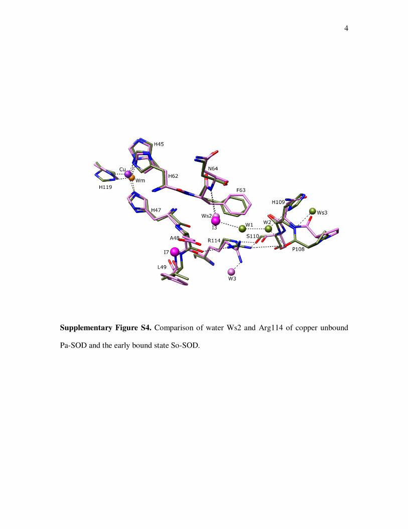
4
Supplementary Figure S4. Comparison of water Ws2 and Arg114 of copper unbound
Pa-SOD and the early bound state So-SOD.

5
Supplementary Figure S5. Movements of the structural water and the C-terminal Greek
key loop between copper unbound Pa-SOD (Olive green) and the early copper bound
state So-SOD (Orchid).

6
(a) (b)
Supplementary Figure S6. Comparison of active site water molecules of copper
unbound Pa-SOD with early chelation state So-SOD. Views of (a) Superposition of Cu
unbound Pa-SOD (Olive green) and the early copper bound state So-SOD (Orchid) and
(d) Superposition of molecules A and B of Pa-SOD. Additional five-membered ring
water structures are observed for molecule B of Pa-SOD.

7
Supplementary Figure S7. Comparison of dimeric interfaces of Pa-SOD (Olive green) and So-SOD (Orchid).

8
Conserved secondary shell interactions in solution and crystal structures of
CuZnSODs
(i) backbone N of His62 with carbonyl O of 135 (Lys/Ser/Thr/Val)
(ii) backbone N of His70 and carbonyl O of 134 (Lys/Leu/Thr/Pro)
(iii) backbone N of His79 with the side chain Oδ2 of Asp82
(iv) backbone N of Asp82 with carbonyl O of His79
(v) backbone N of His119 and carbonyl O of Gly43
(vi) carbonyl O of His119 with Nδ1 of His42 [except Sc-SOD where His is replaced by
Arg (Djinović et al., 1992)]
(vii) carbonyl O of His79 also with Nε of Arg78
(viii) carbonyl O of His62 with water molecule
(ix) carbonyl O of His70 with water molecule
(x) Nε2 of His45 with side chain Oδ1 or Oδ2 of Asp123
(xi) Nδ1 of His119 with carbonyl O of Gly140
(xii) Nε2 of His70 with side chain Oδ1 and Oδ2 of Asp123
(xiii) Nδ1 of His47 with carbonyl O of Gly60
(xiv) Nε2 of His79 with carbonyl O of 68 (Lys/Arg/Gln) through water molecule.
(xv) Residues His45 and His47 are part of β-strand 4f and interact with residues Val117
and Ala/Ser/Thr115 in the β-strand 7g. A slight movement in the carbonyl O of His47 is
found for most of the SOD structures studied so far and thus, the distance between
carbonyl O of His47 with N of 115 is >3.0 Å for all the structures.

9
Supplementary Table 1. Conserved hydrogen bonding interactions in the zinc binding loop
1. Salt Bride
i. Arg78 Asp100
Nη1 Arg78 Oδ1 Asp100
Nη2 Arg78 Oδ2 Asp100
2. Main-chain - main-chain interaction
i. N (Gly/Ser/Asn/Lys)67 O Asn64
ii. N Gly81 O Phe63
iii. N Asp82 O His79
3. Main-chain - side-chain interaction
i. N Gly71 Oδ2 Asp82
ii. N (Lys/Arg/Gln)68 Oδ1 Asn64
iii. O (Lys/Arg/Gln)68 Nδ2 Asn64
iv. O His79 Nε Arg78
v. O Pro73 Nη1 Arg78
vi. O (Ala/Val)80 Nη2 Arg78
vii. N His79 Oδ2 Asp82
4. Water mediated interactions
i. N Gly84 Water 14 (PA numbering)
ii. O Gly71 Water 14, Ws1
iii. Oδ1 Asp123 Water 14, Ws1
Iv N Ala/Leu/Thr/Asp/Glu/Phe66 Water 13, Ws5
v. N Ala/Val80 Water 13, Ws5
Vi N Asn64 Water/I3, Ws2
vii. Oδ1 Asn64 Water 13, Ws5
viii. O Arg78 Water 87, Ws6
ix. Water 13, Ws5 Water 87, Ws6