Detection and Characterization of Partially Unfolded Oligomers of the SH3 Domain of alpha-Spectrin
11
Detection and Characterization of Partially Unfolded Oligomers of the SH3 Domain of a-Spectrin Salvador Casares,* Mourad Sadqi,* Obdulio Lo ´ pez-Mayorga,* Francisco Conejero-Lara,* and Nico A. J. van Nuland y *Departamento de Quı ´mica Fı ´sica e Instituto de Biotecnologı ´a, Facultad de Ciencias, Universidad de Granada, 18071 Granada, Spain; and y Bijvoet Centre, Department of NMR Spectroscopy, University of Utrecht, Padualaan 8, 3584 CH, Utrecht, The Netherlands ABSTRACT For the purpose of equilibrium and kinetic folding-unfolding studies, the SH3 domain of a-spectrin (spc-SH3) has long been considered a classic two-state folding protein. In this work we have indeed observed that the thermal unfolding curves of spc-SH3 measured at pH 3.0 by differential scanning calorimetry, circular dichroism, and NMR follow apparently the two-state model when each unfolding profile is considered individually. Nevertheless, we have found that protein concentration has a marked effect upon the thermal unfolding profiles. This effect cannot be properly explained in terms of the two-state unfolding model and can only be interpreted in terms of the accumulation of intermediate associated states in equilibrium with the monomeric native and unfolded states. By chemical cross-linking and pulsed-field gradient NMR diffusion experiments we have been able to confirm the existence of associated states formed during spc-SH3 unfolding. A three-state model, in which a dimeric intermediate state is assumed to be significantly populated, provides the simplest interpretation of the whole set of thermal unfolding data and affords a satisfactory explanation for the concentration effects observed. Whereas at low concentrations the population of the associated intermediate state is negligible and the unfolding process consequently takes place in a two-state fashion, at concentrations above ;0.5 mM the population of the intermediate state becomes significant at temperatures between 458C and 808C and reaches up to 50% at the largest concentration investigated. The thermodynamic properties of the intermediate state implied by this analysis fall in between those of the unfolded state and the native ones, indicating a considerably disordered conformation, which appears to be stabilized by oligomerization. INTRODUCTION A few dozen proteins have to date been reported as folding in a two-state process (Jackson, 1998), amongst which the folding-unfolding properties of the Src-homology region 3 (SH3) domains have probably been the most extensively studied (Viguera et al., 1994; Grantcharova and Baker, 1997; Grantcharova et al., 1998; Guijarro et al., 1998a; Martinez et al., 1998; Plaxco et al., 1998; Martinez and Serrano, 1999). Kinetic and calorimetric criteria are usually applied to determine whether or not a folding reaction follows a two- state model (Privalov, 1979; Jackson and Fersht, 1991). For the SH3 domain of a-spectrin (spc-SH3) these two criteria have been fulfilled under all the conditions investigated so far (Viguera et al., 1994) and no equilibrium or kinetic intermediates have been detected to date. In contrast to the two-state behavior of the folding-unfolding reactions of spc- SH3, the study of the amide hydrogen-deuterium exchange (HX) measured by NMR under native conditions indicates that this domain, similar to that in other proteins, undergoes a considerable variety of conformational fluctuations, ranging from local motions to large structural disruptions affecting the domain’s core and even extending above the transition-state energy barrier (Sadqi et al., 1999, 2002a,b; Englander, 2000; Casares et al., 2003). It has been reported for several proteins that as destabilizing conditions are approached, e.g., by a change in pH, temperature, pressure, or concentration of denaturants, the large conformational heterogeneity of native fluctuations, as probed by HX measurements, is progressively replaced by a few confor- mational states, including partially folded states, or just the globally unfolded state (Bai et al., 1995; Chamberlain et al., 1996; Fuentes and Wand, 1998; Milne et al., 1999). In a previous paper we reported the temperature dependence of HX in the spc-SH3 domain at pH 3.0 (Sadqi et al., 2002a). As the temperature approached thermal unfolding conditions the HX data indicated the existence of a significant population of states with Gibbs energies clearly lower than that of the globally unfolded state. These types of state might have remained undetectable by the standard analysis of folding-unfolding data as was suggested previously by Englander and colleagues (Mayne and Englander, 2000). In standard HX experiments the concentration of sample is usually high due to the relatively low sensitivity of NMR. It is of interest therefore to explore the possibility that partially folded intermediates of spc-SH3 might become stabilized by intermolecular association favored by the high concentra- tions used in the NMR experiments. The self-association of folding intermediates and denatured states of proteins and protein fragments has been reported previously (Filimonov et al., 1993; Pecorari et al., 1996; Uversky et al., 1998; Kuznetsova et al., 1999; Liu et al., 1999; Uversky et al., 1999; Ye and Wang, 2001). The oligomeric states have been Submitted July 17, 2003, and accepted for publication December 2, 2003. S. Casares and M. Sadqi contributed equally to this work. Address reprint requests to Francisco Conejero-Lara, Tel.: 134-958- 242371; Fax: 134-958-272879; E-mail: [email protected] Sadqi’s present address is Dept. of Chemistry and Biochemistry, University of Maryland, College Park, MD 20742 USA. Ó 2004 by the Biophysical Society 0006-3495/04/04/2403/11 $2.00 Biophysical Journal Volume 86 April 2004 2403–2413 2403
Transcript of Detection and Characterization of Partially Unfolded Oligomers of the SH3 Domain of alpha-Spectrin

Detection and Characterization of Partially Unfolded Oligomersof the SH3 Domain of a-Spectrin
Salvador Casares,* Mourad Sadqi,* Obdulio Lopez-Mayorga,* Francisco Conejero-Lara,*and Nico A. J. van Nulandy
*Departamento de Quımica Fısica e Instituto de Biotecnologıa, Facultad de Ciencias, Universidad de Granada, 18071 Granada, Spain;and yBijvoet Centre, Department of NMR Spectroscopy, University of Utrecht, Padualaan 8, 3584 CH, Utrecht, The Netherlands
ABSTRACT For the purpose of equilibrium and kinetic folding-unfolding studies, the SH3 domain of a-spectrin (spc-SH3) haslong been considered a classic two-state folding protein. In this work we have indeed observed that the thermal unfolding curvesof spc-SH3 measured at pH 3.0 by differential scanning calorimetry, circular dichroism, and NMR follow apparently the two-statemodel when each unfolding profile is considered individually. Nevertheless, we have found that protein concentration hasa marked effect upon the thermal unfolding profiles. This effect cannot be properly explained in terms of the two-state unfoldingmodel and can only be interpreted in terms of the accumulation of intermediate associated states in equilibrium with themonomeric native and unfolded states. By chemical cross-linking and pulsed-field gradient NMR diffusion experiments we havebeen able to confirm the existence of associated states formed during spc-SH3 unfolding. A three-state model, in whicha dimeric intermediate state is assumed to be significantly populated, provides the simplest interpretation of the whole set ofthermal unfolding data and affords a satisfactory explanation for the concentration effects observed. Whereas at lowconcentrations the population of the associated intermediate state is negligible and the unfolding process consequently takesplace in a two-state fashion, at concentrations above ;0.5 mM the population of the intermediate state becomes significant attemperatures between 458C and 808C and reaches up to 50% at the largest concentration investigated. The thermodynamicproperties of the intermediate state implied by this analysis fall in between those of the unfolded state and the native ones,indicating a considerably disordered conformation, which appears to be stabilized by oligomerization.
INTRODUCTION
A few dozen proteins have to date been reported as folding in
a two-state process (Jackson, 1998), amongst which the
folding-unfolding properties of the Src-homology region 3
(SH3) domains have probably been the most extensively
studied (Viguera et al., 1994; Grantcharova and Baker, 1997;
Grantcharova et al., 1998; Guijarro et al., 1998a; Martinez
et al., 1998; Plaxco et al., 1998; Martinez and Serrano,
1999). Kinetic and calorimetric criteria are usually applied to
determine whether or not a folding reaction follows a two-
state model (Privalov, 1979; Jackson and Fersht, 1991). For
the SH3 domain of a-spectrin (spc-SH3) these two criteria
have been fulfilled under all the conditions investigated so
far (Viguera et al., 1994) and no equilibrium or kinetic
intermediates have been detected to date. In contrast to the
two-state behavior of the folding-unfolding reactions of spc-
SH3, the study of the amide hydrogen-deuterium exchange
(HX) measured by NMR under native conditions indicates
that this domain, similar to that in other proteins, undergoes
a considerable variety of conformational fluctuations,
ranging from local motions to large structural disruptions
affecting the domain’s core and even extending above the
transition-state energy barrier (Sadqi et al., 1999, 2002a,b;
Englander, 2000; Casares et al., 2003). It has been reported
for several proteins that as destabilizing conditions are
approached, e.g., by a change in pH, temperature, pressure,
or concentration of denaturants, the large conformational
heterogeneity of native fluctuations, as probed by HX
measurements, is progressively replaced by a few confor-
mational states, including partially folded states, or just the
globally unfolded state (Bai et al., 1995; Chamberlain et al.,
1996; Fuentes and Wand, 1998; Milne et al., 1999). In
a previous paper we reported the temperature dependence of
HX in the spc-SH3 domain at pH 3.0 (Sadqi et al., 2002a).
As the temperature approached thermal unfolding conditions
the HX data indicated the existence of a significant
population of states with Gibbs energies clearly lower than
that of the globally unfolded state. These types of state might
have remained undetectable by the standard analysis of
folding-unfolding data as was suggested previously by
Englander and colleagues (Mayne and Englander, 2000).
In standard HX experiments the concentration of sample is
usually high due to the relatively low sensitivity of NMR. It
is of interest therefore to explore the possibility that partially
folded intermediates of spc-SH3 might become stabilized by
intermolecular association favored by the high concentra-
tions used in the NMR experiments. The self-association of
folding intermediates and denatured states of proteins and
protein fragments has been reported previously (Filimonov
et al., 1993; Pecorari et al., 1996; Uversky et al., 1998;
Kuznetsova et al., 1999; Liu et al., 1999; Uversky et al.,
1999; Ye and Wang, 2001). The oligomeric states have been
Submitted July 17, 2003, and accepted for publication December 2, 2003.
S. Casares and M. Sadqi contributed equally to this work.
Address reprint requests to Francisco Conejero-Lara, Tel.: 134-958-
242371; Fax: 134-958-272879; E-mail: [email protected] Sadqi’s
present address is Dept. of Chemistry and Biochemistry, University of
Maryland, College Park, MD 20742 USA.
� 2004 by the Biophysical Society
0006-3495/04/04/2403/11 $2.00
Biophysical Journal Volume 86 April 2004 2403–2413 2403

often described as an association of folding intermediates
with residual super-secondary structure (Filimonov et al.,
1993), which in certain cases appears as native-like
(Alexandrescu et al., 2000; Ye and Wang, 2001). Even
CI2, another classic two-state protein, can form transient
aggregates during folding under suitable conditions (Silow
et al., 1999). The association of folding intermediates has
special relevance in that with some proteins the association
process proceeds further to the formation of large aggregates
and amyloid fibrils (Dobson, 1999; Rochet and Lansbury,
2000), even in many proteins that are not related to any
amyloid-related disease, including the PI3-SH3 domain.
Fibril formation appears to depend upon the accumulation of
unstable, partially structured, intermediate states stabilized
by self-assembly into an oligomerization nucleus (Rochet
and Lansbury, 2000). Studies of association of folding
intermediates are, therefore, of increasing importance to
understand the details of these misfolding processes.
We present here the results of a detailed study into the
effects of increasing protein concentration upon the ther-
mal unfolding equilibrium of the spc-SH3 domain using
a combination of calorimetry and spectroscopic techniques.
We have also tested the presence of oligomeric species using
chemical cross-linking and pulse-field gradient NMR
diffusion measurements. The results confirm the existence
of partially unfolded oligomeric states in equilibrium with
the native and unfolded states. Using a simple mathematical
model we have estimated the thermodynamic properties of
these intermediates, which may help to shed light upon their
conformational properties and their possible role in the
mechanisms of aggregation of these small domains.
MATERIAL AND METHODS
Protein samples and chemicals
Wild-type chicken a-spectrin SH3 domain was obtained as described
elsewhere (Sadqi et al., 1999). Samples were prepared for experimental
work by extensive dialysis against a large volume of 20 mM glycine pH 3.0,
except for the samples in deuterated buffer, which were prepared by
dissolving the lyophilized protein directly in the buffer and subsequently
readjusting their pH (direct pH-meter reading). Sample concentration was
measured by UV absorption at 280 nm using an extinction coefficient of
16,147 for the native protein (Viguera et al., 1994).
Perdeuterated glycine for NMR samples came from Cambridge Isotope
(Andover, MA). Deuterium oxide and other deuterated compounds were
from Sigma (St. Louis, MO). Glutaraldehyde for cross-linking experiments
was grade I (Sigma). The remaining chemicals used were all of analytical
grade.
Differential scanning calorimetry
High-sensitivity differential scanning calorimetry (DSC) was done with
a VP-DSC microcalorimeter (Microcal, Northampton, MA). The scan rate
was usually 18C/min, except for experiments made specifically to test any
possible effect of scan rate, where it was varied between 0.58C/min and
1.58C/min. The experiments were performed at pH 3.0 in 20 mM glycine
buffer and the concentration of protein was varied from 0.03 mM to 4.85
mM. After baseline subtraction, the temperature dependence of the molar
partial heat capacity (Cp) of spc-SH3 was calculated from the DSC
thermograms using Origin (OriginLab, Northampton, MA) as described
elsewhere (Privalov and Potekhin, 1986). Preliminary fittings of the Cp
curves using the two-state unfolding model were made as described
elsewhere (Viguera et al., 1994).
Thermal unfolding followed by circular dichroism
Circular dichroism (CD) measurements were made in a Jasco J-715
spectropolarimeter (Jasco, Easton, MD) equipped with a temperature-
controlled cell holder. The cell path length was 1 mm or 10 mm, depending
upon the protein concentration used and the spectral region to be observed.
Thermal unfolding was studied by monitoring the CD signal at 222 nm in the
far-ultraviolet (UV) wavelength range, and at 270 nm and 294 nm in the
near-UV range, during temperature scans at a scan rate of 18C/min. The
actual temperature of the sample was measured by immersing a PT-100
probe inside the cell and registered by the instrument’s computer.
Experimental conditions were identical to those of the DSC experiments.
The concentration of protein was 0.028 mM or 0.14 mM for measurements
at 222 nm, and 0.14 mM or 0.69 mM for measurements at both 270 nm and
294 nm. Baseline CD temperature scans were recorded with pure buffer
solutions and subtracted from the CD traces obtained with the protein
solutions. Preliminary fitting of the CD thermal traces using a two-state
unfolding model was made as described elsewhere (van Nuland et al., 1998).
Thermal unfolding followed by nuclearmagnetic resonance
Thermal unfolding was monitored using NMR on a Bruker AMX-500
spectrometer (Billerica, MA). Protein solutions were prepared in 10% D2O/
90% H2O, 20 mM perdeuterated glycine, pH 3.0 (direct pH-meter readings
without isotopic correction). Sample concentration was either 0.68 mM or
5.4 mM. A set of one-dimensional NMR spectra at different temperatures
between 20.78C and 81.38C was acquired using a sweep width of 6024 Hz
and 4096 data points. Spectra were sine-bell apodized, Fourier transformed,
and baseline corrected using the MestRe-C program (University of Santiago
de Compostela, Spain). For each NMR spectrum the intensity of several
well-resolved NMR signals was determined by fitting the peaks to
Lorentzian functions. This procedure allowed us to measure accurately the
intensities of partially overlapping signals. The intensity of the signals of
Ala55-CbH3, Leu33-CdH3, and Trp41-NeH, corresponding to the native state,
was monitored as a function of temperature. Two signals corresponding to
Trp41-NeH and Trp42-NeH in nonnative states were also observed. The data
were initially fitted on the basis of the two-state unfolding model using the
same procedure as for the CD thermal unfolding experiments.
Chemical cross-linking
Cross-linking experiments with spc-SH3 in 20 mM glycine, pH 3.0, at four
sample concentrations of between;0.04 mM and 3.5 mM and temperatures
of 208C and 508C were conducted to identify the presence of spc-SH3
oligomers in solution. Glutaraldehyde was used as cross-linking reagent
according to the procedure described elsewhere (Jaenicke and Rudolph,
1989). An identical procedure was repeated with lysozyme solutions in 20
mM glycine, pH 2.0, at similar sample concentrations. This monomeric
protein was used as a control for unspecific cross-linking. Samples were
incubated at the desired temperature in Eppendorf tubes in a thermostatic
bath for 10 min. Glutaraldehyde was added to a final concentration of 1%
w/v and the samples were left to incubate in the bath for 3 min. Cross-linking
was fixed by adding freshly prepared NaBH4 solution in 0.1 M NaOH to
a final concentration of 50 mM. Samples were left to incubate for a further 20
min. Protein was precipitated by the addition of a 10% sodium deoxycholate
solution to a final 0.01% w/v and a 78% trichloracetic acid solution to a final
2404 Casares et al.
Biophysical Journal 86(4) 2403–2413
1.5% w/v, followed by further incubation in ice for 10 min. Protein
precipitates were collected by centrifugation; the pellets were washed
carefully with water and redissolved for SDS-PAGE analysis.
Pulse-field gradient NMR spectroscopy
Pulse-field gradient NMR (PFG-NMR) diffusion measurements were made
with the pulse gradient stimulated echo longitudinal encode-decode (PG-
SLED) sequence (Wilkins et al., 1999) on a Bruker Avance DRX750
spectrometer. The protein concentrations were �0.5 mM and �5 mM.
Samples were prepared in 20 mM perdeuterated glycine, pH 3.0, either in
7% D2O/93% H2O or in 100% D2O. Thirty microliters of a 1% dioxan
solution was added to the sample. 1H chemical shifts are expressed relatively
to dioxan (3.75 ppm). Each diffusion data set consisted of a series of 40 one-
dimensional 1H-NMR spectra with 2.5% increments of the maximum
gradient strength from 2.5 to 100% collected with a three-axis gradient
probe. The gradient pulse length, d, was optimized to obtain a total decay of
80–90% of the initial signal, a typical value being 4 ms. The echo time, D,
for diffusion was set to 100 ms and NMR spectra were acquired with 8 K
complex points and a sweep width of 10,000 Hz. The water resonance was
suppressed by weak presaturation during the 1-s relaxation delay. The data
were processed with Felix from Biosym Technologies (San Diego, CA).
The intensity of the signals of the protein (I ) in different spectral regions
(e.g., amide/aromatic (7.8–6.4 ppm) and aliphatic groups (from 3.5 to �0.5ppm)) as a function of gradient strength (g) were fitted using Origin
(OriginLab) to
IðgÞ ¼ Ae�dg2
: (1)
This equation enables us to obtain the observed decay rate, d, which is
proportional to the translational diffusion coefficient, D. The resulting decay
rate of the protein, dprot, was then used to fit the change in intensity of the
dioxan signal at 3.75 ppm to a double Gaussian function. This procedure was
motivated by the need of eliminating the contribution of protein signals to
the dioxan signal intensity due to overlap.
The protein hydrodynamic radius (Rproth ) was calculated as described
elsewhere (Wilkins et al., 1999) using the following equation:
Rprot
h ¼ ddioxandprot
Rdioxan
h
� �; (2)
where ddioxan and Rdioxanh are, respectively, the decay rate and the radius of
the dioxan molecule, which was calculated as being 2.12 A (Wilkins et al.,
1999).
RESULTS
Thermal unfolding of spc-SH3 monitored by CDand 1H-NMR spectroscopy
The existence of equilibrium folding intermediates of
proteins has been revealed on many occasions by discrep-
ancies between the thermal unfolding profiles obtained when
using different techniques. We have investigated the thermal
unfolding of spc-SH3 at pH 3.0 by monitoring the tem-
perature dependence of the CD signal at 222 nm, in the far-
UV region of the spectrum. The experiments were made with
sample concentrations of 0.028 mM and 0.14 mM. The
spectral changes induced by temperature at 222 nm probe the
disruption of the secondary structure of the domain. We also
monitored the thermal unfolding of 0.14 mM and 0.69 mM
samples at both 294 nm and 270 nm in the near-UV region.
The changes in this wavelength region are sensitive above all
to the disordering of the tertiary structure. The CD spectra of
samples heated up to 958C and then cooled down to 208C
were indistinguishable from the spectra obtained before
heating, which indicates the reversibility of the unfolding
under these conditions.
We also used one-dimensional 1H-NMR to monitor
unfolding by measuring the intensity of several well-
resolved signals as a function of temperature, as described
in Material and Methods. These experiments were carried
out with protein concentrations of 0.68 mM and 5.4 mM.
The NMR spectra were registered between 20.7 and 81.38C.
After measuring the spectrum at the highest temperature, the
sample was cooled down to 20.78C and another spectrum
was registered. Even at the highest sample concentration of
5.4 mM, the spectra before and after heating were virtually
identical and the sample remained clear with no visible signs
of aggregation. No evidence of either line broadening or
signal displacement due to rapid exchange were observed in
the NMR spectra.
The unfolding profiles were fitted initially using the two-
state unfolding model (see Table 1). When considered
individually all the fittings are excellent (not shown), thus
apparently agreeing with the previously reported two-state
behavior for the unfolding of spc-SH3, although very
strikingly the Tm values obtained for the different experi-
TABLE 1 Thermodynamic parameters resulting from a
two-state analysis of the thermal unfolding profiles of
spc-SH3 at pH 3.0, followed by CD, NMR, and DSC
Observable
Protein concentration
(mg�mL�1)
Tm(8C)
DHm
(kJ 3 mol�1)
CD wavelength (nm)
222 0.2 54.0 6 0.4 176 6 5
222 1.0 53.5 6 0.2 168 6 4
294 1.0 52.8 6 0.3 170 6 9
270 5.0 51.9 6 0.4 172 6 12
294 5.0 51.3 6 0.5 171 6 13
NMR native signal
Ala55-CbH3 4.9 51.3 6 2.5 146 6 31
Leu33-CdH3 4.9 52.0 6 3.5 159 6 46
Trp41-NeH 4.9 51.2 6 3.3 202 6 87
Ala55-CbH3 38.9 47.1 6 2.2 166 6 34
Leu33-CdH3 38.9 45.9 6 3.6 160 6 41
Trp41-NeH 38.9 47.6 6 2.0 223 6 66
DSC
0.22 56.1 6 0.1 182 6 1
1.1 55.9 6 0.1 174 6 1
5.5 55.2 6 0.1 164 6 1
15.5 54.6 6 0.1 149 6 1
35.0 50.2 6 0.3 127 6 1
The heat-capacity change on the unfolding of SH3 has been fixed in the
fittings of the CD and NMR profiles at a value of 1.7 6 0.6 kJ 3 mol�1, as
obtained from the DSC experiments. The uncertainties in the parameters
correspond to the standard errors of the fittings.
Partially Unfolded Oligomers of SH3 2405
Biophysical Journal 86(4) 2403–2413

ments often do not concur. These discrepancies, however,
rather than being related to the use of different techniques,
seem more to be a simple effect of protein concentration. In
fact, the Tm values do agree quite well for experiments made
at similar concentrations but using different observables
(e.g., far-UV and near-UV CD at 0.14 mM, near-UV CD and1H-NMR at ;0.68 mM). Therefore the unfolding Tmdepends to a great extent upon the sample concentration,
ranging from 548C at the lowest concentration to ;478C
at the highest. This result is completely inconsistent with
a simple two-state unfolding model, which does not allow for
any concentration effect, and suggests that under our ex-
perimental conditions the thermal unfolding of spc-SH3
is coupled with some change in the degree of association of
the protein. The increase in concentration has a destabilizing
effect, which would imply that the associated state is a
nonnative state of the domain.
The effect of concentration on the DSCthermograms of spc-SH3
In previous studies into the thermal unfolding of spc-SH3 by
DSC, the sample concentration has not been taken to be
a variable and has in general been kept below 0.5 mM
(Viguera et al., 1994). Thus we have reinvestigated the
thermal unfolding of spc-SH3 using DSC in 20 mM glycine
at pH 3.0 and at different concentrations between 0.03 mM
and 4.9 mM. The partial molar heat capacity (Cp) curves thus
obtained are shown in Fig. 1. The curves were always highly
reproducible in a second consecutive scan with the same
sample and were not affected by a scan rate between 0.58C/
min and 1.58C/min. The percentage of reversibility in terms
of area of the unfolding transitions was[95% in all the DSC
experiments, except at a protein concentration of 4.9 mM,
where reversibility was ;75%. The denaturation peak in the
Cp curves clearly broadens and its maximum shifts toward
lower temperatures concomitantly with a rise in protein
concentration.
Despite these observations we fitted each Cp curve on the
basis of a two-state model. In a similar way as the results
from the unfolding experiments followed by CD or NMR,
the individual Cp curves fit the model very well, except for
the case of the thermogram at the highest concentration (4.9
mM), which shows a small deviation from the model on the
high-temperature side of the transition (data not shown).
Once more, the Tm values obtained from the fittings decrease
concomitantly with a rise in the concentration of the sample,
although to a lesser extent than in the CD or the NMR
experiments. Another important observation is that the Tmvalues found by DSC are generally higher than those
obtained either by CD or NMR at similar concentrations.
This is probably due to the fact that whereas CD (both in the
near and far-UV ranges) and NMR are sensitive to the
disruption of the native structure on unfolding, DSC
monitors the heat capacity of the protein whatever its state,
and nonnative states of the protein that may be undistin-
guishable by CD and NMR could contribute differently to
the enthalpy and the heat capacity of the system.
Taken together, the thermal unfolding profiles of spc-SH3
obtained at pH 3.0 using different techniques and different
sample concentrations contradict the hypothesis that this
small domain is a two-state folding protein under all
conditions. The results suggest the presence of a nonnative
associated state of the domain in the unfolding equilibrium,
favored by high sample concentrations.
A minimalist unfolding model accounting for theeffect of concentration upon the unfoldingprofiles of spc-SH3
The simplest model that might account for the observed
effect can be schematized by
2N ! I2 ! 2U;
where three thermodynamically different states are in
equilibrium: the native, N, the unfolded, U, and a dimeric
intermediate, I2. We assumed an association degree of two
for the sake of simplicity. Two relevant equilibrium con-
stants can thus be defined:
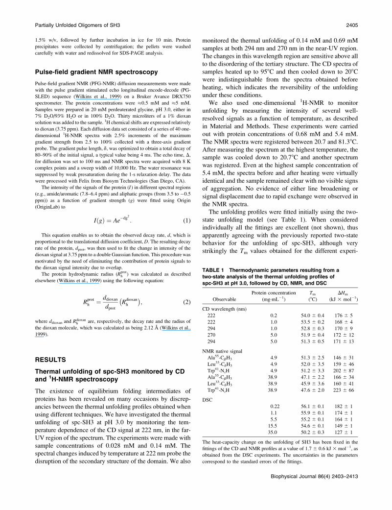
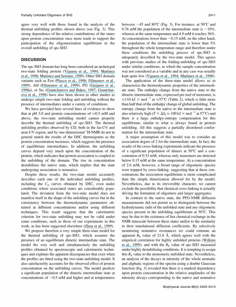
FIGURE 1 Partial molar heat capacity, Cp, versus temperature observed
by DSC for spc-SH3 in 20 mM glycine, pH 3.0, at several concentrations of
protein sample, which are indicated along each curve. Symbols represent the
experimental data. Solid lines correspond to the Cp curves predicted by the
three-state model described in the text using the parameters of Table 2 for
data set A. Dashed lines represent the best prediction of the whole set of Cp
curves using the two-state model.
2406 Casares et al.
Biophysical Journal 86(4) 2403–2413

KI ¼I2½ �N½ �2
KU ¼U½ �N½ � :
The same thermodynamic model, although with different
assumptions concerning the nature of the dimeric interme-
diate state, has been employed elsewhere to describe the
thermal unfolding of the chemotactic protein CheY (Fili-
monov et al., 1993) and of a C-terminal thermolysin
fragment (Azuaga et al., 1995). The equations of the model
that allow us to predict the thermal unfolding profiles are
essentially the same as those described in the above-
mentioned papers and are therefore not included here. States
I2 and U are characterized, respectively, by Gibbs energies
DGI and DGU, enthalpies DHI and DHU, and heat capacities,
DCpI and DCpU, all relative to the native state and expressed
per mol of monomer. We also defined TU as the temperature
at which DGU is zero (which would be the equivalent to the
Tm for a two-state model) and TI as a reference temperature at
which KI has a certain value (Filimonov et al., 1993) taken
here for convenience to be 103.
To apply these equations to an analysis of the thermal
unfolding profiles described here we made two assumptions.
Firstly, we took it that both CD and NMR are only sensitive
to the disappearance of the native state and do not distinguish
between nonnative states. This is strictly true for the
unfolding profiles followed by monitoring the intensity of
a NMR signal corresponding to the native conformation, as it
is the case here. It is also an appropriate assumption for the
near-UV CD signal because folding intermediates or protein
aggregates do not usually exhibit CD in this wavelength
range (Kelly and Price, 1997). In contrast, nonnative states
contribute to the far-UV CD. Nevertheless, the unfolding
profile of spc-SH3 obtained at 222 nm and 0.14 mMmatches
those of the same concentration obtained at 270 nm or 294
nm very closely. The second of our assumptions, which was
also made with the CheY protein (Filimonov et al., 1993),
was that the intermediate and unfolded states have the same
heat capacity functions, i.e., DCpI ¼ DCpU. This approxi-
mation was motivated by the fact that each of these two heat-
capacity changes would be estimated with an extremely large
error if we tried to determine them simultaneously from the
unfolding curves, which do not contain enough information
about them. This approximation implies the assumption that
the amounts of surface exposed to the solvent by the in-
termediate and the unfolded states are similar.
With this model it was possible to predict the unfolding
profiles given the following set of parameters: the two
reference temperatures, TI and TU; the enthalpy values for theintermediate, DHI, and for the unfolded, DHU, states given at
TI and TU, respectively; the heat-capacity change for both theI2 and U states at a certain temperature (508C for con-
venience), DCp; and finally the Cp function of the native
state, considered as being a linear function of temperature.
The Cp function of the unfolded state was calculated as
described elsewhere (Makhatadze and Privalov, 1990).
The fitting of the thermal unfolding profiles using the
model equations was made using two different approaches:
firstly, the global fitting of a set of Cp curves at different
concentrations and secondly, simultaneous fitting of the
unfolding profiles obtained by different techniques at similar
concentrations. Following these two approaches we com-
bined the unfolding profiles in five different data sets (from
A to E), as described in Table 2. The fittings of data sets A
and C are shown in Figs. 1 and 2, respectively. The resulting
parameters of all the fittings are included in Table 2. In the
fittings of data set D and E we needed to fix the parameters
corresponding to the intermediate state, i.e., DHI and TI,which were taken from the fitting of data set A. This was
motivated by the highly indeterminate nature of these
parameters in the fittings, due to the small population of
the intermediate state at these low concentrations, as shown
below. In fact, the fitting of data sets D and E using the two-
state model also gave satisfactory results (data not shown).
As can be seen in Fig. 1, this simple model was capable of
predicting correctly the concentration effects observed in the
Cp curves. This was not possible using the two-state model,
as it is also shown in the same figure. Additionally, the model
could explain simultaneously the unfolding profiles observed
using different techniques (see Fig. 2), which was impossible
using the two-state model at sample concentrations of ;0.5
mM and higher. The resulting thermodynamic parameters,
particularly the two enthalpy values, agree well for the
fittings of different data sets. The enthalpy of the in-
termediate state, which was considered to be a dimer in the
model, is closer to that of the unfolded state than to the native
TABLE 2 Thermodynamic parameters obtained from the
simultaneous fitting of different sets of thermal unfolding
profiles of SH3 at pH 3.0, using the three-state model
described in the text
Data
set*
TI(8C)
TU(8C)
DHI (TI)
(kJ 3 mol�1)
DHU
(TU)
(kJ 3 mol�1)
DCp
(508C)
(kJ 3 K�1
3 mol�1)
A 56.6 6 0.3 55.5 6 0.2 119 6 1 181 6 2 2.2 6 0.2
B 57.0 6 0.2 53.6 6 0.3 120 6 1 166 6 5 2.8 6 0.1
C 56.6 6 0.8 54.6 6 0.2 104 6 4 180 6 2 2.4 6 0.1
D 56.6 (f) 55.6 6 0.2 119 (f) 178 6 1 2.2 6 0.1
E 56.6 (f) 55.9 6 0.1 119 (f) 183 6 1 2.2 6 0.1
For the sake of simplicity, the parameters corresponding to the values of the
different observables for the individual states have not been included. The
uncertainty of each parameter corresponds to the standard error of the fit.
Parameters marked with �f� have been fixed to the corresponding values of
data set A.
*Data set A consisted of five Cp curves obtained at five different sample
concentrations between 0.03 mM and 4.8 mM; data set B consisted of the
Cp curve at 4.8 mM plus an unfolding profile obtained by NMR at 5.4 mM;
data set C consisted of the Cp curve at 0.76 mM, a near-UV CD curve at
0.69 mM, and an NMR profile at 0.68 mM; data set D consisted of a Cp
curve at 0.15 mM, a near-UV CD curve at 0.14 mM, and a far-UV CD
curve at 0.14 mM; data set E consisted of a Cp curve at 0.03 mM and a far-
UV CD curve at 0.028 mM.
Partially Unfolded Oligomers of SH3 2407
Biophysical Journal 86(4) 2403–2413
one, which suggests that it has a low degree of tertiary
structure.
Fig. 3 shows the population of each state of the model as
a function of temperature according to the parameters ob-
tained from the fittings. At low concentrations the population
of the intermediate state is very small throughout the whole
temperature range (#1% at 0.03 mM and#5% at 0.15 mM).
This indicates that at these concentrations the two-state
model describes quite satisfactorily the unfolding of spc-SH3
under these experimental conditions. At 0.7 mM and above,
the population of the intermediate state is quite considerable,
reaching a maximum at temperatures of between 50 and
708C at the highest concentration investigated.
Detection of spc-SH3 oligomers bychemical cross-linking
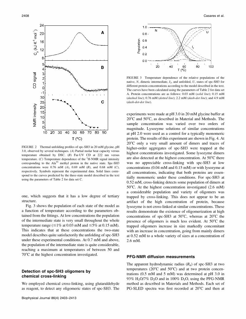
We employed chemical cross-linking, using glutaraldehyde
as reagent, to detect any oligomeric states of spc-SH3. The
experiments were made at pH 3.0 in 20 mM glycine buffer at
208C and 508C, as described in Material and Methods. The
sample concentration was varied over two orders of
magnitude. Lysozyme solutions of similar concentrations
at pH 2.0 were used as a control for a typically monomeric
protein. The results of this experiment are shown in Fig. 4. At
208C only a very small amount of dimers and traces of
higher-order aggregates of spc-SH3 were trapped at the
highest concentrations investigated. Some lysozyme dimers
are also detected at the highest concentration. At 508C there
was no appreciable cross-linking with spc-SH3 at low
concentrations (0.04 mM and 0.15 mM) or with lysozyme at
all concentrations, indicating that both proteins are essen-
tially monomeric under these conditions. For spc-SH3 at
0.52 mM, cross-linking detects some population of dimers at
508C. At the highest concentration investigated (2.6 mM)
a considerable population and variety of oligomers was
trapped by cross-linking. This does not appear to be an
artifact of the high concentration of protein, because
lysozyme is not cross-linked at similar concentrations. These
results demonstrate the existence of oligomerization at high
concentrations of spc-SH3 at 508C, whereas at 208C the
presence of oligomers is much less evident. At 508C the
trapped oligomers increase in size markedly concomitant
with an increase in concentration, going from mainly dimers
at 0.52 mM to a whole variety of sizes at a concentration of
2.6 mM.
PFG-NMR diffusion measurements
The apparent hydrodynamic radius (Rh) of spc-SH3 at two
temperatures (208C and 508C) and at two protein concen-
trations (0.5 mM and 5 mM) was determined at pH 3.0 in
93% H2O/7% D2O and in 100% D2O, using the PFG-NMR
method as described in Materials and Methods. Each set of
PG-SLED spectra was first recorded at 208C and then at
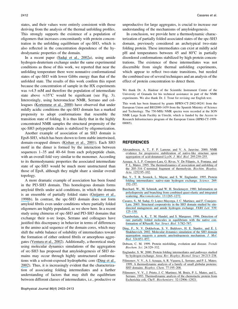
FIGURE 2 Thermal unfolding profiles of spc-SH3 in 20 mM glycine, pH
3.0, observed by several techniques. (A) Partial molar heat capacity versus
temperature obtained by DSC. (B) Far-UV CD at 222 nm versus
temperature. (C) Temperature dependence of the 1H-NMR signal intensity
corresponding to the Ala55 methyl proton in the native state. Spc-SH3
concentrations were 0.76 mM (A), 0.69 mM (B), and 0.68 mM (C),
respectively. Symbols represent the experimental data. Solid lines corre-
spond to the curves predicted by the three-state model described in the text
using the parameters of Table 2 for data set C.
FIGURE 3 Temperature dependence of the relative populations of the
native, N, dimeric intermediate, I2, and unfolded, U, states of spc-SH3 for
different protein concentrations according to the model described in the text.
The curves have been calculated using the parameters of Table 2 for data set
A. Protein concentrations are as follows: 0.03 mM (solid line); 0.15 mM
(dashed line); 0.76 mM (dotted line); 2.2 mM (dash-dot line); and 4.9 mM
(dash-dot-dot line).
2408 Casares et al.
Biophysical Journal 86(4) 2403–2413
508C with the same sample. After cooling the sample down
to 208C a third series of spectra were recorded to check for
reversibility. No differences were seen between the Rh values
obtained at 208C before and after heating the samples to
508C.
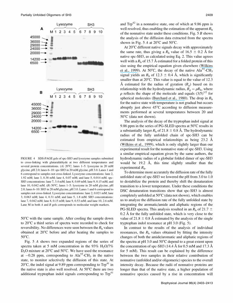
Fig. 5 A shows two expanded regions of the series of
spectra taken at 5 mM concentration in the 93% H2O/7%
D2O mixture at 208C and 508C. We have used the resonance
at �0.28 ppm, corresponding to Ala55-CH3 in the native
state, to monitor selectively the diffusion of this state. At
208C, the indol signal at 9.89 ppm corresponding to Trp41 in
the native state is also well resolved. At 508C there are two
additional tryptophan indol signals corresponding to Trp41
and Trp42 in a nonnative state, one of which at 9.86 ppm is
well resolved, thus enabling the estimation of the apparent Rh
of the nonnative state under these conditions. Fig. 5 B shows
the analysis of the diffusion data extracted from the spectra
shown in Fig. 5 A at 208C and 508C.
At 208C different native signals decay with approximately
the same rate, thus giving a Rh value of 16.5 6 0.2 A for
native spc-SH3, as calculated using Eq. 2. This value agrees
well with a Rh of 15.7 A estimated for a folded protein of this
size using the empirical equation given elsewhere (Wilkins
et al., 1999). At 508C, the decay of the native Ala55-CH3
signal yields an Rh of 12.3 6 0.4 A, which is significantly
smaller than at 208C. This value is equal to the value of 12.3
A estimated for the radius of gyration (Rg) based on its
relationship with the hydrodynamic radius, Rg ¼ rRh, where
r reflects the shape of the molecule and equals (3/5)1/2 for
spherical molecules (Burchard et al., 1980). The drop in Rh
for the native state with temperature is not gradual but occurs
abruptly just above 458C according to diffusion measure-
ments performed at several temperatures between 20 and
508C (data not shown).
The analysis of the decay of the tryptophan indol signal at
9.86 ppm in the series of PG-SLED spectra at 508C results in
a substantially larger Rh of 21.86 0.8 A. The hydrodynamic
radius of the fully unfolded chain of spc-SH3 can be
estimated from empirical relationships as being 23.2 A
(Wilkins et al., 1999), which is only slightly larger than our
experimental result for the nonnative state of spc-SH3. Using
a similar empirical equation given by the same authors, the
hydrodynamic radius of a globular folded dimer of spc-SH3
would be 19.2 A, this time slightly smaller than the
experimental Rh.
To determine more accurately the diffusion rate of the fully
unfolded state of spc-SH3 we lowered the pH from 3.0 to 1.0
to destabilize the protein and thereby shift the denaturation
transition to a lower temperature. Under these conditions the
DSC denaturation transitions show that spc-SH3 is almost
completely unfolded at 508C (data not shown), which enables
us to analyze the diffusion rate of the fully unfolded state by
integrating the aromatic/amide and aliphatic regions of the
PG-SLED spectra. This analysis resulted in an Rh of 21.7 6
0.2 A for the fully unfolded state, which is very close to the
value of 21.86 0.8 A estimated by the analysis of the single
tryptophan indol resonance at pH 3.0 (Fig. 5).
In contrast to the results of the analysis of individual
resonances, the Rh values obtained by fitting the intensity
changes of both the amide/aromatic and aliphatic regions of
the spectra at pH 3.0 and 508C depend to a great extent upon
the concentration of spc-SH3 (14.4 A for 0.5 mM and 17.3 A
for 5 mM). This result can be explained by the difference
between the two samples in their relative contribution of
nonnative (unfolded and/or oligomeric) species to the overall
intensity decay. Because the radii of nonnative proteins are
longer than that of the native state, a higher population of
nonnative species caused by a rise in concentration will
FIGURE 4 SDS-PAGE gels of spc-SH3 and lysozyme samples submitted
to cross-linking with glutaraldehyde at two different temperatures and
several protein concentrations. (A) 208C; lanes 1–5: lysozyme in 20 mM
glycine, pH 2.0; lanes 6–10: spc-SH3 in 20 mM glycine, pH 3.0. Lanes 1 and
6 correspond to samples not cross-linked. Lysozyme concentrations: lane 2,
1.92 mM; lane 3, 0.38 mM; lane 4, 0.07 mM; and lane 5, 0.014 mM. spc-
SH3 concentrations: lane 7, 3.4 mM; lane 8, 0.69 mM; lane 9, 0.15 mM; and
lane 10, 0.042 mM. (B) 508C, lanes 1–5: lysozyme in 20 mM glycine, pH
2.0; lanes 6–10: SH3 in 20 mM glycine, pH 3.0. Lanes 1 and 6 correspond to
samples not cross-linked. Lysozyme concentrations: lane 2, 0.021 mM; lane
3, 0.063 mM; lane 4, 0.31 mM; and lane 5, 1.8 mM. SH3 concentrations:
lane 7, 0.042 mM; lane 8, 0.15 mM; lane 9, 0.53 mM; and lane 10, 2.6 mM.
Lane M in both A and B gels corresponds to molecular weight markers.
Partially Unfolded Oligomers of SH3 2409
Biophysical Journal 86(4) 2403–2413

increase the Rh values obtained in the PG-SLED experi-
ments.
Assuming only two distinguishable states with substan-
tially different hydrodynamic radii at pH 3.0 and 508C
(native and denatured states), and using the experimentally
estimated decay rates for the denatured state (dD) and native
state (dN) from the individually resolved NMR signals, as
described above, the complete amide/aromatic and aliphatic
regions of the PG-SLED spectra can be fitted to the equation:
IðgÞ ¼ ANe�dNg2 1ADe
�dDg2 ; (3)
where AN and AD are the relative contributions to the decay
in intensity of the native and denatured states, respectively
(see also Material and Methods). At 508C and 5 mM this
analysis results in a native state population of 36% and
a denatured state population of 64%, whereas at 508C and 0.5
mM the populations are 80% and 20%, respectively. These
relative populations of native and nonnative states at 508C
FIGURE 5 (A) Expanded regions of a se-
ries of 750 MHz 1D PG-SLED 1H-NMR
spectra of 5 mM spc-SH3 in 93%/7% H2O/
D2O, 20 mM glycine, pH 3.0. Diffusion at
208C and 508C, as indicated in the plot. The
correspondences of each signal with either
native (N) or denatured (D) states of the spc-
SH3 domain are also indicated along the
spectra. (B) Analysis of the PFG-NMR
diffusion measurements shown in panel A.
Solid circles represent the intensity changes
of the native signal at �0.29 ppm at both
temperatures. Open triangles represent the
intensity of the native Trp41 indol signal at
9.89 ppm at 208C. Open squares represent
the intensity of the nonnative tryptophan
indol signal at 9.86 ppm at 508C. The decay
of each signal was fitted using Eq. 1 and the
fits are shown in solid lines.
2410 Casares et al.
Biophysical Journal 86(4) 2403–2413
agree very well with those found in the analysis of the
thermal unfolding profiles shown above (see Fig. 3). This
strong dependence of the relative contributions of the states
upon protein concentration once more tends to support the
participation of the oligomerization equilibrium in the
overall unfolding of spc-SH3.
DISCUSSION
The spc-SH3 domain has long been considered an archetypal
two-state folding protein (Viguera et al., 1994; Martinez
et al., 1998; Martinez and Serrano, 1999). Other SH3 domain
variants such as Fyn (Plaxco et al., 1998; Filimonov et al.,
1999), Abl (Filimonov et al., 1999), PI3 (Guijarro et al.,
1998a), or Src (Grantcharova and Baker, 1997; Grantchar-
ova et al., 1998) have also been shown in other studies to
undergo simple two-state folding and unfolding without the
presence of intermediates under a variety of conditions.
We have provided here several lines of evidence to show
that at pH 3.0 and protein concentrations of �0.5 mM and
above, the two-state unfolding model cannot properly
describe the thermal unfolding of spc-SH3. The thermal
unfolding profiles observed by CD, both in the far-UV and
near-UV region, and by one-dimensional 1H-NMR do not in
general match the results of the DSC thermograms as the
protein concentration increases, which suggests the presence
of equilibrium intermediates. In addition, the unfolding
curves depend very much upon the concentration of the
protein, which indicates that protein association is coupled to
the unfolding of the domain. The rise in concentration
destabilizes the native state, which implies that the state
undergoing association is nonnative.
Despite these results, the two-state model accurately
describes each one of the individual unfolding profiles,
including the Cp curves obtained by DSC, even under
conditions where associated states are considerably popu-
lated. The deviation from the two-state model does not
manifest itself in the shape of the unfolding curves but in the
consistency between the thermodynamic parameters ob-
tained at different concentrations and/or using different
techniques. This result suggests that the calorimetric
criterion for two-state unfolding may not be valid under
some conditions, such as those of our experiments in this
work, as has been suggested elsewhere (Zhou et al., 1999).
We propose therefore a very simple three-state model for
the thermal unfolding of spc-SH3, which assumes the
presence of an equilibrium dimeric intermediate state. The
model fits very well and simultaneously the unfolding
profiles obtained by spectroscopic and calorimetric techni-
ques and explains the apparent discrepancies that exist when
the profiles are fitted using the two-state unfolding model. It
also satisfactorily accounts for the observed effect of protein
concentration on the unfolding curves. The model predicts
a significant population of the dimeric intermediate state at
concentrations of ;0.5 mM and higher and at temperatures
between ;45 and 808C (Fig. 3). For instance, at 508C and
0.76 mM the population of the intermediate state is ;10%,
whereas at the same temperature and 4.9 mM it reaches 36%.
At concentrations lower than ;0.15 mM, on the other hand,
the population of the intermediate state is lower than 5%
throughout the whole temperature range and therefore under
these conditions the unfolding process of spc-SH3 is
adequately described by the two-state model. This agrees
with previous studies of the folding-unfolding of spc-SH3
under similar conditions, in which the sample concentration
was not considered as a variable and in any case was usually
kept quite low (Viguera et al., 1994; Martinez et al., 1998).
The application of the three-state model allows us to
characterize the thermodynamic properties of the intermedi-
ate state. The enthalpy change from the native state to the
dimeric intermediate state, expressed per mol of monomer, is
�110 kJ 3 mol�1 at �578C (Table 2), which is little more
than half that of the enthalpy change of global unfolding. The
entropy change from the native to the intermediate state is
also relatively high (T3 DSI� 100 kJ3mol�1 at 578C) and
there is a large enthalpy-entropy compensation for this
equilibrium, similar to what is always found in protein
unfolding. All this suggests a partially disordered confor-
mation for the intermediate state.
A major assumption of this model was to consider an
association degree of 2 for the intermediate state. In fact, the
results of the cross-linking experiments indicate the presence
of a significant population of dimers at 508C and a con-
centration of 0.53 mM, whereas only monomers are detected
below 0.15 mM at the same temperature. At a concentration
of 2.6 mM, however, a broad range of oligomeric species
were trapped by cross-linking, suggesting that at these con-
centrations the association equilibrium is more complicated
than the simple dimerization allowed for by the model.
Nevertheless, due to its irreversible character, we cannot
exclude the possibility that chemical cross-linking is actually
driving the formation of oligomers and larger aggregates.
In contrast to the native state, the PFG-NMR diffusion
measurements did not permit us to distinguish between the
hydrodynamic radii of the unfolded state and any oligomeric
species present in the unfolding equilibrium at 508C. This
may be due to the existence of fast chemical exchange in the
NMR timescale between these states and/or to the similarity
in their translational diffusion coefficients. By selectively
monitoring nonnative resonances we could estimate an
apparent Rh value of 21.8 A, which agrees well with the
empirical correlation for highly unfolded proteins (Wilkins
et al., 1999), and with the Rh value of spc-SH3 measured
under highly destabilizing conditions. It is tempting to assign
this Rh value to the monomeric unfolded state. Nevertheless,
an analysis of the decays in intensity of the whole aromatic
and aliphatic regions of the spectra using a double Gaussian
function (Eq. 3) revealed that there is a marked dependency
upon protein concentration in the relative amplitudes of the
intensity decays corresponding to the native and nonnative
Partially Unfolded Oligomers of SH3 2411
Biophysical Journal 86(4) 2403–2413
states, and their values were entirely consistent with those
deriving from the analysis of the thermal unfolding profiles.
This strongly supports the existence of a population of
oligomers that increases concomitantly with protein concen-
tration in the unfolding equilibrium of spc-SH3, which is
also reflected in the concentration dependence of the hy-
drodynamic properties of the domain.
In a recent paper (Sadqi et al., 2002a), using amide
hydrogen-deuterium exchange under the same experimental
conditions as those of this work, we reported that near the
unfolding temperature there were nonnative conformational
states of spc-SH3 with lower Gibbs energy than that of the
unfolded state. The results of this work confirm this report
because the concentration of sample in the HX experiments
was �4.5 mM and therefore the population of intermediate
state above �358C would be significant (see Fig. 3).
Interestingly, using heteronuclear NMR, Serrano and col-
leagues (Kortemme et al., 2000) have observed that under
mildly acidic conditions the spc-SH3 domain has a marked
propensity to adopt conformations that resemble the
transition state of folding. It is thus likely that in the highly
concentrated NMR samples the structural propensity of the
spc-SH3 polypeptide chain is stabilized by oligomerization.
Another example of association of an SH3 domain is
Eps8-SH3, which has been shown to form stable intertwined,
domain-swapped dimers (Kishan et al., 2001). Each SH3
motif in the dimer is formed by the interaction between
sequences 1–35 and 40–64 from each polypeptide chain,
with an overall fold very similar to the monomer. According
to its thermodynamic properties the associated intermediate
state of spc-SH3 would be much more unstructured than
those of Eps8, although they might share a similar overall
topology.
A more dramatic example of association has been found
in the PI3-SH3 domain. This homologous domain forms
amyloid fibrils under acid conditions, in which the domain
is an ensemble of partially folded states (Guijarro et al.,
1998b). In contrast, the spc-SH3 domain does not form
amyloid fibrils even under conditions where partially folded
oligomers are highly populated, as we show here. In a recent
study using chimeras of spc-SH3 and PI3-SH3 domains that
exchange their n-src loops, Serrano and colleagues have
justified this discrepancy in behavior in terms of differences
in the amino acid sequence of the domain cores, which may
shift the subtle balance of solubility of intermediates toward
the formation of either ordered fibrils or amorphous aggre-
gates (Ventura et al., 2002). Additionally, a theoretical study
using molecular dynamics simulations of the aggregation
of src-SH3 has proposed that amyloidogenesis of SH3 do-
mains may occur through highly unstructured conforma-
tions with a solvent-exposed hydrophobic core (Ding et al.,
2002). Thus, it is increasingly evident that the characteriza-
tion of associating folding intermediates and a further
understanding of factors that may shift the equilibrium
between different classes of intermediates, i.e., productive or
unproductive for large aggregates, is crucial to increase our
understanding of the mechanisms of amyloidogenesis.
In conclusion, we provide here a thermodynamic charac-
terization of partially folded associated states of the spc-SH3
domain, previously considered an archetypical two-state
folding protein. These intermediates can exist at mildly acid
pH and temperatures between 45 and 808C in partially
disordered conformations stabilized by high protein concen-
trations. The existence of these intermediates was not
discernible from single thermal unfolding experiments,
which appear to reflect two-state transitions, but needed
the combined use of several techniques and an analysis of the
effect of protein concentration to detect them.
We thank Dr. A. Haidour of the Scientific Instrument Centre of the
University of Granada for his technical assistance in part of the NMR
experiments. We also thank Dr. J. Trout for revising our English text.
This work has been financed by grants HPRN-CT-2002-00241 from the
European Union and BIO2000-1459 from the Spanish Ministry of Science
and Technology. The 750-MHz NMR spectra were recorded at the SON
NMR Large Scale Facility in Utrecht, which is funded by the Access to
Research Infrastructures program of the European Union (HPRI-CT-1999-
00005).
REFERENCES
Alexandrescu, A. T., F. P. Lamour, and V. A. Jaravine. 2000. NMRevidence for progressive stabilization of native-like structure uponaggregation of acid-denatured LysN. J. Mol. Biol. 295:239–255.
Azuaga, A. I., F. Conejero-Lara, G. Rivas, V. De Filippis, A. Fontana, andP. L. Mateo. 1995. The thermodynamics of association and unfolding ofthe 205–316 C-terminal fragment of thermolysin. Biochim. Biophys.Acta. 1252:95–102.
Bai, Y., T. R. Sosnick, L. Mayne, and S. W. Englander. 1995. Proteinfolding intermediates: native-state hydrogen exchange. Science. 269:192–197.
Burchard, W., M. Schmidt, and W. H. Stockmayer. 1980. Information onpolydispersity and branching from combined quasi-elastic and integratedscattering. Macromolecules. 13:1265–1272.
Casares, S., M. Sadqi, O. Lopez-Mayorga, J. C. Martinez, and F. Conejero-Lara. 2003. Structural cooperativity in the SH3 domain studied by site-directed mutagenesis and amide hydrogen exchange. FEBS Lett. 539:125–130.
Chamberlain, A. K., T. M. Handel, and S. Marqusee. 1996. Detection ofrare partially folded molecules in equilibrium with the native con-formation of RNaseH. Nat. Struct. Biol. 3:782–787.
Ding, F., N. V. Dokholyan, S. V. Buldyrev, H. E. Stanley, and E. I.Shakhnovich. 2002. Molecular dynamics simulation of the SH3 domainaggregation suggests a generic amyloidogenesis mechanism. J. Mol.Biol. 324:851–857.
Dobson, C. M. 1999. Protein misfolding, evolution and disease. TrendsBiochem. Sci. 24:329–332.
Englander, S. W. 2000. Protein folding intermediates and pathways studiedby hydrogen exchange. Annu. Rev. Biophys. Biomol. Struct. 29:213–238.
Filimonov, V. V., A. I. Azuaga, A. R. Viguera, L. Serrano, and P. L. Mateo.1999. A thermodynamic analysis of a family of small globular proteins:SH3 domains. Biophys. Chem. 77:195–208.
Filimonov, V. V., J. Prieto, J. C. Martinez, M. Bruix, P. L. Mateo, and L.Serrano. 1993. Thermodynamic analysis of the chemotactic protein fromEscherichia coli, CheY. Biochemistry. 32:12906–12921.
2412 Casares et al.
Biophysical Journal 86(4) 2403–2413
Fuentes, E. J., and A. J. Wand. 1998. Local stability and dynamics ofapocytochrome b562 examined by the dependence of hydrogen exchangeon hydrostatic pressure. Biochemistry. 37:9877–9883.
Grantcharova, V. P., and D. Baker. 1997. Folding dynamics of the src SH3domain. Biochemistry. 36:15685–15692.
Grantcharova, V. P., D. S. Riddle, J. V. Santiago, and D. Baker. 1998.Important role of hydrogen bonds in the structurally polarizedtransition state for folding of the src SH3 domain. Nat. Struct. Biol.5:714–720.
Guijarro, J. I., C. J. Morton, K. W. Plaxco, I. D. Campbell, and C. M.Dobson. 1998a. Folding kinetics of the SH3 domain of PI3 kinase byreal-time NMR combined with optical spectroscopy. J. Mol. Biol.276:657–667.
Guijarro, J. I., M. Sunde, J. A. Jones, I. D. Campbell, and C. M. Dobson.1998b. Amyloid fibril formation by an SH3 domain. Proc. Natl. Acad.Sci. USA. 95:4224–4228.
Jackson, S. E. 1998. How do small single-domain proteins fold? Fold. Des.3:R81–R91.
Jackson, S. E., and A. R. Fersht. 1991. Folding of chymotrypsin inhibitor2. 1. Evidence for a two-state transition. Biochemistry. 30:10428–10435.
Jaenicke, R., and R. Rudolph. 1989. Folding proteins. In: Protein Structure:A Practical Approach. T. E. Creighton, editor. IRL Press (OxfordUniversity Press), Oxford, UK. 191–223.
Kelly, S. M., and N. C. Price. 1997. The application of circular dichroismto studies of protein folding and unfolding. Biochim. Biophys. Acta.1338:161–185.
Kishan, K. V., M. E. Newcomer, T. H. Rhodes, and S. D. Guilliot. 2001.Effect of pH and salt bridges on structural assembly: molecular structuresof the monomer and intertwined dimer of the Eps8 SH3 domain. ProteinSci. 10:1046–1055.
Kortemme, T., M. J. Kelly, L. E. Kay, J. Forman-Kay, and L. Serrano.2000. Similarities between the spectrin SH3 domain denatured state andits folding transition state. J. Mol. Biol. 297:1217–1229.
Kuznetsova, I. M., A. G. Biktashev, S. Y. Khaitlina, K. S. Vassilenko,K. K. Turoverov, and V. N. Uversky. 1999. Effect of self-association onthe structural organization of partially folded proteins: inactivated actin.Biophys. J. 77:2788–2800.
Liu, T., P. A. Pemberton, and A. D. Robertson. 1999. Three-state unfoldingand self-association of maspin, a tumor-suppressing serpin. J. Biol.Chem. 274:29628–29632.
Makhatadze, G. I., and P. L. Privalov. 1990. Heat capacity of proteins. I.Partial molar heat capacity of individual amino acid residues in aqueoussolution: hydration effect. J. Mol. Biol. 213:375–384.
Martinez, J. C., M. T. Pisabarro, and L. Serrano. 1998. Obligatory steps inprotein folding and the conformational diversity of the transition state.Nat. Struct. Biol. 5:721–729.
Martinez, J. C., and L. Serrano. 1999. The folding transition state betweenSH3 domains is conformationally restricted and evolutionarily con-served. Nat. Struct. Biol. 6:1010–1016.
Mayne, L., and S. W. Englander. 2000. Two-state vs. multistate proteinunfolding studied by optical melting and hydrogen exchange. ProteinSci. 9:1873–1877.
Milne, J. S., Y. Xu, L. C. Mayne, and S. W. Englander. 1999. Experimentalstudy of the protein folding landscape: unfolding reactions in cytochromec. J. Mol. Biol. 290:811–822.
Pecorari, F., P. Minard, M. Desmadril, and J. M. Yon. 1996. Occurrence oftransient multimeric species during the refolding of a monomeric protein.J. Biol. Chem. 271:5270–5276.
Plaxco, K. W., J. I. Guijarro, C. J. Morton, M. Pitkeathly, I. D. Campbell,and C. M. Dobson. 1998. The folding kinetics and thermodynamics ofthe Fyn-SH3 domain. Biochemistry. 37:2529–2537.
Privalov, P. L. 1979. Stability of proteins: small globular proteins. Adv.Protein Chem. 33:167–241.
Privalov, P. L., and S. A. Potekhin. 1986. Scanning microcalorimetry instudying temperature-induced changes in proteins. Methods Enzymol.131:4–51.
Rochet, J. C., and P. T. Lansbury, Jr. 2000. Amyloid fibrillogenesis: themesand variations. Curr. Opin. Struct. Biol. 10:60–68.
Sadqi, M., S. Casares, M. A. Abril, O. Lopez-Mayorga, F. Conejero-Lara,and E. Freire. 1999. The native state conformational ensemble of the SH3domain from alpha-spectrin. Biochemistry. 38:8899–8906.
Sadqi, M., S. Casares, O. Lopez-Mayorga, and F. Conejero-Lara. 2002a.The temperature dependence of the hydrogen exchange in the SH3domain of alpha-spectrin. FEBS Lett. 527:86–90.
Sadqi, M., S. Casares, O. Lopez-Mayorga, J. C. Martinez, and F. Conejero-Lara. 2002b. pH dependence of the hydrogen exchange in the SH3domain of alpha-spectrin. FEBS Lett. 514:295–299.
Silow, M., Y. J. Tan, A. R. Fersht, and M. Oliveberg. 1999. Formation ofshort-lived protein aggregates directly from the coil in two-state folding.Biochemistry. 38:13006–13012.
Uversky, V. N., A. S. Karnoup, R. Khurana, D. J. Segel, S. Doniach, and A.L. Fink. 1999. Association of partially folded intermediates of staphylo-coccal nuclease induces structure and stability. Protein Sci. 8:161–173.
Uversky, V. N., D. J. Segel, S. Doniach, and A. L. Fink. 1998. Association-induced folding of globular proteins. Proc. Natl. Acad. Sci. USA. 95:5480–5483.
van Nuland, N. A., F. Chiti, N. Taddei, G. Raugei, G. Ramponi, and C. M.Dobson. 1998. Slow folding of muscle acylphosphatase in the absence ofintermediates. J. Mol. Biol. 283:883–891.
Ventura, S., E. Lacroix, and L. Serrano. 2002. Insights into the origin of thetendency of the PI3–SH3 domain to form amyloid fibrils. J. Mol. Biol.322:1147–1158.
Viguera, A. R., J. C. Martinez, V. V. Filimonov, P. L. Mateo, and L. Serrano.1994. Thermodynamic and kinetic analysis of the SH3 domain of spectrinshows a two-state folding transition. Biochemistry. 33:2142–2150.
Wilkins, D. K., S. B. Grimshaw, V. Receveur, C. M. Dobson, J. A. Jones,and L. J. Smith. 1999. Hydrodynamic radii of native and denaturedproteins measured by pulse field gradient NMR techniques. Bio-chemistry. 38:16424–16431.
Ye, K., and J. Wang. 2001. Self-association reaction of denaturedstaphylococcal nuclease fragments characterized by heteronuclearNMR. J. Mol. Biol. 307:309–322.
Zhou, Y., C. K. Hall, and M. Karplus. 1999. The calorimetric criterion fora two-state process revisited. Protein Sci. 8:1064–1074.
Partially Unfolded Oligomers of SH3 2413
Biophysical Journal 86(4) 2403–2413