The in Vivo and in Vitro Aggregation Properties of Globular Proteins Correlate With Their...
16
The in Vivo and in Vitro Aggregation Properties of Globular Proteins Correlate With Their Conformational Stability: The SH3 Case Alba Espargaró 1 †, Virginia Castillo 1 †, Natalia S. de Groot 1 and Salvador Ventura 1,2 ⁎ 1 Departament de Bioquímica i Biologia Molecular, Facultat de Biociències, Universitat Autònoma de Barcelona, E-08193 Bellaterra, Spain 2 Institut de Biotecnologia i de Biomedicina, Universitat Autònoma de Barcelona, E-08193 Bellaterra, Spain Received 16 October 2007; received in revised form 13 March 2008; accepted 13 March 2008 Available online 19 March 2008 Protein misfolding and deposition underlie an increasing number of debilitating human disorders and constitute a problem of major concern in biotechnology. In the last years, in vitro studies have provided valuable insights into the physicochemical principles underlying protein aggregation. Nevertheless, information about the determinants of protein deposition within the cell is scarce and only a few systematic studies comparing in vitro and in vivo data have been reported. Here, we have used the SH3 domain of α-spectrin as a model globular protein in an attempt to understand the relationship between protein aggregation in the test-tube and in the more complex cellular environment. The investigation of the aggregation in Escherichia coli of this domain and a large set of mutants, together with the analysis of their sequential and conformational properties allowed us to evaluate the contribution of different polypeptidic factors to the cellular deposition of globular proteins. The data presented here suggest that the rules that govern in vitro protein aggregation are also valid in in vivo contexts. They also provide relevant insights into intracellular protein deposition in both conformational diseases and recombinant protein production. © 2008 Elsevier Ltd. All rights reserved. Edited by J. Weissman Keywords: protein aggregation; SH3 domains; protein stability; protein folding; protein production Introduction The presence of insoluble protein deposits in human tissues correlates with the development of conformational disorders such as Alzheimer’s dis- ease and Parkinson’s disease. 1–3 Also, the aggrega- tion of globular proteins into insoluble polypeptide chains during protein production in bacteria is of major concern in biotechnology, since it reduces significantly the spectrum of relevant recombinant proteins available for structural genomics or com- mercial use. 4–6 For globular proteins, the formation of aggregates is in most cases an outcome of im- proper folding, resulting in the accumulation of unfolded or partially folded intermediates that are thought to be critical soluble precursors of fibrillar aggregates in amyloid diseases or inclusion bodies in protein production. 7,8 This effect has been characterized extensively in vitro by protein engi- neering using both disease-related and non-related polypeptide models. 9–14 Fewer studies have ad- dressed the influence of polypeptide properties on their aggregation within the cell, 15–19 and only a few have compared systematically the in vitro and in vivo aggregation behaviour of proteins. 17,20–22 The in vivo aggregation of polypeptides does not necessarily have to correlate with their in vitro properties, because the protein quality machinery modulates the accumulation of aggregation-prone polypeptidic chains by facilitating their folding, masking hydro- *Corresponding author. Departament de Bioquímica i Biologia Molecular, Facultat de Biociències, Universitat Autònoma de Barcelona, E-08193 Bellaterra, Spain. E-mail address: [email protected]. † A.E. and V.C. contributed equally to this work. Abbreviations used: ANS, 1-anilinonaphtalene-8- sulfonate; FTIR, Fourier-transformed infrared; HypF-N, N terminus of hydrogenase maturation protein; PI3-kinase, phosphoinositide-3-kinase; SPC-SH3, SH3 domain of α- spectrin; Th-T, thioflavin-T; TEM, transmission electron microscopy. doi:10.1016/j.jmb.2008.03.020 J. Mol. Biol. (2008) 378, 1116–1131 Available online at www.sciencedirect.com 0022-2836/$ - see front matter © 2008 Elsevier Ltd. All rights reserved.
-
Upload
independent -
Category
Documents
-
view
3 -
download
0
Transcript of The in Vivo and in Vitro Aggregation Properties of Globular Proteins Correlate With Their...
doi:10.1016/j.jmb.2008.03.020 J. Mol. Biol. (2008) 378, 1116–1131
Available online at www.sciencedirect.com
The in Vivo and in Vitro Aggregation Properties ofGlobular Proteins Correlate With Their ConformationalStability: The SH3 Case
Alba Espargaró1†, Virginia Castillo1†, Natalia S. de Groot1
and Salvador Ventura1,2⁎
1Departament de Bioquímica iBiologia Molecular, Facultat deBiociències, UniversitatAutònoma de Barcelona,E-08193 Bellaterra, Spain2Institut de Biotecnologia i deBiomedicina, UniversitatAutònoma de Barcelona,E-08193 Bellaterra, Spain
Received 16 October 2007;received in revised form13 March 2008;accepted 13 March 2008Available online19 March 2008
*Corresponding author. DepartamenBiologia Molecular, Facultat de BiocAutònoma de Barcelona, E-08193 Beaddress: [email protected].† A.E. and V.C. contributed equalAbbreviations used: ANS, 1-anilin
sulfonate; FTIR, Fourier-transformedterminus of hydrogenase maturationphosphoinositide-3-kinase; SPC-SH3spectrin; Th-T, thioflavin-T; TEM, trmicroscopy.
0022-2836/$ - see front matter © 2008 E
Protein misfolding and deposition underlie an increasing number ofdebilitating human disorders and constitute a problem of major concern inbiotechnology. In the last years, in vitro studies have provided valuableinsights into the physicochemical principles underlying protein aggregation.Nevertheless, information about the determinants of protein depositionwithin the cell is scarce and only a few systematic studies comparing in vitroand in vivo data have been reported. Here, we have used the SH3 domain ofα-spectrin as a model globular protein in an attempt to understand therelationship between protein aggregation in the test-tube and in the morecomplex cellular environment. The investigation of the aggregation inEscherichia coli of this domain and a large set of mutants, together with theanalysis of their sequential and conformational properties allowed us toevaluate the contribution of different polypeptidic factors to the cellulardeposition of globular proteins. The data presented here suggest that therules that govern in vitro protein aggregation are also valid in in vivo contexts.They also provide relevant insights into intracellular protein deposition inboth conformational diseases and recombinant protein production.
© 2008 Elsevier Ltd. All rights reserved.
Keywords: protein aggregation; SH3 domains; protein stability; proteinfolding; protein production
Edited by J. WeissmanIntroduction
The presence of insoluble protein deposits inhuman tissues correlates with the development ofconformational disorders such as Alzheimer’s dis-ease and Parkinson’s disease.1–3 Also, the aggrega-tion of globular proteins into insoluble polypeptidechains during protein production in bacteria is of
t de Bioquímica iiències, Universitatllaterra, Spain. E-mail
ly to this work.onaphtalene-8-infrared; HypF-N, Nprotein; PI3-kinase,, SH3 domain of α-ansmission electron
lsevier Ltd. All rights reserve
major concern in biotechnology, since it reducessignificantly the spectrum of relevant recombinantproteins available for structural genomics or com-mercial use.4–6 For globular proteins, the formationof aggregates is in most cases an outcome of im-proper folding, resulting in the accumulation ofunfolded or partially folded intermediates that arethought to be critical soluble precursors of fibrillaraggregates in amyloid diseases or inclusion bodiesin protein production.7,8 This effect has beencharacterized extensively in vitro by protein engi-neering using both disease-related and non-relatedpolypeptide models.9–14 Fewer studies have ad-dressed the influence of polypeptide properties ontheir aggregation within the cell,15–19 and only a fewhave compared systematically the in vitro and in vivoaggregation behaviour of proteins.17,20–22 The in vivoaggregation of polypeptides does not necessarilyhave to correlate with their in vitro properties,because the protein quality machinery modulatesthe accumulation of aggregation-prone polypeptidicchains by facilitating their folding, masking hydro-
d.
1117Aggregation Properties of SH3 Domains
phobic regions and targeting improperly foldedproteins towards degradation pathways.23 Ourunderstanding of how protein aggregation proceedswithin the cell will benefit from an integration of thewell-established physicochemical principles under-lying protein self-assembly in vitro with data fromstudies addressing protein deposition in livingorganisms, using consistent protein models.Here, we use the SH3 domain of α-spectrin (SPC-
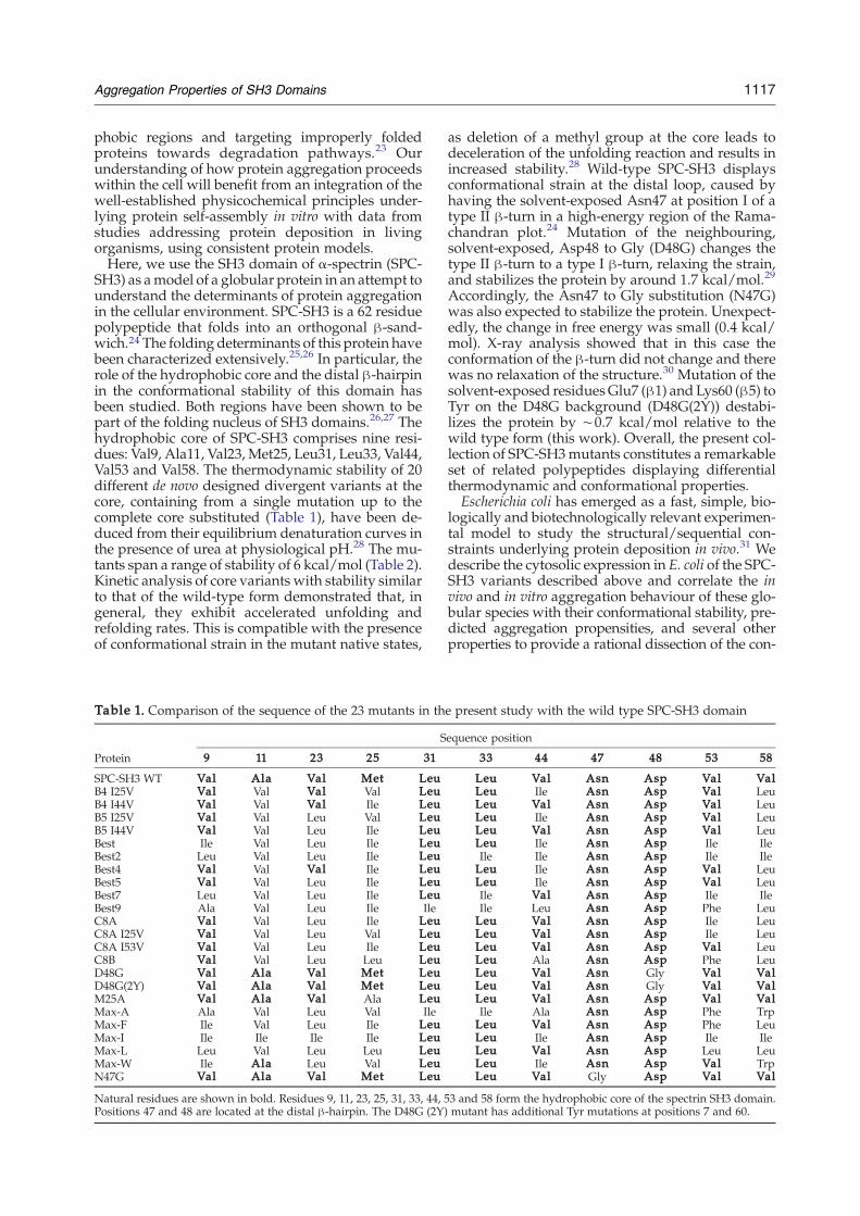
SH3) as amodel of a globular protein in an attempt tounderstand the determinants of protein aggregationin the cellular environment. SPC-SH3 is a 62 residuepolypeptide that folds into an orthogonal β-sand-wich.24 The folding determinants of this protein havebeen characterized extensively.25,26 In particular, therole of the hydrophobic core and the distal β-hairpinin the conformational stability of this domain hasbeen studied. Both regions have been shown to bepart of the folding nucleus of SH3 domains.26,27 Thehydrophobic core of SPC-SH3 comprises nine resi-dues: Val9, Ala11, Val23,Met25, Leu31, Leu33, Val44,Val53 and Val58. The thermodynamic stability of 20different de novo designed divergent variants at thecore, containing from a single mutation up to thecomplete core substituted (Table 1), have been de-duced from their equilibrium denaturation curves inthe presence of urea at physiological pH.28 The mu-tants span a range of stability of 6 kcal/mol (Table 2).Kinetic analysis of core variants with stability similarto that of the wild-type form demonstrated that, ingeneral, they exhibit accelerated unfolding andrefolding rates. This is compatible with the presenceof conformational strain in the mutant native states,
Table 1. Comparison of the sequence of the 23 mutants in th
Protein
S
9 11 23 25 31
SPC-SH3 WT Val Ala Val Met LeuB4 I25V Val Val Val Val LeuB4 I44V Val Val Val Ile LeuB5 I25V Val Val Leu Val LeuB5 I44V Val Val Leu Ile LeuBest Ile Val Leu Ile LeuBest2 Leu Val Leu Ile LeuBest4 Val Val Val Ile LeuBest5 Val Val Leu Ile LeuBest7 Leu Val Leu Ile LeuBest9 Ala Val Leu Ile IleC8A Val Val Leu Ile LeuC8A I25V Val Val Leu Val LeuC8A I53V Val Val Leu Ile LeuC8B Val Val Leu Leu LeuD48G Val Ala Val Met LeuD48G(2Y) Val Ala Val Met LeuM25A Val Ala Val Ala LeuMax-A Ala Val Leu Val IleMax-F Ile Val Leu Ile LeuMax-I Ile Ile Ile Ile LeuMax-L Leu Val Leu Leu LeuMax-W Ile Ala Leu Val LeuN47G Val Ala Val Met Leu
Natural residues are shown in bold. Residues 9, 11, 23, 25, 31, 33, 44,Positions 47 and 48 are located at the distal β-hairpin. The D48G (2Y
as deletion of a methyl group at the core leads todeceleration of the unfolding reaction and results inincreased stability.28 Wild-type SPC-SH3 displaysconformational strain at the distal loop, caused byhaving the solvent-exposed Asn47 at position I of atype II β-turn in a high-energy region of the Rama-chandran plot.24 Mutation of the neighbouring,solvent-exposed, Asp48 to Gly (D48G) changes thetype II β-turn to a type I β-turn, relaxing the strain,and stabilizes the protein by around 1.7 kcal/mol.29
Accordingly, the Asn47 to Gly substitution (N47G)was also expected to stabilize the protein. Unexpect-edly, the change in free energy was small (0.4 kcal/mol). X-ray analysis showed that in this case theconformation of the β-turn did not change and therewas no relaxation of the structure.30 Mutation of thesolvent-exposed residuesGlu7 (β1) and Lys60 (β5) toTyr on the D48G background (D48G(2Y)) destabi-lizes the protein by ∼0.7 kcal/mol relative to thewild type form (this work). Overall, the present col-lection of SPC-SH3mutants constitutes a remarkableset of related polypeptides displaying differentialthermodynamic and conformational properties.Escherichia coli has emerged as a fast, simple, bio-
logically and biotechnologically relevant experimen-tal model to study the structural/sequential con-straints underlying protein deposition in vivo.31 Wedescribe the cytosolic expression in E. coli of the SPC-SH3 variants described above and correlate the invivo and in vitro aggregation behaviour of these glo-bular species with their conformational stability, pre-dicted aggregation propensities, and several otherproperties to provide a rational dissection of the con-
e present study with the wild type SPC-SH3 domain
equence position
33 44 47 48 53 58
Leu Val Asn Asp Val ValLeu Ile Asn Asp Val LeuLeu Val Asn Asp Val LeuLeu Ile Asn Asp Val LeuLeu Val Asn Asp Val LeuLeu Ile Asn Asp Ile IleIle Ile Asn Asp Ile IleLeu Ile Asn Asp Val LeuLeu Ile Asn Asp Val LeuIle Val Asn Asp Ile IleIle Leu Asn Asp Phe LeuLeu Val Asn Asp Ile LeuLeu Val Asn Asp Ile LeuLeu Val Asn Asp Val LeuLeu Ala Asn Asp Phe LeuLeu Val Asn Gly Val ValLeu Val Asn Gly Val ValLeu Val Asn Asp Val ValIle Ala Asn Asp Phe TrpLeu Val Asn Asp Phe LeuLeu Ile Asn Asp Ile IleLeu Val Asn Asp Leu LeuLeu Ile Asn Asp Val TrpLeu Val Gly Asp Val Val
53 and 58 form the hydrophobic core of the spectrin SH3 domain.) mutant has additional Tyr mutations at positions 7 and 60.
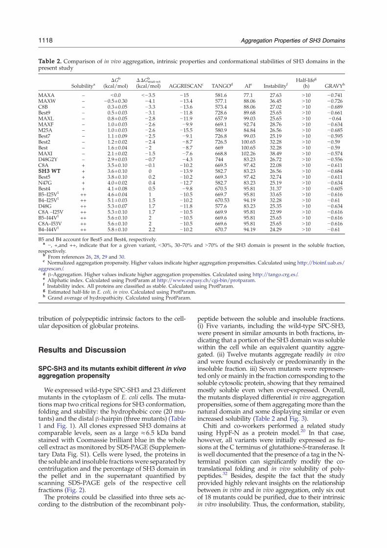
Table 2. Comparison of in vivo aggregation, intrinsic properties and conformational stabilities of SH3 domains in thepresent study
SolubilityaΔGb
(kcal/mol)ΔΔGmut-wt
b
(kcal/mol) AGGRESCANc TANGOd AIe InstabilityfHalf-lifeg
(h) GRAVYh
MAXA – b0.0 b−3.5 −15 581.6 77.1 27.63 N10 −0.741MAXW – −0.5±0.30 −4.1 −13.4 577.1 88.06 36.45 N10 −0.726C8B – 0.3±0.05 −3.3 −13.6 573.4 88.06 27.02 N10 −0.689Best9 – 0.5±0.03 −3.1 −11.8 728.6 89.68 25.65 N10 −0.661MAXL – 0.8±0.05 −2.8 −11.9 657.9 99.03 25.65 N10 −0.64MAXF – 1.0±0.03 −2.6 −9.9 669.1 92.74 28.76 N10 −0.634M25A – 1.0±0.03 −2.6 −15.5 580.9 84.84 26.56 N10 −0.685Best7 – 1.1±0.09 −2.5 −9.1 726.8 99.03 25.19 N10 −0.595Best2 – 1.2±0.02 −2.4 −8.7 726.5 100.65 32.28 N10 −0.59Best – 1.6±0.04 −2 −8.7 669 100.65 32.28 N10 −0.59MAXI – 2.1±0.02 −1.5 −7.6 668.8 102.26 38.49 N10 −0.574D48G2Y – 2.9±0.03 −0.7 −4.3 744 83.23 26.72 N10 −0.556C8A + 3.5±0.10 −0.1 −10.2 669.5 97.42 22.08 N10 −0.611SH3 WT + 3.6±0.10 0 −13.9 582.7 83.23 26.56 N10 −0.684Best5 + 3.8±0.10 0.2 −10.2 669.3 97.42 32.74 N10 −0.611N47G + 4.0±0.02 0.4 −12.7 582.7 83.23 25.19 N10 −0.634Best4 + 4.1±0.08 0.5 −9.8 670.5 95.81 31.37 N10 −0.605B5–I25V1 ++ 4.6±0.04 1 −10.5 669.7 95.81 33.65 N10 −0.616B4–I25V1 ++ 5.1±0.03 1.5 −10.2 670.53 94.19 32.28 N10 −0.61D48G ++ 5.3±0.07 1.7 −11.8 577.6 83.23 25.35 N10 −0.634C8A –I25V ++ 5.3±0.10 1.7 −10.5 669.9 95.81 22.99 N10 −0.616B5–I44V1 ++ 5.6±0.10 2 −10.5 669.6 95.81 25.65 N10 −0.616C8A–I53V ++ 5.6±0.10 2 −10.5 669.6 95.81 25.65 N10 −0.616B4–I44V1 ++ 5.8±0.10 2.2 −10.2 670.7 94.19 24.29 N10 −0.61
B5 and B4 account for Best5 and Best4, respectively.a −, +,and ++, indicate that for a given variant, b30%, 30–70% and N70% of the SH3 domain is present in the soluble fraction,
respectively.b From references 26, 28, 29 and 30.c Normalized aggregation propensity. Higher values indicate higher aggregation propensities. Calculated using http://bioinf.uab.es/
aggrescan/.d β-Aggregation. Higher values indicate higher aggregation propensities. Calculated using http://tango.crg.es/.e Aliphatic index. Calculated using ProtParam at http://www.expasy.ch/cgi-bin/protparam.f Instability index. All proteins are classified as stable. Calculated using ProtParam.g Estimated half-life in E. coli, in vivo. Calculated using ProtParam.h Grand average of hydropathicity. Calculated using ProtParam.
1118 Aggregation Properties of SH3 Domains
tribution of polypeptidic intrinsic factors to the cell-ular deposition of globular proteins.
Results and Discussion
SPC-SH3 and its mutants exhibit different in vivoaggregation propensity
We expressed wild-type SPC-SH3 and 23 differentmutants in the cytoplasm of E. coli cells. The muta-tions map two critical regions for SH3 conformation,folding and stability: the hydrophobic core (20 mu-tants) and the distal β-hairpin (three mutants) (Table1 and Fig. 1). All clones expressed SH3 domains atcomparable levels, seen as a large ≈6.5 kDa bandstained with Coomassie brilliant blue in the wholecell extract as monitored by SDS-PAGE (Supplemen-tary Data Fig. S1). Cells were lysed, the proteins inthe soluble and insoluble fractionswere separated bycentrifugation and the percentage of SH3 domain inthe pellet and in the supernatant quantified byscanning SDS-PAGE gels of the respective cellfractions (Fig. 2).The proteins could be classified into three sets ac-
cording to the distribution of the recombinant poly-
peptide between the soluble and insoluble fractions.(i) Five variants, including the wild-type SPC-SH3,were present in similar amounts in both fractions, in-dicating that a portion of the SH3 domain was solublewithin the cell while an equivalent quantity aggre-gated. (ii) Twelve mutants aggregate readily in vivoand were found exclusively or predominantly in theinsoluble fraction. iii) Seven mutants were represen-ted only ormainly in the fraction corresponding to thesoluble cytosolic protein, showing that they remainedmostly soluble even when over-expressed. Overall,the mutants displayed differential in vivo aggregationpropensities, some of them aggregatingmore than thenatural domain and some displaying similar or evenincreased solubility (Table 2 and Fig. 3).Chiti and co-workers performed a related study
using HypF-N as a protein model.20 In that case,however, all variants were initially expressed as fu-sions at the C terminus of glutathione-S-transferase. Itis well documented that the presence of a tag in the N-terminal position can significantly modify the co-translational folding and in vivo solubility of poly-peptides.32 Besides, despite the fact that the studyprovided highly relevant insights on the relationshipbetween in vitro and in vivo aggregation, only six outof 18 mutants could be purified, due to their intrinsicin vitro insolubility. Thus, the conformation, stability,
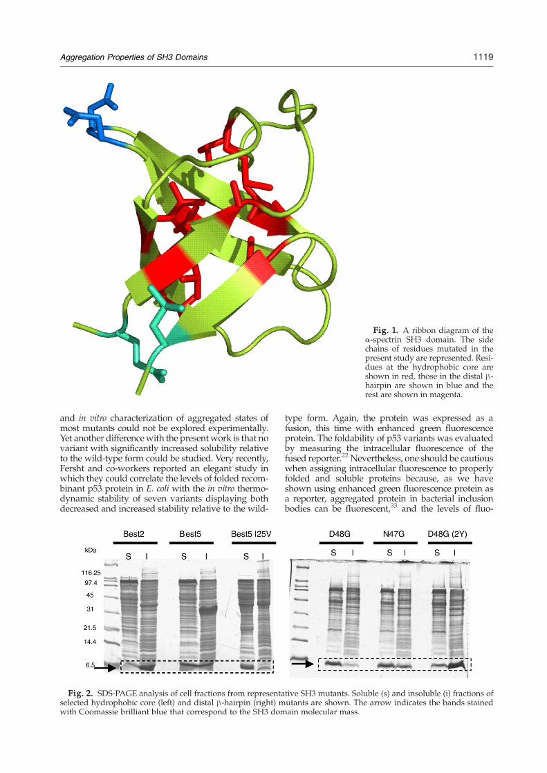
Fig. 1. A ribbon diagram of theα-spectrin SH3 domain. The sidechains of residues mutated in thepresent study are represented. Resi-dues at the hydrophobic core areshown in red, those in the distal β-hairpin are shown in blue and therest are shown in magenta.
1119Aggregation Properties of SH3 Domains
and in vitro characterization of aggregated states ofmost mutants could not be explored experimentally.Yet another differencewith the presentwork is that novariant with significantly increased solubility relativeto the wild-type form could be studied. Very recently,Fersht and co-workers reported an elegant study inwhich they could correlate the levels of folded recom-binant p53 protein in E. coli with the in vitro thermo-dynamic stability of seven variants displaying bothdecreased and increased stability relative to the wild-
Fig. 2. SDS-PAGE analysis of cell fractions from representaselected hydrophobic core (left) and distal β-hairpin (right) mwith Coomassie brilliant blue that correspond to the SH3 dom
type form. Again, the protein was expressed as afusion, this time with enhanced green fluorescenceprotein. The foldability of p53 variants was evaluatedby measuring the intracellular fluorescence of thefused reporter.22 Nevertheless, one should be cautiouswhen assigning intracellular fluorescence to properlyfolded and soluble proteins because, as we haveshown using enhanced green fluorescence protein asa reporter, aggregated protein in bacterial inclusionbodies can be fluorescent,33 and the levels of fluo-
tive SH3 mutants. Soluble (s) and insoluble (i) fractions ofutants are shown. The arrow indicates the bands stainedain molecular mass.
Fig. 3. Correlation between protein conformational stability and solubility. The bars indicate protein stability.Insoluble domains are shown in red and soluble domains are shown in green. Mutants present in similar amounts in thesoluble and insoluble fractions are shown in blue. The line indicates wild-type stability (see the legend in Table 2).
1120 Aggregation Properties of SH3 Domains
rescence of the aggregated protein depend both onintrinsic factors, such as the sequence,34 and extrinsicfactors, such as the temperature.35 In the presentstudy, SH3 domains were expressed without anyfusion, the quantity of soluble and aggregated proteinwas quantified directly and proteins with both in-creased and decreased stability were considered. Asshown earlier, all the proteins could be purified eitherdirectly from the soluble fraction or upon denatu-ration of the insoluble fraction and in vitro refol-ding,28–30 allowing the in vitro characterization of se-lected variants.
In vivo aggregation propensities of SPC-SH3domains do not correlate with intrinsicsequential properties
The composition and arrangement of amino acidsin the proteins has been suggested to be a majorfactor influencing their aggregation propensity.There have been many attempts to determine therelationship between the primary structure and thesolubility status of a protein within the cell in orderto enable rational identification of both natural andmutant protein candidates with a high level of so-lubility on over-expression and substitute commontrial- and -error approaches.36,37 Nevertheless, theexpression conditions and the nature of proteinsanalysed in those studies were different, which isprobably the reason for the dissimilarity between the
results.38 Thus, we reasoned that the present study,in which a large set of variants of a single domainwere analysed in defined expression conditions,might help to decipher the intrinsic features influen-cing in vivo protein aggregation.Among the physicochemical properties that can be
read directly from the primary sequence, the alipha-tic index has been described as a crucial determinantof solubility.39 The aliphatic index corresponds to therelative volume occupied by aliphatic residues andhas usually been associated with thermostability.Nevertheless, there is no correlation between thisfeature and the solubility of the proteins in our dataset (Table 2). A higher level of predicted instabilityhas been suggested to increase the propensity ofproteins to be over-expressed in the soluble form, onthe basis that a polypeptide with a shorter in vivohalf-life has fewer long-lived intermediates.39 How-ever, all the SH3 domains were estimated to havesimilar half-lives in E. coli (N10 h) and no relationshipbetween the instability index and the distribution ofthe different variants between the soluble and inso-luble fractions was found (Table 2). Also, the enrich-ment in global hydrophobicity of the primary se-quence has been suggested to be associated with anincreased propensity to form aggregates inside E.coli;36 yet such correlation does not apply for thepresent set of proteins, at least when hydrophobicityis measured as the grand average of hydropathicityof their sequences (Table 2). In addition, no relation-
1121Aggregation Properties of SH3 Domains
ship between the volume of the hydrophobic coreand SH3 solubility seems to exist.28
In vivo aggregation of globular SPC-SH3domains does not correlate with predictedaggregation propensities of the unfolded state
Several studies indicate that there exist specificcontinuous protein segments that can nucleate theaggregation process when exposed to solvent in fullyor partially unfolded contexts, suggesting a sequence-dependence of aggregation propensities.40,41 Accor-dingly, in the last few years several computationalapproaches for detecting aggregation-prone regionsand predicting relative polypeptide propensities toaggregate have been developed.42,43
We have shown that for the Alzheimer-relatedpeptide Aβ-42, and a set of single-point mutants, invivo aggregation rates upon expression inside bac-terial cells correlated highly significantly with theaggregation propensities predicted theoretically fromthe sequence.34 Thus, we tested whether computer-based approaches were also able to predict the differ-ential in vivo aggregation of the SH3 domains in thepresent study. The sequences of the 24 proteins wereanalysed using AGGRESCAN44 and TANGO45 algo-rithms. No obvious correlation between the predictedglobal aggregations or the relative relevance of thedifferent regions with predicted high aggregationpropensity and the distribution of the protein formsbetween the cytosolic and insoluble fractions of thecell could be observed (Table 2).The discrepancy between the accurate description
of the in-the-cell aggregation propensities of Aβ-42and the failure to predict the behaviour of SH3 do-mains by the above-mentioned, and related, algo-rithms can be rationalized according to the differentconformational properties of both polypeptides. Aβ-42 is a mostly unstructured peptide in which aggre-gation-prone regions are already exposed to solventand available for the establishment of inter-molecu-lar contacts that may finally lead to the formation ofaggregates. Thus, the effect of mutations that faci-litate or hinder the conversion of the soluble un-structured state into oligomeric species have a directimpact on the average aggregation propensity of thepeptide and can be explained in most cases by in-trinsic factors. Because the same physicochemicalprinciples appears to underlie the aggregation pro-pensities of different polypeptides from unfoldedstates,46 the effect of such mutations on aggregationpropensity can be foreseen by most predictive algo-rithms.42,43 These algorithms all assume that theaggregation-prone regions they detect are exposedto solvent at the time the aggregation begins and arethus in an at least partially unfolded context. In con-trast to Aβ-42, SPC-SH3 is a small and compact glo-bular protein in which aggregation-prone regions, ifpresent, are likely to be blocked in the native state ofthe protein because their side chains are either hid-den in the inner hydrophobic core or already in-volved in the network of contacts that stabilizes theprotein. This explains why, once purified, SPC-SH3
is soluble in vitro in its native form at high concen-trations (N10 mg/ml) for weeks in physiologicalconditions. Interestingly, the acid-unfolded state ofthe protein neither assembles into observable aggre-gates on the same time-scale and concentrations.47
This suggests that, within the cell, SPC-SH3 domainsdo not aggregate by coalescence of fully folded orunfolded states and point to the formation and off-pathway association of partially folded intermedi-ates during in vivo protein folding as the responsiblefor the differential intracellular aggregation proper-ties of the SPC-SH3 domain and its mutants. Thiswill explain why the in vivo aggregation propensitiesof SPC-SH3 variants cannot be anticipated directlyfrom the linear information contained in the primarysequence either using simple approaches or employ-ing sophisticated algorithms.
In vivo aggregation levels correlate with theconformational stability of globular SPC-SH3domains
The conformational stability of polypeptides is ex-pected to modulate the ensemble of partially foldedconformations within the cell. Indeed, one shouldexpect that destabilization of the, usually soluble,native state would likely increase the population ofpartially folded species, whereas over-stabilizationof the native conformation would prevent the form-ation of significant levels of partially folded polypep-tides in the cytosol. This implies that, if partially fol-ded intermediates are the aggregation-prone species,protein stability will control the in vivo aggregationfate of globular proteins. To test if this applies to theSPC-SH3 domain, the distribution of the wild typeandmutant proteinswithin the soluble and insolublefractions was compared with their stability mea-sured by urea denaturation at equilibrium in close tophysiological conditions.28 A striking correlationbetween solubility and conformational stability wasfound, independent of the place of mutation (Table 2and Fig. 3). Interestingly, aggregation within the cellwas not necessarily correlated to a large destabiliza-tion of the polypeptides resulting in only a minorpopulation of the native state present at equilibrium.In this way, with the exception of MaxWand MaxA,all the mutants for which the majority of the proteinwas accumulated in the aggregated fraction displayfree energy changes of the unfolding transition in theabsence of denaturant that are significantly differentfrom zero. At the same time, wild-type protein sta-bility, which in its natural context or in vitro underphysiological conditions ensures solubility andfunctionality, rescues only about half of the proteinfrom aggregationwithin the cell. This is in contrast tore-designed SH3 domains in which thermodynamicstability has been improved, since they remain al-most completely in the soluble cytoplasmic fraction.According to our data, stabilization of the SH3 nativestate by more than 1 kcal/mol relative to the wildtype (27%) ensures solubility whereas, in general,destabilization of more than 1 kcal/mol results inmost of the protein being in the aggregated fraction
1122 Aggregation Properties of SH3 Domains
(Fig. 3). Lower levels of stabilization or destabiliza-tion result in a similar distribution of the proteinbetween both fractions. Our data are in excellentagreement with that obtained by using the glu-tathione-S-transferase-HypF-N or enhanced greenfluorescence protein-p53 models.20,22 This confirmsthat in addition to the primary sequence, conforma-tional stability is a major determinant for in vivoprotein aggregation bymodulating the population ofpartially folded intermediates with increased aggre-gation propensities.
SH3 in vivo deposition correlates with in vitroaggregation
SPC-SH3 and its mutants display a two-state tran-sition unfolding curvewhen chemically denatured atlow ionic strength.28 Also, and in contrast to the ho-mologous SH3 domain of PI3-kinase, SPC-SH3 doesnot aggregate into amyloid structures in vitro athighly acidic pH (2.0).47 Nevertheless, it has beenshown recently that destabilization of the SPC-SH3folding nucleus by a point mutation (N47A) resultsin the population of partially folded conformers lea-ding to the formation of amyloid aggregates at mildacidic pH (3.2).48 Most of the residues mutated in thepresent study belong to the SH3 folding nucleus.Hence, we sought to explore whether there is anyrelationship between in vitro aggregation under milddenaturing conditions and the observed in vivo ag-gregation properties of SH3 domains.We selected three representative mutants at the
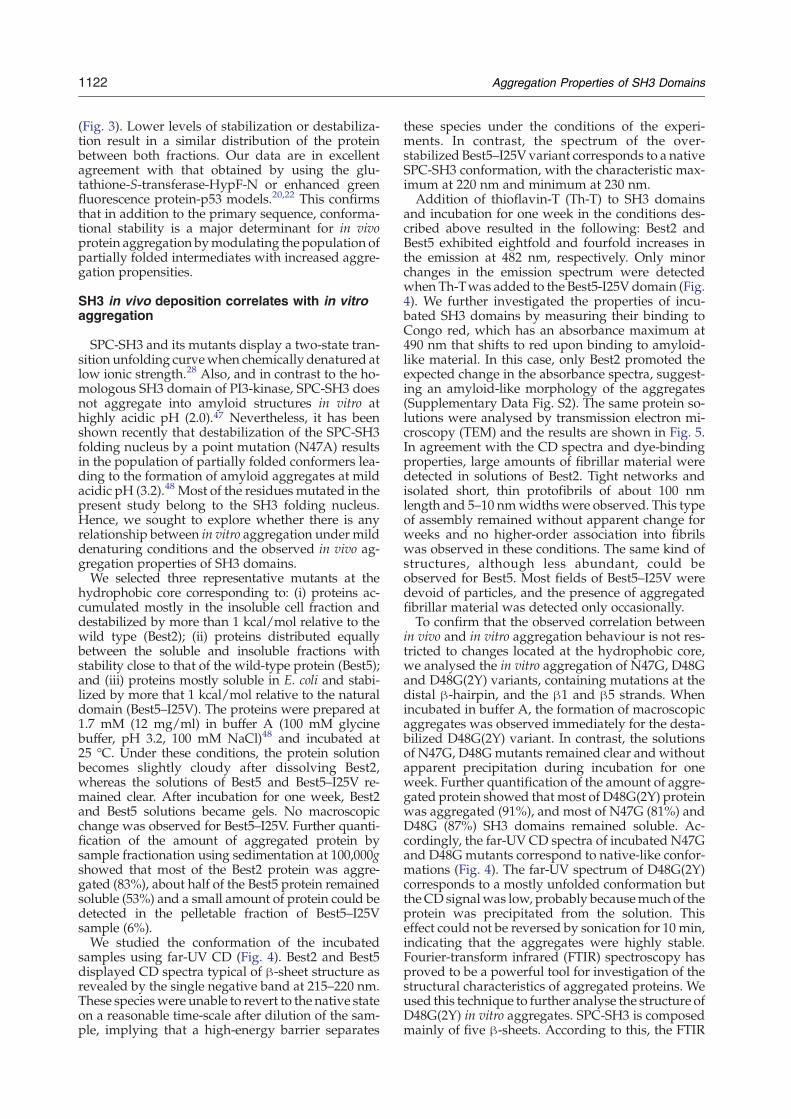
hydrophobic core corresponding to: (i) proteins ac-cumulated mostly in the insoluble cell fraction anddestabilized by more than 1 kcal/mol relative to thewild type (Best2); (ii) proteins distributed equallybetween the soluble and insoluble fractions withstability close to that of the wild-type protein (Best5);and (iii) proteins mostly soluble in E. coli and stabi-lized by more that 1 kcal/mol relative to the naturaldomain (Best5–I25V). The proteins were prepared at1.7 mM (12 mg/ml) in buffer A (100 mM glycinebuffer, pH 3.2, 100 mM NaCl)48 and incubated at25 °C. Under these conditions, the protein solutionbecomes slightly cloudy after dissolving Best2,whereas the solutions of Best5 and Best5–I25V re-mained clear. After incubation for one week, Best2and Best5 solutions became gels. No macroscopicchange was observed for Best5–I25V. Further quanti-fication of the amount of aggregated protein bysample fractionation using sedimentation at 100,000gshowed that most of the Best2 protein was aggre-gated (83%), about half of the Best5 protein remainedsoluble (53%) and a small amount of protein could bedetected in the pelletable fraction of Best5–I25Vsample (6%).We studied the conformation of the incubated
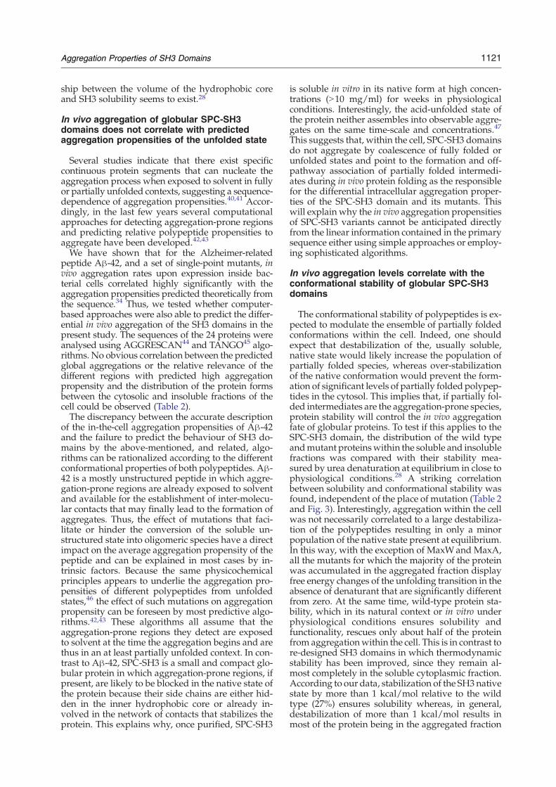
samples using far-UV CD (Fig. 4). Best2 and Best5displayed CD spectra typical of β-sheet structure asrevealed by the single negative band at 215–220 nm.These specieswere unable to revert to the native stateon a reasonable time-scale after dilution of the sam-ple, implying that a high-energy barrier separates
these species under the conditions of the experi-ments. In contrast, the spectrum of the over-stabilized Best5–I25V variant corresponds to a nativeSPC-SH3 conformation, with the characteristic max-imum at 220 nm and minimum at 230 nm.Addition of thioflavin-T (Th-T) to SH3 domains
and incubation for one week in the conditions des-cribed above resulted in the following: Best2 andBest5 exhibited eightfold and fourfold increases inthe emission at 482 nm, respectively. Only minorchanges in the emission spectrum were detectedwhen Th-Twas added to the Best5-I25V domain (Fig.4). We further investigated the properties of incu-bated SH3 domains by measuring their binding toCongo red, which has an absorbance maximum at490 nm that shifts to red upon binding to amyloid-like material. In this case, only Best2 promoted theexpected change in the absorbance spectra, suggest-ing an amyloid-like morphology of the aggregates(Supplementary Data Fig. S2). The same protein so-lutions were analysed by transmission electron mi-croscopy (TEM) and the results are shown in Fig. 5.In agreement with the CD spectra and dye-bindingproperties, large amounts of fibrillar material weredetected in solutions of Best2. Tight networks andisolated short, thin protofibrils of about 100 nmlength and 5–10 nmwidths were observed. This typeof assembly remained without apparent change forweeks and no higher-order association into fibrilswas observed in these conditions. The same kind ofstructures, although less abundant, could beobserved for Best5. Most fields of Best5–I25V weredevoid of particles, and the presence of aggregatedfibrillar material was detected only occasionally.To confirm that the observed correlation between
in vivo and in vitro aggregation behaviour is not res-tricted to changes located at the hydrophobic core,we analysed the in vitro aggregation of N47G, D48Gand D48G(2Y) variants, containing mutations at thedistal β-hairpin, and the β1 and β5 strands. Whenincubated in buffer A, the formation of macroscopicaggregates was observed immediately for the desta-bilized D48G(2Y) variant. In contrast, the solutionsof N47G, D48Gmutants remained clear and withoutapparent precipitation during incubation for oneweek. Further quantification of the amount of aggre-gated protein showed that most of D48G(2Y) proteinwas aggregated (91%), and most of N47G (81%) andD48G (87%) SH3 domains remained soluble. Ac-cordingly, the far-UV CD spectra of incubated N47Gand D48G mutants correspond to native-like confor-mations (Fig. 4). The far-UV spectrum of D48G(2Y)corresponds to a mostly unfolded conformation butthe CD signalwas low, probably becausemuch of theprotein was precipitated from the solution. Thiseffect could not be reversed by sonication for 10 min,indicating that the aggregates were highly stable.Fourier-transform infrared (FTIR) spectroscopy hasproved to be a powerful tool for investigation of thestructural characteristics of aggregated proteins. Weused this technique to further analyse the structure ofD48G(2Y) in vitro aggregates. SPC-SH3 is composedmainly of five β-sheets. According to this, the FTIR
Fig. 4. Conformational properties of 1.7 mM (12 mg/ml) SH3 domains incubated at 25 °C for one week in 100 mMNaCl, 100 mM glycine (pH 3.2). (a and b) CD spectra in the far-UV region. (c) FTIR spectra in the amide I region. Thepositions of the spectral componentswere estimated from the second derivate analysis. (d and e) The fluorescence emissionspectra of 40 μMthioflavin-T in the absence (no symbol) or in the presence of SH3 domains. The excitationwavelengthwas440 nm. (a and d) Hydrophobic core SH3 mutants: Best2 (circles), Best5 (squares) and Best5–I25V (triangles). (b and e)Distal β-hairpin SH3 mutants: D48G(2Y) (circles), N47G (squares) and D48G (triangles). (c) Incubated D48G(2Y)(continuous lines) and native SPC-SH3 (broken line).
1123Aggregation Properties of SH3 Domains
spectrum of native SPC-SH3 in the amide region Iwas dominated by a peak at 1636 cm−1 that arisesfrom CO vibrations in intramolecular β-sheets (Fig.
4). The FTIR spectrum of D48G(2Y) aggregates exhi-bited a main peak at 1623 cm−1 (Fig. 4). This band isindicative of a predominance of extended intermo-
Fig. 5. Representative TEM images of hydrophobiccore SH3 protein mutants incubated at 25 °C for one week.The samples were produced by tenfold dilutions of theoriginal samples used for the incubation. (a) Best2; (b)Best5; and (c) Best5–I25V. The scale bar represents 500 nm.
1124 Aggregation Properties of SH3 Domains
lecular β-sheet structures in the aggregates. Wefurther explored the structure of the incubated solu-tions by assaying their binding to Th-T and Congored. No significant binding to such dyes was detec-ted for N47G and D48G variants. In contrast, D48G
(2Y) bind both Th-T and Congo red (Fig. 4 andSupplementary Data Fig. 2S). The affinity of D48G(2Y) for Th-Twas surprisingly high, promoting a 20-fold increase of the fluorescence signal, suggestingan amyloid-like morphology of the aggregates. Thesame protein solutions were analysed by TEM, andthe Results are shown in Fig. 6. Two different kindsof aggregates were detected in D48G(2Y) prepara-tions: large amorphous aggregates that, in manycases, appear associated to protofibrillar material(Fig. 6a) andwell-structured fibrils displaying a heli-cal twisted morphology with ∼60 nm width (Fig.6b). This highly ordered fibrils have not been ob-served for any spectrin-SH3 domain and are likelyresponsible for the high Th-T affinity of this mutant.In agreement with the CD spectra and dye-bindingproperties, only very small aggregates were detectedfor the D48G mutant, whereas a reduced number ofsmall fibrillar-like aggregates were observed inN47G samples (Fig. 6).We also analysed in vitro aggregation of different
SH3 domains at pH 5.0 and pH 7.0 (100 mM sodiumphosphate buffer). Best5–I25V and D48G as well asBest2 and D48G(2Y) were selected as representativeof stable and unstable mutants at the core and distalβ-hairpin. They were incubated at a concentration of1.7 mM (12 mg/ml) for one week at 25 °C. Macro-scopic aggregates were observed immediately inBest2 and D48G(2Y) solutions at both pH values,whereas the over-stabilized Best5–I25V and D48Gsolutions remained clear. Upon incubation at pH 5.0for one week, most of the Best2 (85%) and D48G(2Y)(81%) protein was aggregated. At pH 7.0, 67% ofBest2 and 59% of D48G(2Y) proteins became in-soluble, indicating that aggregation of unstablemutants occurs at close to physiological conditions.Best2 and D48G(2Y) pH 5.0 aggregates exhibitedsignificant binding to Th-T (Supplementary DataFig. S3), whereas the binding of pH 7.0 aggregateswas much lower than that expected according to theamount of aggregated protein, suggesting that pHaffects the amount of deposited protein and the con-formational properties of the aggregates. No sig-nificant Th-T binding was observed for the over-stabilized Best5–I25V and D48G variants at any pH.Overall, the in vitro properties of the analysed do-
mains are in excellent agreement with their fractio-nation between soluble and insoluble compartmentsobserved in vivo, indicating that, at least in certaincases, the data obtainedusing simplified non-cellularcontexts can be relevant in in vivo environments.
In vivo formed SH3 aggregates display someamyloid-like properties
In order to test if in vitro and in vivo formed aggre-gates share morphological and/or conformationalfeatures, we purified and characterized the in vivoaggregates formed during the expression of themostly insoluble Best2 and D48G(2Y) SH3 domains.Analysis by TEM shows that Best2 in vivo aggregatescorrespond to inclusion bodies (IBs) of around 1 μmsize displaying a typical electro-dense ellipsoidal
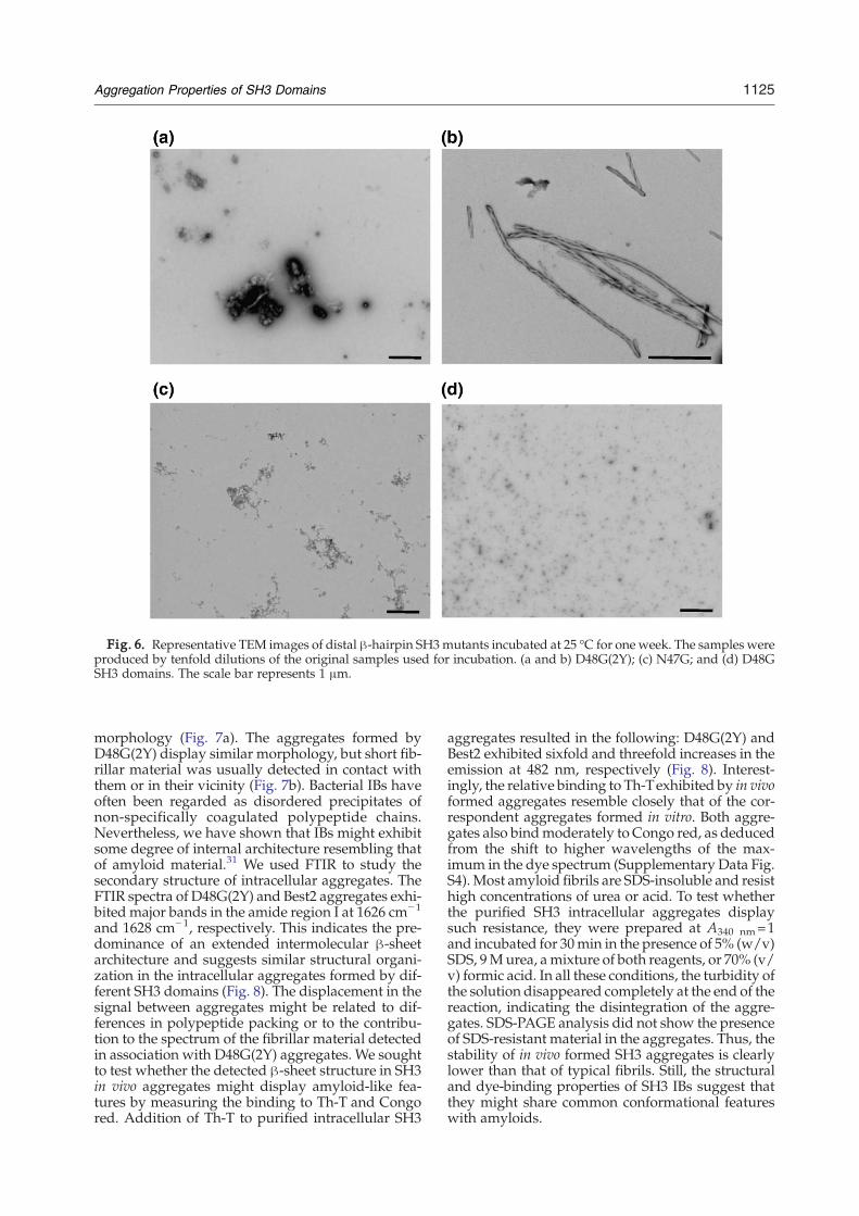
Fig. 6. Representative TEM images of distal β-hairpin SH3mutants incubated at 25 °C for one week. The samples wereproduced by tenfold dilutions of the original samples used for incubation. (a and b) D48G(2Y); (c) N47G; and (d) D48GSH3 domains. The scale bar represents 1 μm.
1125Aggregation Properties of SH3 Domains
morphology (Fig. 7a). The aggregates formed byD48G(2Y) display similar morphology, but short fib-rillar material was usually detected in contact withthem or in their vicinity (Fig. 7b). Bacterial IBs haveoften been regarded as disordered precipitates ofnon-specifically coagulated polypeptide chains.Nevertheless, we have shown that IBs might exhibitsome degree of internal architecture resembling thatof amyloid material.31 We used FTIR to study thesecondary structure of intracellular aggregates. TheFTIR spectra of D48G(2Y) and Best2 aggregates exhi-bited major bands in the amide region I at 1626 cm−1
and 1628 cm−1, respectively. This indicates the pre-dominance of an extended intermolecular β-sheetarchitecture and suggests similar structural organi-zation in the intracellular aggregates formed by dif-ferent SH3 domains (Fig. 8). The displacement in thesignal between aggregates might be related to dif-ferences in polypeptide packing or to the contribu-tion to the spectrum of the fibrillar material detectedin association with D48G(2Y) aggregates. We soughtto test whether the detected β-sheet structure in SH3in vivo aggregates might display amyloid-like fea-tures by measuring the binding to Th-T and Congored. Addition of Th-T to purified intracellular SH3
aggregates resulted in the following: D48G(2Y) andBest2 exhibited sixfold and threefold increases in theemission at 482 nm, respectively (Fig. 8). Interest-ingly, the relative binding to Th-Texhibited by in vivoformed aggregates resemble closely that of the cor-respondent aggregates formed in vitro. Both aggre-gates also bindmoderately to Congo red, as deducedfrom the shift to higher wavelengths of the max-imum in the dye spectrum (Supplementary Data Fig.S4). Most amyloid fibrils are SDS-insoluble and resisthigh concentrations of urea or acid. To test whetherthe purified SH3 intracellular aggregates displaysuch resistance, they were prepared at A340 nm=1and incubated for 30min in the presence of 5% (w/v)SDS, 9Murea, amixture of both reagents, or 70% (v/v) formic acid. In all these conditions, the turbidity ofthe solution disappeared completely at the end of thereaction, indicating the disintegration of the aggre-gates. SDS-PAGE analysis did not show the presenceof SDS-resistant material in the aggregates. Thus, thestability of in vivo formed SH3 aggregates is clearlylower than that of typical fibrils. Still, the structuraland dye-binding properties of SH3 IBs suggest thatthey might share common conformational featureswith amyloids.
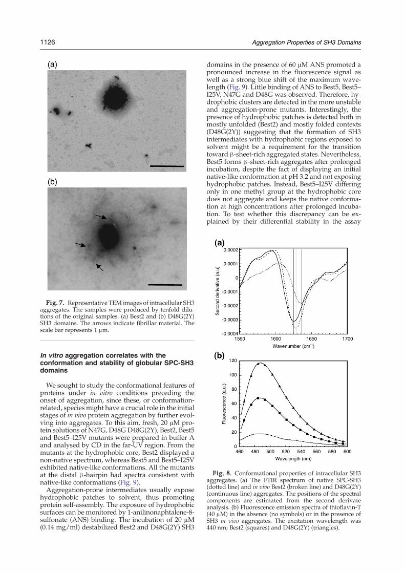
Fig. 8. Conformational properties of intracellular SH3aggregates. (a) The FTIR spectrum of native SPC-SH3(dotted line) and in vivo Best2 (broken line) and D48G(2Y)(continuous line) aggregates. The positions of the spectralcomponents are estimated from the second derivateanalysis. (b) Fluorescence emission spectra of thioflavin-T(40 μM) in the absence (no symbols) or in the presence ofSH3 in vivo aggregates. The excitation wavelength was440 nm; Best2 (squares) and D48G(2Y) (triangles).
Fig. 7. Representative TEM images of intracellular SH3aggregates. The samples were produced by tenfold dilu-tions of the original samples. (a) Best2 and (b) D48G(2Y)SH3 domains. The arrows indicate fibrillar material. Thescale bar represents 1 μm.
1126 Aggregation Properties of SH3 Domains
In vitro aggregation correlates with theconformation and stability of globular SPC-SH3domains
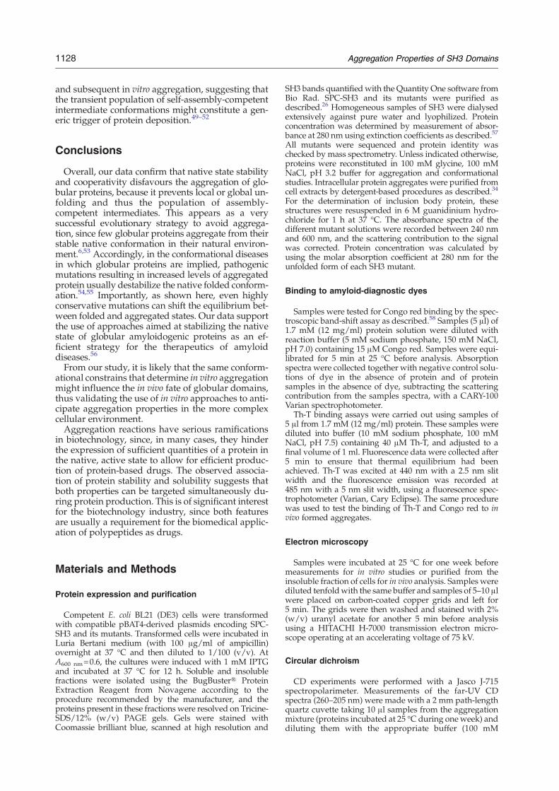
We sought to study the conformational features ofproteins under in vitro conditions preceding theonset of aggregation, since these, or conformation-related, species might have a crucial role in the initialstages of in vivo protein aggregation by further evol-ving into aggregates. To this aim, fresh, 20 μM pro-tein solutions of N47G, D48GD48G(2Y), Best2, Best5and Best5–I25V mutants were prepared in buffer Aand analysed by CD in the far-UV region. From themutants at the hydrophobic core, Best2 displayed anon-native spectrum, whereas Best5 and Best5–I25Vexhibited native-like conformations. All the mutantsat the distal β-hairpin had spectra consistent withnative-like conformations (Fig. 9).Aggregation-prone intermediates usually expose
hydrophobic patches to solvent, thus promotingprotein self-assembly. The exposure of hydrophobicsurfaces can be monitored by 1-anilinonaphtalene-8-sulfonate (ANS) binding. The incubation of 20 μM(0.14 mg/ml) destabilized Best2 and D48G(2Y) SH3
domains in the presence of 60 μM ANS promoted apronounced increase in the fluorescence signal aswell as a strong blue shift of the maximum wave-length (Fig. 9). Little binding of ANS to Best5, Best5–I25V, N47G and D48G was observed. Therefore, hy-drophobic clusters are detected in the more unstableand aggregation-prone mutants. Interestingly, thepresence of hydrophobic patches is detected both inmostly unfolded (Best2) and mostly folded contexts(D48G(2Y)) suggesting that the formation of SH3intermediates with hydrophobic regions exposed tosolvent might be a requirement for the transitiontoward β-sheet-rich aggregated states. Nevertheless,Best5 forms β-sheet-rich aggregates after prolongedincubation, despite the fact of displaying an initialnative-like conformation at pH 3.2 and not exposinghydrophobic patches. Instead, Best5–I25V differingonly in one methyl group at the hydrophobic coredoes not aggregate and keeps the native conforma-tion at high concentrations after prolonged incuba-tion. To test whether this discrepancy can be ex-plained by their differential stability in the assay
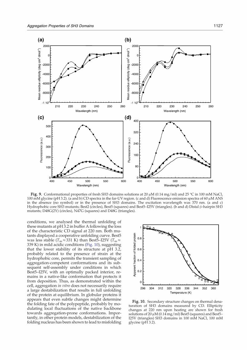
Fig. 9. Conformational properties of fresh SH3 domains solutions at 20 μM (0.14 mg/ml) and 25 °C in 100 mM NaCl,100 mM glycine (pH 3.2). (a and b) CD spectra in the far-UV region. (c and d) Fluorescence emission spectra of 60 μMANSin the absence (no symbol) or in the presence of SH3 domains. The excitation wavelength was 370 nm. (a and c)Hydrophobic core SH3 mutants; Best2 (circles), Best5 (squares) and Best5–I25V (triangles). (b and d) Distal β-hairpin SH3mutants; D48G(2Y) (circles), N47G (squares) and D48G (triangles).
Fig. 10. Secondary structure changes on thermal dena-turation of SH3 domains measured by CD. Ellipticitychanges at 220 nm upon heating are shown for freshsolutions of 20 μM(0.14mg/ml) Best5 (squares) and Best5–I25V (triangles) SH3 domains in 100 mM NaCl, 100 mMglycine (pH 3.2).
1127Aggregation Properties of SH3 Domains
conditions, we analysed the thermal unfolding ofthesemutants at pH 3.2 in buffer A following the lossof the characteristic CD signal at 220 nm. Both mu-tants displayed a cooperative unfolding curve. Best5was less stable (Tm≈331 K) than Best5–I25V (Tm≈339 K) in mild acidic conditions (Fig. 10), suggestingthat the lower stability of its structure at pH 3.2,probably related to the presence of strain at thehydrophobic core, permits the transient sampling ofaggregation-competent conformations and its sub-sequent self-assembly under conditions in whichBest5–I25V, with an optimally packed interior, re-mains in a native-like conformation that protects itfrom deposition. Thus, as demonstrated within thecell, aggregation in vitro does not necessarily requirea large destabilization that results in full unfoldingof the protein at equilibrium. In globular proteins itappears that even subtle changes might determinethe folding fate of the polypeptide, probably by mo-dulating local fluctuations of the native backbonetowards aggregation-prone conformations. Impor-tantly, in other protein models, destabilization of thefolding nucleus has been shown to lead tomisfolding
1128 Aggregation Properties of SH3 Domains
and subsequent in vitro aggregation, suggesting thatthe transient population of self-assembly-competentintermediate conformations might constitute a gen-eric trigger of protein deposition.49–52
Conclusions
Overall, our data confirm that native state stabilityand cooperativity disfavours the aggregation of glo-bular proteins, because it prevents local or global un-folding and thus the population of assembly-competent intermediates. This appears as a verysuccessful evolutionary strategy to avoid aggrega-tion, since few globular proteins aggregate from theirstable native conformation in their natural environ-ment.6,53 Accordingly, in the conformational diseasesin which globular proteins are implied, pathogenicmutations resulting in increased levels of aggregatedprotein usually destabilize the native folded conform-ation.54,55 Importantly, as shown here, even highlyconservative mutations can shift the equilibrium bet-ween folded and aggregated states. Our data supportthe use of approaches aimed at stabilizing the nativestate of globular amyloidogenic proteins as an ef-ficient strategy for the therapeutics of amyloiddiseases.56
From our study, it is likely that the same conform-ational constrains that determine in vitro aggregationmight influence the in vivo fate of globular domains,thus validating the use of in vitro approaches to anti-cipate aggregation properties in the more complexcellular environment.Aggregation reactions have serious ramifications
in biotechnology, since, in many cases, they hinderthe expression of sufficient quantities of a protein inthe native, active state to allow for efficient produc-tion of protein-based drugs. The observed associa-tion of protein stability and solubility suggests thatboth properties can be targeted simultaneously du-ring protein production. This is of significant interestfor the biotechnology industry, since both featuresare usually a requirement for the biomedical applic-ation of polypeptides as drugs.
Materials and Methods
Protein expression and purification
Competent E. coli BL21 (DE3) cells were transformedwith compatible pBAT4-derived plasmids encoding SPC-SH3 and its mutants. Transformed cells were incubated inLuria Bertani medium (with 100 μg/ml of ampicillin)overnight at 37 °C and then diluted to 1/100 (v/v). AtA600 nm=0.6, the cultures were induced with 1 mM IPTGand incubated at 37 °C for 12 h. Soluble and insolublefractions were isolated using the BugBuster® ProteinExtraction Reagent from Novagene according to theprocedure recommended by the manufacturer, and theproteins present in these fractions were resolved on Tricine-SDS/12% (w/v) PAGE gels. Gels were stained withCoomassie brilliant blue, scanned at high resolution and
SH3 bands quantified with the Quantity One software fromBio Rad. SPC-SH3 and its mutants were purified asdescribed.26 Homogeneous samples of SH3 were dialysedextensively against pure water and lyophilized. Proteinconcentration was determined by measurement of absor-bance at 280 nm using extinction coefficients as described.57
All mutants were sequenced and protein identity waschecked by mass spectrometry. Unless indicated otherwise,proteins were reconstituted in 100 mM glycine, 100 mMNaCl, pH 3.2 buffer for aggregation and conformationalstudies. Intracellular protein aggregates were purified fromcell extracts by detergent-based procedures as described.34
For the determination of inclusion body protein, thesestructures were resuspended in 6 M guanidinium hydro-chloride for 1 h at 37 °C. The absorbance spectra of thedifferent mutant solutions were recorded between 240 nmand 600 nm, and the scattering contribution to the signalwas corrected. Protein concentration was calculated byusing the molar absorption coefficient at 280 nm for theunfolded form of each SH3 mutant.
Binding to amyloid-diagnostic dyes
Samples were tested for Congo red binding by the spec-troscopic band-shift assay as described.58 Samples (5 μl) of1.7 mM (12 mg/ml) protein solution were diluted withreaction buffer (5 mM sodium phosphate, 150 mM NaCl,pH 7.0) containing 15 μM Congo red. Samples were equi-librated for 5 min at 25 °C before analysis. Absorptionspectra were collected together with negative control solu-tions of dye in the absence of protein and of proteinsamples in the absence of dye, subtracting the scatteringcontribution from the samples spectra, with a CARY-100Varian spectrophotometer.Th-T binding assays were carried out using samples of
5 μl from 1.7 mM (12 mg/ml) protein. These samples werediluted into buffer (10 mM sodium phosphate, 100 mMNaCl, pH 7.5) containing 40 μM Th-T, and adjusted to afinal volume of 1 ml. Fluorescence data were collected after5 min to ensure that thermal equilibrium had beenachieved. Th-T was excited at 440 nm with a 2.5 nm slitwidth and the fluorescence emission was recorded at485 nm with a 5 nm slit width, using a fluorescence spec-trophotometer (Varian, Cary Eclipse). The same procedurewas used to test the binding of Th-T and Congo red to invivo formed aggregates.
Electron microscopy
Samples were incubated at 25 °C for one week beforemeasurements for in vitro studies or purified from theinsoluble fraction of cells for in vivo analysis. Samples werediluted tenfoldwith the same buffer and samples of 5–10 μlwere placed on carbon-coated copper grids and left for5 min. The grids were then washed and stained with 2%(w/v) uranyl acetate for another 5 min before analysisusing a HITACHI H-7000 transmission electron micro-scope operating at an accelerating voltage of 75 kV.
Circular dichroism
CD experiments were performed with a Jasco J-715spectropolarimeter. Measurements of the far-UV CDspectra (260–205 nm) were made with a 2 mm path-lengthquartz cuvette taking 10 μl samples from the aggregationmixture (proteins incubated at 25 °C during one week) anddiluting them with the appropriate buffer (100 mM
1129Aggregation Properties of SH3 Domains
glycine, 100 mM NaCl, pH 3.2) to a final volume of 500 μl(protein concentration of about 35 μM (0.25 mg/ml).Spectra were recorded at 25 °C. Non-incubated sampleswere analysed at 20 μM (0.14 mg/ml) final protein con-centration in the same buffer. The resulting spectrum wasthe average of 20 scans. Thermal denaturation of SH3 do-mains was studied using a Jasco J-715 spectropolarimeterequipped with a thermostat controlled cell holder. Scanswere conducted between 15 °C and 95 °C at a scan rate of1 degC/min. Changes in CD signal at 220 nm were recor-ded. The protein concentration was 20 μM (0.14 mg/ml).The fitting of the experimental data was performed usingthe non-linear, least-squares algorithm provided with thesoftware KaleidaGraph (Abelbeck Software) assuming atwo-state denaturation process.
FTIR spectroscopy analysis
SH3 sampleswere analysed using a Bruker Tensor 27 FT-IR spectrometer (Bruker Optics Inc) with a Golden GateMKII ATR accessory. Each spectrum consists of 125 inde-pendent scans, measured at a spectral resolution of 2 cm−1
within the 1800–1500 cm−1 range. All spectral data wereacquired and normalized using the OPUS MIR Tensor 27software. Second derivatives of the spectra were used todetermine the frequencies at which the different spectralcomponents were located.
ANS binding assay
The fluorescence emission spectra of ANS in the pre-sence and in the absence of protein were recorded with afluorescence spectrophotometer (Varian, Cary Eclipse) at25 °C. Proteins were diluted in 100 mM glycine, 100 mMNaCl, pH 3.2 buffer containing 60 μMANS to obtain a finalprotein concentration of 20 μM (0.14 mg/ml). The ANSfluorescence was then read immediately using an excita-tion wavelength of 370 nm and the fluorescence emissionwas collected between 400 nm and 600 nm, with excitationand emission slit widths of 2.5 nm and 5 nm, respectively.
Acknowledgements
Most mutants were constructed originally in thelaboratory of Dr Luis Serrano at EMBL-Heidelberg.Wethank Prof. Aviles and Dr. Vendrell for lab facilities.This work was supported by grant BIO2007-68046from the Ministerio de Ciencia y Tecnología, Spain,and by grant 2005SGR-00037 fromAGAUR,Catalonia.
Supplementary Data
Supplementary data associated with this articlecan be found, in the online version, at doi:10.1016/j.jmb.2008.03.020
References
1. Tan, S. Y. & Pepys, M. B. (1994). Amyloidosis. Histo-pathology, 25, 403–414.
2. Sunde, M. & Blake, C. C. (1998). From the globular tothe fibrous state: protein structure and structural con-version in amyloid formation. Quart. Rev. Biophys. 31,1–39.
3. Dobson, C. M. (2001). The structural basis of proteinfolding and its links with human disease. Phil. Trans.Roy. Soc. B, 356, 133–145.
4. Fahnert, B., Lilie, H. & Neubauer, P. (2004). Inclusionbodies: formation and utilisation. Adv. Biochem. Eng.Biotechnol. 89, 93–142.
5. Ventura, S. & Villaverde, A. (2006). Protein quality inbacterial inclusion bodies.Trends Biotechnol. 24, 179–185.
6. Ventura, S. (2005). Sequence determinants of proteinaggregation: tools to increase protein solubility.Microbial Cell Factories, 4, 11.
7. Fink, A. L. (1998). Protein aggregation: folding aggre-gates, inclusion bodies and amyloid. Fold. Des. 3,R9–R23.
8. Rinas, U., Hoffmann, F., Betiku, E., Estape, D. &Marten, S. (2007). Inclusion body anatomy and func-tioning of chaperone-mediated in vivo inclusion bodydisassembly during high-level recombinant proteinproduction in Escherichia coli. J. Biotechnol. 127,244–257.
9. Pallares, I., Vendrell, J., Aviles, F. X. & Ventura, S.(2004). Amyloid fibril formation by a partially struc-tured intermediate state of α-chymotrypsin. J. Mol.Biol. 342, 321–331.
10. Guijarro, J. I., Sunde, M., Jones, J. A., Campbell, I. D. &Dobson, C. M. (1998). Amyloid fibril formation by anSH3 domain. Proc. Natl Acad. Sci. USA, 95, 4224–4228.
11. Ivanova, M. I., Thompson, M. J. & Eisenberg, D.(2006). A systematic screen of β(2)-microglobulin andinsulin for amyloid-like segments. Proc. Natl Acad. Sci.USA, 103, 4079–4082.
12. Hammarstrom, P., Sekijima, Y., White, J. T., Wiseman,R. L., Lim, A., Costello, C. E. et al. (2003). D18G trans-thyretin is monomeric, aggregation prone, and notdetectable in plasma and cerebrospinal fluid: a pres-cription for central nervous system amyloidosis? Bio-chemistry, 42, 6656–6663.
13. McParland, V. J., Kad, N. M., Kalverda, A. P., Brown,A., Kirwin-Jones, P., Hunter, M. G. et al. (2000). Partiallyunfolded states of β-2-microglobulin and amyloidformation in vitro. Biochemistry, 39, 8735–8746.
14. Zerovnik, E., Skarabot, M., Skerget, K., Giannini, S.,Stoka, V., Jenko-Kokalj, S. & Staniforth, R. A. (2007).Amyloid fibril formation by human stefin B: influenceof pH and TFE on fibril growth and morphology.Amyloid, 14, 237–247.
15. Ghaemmaghami, S. & Oas, T. G. (2001). Quantitativeprotein stability measurement in vivo. Nature Struct.Biol. 8, 879–882.
16. de Groot, N. S., Aviles, F. X., Vendrell, J. & Ventura, S.(2006). Mutagenesis of the central hydrophobic clusterin Aβ42 Alzheimer’s peptide. Side-chain propertiescorrelate with aggregation propensities. FEBS J. 273,658–668.
17. Wurth, C., Guimard, N. K. & Hecht, M. H. (2002).Mutations that reduce aggregation of the Alzheimer’sAβ42 peptide: an unbiased search for the sequencedeterminants of Aβ amyloidogenesis. J. Mol. Biol. 319,1279–1290.
18. Speed, M. A., Morshead, T., Wang, D. I. & King, J.(1997). Conformation of P22 tailspike folding and ag-gregation intermediates probed by monoclonal anti-bodies. Protein Sci. 6, 99–108.
19. Speed, M. A., Wang, D. I. & King, J. (1996). Specificaggregation of partially folded polypeptide chains: the
1130 Aggregation Properties of SH3 Domains
molecular basis of inclusion body composition.NatureBiotechnol. 14, 1283–1287.
20. Calloni, G., Zoffoli, S., Stefani, M., Dobson, C. M. &Chiti, F. (2005). Investigating the effects of mutationson protein aggregation in the cell. J. Biol. Chem. 280,10607–10613.
21. Ignatova, Z. & Gierasch, L. M. (2005). Aggregation ofa slow-folding mutant of a β-clam protein proceedsthrough a monomeric nucleus. Biochemistry, 44,7266–7274.
22. Mayer, S., Rudiger, S., Ang, H. C., Joerger, A. C. &Fersht, A. R. (2007). Correlation of levels of foldedrecombinant p53 in escherichia coli with thermody-namic stability in vitro. J. Mol. Biol. 372, 268–276.
23. Ma, Y. & Hendershot, L. M. (2001). The unfolding taleof the unfolded protein response. Cell, 107, 827–830.
24. Musacchio, A., Noble, M., Pauptit, R., Wierenga, R. &Saraste, M. (1992). Crystal structure of a Src-homology3 (SH3) domain. Nature, 359, 851–855.
25. Martinez, J. C., Viguera, A. R., Berisio, R., Wilmanns,M., Mateo, P. L., Filimonov, V. V. & Serrano, L. (1999).Thermodynamic analysis of α-spectrin SH3 and twoof its circular permutants with different loop lengths:discerning the reasons for rapid folding in proteins.Biochemistry, 38, 549–559.
26. Martinez, J. C. & Serrano, L. (1999). The folding tran-sition state between SH3 domains is conformationallyrestricted and evolutionarily conserved. Nature Struct.Biol. 6, 1010–1016.
27. Grantcharova, V. P., Riddle, D. S., Santiago, J. V. &Baker, D. (1998). Important role of hydrogen bonds inthe structurally polarized transition state for foldingof the src SH3 domain. Nature Struct. Biol. 5, 714–720.
28. Ventura, S., Vega, M. C., Lacroix, E., Angrand, I.,Spagnolo, L. & Serrano, L. (2002). Conformationalstrain in the hydrophobic core and its implications forprotein folding and design. Nature Struct. Biol. 9,485–493.
29. Martinez, J. C., Pisabarro, M. T. & Serrano, L. (1998).Obligatory steps in protein folding and the conforma-tional diversity of the transition state. Nature Struct.Biol. 5, 721–729.
30. Vega, M. C., Martinez, J. C. & Serrano, L. (2000). Ther-modynamic and structural characterization of Asnand Ala residues in the disallowed II′ region of theRamachandran plot. Protein Sci. 9, 2322–2328.
31. Carrio, M., Gonzalez-Montalban, N., Vera, A., Villa-verde, A. & Ventura, S. (2005). Amyloid-like proper-ties of bacterial inclusion bodies. J. Mol. Biol. 347,1025–1037.
32. Davis, G. D., Elisee, C., Newham, D. M. & Harrison,R. G. (1999). New fusion protein systems designed togive soluble expression in Escherichia coli. Biotechnol.Bioeng. 65, 382–388.
33. Garcia-Fruitos, E., Gonzalez-Montalban, N., Morell,M., Vera, A., Ferraz, R., Aris, A. et al. (2005). Aggre-gation as bacterial inclusion bodies does not implyinactivation of enzymes and fluorescent proteins.Microb. Cell Fact. 4, 27.
34. de Groot, N. S. & Ventura, S. (2006). Protein activity inbacterial inclusion bodies correlates with predictedaggregation rates. J. Biotechnol. 125, 110–113.
35. de Groot, N. S. & Ventura, S. (2006). Effect of tempe-rature on protein quality in bacterial inclusion bodies.FEBS Lett. 580, 6471–6476.
36. Luan, C. H., Qiu, S., Finley, J. B., Carson, M., Gray,R. J., Huang, W. et al. (2004). High-throughput expres-sion of C. elegans proteins. Genome Res. 14, 2102–2110.
37. Goh, C. S., Lan, N., Douglas, S. M., Wu, B., Echols, N.,
Smith, A. et al. (2004). Mining the structural genomicspipeline: identification of protein properties that affecthigh-throughput experimental analysis. J. Mol. Biol.336, 115–130.
38. Idicula-Thomas, S., Kulkarni, A. J., Kulkarni, B. D.,Jayaraman, V. K. & Balaji, P. V. (2006). A supportvector machine-based method for predicting the pro-pensity of a protein to be soluble or to form inclusionbody on overexpression in Escherichia coli. Bioinfor-matics, 22, 278–284.
39. Idicula-Thomas, S. & Balaji, P. V. (2005). Understand-ing the relationship between the primary structure ofproteins and its propensity to be soluble on over-expression in Escherichia coli. Protein Sci. 14, 582–592.
40. Ivanova, M. I., Sawaya, M. R., Gingery, M., Attinger,A. & Eisenberg, D. (2004). An amyloid-formingsegment of β2-microglobulin suggests a molecularmodel for the fibril. Proc. Natl Acad. Sci. USA, 101,10584–10589.
41. Ventura, S., Zurdo, J., Narayanan, S., Parreno, M.,Mangues, R., Reif, B. et al. (2004). Short amino acidstretches can mediate amyloid formation in globularproteins: the Src homology 3 (SH3) case. Proc. NatlAcad. Sci. USA, 101, 7258–7263.
42. Bemporad, F., Calloni, G., Campioni, S., Plakoutsi, G.,Taddei, N. & Chiti, F. (2006). Sequence and structuraldeterminants of amyloid fibril formation. Accts Chem.Res. 39, 620–627.
43. Caflisch, A. (2006). Computational models for theprediction of polypeptide aggregation propensity.Curr. Opin. Chem. Biol. 10, 437–444.
44. Conchillo-Sole, O., de Groot, N. S., Aviles, F. X.,Vendrell, J., Daura, X. & Ventura, S. (2007). AGGRES-CAN: a server for the prediction and evaluation of“hot spots” of aggregation in polypeptides. BMCBioinformatics, 8, 65.
45. Fernandez-Escamilla, A. M., Rousseau, F., Schymko-witz, J. & Serrano, L. (2004). Prediction of sequence-dependent and mutational effects on the aggregationof peptides and proteins. Nature Biotechnol. 22,1302–1306.
46. Chiti, F., Stefani, M., Taddei, N., Ramponi, G. &Dobson, C. M. (2003). Rationalization of the effects ofmutations on peptide and protein aggregation rates.Nature, 424, 805–808.
47. Ventura, S., Lacroix, E. & Serrano, L. (2002). Insightsinto the origin of the tendency of the PI3-SH3 domainto form amyloid fibrils. J. Mol. Biol. 322, 1147–1158.
48. Morel, B., Casares, S. & Conejero-Lara, F. (2006). Asingle mutation induces amyloid aggregation in the α-spectrin SH3 domain: analysis of the early stages offibril formation. J. Mol. Biol. 356, 453–468.
49. Dumoulin, M., Canet, D., Last, A. M., Pardon, E.,Archer, D. B., Muyldermans, S. et al. (2005). Reducedglobal cooperativity is a common feature underlyingthe amyloidogenicity of pathogenic lysozyme muta-tions. J. Mol. Biol. 346, 773–788.
50. Kim, Y., Wall, J. S., Meyer, J., Murphy, C., Randolph,T. W., Manning, M. C. et al. (2000). Thermodynamicmodulation of light chain amyloid fibril formation.J. Biol. Chem. 275, 1570–1574.
51. Ramirez-Alvarado, M., Merkel, J. S. & Regan, L.(2000). A systematic exploration of the influence of theprotein stability on amyloid fibril formation in vitro.Proc. Natl Acad. Sci. USA, 97, 8979–8984.
52. Smith, D. P., Jones, S., Serpell, L. C., Sunde, M. &Radford, S. E. (2003). A systematic investigation intothe effect of protein destabilisation on beta 2-micro-globulin amyloid formation. J. Mol. Biol. 330, 943–954.
1131Aggregation Properties of SH3 Domains
53. Selkoe, D. J. (2003). Folding proteins in fatal ways.Nature, 426, 900–904.
54. Booth, D. R., Sunde, M., Bellotti, V., Robinson, C. V.,Hutchinson, W. L., Fraser, P. E. et al. (1997). Instability,unfolding and aggregation of human lysozyme variantsunderlying amyloid fibrillogenesis.Nature, 385, 787–793.
55. Takeuchi, M., Mizuguchi, M., Kouno, T., Shinohara, Y.,Aizawa, T., Demura, M. et al. (2007). Destabilization oftransthyretin by pathogenic mutations in the DE loop.Proteins: Struct. Funct. Genet. 66, 716–725.
56. Cohen, F. E. & Kelly, J. W. (2003). Therapeutic ap-proaches to protein-misfolding diseases. Nature, 426,905–909.
57. Gill, S. C. & Von Hippel, P. H. (1989). Calculation ofprotein extinction coefficients from amino acid se-quence data. Anal. Biochem. 182, 319–326.
58. Klunk, W. E., Pettegrew, J. W. & Abraham, D. J. (1989).Quantitative evaluation of congo red binding to amy-loid-like proteins with a beta-pleated sheet conforma-tion. J. Histochem. Cytochem. 37, 1273–1281.