An “I” on Cardiac Hypertrophic Remodelling: Imidazoline Receptors and Heart Disease
Upload
independentCategory
view
0download
0
From the ‘‘C
Romania (M.R
C. Iliescu’’ In
B.A.P., C.C.B
Cardiology, U
Conflict of Int
Funding: This
grants: CNCS
1208/2009) an
(contract 199/
Reprint reque
of Medicine a
Diseases, Sxo
bogdan.a.pop
0894-7317/$3
Copyright 201
doi:10.1016/j.
1090
Left Atrial Dysfunction as a Correlate of Heart FailureSymptoms in Hypertrophic Cardiomyopathy
Monica Rosxca, MD, Bogdan A. Popescu, MD, PhD, Carmen C. Beladan, MD, Andreea C�alin, MD,Denisa Muraru, MD, Elena C. Popa, MD, Patrizio Lancellotti, MD, PhD, Roxana Enache, MD,
Ioan M. Coman, MD, PhD, Ruxandra Jurcutx, MD, PhD, Mihai Ghionea, MD, andCarmen Ginghin�a, MD, PhD, Bucharest, Romania; Liege, Belgium
Background: Hypertrophic cardiomyopathy (HCM) represents a generalized myopathic process affecting bothventricular and atrial myocardium. We aimed to assess left atrial (LA) function by two-dimensional speckletracking echocardiography and its relation with left ventricular (LV) function and clinical status in patientswith HCM.
Methods: We prospectively enrolled 37 consecutive patients with HCM and 37 normal subjects with similarage and gender distribution. Longitudinal LV strain (e) and LA e and strain rate (Sr) parameters (systolic, earlydiastolic, and late diastolic during atrial contraction) were assessed.
Results: Peak LAe and LA Sr parameters were significantly lower in patients compared with controls (P # .001for all). In patients, all LA function parameters correlated with LVe (P < .003 for all). Indexed LA volume, LAfunction parameters, and mitral regurgitation degree were the main correlates of New York Heart Associationclass; late diastolic strain rate during atrial contraction was the only independent predictor of symptomaticstatus.
Conclusion: In patients with HCM, LA function is significantly reduced and related to LV dysfunction. More-over, LA booster pump function emerged as an independent correlate of heart failure symptoms in this setting.(J Am Soc Echocardiogr 2010;23:1090-8.)
Keywords: Deformation imaging, Hypertrophic cardiomyopathy, Left atrium, Left ventricle, Speckle trackingechocardiography
Hypertrophic cardiomyopathy (HCM) is characterized by a generalizedmyopathic process affecting both ventricular and atrial myocardium.1,2
Left ventricular (LV) dysfunction and remodeling of the left atrium (LA)are common features of HCM. Moreover, LA dilation has proved to bea powerful determinant of exercise capacity3 and adverse outcome4 inthis setting.
In patients with symptomaticHCM,exertional dyspnea is a commonsymptom. LA function plays a central role in maintaining optimal
arol Davila’’ University of Medicine and Pharmacy, Bucharest,
., B.A.P., C.C.B., A.C., R.E., I.M.C., R.J., M.G., C.G.); ‘‘Prof. Dr. C.
stitute of Cardiovascular Diseases, Bucharest, Romania (M.R.,
., A.C., D.M., E.C.P., I.M.C., R.J., C.G.); and Department of
niversity Hospital Sart Tilman, Liege, Belgium (P.L.).
erest: none
work was supported by Romanian National Research Programme II
IS –UEFISCSU, project number PNII – IDEI 2008 code ID_447 (contract
d CNCSIS –UEFISCSU, project number PNII – IDEI 2007 code ID_222
2007).
sts: Bogdan A. Popescu, MD, PhD, FESC, ‘‘Carol Davila’’ University
nd Pharmacy, ‘‘Prof. Dr. C. C. Iliescu’’ Institute of Cardiovascular
s. Fundeni 258, sector 2, 022328 Bucharest, Romania (E-mail:
6.00
0 by the American Society of Echocardiography.
echo.2010.07.016
cardiac output despite impaired LV relaxation and reduced LV compli-ance.5 It has been demonstrated that the Frank-Starling mechanism isalso operative in the LA and that LA output increases as atrial diameterincreases, which contributes to maintaining a normal stroke volume.6
LV diastolic dysfunction, elevated filling pressure, LV hypertrophy, LVoutflow tract obstruction, mitral regurgitation, and intrinsic atrialmyopathy are all potential contributors to ongoing LA remodeling.7
Increased LA volume may be accompanied by a progressive impair-ment in LA function, and both may precede symptom developmentand adversely affect prognosis. The role of LA dysfunction in thesymptomatic status of patients with HCM has not been addressed.
There is a close interdependence between LVand LA function. LAreservoir function is influenced by LV contraction through the descentof LV base during systole, LA relaxation, and stiffness;8 LA conduitfunction is dependent on LV relaxation and preload; and LA boosterpump function is influenced by LV compliance, LV filling pressures,and intrinsic LA contractility.9 Moreover, despite the existing theoryof a generalized myopathic process affecting both ventricular andatrial myocardium, the relationship between LA myocardial functionand the degree of LV dysfunction in patients with HCM has not beenexamined.
Both LV function and LA function (reservoir, conduit, and activecontractile functions) can be adequately examined by two-dimensional speckle-tracking echocardiography (STE).10 Strainimaging overcomes the main drawbacks of tissue Doppler-derived

Abbreviations
ASr = Late diastolic strain rateduring atrial contraction
ESr = Early diastolic strainrate
HCM = Hypertrophic
cardiomyopathy
LA = Left atrial
LAVi = LA volume indexed tobody surface area
LV = Left ventricular
NYHA = New York HeartAssociation
Sr = Strain rate
SSr = Systolic strain rate
STE = Speckle-tracking
echocardiography
Journal of the American Society of EchocardiographyVolume 23 Number 10
Rosxca et al 1091
myocardial velocities and thusprovides more accurate quantifi-cation of regional myocardialfunction.11
In the present study, we hy-pothesized that 1) LV longitudinaldysfunction is accompanied by animpairment of LA longitudinalfunction and that 2) heart failuresymptoms are related in part toLA dysfunction in patients withHCM.
MATERIALS AND METHODS
Study Population
For enrollment, we prospectivelyscreened consecutive patientswho had been referred to ourechocardiography laboratory andwho met the diagnostic criteria
for HCM: M-mode and two-dimensional echocardiographic evidenceof a hypertrophied (diastolic wall thickness $ 15 mm), nondilated LVin the absence of exercise training history and cardiac or systemicconditions capable of inducing that magnitude of hypertrophy.12
Patients with a poor acoustic window, patients who were technicallyunsuitable for STE analysis, and patients with non-sinus rhythm wereexcluded. The final study population consisted of 37 patients. Thefollowing clinical data were collected: age, gender, history of smoking,hypertension (defined as history of hypertension requiring medical ther-apy), diabetes mellitus, and hypercholesterolemia. The clinical statuswas defined according to the New York Heart Association (NYHA) clas-sification. Information regarding current medication was also obtained.Thirty-seven healthy volunteers with similar age and gender distributionserved as a control group. They had no evidence of heart disease byphysical examination, 12-lead electrocardiogram, and echocardiogra-phy and were taking no medication. All subjects gave their informedconsent to participate in the study.
Echocardiographic Study
A commercially available ultrasound machine (Vivid 7, GeneralElectric Medical Systems, Horten, Norway) equipped with an M4Sprobe was used for all echocardiographic examinations. Standardechocardiographic views were obtained using second-harmonicimaging with frequency, depth, and sector width adjusted forframe-rate optimization (between 60-100 fps). Image settings andframe-rates were kept similar for LV four-chamber, two-chamber,and long-axis apical views, which were recorded immediately oneafter another.13 For LA size measurements (area and volume) anddeformation analysis, a conventional apical four-chamber view wasrecorded with attention to LA cavity optimization and wall definition.The LA appendage and the confluence of the pulmonary veins wereexcluded from the measurements. From the apical four-chamberview, the pre-atrial contraction LA volume and the minimal and max-imal LA volumes were measured using the area-length method. LAactive emptying fraction, LA expansion index, and LA passive empty-ing fraction were calculated as previously described.14 LV volumesand ejection fraction were calculated using Simpson’s biplanemethod.15 LV mass was calculated by the equation of Devereux.16
All volumes and LV mass were normalized to body surface area.The maximal LA volume indexed to body surface area (LAVi) wasused in further statistical analyses. Peak systolic (S) and peak earlydiastolic (E’) mitral annular velocities were obtained by pulse-wavetissue Doppler imaging from the apical four-chamber view usingboth the septal and the lateral sites. The average E’ was used tocalculate the ratio of peak early-diastolic transmitral flow velocity Eto E’, to estimate LV filling pressures.17 LV diastolic dysfunction wasgraded according to the American Society of Echocardiography/European Association of Echocardiography recommendations: gradeI (impaired relaxation), grade II (pseudonormal filling pattern), andgrade III (restrictive filling pattern).16,18,19 LV outflow tract gradientwas measured by continuous-wave Doppler from the apical 5-cham-ber view. LVoutflow tract obstruction was defined as a peak gradient> 30 mm Hg at rest or during Valsalva maneuver.12 Color Dopplerechocardiography was used for the semiquantitative assessment ofmitral regurgitation severity, as recommended.17
Both two-dimensional and Doppler images were digitally stored asthree consecutive cycles recorded during end-expiratory apnea. Datawere analyzed offline using a commercially available softwarepackage (EchoPac PC version BT08; General Electric MedicalSystems) by a single observer experienced in two-dimensional strainquantitation by STE.
Measurement of Left Ventricular Strain and Left Atrial Strainand Strain Rate Parameters
Analysis of LV strain by STE was performed on the four-chamber, two-chamber, and long-axis apical views, as previously described.20 Briefly,after manually tracing the LVendocardium, an automatically generatedregion of interest divided into six segments was provided for each view,which could be adjusted by contour position refinements and widthtuning to fit the LV wall. LV segments with inadequate image qualitywere rejected by the software, leading to subject exclusion from furtherstudy. LV longitudinal strain was measurable from the apical four-chamber view in all patients. Global longitudinal peak systolic LV strainvalues, calculated using a 17-segmental model, were validated by thesoftware in 30 patients. LV longitudinal strain rate (Sr) parameters(systolic Sr, early diastolic Sr, and late diastolic Sr) were also measuredfrom the apical four-chamber view.
Analysis of LA strain and strain rate parameters by STE was per-formed on the same four-chamber view in which LA area and volumemeasurements were performed. Similar to STE-derived LV analysis,longitudinal global LA strain and strain rate parameters were assessedas the average of six segmental values. Peak LA strain (e) and Sr(systolic [SSr], early diastolic [ESr], and late diastolic strain rate duringatrial contraction [ASr]) were measured as LA function parameters:SSr for reservoir function, ESr for conduit function, and ASr forbooster pump function.
Statistical Analysis
Measurements are presented as mean 6 standard deviation. Variableswere compared using Student t test, analysis of variance, or chi-squaretest when appropriate. The relationships between different parameterswere assessed by correlation analysis: Pearson’s method for continuous,normally distributed variables and Spearman’s rho method for ordinalor continuous but skewed variables.
To assess the comparative accuracy of different echocardiographyvariables in identifying symptomatic patients with HCM, receiveroperating characteristic curves and the respective area under the
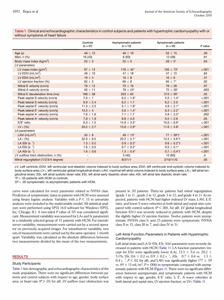
Table 1 Clinical and echocardiographic characteristics in control subjects and patients with hypertrophic cardiomyopathy with orwithout symptoms of heart failure
Controls
(n = 37)
Asymptomatic patients
(n = 12)
Symptomatic patients
(n = 25) P value
Age (y) 48 6 12 46 6 16 53 6 15 .30
Men, n (%) 16 (43) 6 (50) 12 (48) .81
Body mass index (kg/m2) 25 6 3 25 6 5 28 6 5* .04
LV parameters
LV mass index (g/m2) 87 6 13 176 6 42* 193 6 73* <.001
LV EDVi (mL/m2) 49 6 10 47 6 18 47 6 15 .84
LV ESVi (mL/m2) 19 6 4 16 6 8 16 6 6 .31
LV ejection fraction (%) 62 6 3 66 6 8 65 6 7* .02
Mitral E velocity (cm/s) 79 6 12 79 6 16 78 6 26 .97
Mitral A velocity (cm/s) 56 6 11 78 6 24* 72 6 30* .003
Mitral E deceleration time (ms) 168 6 38 203 6 63 213 6 90* .02
Peak septal S velocity (cm/s) 7.6 6 1 6.2 6 1.6* 5.5 6 1.4* <.001
Peak lateral S velocity (cm/s) 9.9 6 2.4 6.2 6 1.7 6.2 6 2.0 <.001Peak septal E’ velocity (cm/s) 11.0 6 2.5 5.1 6 1.8* 4.9 6 2.1* <.001
Peak lateral E’ velocity (cm/s) 15.5 6 4 5.6 6 1.4* 6.3 6 2.2* <.001Peak septal A’ velocity (cm/s) 7.6 6 1.6 7.1 6 1.7 5.8 6 2,2* .002
Peak lateral A’ velocity (cm/s) 7.8 6 1.9 8.8 6 4.0 6.5 6 2.9 .05E/E’ ratio 6.3 6 1.5 14.9 6 3.5* 15.6 6 6.6* <.001
LV e (%) �20.5 6 2.7 �13.8 6 2.9* �11.6 6 3.8* <.001LA parameters
LAVi (mL/m2) 33 6 8 49 6 13* 77 6 39*† <.001
LA e (%) 32.0 6 8.5 20.2 6 5.1* 13.3 6 5.6*† <.001
LA SSr (s�1) 1.3 6 0.2 0.9 6 0.2* 0.6 6 0.2*† <.001
LA ESr (s�1) �1.6 6 0.5 �0.7 6 0.2* �0.5 6 0.1* <.001
LA ASr (s�1) �1.6 6 0.6 �1.3 6 0.4* �0.7 6 0.4*† <.001
LV outflow tract obstruction, n (%) - 5 (42) 15 (60) .3
Mitral regurgitation (1/2/3/4 degree) - 8/2/1/1 3/10/11/0 .05
LV, Left ventricle; EDVi, left ventricular end-diastolic volume indexed to body surface area; ESVi, left ventricular end-systolic volume indexed to
body surface area; LV e, left ventricular global longitudinal strain; LAVi, maximal left atrial volume indexed to body surface area; LA e, left atrial lon-
gitudinal strain; SSr, left atrial systolic strain rate; ESr, left atrial early diastolic strain rate; ASr, left atrial late diastolic strain rate.
*P < .05 patients with HCM vs controls.†P < .05 symptomatic vs asymptomatic patients with HCM.
1092 Rosxca et al Journal of the American Society of EchocardiographyOctober 2010
curve were calculated for every parameter related to NYHA class.Predictors of symptomatic status in patients with HCM were assessedusing binary logistic analysis. Variables with a P < .15 in univariateanalyses were included in the multivariable model. All statistical anal-yses were performed using SPSS 14.0 software for Windows (SPSS,Inc, Chicago, IL). A two-sided P value of .05 was considered signifi-cant. Measurement variability was assessed for LAe and Sr parametersin a randomly selected group of 15 patients with HCM. For interob-server variability, measurements were carried out by a second opera-tor on previously acquired images. For intraobserver variability, twosets of measurements were carried out by the same operator, 1 monthapart. Variability was calculated as the absolute differences betweentwo measurements divided by the mean of the two measurements.
RESULTS
Study Participants
Table 1 lists demographic and echocardiographic characteristics of thestudy population. There were no significant differences between pa-tients and control subjects with respect to age, gender, body surfacearea, or heart rate (P > .05 for all). LV outflow tract obstruction was
present in 20 patients. Thirty-six patients had mitral regurgitation(grade 1 in 11 ; grade 2 in 12; grade 3 in 12; and grade 4 in 1). As ex-pected, patients with HCM had higher indexed LV mass, LAVi, E/E’ratio, and lower S-wave velocities at both lateral and septal sites com-pared with control subjects (P < .001, for all). LV global longitudinalfunction (LVe) was severely reduced in patients with HCM, despitethe slightly higher LV ejection fraction. Twelve patients were asymp-tomatic (NYHA class I), and 25 patients were symptomatic (NYHAclass II in 15, class III in 7, and class IV in 3).
Left Atrial Function Parameters in Patients with HypertrophicCardiomyopathy
Left atrial strain and LA Sr (SSr, ESr, ASr) parameters were severely de-creased in patients with HCM (Table 1). LA function parameters (ex-cept for ESr) were significantly lower (LAe, 13.3 6 5.6 vs. 20.2 6
5.1%; SSr, 0.6 6 0.2 vs. 0.9 6 0.2 s�1; ASr, �0.7 6 0.4 vs. �1.3 6
0.4 s�1, P < .02 for all), and LAVi was significantly higher (77 6 39vs. 49 6 13 mL/m2, P = .006) in symptomatic compared with asymp-tomatic patients with HCM (Figure 1). There were no significant differ-ences between asymptomatic and symptomatic patients with HCMwith respect to age, gender, E/E’ ratio, S, E’, A’-wave velocities atboth lateral and septal sites, LV ejection fraction, or LVe (Table 1).

Figure 1 Comparative display of LA deformation parameters in a control subject (A) and asymptomatic (B) and symptomatic (C) pa-tients with HCM. Upper panels: mean LA longitudinal strain curves (e). Lower panels: mean values of LA SSr, ESr, and ASr. Whencompared with the control subject (A), the asymptomatic (B) and symptomatic (C) patients with HCM have progressively lower lon-gitudinal LA e, SSr, ESr, and ASr.
Table 2 Correlates of left atrial function parameters in patients with hypertrophic cardiomyopathy
LA e SSr ESr ASr
P r P r P r P r
Age .70 .47 .03 0.36 .48LVMi .001 �0.53 .007 �0.45 .13 .01 0.40
Mitral E velocity .16 .23 .67 .05Mitral A velocity .58 .75 .04 0.35 .22
Mitral E deceleration time .47 .87 .04 0.34 .51Peak septal S velocity <.001 0.56 .004 0.47 .01 �0.40 .01 �0.43
Peak lateral S velocity .31 .15 .81 .21Peak septal E’ velocity .25 .51 .005 �0.46 .49
Peak lateral E’ velocity .78 .61 .36 .79Peak septal A’ velocity <.001 0.72 <.001 0.68 .01 �0.41 <.001 �0.77
Peak lateral A’ velocity <.001 0.66 <.001 0.65 .01 �0.45 <.001 �0.82
E/E’ ratio .02 �0.41 .02 �0.40 .007 0.49 .06
LV EF .17 .12 .41 .81LV e <.001 �0.79 <.001 �0.71 .007 0.48 .003 0.53
LAVi .002 �0.51 .008 �0.44 .17 .003 0.48MR degree .04 �0.33 .16 .07 .02 0.36
LVMi, Left ventricular mass indexed to body surface area; LVe, left ventricular global longitudinal strain; LVEF, left ventricular ejection fraction; LA e,left atrial longitudinal strain; SSr, left atrial systolic strain rate; ESr, left atrial early diastolic strain rate; ASr, left atrial late diastolic strain rate; E/E’, the
ratio of peak early-diastolic transmitral flow velocity E to average E’; LAVi, maximal left atrial volume indexed to body surface area; MR, mitral re-
gurgitation.
Journal of the American Society of EchocardiographyVolume 23 Number 10
Rosxca et al 1093
Left atrial strain (LA e), systolic Sr (SSr), and late diastolic Sr (ASr)were significantly related to LAVi and to LV mass in patients withHCM. Early diastolic LA Sr (ESr) decreased with increasing age(Table 2). There was a significant correlation between LA functionalindices derived from volumetric changes and LA-derived strainparameters. LA expansion index correlated significantly with LA e
(r = 0.57, P < .001) and SSr (r = 0.48, P = .003), LA passive emptyingfraction correlated with ESr (r = �0.34, P = .04), and LA activeemptying fraction correlated with ASr (r = �0.62, P < .001).
Intraobserver variability was 4.1% 6 3.4% for LAe, 7.5% 6 6.4%for SSr, 8.1% 6 6.0% for ESr, and 6.4% 6 5.7% for ASr. Interobservervariability for the same parameters was 5.9% 6 4.5%, 8.1% 6 3.2%,12.9% 6 8.4%, and 9.5% 6 7.8%, respectively.
Relationship of Left Atrial Function with Left VentricularSystolic and Diastolic Function in Patients with HypertrophicCardiomyopathy
All atrial function parameters (LAe, SSr, ESr, and ASr) were signifi-cantly related to LV global longitudinal strain (Figure 2). A similarlyclose correlation was found between LV longitudinal strain measuredfrom the apical four-chamber view and LA function parameters(r = �0.72, P < .001 for LAe; r = �0.62, P < .001 for SSr; r = 0.45,P = .005 for ESr; and r = 0.48, P = .003 for ASr). Significant correla-tions were also found between LA function parameters and septal S,but not with lateral S. LVejection fraction was not related to LAe, SSr,ESr, or ASr. Left atrial conduit function (ESr) and LA reservoir

Figure 2 Linear regression analysis and Pearson correlation coefficients for the relationship of LV global longitudinal strain to each ofLA deformation parameters: LA strain (A), SSr (B), ESr (C), and ASr (D) (red circles, symptomatic patients; green circles, asymptom-atic patients).
1094 Rosxca et al Journal of the American Society of EchocardiographyOctober 2010
function (LAe and SSr), were related to E/E’ ratio, in contrast with LAbooster pump function (ASr) (Table 2). Only LA conduit function wasrelated to LV early diastolic longitudinal Sr (r = �0.43, P = .01),whereas LA reservoir and LA booster pump functions were not(P > .05 for both). LV late diastolic longitudinal Sr correlatedwith LAe (r = 0.46, P = .007), SSr (r = 0.42, P = .01), and ASr(r =�0.75, P < .001). There were no significant correlations betweenLV diastolic dysfunction degree and LA function parameters (P > .05for all).
Indexed LA volume was related to indexed LV mass (r = 0.64,P < .001) and LV longitudinal strain (r = 0.56, P = .001). Neither E/E’ ratio nor LV outflow tract gradient was related to LAVi (P > .05 forboth), whereas mitral regurgitation severity had only a weak correlation(r = 0.35, P = .03) with LA volume.
Correlates of New York Heart Association Class in Patientswith Hypertrophic Cardiomyopathy
The main correlates of NYHA class in patients with HCM were LAfunction parameters (LAe, SSr, ESr and ASr), LAVi, and mitralregurgitation degree (Table 3). LA function parameters were the
only significant correlates of NYHA class by analysis of variance. LVmass, LV ejection fraction, S, E’, A’-wave velocities at both lateraland septal sites, and E/E’ ratio were not related to NYHA class. LVoutflow tract obstruction was not significantly different betweenasymptomatic and symptomatic patients and did not correlate withsymptomatic status. Moreover, the resting peak LV outflow tractgradient was not significantly different between asymptomaticand symptomatic patients with HCM (69 6 39 vs 81 6 35 mmHg, P = .51). To comparatively assess the accuracy of LAVi and LAfunction parameters (LAe, SSr, ESr, and ASr) in identifying symptom-atic patients with HCM, receiver operating characteristic curvesand the corresponding area under the curve were calculated. Thebest result has been obtained for ASr (area under the curve: 0.83)with a cutoff of �0.92 s�1 for identifying symptomatic patientswith HCM (sensitivity: 75%, specificity: 83%) (Figure 3). Thecorrelates of symptomatic status are displayed in Table 4. Atmultivariable logistic regression analysis, ASr emerged as the onlyindependent correlate of heart failure symptoms in our studypopulation (odds ratio = 2.63; 95% confidence interval, 1.015-6.922, P = .04).

Table 3 Correlates of New York Heart Association class in patients with hypertrophic cardiomyopathy
Variables
NYHA I
(n = 12)
NYHA II
(n = 15)
NYHA III�IV
(n = 10)
P
ANOVA
Spearman correlation
coefficient
P
Spearman
LAVi (mL/m2) 49 6 13 72 6 45 83 6 28 .06 0.55 <.001LA e (%) 20.2 6 5.1 13.9 6 4.9 12.6 6 6.7 .005 �0.47 .003
LA SSr (s�1) 0.9 6 0.2 0.6 6 0.1 0.6 6 0.4 .004 �0.47 .003LA ESr (s�1) �0.7 6 0.2 �0.5 6 0.1 �0.5 6 0.2 .005 0.46 .005
LA ASr (s�1) �1.3 6 0.4 �0.7 6 0.4 �0.7 6 0.6 .01 0.48 .003LV e (%) �13.9 6 2.9 �12.2 6 3.3 �10.3 6 4.6 .14 0.36 .04
MR degree(1/2/3/4) 8/2/1/1 3/7/5/0 1/3/6/0 .02* 0.39 .01
ANOVA, Analysis of variance; NYHA, New York Heart Association; LAVi, maximal left atrial volume indexed to body surface area; LA e, left atrial
longitudinal strain; LA SSr, left atrial systolic strain rate; LA ESr, left atrial early diastolic strain rate; LA ASr, left atrial late diastolic strain rate; MR,mitral regurgitation; LV e, left ventricular global longitudinal strain.
*P value obtained by chi-square test.
Figure 3 Receiver operating characteristic curve for late diastolicLA strain rate in identifying heart failure symptoms in patients withHCM. Best cutoff value for LA ASr in identifying heart failuresymptoms was�0.92 s�1 (arrow), with an area under the receiveroperating characteristic curve of 0.83.
Table 4 Correlates of symptomatic status in patients withhypertrophic cardiomyopathy
Univariate analysis Multivariable
analysis
Variables OR 95% CI P P
Age 1.027 0.983-1.073 .23 -
LVe 1.215 0.954-1.549 .11 .49
LAVi 1.091 1.018-1.170 .01 .34
ASr 3.377* 1.349-8.616 .009 .04
MR degree 2.277 0.969-5.353 .056 .10
Presence of LV outflow
tract obstruction (Y/N)
0.476 0.118-1.929 .29 -
Peak LV outflowtract gradient
1.010 0.981-1.041 .49 -
OR, Odds ratio; CI, confidence interval; LV, left ventricular; LVe, leftventricular global longitudinal strain; LAVi, maximal left atrial volume
indexed to body surface area; ASr, left atrial late diastolic strain rate;
MR, mitral regurgitation; Y/N, YES or NO.*OR is expressed per standard deviation increment in the variable.
Journal of the American Society of EchocardiographyVolume 23 Number 10
Rosxca et al 1095
DISCUSSION
The main findings of the present study in patients with HCM can besummarized as follows: LA and LV longitudinal deformation aremutually dependent and significantly impaired, despite preservedLV ejection fraction. Although the reduction in LA longitudinalfunction involves all three atrial phases, LA booster pump dysfunctionrepresents the main correlate of functional disability.
Left Atrial Remodeling and Dysfunction in Patients withHypertrophic Cardiomyopathy
The LA acts as a reservoir during LV systole, as a conduit during earlydiastole, and as a booster pump in late diastole, thus modulating LV fill-ing.21 The LA can act to increase LA pressure (in significant atrial dis-ease) and can react to increased LV filling pressure (in significantventricular disease). There is a growing body of evidence demonstratingthat LA enlargement is a marker of significant atrial22 or ventriculardisease.23,24
In patients with HCM, the enlargement of the LA has proved tobe inconsistently related to LV diastolic dysfunction, mitral regurgi-
tation degree, or LV dynamic outflow tract obstruction.25 Thus, theexistence of an additional mechanism involved in atrial remodelingwas postulated. The assessment of LA function using traditionalparameters such as atrial fraction25 or newer parameters derivedfrom tissue Doppler or STE analysis26 brought new insights intothe pathophysiologic mechanisms involved in atrial remodeling inHCM. The poorer LA function was indeed attributed to a possibleatrial myopathic process.25,26 In our study, the three phases of LAfunction (reservoir, conduit, and booster pump) were significantlyreduced. The passive stretching of the LA (reservoir function)and the active LA contraction (booster pump function) wereinversely related to LA enlargement. This is in line with previousstudies showing that the LA active emptying might decrease inthe presence of severe LA dilation as the optimal Frank-Starlingrelationship is exceeded.27 Neither diastolic parameters nor LVoutflow tract gradient was related to LA enlargement, whereasmitral regurgitation severity had only a weak correlation. Thesedata suggest that severe LA remodeling is not only a consequenceof the hemodynamic abnormalities caused by LV dysfunction,mitral regurgitation, or LV outflow tract obstruction.

1096 Rosxca et al Journal of the American Society of EchocardiographyOctober 2010
Relation of Left Atrial Function Parameters to Left VentricularDysfunction in Hypertrophic Cardiomyopathy
LV longitudinal function, as assessed by systolic annular velocities,strain, or Sr, is commonly reduced in patients with HCM despitepreserved LVejection fraction28 and is related to the degree of hyper-trophy, myocardial disarray, and fibrosis.29 All these patients have LVdiastolic dysfunction, even those with preclinical disease, withoutLV hypertrophy.30
The present study confirms and extends these data by showing thatLV longitudinal function is strongly correlated to the three compo-nents of LA function. The greater the LV longitudinal dysfunction,the more severe the LA dysfunction. LA conduit function seems tobe governed mainly by LV relaxation capacity, being the only LAfunction parameter correlated to LV early diastolic longitudinal Srand to early diastolic septal velocity (E’). Whether the reduction inLA reservoir function in patients with HCM is more due to LV longi-tudinal dysfunction with a decrease in the systolic descent of LVbase or to an impaired LA relaxation and stiffness (as a result of amyopathic process affecting both the ventricle and the atrium) needsfurther study. However, the E/E’ ratio and LVearly diastolic longitudi-nal Sr were not related to LA booster pump function suggesting thatin patients with HCM LA contraction is only partially modulated byLV diastolic performance and LV filling pressures.
Left Atrial Function and Heart Failure Symptoms inHypertrophic Cardiomyopathy
The clinical course of HCM is characterized by an extreme heteroge-neity with unpredictable development of heart failure symptoms inthe presence of normal or supranormal LV ejection fraction andregardless of whether outflow tract obstruction is present or not.7
The hemodynamic mechanisms for impaired exercise tolerance inpatients with HCM are still poorly defined. However, increased LVchamber stiffness, impaired LV relaxation, and compromised LAfunction with elevated LV filling pressures have been postulated asthe main mechanisms of heart failure symptoms in HCM.Myocardial ischemia and LV outflow tract obstruction associatedwith mitral regurgitation can further increase LV stiffness, leading tomore severe LV diastolic dysfunction and more severe symptoms.
The LA plays an important role in maintaining LV filling and con-sequently LV stroke volume, especially when the LV is dysfunctional.5
The enlargement of the LA and the increase in LA emptying fractionare adaptive responses to impaired LV diastolic function to maintainnormal LV filling pressures.31 LA remodeling predicts exercise capac-ity3 and development of severe symptoms in patients with HCM.32
LA fractional shortening is related to peak oxygen consumption inthese patients.33 Decreased LA compliance with reduced reservoirand contractile pump functions can counteract this adaptive mecha-nism and promote symptom occurrence. However, there are nodata showing the relationship between LA function parameters, asassessed by STE, and symptoms in patients with HCM.
In the current study, LA reservoir, conduit, and booster pumpfunctions correlated significantly with NYHA class. Specifically, theseverity of heart failure symptoms increased with the severity of LAdysfunction. Left atrial enlargement and mitral regurgitation werealso related to heart failure symptoms, whereas LV outflow tractobstruction was not significantly different between asymptomaticand symptomatic patients and did not correlate with symptomaticstatus. However, we cannot exclude the potential influence ofexercise-induced increases in LV outflow tract gradient on heartfailure symptoms in these patients.
LV filling pressures, assessed by the E/E’ ratio, failed to correlate withNYHA class in our patients. This may seem to contradict previousfindings. However, the reported correlations between E/E’ ratio andexercise capacity in patients with HCM are relatively modest,34,35
leading to the hypothesis that other factors also influence exercisecapacity in this setting. Moreover, estimates of LV filling pressurebased on the E/E’ ratio correlated weakly with direct measurementsof LA pressure in symptomatic patients with HCM.36,37
Clinical Perspective
In patients with HCM, LA enlargement independently predicts long-term prognosis,4,38 adverse cardiovascular events,1 and post-myectomy survival.39 In these patients, not only LA size but also myo-cardial fibrosis correlated with the presence of atrial fibrillation,40 andthe relationship of myocardial fibrosis burden with a more decreasedLV longitudinal strain has recently been acknowledged.29 Therefore,the noninvasive assessment of LA longitudinal deformation may addincremental information to LA size for predicting atrial fibrillation oc-currence or response to therapy in HCM. Moreover, the potentialprognostic implications of extensive LA structural and functional ab-normalities and whether these alterations can be modified by treat-ment remain to be determined.
STUDY LIMITATIONS
Because this study was performed in a tertiary center, our study pop-ulation may not reflect the typical patient seen in the community.However, one third of our patients were asymptomatic, allowingthe identification of parameters related to symptoms. BecauseHCM is not a common disease, the study sample size was relativelysmall, particularly for the subanalyses of symptomatic versus asymp-tomatic patients. However, identifying factors associated with thesymptomatic status is of clinical importance. Patients did not undergomutation analysis for HCM diagnosis. Twenty-three of the 37 patientshad asymmetric LV hypertrophy. In the remaining 14 patients withsymmetric hypertrophy, wall thickness was > 15 mm in the absenceof exercise training history and of cardiac or systemic conditionscapable of inducing such a degree of hypertrophy. LA deformationwas assessed only in the apical four-chamber view, as previouslyperformed by D’Andrea et al.41 However, an average strain valuefor all LA segments in this view was assessed. Patients remained ontheir routine medication, which may have had some impact on theirLA function or on the true extent of LV dysfunction. However, therewas no significant difference in treatment between asymptomatic andsymptomatic patients in this study.
CONCLUSIONS
In patients with HCM, LA reservoir, conduit, and booster pump func-tions were significantly reduced and closely related to LV longitudinalmyocardial deformation. In this cohort, symptoms of heart failurewere related to the severity of LA dilation and of LA dysfunction.The assessment of LA function can provide further insights into thepathophysiology and symptoms occurrence in HCM.
ACKNOWLEDGMENT
The authors acknowledge the support of colleagues who have referredpatients for this study.
Journal of the American Society of EchocardiographyVolume 23 Number 10
Rosxca et al 1097
REFERENCES
1. Yang H, Woo A, Monakier D, Jamorski M, Fedwick K, Wigle ED, et al.Enlarged left atrial volume in hypertrophic cardiomyopathy: a markerfor disease severity. J Am Soc Ecocardiogr 2005;18:1074-82.
2. Afonso LC, Bernal J, Bax JJ, Abraham TP. Echocardiography in hypertro-phic cardiomyopathy. JACC Cardiovasc Imaging 2008;1:787-800.
3. Sachdev V, Shizukuda Y, Brenneman CL, Birdsall CW, Waclawiw MA,Arai AE, et al. Left atrial volumetric remodeling is predictive of functionalcapacity in nonobstructive hypertrophic cardiomyopathy. Am Heart J2005;149:730-6.
4. Nistri S, Olivotto I, Betocchi S, Losi MA, Valsecchi G, Pinamonti B, et al.Prognostic significance of left atrial size in patients with hypertrophiccardiomyopathy (from the Italian Registry for Hypertrophic Cardiomyop-athy). Am J Cardiol 2006;98:960-5.
5. Matsuda Y, Toma Y, Ogawa H, Matsuzaki M, Katayama K, Fujii T, et al.Importance of left atrial function in patients with myocardial infarction.Circulation 1983;67:565-71.
6. Payne RM, Stone HL, Engelken EJ. Atrial function during volume loading.J Appl Physiol 1971;31:326.
7. Maron BJ. Hypertrophic cardiomyopathy. A systematic review. JAMA2002;287:1308-20.
8. Barbier P, Solomon SB, Schiller NB, Glantz SA. Left atrial relaxation andleft ventricular systolic function determine left atrial reservoir function.Circulation 1999;100:427-36.
9. Toma Y, Matsuda Y, Moritani K, Ogawa H, Matsuzaki M, Kusukawa R.Left atrial filling in normal human subjects: relation between left atrialcontraction and left atrial early filling. Cardiovasc Res 1987;21:255-9.
10. Sirbu C, Herbots L, D’hooge J, Claus P, Marciniak A, Langeland T, et al.Feasibility of strain and strain rate imaging for the assessment of regionalleft atrial deformation: a study in normal subjects. Eur J Echocardiogr2006;7:199-208.
11. Amundsen BH, Helle-Valle T, Edvardsen T, Torp H, Crosby J, Lyseggen E,et al. Noninvasive myocardial strain measurement by speckle trackingechocardiography: validation against sonomicrometry and tagged mag-netic resonance imaging. J Am Coll Cardiol 2006;47:789-93.
12. Maron BJ, McKenna WJ, Danielson GK, Kappenberger LJ, Kuhn HJ,Seidman CE, et al. Task Force on Clinical Expert Consensus Documents.American College of Cardiology; Committee for Practice Guidelines.European Society of Cardiology. American College of Cardiology/ Euro-pean Society of Cardiology clinical expert consensus document on hyper-trophic cardiomyopathy. A report of the American College of CardiologyFoundation Task Force on Clinical Expert Consensus Documents and theEuropean Society of Cardiology Committee for Practice Guidelines. J AmColl Cardiol 2003;42:1687-713.
13. Popescu BA, Beladan CC, Calin A, Muraru D, Deleanu D, Rosca M, et al.Left ventricular remodelling and torsional dynamics in dilated cardiomy-opathy: reversed apical rotation as a marker of disease severity. Eur J HeartFail 2009;11:945-51.
14. Nikitin NP, Witte KK, Thackray SD, Goodge LJ, Clark AL, Cleland JG. Ef-fect of age and sex on left atrial morphology and function. Eur J Echocar-diogr 2003;4:36-42.
15. Lang RM, Bierig M, Devereux RB, Flachskampf F, Foster E, Pellikka P, et al.Recommendations for chamber quantification. Eur J Echocardiogr 2006;7:79-108.
16. Devereux RB, Alonso DR, Lutas EM, Gottlieb GJ, Campo E, Sachs I, et al.Echocardiographic assessment of left ventricular hypertrophy: comparisonto necropsy findings. Am J Cardiol 1986;57:450-8.
17. Ommen SR, Nishimura RA, Appleton CP, Miller FA, Oh JK, Redfield MM,et al. Clinical utility of Doppler echocardiography and tissue Dopplerimaging in the estimation of left ventricular filling pressures: a comparativesimultaneous Doppler-catheterization study. Circulation 2000;102:1788-94.
18. Nagueh SF, Appleton CP, Gillebert TC, Marino PN, Oh JK, Smiseth OA,et al. Recommendations for the evaluation of left ventricular diastolicfunction by echocardiography. J Am Soc Echocardiogr 2009;22:107-33.
19. Lancellotti P, Moura L, Pierard LA, Agricola E, Popescu BA, Tribouilloy C,et al. European Association of Echocardiography recommendations forthe assessment of valvular regurgitation. Part 2: mitral and tricuspidregurgitation (native valve disease). Eur J Echocardiogr 2010;11:307-32.
20. Serri K, Reant P, Lafitte M, Berhouet M, Le Bouffos V, Roudaut R, et al.Global and regional myocardial function quantification by two-dimensional strain; application in hypertrophic cardiomyopathy. J AmColl Cardiol 2006;47:1175-81.
21. Triposkiadis F, Tentolouris K, Androulakis A, Trikas A, Toutouzas K,Kyriakidis M, et al. Left atrial mechanical function in the healthy elderly:new insights from a combined assessment of changes in atrial volumeand transmitral flow velocity. J Am Soc Echocardiogr 1995;8:801-9.
22. Brodsky MA, Allen BJ, Capparelli EV, Luckett CR, Morton R, Henry WL.Factors determining maintenance of sinus rhythm after chronic atrial fibril-lation with left atrial dilatation. Am J Cardiol 1989;63:1065-8.
23. Kannel WB, Abbott RD, Savage DD, McNamara PM. Epidemiologicfeatures of chronic atrial fibrillation: the Framingham Study. N Engl JMed 1982;306:1018-22.
24. Popescu BA, Macor F, Antonini-Canterin F, Giannuzzi P, Temporelli PL,Bosimini E, et al. GISSI-3 Echo Substudy Investigators. Left atrium remod-eling after acute myocardial infarction (results of the GISSI-3 EchoSubstudy). Am J Cardiol 2004;93:1156-9.
25. Eshoo S, Semsarian C, Ross DL, Thomas L. Left atrial phasic volumes aremodulated by the type rather than the extent of left ventricular hypertro-phy. J Am Soc Echocardiogr 2010 Mar 10 [Epub ahead of print].
26. Paraskevaidis IA, Panou F, Papadopoulos C, Farmakis D, Parissis J,Ikonomidis I, et al. Evaluation of left atrial longitudinal function in patientswith hypertrophic cardiomyopathy: a tissue Doppler imaging andtwo-dimensional strain study. Heart 2009;95:483-9.
27. Pagel PS, Kehl F, Gare M, Hettrick DA, Kersten JR, Warltier DC. Mechan-ical function of the left atrium: new insights based on analysis of pressure-volume relations and Doppler echocardiography. Anesthesiology 2003;98:975-94.
28. Kato TS, Noda A, Izawa H, Yamada A, Obata K, Nagata K, et al. Discrim-ination of nonobstructive hypertrophic cardiomyopathy from hyperten-sive left ventricular hypertrophy on the basis of strain rate imaging bytissue Doppler ultrasonography. Circulation 2004;110:3808-14.
29. Popovic ZB, Kwon DH, Mishra M, Buakhamsri A, Greenberg NL,Thamilarasan M, et al. Association between regional ventricular functionand myocardial fibrosis in hypertrophic cardiomyopathy assessed byspeckle tracking echocardiography and delayed hyperenhancementmagnetic resonance imaging. J Am Soc Echocardiogr 2008;21:1299-305.
30. Ho CY, Sweitzer NK, McDonough B, Maron BJ, Casey SA, Seidman JG,et al. Assessment of diastolic function with Doppler tissue imaging topredict genotype in preclinical hypertrophic cardiomyopathy. Circulation2002;105:2992-7.
31. Leung DY, Boyd A, Ng AA, Chi C, Thomas L. Echocardiographic evaluationof left atrial size and function: current understanding, pathophysiologiccorrelates, and prognostic implications. Am Heart J 2008;156:1056-64.
32. Losi MA, Betocchi S, Barbati G, Parisi V, Tocchetti CG, Pastore F, et al.Prognostic significance of left atrial volume dilatation in patients withhypertrophic cardiomyopathy. J Am Soc Echocardiogr 2009;22:76-81.
33. Briguori C, Betocchi S, Romano M, Manganelli F, Angela Losi M,Ciampi Q, et al. Exercise capacity in hypertrophic cardiomyopathy de-pends on left ventricular diastolic function. Am J Cardiol 1999;84:309-15.
34. Matsumura Y, Elliott PM, Virdee MS, Sorajja P, Doi Y, McKenna WJ. Leftventricular diastolic function assessed using Doppler tissue imaging inpatients with hypertrophic cardiomyopathy: relation to symptoms andexercise capacity. Heart 2002;87:247-51.
35. Ha JW, Cho JR, Kim JM, Ahn JA, Choi EY, Kang SM, et al. Tissue Doppler-derived indices predict exercise capacity in patients with apical hypertro-phic cardiomyopathy. Chest 2005;128:3428-33.
36. Geske JB, Sorajja P, Nishimura RA, Ommen SR. Evaluation of left ventric-ular filling pressures by Doppler echocardiography in patients with hyper-trophic cardiomyopathy: correlation with direct left atrial pressuremeasurement at cardiac catheterization. Circulation 2007;116:2702-8.

1098 Rosxca et al Journal of the American Society of EchocardiographyOctober 2010
37. Geske JB, Sorajja P, Nishimura RA, Ommen SR. The relationship of leftatrial volume and left atrial pressure in patients with hypertrophiccardiomyopathy: an echocardiographic and cardiac catheterization study.J Am Soc Echocardiogr 2009;22:961-6.
38. Yang WI, Shim CY, Kim YJ, Kim SA, Rhee SJ, Choi EY, et al. Left atrialvolume index: a predictor of adverse outcome in patients withhypertrophic cardiomyopathy. J Am Soc Echocardiogr 2009;22:1338-43.
39. Woo A, Williams WG, Choi R, Wigle ED, Rozenblyum E, Fedwick K, et al.Clinical and echocardiographic determinants of long-term survival after
surgical myectomy in obstructive hypertrophic cardiomyopathy. Circula-tion 2005;111:2033-41.
40. Pujadas S, Vidal-Perez R, Hidalgo A, Leta R, Carreras F, Barros A, et al.Correlation between myocardial fibrosis and the occurrence of atrialfibrillation in hypertrophic cardiomyopathy: a cardiac magnetic resonanceimaging study. Eur J Radiol 2010 Jan 14 [Epub ahead of print].
41. D’Andrea A, Caso P, Romano S, Scarafile R, Riegler L, Salerno G, et al.Different effects of cardiac resynchronization therapy on left atrial functionin patients with either idiopathic or ischaemic dilated cardiomyopathy:a two-dimensional speckle strain study. Eur Heart J 2007;28:2738-48.
Copyright © 2022 FDOKUMEN