
Thermodynamics and Folding Kinetics Analysis of the SH3 Domain from Discrete Molecular Dynamics
14
Thermodynamics and Folding Kinetics Analysis of the SH3 Domain from Discrete Molecular Dynamics Jose M. Borreguero 1 *, Nikolay V. Dokholyan 2 , Sergey V. Buldyrev 1 Eugene I. Shakhnovich 2 and H. Eugene Stanley 1 1 Center for Polymer Studies and Department of Physics Boston University, Boston MA 02215, USA 2 Department of Chemistry and Chemical Biology, Harvard University, Cambridge MA 02138, USA We perform a detailed analysis of the thermodynamics and folding kinetics of the SH3 domain fold with discrete molecular dynamic simu- lations. We propose a protein model that reproduces some of the experi- mentally observed thermodynamic and folding kinetic properties of proteins. Specifically, we use our model to study the transition state ensemble of the SH3 fold family of proteins, a set of unstable confor- mations that fold to the protein native state with probability 1/2. We analyze the participation of each secondary structure element formed at the transition state ensemble. We also identify the folding nucleus of the SH3 fold and test extensively its importance for folding kinetics. We predict that a set of amino acid contacts between the RT-loop and the distal hairpin are the critical folding nucleus of the SH3 fold and propose a hypothesis that explains this result. q 2002 Elsevier Science Ltd. All rights reserved Keywords: protein folding; SH3; transition state; folding nucleus; molecular dynamics *Corresponding author Introduction Studying the protein folding kinetics is a chal- lenging task because it involves the identification of the transition state ensemble (TSE), a set of unstable conformations that form at the top of the free energy barrier separating the unique folded state from the misfolded and unfolded states. 1 The TSE has been the subject of numerous experiments 2–18 and theoretical studies 19 – 25 of globular proteins. The TSE is defined as the set of protein conformations with a probability to fold, p FOLD , equal to 1/2. 1 Protein engineering experiments 8–18 suggest that in two-state proteins, there is a specific set of amino acid residues that determines the folding properties. Theoretical studies 19 – 25 of the TSE support the hypothesis of a specific folding nucleus scenario. Passing through the TSE with the subsequent rapid assembly of the native conformation requires the formation of a set of specific obligatory contacts, which are called the protein folding nucleus. 1 Lattice simulations 19,23 suggest that the folding nucleus location is identical for two different pro- tein sequences, designed with various potentials to fold into the same structure. In protein engineer- ing experiments 26 on the Src SH3 domain 14,15,27 – 33 and the a-spectrin SH3 domain, 11,13,34 authors find the same structural characteristics for the TSE of the two homologous proteins. These studies suggest that the location of the TSE and the folding nucleus in the native structure of the protein depends more on the topology of the native struc- ture than on the specific protein amino acid sequence folding into that structure. Several groups 35 – 38 attempted to identify the TSE of some well characterized globular proteins and obtained a significant correlation with experimental kinetic data. However, these models assume the number of ordered residues as an approximation to the reaction coordinate for the folding process. The main difficulty with these studies is that the num- ber of ordered residues does not characterize the TSE. Ding et al. (unpublished results) studied the TSE of the Src SH3 domain and found that differ- ent conformations with the same number of native contacts as TSE conformations had drastically different probabilities to fold. Another simplifica- tion is the assumption that the folding process occurs through the meeting and adoption of native structure of only two fragments of the protein. In contrast, our method to select TSE conformations does not depend on the number of ordered 0022-2836/02/$ - see front matter q 2002 Elsevier Science Ltd. All rights reserved E-mail address of the corresponding author: [email protected] Abbreviations used: TSE, transition state ensemble; rmsd, root mean square displacement. doi: 10.1016/S0022-2836(02)00136-5 available online at http://www.idealibrary.com on B w J. Mol. Biol. (2002) 318, 863–876
Transcript of Thermodynamics and Folding Kinetics Analysis of the SH3 Domain from Discrete Molecular Dynamics

Thermodynamics and Folding Kinetics Analysis of theSH3 Domain from Discrete Molecular Dynamics
Jose M. Borreguero1*, Nikolay V. Dokholyan2, Sergey V. Buldyrev1
Eugene I. Shakhnovich2 and H. Eugene Stanley1
1Center for Polymer Studiesand Department of PhysicsBoston University, BostonMA 02215, USA
2Department of Chemistry andChemical Biology, HarvardUniversity, CambridgeMA 02138, USA
We perform a detailed analysis of the thermodynamics and foldingkinetics of the SH3 domain fold with discrete molecular dynamic simu-lations. We propose a protein model that reproduces some of the experi-mentally observed thermodynamic and folding kinetic properties ofproteins. Specifically, we use our model to study the transition stateensemble of the SH3 fold family of proteins, a set of unstable confor-mations that fold to the protein native state with probability 1/2. Weanalyze the participation of each secondary structure element formed atthe transition state ensemble. We also identify the folding nucleus ofthe SH3 fold and test extensively its importance for folding kinetics. Wepredict that a set of amino acid contacts between the RT-loop and thedistal hairpin are the critical folding nucleus of the SH3 fold and proposea hypothesis that explains this result.
q 2002 Elsevier Science Ltd. All rights reserved
Keywords: protein folding; SH3; transition state; folding nucleus; moleculardynamics*Corresponding author
Introduction
Studying the protein folding kinetics is a chal-lenging task because it involves the identificationof the transition state ensemble (TSE), a set ofunstable conformations that form at the top of thefree energy barrier separating the unique foldedstate from the misfolded and unfolded states.1
The TSE has been the subject of numerousexperiments2–18 and theoretical studies19–25 ofglobular proteins. The TSE is defined as the set ofprotein conformations with a probability tofold, pFOLD, equal to 1/2.1 Protein engineeringexperiments8–18 suggest that in two-state proteins,there is a specific set of amino acid residues thatdetermines the folding properties. Theoreticalstudies19–25 of the TSE support the hypothesis of aspecific folding nucleus scenario. Passing throughthe TSE with the subsequent rapid assembly ofthe native conformation requires the formation ofa set of specific obligatory contacts, which arecalled the protein folding nucleus.1
Lattice simulations19,23 suggest that the foldingnucleus location is identical for two different pro-
tein sequences, designed with various potentialsto fold into the same structure. In protein engineer-ing experiments26 on the Src SH3 domain14,15,27–33
and the a-spectrin SH3 domain,11,13,34 authors findthe same structural characteristics for the TSE ofthe two homologous proteins. These studiessuggest that the location of the TSE and the foldingnucleus in the native structure of the proteindepends more on the topology of the native struc-ture than on the specific protein amino acidsequence folding into that structure. Severalgroups35–38 attempted to identify the TSE of somewell characterized globular proteins and obtaineda significant correlation with experimental kineticdata. However, these models assume the numberof ordered residues as an approximation to thereaction coordinate for the folding process. Themain difficulty with these studies is that the num-ber of ordered residues does not characterize theTSE. Ding et al. (unpublished results) studied theTSE of the Src SH3 domain and found that differ-ent conformations with the same number of nativecontacts as TSE conformations had drasticallydifferent probabilities to fold. Another simplifica-tion is the assumption that the folding processoccurs through the meeting and adoption of nativestructure of only two fragments of the protein. Incontrast, our method to select TSE conformationsdoes not depend on the number of ordered
0022-2836/02/$ - see front matter q 2002 Elsevier Science Ltd. All rights reserved
E-mail address of the corresponding author:[email protected]
Abbreviations used: TSE, transition state ensemble;rmsd, root mean square displacement.
doi: 10.1016/S0022-2836(02)00136-5 available online at http://www.idealibrary.com onBw
J. Mol. Biol. (2002) 318, 863–876
residues, and we employ discrete moleculardynamic simulations that do not constrain thenumber of native elements of structure.
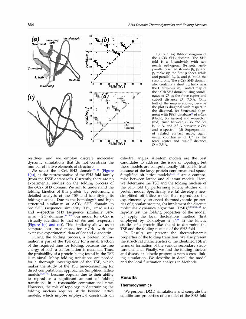
We select the c-Crk SH3 domain39–41 (Figure1(a)), as the representative of the SH3 fold family(from the FSSP database42). Currently, there are noexperimental studies on the folding process ofthe c-Crk SH3 domain. We aim to understand thefolding kinetics of this protein by performing adetailed analysis of the TSE and identifying itsfolding nucleus. Due to the homology43 and highstructural similarity of c-Crk SH3 domain toSrc SH3 (sequence similarity 33%, rmsd ¼ 1:4)and a-spectrin SH3 (sequence similarity 34%,rmsd ¼ 2:3) domains,9–14,40 our model for c-Crk isvirtually identical to that of Src and a-spectrin(Figure 1(c) and (d)). This similarity allows us tocompare our predictions for c-Crk with theextensive experimental data of Src and a-spectrin.
During the folding process, a protein confor-mation is part of the TSE only for a small fractionof the required time for folding, because the freeenergy of such a conformation is maximal. Thus,the probability of a protein being found in the TSEis minimal. Many folding transitions are neededfor a thorough investigation of the TSE, whichmakes the study of the TSE time-consuming fordirect computational approaches. Simplified latticemodels20,44–52 became popular due to their abilityto reproduce a significant amount of foldingtransitions in a reasonable computational time.However, the role of topology in determining thefolding nucleus requires study beyond latticemodels, which impose unphysical constraints on
dihedral angles. All-atom models are the bestcandidates to address the issue of topology, butthese models are computationally difficult to treatbecause of the large protein conformational space.Simplified off-lattice models25,53–57 are a compro-mise between lattice and all-atom models. Here,we determine the TSE and the folding nucleus ofthe SH3 fold by performing kinetic studies of aprotein model. Specifically, we: (a) develop a new,simplified off-lattice model that reproduces theexperimentally observed thermodynamic proper-ties of globular proteins, (b) implement the discretemolecular dynamics algorithm (DMD)25,53,58–62 torapidly test the folding properties of the model;(c) apply the local fluctuations method (firstemployed by Dokholyan et al.25 in the kineticstudies of a protein-like chain) to determine theTSE and the folding nucleus of the SH3 fold.
In Results we present the thermodynamicproperties of the folding transition. We also presentthe structural characteristics of the identified TSE interms of formation of the various secondary struc-ture elements. Finally, we find the folding nucleusand discuss its kinetic properties with a cross-link-ing simulation. We describe in detail the modeland the local fluctuation analysis in Methods.
Results
Thermodynamics
We perform DMD simulations and compute theequilibrium properties of a model of the SH3 fold
Figure 1. (a) Ribbon diagram ofthe c-Crk SH3 domain. The SH3fold is a b-sandwich with twonearly orthogonal b-sheets. Anti-parallel oriented strands b1, b2 andb5 make up the first b-sheet, whileanti-parallel b2, b3 and b4 build thesecond one. The c-Crk SH3 domainalso contains a short 310 helix nearthe C terminus. (b) Contact map ofthe c-Crk SH3 domain using coordi-nates of Cb as the force center andcut-off distance D ¼ 7:5 �A: Onlyhalf of the map is shown, becausethe plot is diagonal with respect tothe diagonal. (c) Structural align-ment with FSSP database42 of c-Crk(black), Src (green) and a-spectrin(red). rmsd between c-Crk and Srcis 1.4 A, and 2.3 A between c-Crkand a-spectrin. (d) Superpositionof related contact maps, againusing coordinates of Cb as theforce center and cut-off distanceD ¼ 7:5 �A:
864 SH3 Domain Thermodynamics and Folding Kinetics
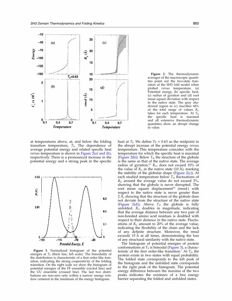
at temperatures above, at, and below the foldingtransition temperature, TF. The dependence ofaverage potential energy and related specific heatversus temperature is shown in Figure 2(a) and (b),respectively. There is a pronounced increase in thepotential energy and a strong peak in the specific
heat at TF. We define TF ¼ 0:63 as the midpoint inthe abrupt increase of the potential energy versustemperature. This temperature coincides with thetemperature for which the specific heat is maximal(Figure 2(b)). Below TF, the structure of the globuleis the same as that of the native state. The averageradius of gyration,63 RG, does not exceed 10% ofthe value of RG in the native state (10 A), markingthe stability of the globular shape (Figure 2(c)). Ateach studied temperature below TF, fluctuations ofRG around the average value do not exceed 3%,showing that the globule is never disrupted. Theroot mean square displacement64 (rmsd ) withrespect to the native state is never greater than2 A, showing that the structure of the globule doesnot deviate from the structure of the native state(Figure 2(d)). Above TF, the globule is fullyunfolded. RG doubles in magnitude, indicatingthat the average distance between any two pair ofnon-bonded amino acid residues is doubled withrespect to their distance in the native state. Fluctu-ations of RG amount to 20% of the average value,indicating the flexibility of the chain and the lackof any definite structure. Moreover, the rmsdexceeds 15 A at all times, demonstrating the lossof any structural similarity with the native state.The histogram of potential energies of protein
conformations at TF is bimodal (Figure 3), a charac-teristic of the first order-like transition.1 At TF, theprotein exists in two states with equal probability.The folded state corresponds to the left peak ofthe histogram and the unfolded state correspondsto the right peak of the histogram. The potentialenergy difference between the maxima of the twopeaks indicates the existence of a free energybarrier separating the folded and unfolded states.
Figure 2. The thermodynamicaverages of the macroscopic quanti-ties point out the two-state tran-sition of the SH3 fold model whenplotted versus temperature. (a)Potential energy, (b) specific heat,(c) radius of gyration and (d) rootmean square deviation with respectto the native state. The gray sha-dowed region in (c) inscribes 68%of the total range of values RG
takes for each temperature. At TF,the specific heat is maximaland all extensive thermodynamicquantities show an abrupt changein value.
Figure 3. Normalized histogram of the potentialenergies at TF (thick line, left scale). The bimodality ofthe distribution is characteristic of a first order-like tran-sition, indicating the strong cooperativity of the foldingtransition. On the right scale we show the histogram ofpotential energies of the FF ensemble (circled line) andthe UU ensemble (crossed line). The last two distri-butions are non-zero only within a narrow energy win-dow centered in the minimum of the energy histogram.
SH3 Domain Thermodynamics and Folding Kinetics 865
Kinetics
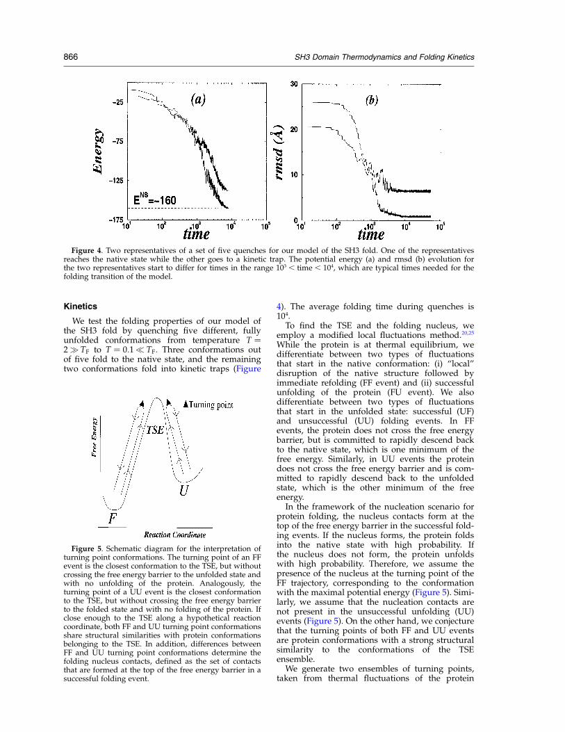
We test the folding properties of our model ofthe SH3 fold by quenching five different, fullyunfolded conformations from temperature T ¼2q TF to T ¼ 0:1p TF: Three conformations outof five fold to the native state, and the remainingtwo conformations fold into kinetic traps (Figure
4). The average folding time during quenches is104.
To find the TSE and the folding nucleus, weemploy a modified local fluctuations method.20,25
While the protein is at thermal equilibrium, wedifferentiate between two types of fluctuationsthat start in the native conformation: (i) “local”disruption of the native structure followed byimmediate refolding (FF event) and (ii) successfulunfolding of the protein (FU event). We alsodifferentiate between two types of fluctuationsthat start in the unfolded state: successful (UF)and unsuccessful (UU) folding events. In FFevents, the protein does not cross the free energybarrier, but is committed to rapidly descend backto the native state, which is one minimum of thefree energy. Similarly, in UU events the proteindoes not cross the free energy barrier and is com-mitted to rapidly descend back to the unfoldedstate, which is the other minimum of the freeenergy.
In the framework of the nucleation scenario forprotein folding, the nucleus contacts form at thetop of the free energy barrier in the successful fold-ing events. If the nucleus forms, the protein foldsinto the native state with high probability. Ifthe nucleus does not form, the protein unfoldswith high probability. Therefore, we assume thepresence of the nucleus at the turning point of theFF trajectory, corresponding to the conformationwith the maximal potential energy (Figure 5). Simi-larly, we assume that the nucleation contacts arenot present in the unsuccessful unfolding (UU)events (Figure 5). On the other hand, we conjecturethat the turning points of both FF and UU eventsare protein conformations with a strong structuralsimilarity to the conformations of the TSEensemble.
We generate two ensembles of turning points,taken from thermal fluctuations of the protein
Figure 4. Two representatives of a set of five quenches for our model of the SH3 fold. One of the representativesreaches the native state while the other goes to a kinetic trap. The potential energy (a) and rmsd (b) evolution forthe two representatives start to differ for times in the range 103 , time , 104, which are typical times needed for thefolding transition of the model.
Figure 5. Schematic diagram for the interpretation ofturning point conformations. The turning point of an FFevent is the closest conformation to the TSE, but withoutcrossing the free energy barrier to the unfolded state andwith no unfolding of the protein. Analogously, theturning point of a UU event is the closest conformationto the TSE, but without crossing the free energy barrierto the folded state and with no folding of the protein. Ifclose enough to the TSE along a hypothetical reactioncoordinate, both FF and UU turning point conformationsshare structural similarities with protein conformationsbelonging to the TSE. In addition, differences betweenFF and UU turning point conformations determine thefolding nucleus contacts, defined as the set of contactsthat are formed at the top of the free energy barrier in asuccessful folding event.
866 SH3 Domain Thermodynamics and Folding Kinetics
near TF: one ensemble for FF events and the otherfor UU events. If a native contact between aminoacids i and j has a high contact probability, fij, inthe FF ( fij
FF) and UU ( fijUU) ensembles, the contact
is a candidate to be present in the TSE. We assumethat contacts with a positive difference of ðf FFij 2
fUUij Þ form at the top of the free energy barrier asthe protein folds, and thus may be identified asthe folding nucleus. We test this assumption exten-sively in Methods.
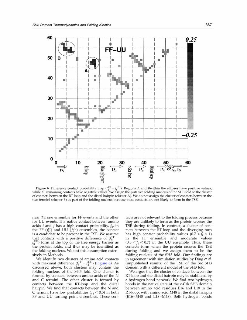
We identify two clusters of amino acid contactswith maximal difference ðf FFij 2 fUU
ij Þ (Figure 6). Asdiscussed above, both clusters may contain thefolding nucleus of the SH3 fold. One cluster isformed by contacts between amino acids of the Nand C termini. The other cluster is formed bycontacts between the RT-loop and the distalhairpin. We find that contacts between the N andC termini have low probabilities ( fij , 0.5) in bothFF and UU turning point ensembles. These con-
tacts are not relevant to the folding process becausethey are unlikely to form as the protein crosses theTSE during folding. In contrast, a cluster of con-tacts between the RT-loop and the diverging turnhas high contact probability values (0.7 , fij , 1)in the FF ensemble and moderate values(0.5 , fij , 0.7) in the UU ensemble. Thus, thesecontacts form when the protein crosses the TSEduring folding and we assign them to be thefolding nucleus of the SH3 fold. Our findings arein agreement with simulation studies by Ding et al.(unpublished results) of the TSE of the Src SH3domain with a different model of the SH3 fold.We argue that the cluster of contacts between the
RT-loop and the distal hairpin may be stabilized bya hydrogen bond network. We find two hydrogenbonds in the native state of the c-Crk SH3 domainbetween amino acid residues E16 and L18 in theRT-loop, with amino acid M48 in the distal hairpin(E16–M48 and L18–M48). Both hydrogen bonds
Figure 6. Difference contact probability map ðf FFij 2 fUUij Þ: Regions A and Bwithin the ellipses have positive values,
while all remaining contacts have negative values. We assign the putative folding nucleus of the SH3 fold to the clusterof contacts between the RT-loop and the distal hairpin (cluster A). We do not assign the cluster of contacts between thetwo termini (cluster B) as part of the folding nucleus because these contacts are not likely to form in the TSE.
SH3 Domain Thermodynamics and Folding Kinetics 867
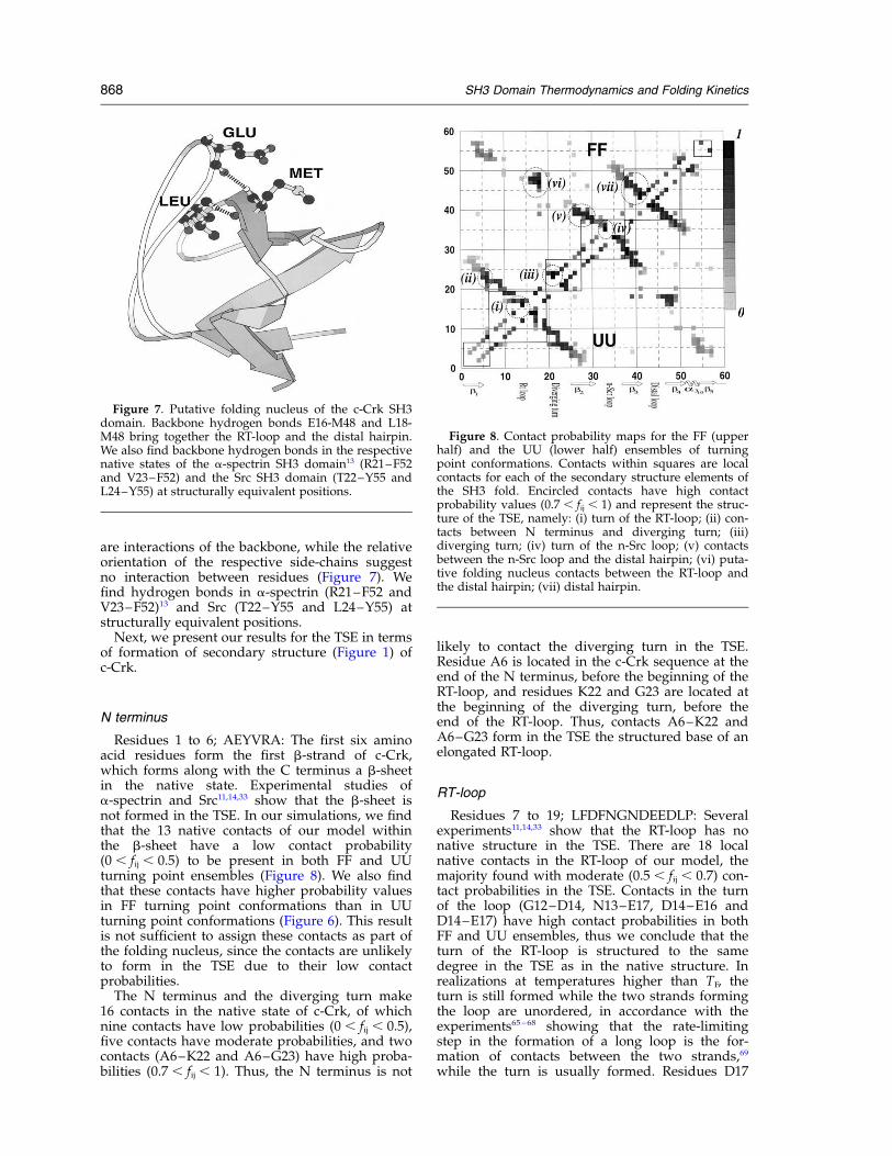
are interactions of the backbone, while the relativeorientation of the respective side-chains suggestno interaction between residues (Figure 7). Wefind hydrogen bonds in a-spectrin (R21–F52 andV23–F52)13 and Src (T22–Y55 and L24–Y55) atstructurally equivalent positions.
Next, we present our results for the TSE in termsof formation of secondary structure (Figure 1) ofc-Crk.
N terminus
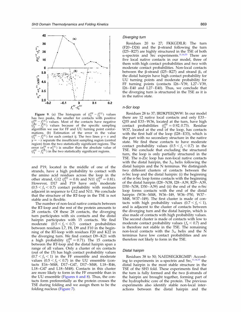
Residues 1 to 6; AEYVRA: The first six aminoacid residues form the first b-strand of c-Crk,which forms along with the C terminus a b-sheetin the native state. Experimental studies ofa-spectrin and Src11,14,33 show that the b-sheet isnot formed in the TSE. In our simulations, we findthat the 13 native contacts of our model withinthe b-sheet have a low contact probability(0 , fij , 0.5) to be present in both FF and UUturning point ensembles (Figure 8). We also findthat these contacts have higher probability valuesin FF turning point conformations than in UUturning point conformations (Figure 6). This resultis not sufficient to assign these contacts as part ofthe folding nucleus, since the contacts are unlikelyto form in the TSE due to their low contactprobabilities.
The N terminus and the diverging turn make16 contacts in the native state of c-Crk, of whichnine contacts have low probabilities (0 , fij , 0.5),five contacts have moderate probabilities, and twocontacts (A6–K22 and A6–G23) have high proba-bilities (0.7 , fij , 1). Thus, the N terminus is not
likely to contact the diverging turn in the TSE.Residue A6 is located in the c-Crk sequence at theend of the N terminus, before the beginning of theRT-loop, and residues K22 and G23 are located atthe beginning of the diverging turn, before theend of the RT-loop. Thus, contacts A6–K22 andA6–G23 form in the TSE the structured base of anelongated RT-loop.
RT-loop
Residues 7 to 19; LFDFNGNDEEDLP: Severalexperiments11,14,33 show that the RT-loop has nonative structure in the TSE. There are 18 localnative contacts in the RT-loop of our model, themajority found with moderate (0.5 , fij , 0.7) con-tact probabilities in the TSE. Contacts in the turnof the loop (G12–D14, N13–E17, D14–E16 andD14–E17) have high contact probabilities in bothFF and UU ensembles, thus we conclude that theturn of the RT-loop is structured to the samedegree in the TSE as in the native structure. Inrealizations at temperatures higher than TF, theturn is still formed while the two strands formingthe loop are unordered, in accordance with theexperiments65–68 showing that the rate-limitingstep in the formation of a long loop is the for-mation of contacts between the two strands,69
while the turn is usually formed. Residues D17
Figure 7. Putative folding nucleus of the c-Crk SH3domain. Backbone hydrogen bonds E16-M48 and L18-M48 bring together the RT-loop and the distal hairpin.We also find backbone hydrogen bonds in the respectivenative states of the a-spectrin SH3 domain13 (R21–F52and V23–F52) and the Src SH3 domain (T22–Y55 andL24–Y55) at structurally equivalent positions.
Figure 8. Contact probability maps for the FF (upperhalf) and the UU (lower half) ensembles of turningpoint conformations. Contacts within squares are localcontacts for each of the secondary structure elements ofthe SH3 fold. Encircled contacts have high contactprobability values (0.7 , fij , 1) and represent the struc-ture of the TSE, namely: (i) turn of the RT-loop; (ii) con-tacts between N terminus and diverging turn; (iii)diverging turn; (iv) turn of the n-Src loop; (v) contactsbetween the n-Src loop and the distal hairpin; (vi) puta-tive folding nucleus contacts between the RT-loop andthe distal hairpin; (vii) distal hairpin.
868 SH3 Domain Thermodynamics and Folding Kinetics
and P19, located in the middle of one of thestrands, have a high probability to contact withthe amino acid residues across the loop in theother strand, G12 ðf FFij ¼ 0:8Þ and N11 ðf FFij ¼ 0:81Þ:However, D17 and P19 have only moderate(0.5 , fij , 0.7) contact probability with residuesadjacent in sequence to G12 and N11. We concludethat the structure of the RT-loop in the TSE is notstable and is flexible.
The number of non-local native contacts betweenthe RT-loop and the rest of the protein amounts to28 contacts. Of these 28 contacts, the divergingturn participates with six contacts and the distalhairpin participates with 15 contacts. We findmoderate (0.5 , fij , 0.7) contact probabilitiesbetween residues L7, F8, D9 and F10 in the begin-ning of the RT-loop with residues F20 and K22 inthe diverging turn. We find contact D9–K21 witha high probability ðf FFij ¼ 0:71Þ. The 15 contactsbetween the RT-loop and the distal hairpin span arange of all values. Only a cluster of six contacts(out of the 15) has high contact probability values(0.7 , fij , 1) in the FF ensemble and moderatevalues (0.5 , fij , 0.7) in the UU ensemble (con-tacts E16–M48, D17–G47, D17–M48, L18–R46,L18–G47 and L18–M48). Contacts in this clusterare more likely to form in the FF ensemble than inthe UU ensemble (Figures 6 and 8). Thus, the con-tacts form preferentially as the protein crosses theTSE during folding and we assign them to be thefolding nucleus (Figure 7).
Diverging turn
Residues 20 to 27; FKKGDILR: The turn(F20–D24) and the b-strand following the turn(I25–R27) are highly structured in the TSE of botha-spectrin and Src experiments.11,14,33 There arefive local native contacts in our model, three ofthem with high contact probabilities and two withmoderate contact probabilities. Non-local contactsbetween the b-strand (I25–R27) and strand b3 ofthe distal hairpin have high contact probability forUU turning points and moderate probability forFF turning points (contacts I26–V39, L27–V39,I26–E40 and L27–E40). Thus, we conclude thatthe diverging turn is structured in the TSE as it isin the native state.
n-Scr loop
Residues 28 to 37; IRDKPEEQWW: In our modelthere are 12 native local contacts and only E33–Q35 and E33–W36, located at the turn, have highcontact probabilities ðf FFij ¼ 0:92; 0:73Þ: ResidueW37, located at the end of the loop, has contactswith the first half of the loop (I28–E33), which isthe part with no secondary structure in the nativestate. We find these contacts to have moderatecontact probability values (0.5 , fij , 0.7) in theTSE. We conclude that excluding the structuredturn, the loop is only partially structured in theTSE. The n-Zrc loop has non-local native contactswith the distal hairpin, the 310 helix following thedistal hairpin and the N terminus. We distinguishtwo different clusters of contacts between then-Src loop and the distal hairpin: (i) the beginningof the n-Src loop forms contacts with the beginningof the distal hairpin (I28–N38, I28–A39, R29–A39,D30–N38, D30–A39) and (ii) the end of the n-Srcloop forms contacts with the end of the distalhairpin (W36–M48, W36–I49, W36–P50, W37–M48, W37–I49). The first cluster is made of con-tacts with high probability values (0.7 , fij , 1),and is adjacent to the cluster of contacts betweenthe diverging turn and the distal hairpin, which isalso made of contacts with high probability values.The second cluster is made of contacts with low tomoderate contact probability values ( fij , 0.7) andis therefore not stable in the TSE. The remainingnon-local contacts with the 310 helix and the Nterminus have low contact probabilities and aretherefore not likely to form in the TSE.
Distal hairpin
Residues 38 to 50; NAEDSEGKRGMIP: Accord-ing to experiments in a-spectrin and Src,11,14,33 thedistal hairpin is the most stable structure in theTSE of the SH3 fold. These experiments find thatthe turn is fully formed and the two b-strands ofthe hairpin are brought together, forming part ofthe hydrophobic core of the protein. The previousexperiments also identify stable non-local inter-actions between the distal hairpin and the
Figure 9. (a) The histogram of ðf FFij 2 fUUij Þ values
has two peaks, the smaller for contacts with positiveðf FFij 2 fUU
ij Þ values. Most of the contacts have negativeðf FFij 2 fUU
ij Þ values because of the specific samplingalgorithm we use for FF and UU turning point confor-mations. (b) Estimation of the error in the valueðf FFij 2 fUU
ij Þ for each contact ij. The two lines y ¼ x andy ¼ 2x separate the insufficient sampling region (centralregion) from the two statistically significant regions. Theerror ðsFF
ij þ sUUij Þ is smaller than the absolute value of
ðf FFij 2 fUUij Þ in the two statistically significant regions.
SH3 Domain Thermodynamics and Folding Kinetics 869
diverging turn and partially stable interactionswith the n-Src loop. From our simulations, outof a total of 18 local native contacts, we find 12contacts with high contact probability values(0.7 , fij , 1) and four contacts with moderatevalues (0.5 , fij , 0.7). The turn (S42, E43 andG44) has the highest values, making the turn themost stable fragment of the hairpin. Thus, we findthat the hairpin is the most stable fragment of thesecondary structure in the TSE of the SH3 fold.Non-local native contacts with the diverging turn(11 contacts), the n-Scr loop (13 contacts) and theRT-loop (13 contacts) have already been discussedin the previous respective subsections.
C terminus
Residues 54 to 57; VEKY: The C terminus makescontacts only with the N terminus, already dis-cussed in the N terminus subsection.
Discussion
Error analysis
We address the question of whether our numeri-cal results for the different ðf FFij 2 fUU
ij Þ values arestatistically significant or if they are the result ofinsufficient sampling. First, we compute the histo-gram of ðf FFij 2 fUU
ij Þ values, which has two peaks,the smaller one corresponding to the positivevalues of ðf FFij 2 fUU
ij Þ (Figure 9(a)). Next, we
estimate the expected error for each ðf FFij 2 fUUij Þ
value. We assume that the presence of a specificcontact ij at any time is a random variable, withan output of 0 or 1. We analyze uncorrelated FFand UU events separated by at least an interval of2500 time units, which is longer than the typicalfolding transition time. We collect NFF ¼ 153 FFevents and NUU ¼ 181 UU events, then estimatethe error in ðf FFij 2 fUU
ij Þ as sFFij þ sUU
ij ; where
sFFðUUÞij ¼
pðf FFðUUÞij ð12 f FFðUUÞ
ij Þ=NFFðUUÞÞ: We define
ðf FFij 2 fUUij Þ to be statistically significant if lf FFij 2
fUUij l . sFF
ij þ sUUij and find ðf FFij 2 fUU
ij Þ . 0.06 to be
statistically significant (Figure 9(b)).
pFOLD Analysis
We test the hypothesis that the FF and UUensembles have structural similarities with theTSE in two complimentary tests. In the first test,we assume that the energy is the reaction coordi-nate for the folding–unfolding transition. Thisassumption is supported by the bimodality of thepotential energy histogram at TF (Figure 3). Wethen estimate the TSE as the set of conformationswith energies within a narrow energy windowcentered in the minimum of the histogram, Emin ¼282: According to this criterion, both FF and UUensembles are candidates to represent the TSE(Figure 3). Even though we accept FF turningpoint conformations with energies far above theminimum of the histogram ðEth;f ¼ 256Þ; we do
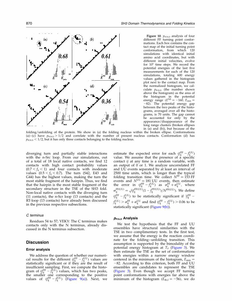
Figure 10. pFOLD analysis of fourdifferent FF turning point confor-mations. Each box contains the con-tact map of the initial turning pointconformation, from which 120simulations with identical initialamino acid coordinates, but withdifferent initial velocities, evolvefor 104 time steps. We record thepotential energies of the last fivemeasurements for each of the 120simulations, totaling 600 energyvalues gathered in the histogramplot next to the contact map. Fromthe normalized histogram, we cal-culate pFOLD (the number shownabove the histogram) as the area ofthe histogram in the potentialenergy range ðENS ¼ 2160; Emin ¼282Þ: The potential energy gapbetween the two peaks of the histo-grams, averaged over all the histo-grams, is 70 units. The gap cannotbe accounted for only by theappearance/disappearance of thelong range clusters (broken ellipsesin (a) and (b)), but because of the
folding/unfolding of the protein. We show in (a) the folding nucleus within the broken ellipse. Conformations(a)–(c) have pFOLD . 1/2 and correlate with the number of present nucleus contacts. Conformation (d) haspFOLD , 1/2, but it has only three contacts belonging to the folding nucleus.
870 SH3 Domain Thermodynamics and Folding Kinetics
not find any FF turning point with an energybigger than E ¼ 268; out of an ensemble of 2620FF turning point conformations. We retrieve 95%of the FF ensemble with an energy threshold evencloser to Emin, Eth,f ¼ 276. We also find analogousresults for the UU ensemble (Figure 3).
The second test is to apply the pFOLD criterionaccording to which a conformation belongs to theTSE if it has a probability pFOLD ¼ 1=2 to evolveeither to the folded or unfolded state. If a turningpoint conformation is structurally similar to theTSE conformations, then pFOLD is close to 1/2.Moreover, if the turning point conformationbelongs to a FF event, the nucleus is likely to bepresent and we expect pFOLD to be larger than 1/2.Analogously, if the turning point conformationbelongs to a UU event, the nucleus is likely to beabsent and we expect pFOLD to be less than 1/2.Thus, pFOLD is a measure of the presence or absenceof the folding nucleus in a turning pointconformation.
The pFOLD analysis for one turning point confor-mation has five steps: (i) random selection of theturning point conformation; (ii) change of initialconditions by replacing all amino acids and heatbath particle velocities with random velocityvalues from a Maxwell velocity distribution corre-sponding to thermal equilibrium at TF (we performthe velocity replacement 120 times, creating 120different initial conditions); (iii) subsequent timeevolution for 104 time steps, which is above thetypical time of folding or unfolding; (iv) collectthe potential energy values of the last fivemeasurements, giving a total of 5 £ 120 ¼ 600energy values that we plot in a normalized histo-gram; (v) compute pFOLD as the area below the
histogram curve in the range of potential energies(ENS ¼ 2160, Emin ¼ 282).We compute an estimate of the average pFOLD for
the FF ensemble, kpFOLDlFF; with the analysis of 16randomly selected FF turning point conformationstotaling 16 £ 120 ¼ 1920 different conformationsplus initial conditions. Once simulations are over,we find that 1113 fold and 807 unfold, thuskpFOLDlFF ¼ 0:58: The same analysis on 13 ran-domly selected UU turning point conformationsproduces 1560 different conformations plus initialconditions, of which 622 fold and 938 unfold, thuskpFOLDlUU ¼ 0:40: The difference of kpFOLDl withrespect to 1/2 states the preference of the FF (UU)ensemble toward the folded (unfolded) state. Asexpected, we find that both pFOLD values are closeto 1/2, indicating the closeness of both FF and UUensembles to the TSE.In addition, pFOLD analyses quantify the ability of
the various structural elements of the TSE to biasthe protein toward the folded or the unfoldedstates. For this study, we randomly select eight FFturning point conformations and eight UU turningpoint conformations with different contact mapsand perform pFOLD analysis for each of them.We find seven out of the eight FF turning point
conformations with pFOLD $ 1=2 (Figure 10), andthe correlation coefficient between pFOLD and thenumber of present folding nucleus contacts is 0.98.The coefficient drops to 0.86 when we include theturning point conformation with pFOLD , 1/2(Figure 10(d)). A similar analysis of pFOLD versusnumber of present contacts between the twotermini gives a correlation coefficient of 0.0. In theanalogous analysis of the UU ensemble, we findtwo turning point conformations with pFOLD . 1/2.
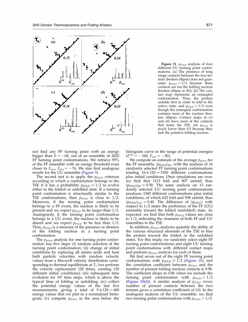
Figure 11. pFOLD analysis of fourdifferent UU turning point confor-mations. (a) The presence of longrange contacts between the two ter-mini (broken ellipse) does not guar-antee pFOLD . 1/2 because thesecontacts are not the folding nucleus(broken ellipse in (b)). (b) The con-tact map represents an entangledconformation. Thus, the proteinunfolds first in order to fold to thenative state, and pFOLD , 1/2 eventhough the entangled conformationcontains most of the nucleus (bro-ken ellipse). Contact maps in (c)and (d) have most of the contactsthat make the TSE, yet pFOLD ismuch lower than 0.5 because theylack the putative folding nucleus.
SH3 Domain Thermodynamics and Folding Kinetics 871
The two conformations with pFOLD . 1/2 containthe folding nucleus, thus belonging rather to theFF ensemble than to the UU ensemble. We findthat conformations with no folding nucleuscontacts and with no contacts between terminihave the lowest pFOLD values (Figure 11). It is inter-esting to observe that one of the eight UU turningpoint conformations contains most of the foldingnucleus contacts, yet pFOLD equals only 0.24 (Figure11(b)). Inspection of the structure reveals that thisconformation is entangled and it is necessary forthe protein to unfold first in order to fold to thenative state. In summary, pFOLD analysis confirmsthat the putative folding nucleus plays an impor-tant role in the folding transition.
Cross-linking of folding nucleus
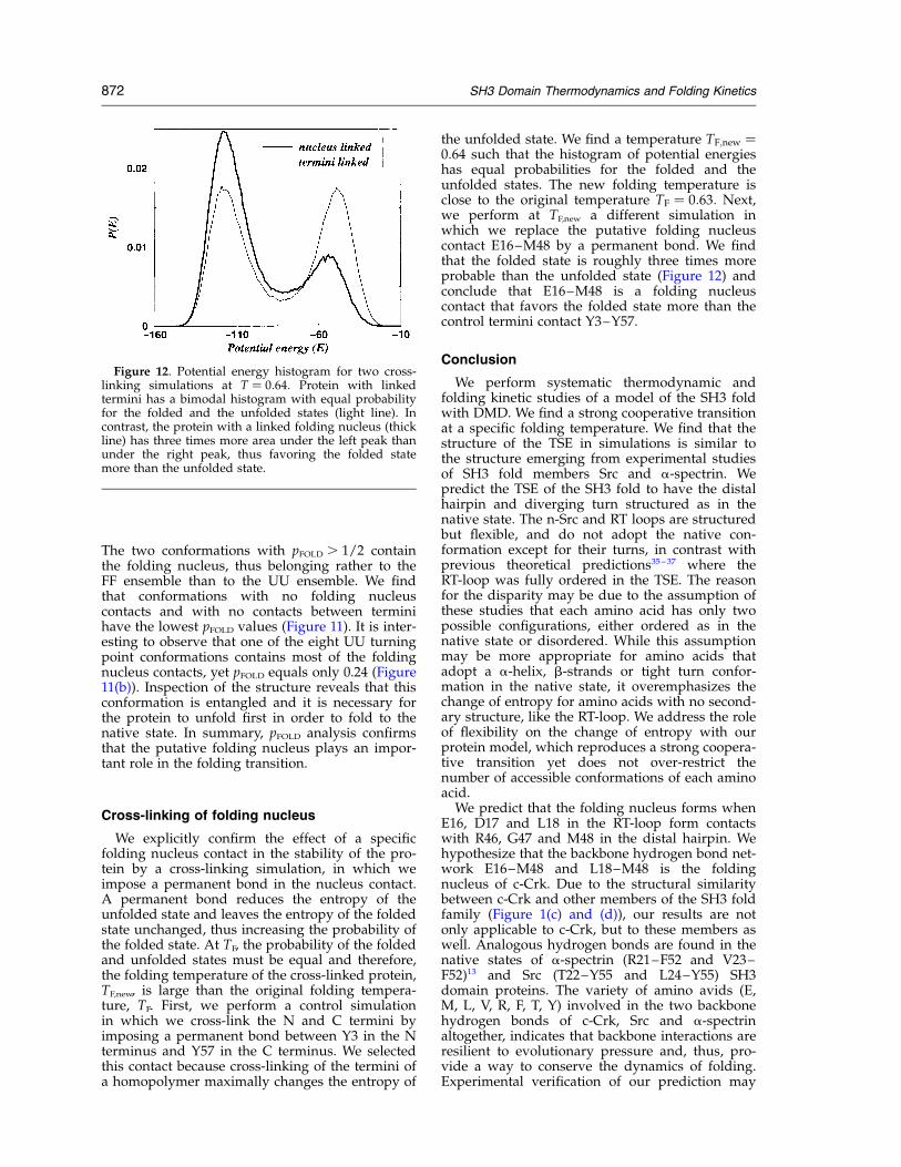
We explicitly confirm the effect of a specificfolding nucleus contact in the stability of the pro-tein by a cross-linking simulation, in which weimpose a permanent bond in the nucleus contact.A permanent bond reduces the entropy of theunfolded state and leaves the entropy of the foldedstate unchanged, thus increasing the probability ofthe folded state. At TF, the probability of the foldedand unfolded states must be equal and therefore,the folding temperature of the cross-linked protein,TF,new, is large than the original folding tempera-ture, TF. First, we perform a control simulationin which we cross-link the N and C termini byimposing a permanent bond between Y3 in the Nterminus and Y57 in the C terminus. We selectedthis contact because cross-linking of the termini ofa homopolymer maximally changes the entropy of
the unfolded state. We find a temperature TF;new ¼0:64 such that the histogram of potential energieshas equal probabilities for the folded and theunfolded states. The new folding temperature isclose to the original temperature TF ¼ 0:63: Next,we perform at TF,new a different simulation inwhich we replace the putative folding nucleuscontact E16–M48 by a permanent bond. We findthat the folded state is roughly three times moreprobable than the unfolded state (Figure 12) andconclude that E16–M48 is a folding nucleuscontact that favors the folded state more than thecontrol termini contact Y3–Y57.
Conclusion
We perform systematic thermodynamic andfolding kinetic studies of a model of the SH3 foldwith DMD. We find a strong cooperative transitionat a specific folding temperature. We find that thestructure of the TSE in simulations is similar tothe structure emerging from experimental studiesof SH3 fold members Src and a-spectrin. Wepredict the TSE of the SH3 fold to have the distalhairpin and diverging turn structured as in thenative state. The n-Src and RT loops are structuredbut flexible, and do not adopt the native con-formation except for their turns, in contrast withprevious theoretical predictions35–37 where theRT-loop was fully ordered in the TSE. The reasonfor the disparity may be due to the assumption ofthese studies that each amino acid has only twopossible configurations, either ordered as in thenative state or disordered. While this assumptionmay be more appropriate for amino acids thatadopt a a-helix, b-strands or tight turn confor-mation in the native state, it overemphasizes thechange of entropy for amino acids with no second-ary structure, like the RT-loop. We address the roleof flexibility on the change of entropy with ourprotein model, which reproduces a strong coopera-tive transition yet does not over-restrict thenumber of accessible conformations of each aminoacid.
We predict that the folding nucleus forms whenE16, D17 and L18 in the RT-loop form contactswith R46, G47 and M48 in the distal hairpin. Wehypothesize that the backbone hydrogen bond net-work E16–M48 and L18–M48 is the foldingnucleus of c-Crk. Due to the structural similaritybetween c-Crk and other members of the SH3 foldfamily (Figure 1(c) and (d)), our results are notonly applicable to c-Crk, but to these members aswell. Analogous hydrogen bonds are found in thenative states of a-spectrin (R21–F52 and V23–F52)13 and Src (T22–Y55 and L24–Y55) SH3domain proteins. The variety of amino avids (E,M, L, V, R, F, T, Y) involved in the two backbonehydrogen bonds of c-Crk, Src and a-spectrinaltogether, indicates that backbone interactions areresilient to evolutionary pressure and, thus, pro-vide a way to conserve the dynamics of folding.Experimental verification of our prediction may
Figure 12. Potential energy histogram for two cross-linking simulations at T ¼ 0:64: Protein with linkedtermini has a bimodal histogram with equal probabilityfor the folded and the unfolded states (light line). Incontrast, the protein with a linked folding nucleus (thickline) has three times more area under the left peak thanunder the right peak, thus favoring the folded statemore than the unfolded state.
872 SH3 Domain Thermodynamics and Folding Kinetics
come from an attempt to inhibit formation ofhydrogen bonds. According to our prediction, inhi-bition of formation of the hydrogen bond networkE16–M48, D17–M48 in c-Crk, R21–F52, V23–F52
in a-spectrin and T22–Y55, L24–Y55 in Src wouldreduce the folding rate of these proteins.
Methods
Model
As the unfolded protein approaches the TSE, thenumber of allowed conformations reduces drastically,resulting in an abrupt decrease of entropy.1 A successfulmodel for protein folding must reproduce this abruptentropy reduction. Flexibility is the main factor contri-buting to entropy and thus, it is crucial to define anappropriate set of effective constraints among aminoacids. First, we study the folding transition of the SH3fold with the beads-on-a-string model.25 We find thatthe protein folds into the native state by passing througha series of meta-stable intermediates, giving the beads-on-a-string model a flexibility that does not reproducethe cooperative transition experimentally observed inglobular proteins. Next, we introduce a set of additionalconstraints among amino acids in order to make the fold-ing transition strongly cooperative.
We use the Go model70–72 of interactions (Figure 1(b)),determined with the knowledge of the native topologyof the protein under study. We represent each of the 57amino acid residue by beads centered in their respectiveCb coordinates41 (Ca in case of Gly). We model the pep-tide bond between to neighboring amino acids i andi þ 1 along the sequence by a narrow, infinitely highpotential well (Figure 13(a)), so that the bond length canfluctuate within 2% of the distance in the native state,dNSi;iþ1; between the Cb coordinates of the two aminoacids. We define a contact between two non-bondedamino acids i and j to be a native contact when the dis-tance between their respective Cb0 is in the native state,dNSij ; is smaller than some predefined cut-off distance D.We use Ca in case of Gly. We plot the resulting set ofnative contacts on the contact map (Figure 1). We modela native contact between amino acids i and j by an attrac-tive square-well potential53,70,71 of depth 1 with a hard-core distance 0:99 £ dNS
ij and an attraction distance D
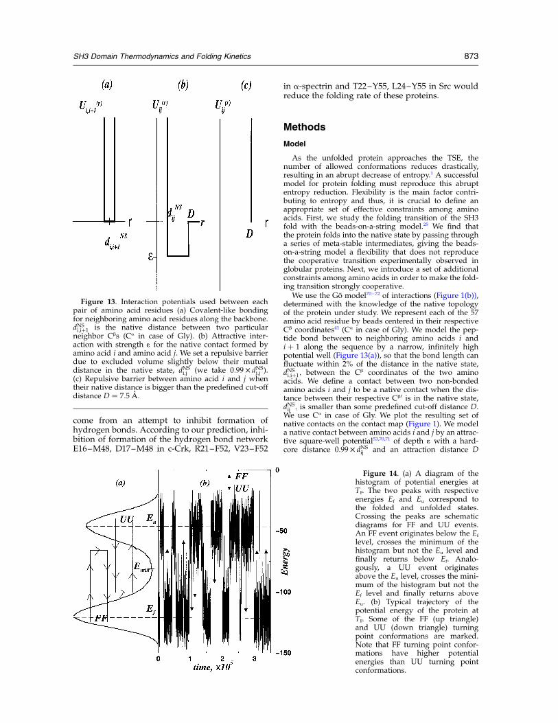
Figure 13. Interaction potentials used between eachpair of amino acid residues (a) Covalent-like bondingfor neighboring amino acid residues along the backbone.dNSi;iþ1 is the native distance between two particularneighbor Cbs (Ca in case of Gly). (b) Attractive inter-action with strength 1 for the native contact formed byamino acid i and amino acid j. We set a repulsive barrierdue to excluded volume slightly below their mutualdistance in the native state, dNS
i;j (we take 0:99 £ dNSi;j ).
(c) Repulsive barrier between amino acid i and j whentheir native distance is bigger than the predefined cut-offdistance D ¼ 7:5 �A:
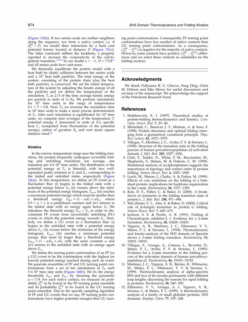
Figure 14. (a) A diagram of thehistogram of potential energies atTF. The two peaks with respectiveenergies Ef and Eu correspond tothe folded and unfolded states.Crossing the peaks are schematicdiagrams for FF and UU events.An FF event originates below the Ef
level, crosses the minimum of thehistogram but not the Eu level andfinally returns below Ef. Analo-gously, a UU event originatesabove the Eu level, crosses the mini-mum of the histogram but not theEf level and finally returns aboveEu. (b) Typical trajectory of thepotential energy of the protein atTF. Some of the FF (up triangle)and UU (down triangle) turningpoint conformations are marked.Note that FF turning point confor-mations have higher potentialenergies than UU turning pointconformations.
SH3 Domain Thermodynamics and Folding Kinetics 873
(Figure 13(b)). If two amino acids are neither neighborsalong the sequence, nor form a native contact, i.e. ifdNSij . D; we model their interaction by a hard corepotential barrier located at distance D (Figure 13(c)).The latter constraint stiffens the backbone, a propertyreported to increase the cooperativity of the coil-to-globule transition.1,73,74 In our model 1 ¼ 21; D ¼ 7:5 �A75
and all amino acids have unit mass.We thermally equilibrate the protein model with a
heat bath by elastic collisions between the amino acidsand a 103 heat bath particles. The total energy of thesystem, consisting of the protein chain plus the heatbath particles, is conserved. We set the initial tempera-ture of the system by adjusting the kinetic energy of allthe particles and we define the temperature of thesimulation, T, as 2/3 of the time average kinetic energyper particle in units of l1l=kB: We perform simulationsfor 105 time units in the range of temperatures0.1 , T , 0.8. Near TF, we increase the simulation timeto 106 time units to make a more precise determinationof TF. After each simulation is equilibrated for 104 timeunits, we compute time averages of the temperature T,potential energy E (measured in units of l1l), specificheat Cv (computed from fluctuations of the potentialenergy), radius of gyration Rg and root mean squaredistance rmsd.64
Kinetics
In the narrow temperature range near the folding tran-sition, the protein frequently undergoes reversible fold-ing and unfolding transitions (on average, onetransition per 4 £ 105 time units). The distribution of thepotential energy at TF is bimodal, with two well-separated peaks centered at Ef and Eu, corresponding tothe folded and unfolded states, respectively (Figure14(a)). At this temperature, we define an FF event as afluctuation that: (i) begins in the folded state with apotential energy below Ef; (ii) crosses above the mini-mum of the potential energy histogram, Emin; (iii) reachesa maximum potential energy which must be smaller thana threshold energy Eth;f ¼ ð12 aÞEf þ aEu; where0.5 , a , 1 is a predefined constant and (iv) returns tothe folded state with an energy again below Ef. Weintroduce the thresold energy Eth,f , Eu in order to dis-criminate FF events from successfully unfolding (FU)events in which the potential energy exceeds Eu. Simi-larly, we define a UU event as a fluctuation that: (i)begins in the unfolded state with a potential energyabove Eu; (ii) crosses below the minimum of the energyhistogram, Emin; (iii) reaches a minimum potentialenergy, that must be larger than a threshold energyEth;u ¼ ð12 aÞEu þ aEf with the same constant a and(iv) returns to the unfolded state with an energy againabove Eu.
We define the turning point conformation of an FF (ora UU) event to be the confirmation with the highest (orlowest) potential energy reached during such an event.We generate ensembles of FF and UU turning point con-formations from a set of ten simulations at TF, each5 £ 106 time step units (Figure 14(b)). We fix the energythresholds Eth,f and Eth,u by choosing the parametera ¼ 7/8. For each native contact, we measure its prob-ability f FFij to be found in the FF turning point ensembleand its probability fUU
ij to be found in the UU turningpoint ensemble. Due to the specific sampling algorithmof FF and UU events that we use, FF turning point con-formations have higher potential energies that UU turn-
ing point conformations. Consequently, FF turning pointconformations have less number of native contacts thanUU turning point conformations. As a consequence,ð f FFij 2 fUU
ij Þ is negative for the majority of native contacts.However, some contacts have positive ð f FFij 2 fUU
ij Þ differ-ences and we select these contacts as candidates for thefolding nucleus.
Acknowledgments
We thank Pollyanna R. G. Chavez, Feng Ding, ChrisM. Dobson and Ilike Merey for useful discussions andrevision of the manuscript. We acknowledge the supportof the Petroleum Research Fund.
References
1. Shakhnovich, E. I. (1997). Theoretical studies ofprotein-folding thermodynamics and kinetics. Curr.Opin. Struct. Biol. 7, 29–40.
2. Micheletti, C., Banavar, J. T., Martian, A. & Seno, F.(1998). Protein structures and optimal folding emer-ging from a geometrical variational principle. Phys.Rev. Letters, 82, 3372–3375.
3. Villegas, V., Martinez, J. C., Aviles, F. X. & Serrano, L.(1998). Structure of the transition state in the foldingprocess of human procarboxypeptidase a2 activationdomain. J. Mol. Biol. 283, 1027–1036.
4. Chiti, F., Taddei, N., White, P. M., Bucciantini, M.,Megherini, F., Stefani, M. & Dobson, C. M. (1999).Mutational analysis of acylphosphatase suggests theimportance of topology and contact order in proteinfolding. Nature Struct. Biol. 6, 1005–1009.
5. Lorch, M., Mason, J., Clarke, A. & Parker, M. (1999).Effects of core mutations on the folding of a beta-sheet protein: implications for backbone organizationin the i-state. Biochemistry, 38, 1377–1385.
6. Kim, D. E., Fisher, C. & Baker, D. (2000). A break-down of symmetry in the folding transition state ofprotein L. J. Mol. Biol. 298, 971–984.
7. McCallister, E. L., Alm, E. & Baker, D. (2000). Criticalrole of b-hairpin formation in protein G folding.Nature Struct. Biol. 7, 669–673.
8. Jackson, S. E. & Fersht, A. R. (1991). Folding ofChymotrypsin inhibitor-2. I. Evidence for a 2-statetransition. Biochemistry, 30, 10428–10435.
9. Viguera, A. R., Martinez, J. C., Filimonov, V. V.,Mateo, P. L. & Serrano, L. (1994). Thermodynamicand kinetic-analysis of the SH3 domain of Spectrinshows a 2-state folding transition. Biochemistry, 33,10925–10933.
10. Villegas, V., Azuaga, A., Catasus, L., Reverter, D.,Mateo, P. L., Aviles, F. X. & Serrano, L. (1995).Evidence for a 2-state transition in the folding pro-cess of the activation domain of human procarboxy-peptidase-a2. Biochemistry, 46, 15105–15110.
11. Martinez, J. C., Viguera, A. R., Berisio, R., Wilmanns,M., Mateo, P. L., Filimonov, V. V. & Serrano, L.(1999). Thermodynamic analysis of alpha-spectrinSH3 and two of its circular permutants with differentloop lengths: discerning the reasons for rapid foldingin proteins. Biochemistry, 38, 549–559.
12. Filimonov, V. V., Azuaga, A. I., Viguera, A. R.,Serrano, L. & Mateo, P. L. (1999). A thermodynamicanalysis of a family of small globular proteins: SH3domains. Biophys. Chem. 77, 195–208.
874 SH3 Domain Thermodynamics and Folding Kinetics
13. Viguera, A. R., Serrano, L. & Wilmanns, M. (1996).Different folding transition states may result inthe same native structure. Nature Struct. Biol. 10,874–880.
14. Grantcharova, V. P., Riddle, D. S., Santiago, J. N. &Baker, D. (1998). Important role of hydrogen bondsin the structurally polarized transition state forfolding of the src SH3 domain. Nature Struct. Biol. 8,714–720.
15. Grantcharova, V. P. & Baker, D. (1997). Foldingdynamics of the Src SH3 domain. Biochemistry, 36,15685–15692.
16. Otzen, D. E., Itzhaki, L. S., Elmasry, N. F., Jackson,S. E. & Fersht, A. R. (1994). Structure of the tran-sition-state for the folding/unfolding of the barleyChymotrypsin inhibitor-2 and its implications formechanisms of protein-folding. Proc. Natl Acad. Sci.USA, 22, 10426–10429.
17. Knapp, S., Mattson, P. T., Christova, P., Berndt, K. D.,Karshikoff, A., Vihinen, M. et al. (1998). Thermalunfolding of small proteins with SH3 domain foldingpattern. Proteins: Struct. Funct. Genet. 23, 309–319.
18. Jackson, S. E., Elmasry, N. & Fersht, A. R. (1993).Structure of the hydrophobic core in the transition-state for folding of Chymotrypsin inhibitor-2: acritical test of the protein engineering method ofanalysis. Biochemistry, 32, 11270–11278.
19. Shakhnovich, E. I., Abkevich, V. I. & Ptitsyn, O.(1996). Conserved residues and the mechanism ofprotein folding. Nature, 379, 96–98.
20. Abkevich, V. I., Gutin, A. M. & Shakhnovich, E. I.(1994). Specific nucleus as the transition state forprotein folding: evidence from the lattice model.Biochemistry, 33, 10026–10036.
21. Pande, V. S., Grosberg, A. Y., Tanaka, T. & Rokhsar,D. S. (1998). Pathways for protein folding: is a newview needed? Curr. Opin. Struct. Biol. 1, 68–79.
22. Mirny, L. A., Abkevich, V. I. & Shakhnovich, E. I.(1998). How evolution makes proteins fold quickly.Proc. Natl Acad. Sci. USA, 95, 4976–4981.
23. Shakhnovich, E. I. (1998). Folding nucleus: specific ormultiple? Folding Des. 3, R108–R111.
24. Gutin, A. M., Abkevich, V. I. & Shakhnovich, E. I.(1998). A protein engineering analysis of the tran-sition state for protein folding: simulation in thelattice model. Folding Des. 3, 459–480.
25. Dokholyan, N. V., Buldyrev, S. V., Stanley, H. E. &Shakhnovich, E. I. (2000). Identifying the proteinfolding nucleus using molecular dynamics. J. Mol.Biol. 296, 1183–1188.
26. Fersht, A. R. (1995). Characterizing transition-statesin protein folding: an essential step in the puzzle.Curr. Opin. Struct. Biol. 1, 79–84.
27. Yu, H., Rosen, M. K., Shin, T. B., Seidel-Dugan, C. &Brugde, J. S. (1992). Solution structure of the SH3domain of Src and identification of its ligand-bindingsite. Science, 258, 1665–1668.
28. Yu, H., Chen, J. K., Feng, S., Dalgarno, D. C. &Brauer, A. W. (1994). Structural basis for the bindingof proline-rich peptides to SH3 domains. Cell, 76,933–945.
29. Feng, S., Chen, J. K., Yu, H., Simons, J. A. & Schreiber,S. L. (1994). Two binding orientations for peptides tothe Src SH3 domain: development of a generalmodel for SH3–ligand interactions. Science, 266,1241–1247.
30. Feng, S., Kasahara, C., Rickles, R. J. & Schreiber, S. L.(1995). Specific interactions outside the proline-rich
core of two classes of Src homology 3 ligands. Proc.Natl Acad. Sci. USA, 92, 12408–12415.
31. Combs, A. P., Kapoor, T. M., Feng, S. & Chen, J. K.(1996). Protein structure-based combinatorialchemistry: discovery of non-peptide bindingelements to Src SH3 domain. J. Am. Chem. Soc. 118,287–288.
32. Feng, S. B., Kapoor, T. M., Shirai, F., Combs, A. P. &Schreiber, S. L. (1996). Molecular basis for the bind-ing of SH3 ligands with non-peptide elementsidentified by combinatorial synthesis. Chem. Biol. 8,661–670.
33. Riddle, D. S., Grantcharova, V. P., Santiago, J. V.,Ruczinski, A. E. & Baker, D. (1999). Experiment andtheory highlight role of native state topology in SH3folding. Nature Struct. Biol. 6, 1016–1024.
34. Musacchio, A., Noble, M., Pauptit, R., Wierenga, R. &Saraste, M. (1992). Crystal-structure of a src-hom-ology-3 (SH3) domain. Nature, 359, 851.
35. Munoz, V. & Eaton, W. A. (1999). A simple model forcalculating the kinetics of protein folding from three-dimensional structures. Proc. Natl Acad. Sci. USA, 96,11311–11316.
36. Galzitskaya, O. V. & Finkelstein, A. V. (1999). Atheoretical search for folding/unfolding nuclei inthree-dimensional protein structures. Proc. NatlAcad. Sci. USA, 96, 11299–11304.
37. Alm, E. & Baker, D. (1999). Prediction of protein-folding mechanisms from free-energy landscapesderived from native structures. Proc. Natl Acad. Sci.USA, 96, 11305–11310.
38. Guerois, R. & Serrano, L. (2000). The SH3-foldfamily: experimental evidence and prediction ofvariations in the folding pathways. J. Mol. Biol. 304,967–982.
39. Wu, X. D., Knudsen, B., Feller, S. M., Sali, J.,Cowburn, D. & Hanafusa, H. (1995). Structural basisfor the specific interaction of lysine-containing pro-line-rich peptides with the n-terminal SH3 domainof c-Crk. Structure, 2, 215–226.
40. Branden, C. & Tooze, J. (1999). Introduction to ProteinStructure, Garland Publishing Inc, New York.
41. Berman, H. M., Westbrook, J., Feng, Z., Gilliland, G.,Bhat, T. N., Weissig, H. et al. (2000). The protein databank. Nucl. Acids Res. 28, 235–242.
42. Holm, L. & Sander, C. (1996). Mapping the proteinuniverse. Science, 273, 595–602.
43. Dokholyan, N. V. & Shakhnovich, E. I. (2001). Under-standing hierarchical protein evolution from firstprinciples. J. Mol. Biol. 312, 289–307.
44. Kolinski, A., Galazka, W. & Skolnick, J. (1996). On theorigin of the cooperativity of protein folding: impli-cations from model simulations. Proteins: Struct.Funct. Genet. 26, 271–287.
45. Doye, J. P. K. & Wales, D. J. (1996). On potentialenergy surfaces and relaxation to the globalminimum. J. Chem. Phys. 105, 8428–8445.
46. Abkevich, V. I., Gutin, A. M. & Shakhnovich, E. I.(1995). Domains in folding of model proteins. ProteinSci. 4, 1167–1177.
47. Abkevich, V. I., Gutin, A. M. & Shakhnovich, E. I.(1994). Free-energy landscape for protein-foldingkinetic: intermediates, traps, and multiple pathwaysin theory and lattice model simulations. J. Chem.Phys. 7, 6052–6062.
48. Shakhnovich, E. I. (1994). Proteins with selectedsequences fold into unique native conformation.Phys. Rev. Letters, 72, 3907–3910.
SH3 Domain Thermodynamics and Folding Kinetics 875
49. Hao, M. H. & Scheraga, H. A. (1994). Monte Carlosimulation of a first-order transition for protein-folding. J. Phys. Chem. 98, 4940–4948.
50. Camacho, C. J. & Thirumalai, D. (1993). Kinetics andthermodynamics of folding in model proteins. Proc.Natl Acad. Sci. USA, 90, 6369–6372.
51. Skolnick, J., Kolinski, A., Brooks, C. L., Godzik, A. &Rey, A. (1993). A method for predicting protein-structure from sequence. Curr. Biol. 3, 414–423.
52. Sali, A., Shakhnovich, E. & Karplus, M. (1994). Howdoes a protein fold. Nature, 6477, 248–251.
53. Dokholyan, N. V., Buldyrev, S. V., Stanley, H. E. &Shakhnovich, E. I. (1998). Molecular dynamicsstudies of folding of a protein-like model. FoldingDes. 3, 577–587.
54. Clementi, C., Nymeyer, H. & Onuchic, J. N. (2000).Topological and energetic factors: what determinesthe structural details of the transition state ensembleand en-route intermediates for protein folding? Aninvestigation for small globular proteins. J. Mol. Biol.5, 937–953.
55. Irback, A. & Schwarze, H. (1995). Sequence depen-dence of self-interacting random chains. J. Phys. A:Math. Gen. 28, 2121–2132.
56. Berriz, G. F., Gutin, A. M. & Shakhnovich, E. I.(1997). Cooperativity and stability in a Langevinmodel of protein like folding. J. Chem. Phys. 106,9276–9285.
57. Guo, Z. & Brooks, C. L. (1997). Thermodynamics ofprotein folding: a statistical mechanical study of asmall all-b protein. Biopolymers, 42, 745–757.
58. Zhou, Y. Q., Karplus, M., Wichert, J. M. & Hall,C. K. (1997). Equilibrium thermodynamics of homo-polymers and clusters: molecular dynamics andMonte Carlo simulations of systems with square-well interactions. J. Chem. Phys. 24, 10691–10708.
59. Alder, B. J. & Wainwright, T. E. (1959). Studies inmolecular dynamics. I. General method. J. Chem.Phys. 31, 459–466.
60. Allen, M. P. & Tildesley, D. J. (1987). ComputerSimulation of Liquids, Clarendon Press, Oxford.
61. Rapaport, D. C. (1997). The Art Of Molecular DynamicsSimulation, Cambridge University Press, Cambridge.
62. Grosberg, A. Y. & Khokhlov, A. R. (1997). GiantMolecules, Academic Press, Boston, MA.
63. Doi, M. (1997). Introduction to Polymer Physics, OxfordUniversity Press, New York.
64. Kabsch, W. (1978). A discussion of the solution forthe best rotation to relate two sets of vectors. ActaCrystallog. sect. A, 34, 327–380.
65. Matouschek, A., Serrano, L. & Fersht, A. R. (1992).The folding of an enzyme. IV. Structure of an inter-mediate in the refolding of barnase analyzed by aprotein engineering procedure. J. Mol. Biol. 224,819–835.
66. Matouschek, A., Serrano, L., Meiering, E. M., Bycroft,M. & Fersht, A. R. (1992). The folding on an enzyme.V. H/H-2 exchange nuclear-magnetic-resonancestudies on the folding pathway of barnase: comple-mentarity to and agreement with protein engineeringstudies. J. Mol. Biol. 224, 837–845.
67. Serrano, L., Kellis, J. T., Cann, P., Matouschek, A. &Fersht, A. R. (1992). The folding of an enzyme. II.Substructure of barnase and the contribution ofdifferent interactions to protein stability. J. Mol. Biol.224, 783–804.
68. Serrano, L., Matouschek, A. & Fersht, A. R. (1992).The folding of an enzyme. III. Structure of the tran-sition state for unfolding of barnase analyzed by aprotein engineering. J. Mol. Biol. 224, 805–818.
69. Viguera, A. R., Blanco, F. J. & Serrano, L. (1995). Theorder of secondary structure elements does notdetermine the structure of a protein but does affectits folding kinetics. J. Mol. Biol. 247, 670–681.
70. Taketomi, H., Ueda, Y. & Go, N. (1975). Studies onprotein folding, unfolding and fluctuations by com-puter simulations. Int. J. Pept. Protein Res. 7, 445–459.
71. Go, N. & Abe, H. (1975). Non-interacting local-struc-ture model of folding and unfolding transition inglobular proteins. I. Formulation. Biopolymers, 20,991–1011.
72. Abe, H. & Go, N. (1981). Noninteracting local-struc-ture model of folding and unfolding transition inglobular proteins. ii. Application to two-dimensionallattice proteins. Biopolymers, 20, 1013–1031.
73. Lifshitz, I. M., Grosberg, A. Y. & Khohlov, A. R.(1978). Some problems of statistical physics ofpolymers with volume interactions. Rev. Mod. Phys.50, 683–713.
74. Sfatos, C. M., Gutin, A. M. & Shakhnovich, E. I.(1993). Phase diagram of random copolymers. Phys.Rev. E, 48, 465–475.
75. Jernigan, R. L. & Bahar, I. (1996). Structure-derivedpotentials and protein simulations. Curr. Opin. Struct.Biol. 2, 195–209.
Edited by A. R. Fersht
(Received 5 November 2001; received in revised form 21 February 2002; accepted 21 February 2002)
876 SH3 Domain Thermodynamics and Folding Kinetics