Hip Arthroscopy: Prevalence of Intra-articular Pathologic Findings After Traumatic Injury of the Hip
Upload
independentCategory
view
1download
0
Hum Genet (1985) 71 : 351-357
© Springer-Verlag 1985
Pathologic and nonpathologic variants of the spectrin molecule in two black families with hereditary elliptocytosis
Marie-Christine Lecomte 1, D. Dhermy 1, M. Garbarz 1, C. Feo 2, Huguette Gautero t, Odile Bournier 1, Christiane Picat 1, Isabelle Chaveroche 1, Anna Ester 3, Colette Galand 1, and P. Boivin 1
1 Laboratoire d'Enzymologie des Cellules Sanguines (INSERM U160) H6pital Beaujon, F-92118 Clichy C6dex, France z Institut de Pathologic Cellulaire, INSERM U48, H6pital de Bic~tre, 94 Le Kremlin Bic6tre, France 3 Laboratori d'Hematologia, Hospital Clinic I Provincial de Barcelona, Spain
Summary. Five patients with hereditary elliptocytosis (HE) from two unrelated black families were studied. The patients had prominent elliptocytosis and a decreased erythrocyte re- sistance to heat treatment. In one infant blood smears showed elliptocytosis and poikilocytosis; his erythrocytes fagmented at a lower temperature than those of his mother and sister, both having typical mild HE. Defective dimer-dimer associa- tion was present in all patients. Limited tryptic digestion of spectrin and subsequent analysis by one- and two-dimensional electrophoresis revealed a similar and reproducible decrease in the 80,000-dalton peptide (M domain) and the concomitant appearance of a 46,000-dalton peptide. All the patients had the polymorphism of the spectrin MI domain commonly ob- served in black populations. In addition, modifications rela- tive to the c~III domain were detected; similar variants were found in one black control subject out of 136 and are likely re- lated to a genetic polymorphism of the ctIII domain. No differ- ences were observed between the peptide patterns in the in- fant with poikilocytosis and those of his HE sister and mother.
Introduction
Recent studies of the erythrocyte membrane have shown that red cell morphology and stability depend, in part, on a sub- membranous protein network named "membrane skeleton" (for review see Cohen 1983). This network consists of inter- connected proteins mainly spectrin, actin, and protein 4.1. Spectrin (Sp) is composed of two nonidentical subunits, o, and ]3, twisted around each other to form heterodimers (Shot- ton et al. 1979; Speicher et al. 1980). Restricted tryptic cle- avage of the two chains produces several specific chemical do- mains named c~I to aV, and 13I to 13IV (Speicher et al. 1982). Sp exists in the membrane in a tetrametric or more likely in a higher polymeric form due to head-to-head self-association of heterodimers (Liu and Palek 1980; Morrow et al. 1981); this association involves ctI and J3I domains (Morrow et al. 1980; Hanspal and Ralston 1982; Dhermy et al. 1982).
Offprint requests to: M. C. Lecomte, Laboratoire d'Enzymologie des Cellules Sanguines (INSERM U160) H6pital Beaujon, F-92118 Clichy C6dex, France
Recent results indicate that the abnormal morphology and predisposition to hemolysis observed in such congenital an- emias as hereditary spherocytosis, hereditary elliptocytosis (HE), and hereditary pyropoikilocytosis (HPP) may be associ- ated with molecular defects or deficiencies in skeletal compo- nents (for review see Palek and Lux 1983). HE, which pre- sents several clinical phenotypes, is also a heterogeneous dis- order from a biochemical point of view. HE can be associated with a complete or partial deficiency of protein band 4.1, a de- fective binding of Sp to membrane vesicles (Zail and Coetzer 1984), but in most cases HE is associated with defective Sp self- association and is named type I HE (Liu et al. 1982; Coetzer and Zail 1982; Evans et al. 1983). In one white kindred, this defective Sp self-association was related to a defect within the ~3I domain (Dhermy et al. 1982). In other cases, limited tryptic digestion of Sp has revealed a decreased amount of an 80,000- dalton peptide which corresponds to the uI-domain associated with: (i) a concomitant increase in a 74,000-dalton peptide, HE (SpD c~ v74) in four HE families (Lawler et al. 1984a; Dhermy et al. 1984; Lecomte et al. 1984a); (ii) the presence of an unusual 65,000-dalton peptide, HE (SpD a 1/65) (Lecomte et al. 1984b, 1985; LaMer et al. 1984b) in seven black HE families; (iii) the presence of a 46,000-dalton peptide in one child suffering from HE with infantile poikilocytosis (Lawler et al. 1984a) and a 50,000-dalton variant in two HE black families (Marchesi et al. 1983).
In this paper we report studies of five new HE patients from two unrelated black families with defective dimer- tetramer equilibrium. Peptide maps showed a decrease in the 80,000-dalton peptide and the presence of a 46,000-dalton peptide. The patients had ctII variants commonly observed in the black population. In addition, new variations relative to a I I I domains were observed.
Materials and methods
Clinical materials
Blood samples from normal donors and affected patients were collected in heparin and analyzed within 24 h. The two black families studied originated from the Antilles and were un- related.
352
In family N (Fig. 1) hemolytic anemia was discovered for- tuitously in a male baby, GN, at the age of 2 months. When he was 5 months old (date of our first study) blood smears show- ed predominant elliptocytosis and poikilocytosis with schizo- cytes (5%) and microelliptocytes. Hemoglobin (Hb) was 9.9 g/dl and improved to 11.6 g/dl over a four month period (date of our second study), while the reticulocyte count decreased from 450,000 to 140,000/mm 3. Pronounced elliptocytosis (95%) was observed in his 7-year-old sister (NN). In his mother (YRN), 60% of the cells were slightly elliptical; both sister and mother were not anemic. The father (JCN) was clin- ically and hematologically normal. Detailed hematologic data are summarized in Table 1.
In family B (Fig. 1), EB, a 2-year-old female child, and her mother (CB) presented with typical mild HE with predominant elliptocytosis (80-100%). Red cell indices are summarized in Table 1. The father (SB) was found to be normal.
Thermal sensitivity and morphology of erythrocytes
The thermal sensitivity of patient and control erythrocytes was studied as previously decribed (Zarkowski et al. 1975; Dhermy et al. 1982). The cells were examined by light phase contrast microscopy, after fixation in 1% glutaraldehyde, 5 mM phos- phate buffer, 150 mM NaC1, pH 7.4.
Preparation of erythrocyte membranes The erythrocytes were washed three times in phosphate-buf- fered saline (PBS), pH 8. The ghosts were prepared according
0(I" VARIANT (XII (X III
JCN YRN
NN GN
SB CB
EB
Fig.1. Pedigree of N and B families: [] ~ Distribution of HE with ctI va r i an t (UI/46), nII variant (nonpathologic variant found in 50% of black population), and aIII variant
to the method of Litman et al. (1980) except that 0.1mM phenylmethyl sulfonyl fluoride (PMSF) was added to the lysis buffer.
Spectrin dimer-tetramer equilibrium studies
As previously reported (Liu et al. 1981) the Sp was extracted by incubating the membranes in low ionic strength buffer pH 8 either for 30 min at 37°C, or overnight at 4°C. The spectrin ex- tracts, separated from the membrane residues by centrifuga- tion, were dialized at 4°C against saline phosphate buffer, pH 8 containing 0.1mM EDTA, 0.1raM 13 mercaptoethanol, 0.3 mM PMSF.
The spectrin dimer-dimer association in solution was per- formed as described in Dhermy et al. (1984). The spectrin dimers (Sp-D) from 37°C extracts were incubated at 30°C under isotonic conditions for 240 rain to induce tetramer for- mation. The distribution of spectrin species in the 4°C extracts and obtained after the dimer-tetramer conversion was deter- mined by ultra-centrifugation on a linear 10-30% sucrose gra- dient.
Limited tryptic digestion of spectrin Tryptic digestion was done at 0°C for 20 h in PBS, pH 8 with an enzyme-substrate ratio of 1 : 100. The substrate concentra- tions were always adjusted to i mg/ml for spectrinextracts and 0.1 mg/ml for SpD and SpT assuming an absorbance at 280 nm, E 1%/1 cm = 10.1 (Clarke 1971). The digestion was ended by addition of diisopropyl fluorophosphate (DFP) (final concen- tration: i raM). The assays were done in duplicate.
Polyacrylamide slab gel electrophoresis
Sodium dodecyl sulfate (SDS)-gel electrophoresis was carried out using the discontinuous system described by Laemmli (1970). The electrophoresis of the membrane proteins was carried out with a 5-15% polyacrylamide gradient, the spec- trin peptides from the tryptic digestion were separated on 140 × 160 × 1.5ram gels with a 7-15% polyacrylamide gra- dient. Gels were stained with Coomassie blue and scanned at 550 nm in a DU8 Beckman spectrophotometer.
Isoelectric focusing Isoelectric focusing followed by SDS-polyacrylamide gel elec- trophoresis were performed using O'Farrell's method (1975) with modifications described by Speicher (1982). Samples
Table 1. Hematologic indices
Red cell Packed cell Hb count volume (g/dl) (× 1012/1) (PCV)
(%)
Mean Mean cor- Reticulo- Elliptocytes corpuscular pusctflar cytes count (%) volume hemoglobin (/mm 3) (MCV) (fl) concentra-
tion (MCHC)
Fam//y N GN (HE-poikilocytosis) 4.5--+4.8 3 2 - - + 3 4 9.9--+11.6 NN (HE) 4.8 37 13.6 Mother YRN (HE) 4.3 37 12.4 Father JCN 4.9 45 15.2
Family B EB (HE) 6.5 49.3 15.5 Mother CB (HE) 5.0 41 13.2 Father SB 5.4 52.8 16.6
71 80 89 94
75 81 97
31.4 156,000 32.2 141,000 31.5 54,000
450,000--+ 144,000 95 + schizocytes 95 6O
None
80 100 None

were electrofocused for 16 h at 340 V in 4% polyacrylamide tube gels (0.15 x 13cm) containing 2.4% ampholytes. The second dimension was performed on 7-15% linear gradient slab gels (140 × 160 × 1.5 ram) as described by Laemmli (1970).
Chemical and reagents
Acrylamide, NN'-methylene bisacrylamide and TEMED were purchased from Eastman Kodak. Sodium dodecyl sulfate (SDS), diisopropylfluorophosphate (DFP), and Coomassie brilliant blue were from Sigma Chemical Co. Standards for molecular weight determination were from Pharmacia fine chemicals. TPCK trypsin and other reagents were obtained from Merck.
Results
1. Thermal sensitivity of erythrocytes
In controls, budding and fragmentation occur at 49°C and in- crease with time of incubation as previously reported (To-
Table 2. Percentage of Sp-D in 4°C extracts and association constant (Ka) of SpD from 37°C extracts
% SpD in 4°C Ka in solution extracts (x 105 M-l)
Controls 13.2 _+ 3.4 (n = 39) 6.0 _+ 0.4 (n = 42) GN(HE) 34 (n= 2) 0.5 (n= 2) NN (HE) 52 (n = 1)
YRN (HE) 35 (n = 1) JCN 17 (n= 1)
EB(HE) 30 (n= 1) 1.3 (n= 1) CB(HE) 28 (n= 2) 1.25 (n= 2) SB 10.5 (n= 1) 6.1 (n= 1)
353
maselli et al. 1981). Erythrocytes from the five HE patients were abnormally heat sensitive and fragmented at 45-47°C. The most fragmentable cells were found in patient GN, an in- fant with elliptocytosis and pyknocytosis. When he was 5 months old, 50% of the cells fragmented after 10min incuba- tion at 45°C, the percentage of fragmented cells increasing with the time of incubation (80% at 20min). Similar results were obtained four months later but to a lesser extent; frag- mentation appeared at 45°C and was only significant after 20rain incubation. His sister's erythrocyte (NN) began to frag- ment at 45°C but only after 40rain incubation; fragmentation was obvious at 47°C. Identical results were obtained in the child EB. In the two HE mothers (YRN and CB), the critical temperature was 47°C. In the two normal fathers (JCN and SB) fragmentation occurred at 49°C as in controls.
2. Electrophoresis of membrane proteins
Neither qualitative nor quantitative abnormalities of mem- brane proteins were observed by SDS-polyacrylamide gel electrophoresis (data not shown).
3. Analysis of spectrin dimer-tetramer equilibrium
The relative proportion of Sp dimer (SpD) and Sp tetramer (SpT) extracted at 4°C reflects the native state of Sp in the membrane (Liu and Palek 1980). In normal subjects, the per- centage of SpD is 13.2 + 3.4 (n = 39). All five patients ex- hibited an increased amount of SpD in 4°C extracts ranging from 28% to 52% (Table 2), indicating a defective SpD-SpT equilibrium.
As reported previously (Liu et al. 1981), the conversion of SpD to SpT can be directly studied in 37°C extracts. The equilibrium state is reached after incubation of the 37°C ex- tracts at 30°C for 240 min. The SpD self-association in solu- tion determined in three patients (GN, EB, CB) was found to be defective with an association constant (Ka) of 1.25-1.3 x 10⁵M -~ in patients EB and CB, and 0.5 × 10⁵M -~ in patient GN (normal Ka value 6.0 + 0.4 x 10⁵M -1) (Table 2).
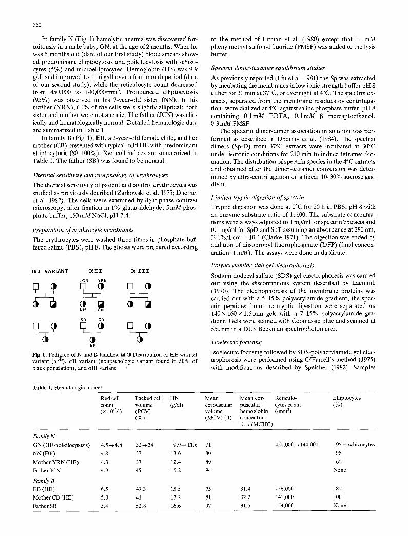
Fig.2. Sodium dodecyl sulfate polyacrylamide gel electrophoresis (SDS-PAGE) of limited tryptic digests of spectrin from controls (C), children with HE (EB, GN, NN), HE parents (CB, YRN), and normal parents (SB, JCN)

354
4. Limited tryptic digests of Sp extracts
To control minor variations due to experimental conditions, Sp from a normal subject was always prepared, digested, and electrophoresed concurrently with the patient samples. Li- mited tryptic digestion of Sp extracts from the two mothers and the three children produced similar and reproducible peptide patterns which were different from controls (46 white and 90 black control subjects have been studied). These pat- terns were characterized by a marked decrease in the 80,000- dalton peptide and the appearance of two additional peptides of 46,000 and 22,000 daltons (Fig. 2). Densitometer tracings of one-dimensional slab gel indicated a diminution in the 80,000- dalton peptide of 40 to 50% in the three children and 35 to 40% in the two mothers.
A decrease in the 35,000-dalton peptide and a concomitant presence of a 37,000-dalton peptide were also observed in all the patients, but these variations were commonly observed in black control populations and are related to a polymorphism of the aII domain (Knowles et al. 1984). These variants are usually associated with a decrease in a 48,000-dalton peptide and an increase in a 50,000-dalton peptide. In these patients, such differences were difficult to detect on one-dimensional gels because other modifications seemed to occur in this area; however we have observed the presence of an additional band at 54,000 daltons.
Tryptic digests separated by two-dimensional electro- phoresis (isoelectric focusing followed by SDS-PAGE) gave a better resolution. Peptide maps obtained from the five pa- tients confirmed results observed in one-dimensional slab gel: the decrease in the 80,000-dalton peptide (pI = 5.2-5.3) and the presence of two additional peptides: the 46,000-dalton peptide which focused in three closed spots from pH 5.20 to pH 5.35, and the 22,000-dalton peptide with a isoelectric point of 5.35 (Fig. 3). Two-dimensional analysis allowed a good sep- aration in multiple spots of the different peptides from the 55,000- to 46,000-dalton range. Some other modifications were detectable in the peptide maps from the five patients: the common variants related to the cdI domain, characterized by the decrease in the two spots at 48,000 daltons and the spot at 35,000 daltons with the concomitant appearance of two spots
at 50,000 daltons and one spot at 37,000 daltons, respectively (Fig. 3). Both spots at 50,000 daltons had a modified isoelec- tric point, which is slightly more basic. These modifications in molecular weight and isoelectric point correspond with the type 1/2 described by Knowles et al. (1984). The last modifica- tions occurring in these five patients were the decreased stain- ing intensity of the two spots related to the aIII domains (ar- rows in Fig. 3). Two additional spots with higher molecular weight and slightly acidic isoelectric point were observed. Similar findings relative to the aIII domain were also ob- served in a black control subject. Peptide maps from both nor- mal fathers were indistinguishable from those obtained from white control subjects,
5. Limited tryptic digest of SpD and SpT
To examine the relationship between the defect in Sp self- association and abnormal tryptic digest patterns from HE pa- tients, SpD was separated from SpT after S p D ~ SpT conver- sion in solution and subjected to tryptic digestion. The de- crease in the 80,000-dalton peptide and the presence of the 46,000-dalton peptide were mainly observed in SpD of HE pa- tients while traces of 46,000-dalton peptide were observed in SpT (Fig. 4). The amount of 80,000-dalton peptide in HE SpT was similar to control SpD or SpT. In both HE mothers as well as in the three children, the variants relative to the c~II domain (the decrease in 48,000 and 35,000-dalton peptide and the apparition of the 37,000-dalton peptide) were essentially observed in SpD. Traces of a 54,000-dalton peptide (c~III do- main) could also be detected in SpD.
6. Kinetic studies of tryptic digestion of HE spectrin
To examine the relationship between the decrease of the 80,000-dalton fragment and the production of the 46,000-dal- ton fragment, kinetic studies of tryptic digestion of HE were performed. After a 30 min reaction, much 80,000-dalton pep- tide was produced. As digestion further proceeded we ob- served a gradual decrease in the 80,000-dalton peptide with a concomitant increase in the 46,000-dalton peptide (Fig. 5).
Fig.3. Two-dimensional separation (isoelectric focusing/SDS-PAGE) of tryptic digests of spectrin from control and HE patients: EB and CB (EB's mother). Encircled area indicates the cdI peptides: triangles indicate the 80 KD peptide (ctI domain); arrows indicate cdII domain

355
Fig.4. SDS-PAGE of limited tryptic digests of Sp-dimer and Sp- tetramer from EB's parents: normal father (SB); HE's mother (CB)
Discussion
On the basis of clinical evaluations, the disease observed in the four patients (NN, YRN, EB, CB) corresponded to the common form of HE with mild and compensated hemolysis. As previously reported in a subset of HE (Tomaselli et al. 1981), erythrocytes of these four patients were abnormally heat sensitive (47°C). The clinical features of patient GN is consistent with the diagnosis of HE with pyknocytosis in in- fants. As reported by Zarkowsky (1979) in such cases, ery- throcytes were particularly heat sensitive (45°C).
The three children and their mothers had a defect in Sp dimer-tetramer equilibrium which showed up as an excess of dimer species in the 4°C extracts together with a decreased Sp-dimer association constant in solution when determined. These HE should be classified as type I HE. As we have previ- ously observed (Lecomte et al. 1985), HE patients who pre- sented abnormal thermal sensitivity of erythrocytes displayed defective Sp self-association.
Limited tryptic cleavage of normal Sp at 0°C allows one to characterize an 80,000-dalton peptide called the cd domain, which contains the domain involved in the Sp self-association process. The decrease in the 80,000-dalton peptide, observed in the peptide pattern of the five HE patients suggests a struc-
Fig.5. Kinetic studies of tryptic digestion of Sp extracts from HE pa- tient. Sp extracts (1 mg/ml) were treated with TPCK trypsin (enzyme/ substrate ratio: 1/100) at 0°C for 0.5 h, 1 h, 5 h, 6 h
tural alteration in the c~I domain affecting the association state of Sp. Such a decrease in the 80,000-dalton peptide has previ- ously been described in some cases of HE and in HPP (Lawler et al. 1982, 1983; Knowles et al. 1983) and it has always been found to be associated with a defect of the Sp self-association. The 30 to 50% decrease in 80,000-dalton peptide found in the five patients is consistent with a heterozygous state.
Like the decrease in the 80,000-dalton peptide, the 46,000- dalton peptide was never observed in any of the control sub- jects (n = 136) or in either of the normal fathers. This frag- ment together with the decrease in the 80,000-dalton peptide, were observed mainly in defective Sp dimers as compared to Sp tetramers. Kinetic studies of Sp tryptic digestion showed that the gradual decrease in the 80,000-dalton peptide and a concomitant increase in the 46,000-dalton peptide were time related. These results indicated that the presence of the 46,000 fragment found in peptide maps of these five HE patients is related to a defect of the aI domain. The 46,000-dalton pep- tide observed in these patients is similar to those reported by Lawler and coworkers (1983, 1984a) in two HPP kindred and in one child with HE and transient poikilocytosis: it was found to be derived from the ct chain. The 50,000-dalton fragments reported in two HPP kindreds (Knowles et al. 1983) and in preliminary studies in two HE kindreds (Marchesi et al. 1983) could be similar to the 46,000-dalton fragment. However, the comparison between the two-dimensional maps showed by Knowles et al. (1983) and our two-dimensional maps seems to

356
indicate that the location of the 50,000-dalton peptide differs from that of the 46,000-dalton peptide.
Both mothers and the children exhibited the variants re- lated to a I I in the heterozygous form and referred to as type 1/2 as proposed by Knowles et ah (1984). These variants are commonly observed in black populations with a frequency of 50%. Subjects who have this polymorphism in either hetero- zygous or homozygous forms (Dhermy et al. 1984) do not ex- hibit abnormal shaped cells and defective Sp self-association.
In these patients we observed new modifications relative to the a I I I domain: the decrease in two spots of the a I I I domain and the presence of two addi t iona l spots with higher molecu- lar weight and similar isoelectric point; they were also ob- served in a black control subject. Sp tryptic digests from this black subject who has normal discoid-shaped erythrocytes had a normal amount of 80,000-dalton peptide. These modifica- tions of the a I I I domain were probably due to another polymorphism, which is less frequent than the variants rela- tive to the a I I domain. In conclusion, two types of modifica- t ion were observed in the peptide maps of these patients: alterations relative to the a I domain that are always associated with a functional defect of Sp and alterations associated with asymptomatic variants of Sp and relative to a I I and a I I I do- mains. However, tryptic digestion studies of Sp dimer and tetramer in both mothers have shown that the modifications of c~I, a I I , and a I I I domains were localized on the defective molecule of Sp dimer and transmit ted together to their chil- dren. These results suggest a l inked transmission of these mu- tated domains.
The infant GN with elliptocytosis and transient poikilocyto- sis had functional and molecular alterations to the same extent as his mother and sister. As suggested by Mentzer et al. (1984) the hemolysis and the high erythrocyte instability to heat ob- served in this child could be related to the transient high level of free 2-3 diphosphoglycerate (2-3 DPG) , a common feature of neonata l erythrocytes. 2-3 D P G is known to dissociate membrane skeleton in vitro (Sheetz and Casaly 1980) and could act in concert with the inheri ted Sp defect to increased membrane fragility.
Acknowledgements. We are indebted to Dr. Boucheix and Dr. Gretil- lat for permission to study their patients, and to N. Lemaire for pre- paring the manuscript. This work was supported by Minist~re de la Recherche et de l'Industrie (MIR 82 L. 1115).
References
Clarke M (1971) Isolation and characterization of a water soluble pro- tein for bovine erythrocyte membranes. Biochem Biophys Res Commun 45 : 1063-1070
Coetzer T, Zail S (1982) Spectrin tetramer-dimer equilibrium in hereditary elliptocytosis. Blood 59 : 900-905
Cohen MC (1983) The molecular organization of the red cell mem- brane skeleton. Semin Hematol 20 : 141-158
Dhermy D, Lecomte MC, Garbarz M, Bournier O, Galand C, Gau- tero H, F6o C, Alloisio N, Delauny J, Boivin P (1982) Spectrin- 13 chain variant associated with hereditary elliptocytosis. J Clin Invest 70 : 707-715
Dhermy D, Lecomte MC, Garbarz M, F6o C, Gautero H, Bournier O, Galand C, Herrera A, Gretillat F, Boivin P (1984) Molecular defect of spectrin in the family of a child with congenital hemolytic poikilocytic anemia. Pediatr Res 18 : 1005-1012
Evans JPM, Baines AJ, Hann IM, A1-Hakim I, Knowles SM, Hoff- brand AV (1983) Defective spectrin dimer-dimer association in a family with transfusion-dependent homozygous hereditary ellipto- cytosis. Br J Haematol 54:163-172
Hanspal M, Ralston GB (1982) Binding of an 80,000 dalton trypsin fragment of spectrin to intact spectrin. Biochim Biophys Acta 709 : 105-109
Knowles WJ, Morrow JS, Speicher DW, Zarkowsky HS, Mohandas N, Mentzer WC, Shohet SB, Marchesi VT (1983) Molecular and functional changes in spectrin from patients with hereditary pyropoikilocytosis. J Clin Invest 71 : 1867-1877
Knowles WJ, Bologna ML, Chasis JA, Marchesi SL, Marchesi VT (1984) Common structural polymorphisms in human erythrocyte spectrin. J Clin Invest 73 : 973-979
Laemmli UK (1970) Cleavage of structural proteins during the assem- bly of the head of bacteriophage T4. Nature 227 : 680-685
LaMer J, Liu SC, Palek J (1982) Molecular defect of spectrin in hereditary pyropoikilocytosis. J Clin Invest 70 : 1019-1030
Lawler J, Palek J, Liu SC, Prchal J, Butler WM (1983) Molecular heterogeneity of hereditary pyropoikilocytosis: identification of a second variant of the spectrin-subunit. Blood 62:1182-1189
Lawler J, Liu SC, Palek J (1984a) A molecular defect of spectrin in a subset of patients with hereditary elliptocytosis. J Clin Invest 73 : 1688-1695
Lawler J, Coetzer TL, Palek J, Jacob HS, Luban NLC (1984b) Sp I/ 65: an abnormal spectrin associated with defective spectrin hetero- dimer self-associafion, membrane skeleton instability and in- creased susceptibility to hemolysis. Blood 64 [Suppl 1]:27a (Abstr)
Lecomte MC, Dhermy D, Garbarz M, Gautero H, Bournier O, Ga- land C, Boivin P (1984a) Hereditary elliptocytosis with a spectrin molecular defect in a white patient. Acta Haematol (Basel) 71: 235-240
Lecomte MC, Dhermy D, Garbarz M, Solis-ViUa C, Ester A, F6o C, Gautero H, Bournier O, Galand C, Boivin P (1984b) Hetero- geneity in the molecular defect of spectrin in type I hereditary elliptocytosis: identification of a third variant within u chain. Blood 64 [Suppl] : 28a (Abstr)
Lecomte MC, Dhermy D, Solis C, Ester A, F6o C, Gautero H, Bour- nier O, Boivin P (1985) A new abnormal variant of spectrin in black patients with hereditary elliptocytosis. Blood 65 : 1208-1217
Litman D, Hsu CJ, Marchesi VT (1980) Evidence that spectrin binds to macromolecular complexes on the inner surface of the red cell membrane. J Cell Sei 42: 1-22
Liu SC, Palek J (1980) Spectrin tetramer dimer-equilibrium and the stability of erythrocyte membrane skeletons. Nature 285 : 586-588
Liu SC, Palek J, Prchal J, Castleberry RP (1981) Altered spectrin dimer-dimer association and instability of erythrocyte membrane skeletons in hereditary pyropoikilocytosis. J Clin Invest 68:597- 605
Liu SC, Palek J, Prchal J (1982) Defective speetrin dimer-dimer as- sociation in hereditary elliptocytosis. Proc Natl Acad Sci USA 79 : 2072-2076
Marchesi SL, Knowles WJ, Morrow JS, Marchesi VT (1983) Abnor- mal spectrin in hereditary elliptocytosis similar to HPP spectrin. Blood 62 [Suppl 1] :48a (Abstr)
Mentzer WC, Iarocci TA, Mohandas N (1984) Modulation of RBC membrane mechanical fragility by 2,3 DPG in transient neonatal poikilocytosis. Blood 64 [Suppl 1] :28a (Abstr)
Morrow JS, Speicher DW, Knowles W J, Hsu C J, Marchesi VT (1980) Identification of functional domains of human erythrocyte spec- trin. Proc Natl Acad Sci USA 77 : 6592-6596
Morrow JS, Haigh WB, Marchesi VT (1981) Spectrin oligomers: a structural feature of the erythrocyte cytoskeleton. J Supralmol Struct Cell Biochem 17 : 275-287
O'Farrell P (1975) High resolution two-dimensional electrophoresis of proteins. J Biol Chem 250: 4007-4021
Palek J, Lux SE (1983) Red cell membrane skeletal defects in here- ditary and acquired hemolytic anemias. Semin Hematol 20:189- 224
Sheetz MP, Casaly J (1980) 2-3 Diphosphoglycerate and ATP dis- sociate erythrocyte membrane skeletons. J Biol Chem 255 : 9955- 9960

357
Shotton DM, Burke BE, Branton D (1979) The molecular structure of human erythrocyte spectrin. J Mol Biol 131 : 303-329
Speicher DW, Morrow JS, Knowles WJ, Marchesi VT (1980) Identifi- cation of proteolytically resistant domains of human erythrocyte spectrin. Proc Natl Acad Sci USA 77 : 5673-5677
Speicher DW, Morrow JS, Knowles WJ, Marchesi VT (1982) A struc- tural model of human erythrocyte spectrin alignment of chemical and functional domains. J Biol Chem 257:9093-9101
Tomaselli MG, John KM, Lux SE (1981) Elliptical erythrocyte mem- brane skeletons and heat sensitive spectrin in hereditary ellipto- cytosis. Proc Natl Acad Sci USA 78 : 1911-1915
Zail SS, Goetzer TL (1984) Defective binding of spectrin to ankyrin in a kindred with recessively inherited hereditary elliptocytosis. J Clin Invest 74 : 753-762
Zarkowsky HS (1979) Heat-induced erythrocyte fragmentation in neonatal eUiptocytosis. Br J Haematol 41:515-518
Zarkowsky HS, Mohandas N, Speicher CB, Shohet SB (1975) A con- genital haemolytic anaemia with thermal sensitivity of the erythro- cyte membrane. Br J Haematol 29 : 537-543
Received May 25, 1985 / Revised August 1, 1985
Copyright © 2022 FDOKUMEN