Clinical and Pathologic Findings of Spitz Nevi and Atypical Spitz Tumors With ALK Fusions
9
Copyright © Lippincott Williams & Wilkins. Unauthorized reproduction of this article is prohibited. Clinical and Pathologic Findings of Spitz Nevi and Atypical Spitz Tumors With ALK Fusions Klaus J. Busam, MD,* Heinz Kutzner, MD,w Lorenzo Cerroni, MD,z and Thomas Wiesner, MDzy Abstract: Spitz tumors represent a group of melanocytic neo- plasms that typically affect young individuals. Microscopically, the lesions are composed of cytologically distinct spindle and ep- ithelioid melanocytes, with a range in the architectural display or the cells, their nuclear features, and secondary epidermal or stromal changes. Recently, kinase fusions have been documented in a subset of Spitz tumors, but there is limited information on the clinical and pathologic features associated with those lesions. Here, we report a series of 17 patients (9 male, 8 female) with spitzoid neoplasms showing ALK fusions (5 Spitz nevi and 12 atypical Spitz tumors). The patients’ ages ranged from 2 years to 35 years (mean = 17 y; median = 16 y). Most lesions were located on the lower extremities and presented clinically as polypoid nodules. All tumors were compound melanocytic proliferations with a predominant intradermal growth. Tumor thickness ranged from 1.1 to 6 mm (mean = 2.9 mm; median = 2.5 mm). The most characteristic histopathologic feature of the tumors (seen in all but 2 lesions) was a plexiform dermal growth of intersecting fascicles of fusiform melanocytes. All but 2 tumors were amelanotic. All tumors were strongly immunoreactive for ALK. The ALK re- arrangements were confirmed in all cases by fluorescence in situ hybridization (FISH), and the fusion partner was determined by quantitative polymerase chain reaction as TPM3 (tropomyosin 3) in 11 cases and DCTN1 (dynactin 1) in 6 cases. None of the 8 tumors that were analyzed by FISH for copy number changes of 6p, 6q, 9p, or 11q met criteria for melanoma. Two patients un- derwent a sentinel lymph node biopsy, and in both cases mela- nocyte nests were found in the subcapsular sinus of the node. Array comparative genomic hybridization of these 2 tumors re- vealed no chromosomal gains or losses. In conclusion, our study revealed that Spitz nevi/tumors with ALK rearrangement show a characteristic plexiform morphology and that ALK im- munohistochemistry and FISH enable the accurate identification of this morphologic and genetic distinct subset of spitzoid neo- plasms. Key Words: kinase fusion, melanocytic nevus, Spitz nevus, ALK (Am J Surg Pathol 2014;00:000–000) S pitzoid melanocytic neoplasms include Spitz nevi (be- nign tumors), so-called “atypical Spitz tumors” (tumors of uncertain malignant potential), and “spitzoid” melano- mas (malignant tumors, with metastasizing and lethal po- tential 1 ). They remain a problem area in the field of dermatopathology because of the difficulty in distinguishing the relatively more common indolent lesions from the rare metastasizing and lethal spitzoid melanoma. However, significant progress has been made over the past decade regarding cytogenetic and/or molecular alterations of these tumors. 2–7 A number of subsets of Spitz tumors have been identified with distinct, clinical, pathologic, and genetic features. They include the HRAS-mutant sclerosing Spitz nevi, 2 the BAP1 loss Spitz tumors, 7–9 and the spitzoid mel- anomas with homozygous deletions of p16. 6 An important milestone in our understanding of Spitz tumors is the recent discovery that some of them harbor gene fusions involving receptor tyrosine kinases ALK, ROS1, NTRK1, and RET or the serine-threonine kinase BRAF. 10 Such gene rearrangements were found in 72 of 140 (51.4%) spitzoid neoplasms. Fourteen of these 140 (10%) spitzoid neoplasms showed ALK fusions. All kinase fusions were mutually exclusive and occurred only in tumors without HRAS mutations or loss of BAP1. Kinase fusions were detected across the entire spectrum of Spitz lesions (benign Spitz nevi, atypical Spitz tumors, and rare spitzoid melanomas), which suggests that the fusions likely occur early in the pathogenesis of the tumors and are per se not sufficient for malignant transformation. This observation is analogous to that of mutations in oncogenes (such as BRAF, NRAS, GNAQ, and GNA11) commonly found in melanocytic neoplasms. 4,7,11,12 Currently, it is unknown whether or not any of the translocation-associated Spitz tumors have distinct clin- ical and/or pathologic features. It is also unknown whether the presence or absence of a kinase fusion of a particular type is associated with a more indolent or aggressive clinical course. In this study, we document clinical findings and describe the spectrum of microscopic From the *Department of Pathology; yHuman Oncology and Patho- genesis Program, Memorial Sloan-Kettering Cancer Center, New York, NY; wDermatopathologie Friedrichshafen, Friedrich- shafen, Germany; and zDepartment of Dermatology, Medical University Graz, Graz, Austria. Conflicts of Interest and Source of Funding: Funded by grants from the National Institutes of Health P30 CA008748 to Memorial Sloan- Kettering Cancer Center. T.W. is funded by the Harry J. Lloyd Trust, by the Jubilaeumsfonds of the Oesterreichische Nationalbank, and by a Charles H. Revson Senior Fellowship. The authors have disclosed that they have no significant relationships with, or financial interest in, any commercial companies pertaining to this article. Correspondence: Klaus J. Busam, MD, Department of Pathology, Memorial Sloan-Kettering Cancer Center, 1275 York Ave, New York, NY 10065 (e-mail: [email protected]). Copyright r 2014 by Lippincott Williams & Wilkins ORIGINAL ARTICLE Am J Surg Pathol Volume 00, Number 00, ’’ 2014 www.ajsp.com | 1
-
Upload
independent -
Category
Documents
-
view
0 -
download
0
Transcript of Clinical and Pathologic Findings of Spitz Nevi and Atypical Spitz Tumors With ALK Fusions

Copyright © Lippincott Williams & Wilkins. Unauthorized reproduction of this article is prohibited.
Clinical and Pathologic Findings of Spitz Nevi andAtypical Spitz Tumors With ALK Fusions
Klaus J. Busam, MD,* Heinz Kutzner, MD,w Lorenzo Cerroni, MD,z and Thomas Wiesner, MDzy
Abstract: Spitz tumors represent a group of melanocytic neo-
plasms that typically affect young individuals. Microscopically,
the lesions are composed of cytologically distinct spindle and ep-
ithelioid melanocytes, with a range in the architectural display or
the cells, their nuclear features, and secondary epidermal or
stromal changes. Recently, kinase fusions have been documented
in a subset of Spitz tumors, but there is limited information on the
clinical and pathologic features associated with those lesions.
Here, we report a series of 17 patients (9 male, 8 female) with
spitzoid neoplasms showing ALK fusions (5 Spitz nevi and 12
atypical Spitz tumors). The patients’ ages ranged from 2 years to
35 years (mean=17y; median=16y). Most lesions were located
on the lower extremities and presented clinically as polypoid
nodules. All tumors were compound melanocytic proliferations
with a predominant intradermal growth. Tumor thickness ranged
from 1.1 to 6mm (mean=2.9mm; median=2.5mm). The most
characteristic histopathologic feature of the tumors (seen in all but
2 lesions) was a plexiform dermal growth of intersecting fascicles
of fusiform melanocytes. All but 2 tumors were amelanotic. All
tumors were strongly immunoreactive for ALK. The ALK re-
arrangements were confirmed in all cases by fluorescence in situ
hybridization (FISH), and the fusion partner was determined by
quantitative polymerase chain reaction as TPM3 (tropomyosin 3)
in 11 cases and DCTN1 (dynactin 1) in 6 cases. None of the 8
tumors that were analyzed by FISH for copy number changes of
6p, 6q, 9p, or 11q met criteria for melanoma. Two patients un-
derwent a sentinel lymph node biopsy, and in both cases mela-
nocyte nests were found in the subcapsular sinus of the node.
Array comparative genomic hybridization of these 2 tumors re-
vealed no chromosomal gains or losses. In conclusion, our study
revealed that Spitz nevi/tumors with ALK rearrangement show a
characteristic plexiform morphology and that ALK im-
munohistochemistry and FISH enable the accurate identification
of this morphologic and genetic distinct subset of spitzoid neo-
plasms.
Key Words: kinase fusion, melanocytic nevus, Spitz nevus, ALK
(Am J Surg Pathol 2014;00:000–000)
Spitzoid melanocytic neoplasms include Spitz nevi (be-nign tumors), so-called “atypical Spitz tumors” (tumors
of uncertain malignant potential), and “spitzoid” melano-mas (malignant tumors, with metastasizing and lethal po-tential1). They remain a problem area in the field ofdermatopathology because of the difficulty in distinguishingthe relatively more common indolent lesions from the raremetastasizing and lethal spitzoid melanoma. However,significant progress has been made over the past decaderegarding cytogenetic and/or molecular alterations of thesetumors.2–7 A number of subsets of Spitz tumors have beenidentified with distinct, clinical, pathologic, and geneticfeatures. They include the HRAS-mutant sclerosing Spitznevi,2 the BAP1loss Spitz tumors,7–9 and the spitzoid mel-anomas with homozygous deletions of p16.6
An important milestone in our understanding of Spitztumors is the recent discovery that some of them harborgene fusions involving receptor tyrosine kinases ALK,ROS1, NTRK1, and RET or the serine-threonine kinaseBRAF.10 Such gene rearrangements were found in 72 of 140(51.4%) spitzoid neoplasms. Fourteen of these 140 (10%)spitzoid neoplasms showed ALK fusions. All kinase fusionswere mutually exclusive and occurred only in tumorswithout HRAS mutations or loss of BAP1. Kinasefusions were detected across the entire spectrum of Spitzlesions (benign Spitz nevi, atypical Spitz tumors, and rarespitzoid melanomas), which suggests that the fusions likelyoccur early in the pathogenesis of the tumors and areper se not sufficient for malignant transformation. Thisobservation is analogous to that of mutations in oncogenes(such as BRAF, NRAS, GNAQ, and GNA11) commonlyfound in melanocytic neoplasms.4,7,11,12
Currently, it is unknown whether or not any of thetranslocation-associated Spitz tumors have distinct clin-ical and/or pathologic features. It is also unknownwhether the presence or absence of a kinase fusion of aparticular type is associated with a more indolent oraggressive clinical course. In this study, we documentclinical findings and describe the spectrum of microscopic
From the *Department of Pathology; yHuman Oncology and Patho-genesis Program, Memorial Sloan-Kettering Cancer Center,New York, NY; wDermatopathologie Friedrichshafen, Friedrich-shafen, Germany; and zDepartment of Dermatology, MedicalUniversity Graz, Graz, Austria.
Conflicts of Interest and Source of Funding: Funded by grants from theNational Institutes of Health P30 CA008748 to Memorial Sloan-Kettering Cancer Center. T.W. is funded by the Harry J. LloydTrust, by the Jubilaeumsfonds of the Oesterreichische Nationalbank,and by a Charles H. Revson Senior Fellowship. The authors havedisclosed that they have no significant relationships with, or financialinterest in, any commercial companies pertaining to this article.
Correspondence: Klaus J. Busam, MD, Department of Pathology,Memorial Sloan-Kettering Cancer Center, 1275 York Ave,New York, NY 10065 (e-mail: [email protected]).
Copyright r 2014 by Lippincott Williams & Wilkins
ORIGINAL ARTICLE
Am J Surg Pathol � Volume 00, Number 00, ’’ 2014 www.ajsp.com | 1

Copyright © Lippincott Williams & Wilkins. Unauthorized reproduction of this article is prohibited.
features associated with Spitz nevi and atypical Spitz tu-mors carrying ALK fusions.
METHODS
Case SelectionEight cases were from the personal consultation files
of one of the authors (K.J.B.), 8 cases from another au-thor (H.K.), and 1 case from a third author (L.C.). Onlycases diagnosed as a Spitz nevus or an “atypical Spitztumor,” but not tumors reported as or favored to bemalignant melanomas, were included in this series. Thir-teen of the 17 analyzed tumors were included in the initialstudy reporting kinase fusions in spitzoid tumors10; cases2, 3, 10, and 11 have not been previously reported.
Light Microscopic AnalysisThe following histopathologic parameters were re-
corded: polypoid silhouette, ulceration, epidermal hyper-plasia, tumor thickness, anatomic (Clark) level, tumormitotic rate (number of mitotic figures in dermal or sub-cutaneous melanocytes per mm2), plexiform growth pat-tern, inflammation, and the presence of melanin pigment.
ImmunohistochemistryImmunohistochemical studies were conducted using
5-mm-thick unstained sections of formalin-fixed, paraffin-embedded archival material. We used the ALK antibody(clone D5F3) from Cell Signaling (Danvers, MA) on aDiscovery Ultra instrument with a multimer/DAB de-tection system (Ventana Medical Systems Inc., Tucson,AZ) with appropriate negative and positive controls.
Quantitative Polymerase Chain Reaction andSanger Sequencing
RNA was extracted from 20-mm-thick formalin-fixed, paraffin-embedded tissue sections (High PuremiRNA Isolation Kit; Roche) and was reverse-tran-scribed with random hexamer primers using the Tran-scriptor First Strand cDNA Synthesis Kit (Roche). TheALK fusion partners were determined by quantitativepolymerase chain reaction (PCR) using break-pointspanning primers reported previously.10 Specific PCRamplicons were only detected with the appropriate com-bination of primers and template and not with negativecontrols. The nucleotide sequence of the PCR productswas confirmed by Sanger sequencing.
Fluorescence In Situ HybridizationTwo sets of fluorescence in situ hybridization
(FISH) tests were performed. To confirm the ALK fu-sions, a commercially available break-apart probe wasused according to the manufacturer’s protocol (AbbottMolecular, Des Plaines, IL). The probes were hybridizedon 5-mm-thick tissue sections, and the number and lo-calization of the hybridization signals was assessed in aminimum of 100 interphase nuclei with well-delineatedcontours. At least 50% of neoplastic cells had to show asplit signal to report a rearrangement of a kinase.
The so-called “melanoma FISH test” was used in theworkup of some atypical Spitz tumors to identify chromo-somal copy number changes typical of melanoma (AbbottMolecular). In this test, a set of 4 probes targeting Ras re-sponsive element-binding protein-1 (VysisLSI RREB1-
TABLE 1. Clinical and Pathologic Features of Patients and Their Tumors
Patient
Age/
Sex Site Dx
Breslow
Thickness
Clark
Level TMR
Poly-
poid
Pigment-
ation Ulcer
Plexi-
form
Inflamm-
ation
IHC-
ALK
FISH-
ALK
Fusion
Partner
Cyto-
genetics
1 5/M Chin CSN 2.5 IV 0 Yes + + + � + + TPM3 �(FISH)
2 25/M Lt low
leg
CSN 4 III 0 Yes � � + � + + TPM3 �(FISH)
3 11/F Rt ant
thigh
CSN 2.5 IV 0 No + � + Mild + + TPM3 �(FISH)
4 35/M Lt ear AST 2 IV 0 Yes � � + Mild + + TPM3 �(CGH)
5 14/M Rt
buttock
AST 3.7 IV 3 Yes � � + Mild + + TPM3 ND
6 2/M Lt arm AST 4.5 IV 3 Yes � � + Mild + + TPM3 ND
7 16/F Lt
buttock
AST 2 IV 1 Yes � � + Mild + + TPM3 ND
8 14/F NA CSN 3.5 IV 1 Yes � � + Mild + + TPM3 ND
9 17/F Lt ankle AST 6 IV 4 Yes � � + � + + TMP3 ND
10 23/F Rt toe AST 2.5 V 1 No � � + � + + TPM3 �(FISH)
11 19/F Lt
abdomen
CSN 1.8 IV 0 Yes � � + � + + TPM3 �(FISH)
12 14/M Rt ear AST 3 IV 1 Yes � + + Mild + + DCTN1 ND
13 9/F Rt
lat.thigh
AST 4.4 V 2 Yes � � + Mild + + DCTN1 �(CGH)
14 28/F Rt back AST 2.5 IV 0 No � � (+) Mild + + DCTN1 ND
15 9/M NA AST 1.8 IV 1 Yes � � + Moderate + + DCTN1 ND
16 19/M Lt arm AST 1.5 IV 0 Yes � � (+) Mild + + DCTN1 ND
17 35/M Lt leg AST 1.1 IV 0 No � � + Mild + + DCTN1 �(FISH)
� indicates negative or absent; (+) only focally present; +, positive or present; AST, atypical Spitz tumor; CSN, compound Spitz nevus; DCTN1, dynactin 1; Dx,diagnosis; F, female; lt, left; M, male; NA, information not available; ND, not done; rt, right; TPM3, tropomyosin 3.
Busam et al Am J Surg Pathol � Volume 00, Number 00, ’’ 2014
2 | www.ajsp.com r 2014 Lippincott Williams & Wilkins

Copyright © Lippincott Williams & Wilkins. Unauthorized reproduction of this article is prohibited.
Spectrum Red), myeloblastosis (VysisLSI MYB-S Gold),cyclin D1 or chromosome 11q (VysisLSI CCND1-SpectrumGreen), and centromeric enumeration probe control forchromosome 6 (VysisLSI CEP6-Spectrum Aqua) was ini-tially used, followed by a probe set assessing aberrations ofchromosome 9p (VysisLSI CDKN2A/CEP 9 FISH probekit). The protocol used for these FISH tests has previouslybeen described.13–15 A lesion was considered as having apositive FISH result if any of the following criteria were met:(1) gain in 6p25(RREB1) relative to CEP6 >55% or gain in6p25(RREB1) >29%; (2) loss in 6q23(MYB) relative toCEP6 >40%; (3) gain in 11q13(CCND1) >38% or ho-mozygous loss of 9p(CDNK2A) in >30% of the tumor cells.
aCGHTumor and reference DNA were differentially la-
beled with dCTP-Cy5 and dCTP-Cy3 (GE Healthcare,Piscataway, NJ) using a Bioprime Array CGH GenomicLabeling Kit (Invitrogen, Carlsberg, CA) according to themanufacturer’s instructions. Genome-wide analysis ofDNA copy number changes was conducted using an oli-gonucleotide array containing 180k probes according tothe manufacturer’s protocol (SurePrint G3 Human CGHMicroarray Kit, 1�180k; Agilent, Santa Clara, CA).Slides were scanned using Agilent’s microarray scannerG2505B and analyzed using Agilent Genomic Work-bench.
FIGURE 1. Spitz nevus with a TPM3-ALK fusion from the chin of a 5-year-old boy (case 1). A, Silhouette of an irritated nevus withepidermal hyperplasia and hemorrhagic crust associated with focal ulceration. B, Proliferation of cytologically bland spindle cells(hematoxylin and eosin–stained section). C, The tumor cells are immunoreactive for ALK. D, FISH demonstrates the ALK generearrangement by the individual green and orange signals using break-point flanking probes. E, TPM3-ALK fusion. ALK is locatedon chromosome 2p23. Because of genomic rearrangements, exon 1-8 of TPM3 is fused with exon 20-29 of ALK, which containsthe tyrosine kinase domain.
Am J Surg Pathol � Volume 00, Number 00, ’’ 2014 Spitz Tumors With ALK Fusions
r 2014 Lippincott Williams & Wilkins www.ajsp.com | 3

Copyright © Lippincott Williams & Wilkins. Unauthorized reproduction of this article is prohibited.
RESULTS
Clinical Findings
The group of individuals with ALK-fusion Spitznevi or tumors included 9 male and 8 female patients.Their ages ranged from 2 to 35 years (mean=17 y;median=16 y). None of them reported a family historyof melanoma. None of the clinical lesions was suspectedto be melanoma. All were solitary lesions. They were re-moved as “irritated nevus,” “atypical nevus,” “angioma,”or “verruca.” Six lesions were located on the lowerextremities, 4 were from the trunk and buttocks,3 were from the head and neck region, and 2 lesionswere located on the upper extremities (Table 1). Theanatomic site was not specified in 2 cases. Five tumorswere reported as Spitz nevus and 12 lesions as atypicalSpitz tumor. All patients with this diagnosis had acomplete excision of the primary tumor with negativemargins. Two patients underwent a sentinel lymphnode (SLN) biopsy, and in both cases nests of spitzoidmelanocytes were found in the subcapsular sinus of thenode. A complete lymph node dissection was not per-formed. All patients are alive and well with no evidenceof disease at last follow-up. The follow-up ranged from2 months to 4 years.
Histopathologic FindingsHistopathologic and immunohistochemical findings
are documented in Table 1 and illustrated in Figures 1–6.All lesions were compound melanocytic proliferations withboth junctional and intradermal components. Tumorthickness of all lesions ranged from 1.1 to 6mm (mean=2.9mm; median=2.5mm). One tumor was confined tothe epidermis and papillary dermis (Clark level III). Thir-teen tumors extended into the reticular dermis (Clark levelIV) and 2 into the superficial subcutis (Clark level V). Allbut 2 tumors were amelanotic (no detectable melanin pig-ment on hematoxylin and eosin–stained sections). Epi-dermal hyperplasia was common (13 of 17 cases). Twolesions were focally ulcerated (excoriated).
Spitzoid melanocytic proliferations with TPM3(gene for tropomyosin 3) as the ALK fusion partner in-cluded 5 Spitz nevi and 7 Spitz tumors. Mean and mediantumor thicknesses of lesions with TPM3-ALK fusionswere 3.1 and 2.5mm, respectively. All tumors of thisseries displayed a characteristic plexiform growth patternof intersecting fascicles of predominantly fusiform mela-nocytes in the dermis (Figs. 1–4, Table 1). The nucleishowed smooth nuclear contours and a slightly vesicularchromatin pattern. Marked pleomorphism was lacking.Two lesions showed prominent dermal sclerosis. Mitoses
FIGURE 2. Spitz nevus with a TPM3-ALK fusion from the leg of a 25-year-old man (case 2). A, Silhouette of an amelanoticpolypoid compound Spitz nevus (hematoxylin and eosin–stained section) with evidence of maturation. B, Proliferation of cyto-logically bland spindle and epithelioid melanocytes. C, The tumor cells are positive for ALK. D, FISH confirms the ALK re-arrangements using break-point flanking probes by the individual green and red signals.
Busam et al Am J Surg Pathol � Volume 00, Number 00, ’’ 2014
4 | www.ajsp.com r 2014 Lippincott Williams & Wilkins

Copyright © Lippincott Williams & Wilkins. Unauthorized reproduction of this article is prohibited.
were absent or rare. Inflammation was absent in 5 andmild in 6 of 11 tumors. One patient with a Spitz tumorharboring a TPM3-ALK fusion underwent an SLN bi-opsy. A small cluster of spitzoid melanocytes similar inappearance to the primary tumor was found in the sub-capsular sinus of the node (Fig. 4).
All spitzoid melanocytic proliferations with DCTN1(gene for dynactin 1) as the ALK fusion partner had beenclassified as atypical Spitz tumors. Mean and median tumorthicknesses of lesions with DCTN1-ALK fusions were 2.3and 2.2mm, respectively. The mitotic rate ranged from0 to 2/mm2 (mean=1/mm2; median=1/mm2). Whereas
FIGURE 3. Partly pigmented compound Spitz nevus with a TPM3-ALK fusion from the thigh of an 11-year-old girl (case 3). A,Wedge-shaped silhouette of a compound spindle cell melanocytic proliferation with epidermal hyperplasia (hematoxylin andeosin–stained section). B, The junctional component shows features of a pigmented spindle cell nevus. C, Deeper section of thelesion, which was adjacent to the section used for immunohistochemistry. The junctional melanocytic proliferation shows apredominant nested pattern and is pigmented. The intradermal melanocytes are amelanotic and display a plexiform growthpattern. D, The tumor cells are positive for ALK in immunohistochemistry. E, FISH confirms the ALK rearrangement.
Am J Surg Pathol � Volume 00, Number 00, ’’ 2014 Spitz Tumors With ALK Fusions
r 2014 Lippincott Williams & Wilkins www.ajsp.com | 5

Copyright © Lippincott Williams & Wilkins. Unauthorized reproduction of this article is prohibited.
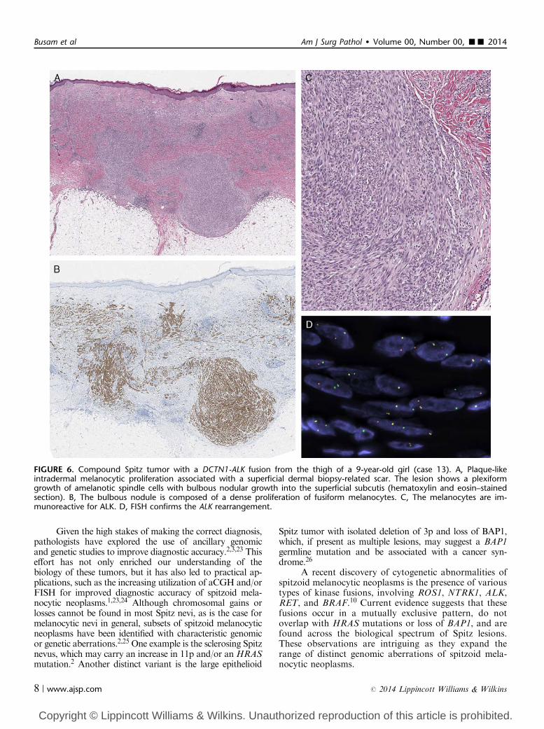
5 lesions showed a plexiform growth of intersecting fasciclesof fusiform melanocytes similar to the Spitz tumors withTPM3-ALK fusions, 2 tumors were composed of a pre-dominant large epithelioid cell proliferation with enlargednuclei and nuclear pleomorphism (Fig. 5). These 2 lesionsdisplayed a plexiform growth pattern only focally. Mildinflammation was observed in 5 of 6 tumors and moderateinflammation in 1 case. One patient with a Spitz tumorharboring a DCTN1-ALK fusion underwent an SLN bi-opsy with detection of microscopic melanocyte clusters inthe subcapsular sinus. This lesion was remarkable for abulbous nodular growth component in the deep dermis/superficial subcutis (Fig. 6). This lesion was analyzed by
array comparative genomic hybridization (aCGH), and nocopy number gain or loss was detected.
Immunohistochemical FindingsAs implied by the case selection, all lesions were
immunohistochemically positive for ALK. Immuno-reactivity was strong and homogenous through the entiretumor cell population.
Genetic FindingsFISH with a break-apart probe revealed in all cases
an ALK rearrangement. Eight lesions were also analyzedby “melanoma-FISH” using probes for 6p, 6q, 6cent, 11q,
FIGURE 4. Compound Spitz tumor with a TPM3-ALK fusion and positive SLN from the ear of a 35-year-old man (case 4). A,Silhouette of a polypoid compound melanocytic proliferation (hematoxylin and eosin–stained section). B, Amelanotic spindle andepithelioid melanocytic proliferation with evidence of maturation. C, The tumor cells are immunoreactive for ALK. D, FISHconfirms the ALK rearrangement. E, Spitzoid melanocyte deposits were found in the subcapsular sinus of the SLN(immunohistochemical stain for S100 protein).
Busam et al Am J Surg Pathol � Volume 00, Number 00, ’’ 2014
6 | www.ajsp.com r 2014 Lippincott Williams & Wilkins

Copyright © Lippincott Williams & Wilkins. Unauthorized reproduction of this article is prohibited.
and 9p. None of the lesions met the FISH criteria formelanoma (Table 1). None of the lesions showed homo-zygous deletions of 9p.
Using quantitative PCR and Sanger sequencing, 2types of 50 fusion partners for ALK were identified: TPM3(Fig. 1) and DCTN1 (Fig. 5). TPM3-ALK fusions weredetected in 11 Spitz lesions (5 Spitz nevi and 6 atypicalSpitz tumors). DCTN1-ALK was found in 6 atypical Spitztumors and resulted from a balanced translocation be-tween homologous copies of chromosome 2.
aCGH was performed retrospectively on lesions from2 patients. Both patients had undergone SLN biopsy, andmicroscopic clusters of spitzoid melanocytes were detectedin the subcapsular sinus of the node. No chromosomalgains or losses were detected by aCGH in both cases.
DISCUSSIONSophie Spitz16 reported in 1948 a series of melano-
cytic tumors (then termed “melanoma of childhood”)composed of spindled or epithelioid melanocytes thatdeveloped predominantly in children and adolescents. Itsubsequently became apparent that these tumors couldalso arise later in life and that the majority of these ne-oplasms behaved in an indolent manner, which led to theterm “Spitz nevus.”17–19 Although criteria have been de-veloped to distinguish Spitz nevi from melanoma, it canat times be very difficult or impossible to make a definitivedetermination by light microscopic analysis alone, whichhas led to the category of “borderline” Spitz tumors orspitzoid melanocytic neoplasms of uncertain malignantpotential.1,17–22
FIGURE 5. Compound Spitz tumor with a DCTN1-ALK fusion from the arm of a 19-year-old man (case 16). A, Polypoid com-pound melanocytic tumor, focally ulcerated (hematoxylin and eosin–stained section). B, Proliferation of amelanotic predom-inantly large epithelioid melanocytes with nuclear atypia. C, DCTN1-ALK kinase fusion. ALK is located on chromosome 2p23 andDCTN1 on chromosome 2p13. Because of genomic rearrangements, exon 1-26 of DCTN1 is fused with exon 20-29 of ALK, whichcontains the tyrosine kinase domain. The in-frame junction of the fusion transcript was confirmed with Sanger sequencing.
Am J Surg Pathol � Volume 00, Number 00, ’’ 2014 Spitz Tumors With ALK Fusions
r 2014 Lippincott Williams & Wilkins www.ajsp.com | 7
Copyright © Lippincott Williams & Wilkins. Unauthorized reproduction of this article is prohibited.
Given the high stakes of making the correct diagnosis,pathologists have explored the use of ancillary genomicand genetic studies to improve diagnostic accuracy.2,3,23 Thiseffort has not only enriched our understanding of thebiology of these tumors, but it has also led to practical ap-plications, such as the increasing utilization of aCGH and/orFISH for improved diagnostic accuracy of spitzoid mela-nocytic neoplasms.1,23,24 Although chromosomal gains orlosses cannot be found in most Spitz nevi, as is the case formelanocytic nevi in general, subsets of spitzoid melanocyticneoplasms have been identified with characteristic genomicor genetic aberrations.2,25 One example is the sclerosing Spitznevus, which may carry an increase in 11p and/or an HRASmutation.2 Another distinct variant is the large epithelioid
Spitz tumor with isolated deletion of 3p and loss of BAP1,which, if present as multiple lesions, may suggest a BAP1germline mutation and be associated with a cancer syn-drome.26
A recent discovery of cytogenetic abnormalities ofspitzoid melanocytic neoplasms is the presence of varioustypes of kinase fusions, involving ROS1, NTRK1, ALK,RET, and BRAF.10 Current evidence suggests that thesefusions occur in a mutually exclusive pattern, do notoverlap with HRAS mutations or loss of BAP1, and arefound across the biological spectrum of Spitz lesions.These observations are intriguing as they expand therange of distinct genomic aberrations of spitzoid mela-nocytic neoplasms.
FIGURE 6. Compound Spitz tumor with a DCTN1-ALK fusion from the thigh of a 9-year-old girl (case 13). A, Plaque-likeintradermal melanocytic proliferation associated with a superficial dermal biopsy-related scar. The lesion shows a plexiformgrowth of amelanotic spindle cells with bulbous nodular growth into the superficial subcutis (hematoxylin and eosin–stainedsection). B, The bulbous nodule is composed of a dense proliferation of fusiform melanocytes. C, The melanocytes are im-munoreactive for ALK. D, FISH confirms the ALK rearrangement.
Busam et al Am J Surg Pathol � Volume 00, Number 00, ’’ 2014
8 | www.ajsp.com r 2014 Lippincott Williams & Wilkins

Copyright © Lippincott Williams & Wilkins. Unauthorized reproduction of this article is prohibited.
In this study, we sought to document the morpho-logic spectrum associated with Spitz nevi/tumors with ALKrearrangements and to explore a possible association be-tween histopathologic appearance and fusion partner sub-type. Spitz nevi/tumors with ALK fusions tended to bepolypoid and amelanotic. A plexiform growth of inter-secting fascicles of fusiform melanocytes was the mostcommon and characteristic feature. It was seen in all lesionsof this series with TPM3-ALK fusions and in 4 of 6 tumorswith DCTN1-ALK fusions. Lesions with the latter fusionsubtype also included tumors with a higher proportion oflarge epithelioid melanocytes with nuclear pleomorphism.As we report our findings, we want to emphasize that theyare preliminary. A larger number of cases will be necessaryfor a more comprehensive assessment of the association ofphenotype with genotype.
An important question is the potential clinical sig-nificance of the type of kinase fusion and prognosis. As thedetection of kinase fusions in Spitz tumors is a very recentdiscovery, there is at this point insufficient knowledge onthis issue. Although none of the patients of this series ex-perienced a tumor recurrence, the number of cases is toosmall, and the currently available follow-up is too short forany meaningful conclusions. However, it may be of interestin this regard to point out that 2 patients of this seriesunderwent an SLN biopsy after a diagnosis of atypicalSpitz tumor was made. Small tumor deposits were found inthe SLN of both patients. When the primary tumors wereanalyzed retrospectively by array CGH, no copy numbergains or losses were detected. Whereas clinical follow-up of1 patient is <1 year, the other patient is alive and well 4years after the excision of the primary Spitz tumor anddetection of melanocyte deposits in the SLN. That patienthad undergone only an SLN biopsy with no subsequentcomplete lymph node dissection.
In conclusion, we present herein 17 cases of Spitznevi/tumors with ALK fusions and compare the histo-pathologic findings with the fusion partner subtype. Aplexiform growth pattern of intersecting fascicles ofamelanotic spindle cells was found as the most charac-teristic feature, especially in tumors with TPM3-ALKfusions, but it was not exclusive. Future studies areneeded to determine the strength of the association be-tween the light microscopic appearance of a lesion andthe type of kinase fusion and, most importantly, to learnmore about the biological and clinical significance ofkinase fusions in spitzoid melanocytic neoplasms.
ACKNOWLEDGMENTSThe authors thank Dr Margaret Leversha, Dr Gouri
Nanjangud, Kalyani Chadalavada, and Yuqiang Fang forassisting with FISH. The authors also thank Isla Otap forher assistance in preparing the manuscript.
REFERENCES1. Zedek DC, McCalmont TH. Spitz nevi, atypical spitzoid neoplasms,
and spitzoid melanoma. Clin Lab Med. 2011;31:311–320.
2. Bastian BC, LeBoit PE, Pinkel D. Mutations and copy numberincrease of HRAS in Spitz nevi with distinctive histopathologicalfeatures. Am J Pathol. 2000;157:967–972.
3. Bastian BC, Wesselmann U, Pinkel D, et al. Molecular cytogeneticanalysis of Spitz nevi shows clear differences to melanoma. J InvestDermatol. 1999;113:1065–1069.
4. Fullen DR, et al. BRAF and NRAS mutations in spitzoidmelanocytic lesions. Mod Pathol. 2006;19:1324–1332.
5. Gerami P, Busam KJ. Cytogenetic and mutational analyses ofmelanocytic tumors. Dermatol Clin. 2012;30:555–566.
6. Gerami P, et al. Risk assessment for atypical spitzoid melanocyticneoplasms using FISH to identify chromosomal copy numberaberrations. Am J Surg Pathol. 2013;37:676–684.
7. Murali R, et al. GNAQ and GNA11 mutations in melanocytomasof the central nervous system. Acta Neuropathol. 2012;123:457–459.
8. Busam KJ, et al. Combined BRAF(V600E)-positive melanocyticlesions with large epithelioid cells lacking BAP1 expressionand conventional nevomelanocytes. Am J Surg Pathol. 2013;37:193–199.
9. Busam KJ, Wanna M, Wiesner T. Multiple epithelioid Spitz nevi ortumors with loss of BAP1 expression: a clue to a hereditary tumorsyndrome. JAMA Dermatol. 2013;149:335–339.
10. Wiesner T, He J, Yelensky R, et al. Kinase fusions are frequent inSpitz tumours and spitzoid melanomas. Nat Commun. 2014;5:3116.
11. Van Raamsdonk CD, et al. Mutations in GNA11 in uvealmelanoma. N Engl J Med. 2010;363:2191–2199.
12. van Dijk MC, Bernsen MR, Ruiter DJ. Analysis of mutations inB-RAF, N-RAS, and H-RAS genes in the differential diagnosis ofSpitz nevus and spitzoid melanoma. Am J Surg Pathol. 2005;29:1145–1151.
13. Fang Y, et al. Fluorescence in situ hybridization (FISH) analysis ofmelanocytic nevi and melanomas: sensitivity, specificity, and lack ofassociation with sentinel node status. Int J Surg Pathol. 2012;20:434–440.
14. Gerami P, et al. Fluorescence in situ hybridization for distinguishingnevoid melanomas from mitotically active nevi. Am J Surg Pathol.2009;33:1783–1788.
15. Gerami P, et al. Fluorescence in situ hybridization (FISH) as anancillary diagnostic tool in the diagnosis of melanoma. Am J SurgPathol. 2009;33:1146–1156.
16. Spitz S. Melanomas of childhood. Am J Pathol. 1948;24:591–609.17. Ackerman AB. Spitz nevus. Am J Dermatopathol. 1997;19:
419–421.18. Dahlstrom JE, et al. Spitz naevus: diagnostic problems and their
management implications. Pathology. 2004;36:452–457.19. LeBoit P. Spitz nevus: a look back and a look ahead. Adv Dermatol.
2000;16:81–109; discussion 110.20. Barnhill RL. The Spitzoid lesion: rethinking Spitz tumors, atypical
variants, ‘Spitzoid melanoma’ and risk assessment. Mod Pathol.2006;19(suppl 2):S21–S33.
21. Connors RC, Chalet MD, Ackerman AB. Benign juvenile melano-ma (Spitz nevus) vs. superficial spreading malignant melanoma:criteria for histologic differentiation. J Dermatol Surg. 1975;1:14–15.
22. Spatz A, Barnhill RL. The Spitz tumor 50 years later: revisiting alandmark contribution and unresolved controversy. J Am AcadDermatol. 1999;40(pt 1):223–228.
23. Gerami P, et al. Outcomes of atypical Spitz tumors withchromosomal copy number aberrations and conventional melano-mas in children. Am J Surg Pathol. 2013;37:1387–1394.
24. McCalmont TH, et al. Molecular-microscopical correlation indermatopathology. J Cutan Pathol. 2011;38:324–326. 323.
25. Bauer J, Bastian BC. Distinguishing melanocytic nevi frommelanoma by DNA copy number changes: comparative genomichybridization as a research and diagnostic tool. Dermatol Ther.2006;19:40–49.
26. Wiesner T, et al. A distinct subset of atypical Spitz tumors ischaracterized by BRAF mutation and loss of BAP1 expression. AmJ Surg Pathol. 2012;36:818–830.
Am J Surg Pathol � Volume 00, Number 00, ’’ 2014 Spitz Tumors With ALK Fusions
r 2014 Lippincott Williams & Wilkins www.ajsp.com | 9