Exploiting Amino Acid Composition for Predicting Protein-Protein Interactions
Microbiology (2000), 146, 415–426 Printed in Great Britain
Glucoamylase::green fluorescent proteinfusions to monitor protein secretion inAspergillus niger
Caroline L. Gordon,1 Vahid Khalaj,1 Arthur F. J. Ram,2 David B. Archer,3
Jayne L. Brookman,1 Anthony P. J. Trinci,1 David J. Jeenes,3
John H. Doonan,5 Brian Wells,5 Peter J. Punt,4
Cees A. M. J. J. van den Hondel4 and Geoffrey D. Robson1
Author for correspondence: Geoffrey D. Robson. Tel : 44 161 275 5048. Fax: 44 161 275 5656.e-mail : Geoff.Robson!man.ac.uk
1 School of BiologicalSciences, StopfordBuilding, University ofManchester, ManchesterM13 9PT, UK
2 Centre forPhytotechnology, Institutefor Molecular PlantSciences, ClusiusLaboratory,Wassenaarseweg 64, 2333Al Leiden, The Netherlands
3 Department of Geneticsand Microbiology, Instituteof Food Research, NorwichLaboratory, NorwichResearch Park, Colney,Norwich NR4 7UA, UK
4 TNO Nutrition and FoodResearch Institute,Department of MolecularGenetics and GeneTechnology, Utrechtseweg48, PO Box 360, 3700 AJZeist, The Netherlands
5 Department of CellBiology, John Innes Centre,Colney Lane, NorwichNR4 7UH, UK
A glucoamylase::green fluorescent protein fusion (GLA::sGFP) wasconstructed which allows the green fluorescent protein to be used as an invivo reporter of protein secretion in Aspergillus niger. Two secretory fusionswere designed for secretion of GLA::sGFP which employed slightly differentlengths of the glucoamylase protein (GLA499 and GLA514). Expression ofGLA::sGFP revealed that fluorescence was localized in the hyphal cell wallsand septa, and that fluorescence was most intense at hyphal apices.Extracellular GLA::sGFP was detectable by Western blotting only in thesupernatant of young cultures grown in soya milk medium. In older cultures,acidification of the medium and induction of proteases were probablyresponsible for the loss of extracellular and cell wall fluorescence and theinability to detect GLA::sGFP by Western analysis. A strain containing theGLA: :sGFP construct was subjected to UV mutagenesis and survivors screenedfor mutations in the general secretory pathway. Three mutants were isolatedthat were unable to form a halo on either starch or gelatin medium. All threemutants grew poorly compared to the parental strain. Fluorescencemicroscopy revealed that for two of the mutants, GLA::sGFP accumulatedintracellularly with no evidence of wall fluorescence, whereas for the thirdmutant, wall fluorescence was observed with no evidence of intracellularaccumulation. These results indicate that the GLA::sGFP fusion constructs canbe used as convenient fluorescent markers to study the dynamics of proteinsecretion in vivo and as a tool in the isolation of mutants in the generalsecretory pathway.
Keywords : Aspergillus niger, protein secretion, glucoamylase, green fluorescent protein(GFP), heterologous protein production
INTRODUCTION
Filamentous fungi have a high capacity for secretion ofextracellular enzymes. Members of the genera Asper-gillus and Trichoderma have been reported to produce,under optimal fermentation conditions, 20 g gluco-amylase l−" and 40 g cellulases l−", respectively (Finkel-stein, 1987; Durand et al., 1988). In addition, the GRAS.................................................................................................................................................
Abbreviations: ER, endoplasmic reticulum; GFP, green fluorescent pro-tein; sGFP, synthetic GFP(S65T) ; GLA, glucoamylase.
(Generally Regarded As Safe, US Food and DrugAdministration) status of many industrially exploitedstrains makes them attractive hosts for the production ofheterologous proteins. In many cases however, yields ofheterologous proteins rarely exceed more than a fewmilligrams per litre (Gouka et al., 1997a). Despite lowlevels of secreted heterologous proteins, high mRNAlevels have been recorded in some cases, suggesting thatbottlenecks exist at the post-transcriptional level, poss-ibly within the secretory pathway (Jeenes et al., 1994;Nyysso$ nen & Kera$ nen, 1995; Gouka et al., 1996). The
0002-3424 # 2000 SGM 415

C. L. GORDON and OTHERS
development of a gene fusion strategy, whereby ahomologous, well-secreted protein is fused to theheterologous protein of interest, has been shown to helpalleviate post-translational limitations, although levelsof secreted protein are often still much lower than thoseobtained for homologous proteins (Ward et al., 1990;Contreras et al., 1991; Broekhuijsen et al., 1993; Archeret al., 1994; Gouka et al., 1997b).
In filamentous fungi, protein secretion is thought tooccur at growing hyphal apices where released proteinscan pass by ‘bulk flow’ through the newly synthesizedcell wall (Wessels, 1994). Wo$ sten et al. (1991) demon-strated indirectly that secretion was coupled to hyphalgrowth and that glucoamylase appeared to be secretedprimarily at the apices of Aspergillus niger hyphae,although subapical localization of glucoamylase in A.niger (A. Keizer-Gunnink, personal communication)and lignin peroxidase in Phanerochaete chrysosporium(Moukha et al., 1993) has also been reported.
Green fluorescent protein (GFP) from the jellyfishAequorea victoria has been used as a reporter formonitoring gene expression and protein localizationstudies in many organisms (Chalfie, 1995). Red-shiftedvariants of GFP have been developed, in which theexcitation maximum peak is shifted to 490 nm, resultingin much brighter fluorescence than the wild-type (Heimet al., 1995). A synthetic version of one of these variants,S65T (sGFP), was developed for use in plants (Chiu etal., 1996; Haas et al., 1996) and has been expressedsuccessfully in a number of fungi, including Candidaalbicans, Aspergillus nidulans, Aureobasidium pullulansand Ustilago maydis, (Spellig et al., 1996; Cormack etal., 1997; Suelmann et al., 1997; van den Wymelenberget al., 1997; Ferna! ndez-A! balos et al., 1998; Siedenberg etal., 1999).
In this study, we describe the expression of a gluco-amylase (GLA): : sGFP (GLA::sGFP) fusion protein inA. niger as an in vivo reporter of protein secretion ingrowing hyphae. The results demonstrate the use ofGLA::sGFP fusion proteins as in vivo reporters ofprotein trafficking and secretion in A. niger and as a toolin the isolation of mutants in the general secretorypathway.
METHODS
Strains and plasmids. A. niger AB4.1 (van Hartingsveldt et al.,1987) is a pyrG mutant derived from A. niger N402. A. nigerD15 is a mutant derived from a protease-deficient strain ofAB1.13 (Mattern et al., 1992) which acidifies the culturemedium far less (P. Punt, unpublished observation). An ile19A. niger mutant was kindly provided by David Gems (JohnInnes Centre, Norwich, UK). The synthetic sgfp gene encodingthe S65T variant of GFP (Chiu et al., 1996; Haas et al., 1996)was used in all constructs. Plasmids pMCB30 (Ferna! ndez-A! balos et al., 1998) and pBluescript-sGFP-TyG-nos-hs (Sheenet al., 1995) both contained sgfp. Plasmid pIGF contains the A.niger glaA promoter and truncated glaA gene (equivalent to499 amino acids, G499) and glaA terminator region (Archer etal., 1994). Plasmid pAN56-2 contains the glaA promoterregion and truncated glaA (equivalent to 514 amino acids,
G514) and A. nidulans trpC termination sequences (EMBLZ-32690). Plasmid pAB4.1 containing the A. niger pyrG gene(van Hartingsveldt et al., 1987) and plasmid pAN7.1 con-taining the bacterial hygromycin resistance marker (Punt etal., 1987; Punt & van den Hondel, 1992) were used forselection purposes.
Growth and culture conditions. A. niger cytoplasmic sGFPand GLA514 fusion strains were grown on BenneH}Lasuremedia, whereas the GLA499 fusion strains were grown onVogel’s medium (Vogel, 1956) (modified by substitution of10 g maltodextrin or glucose l−" for sucrose), solidified with1±5% (w}v) agar when required. For microscopy, spores of thecytoplasmic sGFP and GLA514 fusion strains were adhered tocoverslips and grown under a thin liquid layer in a Petri dish(Harris et al., 1994). A modified version of soya milk medium(MacKenzie et al., 1994) was also used. Media for A. nigerpyrG strains contained 10 mM uridine. Conidia for sporeinocula were grown on potato dextrose agar (PDA), harvestedafter 5–6 d with 0±1% (w}v) Tween 20 and washed twice withdistilled water. All cultures were grown at 30 °C and agitatedat 250 r.p.m. when necessary.
Construction of gene fusions and transformant strains.Molecular methods for plasmid isolation, restriction enzymeanalysis, ligation of DNA fragments and transformation ofEscherichia coli were essentially as described by Sambrooket al. (1989). Constructs are shown in Fig. 1. For theGLA499: :sGFP-encoding construct, a full-length 742 bp sgfpfragment was generated by PCR using pMCB30 as a template.Primers used were (1) 5«-CGATATCTAGAATGGTGGCA-AGGGCGAGGA, and (2) 5«-TGACTTCTAGATTACTTG-TACAGCTCGTCCA. The sgfp fragments were digested andligated into the XbaI cloning site of pIGF between thetruncated glaA gene and the glaA terminator. TheGLA514: :sGFP-encoding gene fusion was generated bycloning sgfp as an NcoI (blunt)–BamHI fragment frompBluescript-sGFP-TyG-nos-hs (Sheen et al., 1995) between theEheI and BglII (partial) sites of pAN 56-2Not, resulting in thevector pAN56-2sGFP. For the GLA514: :sGFP-HDEL-en-coding gene fusion, pBluescript-sGFP-TyG-nos-hs (Sheen etal., 1995) was used as the template. Primers used were (1)reverse primer on pBluescript 5«-GGAAACAGCTATGACC-ATG, and (2) sGFP-HDEL-reverse primer 5«-CGGGATCCT-TACAGCTCGTCGTGCTTGTACAGCTCGTCCATGCC.The 882 base pair sGFP-HDEL fragment was digested withNcoI and BamHI and cloned into pAN56-2 to give pAN56-2(sGFP-HDEL). All constructs were verified by sequencing.The cytoplasmic sGFP was constructed as described bySiedenberg et al. (1999).
For transformation experiments, A. niger cultures were grownfor 18–20 h in Vogel’s medium (supplemented with 10 mMuridine for strain AB4.1). Protoplasts were prepared with20 mg Novozyme 234 (Novo Nordisk) (g wet wt mycelia)−"and sgfp-containing constructs were co-transformed into A.niger AB4.1 with pAB4.1 and into A. niger N402 with pAN7.1using polyethylene glycol as described previously (Punt & vanden Hondel, 1992). Transformants containing the sgfp fusionconstructs were purified and the presence of sgfp was verifiedby Southern blotting (Southern, 1975) and colony PCR (vanZeijl et al., 1998).
Microscopy. Fluorescence microscopy with the A. nigercytoplasmic sGFP and GLA514 fusion strains was conductedusing a Zeiss axioplan microscope. Standard FITC filters wereused. Fixation of mycelia for electron microscopy was carriedout overnight at room temperature with 2±5% (v}v) glutar-aldehyde in 0±05 M sodium cacodylate with 0±05% (v}v)
416
GFP fusions in Aspergillus niger
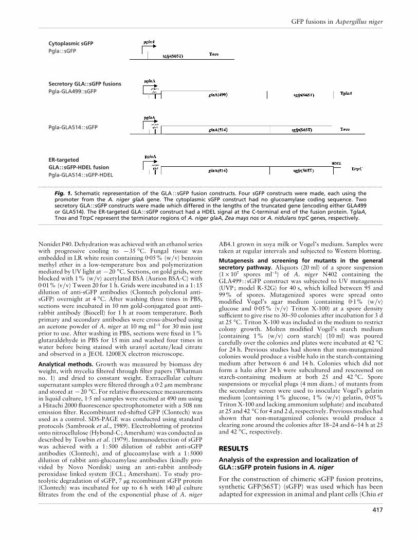
Cytoplasmic sGFPPgla: :sGFP
Secretory GLA::sGFP fusionsPgla-GLA499::sGFP
ER-targetedGLA::sGFP-HDEL fusionPgla-GLA514::sGFP-HDEL
Pgla-GLA514::sGFP
.................................................................................................................................................................................................................................................................................................................
Fig. 1. Schematic representation of the GLA ::sGFP fusion constructs. Four sGFP constructs were made, each using thepromoter from the A. niger glaA gene. The cytoplasmic sGFP construct had no glucoamylase coding sequence. Twosecretory GLA::sGFP constructs were made which differed in the lengths of the truncated gene (encoding either GLA499or GLA514). The ER-targeted GLA::sGFP construct had a HDEL signal at the C-terminal end of the fusion protein. TglaA,Tnos and TtrpC represent the terminator regions of A. niger glaA, Zea mays nos or A. nidulans trpC genes, respectively.
Nonidet P40. Dehydration was achieved with an ethanol serieswith progressive cooling to ®35 °C. Fungal tissue wasembedded in LR white resin containing 0±05% (w}v) benzoinmethyl ether in a low-temperature box and polymerizationmediated by UV light at ®20 °C. Sections, on gold grids, wereblocked with 1% (w}v) acetylated BSA (Aurion BSA-C) with0±01% (v}v) Tween 20 for 1 h. Grids were incubated in a 1:15dilution of anti-sGFP antibodies (Clontech polyclonal anti-sGFP) overnight at 4 °C. After washing three times in PBS,sections were incubated in 10 nm gold-conjugated goat anti-rabbit antibody (Biocell) for 1 h at room temperature. Bothprimary and secondary antibodies were cross-absorbed usingan acetone powder of A. niger at 10 mg ml−" for 30 min justprior to use. After washing in PBS, sections were fixed in 1%glutaraldehyde in PBS for 15 min and washed four times inwater before being stained with uranyl acetate}lead citrateand observed in a JEOL 1200EX electron microscope.
Analytical methods. Growth was measured by biomass dryweight, with mycelia filtered through filter papers (Whatmanno. 1) and dried to constant weight. Extracellular culturesupernatant samples were filtered through a 0±2 µm membraneand stored at ®20 °C. For relative fluorescence measurementsin liquid culture, 1±5 ml samples were excited at 490 nm usinga Hitachi 2000 fluorescence spectrophotometer with a 508 nmemission filter. Recombinant red-shifted GFP (Clontech) wasused as a control. SDS-PAGE was conducted using standardprotocols (Sambrook et al., 1989). Electroblotting of proteinsonto nitrocellulose (Hybond-C; Amersham) was conducted asdescribed by Towbin et al. (1979). Immunodetection of sGFPwas achieved with a 1:500 dilution of rabbit anti-sGFPantibodies (Clontech), and of glucoamylase with a 1:5000dilution of rabbit anti-glucoamylase antibodies (kindly pro-vided by Novo Nordisk) using an anti-rabbit antibodyperoxidase linked system (ECL; Amersham). To study pro-teolytic degradation of sGFP, 7 µg recombinant sGFP protein(Clontech) was incubated for up to 6 h with 140 µl culturefiltrates from the end of the exponential phase of A. niger
AB4.1 grown in soya milk or Vogel’s medium. Samples weretaken at regular intervals and subjected to Western blotting.
Mutagenesis and screening for mutants in the generalsecretory pathway. Aliquots (20 ml) of a spore suspension(1¬10( spores ml−") of A. niger N402 containing theGLA499: :sGFP construct was subjected to UV mutagenesis(UVP; model R-52G) for 40 s, which killed between 95 and99% of spores. Mutagenized spores were spread ontomodified Vogel’s agar medium (containing 0±1% (w}v)glucose and 0±05% (v}v) Triton X-100) at a spore densitysufficient to give rise to 30–50 colonies after incubation for 3 dat 25 °C. Triton X-100 was included in the medium to restrictcolony growth. Molten modified Vogel’s starch medium[containing 1% (w}v) corn starch] (10 ml) was pouredcarefully over the colonies and plates were incubated at 42 °Cfor 24 h. Previous studies had shown that non-mutagenizedcolonies would produce a visible halo in the starch-containingmedium after between 6 and 14 h. Colonies which did notform a halo after 24 h were subcultured and rescreened onstarch-containing medium at both 25 and 42 °C. Sporesuspensions or mycelial plugs (4 mm diam.) of mutants fromthe secondary screen were used to inoculate Vogel’s gelatinmedium [containing 1% glucose, 1% (w}v) gelatin, 0±05%Triton X-100 and lacking ammonium sulphate) and incubatedat 25 and 42 °C for 4 and 2 d, respectively. Previous studies hadshown that non-mutagenized colonies would produce aclearing zone around the colonies after 18–24 and 6–14 h at 25and 42 °C, respectively.
RESULTS
Analysis of the expression and localization ofGLA::sGFP protein fusions in A. niger
For the construction of chimeric sGFP fusion proteins,synthetic GFP(S65T) (sGFP) was used which has beenadapted for expression in animal and plant cells (Chiu et
417

C. L. GORDON and OTHERS
(a)
(b)
(c)
.................................................................................................................................................
Fig. 2. Analysis of A. niger AB4.1 transformants expressing sGFPfusion proteins. Hyphae of transformants expressing (a) thesecretory construct G514::sGFP, (b) the ER-directed constructG514::sGFP-HDEL and (c) the cytoplasmic sGFP construct.Transformants were grown at 30 °C for 20 h. Bar, 20 µm.
al., 1996; Haas et al., 1996), and has been successfullyexpressed in filamentous fungi. For the cytoplasmicexpression of gfp in A. niger, the gfp gene was fused to
the A. niger glaA promoter (Fig. 1). The glaA promoteris known to direct production of high levels of gluco-amylase in the presence of starch, maltodextrin andglucose, and to be repressed when xylose is used as acarbon source (Fowler et al., 1990; Schrickx et al.,1993; Verdoes et al., 1994; Siedenberg et al., 1999).Analysis of A. niger transformants containing the gfpgene by fluorescence microscopy showed high ex-pression levels of cytoplasmic sGFP when grown onglucose. sGFP was present throughout the cytoplasmand appeared to be excluded from the vacuoles, whichwere visible as dark spots within hyphal compartments(Fig. 2c). Very low background fluorescence was ob-served in transformants that only contained the select-able marker (results not shown) or in medium withxylose as the sole carbon source, indicating that sGFPexpression was still under control of the glaA promoter(Siedenberg et al., 1999).
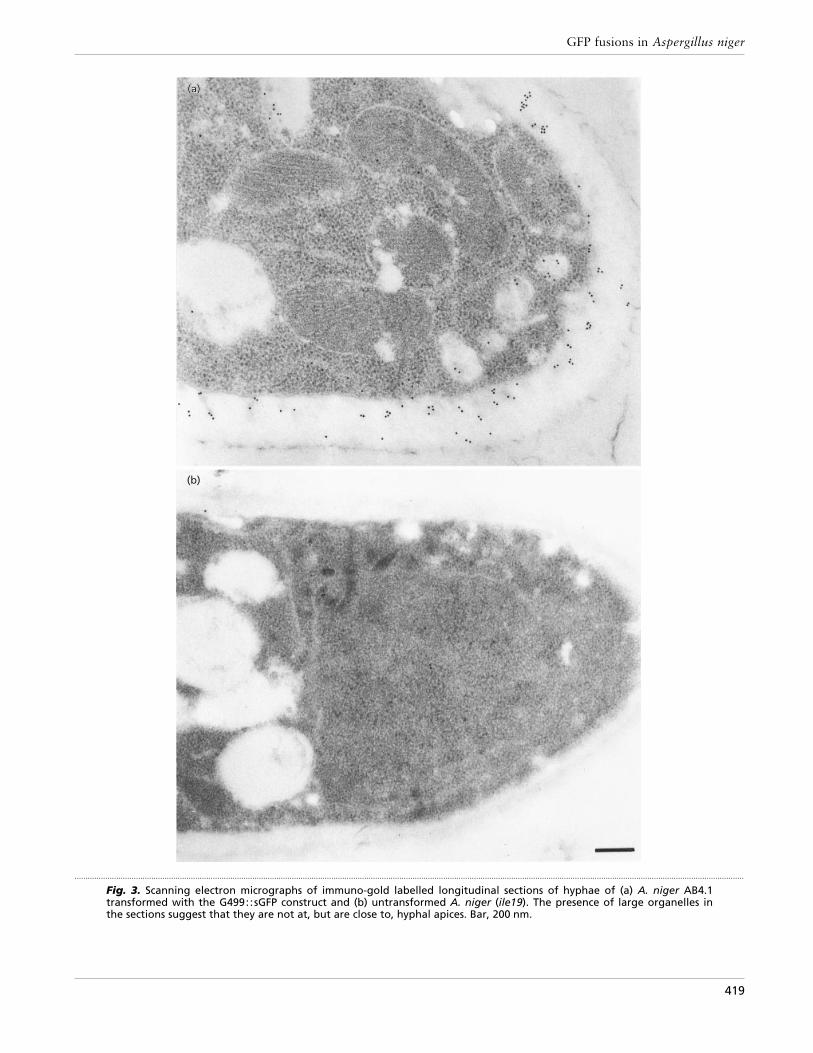
To monitor protein secretion in vivo, the sGFP gene wasfused to glaA. Two different constructs were made usingeither the coding sequence for the first 499 aminoacids or the first 514 amino acids of glucoamylase(GLA499: :sGFP and GLA514: :sGFP, respectively ; Fig.1). Expression of both fusion proteins inA.niger resultedin similar patterns of localization. Most fluorescencewas observed in the cell walls and in septa, indicatingthat both fusion proteins are secreted and at leastpartially retained within the cell wall (Fig. 2a). Nosignificant intracellular fluorescence was observed andthe pattern of fluorescence contrasted sharply to thegranular intracellular fluorescence observed with theGLA::sGFP-HDEL [endoplasmic reticulum (ER)-reten-tion signal] strain (Fig. 2b) and the more homogeneousfluorescence observed in the cytoplasmic-sGFP-express-ing transformant (Fig. 2c). Immunogold labelling oflongitudinal sections of hyphae of the GLA::sGFP-expressing strain by scanning electron microscopyconfirmed the presence of sGFP within the hyphal cellwall (Fig. 3). In addition, we were able to removeGLA::sGFP fusion protein from the cell wall using amild SDS-extraction of intact mycelium [0±05% SDS in50 mM sodium phosphate buffer (pH 7±0) for 5 min at95 °C]. This treatment did not cause lysis of the myceliaas no glucose-6-phosphate dehydrogenase, a cytoplas-mic protein, was detected on Western blots (results notshown).
GLA::sGFP expression and localization in batchculture
Initially in shake-flask cultures, GLA::sGFP was visibleas bright fluorescence in the cell walls and septa;however fluorescence was typically lost after 16–20 hgrowth (Fig. 4a). In contrast, intracellular fluorescencewas still observed in the cytoplasmic sGFP strain (resultsnot shown) and the GLA::sGFP-HDEL transformant(Fig. 4b). As GFP fluorescence is sensitive to low pH(Patterson et al., 1997), the rapid acidification of themedium in shake-flask cultures during the growth of A.niger probably accounts for the observed loss offluorescence in the hyphal wall. Loss of fluorescence in
418
GFP fusions in Aspergillus niger
(a)(a)(a)
(b)
.................................................................................................................................................................................................................................................................................................................
Fig. 3. Scanning electron micrographs of immuno-gold labelled longitudinal sections of hyphae of (a) A. niger AB4.1transformed with the G499::sGFP construct and (b) untransformed A. niger (ile19). The presence of large organelles inthe sections suggest that they are not at, but are close to, hyphal apices. Bar, 200 nm.
419

C. L. GORDON and OTHERS
(a) (b)
(c) (d)
.................................................................................................................................................................................................................................................................................................................
Fig. 4. A. niger AB4.1 and D15 transformants expressing sGFP fusion proteins in shake-flask culture. (a, b) A. niger AB4.1containing (a) the G514::sGFP construct and (b) the G514::sGFP-HDEL construct. (c, d) A. niger D15 containing (c) theG514: :sGFP construct and (d) the G514::sGFP-HDEL construct. All strains were grown on minimal medium at 30 °C withan initial pH of 6±0. Images were taken after 16 h growth. Bar, 20 µm.
the cell wall was not observed when the non-acidifyingA. niger D15 mutant expressing GLA::sGFP was grownin shake-flask culture (Fig. 4c, d). After 16 h growth, thepH of the culture medium of this transformant remainedat the initial value of 6±0. The effect of extracellular pHon the fluorescence of sGFP in the hyphal wall could befurther demonstrated by studying the effect of changingthe extracellular pH on cell wall fluorescence. The cellwalls of A. niger D15 GLA::sGFP were fluorescentwhen at pH 6±0 but lost fluorescence over a period of90 min when transferred to pH 3±5 (Fig. 5a, b). Aspredicted, the intracellular fluorescence of A. niger D15expressing GLA::sGFP-HDEL was not affected bychanging the external pH (Fig. 5c, d).
Secretion of GLA::sGFP takes place at the hyphal tips
Newly synthesized GFP molecules need to be processedinto their mature form, i.e. the chromophore, beforebeing able to emit fluorescence. Although sGFP(S65T)has been shown to fold more rapidly than wild-typeGFP, this may still take up to 45 min (Heim et al., 1995).When freshly grown mycelia were directly observed
using the fluorescence microscope, a lower fluorescentsignal was often observed in the hyphal apices comparedto more subapical regions and septa. It is possible thatthe time required for chromophore formation is re-sponsible for this observation. To overcome this prob-lem, young germlings were held at a low temperature(6 °C) for at least 2 h before observations, to slow downprotein secretion and to allow newly secreted sGFP tobecome fluorescent. In doing so, intense fluorescencewas observed at most hyphal apices compared tosubapical regions (Fig. 6a). Loss of fluorescence from thesubapical hyphal wall was also observed after 28 h (Fig.6b), probably due to the increased acidification of themedium.
Localization of secreted GLA::sGFP fusion proteins
To investigate whether the GLA::sGFP fusion proteincould be detected in the culture medium, samples ofculture media were taken at different times duringgrowth in shake-flask cultures and analysed for thepresence of sGFP by Western blotting and fluorescencespectrophotometry. Neither intact nor degraded
420

GFP fusions in Aspergillus niger
(a) (b)
(c) (d)
.................................................................................................................................................................................................................................................................................................................
Fig. 5. Effect of ambient pH on extracellular GLA::sGFP fluorescence. (a, b) A. niger D15 transformed with the G514::sGFPconstruct incubated (a) in 0±9% NaCl pH 6±0 or (b) after 90 min incubation in 0±9% NaCl adjusted to pH 3±5 with HCl. (c, d)A. niger D15 transformed with the G514::sGFP-HDEL construct incubated (c) at pH 6±0 or (d) at pH 3±5. Bar, 20 µm.
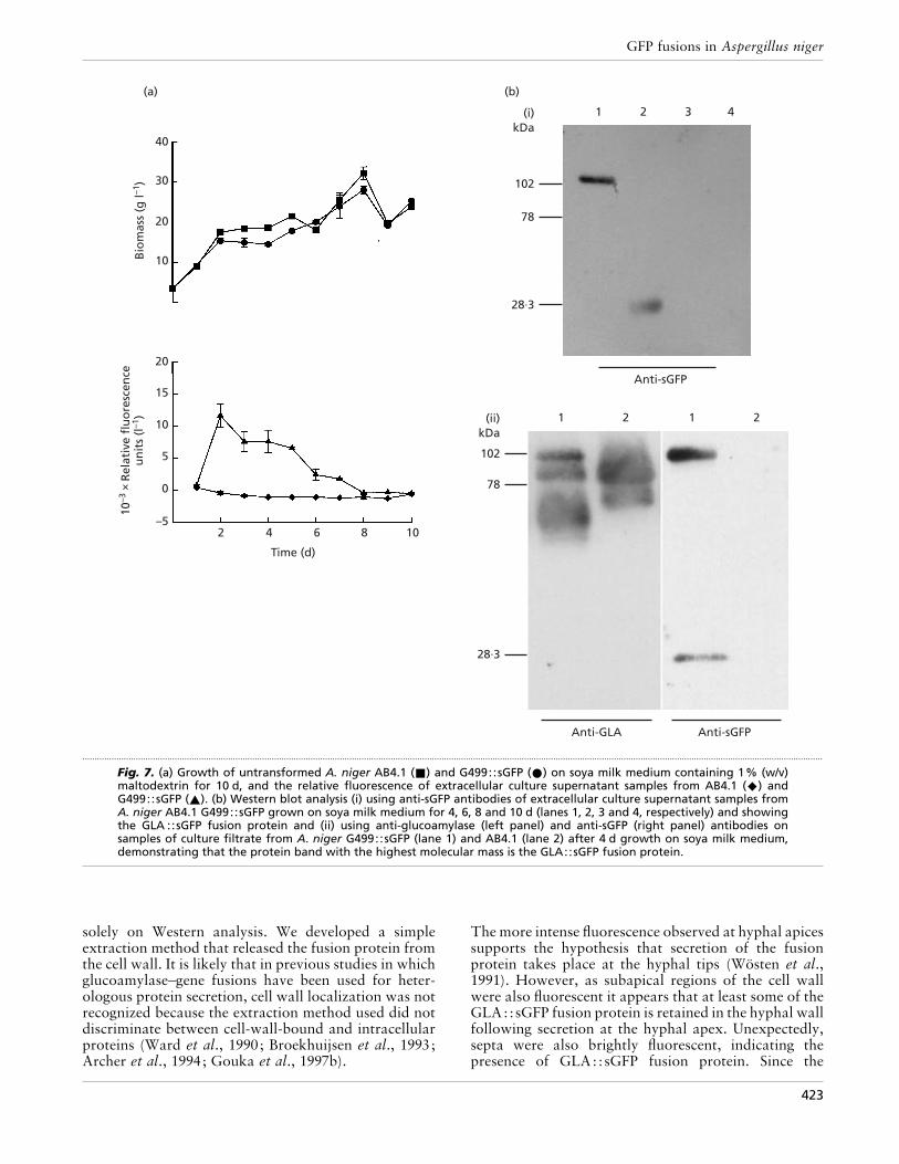
GLA::sGFP could be detected at any point duringgrowth on minimal medium. This suggests that theGLA::sGFP fusion protein is either efficiently retainedwithin the cell wall or is rapidly degraded on release intothe culture medium. In contrast, extracellular fluor-escence was detected when the GLA::sGFP trans-formant was grown in shake-flask cultures in soya milkmedium (Fig. 7a). Fluorescence was highest 2 d afterinoculation and thereafter declined steadily until after8 d no significant fluorescence was detectable.
Western analysis of medium samples with anti-sGFPantibody after 4 d growth on soya milk medium revealedthe presence of a single protein species with a molecularmass of approximately 102 kDa, the predicted size of theGLA::sGFP fusion protein (Fig. 7bi, lane 1) or thepresence of both a 102 kDa and a 27 kDa protein (7bii,right panel, lane 1). Detection of a 102 kDa proteinusing anti-glucoamylase (Fig. 7bii, left panel, lane 1),which was absent in the parental strain (Fig. 7bii, leftpanel, lane 2), confirmed that the medium contained aGLA::sGFP fusion protein. After 6 d growth, no intactGLA::sGFP fusion protein could be detected, but somedegraded sGFP was still present (Fig. 7bi, lane 2). The
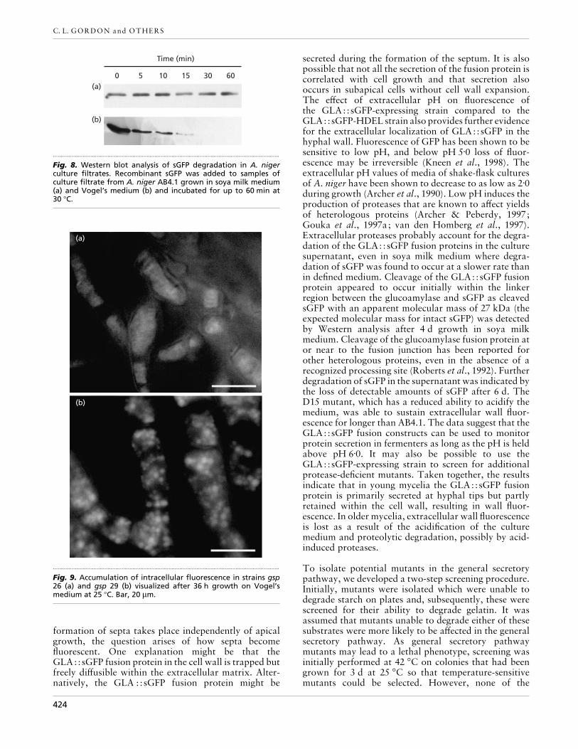
size of this degradation product, with an apparentmolecular mass of 27 kDa, the predicted size of sGFP,suggests that proteolytic cleavage occurred close to thelinker region between the glucoamylase and sGFP.Beyond 6 d, neither GLA::sGFP nor sGFP could bedetected (Fig. 7bi, lanes 3 and 4) indicating that initialcleavage of the GLA::sGFP fusion protein was followedby subsequent degradation of sGFP, probably due toproteases present in the culture medium. To confirmthat proteases present in the culture medium werecapable of degrading sGFP, medium from cultures of A.niger AB4.1 grown on defined and soya milk mediumwere spiked with recombinant sGFP and analysed byWestern blotting over a period of 60 min. RecombinantsGFP was shown to be more rapidly degraded in culturefiltrates obtained from A. niger AB4.1 grown in minimalmedium compared to that from soya milk medium(Fig. 8).
Isolation of mutants in the general secretorypathway
A total of approximately 5000 colonies from UV-mutagenized spores (95–99% kill) of a transformant of
421

C. L. GORDON and OTHERS
(a)
(b)
.................................................................................................................................................
Fig. 6. Secretion occurs at the hyphal tips. Young germlingsof A. niger AB4.1 G514::sGFP grown for (a) 20 h and (b)28 h showing apical localization and loss of subapical wallfluorescence. Bar, 20 µm.
A. niger N402 expressing the GLA499: :sGFP fusionprotein were screened for defects in the general secretorypathway. Twenty-nine colonies were recovered whichwere unable to form a clearing zone on starch platesafter 24 h at 42 °C. None of the colonies isolatedproduced a clearing zone on starch plates at 25 °C.When screened on gelatin medium, 10 putative generalsecretory pathway (gsp) mutants were isolated whichwere unable to form a clearing zone on gelatin-containing medium at either 25 or 42 °C. Seven of theseputative general secretory mutants were found to growat a similar rate to the parental strain but with reducedsporulation (results not shown). Examination of thesemutants revealed fluorescence localized in the walls andsepta with no evidence of intracellular accumulation.The three remaining mutants (gsp 26, 29 and 31) all grewmore slowly than the parental strain, displayed ab-normally wide and swollen hyphae and lacked or hadreduced levels of sporulation (results not shown). Onexamination under fluorescence microscopy, gsp 26 and29 displayed intracellular accumulation of GLA::sGFPwith no fluorescence visible in either the cell walls or
septa (Fig. 9). However, gsp 31 displayed a pattern offluorescence similar to the parental strain with nointracellular accumulation and fluorescence present inboth the cell walls and septa (results not shown).
DISCUSSION
A. niger is used commercially in the production of awide range of secreted enzymes and is being developedas a host for the secretion of heterologous enzymes(Archer & Peberdy, 1997; Gouka et al., 1997a). Despitethe biotechnological importance of protein secretion infilamentous fungi, details of the secretory pathway arelargely unknown. Much of our current knowledgeregarding protein secretion has come from the study oftemperature-sensitive secretion mutants in the yeastSaccharomyces cerevisiae. Identification of severalhomologues in other eukaryotes, including A. niger(Veldhuisen et al., 1997), indicates that the process ofprotein secretion is highly conserved. It is a reasonableassumption therefore, that protein secretion in yeastsand filamentous fungi will share many features. How-ever, the biological and morphological differences be-tween these two groups suggest that additional proteinsmay be involved in the process of protein secretion in thefungal mycelium. This has been highlighted recently bythe identification of an annexin homologue in Neuro-spora crassa which shares homology to the annexin genefamily in higher eukaryotes but appears to be absent inS. cerevisiae (Braun et al., 1998).
In a series of gene fusions we replaced the starch-bindingdomain of glucoamylase with sGFP to create fluorescentmarkers for the study of the secretion process in A.niger. Three glucoamylase sGFP fusions were made.Two of them were designed for secretion of the fusionprotein and employed two different lengths of theglucoamylase protein (GLA499 and GLA514), althoughboth constructs lacked the starch-binding domain. Thisapproach has been used successfully to secrete heter-ologous proteins in both cases. The third constructincluded a C-terminal HDEL motif designed to retainthe fusion protein within the lumen of the ER. Thedifferent lengths of glucoamylase in the secretory fusionswere found to have no significant effect as bothGLA499: :sGFP and GLA514: :sGFP produced the samepatterns of fluorescence in A. niger, and were distinctfrom that of the ER-retained GLA514: :sGFP-HDELand the cytoplasmic-sGFP-containing strains. In youngmycelia expressing the GLA::sGFP fusion protein,bright fluorescence was observed in the hyphal wallsindicating that the fusion protein was secreted, butretained within the cell wall. The presence of the fusionprotein in the cell wall was confirmed by immuno-goldlabelling. Retention of extracellular proteins in thehyphal wall has been reported previously for glucoseoxidase in A. niger (Witteveen et al., 1992) and invertasein N. crassa (Trevithick & Metezenberg, 1966) and fora variety of secreted proteins in S. cerevisiae (de Nobel& Barnett, 1991). Using sGFP, we were able to directlyobserve in vivo the presence of the fusion protein withinthe cell wall, which would have been difficult based
422
GFP fusions in Aspergillus niger
40
30
20
10
20
15
10
5
0
–52 4 6 8 10
Time (d)
Bio
mas
s (g
l–1)
10–3
× R
elat
ive
flu
ore
scen
ceu
nit
s (l
–1)
1 2 3 4
102
78
28·3
Anti-sGFP
1 2 1 2
102
78
28·3
Anti-sGFPAnti-GLA
(a) (b)
(i)kDa
(ii)kDa
.................................................................................................................................................................................................................................................................................................................
Fig. 7. (a) Growth of untransformed A. niger AB4.1 (+) and G499::sGFP (E) on soya milk medium containing 1% (w/v)maltodextrin for 10 d, and the relative fluorescence of extracellular culture supernatant samples from AB4.1 (U) andG499::sGFP (_). (b) Western blot analysis (i) using anti-sGFP antibodies of extracellular culture supernatant samples fromA. niger AB4.1 G499::sGFP grown on soya milk medium for 4, 6, 8 and 10 d (lanes 1, 2, 3 and 4, respectively) and showingthe GLA ::sGFP fusion protein and (ii) using anti-glucoamylase (left panel) and anti-sGFP (right panel) antibodies onsamples of culture filtrate from A. niger G499::sGFP (lane 1) and AB4.1 (lane 2) after 4 d growth on soya milk medium,demonstrating that the protein band with the highest molecular mass is the GLA::sGFP fusion protein.
solely on Western analysis. We developed a simpleextraction method that released the fusion protein fromthe cell wall. It is likely that in previous studies in whichglucoamylase–gene fusions have been used for heter-ologous protein secretion, cell wall localization was notrecognized because the extraction method used did notdiscriminate between cell-wall-bound and intracellularproteins (Ward et al., 1990; Broekhuijsen et al., 1993;Archer et al., 1994; Gouka et al., 1997b).
The more intense fluorescence observed at hyphal apicessupports the hypothesis that secretion of the fusionprotein takes place at the hyphal tips (Wo$ sten et al.,1991). However, as subapical regions of the cell wallwere also fluorescent it appears that at least some of theGLA::sGFP fusion protein is retained in the hyphal wallfollowing secretion at the hyphal apex. Unexpectedly,septa were also brightly fluorescent, indicating thepresence of GLA::sGFP fusion protein. Since the
423
C. L. GORDON and OTHERS
0 5 10 15 30 60
Time (min)
(a)
(b)
.................................................................................................................................................
Fig. 8. Western blot analysis of sGFP degradation in A. nigerculture filtrates. Recombinant sGFP was added to samples ofculture filtrate from A. niger AB4.1 grown in soya milk medium(a) and Vogel’s medium (b) and incubated for up to 60 min at30 °C.
(a)
(b)
.................................................................................................................................................
Fig. 9. Accumulation of intracellular fluorescence in strains gsp26 (a) and gsp 29 (b) visualized after 36 h growth on Vogel’smedium at 25 °C. Bar, 20 µm.
formation of septa takes place independently of apicalgrowth, the question arises of how septa becomefluorescent. One explanation might be that theGLA::sGFP fusion protein in the cell wall is trapped butfreely diffusible within the extracellular matrix. Alter-natively, the GLA ::sGFP fusion protein might be
secreted during the formation of the septum. It is alsopossible that not all the secretion of the fusion protein iscorrelated with cell growth and that secretion alsooccurs in subapical cells without cell wall expansion.The effect of extracellular pH on fluorescence ofthe GLA::sGFP-expressing strain compared to theGLA::sGFP-HDEL strain also provides further evidencefor the extracellular localization of GLA::sGFP in thehyphal wall. Fluorescence of GFP has been shown to besensitive to low pH, and below pH 5±0 loss of fluor-escence may be irreversible (Kneen et al., 1998). Theextracellular pH values of media of shake-flask culturesof A. niger have been shown to decrease to as low as 2±0during growth (Archer et al., 1990). Low pH induces theproduction of proteases that are known to affect yieldsof heterologous proteins (Archer & Peberdy, 1997;Gouka et al., 1997a; van den Homberg et al., 1997).Extracellular proteases probably account for the degra-dation of the GLA::sGFP fusion proteins in the culturesupernatant, even in soya milk medium where degra-dation of sGFP was found to occur at a slower rate thanin defined medium. Cleavage of the GLA::sGFP fusionprotein appeared to occur initially within the linkerregion between the glucoamylase and sGFP as cleavedsGFP with an apparent molecular mass of 27 kDa (theexpected molecular mass for intact sGFP) was detectedby Western analysis after 4 d growth in soya milkmedium. Cleavage of the glucoamylase fusion protein ator near to the fusion junction has been reported forother heterologous proteins, even in the absence of arecognized processing site (Roberts et al., 1992). Furtherdegradation of sGFP in the supernatant was indicated bythe loss of detectable amounts of sGFP after 6 d. TheD15 mutant, which has a reduced ability to acidify themedium, was able to sustain extracellular wall fluor-escence for longer than AB4.1. The data suggest that theGLA::sGFP fusion constructs can be used to monitorprotein secretion in fermenters as long as the pH is heldabove pH 6±0. It may also be possible to use theGLA::sGFP-expressing strain to screen for additionalprotease-deficient mutants. Taken together, the resultsindicate that in young mycelia the GLA::sGFP fusionprotein is primarily secreted at hyphal tips but partlyretained within the cell wall, resulting in wall fluor-escence. In older mycelia, extracellular wall fluorescenceis lost as a result of the acidification of the culturemedium and proteolytic degradation, possibly by acid-induced proteases.
To isolate potential mutants in the general secretorypathway, we developed a two-step screening procedure.Initially, mutants were isolated which were unable todegrade starch on plates and, subsequently, these werescreened for their ability to degrade gelatin. It wasassumed that mutants unable to degrade either of thesesubstrates were more likely to be affected in the generalsecretory pathway. As general secretory pathwaymutants may lead to a lethal phenotype, screening wasinitially performed at 42 °C on colonies that had beengrown for 3 d at 25 °C so that temperature-sensitivemutants could be selected. However, none of the
424

GFP fusions in Aspergillus niger
mutants isolated from either the first starch plate screenor the subsequent gelatin plate screen displayed tem-perature sensitivity, i.e. the inability to degrade eithersubstrate was displayed at both 25 and 42 °C. Of the 10putative general secretory mutants isolated, 7 appearedto grow at the same rate as the parental strain but allshowed reduced sporulation. None of these mutantswhen examined by fluorescence microscopy showedintracellular accumulation and all had fusion proteinpresent in the walls and septa. These mutants weretherefore disregarded. The lack of halo formation onstarch and gelatin media may reflect a reduction in theoverall ability of these strains to secrete proteins,resulting in delayed halo formation compared to theparent strain rather than a block in secretion per se. Theremaining three putative general secretory pathwaymutants all displayed poor growth at both 25 and 42 °Ccompared to the parental strain, a complete loss ofsporulation and abnormal hyphal morphology. Whenexamined under fluorescence microscopy, two mutants(gsp 26 and 29) displayed intracellular accumulation ofGLA::sGFP fusion protein with no detectable fluor-escence in the walls or septa. The third mutant (gsp 31)showed no accumulation and fluorescence in the wall orsepta and was disregarded. The pattern of accumulationof GLA::sGFP fusion protein in gsp 26 and 29 differedsignificantly from each other. In gsp 26 (Fig. 9a),fluorescence was diffuse and evenly located throughoutthe hyphae, whereas in gsp 29 (Fig. 9b), accumulation incircular bodies ranging in size from approximately 0±5 to2 µm diameter could clearly be seen. Thus, for both gsp26 and 29, preliminary evidence strongly suggests thepresence of defects in the general secretory pathway,leading to intracellular accumulation, and that thesedefects are likely to be affecting different points withinthe pathway. Further characterization of both gsp 26and 29 is currently being undertaken to identify the sitesat which accumulation occurs as well as examining theeffects of inhibitors of secretion on the dynamicsand organization of the secretory pathway. Thus theGLA::sGFP constructs allow the direct visualization ofprotein secretion and localization in vivo in growinghyphae and have proved invaluable in the charac-terization of secretory mutants, allowing a quick andreliable method for confirming blocks in the secretorypathway and providing additional visual information onthe sites of accumulation within the hyphae.
ACKNOWLEDGEMENTS
We would like to thank Jen Sheen for sGFP(S65T), RoyMontijn for help in the analysis of transformants and GerdaLamers for assistance with the microscopical techniques.
The work carried out in the UK was funded by a BBSRCstudentship to C.G. and a scholarship from the PasteurInstitute of Iran to V.K.
REFERENCES
Archer, D. B. & Peberdy, J. F. (1997). The molecular biology ofsecreted enzyme production by fungi. Crit Rev Biotechnol 17,273–306.
Archer, D. B., Roberts, I. N. & MacKenzie, D. A. (1990). Heter-ologous protein secretion from Aspergillus niger in phosphate-buffered batch culture. Appl Microbiol Biotechnol 34, 313–315.
Archer, D. B., Jeenes, D. J. & MacKenzie, D. A. (1994). Strategiesfor improving heterologous protein production from filamentousfungi. Antonie Leeuwenhoek 65, 245–250.
Braun, E. L., Kang, S. C., Nelson, M. A. & Natvig, D. O. (1998).Identification of the first fungal annexin: analysis of annexin geneduplications and implications for eukaryotic evolution. J MolEvol 47, 531–543.
Broekhuijsen, M. P., Mattern, I. E., Contreras, R., Kinghorn, J. R.& van den Hondel, C. A. M. J. J. (1993). Secretion of heterologousproteins by Aspergillus niger : production of active humaninterleukin-6 in a protease-deficient mutant by KEX2-like pro-cessing of a glucoamylase–hIL6 fusion protein. J Biotechnol 31,135–145.
Chalfie, M. (1995). Green fluorescent protein. Photochem Photo-biol 62, 651–656.
Chiu, W.-L., Niwa, Y., Zeng, W., Hirano, T., Kobayashi, H. &Sheen, J. (1996). Engineered GFP as a vital reporter in plants. CurrBiol 6, 325–330.
Contreras, R., Carrez, D., Kinghorn, J. R., van den Hondel,C. A. M. J. J. & Fiers, W. (1991). Efficient KEX2-like processingof a glucoamylase–interleukin-6 fusion protein by Aspergillusnidulans and secretion of mature interleukin-6. Biotechnology 9,378–381.
Cormack, B. P., Bertram, G., Egerton, M., Gow, N. A. R., Falkow,S. & Brown, A. J. P. (1997). Yeast-enhanced green fluorescentprotein (yEGFP) : a reporter of gene expression in Candidaalbicans. Microbiology 143, 303–311.
Durand, H., Clanet, M. & Tiraby, G. (1988). Genetic improvementof Trichoderma reesei for large scale cellulase production.Enzyme Microb Technol 10, 341–345.
Ferna! ndez-A! balos, J. M., Fox, H., Pitt, C., Wells, B. & Doonan,J. H. (1998). Plant-adapted green fluorescent protein is a versatilevital reporter for gene expression, protein localization and mitosisin the filamentous fungus Aspergillus nidulans. Mol Microbiol 27,121–130.
Finkelstein, D. B. (1987). Improvement of enzyme production inAspergillus. Antonie Leeuwenhoek 53, 349–352.
Fowler, T., Berka, R. M. & Ward, M. (1990). Regulation of the glaAgene in Aspergillus niger. Curr Genet 18, 537–545.
Gouka, R. J., Punt, P. J., Hessing, J. G. M. & van den Hondel,C. A. M. J. J. (1996). Analysis of heterologous protein productionin defined recombinant Aspergillus awamori strains. ApplEnviron Microbiol 62, 1951–1957.
Gouka, R. J., Punt, P. J. & van den Hondel, C. A. M. J. J. (1997a).Efficient production of secreted proteins by Aspergillus : progress,limitations and prospects. Appl Microbiol Biotechnol 47, 1–11.
Gouka, R. J., Punt, P. J. & van den Hondel, C. A. M. J. J. (1997b).Glucoamylase gene fusions alleviate limitations for proteinproduction in Aspergillus awamori at the transcriptional and(post) translational levels. Appl Environ Microbiol 63, 488–497.
Haas, J., Park, E.-C. & Seed, B. (1996). Codon usage limitation inthe expression of HIV-1 envelope glycoprotein. Curr Biol 6,315–324.
Harris, S. D., Morrell, J. L. & Hamer, J. E. (1994). Identification andcharacterization of Aspergillus nidulans mutants defective incytokinesis. Genetics 136, 517–532.
van Hartingsveldt, W., Mattern, I. E., van Zeijl, C. M. J., Pouwels,P. H. & van den Hondel, C. A. M. J. J. (1987). Development of a
425

C. L. GORDON and OTHERS
homologous transformation system for Aspergillus niger basedon the pyrG gene. Mol Gen Genet 206, 71–75.
van den Homberg, J. P. T. W., van de Vondervoort, P. J. I.,Fraissinet-Tachet, L. & Visser, J. (1997). Aspergillus as a host forheterologous protein production, the problem of proteases.Trends Biotechnol 15, 256–263.
Heim, R., Cubitt, A. B. & Tsien, R. Y. (1995). Improved greenfluorescence. Nature 373, 663–664.
Jeenes, D. J., MacKenzie, D. A. & Archer, D. B. (1994). Tran-scriptional and post-translational events affect the production ofsecreted hen egg white lysozyme by Aspergillus niger. TransgenicRes 3, 297–303.
Kneen, M., Farinas, J., Li, Y. & Verkman, A. S. (1998). Greenfluorescent protein as a noninvasive intracellular pH indicator.Biophys J 74, 1591–1599.
MacKenzie, D. A., Gendron, L. C. G., Jeenes, D. J. & Archer, D. B.(1994). Physiological optimization of secreted protein productionby Aspergillus niger. Enzyme Microb Technol 16, 276–280.
Mattern, I. E., van Noort, J. M., van den Berg, P., Roberts, I. N.,Archer, D. B. & van den Hondel, C. A. M. J. J. (1992). Isolation andcharacterization of mutants of Aspergillus niger deficient inextacellular proteases. Mol Gen Genet 234, 332–336.
Moukha, S. M., Wo$ sten, H. A. B., Asther, M. & Wessels, J. G. H.(1993). In situ localization of the secretion of lignin peroxidases incolonies of Phanerochaete chrysosporium using a sandwichedmode of culture. J Gen Microbiol 139, 969–978.
de Nobel, J. G. & Barnett, J. A. (1991). Passage of moleculesthrough yeast cell walls – a brief essay review. Yeast 7, 313–323.
Nyysso$ nen, E. & Kera$ nen, S. (1995). Multiple roles of the cellulaseCBHI in enhancing production of fusion antibodies by thefilamentous fungus Trichoderma reesei. Curr Genet 28, 71–79.
Patterson, G. H., Knobel, S. M., Sharif, W. D., Kain, S. R. & Piston,D. W. (1997). Use of the green fluorescent protein and its mutantsin quantitative fluorescence microscopy. Biophys J 73, 2782–2790.
Punt, P. J., Oliver, R. P., Dingemanse, M. A., Pouwels, P. H. & vanden Hondel, C. A. M. J. J. (1987). Transformation of filamentousfungi based on the hygromycin B resistance marker fromEscherichia coli. Gene 56, 117–124.
Punt, P. J. & van den Hondel, C. A. M. J. J. (1992). Transformationof filamentous fungi based on hygromycin B and phleomycinresistance markers. Methods Enzymol 216, 447–457.
Roberts, I. N., Jeenes, D. J., MacKenzie, D. A., Wilkinson, A. P.,Sumner, I. G. & Archer, D. B. (1992). Heterologous gene expressionin Aspergillus niger, a glucoamylase–porcine pancreatic prophos-pholipase A2 fusion protein is secreted and processed to yieldmature enzyme. Gene 122, 155–161.
Sambrook, J., Fritsch, E. F. & Maniatis, T. (1989). MolecularCloning: a Laboratory Manual, 2nd edn. Cold Spring Harbor,NY: Cold Spring Harbor Laboratory.
Schrickx, J. M., Krave, A. S., Verdoes, J. C. & van den Hondel,C. A. M. J. J. (1993). Growth and product formation in chemostatand recycling cultures by Aspergillus niger N402 and a gluco-amylase overproducing transformant, provided by multiplecopies of the glaA gene. J Gen Microbiol 139, 2801–2810.
Sheen, J., Hwang, S., Niwa, Y., Kobayashi, H. & Galbraith, D. W.(1995). Green-fluorescent protein as a new vital marker in plantcells. Plant J 8, 777–784.
Siedenberg, D., Mestric, S., Ganzlin, M., Schmidt, M., Punt, P. J.,van den Hondel, C. A. M. J. J. & Rinas, U. (1999). GlaA promotercontrolled production of a mutant green fluorescent protein(S65T) by recombinant Aspergillus niger during growth ondefined medium in batch and fed-batch cultures. Biotechnol Prog15, 43–50.
Southern, E. M. (1975). Detection of specific sequences amongDNA fragments separated by gel electrophoresis. J Mol Biol 98,503–517.
Spellig, T., Bottin, A. & Kahmann, R. (1996). Green fluorescentprotein (GFP) as a new vital marker in the phytopathogenicfungus Ustilago maydis. Mol Gen Genet 252, 503–509.
Suelmann, R., Sievers, N. & Fischer, R. (1997). Nuclear traffic infungal hyphae: in vivo study of nuclear migration and positioningin Aspergillus nidulans. Mol Microbiol 25, 757–769.
Towbin, H., Staehelin, T. & Gordon, J. (1979). Electrophoretictransfer of proteins from polyacrylamide gels to nitrocellulosesheets : procedure and some applications. Proc Natl Acad Sci USA76, 4350–4354.
Trevithick, J. R. & Metezenberg, R. L. (1966). Molecular sieving byNeurospora crassa cell walls during secretion of invertaseisoenzymes. J Bacteriol 92, 1010–1015.
Veldhuisen, G., Saloheimo, M., Fiers, M. A., Punt, P. J., Contreras,R., Penttila, M. & van den Hondel, C. A. M. J. J. (1997). Isolationand analysis of functional homologues of the secretion-relatedSAR1 gene of Saccharomyces cerevisiae from Aspergillus nigerand Trichoderma reesei. Mol Gen Genet 256, 446–455.
Verdoes, J. C., Punt, P. J., Strouthamer, A. H. & van den Hondel,C. A. M. J. J. (1994). The effect of multiple copies of the upstreamregion on expression of the Aspergillus niger glucoamylase-encoding gene. Gene 145, 179–187.
Vogel, H. J. (1956). A convenient growth medium for Neurospora(medium N). Microb Genet Bull 13, 42–44.
Ward, M., Wilson, L. J., Kodama, K. H., Rey, M. W. & Berka, R. M.(1990). Improved production of chymosin in Aspergillus byexpression as a glucoamylase–chymosin fusion. Biotechnology 8,435–440.
Wessels, J. G. H. (1994). Developmental regulation of fungal cellwall formation. Annu Rev Phytopathol 32, 413–437.
Witteveen, C. F. B., Veenhuis, M. & Visser, J. (1992). Localisationof glucose oxidase and catalase activities in Aspergillus niger.Appl Environ Microbiol 58, 1190–1194.
Wo$ sten, H. A. B., Moukha, S. M., Sietsma, J. H. & Wessels, J. G. H.(1991). Localization of growth and secretion of proteins inAspergillus niger. J Gen Microbiol 137, 2017–2023.
van den Wymelenberg, A. J., Cullen, D., Spear, R. N., Schoenike,B. & Andrews, J. H. (1997). Expression of green fluorescentprotein in Aureobasidium pullulans and quantification of thefungus on leaf surfaces. Biotechniques 23, 686–690.
van Zeijl, C. M. J., van de Kamp, E. H. M., Punt, P. J., Selten,G. C. M., Hauer, B., van Gorcom, R. F. M. & van den Hondel,C. A. M. J. J. (1998). An improved colony-PCR method forfilamentous fungi for amplication of PCR fragments of severalkilobases. J Biotechnol 59, 221–224.
.................................................................................................................................................
Received 9 April 1999; revised 11 October 1999; accepted 28 October1999.
426
Copyright © 2022 FDOKUMEN