Absence of TMPRSS2:ERG fusions and PTEN losses in prostate cancer is associated with a favorable...
10
Absence of TMPRSS2:ERG fusions and PTEN losses in prostate cancer is associated with a favorable outcome Maisa Yoshimoto 1 , Anthony M Joshua 1 , Isabela W Cunha 2 , Renata A Coudry 2 , Francisco P Fonseca 3 , Olga Ludkovski 1 , Maria Zielenska 4 , Fernando A Soares 2 and Jeremy A Squire 1,5 1 Division of Applied Molecular Oncology, Ontario Cancer Institute, Toronto, ON, Canada; 2 Department of Pathology, Hospital do Ca ˆncer, Sa ˜o Paulo, Brazil; 3 Department of Pelvic Surgery, Hospital do Ca ˆncer, Sa ˜o Paulo, Brazil; 4 Department of Pathology and Laboratory Medicine, The Hospital for Sick Children, Toronto, ON, Canada and 5 Department of Medical Biophysics, Faculty of Medicine, University of Toronto, Toronto, ON, Canada TMPRSS2:ERG gene fusions and PTEN deletions are the most common genomic aberrations in prostate cancer. Recent work has suggested that the TMPRSS2:ERG fusion is associated with a more aggressive phenotype. Similarly, PTEN deletion has been associated with biochemical recurrence and lymph node metastasis. To date, there has been no systematic analysis of the combined influence of genomic PTEN deletion with TMPRSS2:ERG gene fusions on clinical parameters of prostate cancer progression. We carried out a retrospective analysis of 125 prostate cancers with known clinical outcome using interphase fluorescence in situ hybridization to detect the relative prevalence of TMPRSS2:ERG rearrangements and/or PTEN genomic deletions. TMPRSS2:ERG rearrangement was found in 60 of 125 (48%) prostate cancers. Duplication of TMPRSS2:ERG fusion was observed in seven (6%) tumors. Gleason grade (P ¼ 0.0002)/score (P ¼ 0.001), median tumor volume (P ¼ 0.0024), preoperative PSA (P ¼ 0.001) and perineural invasion (P ¼ 0.0304) were significantly associated with biochemical recurrence by univariate analysis with TMPRSS2:ERG approaching significance (P ¼ 0.0523). By multivariate analysis, relevant factors associated with recurrence were Gleason scores 7 (P ¼ 0.001) and 8–10 (P ¼ 0.015), PTEN homozygous deletion (P ¼ 0.013) and concurrent TMPRSS2:ERG fusion and PTEN deletion (P ¼ 0.036). Kaplan–Meier analysis indicated that the presence of TMPRSS2:ERG fusion was marginally less favorable in comparison to no fusion. Duplication of fusion gene showed worse prognosis. It was possible to determine the relative frequencies of PTEN deletion and/or TMPRSS2:ERG fusions in 82 of 125 prostate cancers. With biochemical recurrence as an endpoint, the genomic biomarkers identified three patient groups: (1) ‘poor genomic grade’ characterized by both PTEN deletion and TMPRSS2:ERG fusions (23/82, 28%); (2) ‘intermediate genomic grade’ with either PTEN deletion or TMPRSS2:ERG fusion (35/82, 43%) and (3) ‘favorable genomic grade’ in which neither rearrangement was present (24/82, 29%). Kaplan–Meier and multivariate analysis indicate that TMPRSS2:ERG fusion and PTEN loss together are a predictor of earlier biochemical recurrence of disease. Modern Pathology (2008) 21, 1451–1460; doi:10.1038/modpathol.2008.96; published online 23 May 2008 Keywords: interphase FISH; PTEN haploinsufficiency; prognosis; biomarker; biochemical recurrence The identification of prognostic molecular biomar- kers is recognized as critically important in the future clinical management of prostate cancer. Despite the clinical utility of Gleason score, patho- logical stage and serum PSA in assessing prognosis and guiding management, further molecular deter- minants are needed that more accurately address pathways that underlie tumorigenesis of prostate cancer. 1 Chromosomal deletions of 10q suggested that the phosphatase and tensin homologue (PTEN) gene at cytoband 10q23.3 is strongly associated with the progression of prostate cancer. 2–4 PTEN plays an important role in the modulation of the phosphati- dylinositol-3-kinase (PI3K) pathway by catalyzing degradation of phosphatidylinositol-(3,4,5)-tripho- sphate (PIP3) generated by PI3K. 5 PIP3 activates the protein kinase AKT which then modulates a number of downstream targets with important roles in apoptosis and the cell-cycle progression, including BAD, 6 CASP3 and CASP9, 7 MDM2, 8 mTOR, 9 FKHR 10 and FOXO3A, 11 p27 12 and the recently recognized Received 14 March 2008; revised 25 April 2008; accepted 26 April 2008; published online 23 May 2008 Correspondence: Dr JA Squire, PhD, Division of Applied Molecular Oncology, Ontario Cancer Institute, Princess Margaret Hospital, 610 University Avenue, Room 9-721, Toronto, ON M5G 2M9 Canada. E-mail: [email protected] Modern Pathology (2008) 21, 1451–1460 & 2008 USCAP, Inc All rights reserved 0893-3952/08 $30.00 www.modernpathology.org
-
Upload
independent -
Category
Documents
-
view
1 -
download
0
Transcript of Absence of TMPRSS2:ERG fusions and PTEN losses in prostate cancer is associated with a favorable...
Absence of TMPRSS2:ERG fusions and PTENlosses in prostate cancer is associatedwith a favorable outcome
Maisa Yoshimoto1, Anthony M Joshua1, Isabela W Cunha2, Renata A Coudry2, Francisco PFonseca3, Olga Ludkovski1, Maria Zielenska4, Fernando A Soares2 and Jeremy A Squire1,5
1Division of Applied Molecular Oncology, Ontario Cancer Institute, Toronto, ON, Canada; 2Department ofPathology, Hospital do Cancer, Sao Paulo, Brazil; 3Department of Pelvic Surgery, Hospital do Cancer, SaoPaulo, Brazil; 4Department of Pathology and Laboratory Medicine, The Hospital for Sick Children, Toronto,ON, Canada and 5Department of Medical Biophysics, Faculty of Medicine, University of Toronto, Toronto,ON, Canada
TMPRSS2:ERG gene fusions and PTEN deletions are the most common genomic aberrations in prostate cancer.Recent work has suggested that the TMPRSS2:ERG fusion is associated with a more aggressive phenotype.Similarly, PTEN deletion has been associated with biochemical recurrence and lymph node metastasis. To date,there has been no systematic analysis of the combined influence of genomic PTEN deletion with TMPRSS2:ERGgene fusions on clinical parameters of prostate cancer progression. We carried out a retrospective analysis of125 prostate cancers with known clinical outcome using interphase fluorescence in situ hybridization to detectthe relative prevalence of TMPRSS2:ERG rearrangements and/or PTEN genomic deletions. TMPRSS2:ERGrearrangement was found in 60 of 125 (48%) prostate cancers. Duplication of TMPRSS2:ERG fusion wasobserved in seven (6%) tumors. Gleason grade (P¼ 0.0002)/score (P¼ 0.001), median tumor volume (P¼ 0.0024),preoperative PSA (P¼ 0.001) and perineural invasion (P¼ 0.0304) were significantly associated withbiochemical recurrence by univariate analysis with TMPRSS2:ERG approaching significance (P¼ 0.0523). Bymultivariate analysis, relevant factors associated with recurrence were Gleason scores 7 (P¼ 0.001) and 8–10(P¼ 0.015), PTEN homozygous deletion (P¼ 0.013) and concurrent TMPRSS2:ERG fusion and PTEN deletion(P¼ 0.036). Kaplan–Meier analysis indicated that the presence of TMPRSS2:ERG fusion was marginally lessfavorable in comparison to no fusion. Duplication of fusion gene showed worse prognosis. It was possible todetermine the relative frequencies of PTEN deletion and/or TMPRSS2:ERG fusions in 82 of 125 prostate cancers.With biochemical recurrence as an endpoint, the genomic biomarkers identified three patient groups: (1) ‘poorgenomic grade’ characterized by both PTEN deletion and TMPRSS2:ERG fusions (23/82, 28%); (2) ‘intermediategenomic grade’ with either PTEN deletion or TMPRSS2:ERG fusion (35/82, 43%) and (3) ‘favorable genomic grade’in which neither rearrangement was present (24/82, 29%). Kaplan–Meier and multivariate analysis indicate thatTMPRSS2:ERG fusion and PTEN loss together are a predictor of earlier biochemical recurrence of disease.Modern Pathology (2008) 21, 1451–1460; doi:10.1038/modpathol.2008.96; published online 23 May 2008
Keywords: interphase FISH; PTEN haploinsufficiency; prognosis; biomarker; biochemical recurrence
The identification of prognostic molecular biomar-kers is recognized as critically important in thefuture clinical management of prostate cancer.Despite the clinical utility of Gleason score, patho-logical stage and serum PSA in assessing prognosisand guiding management, further molecular deter-minants are needed that more accurately address
pathways that underlie tumorigenesis of prostatecancer.1
Chromosomal deletions of 10q suggested that thephosphatase and tensin homologue (PTEN) gene atcytoband 10q23.3 is strongly associated with theprogression of prostate cancer.2–4 PTEN plays animportant role in the modulation of the phosphati-dylinositol-3-kinase (PI3K) pathway by catalyzingdegradation of phosphatidylinositol-(3,4,5)-tripho-sphate (PIP3) generated by PI3K.5 PIP3 activates theprotein kinase AKT which then modulates a numberof downstream targets with important roles inapoptosis and the cell-cycle progression, includingBAD,6 CASP3 and CASP9,7 MDM2,8 mTOR,9 FKHR10
and FOXO3A,11 p2712 and the recently recognizedReceived 14 March 2008; revised 25 April 2008; accepted 26 April2008; published online 23 May 2008
Correspondence: Dr JA Squire, PhD, Division of AppliedMolecular Oncology, Ontario Cancer Institute, Princess MargaretHospital, 610 University Avenue, Room 9-721, Toronto, ON M5G2M9 Canada.E-mail: [email protected]
Modern Pathology (2008) 21, 1451–1460& 2008 USCAP, Inc All rights reserved 0893-3952/08 $30.00
www.modernpathology.org
JNK pathway.13 PTEN deletion is associated withtumor progression14–16 and more predictive of short-er time to biochemical recurrence of disease,17
emphasizing the crucial role of PTEN as marker oftumor behavior in prostate cancer.
The discovery of recurrent translocations in B40–60% of prostate carcinoma,4,18–24 involving theTMPRSS2 gene at 21q22.3 with members of theerythroblast transformation specific (ETS) transcrip-tion factor family, such as ERG (21q22.2), ETV1(7p21.2), ETV4 (17q21) or ETV5 (3q28) genes, hasprovided new insights into prostatic carcinogenesis.Although the downstream molecular pathways ofETS fusion genes are only beginning to be clarified,clinical associations have been reported in earlystudies.19,22,25–28 For instance, TMPRSS2:ERG-rear-ranged prostate cancers have been associated with agreater likelihood of lethal prostate cancer,26,27
moderate to poorly differentiated tumors25 andhigher stage diseases with pelvic lymph nodemetastases.19 Evidence of specific TMPRSS2:ERGisoforms were also described to be associated withhigh levels of fusion mRNA expression and earlybiochemical recurrence following radical prostatect-omy.22 However, there are a number of conflictingstudies29,30 about the prognostic importance of thegene fusion; so the clinical significance ofTMPRSS2:ERG fusions remains to be clarified.
Given the potential of both TMPRSS2:ERG fusionsand PTEN genomic deletions to contribute toprognosis prediction in prostate cancer, additionalgenotype–phenotype correlation studies will behelpful. In addition, it is unclear whether theTMPRSS2:ERG rearrangements may be accompa-nied by PTEN genomic loss, or in contrast, these aremutually exclusive events. There has been nosystematic analysis to date of the frequency atwhich PTEN and TMPRSS2:ERG rearrangementsoccur simultaneously in prostate cancer, or toexamine clinical phenotypes associated with geno-mic mutations affecting both pathways. Therefore,this study was designed to retrospectively assessthe overall frequency and clinical impact ofTMPRSS2:ERG rearrangements using a clinically-annotated tissue microarray. We also provide theresults of clinical impact of cooperation betweenTMPRSS2:ERG gene rearrangements and PTENgenomic deletion in prostate cancer samples thatbecame available after the publication of ourprevious study.17 Our results further implicateTMPRSS2:ERG rearrangements as a prognostic valuein the primary tumor, in addition to providinginsights into prostate cancer pathophysiology.
Materials and methods
Tissue Specimens
The collection of all tissue specimens, clinical andpatient follow-up data was obtained after informedconsent in accordance with the Hospital do Cancer
Research Ethics guidelines (Sao Paulo, Brazil).Archival formalin-fixed, paraffin-embedded tissueswere obtained from 125 radical prostatectomiesperformed between 1997 and 2000 at the Hospitaldo Cancer, AC Camargo, Sao Paulo, Brazil. Forcontrol purposes, 10 non-neoplastic prostate tissuesamples were obtained from patients undergoingsurgery solely for benign prostate hyperplasia. Theprostate cancer cohort and control specimens weresampled using a 0.6-mm diameter tissue coredistributed on tissue microarray slide. Adjacenthematoxylin and eosin (H&E) stained section wasreviewed by two pathologists to determine thepresence and extent of morphologically representa-tive areas of the original tumors in each tissue core.Reassessment of Gleason grading in a contiguousH&E stained tissue microarray section assured thepresence of prostate adenocarcinoma and the fide-lity of the intended tissue microarray core. The sizeof tumor was based on assessment of total surfacearea of gland examined histologically involved bycarcinoma. Preoperative PSA level was available forall patients and the PSA nonfailure was definedas PSA remaining below 0.2 ng/ml after radicalprostatectomy.
Fluorescence In Situ Hybridization
Break-apart fluorescence in situ hybridization(FISH) was used for studying the TMPRSS2:ERGgene rearrangement as previously described.31 Thefollowing bacterial artificial chromosome (BAC)clones were used. BACs located at: (a) the 30 ERGgene locus (RP11-476D17, 30ERG sequence extend-ing inward past exon 4), (b) the 50 ERG gene locus(RP11-95I21, 50ERG sequence extending inward intoexon 10), (c) the TMPRSS2 locus (RP11-535H11) and(d) the telomeric BACs to the 50 end of TMPRSS2locus (RP11-35C4, RP11-891L10 and RP11-260O11,325 kb downstream from the 50 end of the TMPRSS2gene; Figure 1a). The BAC clones were obtainedfrom The Center for Applied Genomics(Toronto, Canada). DNA was extracted and labeledwith SpectrumGreen-dUTP, SpectrumOrange-dUTP(Vysis Inc., Downers Grove, IL, USA) or diethyl-aminocoumarin (DEAC)-dUTP (PerkinElmer Lifeand Analytical Sciences, Boston, MA, USA) usingthe Vysis nick-translation kit according to manufac-turer’s instructions (Vysis Inc.). The integrity andcorrect chromosome localization of all BAC cloneswere verified by hybridization to metaphase spreadsof normal peripheral lymphocytes.
Sequential dual-color FISH method wasapplied to the prostate cancer tissue microarray toinvestigate the occurrence of PTEN genomicdeletion in addition to TMPRSS2:ERG gene rearran-gements in 82 of 125 prostate cancer cohort.Dual-color FISH on paraffin-embedded tissue micro-array tissue was performed using commerciallyavailable DNA probes for cytoband 10q23 (Spectrum
PTEN and TMPRSS2:ERG in prostate cancerM Yoshimoto et al
1452
Modern Pathology (2008) 21, 1451–1460
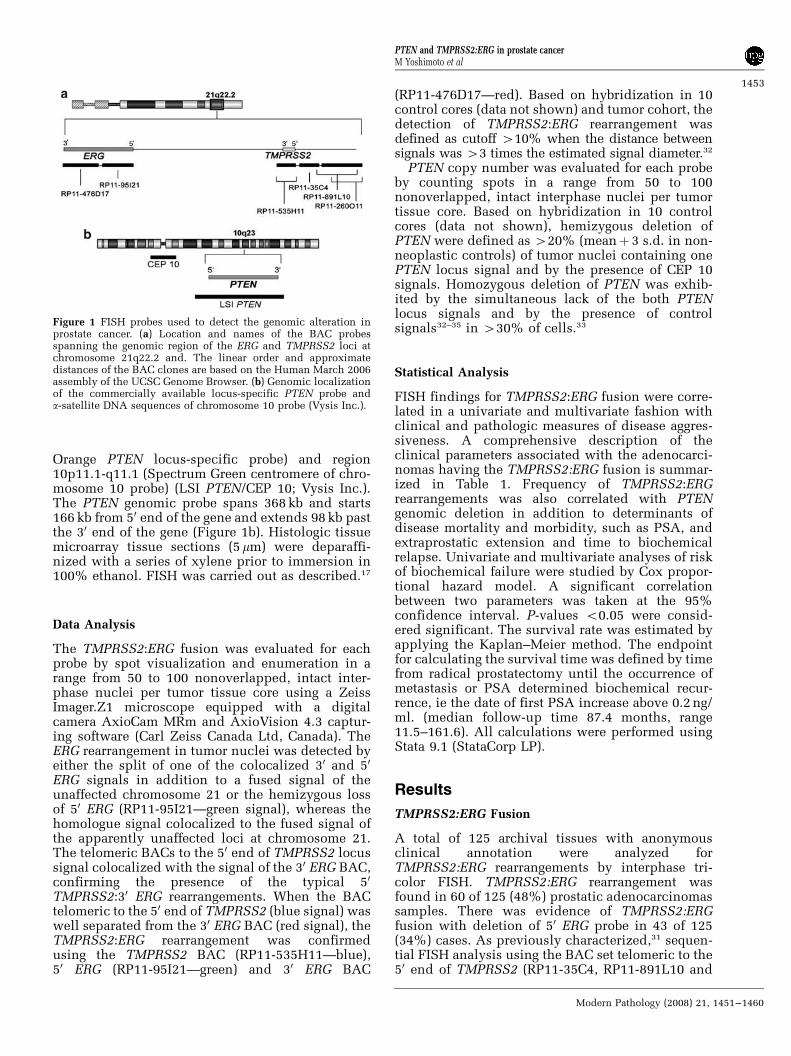
Orange PTEN locus-specific probe) and region10p11.1-q11.1 (Spectrum Green centromere of chro-mosome 10 probe) (LSI PTEN/CEP 10; Vysis Inc.).The PTEN genomic probe spans 368 kb and starts166 kb from 50 end of the gene and extends 98 kb pastthe 30 end of the gene (Figure 1b). Histologic tissuemicroarray tissue sections (5 mm) were deparaffi-nized with a series of xylene prior to immersion in100% ethanol. FISH was carried out as described.17
Data Analysis
The TMPRSS2:ERG fusion was evaluated for eachprobe by spot visualization and enumeration in arange from 50 to 100 nonoverlapped, intact inter-phase nuclei per tumor tissue core using a ZeissImager.Z1 microscope equipped with a digitalcamera AxioCam MRm and AxioVision 4.3 captur-ing software (Carl Zeiss Canada Ltd, Canada). TheERG rearrangement in tumor nuclei was detected byeither the split of one of the colocalized 30 and 50
ERG signals in addition to a fused signal of theunaffected chromosome 21 or the hemizygous lossof 50 ERG (RP11-95I21—green signal), whereas thehomologue signal colocalized to the fused signal ofthe apparently unaffected loci at chromosome 21.The telomeric BACs to the 50 end of TMPRSS2 locussignal colocalized with the signal of the 30 ERG BAC,confirming the presence of the typical 50
TMPRSS2:30 ERG rearrangements. When the BACtelomeric to the 50 end of TMPRSS2 (blue signal) waswell separated from the 30 ERG BAC (red signal), theTMPRSS2:ERG rearrangement was confirmedusing the TMPRSS2 BAC (RP11-535H11—blue),50 ERG (RP11-95I21—green) and 30 ERG BAC
(RP11-476D17—red). Based on hybridization in 10control cores (data not shown) and tumor cohort, thedetection of TMPRSS2:ERG rearrangement wasdefined as cutoff 410% when the distance betweensignals was 43 times the estimated signal diameter.32
PTEN copy number was evaluated for each probeby counting spots in a range from 50 to 100nonoverlapped, intact interphase nuclei per tumortissue core. Based on hybridization in 10 controlcores (data not shown), hemizygous deletion ofPTEN were defined as 420% (meanþ 3 s.d. in non-neoplastic controls) of tumor nuclei containing onePTEN locus signal and by the presence of CEP 10signals. Homozygous deletion of PTEN was exhib-ited by the simultaneous lack of the both PTENlocus signals and by the presence of controlsignals32–35 in 430% of cells.33
Statistical Analysis
FISH findings for TMPRSS2:ERG fusion were corre-lated in a univariate and multivariate fashion withclinical and pathologic measures of disease aggres-siveness. A comprehensive description of theclinical parameters associated with the adenocarci-nomas having the TMPRSS2:ERG fusion is summar-ized in Table 1. Frequency of TMPRSS2:ERGrearrangements was also correlated with PTENgenomic deletion in addition to determinants ofdisease mortality and morbidity, such as PSA, andextraprostatic extension and time to biochemicalrelapse. Univariate and multivariate analyses of riskof biochemical failure were studied by Cox propor-tional hazard model. A significant correlationbetween two parameters was taken at the 95%confidence interval. P-values o0.05 were consid-ered significant. The survival rate was estimated byapplying the Kaplan–Meier method. The endpointfor calculating the survival time was defined by timefrom radical prostatectomy until the occurrence ofmetastasis or PSA determined biochemical recur-rence, ie the date of first PSA increase above 0.2 ng/ml. (median follow-up time 87.4 months, range11.5–161.6). All calculations were performed usingStata 9.1 (StataCorp LP).
Results
TMPRSS2:ERG Fusion
A total of 125 archival tissues with anonymousclinical annotation were analyzed forTMPRSS2:ERG rearrangements by interphase tri-color FISH. TMPRSS2:ERG rearrangement wasfound in 60 of 125 (48%) prostatic adenocarcinomassamples. There was evidence of TMPRSS2:ERGfusion with deletion of 50 ERG probe in 43 of 125(34%) cases. As previously characterized,31 sequen-tial FISH analysis using the BAC set telomeric to the50 end of TMPRSS2 (RP11-35C4, RP11-891L10 and
Figure 1 FISH probes used to detect the genomic alteration inprostate cancer. (a) Location and names of the BAC probesspanning the genomic region of the ERG and TMPRSS2 loci atchromosome 21q22.2 and. The linear order and approximatedistances of the BAC clones are based on the Human March 2006assembly of the UCSC Genome Browser. (b) Genomic localizationof the commercially available locus-specific PTEN probe anda-satellite DNA sequences of chromosome 10 probe (Vysis Inc.).
PTEN and TMPRSS2:ERG in prostate cancerM Yoshimoto et al
1453
Modern Pathology (2008) 21, 1451–1460
RP11-260O11—blue) identified a split of the typicalcolocalized 50 end of TMPRSS2:30 ERG probe signalsin 3 of these 43 samples. This is indicative of a morecomplex genomic alteration, involving an unknownchromosomal partner(s). Confirmation of theTMPRSS2:ERG fusion in these three samples show-ing the atypical FISH pattern was obtained by the
BAC set consisting of the 30 ERG BAC (RP11-476D17), 50 ERG (RP11-95I21) and the TMPRSS2locus (RP11-535H11). Interestingly, an extra copy ofTMPRSS2:ERG fusion associated with deletion of 50
ERG probe was observed in 7 of the 43 samplesshowing TMPRSS2:ERG fusion with deletion of 50
ERG (Table 2). There was evidence of FISH
Table 1 Clinicopathological parameters from 122 of the 125 prostatic adenocarcinoma patients
Clinicopathological parameters Number of cases TMPRSS2:ERG P-value
Not fused Fusion
Preoperative PSA (ng/ml)a 0.6120.9–4.0 9 3 (33.33) 6 (66.67)4.0–10.0 58 31 (53.45) 27 (46.55)10.1–20.0 37 21 (56.76) 16 (43.24)20.1–84.0 15 7 (46.67) 8 (53.33)
Median tumor volume (%)a 0.8860–10.0 35 19 (54.29) 16 (45.71)10.1–20.0 28 14 (50.00) 14 (50.00)20.1–85.0 49 24 (48.98) 25 (51.02)
Gleason score 0.6364–6 74 39 (52.70) 35 (47.30)7 35 18 (51.43) 17 (48.57)8–9 13 5 (38.46) 8 (61.54)
Pathologic stage 0.966pT2a 10 6 (60.00) 4 (40.00)pT2b 58 30 (51.72) 28 (48.28)pT3a 39 19 (48.72) 20 (51.28)pT3b 9 4 (44.44) 5 (55.56)pT4 6 3 (50.00) 3 (50.00)
Seminal vesicle invasiona 0.337Negative 108 54 (50.00) 54 (50.00)Positive 9 6 (66.67) 3 (33.33)
Perineural infiltration 0.344Negative 18 11 (61.11) 7 (38.89)Positive 104 51 (49.04) 53 (50.96)
Angiolymphatic invasiona 0.400Negative 89 43 (48.31) 46 (80.70)Positive 26 15 (57.69) 11 (19.30)
Capsular invasiona 0.387Negative 52 29 (55.77) 23 (44.23)Positive 69 33 (47.83) 36 (52.17)
Extraprostatic extensiona 0.835Negative 92 46 (50.00) 50 (50.00)Positive 19 9 (47.37) 10 (52.63)
Lymphonodal invasiona 0.154Negative 108 53 (49.07) 55 (50.93)Positive 2 2 (100.00) 0 (0.00)
Biochemical recurrence 0.047Negative 62 37 (59.68) 25 (40.32)Positive 60 25 (41.67) 35 (58.33)
aValues not available for all 122 cases.
The three atypical cases were excluded from the clinicopathological correlation analysis given that complex genomic rearrangement involving anunknown chromosomal partner(s) could not be elucidated as previously described.Median overall survival was 87.4 months (11.5–161.6).Values in parentheses indicate the percentage of sample in each category.P-value¼ w2-analysis.
PTEN and TMPRSS2:ERG in prostate cancerM Yoshimoto et al
1454
Modern Pathology (2008) 21, 1451–1460
TMPRSS2:ERG fusion with no deletion of 50 ERGprobe in 17 of 125 (14%) cases. Only 1 of the 17cases showed the BAC set telomeric to the 50 end ofTMPRSS2 (RP11-35C4, RP11-891L10 and RP11-260O11—blue) well separated from the 30 ERGBAC (RP11-476D17—red) (Table 2).
In addition to the 60 TMPRSS2:ERG fused cases, 3atypical cases had abnormal TMPRSS2:ERG FISHcolocalization pattern. The 30 ERG RP11-476D17(red signal) did remain juxtaposed to the 50 ERGRP11-95I21 (green signal), but failed to exhibit theexpected colocalization of the TMPRSS2 locus(RP11-535H11—blue) with the ERG gene probes.We were not able to detect FISH fusions betweenTMPRSS2 and ETV1 or ETV4 in any of these threecases with abnormal FISH TMPRSS2:ERG break-apart results. Furthermore, extra copies of theTMPRSS2 locus (RP11-535H11—blue) were ob-served in the atypical samples. Such findingsindicate that: (a) fusion events between TMPRSS2and other genes are possible and (b) TMPRSS2:ERGfusions may sometimes have concurrentcomplex genomic rearrangements within theB2.9 Mb that separates these two genes. However,the three atypical cases were excluded fromthe clinicopathological correlation analysis giventhat complex genomic rearrangement involving anunknown chromosomal partner(s) could not beelucidated.
Among the 60 rearranged TMPRSS2:ERG tumorsdetected, the Gleason scores were 4–6 (35 tumors), 7(17 tumors) and 8–9 (8 tumors). A median tumorvolume of 420% was found in 25/60 rearrangedTMPRSS2:ERG tumors. In addition, early biochem-ical recurrence was detected in all seven sampleswith an extra copy of TMPRSS2:ERG fusion asso-ciated with deletion of 50 ERG probe.
Concomitant Presence of TMPRSS2:ERG Fusion andPTEN Genomic Deletion
After acquisition of FISH data, the caseswere reviewed to search for potential associations
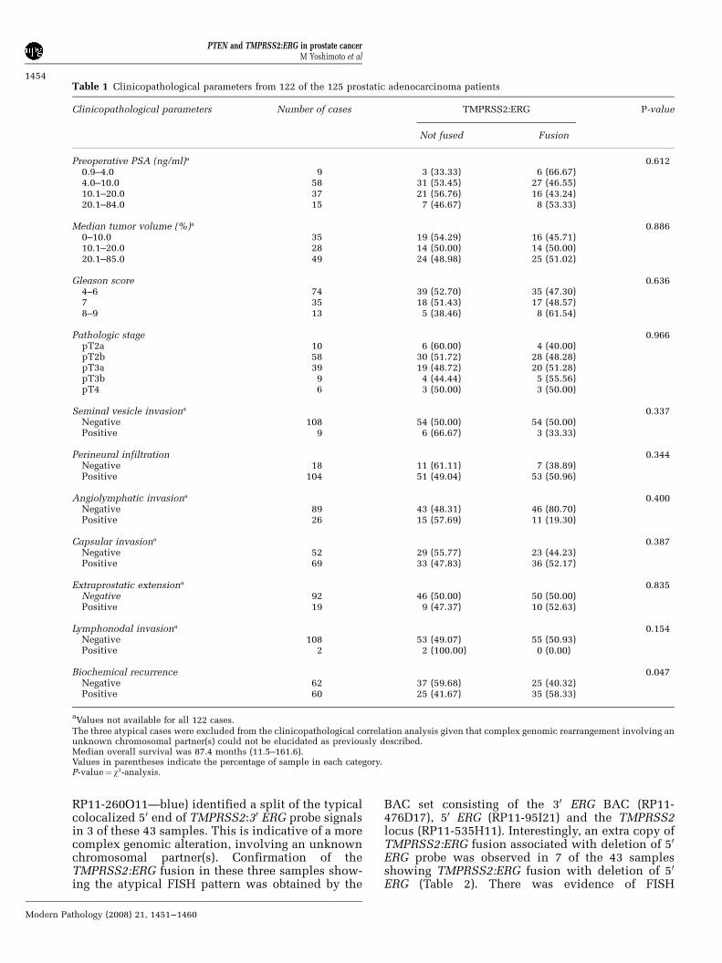
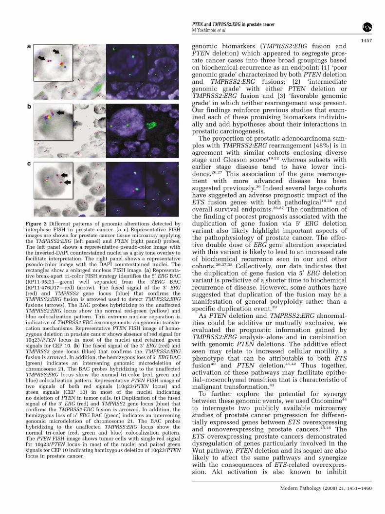
between PTEN deletion as studied previously,17 andTMPRSS2:ERG rearrangements. Therefore, weexamined differential status of PTEN (deleted ornot deleted) and presence of TMPRSS2:ERG fusionin 82 of the 125 tumor samples to assess its utility asbiomarker of prognosis. Overall, PTEN deletion inaddition to the presence of TMPRSS2:ERG rearran-gement was observed in 23 of 82 (28%) prostaticadenocarcinomas. PTEN deletion was also found in14 prostate adenocarcinoma samples showing ab-sence of TMPRSS2:ERG rearrangement (14/82,17%). There was evidence of no copy change ofPTEN locus with TMPRSS2:ERG fusion in 21 of 82(26%) cases and no copy change of PTEN locus withabsence of TMPRSS2:ERG fusion in 24 of 82 (29%)cases. The description of the TMPRSS2:ERG fusionconcomitantly with PTEN deletion is summarizedin Table 3. A comprehensive description of theclinicopathological parameters associated with 47adenocarcinomas (23 cases showing both PTENdeletion and TMPRSS2:ERG fusion, and 24 casesin which neither rearrangement was present) issummarized in Table 4. Representative images ofTMPRSS2:ERG rearrangement and PTEN deletionare shown in Figure 2.
Statistical Analysis of Clinical Parameters andGenomic Alterations
High Gleason score and clinical parameters ofaggressive disease such as extraprostatic extension(P¼ 0.0002), seminal vesicle invasion (P¼ 0.0023),margin status (P¼ 0.0008), neoadjuvant hormonetherapy (P¼ 0.0004), Gleason grade (P¼ 0.0002)/score (P¼ 0.001), median tumor volume(P¼ 0.0024), preoperative PSA (P¼ 0.001), PTENdeletion (P¼ 0.009), concurrent TMPRSS2:ERG fu-sion and PTEN deletion (0.001) and perineuralinvasion (P¼ 0.0304) were significantly associatedwith biochemical recurrence by univariate analysiswith TMPRSS2:ERG approaching significance(P¼ 0.0523; Table 5). By multivariate analysis,relevant factors to explain biochemical failureincluded Gleason score (7 and 8–10, P¼ 0.001 andP¼ 0.015, respectively), concurrent TMPRSS2:ERGfusion and PTEN deletion (0.036) and PTEN homo-zygous deletion (0.013). The PTEN findings reflectthose previously reported for this cohort17 (Table 6).For comparison purpose, Kaplan–Meier survivalanalysis applying established clinical markers, such
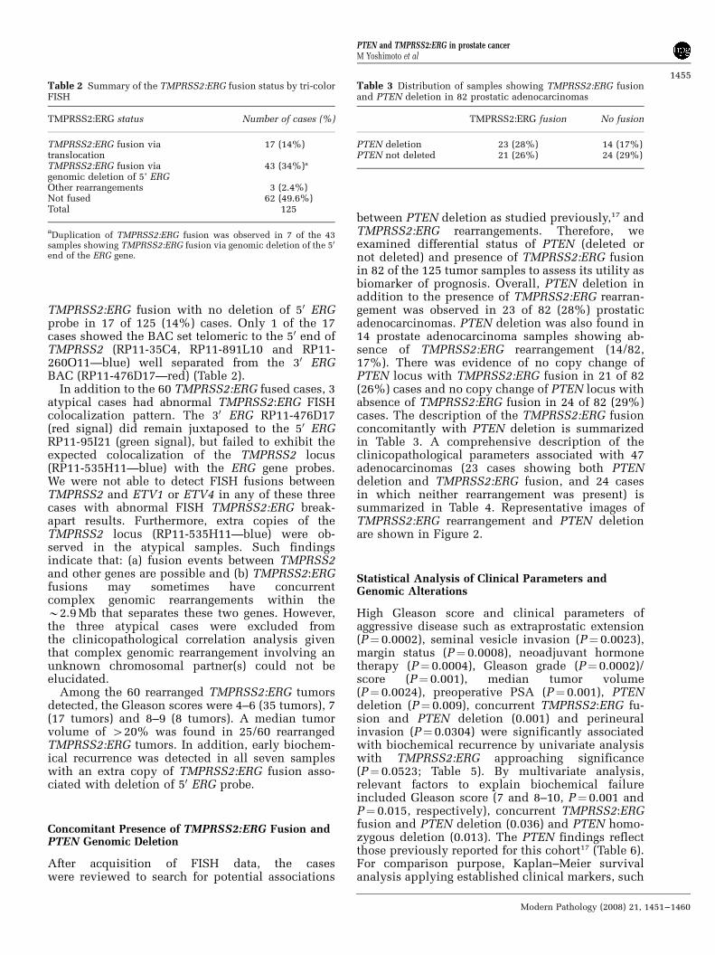
Table 2 Summary of the TMPRSS2:ERG fusion status by tri-colorFISH
TMPRSS2:ERG status Number of cases (%)
TMPRSS2:ERG fusion viatranslocation
17 (14%)
TMPRSS2:ERG fusion viagenomic deletion of 5’ ERG
43 (34%)a
Other rearrangements 3 (2.4%)Not fused 62 (49.6%)Total 125
aDuplication of TMPRSS2:ERG fusion was observed in 7 of the 43
samples showing TMPRSS2:ERG fusion via genomic deletion of the 50
end of the ERG gene.
Table 3 Distribution of samples showing TMPRSS2:ERG fusionand PTEN deletion in 82 prostatic adenocarcinomas
TMPRSS2:ERG fusion No fusion
PTEN deletion 23 (28%) 14 (17%)PTEN not deleted 21 (26%) 24 (29%)
PTEN and TMPRSS2:ERG in prostate cancerM Yoshimoto et al
1455
Modern Pathology (2008) 21, 1451–1460
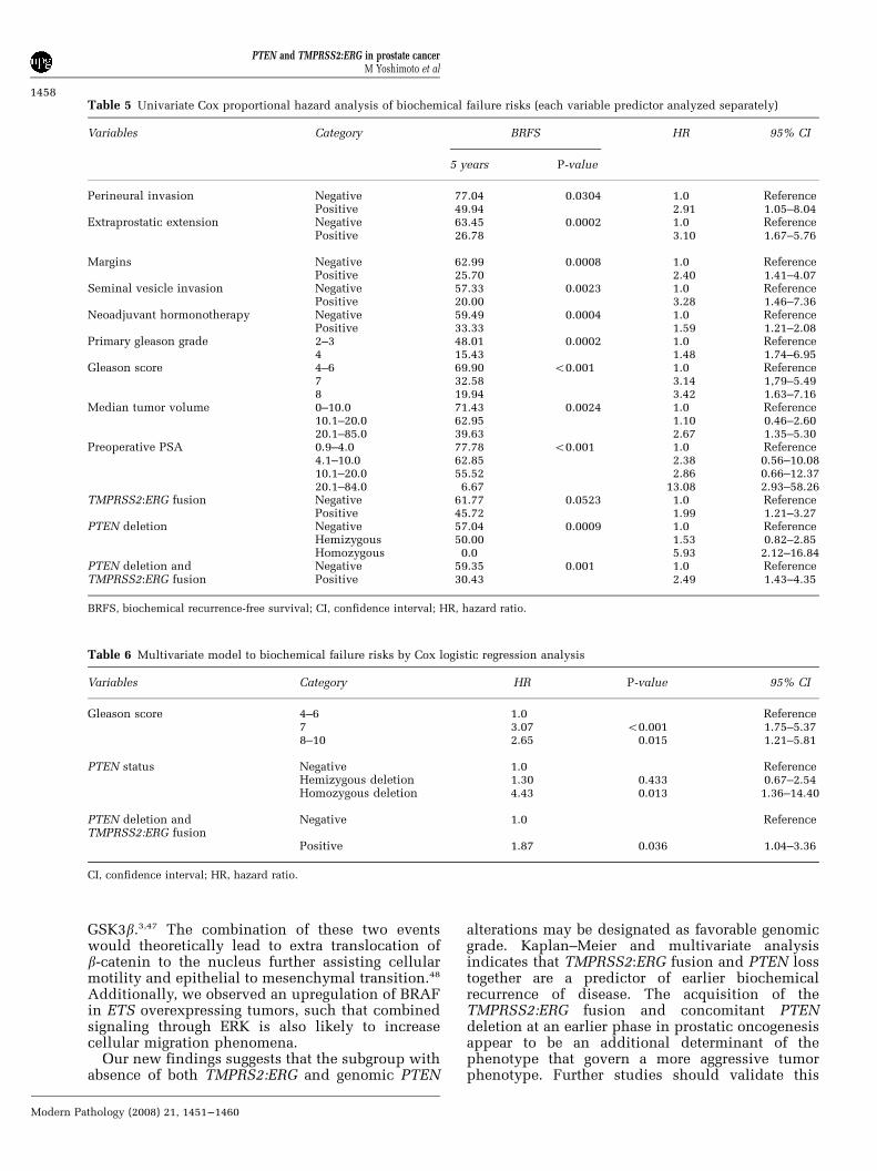
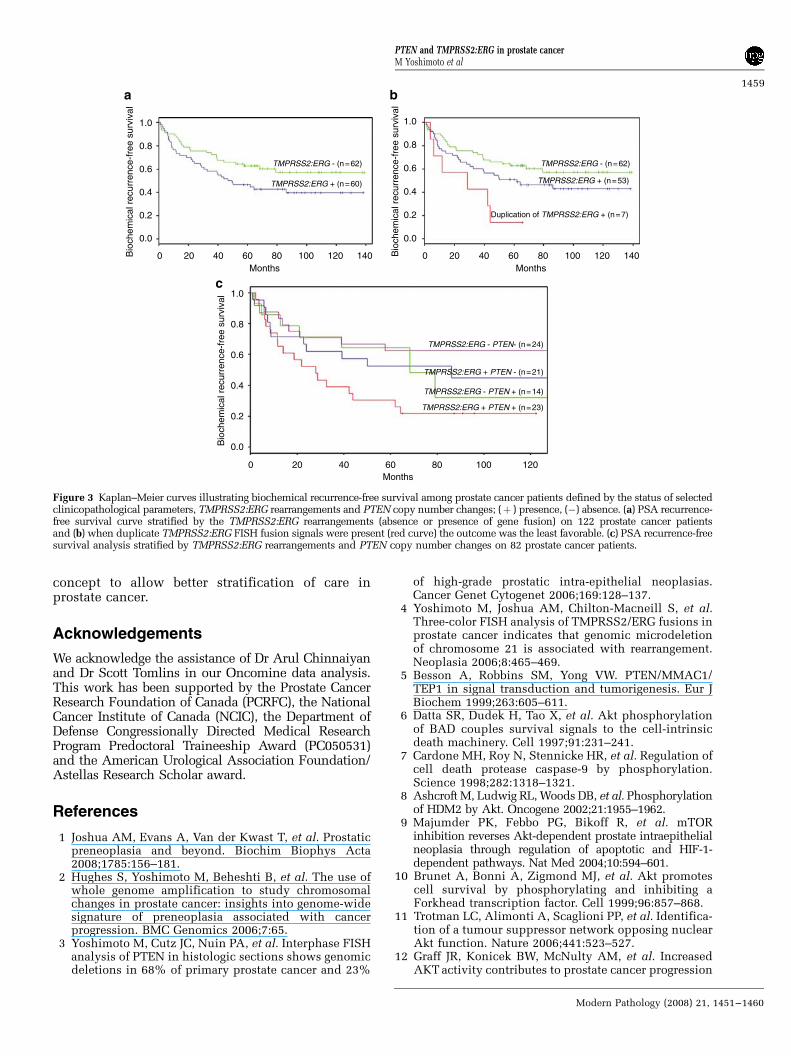
as the preoperative PSA, seminal vesicle invasionand surgical margins status, was considered toidentify subgroups with different prognosis withrespect to time to relapse after surgery. Theestimated disease-free survival curves demonstratedassociation between TMPRSS2:ERG fusion andshort time based on PSA recurrence intervals(Figure 3a). Significantly, the occurrence of duplica-tion of the TMPRSS2:ERG fusion was associatedwith a much earlier onset of biochemical recurrencebased on PSA values (Figure 3b). Furthermore, theestimated disease-free survival curves demonstratedconsiderable association of coexisting PTEN dele-tion and TMPRSS2:ERG rearrangements with shorttime based on PSA recurrence intervals (Figure 3c).This analysis allowed for three broad groupings ofdifferential patient outcome based on time to
biochemical recurrence: (1) a poor prognosticgroup characterized by both PTEN deletion andTMPRSS2:ERG fusions; (2) an intermediate groupwith either PTEN deletion or TMPRSS2:ERG fusionand (3) a favorable prognostic group with neitherevent.
Discussion
The search for accurate biomarkers in prostatecancer is critical for evolution of accurate manage-ment of prostate cancer. This study evaluated thetwo leading genomic biomarkers (PTEN deletionand TMPRSS2:ERG rearrangements) for their con-tribution to prostate cancer prognosis in a largeBrazilian cohort. Figure 3c illustrates the two
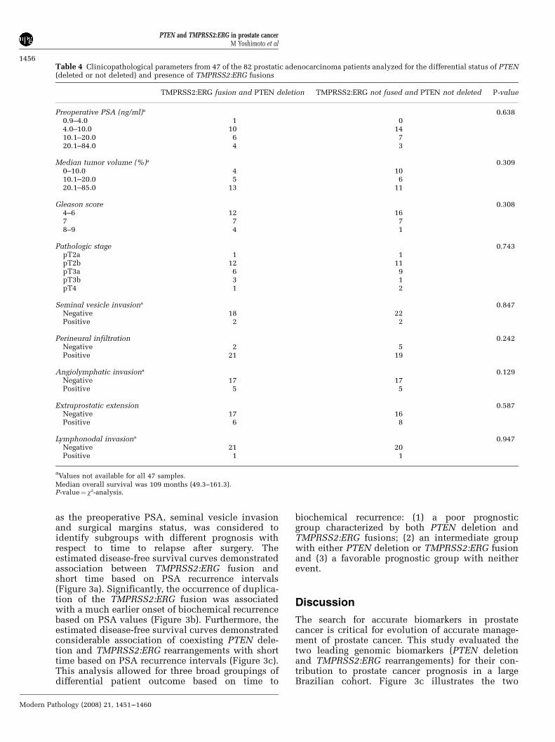
Table 4 Clinicopathological parameters from 47 of the 82 prostatic adenocarcinoma patients analyzed for the differential status of PTEN(deleted or not deleted) and presence of TMPRSS2:ERG fusions
TMPRSS2:ERG fusion and PTEN deletion TMPRSS2:ERG not fused and PTEN not deleted P-value
Preoperative PSA (ng/ml)a 0.6380.9–4.0 1 04.0–10.0 10 1410.1–20.0 6 720.1–84.0 4 3
Median tumor volume (%)a 0.3090–10.0 4 1010.1–20.0 5 620.1–85.0 13 11
Gleason score 0.3084–6 12 167 7 78–9 4 1
Pathologic stage 0.743pT2a 1 1pT2b 12 11pT3a 6 9pT3b 3 1pT4 1 2
Seminal vesicle invasiona 0.847Negative 18 22Positive 2 2
Perineural infiltration 0.242Negative 2 5Positive 21 19
Angiolymphatic invasiona 0.129Negative 17 17Positive 5 5
Extraprostatic extension 0.587Negative 17 16Positive 6 8
Lymphonodal invasiona 0.947Negative 21 20Positive 1 1
aValues not available for all 47 samples.
Median overall survival was 109 months (49.3–161.3).P-value¼ w2-analysis.
PTEN and TMPRSS2:ERG in prostate cancerM Yoshimoto et al
1456
Modern Pathology (2008) 21, 1451–1460
genomic biomarkers (TMPRSS2:ERG fusion andPTEN deletion) which appeared to segregate pros-tate cancer cases into three broad groupings basedon biochemical recurrence as an endpoint: (1) ‘poorgenomic grade’ characterized by both PTEN deletionand TMPRSS2:ERG fusions; (2) ‘intermediategenomic grade’ with either PTEN deletion orTMPRSS2:ERG fusion and (3) ‘favorable genomicgrade’ in which neither rearrangement was present.Our findings reinforce previous studies that exam-ined each of these promising biomarkers individu-ally and add hypotheses about their interactions inprostatic carcinogenesis.
The proportion of prostatic adenocarcinoma sam-ples with TMPRSS2:ERG rearrangement (48%) is inagreement with similar cohorts enclosing diversestage and Gleason scores19,22 whereas subsets withearlier stage disease tend to have lower inci-dence.26,27 This association of the gene rearrange-ment with more advanced disease has beensuggested previously.36 Indeed several large cohortshave suggested an adverse prognostic impact of theETS fusion genes with both pathological19,28 andoverall survival endpoints.26,27 The confirmation ofthe finding of poorest prognosis associated with theduplication of gene fusion via 50 ERG deletionvariant also likely highlight important aspects ofthe pathophysiology of prostate cancer. The effec-tive double dose of ERG gene alteration associatedwith this variant is likely to lead to an increased rateof biochemical recurrence seen in our and othercohorts.26,37,38 Collectively, our data indicates thatthe duplication of gene fusion via 50 ERG deletionvariant is predictive of a shorter time to biochemicalrecurrence of disease. However, some authors havesuggested that duplication of the fusion may be amanifestation of general polyploidy rather than aspecific duplication event.39
As PTEN deletion and TMPRSS2:ERG abnormal-ities could be additive or mutually exclusive, weevaluated the prognostic information gained byTMPRSS2:ERG analysis alone and in combinationwith genomic PTEN deletions. The additive effectseen may relate to increased cellular motility, aphenotype that can be attributable to both ETSfusion40 and PTEN deletion.41,42 Thus together,activation of these pathways may facilitate epithe-lial–mesenchymal transition that is characteristic ofmalignant transformation.43
To further explore the potential for synergybetween these genomic events, we used Oncomine44
to interrogate two publicly available microarraystudies of prostate cancer progression for differen-tially expressed genes between ETS overexpressingand nonoverexpressing prostate cancers.45,46 TheETS overexpressing prostate cancers demonstrateddysregulation of genes particularly involved in theWnt pathway. PTEN deletion and its sequel are alsolikely to affect the same pathways and synergizewith the consequences of ETS-related overexpres-sion. Akt activation is also known to inhibit
Figure 2 Different patterns of genomic alterations detected byinterphase FISH in prostate cancer. (a–c) Representative FISHimages are shown for prostate cancer tissue microarray applyingthe TMPRSS2:ERG (left panel) and PTEN (right panel) probes.The left panel shows a representative pseudo-color image withthe inverted-DAPI counterstained nuclei as a gray tone overlay tofacilitate interpretation. The right panel shows a representativepseudo-color image with the DAPI counterstained nuclei. Therectangles show a enlarged nucleus FISH image. (a) Representa-tive break-apart tri-color FISH strategy identifies the 50 ERG BAC(RP11-95I21—green) well separated from the 30ERG BAC(RP11-476D17—red) (arrow). The fused signal of the 30 ERG(red) and TMPRSS2 gene locus (blue) that confirms theTMPRSS2:ERG fusion is arrowed used to detect TMPRSS2:ERGfusions (arrows). The BAC probes hybridizing to the unaffectedTMPRSS2:ERG locus show the normal red-green (yellow) andblue colocalization pattern. This extreme nuclear separation isindicative of TMPRSS2:ERG rearrangements via genomic translo-cation mechanisms. Representative PTEN FISH image of homo-zygous deletion in prostate cancer shows absence of red signal for10q23/PTEN locus in most of the nuclei and retained greensignals for CEP 10. (b) The fused signal of the 30 ERG (red) andTMPRSS2 gene locus (blue) that confirms the TMPRSS2:ERGfusion is arrowed. In addition, the hemizygous loss of 50 ERG BAC(green) indicates an intervening genomic microdeletion ofchromosome 21. The BAC probes hybridizing to the unaffectedTMPRSS:ERG locus show the normal tri-color (red, green andblue) colocalization pattern. Representative PTEN FISH image oftwo signals of both red signals (10q23/PTEN locus) andgreen signals (CEP 10) in most of the nuclei indicatingno deletion of PTEN in tumor cells. (c) Duplication of the fusedsignal of the 30 ERG (red) and TMPRSS2 gene locus (blue) thatconfirms the TMPRSS2:ERG fusion is arrowed. In addition, thehemizygous loss of 50 ERG BAC (green) indicates an interveninggenomic microdeletion of chromosome 21. The BAC probeshybridizing to the unaffected TMPRSS:ERG locus show thenormal tri-color (red, green and blue) colocalization pattern.The PTEN FISH image shows tumor cells with single red signalfor 10q23/PTEN locus in most of the nuclei and paired greensignals for CEP 10 indicating hemizygous deletion of 10q23/PTENlocus in prostate cancer.
PTEN and TMPRSS2:ERG in prostate cancerM Yoshimoto et al
1457
Modern Pathology (2008) 21, 1451–1460
GSK3b.3,47 The combination of these two eventswould theoretically lead to extra translocation ofb-catenin to the nucleus further assisting cellularmotility and epithelial to mesenchymal transition.48
Additionally, we observed an upregulation of BRAFin ETS overexpressing tumors, such that combinedsignaling through ERK is also likely to increasecellular migration phenomena.
Our new findings suggests that the subgroup withabsence of both TMPRS2:ERG and genomic PTEN
alterations may be designated as favorable genomicgrade. Kaplan–Meier and multivariate analysisindicates that TMPRSS2:ERG fusion and PTEN losstogether are a predictor of earlier biochemicalrecurrence of disease. The acquisition of theTMPRSS2:ERG fusion and concomitant PTENdeletion at an earlier phase in prostatic oncogenesisappear to be an additional determinant of thephenotype that govern a more aggressive tumorphenotype. Further studies should validate this
Table 5 Univariate Cox proportional hazard analysis of biochemical failure risks (each variable predictor analyzed separately)
Variables Category BRFS HR 95% CI
5 years P-value
Perineural invasion Negative 77.04 0.0304 1.0 ReferencePositive 49.94 2.91 1.05–8.04
Extraprostatic extension Negative 63.45 0.0002 1.0 ReferencePositive 26.78 3.10 1.67–5.76
Margins Negative 62.99 0.0008 1.0 ReferencePositive 25.70 2.40 1.41–4.07
Seminal vesicle invasion Negative 57.33 0.0023 1.0 ReferencePositive 20.00 3.28 1.46–7.36
Neoadjuvant hormonotherapy Negative 59.49 0.0004 1.0 ReferencePositive 33.33 1.59 1.21–2.08
Primary gleason grade 2–3 48.01 0.0002 1.0 Reference4 15.43 1.48 1.74–6.95
Gleason score 4–6 69.90 o0.001 1.0 Reference7 32.58 3.14 1,79–5.498 19.94 3.42 1.63–7.16
Median tumor volume 0–10.0 71.43 0.0024 1.0 Reference10.1–20.0 62.95 1.10 0.46–2.6020.1–85.0 39.63 2.67 1.35–5.30
Preoperative PSA 0.9–4.0 77.78 o0.001 1.0 Reference4.1–10.0 62.85 2.38 0.56–10.0810.1–20.0 55.52 2.86 0.66–12.3720.1–84.0 6.67 13.08 2.93–58.26
TMPRSS2:ERG fusion Negative 61.77 0.0523 1.0 ReferencePositive 45.72 1.99 1.21–3.27
PTEN deletion Negative 57.04 0.0009 1.0 ReferenceHemizygous 50.00 1.53 0.82–2.85Homozygous 0.0 5.93 2.12–16.84
PTEN deletion and Negative 59.35 0.001 1.0 ReferenceTMPRSS2:ERG fusion Positive 30.43 2.49 1.43–4.35
BRFS, biochemical recurrence-free survival; CI, confidence interval; HR, hazard ratio.
Table 6 Multivariate model to biochemical failure risks by Cox logistic regression analysis
Variables Category HR P-value 95% CI
Gleason score 4–6 1.0 Reference7 3.07 o0.001 1.75–5.378–10 2.65 0.015 1.21–5.81
PTEN status Negative 1.0 ReferenceHemizygous deletion 1.30 0.433 0.67–2.54Homozygous deletion 4.43 0.013 1.36–14.40
PTEN deletion andTMPRSS2:ERG fusion
Negative 1.0 Reference
Positive 1.87 0.036 1.04–3.36
CI, confidence interval; HR, hazard ratio.
PTEN and TMPRSS2:ERG in prostate cancerM Yoshimoto et al
1458
Modern Pathology (2008) 21, 1451–1460
concept to allow better stratification of care inprostate cancer.
Acknowledgements
We acknowledge the assistance of Dr Arul Chinnaiyanand Dr Scott Tomlins in our Oncomine data analysis.This work has been supported by the Prostate CancerResearch Foundation of Canada (PCRFC), the NationalCancer Institute of Canada (NCIC), the Department ofDefense Congressionally Directed Medical ResearchProgram Predoctoral Traineeship Award (PC050531)and the American Urological Association Foundation/Astellas Research Scholar award.
References
1 Joshua AM, Evans A, Van der Kwast T, et al. Prostaticpreneoplasia and beyond. Biochim Biophys Acta2008;1785:156–181.
2 Hughes S, Yoshimoto M, Beheshti B, et al. The use ofwhole genome amplification to study chromosomalchanges in prostate cancer: insights into genome-widesignature of preneoplasia associated with cancerprogression. BMC Genomics 2006;7:65.
3 Yoshimoto M, Cutz JC, Nuin PA, et al. Interphase FISHanalysis of PTEN in histologic sections shows genomicdeletions in 68% of primary prostate cancer and 23%
of high-grade prostatic intra-epithelial neoplasias.Cancer Genet Cytogenet 2006;169:128–137.
4 Yoshimoto M, Joshua AM, Chilton-Macneill S, et al.Three-color FISH analysis of TMPRSS2/ERG fusions inprostate cancer indicates that genomic microdeletionof chromosome 21 is associated with rearrangement.Neoplasia 2006;8:465–469.
5 Besson A, Robbins SM, Yong VW. PTEN/MMAC1/TEP1 in signal transduction and tumorigenesis. Eur JBiochem 1999;263:605–611.
6 Datta SR, Dudek H, Tao X, et al. Akt phosphorylationof BAD couples survival signals to the cell-intrinsicdeath machinery. Cell 1997;91:231–241.
7 Cardone MH, Roy N, Stennicke HR, et al. Regulation ofcell death protease caspase-9 by phosphorylation.Science 1998;282:1318–1321.
8 Ashcroft M, Ludwig RL, Woods DB, et al. Phosphorylationof HDM2 by Akt. Oncogene 2002;21:1955–1962.
9 Majumder PK, Febbo PG, Bikoff R, et al. mTORinhibition reverses Akt-dependent prostate intraepithelialneoplasia through regulation of apoptotic and HIF-1-dependent pathways. Nat Med 2004;10:594–601.
10 Brunet A, Bonni A, Zigmond MJ, et al. Akt promotescell survival by phosphorylating and inhibiting aForkhead transcription factor. Cell 1999;96:857–868.
11 Trotman LC, Alimonti A, Scaglioni PP, et al. Identifica-tion of a tumour suppressor network opposing nuclearAkt function. Nature 2006;441:523–527.
12 Graff JR, Konicek BW, McNulty AM, et al. IncreasedAKT activity contributes to prostate cancer progression
TMPRSS2:ERG - PTEN- (n=24)
TMPRSS2:ERG + PTEN - (n=21)
TMPRSS2:ERG - PTEN + (n=14)
TMPRSS2:ERG + PTEN + (n=23)
0 20 40 60Months
80 100 120
1.0
0.8
0.6
0.4
0.2
0.0Bio
chem
ical
rec
urre
nce-
free
sur
viva
l
1.0
0.8
0.6
0.4
0.2
0.0
1.0
0.8
0.6
0.4
0.2
0.0
0
TMPRSS2:ERG - (n=62) TMPRSS2:ERG - (n=62)
TMPRSS2:ERG + (n=60)
Duplication of TMPRSS2:ERG + (n=7)
TMPRSS2:ERG + (n=53)
20 40 60Months
80 100 120 140 0 20 40 60Months
80 100 120 140Bio
chem
ical
rec
urre
nce-
free
sur
viva
l
Bio
chem
ical
rec
urre
nce-
free
sur
viva
l
Figure 3 Kaplan–Meier curves illustrating biochemical recurrence-free survival among prostate cancer patients defined by the status of selectedclinicopathological parameters, TMPRSS2:ERG rearrangements and PTEN copy number changes; (þ ) presence, (�) absence. (a) PSA recurrence-free survival curve stratified by the TMPRSS2:ERG rearrangements (absence or presence of gene fusion) on 122 prostate cancer patientsand (b) when duplicate TMPRSS2:ERG FISH fusion signals were present (red curve) the outcome was the least favorable. (c) PSA recurrence-freesurvival analysis stratified by TMPRSS2:ERG rearrangements and PTEN copy number changes on 82 prostate cancer patients.
PTEN and TMPRSS2:ERG in prostate cancerM Yoshimoto et al
1459
Modern Pathology (2008) 21, 1451–1460
by dramatically accelerating prostate tumor growthand diminishing p27Kip1 expression. J Biol Chem2000;275:24500–24505.
13 Vivanco I, Palaskas N, Tran C, et al. Identification of theJNK signaling pathway as a functional target of the tumorsuppressor PTEN. Cancer Cell 2007;11:555–569.
14 Koksal IT, Dirice E, Yasar D, et al. The assessment ofPTEN tumor suppressor gene in combination withGleason scoring and serum PSA to evaluate progressionof prostate carcinoma. Urol Oncol 2004;22:307–312.
15 Bertram J, Peacock JW, Fazli L, et al. Loss of PTEN isassociated with progression to androgen indepen-dence. Prostate 2006;66:895–902.
16 Schmitz M, Grignard G, Margue C, et al. Complete lossof PTEN expression as a possible early prognosticmarker for prostate cancer metastasis. Int J Cancer2007;120:1284–1292.
17 Yoshimoto M, Cunha IW, Coudry RA, et al. FISHanalysis of 107 prostate cancers shows that PTENgenomic deletion is associated with poor clinicaloutcome. Br J Cancer 2007;97:678–685.
18 Tomlins SA, Mehra R, Rhodes DR, et al.TMPRSS2:ETV4 gene fusions define a third molecularsubtype of prostate cancer. Cancer Res 2006;66:3396–3400.
19 Perner S, Demichelis F, Beroukhim R, et al.TMPRSS2:ERG fusion-associated deletions provideinsight into the heterogeneity of prostate cancer.Cancer Res 2006;66:8337–8341.
20 Cerveira N, Ribeiro FR, Peixoto A, et al. TMPRSS2-ERGgene fusion causing ERG overexpression precedes chro-mosome copy number changes in prostate carcinomasand paired HGPIN lesions. Neoplasia 2006;8:826–832.
21 Tomlins SA, Rhodes DR, Perner S, et al. Recurrentfusion of TMPRSS2 and ETS transcription factor genesin prostate cancer. Science 2005;310:644–648.
22 Wang J, Cai Y, Ren C, et al. Expression of variantTMPRSS2/ERG fusion messenger RNAs is associated withaggressive prostate cancer. Cancer Res 2006;66:8347–8351.
23 Ahlers CM, Figg WD. ETS-TMPRSS2 fusion gene productsin prostate cancer. Cancer Biol Ther 2006;5:254–255.
24 Soller MJ, Isaksson M, Elfving P, et al. Confirmation ofthe high frequency of the TMPRSS2/ERG fusion genein prostate cancer. Genes Chromosomes Cancer 2006;45:717–719.
25 Rajput AB, Miller MA, De Luca A, et al. Frequency ofthe TMPRSS2:ERG gene fusion is increased in moder-ate to poorly differentiated prostate cancers. J ClinPathol 2007;60:1238–1243.
26 Attard G, Clark J, Ambroisine L, et al. Duplication ofthe fusion of TMPRSS2 to ERG sequences identifies fatalhuman prostate cancer. Oncogene 2008;27:253–263.
27 Demichelis F, Fall K, Perner S, et al. TMPRSS2:ERGgene fusion associated with lethal prostate cancer in awatchful waiting cohort. Oncogene 2007;26:4596–4599.
28 Nami RK, Sugar L, Wang Z, et al. Expression ofTMPRSS2:ERG gene fusion in prostate cancer cells isan important prognostic factor for cancer progression.Cancer Biol Ther 2007;6:40–45.
29 Winnes M, Lissbrant E, Damber JE, et al. Moleculargenetic analyses of the TMPRSS2-ERG and TMPRSS2-ETV1 gene fusions in 50 cases of prostate cancer.Oncol Rep 2007;17:1033–1036.
30 Petrovics G, Liu A, Shaheduzzaman S, et al. Frequentoverexpression of ETS-related gene-1 (ERG1) inprostate cancer transcriptome. Oncogene 2005;24:3847–3852.
31 Yoshimoto M, Ludkovski O, Bayani J, et al.Microdeletion and concurrent translocation associatedwith a complex TMPRSS2:ERG prostate cancer genefusion. Genes Chromosomes Cancer 2007;46:861–863.
32 Ventura RA, Martin-Subero JI, Jones M, et al.FISH analysis for the detection of lymphoma-associatedchromosomal abnormalities in routine paraffin-embeddedtissue. J Mol Diagn 2006;8:141–151.
33 Korshunov A, Sycheva R, Gorelyshev S, et al. Clinicalutility of fluorescence in situ hybridization (FISH) innonbrainstem glioblastomas of childhood. Mod Pathol2005;18:1258–1263.
34 Kawai T, Hiroi S, Nakanishi K, et al. Abnormalities inchromosome 17 and p53 in lung carcinoma cellsdetected by fluorescence in situ hybridization. PatholInt 2004;54:413–419.
35 Mezzelani A, Alasio L, Bartoli C, et al. c-erbB2/neugene and chromosome 17 analysis in breast cancer byFISH on archival cytological fine-needle aspirates. Br JCancer 1999;80:519–525.
36 Mehra R, Tomlins SA, Shen R, et al. Comprehensiveassessment of TMPRSS2 and ETS family gene aberra-tions in clinically localized prostate cancer. ModPathol 2007;20:538–544.
37 Tomlins SA, Laxman B, Dhanasekaran SM, et al.Distinct classes of chromosomal rearrangements createoncogenic ETS gene fusions in prostate cancer. Nature2007;448:595–599.
38 Tomlins SA, Laxman B, Varambally S, et al. Role of theTMPRSS2-ERG gene fusion in prostate cancer. Neo-plasia 2008;10:177–188.
39 Gopalan A LM, Satagopan JM, Zhou QC, et al. 97thAnnual Meeting of the United States and CanadianAcademy of Pathology. The United States and Cana-dian Academy of Pathology: Denver, Colorado, 2008.
40 Hsu T, Trojanowska M, Watson DK. Ets proteins inbiological control and cancer. J Cell Biochem2004;91:896–903.
41 Shukla S, Maclennan GT, Hartman DJ, et al. Activationof PI3K-Akt signaling pathway promotes prostatecancer cell invasion. Int J Cancer 2007;121:1424–1432.
42 Kotelevets L, van Hengel J, Bruyneel E, et al. The lipidphosphatase activity of PTEN is critical for stabilizingintercellular junctions and reverting invasiveness.J Cell Biol 2001;155:1129–1135.
43 Larue L, Bellacosa A. Epithelial-mesenchymal transi-tion in development and cancer: role of phosphatidy-linositol 30 kinase/AKT pathways. Oncogene 2005;24:7443–7454.
44 Rhodes DR, Kalyana-Sundaram S, Mahavisno V, et al.Oncomine 3.0: genes, pathways, and networks in acollection of 18 000 cancer gene expression profiles.Neoplasia 2007;9:166–180.
45 Lapointe J, Li C, Higgins JP, et al. Gene expressionprofiling identifies clinically relevant subtypes of prostatecancer. Proc Natl Acad Sci USA 2004;101:811–816.
46 Glinsky GV, Glinskii AB, Stephenson AJ, et al. Geneexpression profiling predicts clinical outcome ofprostate cancer. J Clin Invest 2004;113:913–923.
47 Maira SM, Galetic I, Brazil DP, et al. Carboxyl-terminalmodulator protein (CTMP), a negative regulator ofPKB/Akt and v-Akt at the plasma membrane. Science2001;294:374–380.
48 Polette M, Mestdagt M, Bindels S, et al. Beta-cateninand ZO-1: shuttle molecules involved in tumorinvasion-associated epithelial-mesenchymal transitionprocesses. Cells Tissues Organs 2007;185:61–65.
PTEN and TMPRSS2:ERG in prostate cancerM Yoshimoto et al
1460
Modern Pathology (2008) 21, 1451–1460