The hereditary dystrophies of the posterior pole of the eye
502
The hereditary dystrophies of the posterior pole of the eye
-
Upload
khangminh22 -
Category
Documents
-
view
0 -
download
0
Transcript of The hereditary dystrophies of the posterior pole of the eye

The hereditary dystrophies of the posterior pole of the eye


THE HEREDITARY DYSTROPHIES
OF THE POSTERIOR POLE OF THE EYE
PROEFSCHRIFT
ter verkrijging van de graad van doctor in de geneeskunde
aan de Medische Faculteit te Rotterdam,
op gezag van de decaan Prof. D. C. den Haan,
hoogleraar in de faculteit der geneeskunde,
tegen de bedenkingen van de faculteit der geneeskunde
te verdedigen op 13 januari 1971 te r6.oo uur door
AUGUST FRANS DEUTMAN
geboren te Leiden in 1939
TE ASSEN BIJ
VAN GORCUM & COMP. N.V. - DR. H. J· PRAKKE & H. M.G. PRAKKE

PROMOTOR: PROF. DR. H. E. HENKES
CO-REFERENTEN: DR. P- J· WAARDENBURG
PROF. DR. J. A. OOSTERHUIS
The publication of this book was made possible through grants from the Netherlands Organization for the Advancement of Pure Research(Z.W.O.), the Netherlands General Association for Prevention of Blindness, the Flieringa Stichting, the foundation "De Dric Lichten" and the "Bataafsch Genootschap dcr Proef-
ondcrvindelijke Wijsbegeerte".

Aan mijn ouders, vrouw en kinderen

""So there he stands, ow: vertical, hunting, weapontoting, territorial, neotenous, brainy, Naked Ape, a primate by ancestry and a carnivore by adoption, ready to conquer the world. But he is a very new and e~-perimental departure, and new models frequently have imperfections. For him the main troubles will stem from the fact that his culturally operated advances will race ahead of any further genetic ones. His genes will lag behind, and he will be constantly :reminded that, for all his environment~moulding achievements, he is still at heart a very naked ape".
DESMOND MORRIS: "THE NAKED APE"

Acknowledgements
I am greatly indebted to Prof. Dr. H. E. Henkes, who inspired me with his dynamic enthusiasm over the years in which it was my privilege to receive my training as ophthalmologist under his supervision. He created the opportunity to use modern, refined methods of examination. Dr. P. J. Waardenburg, assessor, gave his valuable advice on questions of genetics and ophthalmology. His wise and encouraging criticisms were indispensable and unforgettable. My deep gratitude goes to Prof. Dr. J. A. Oosterhuis, also assessor, for his critical analysis and stimulating interest, particularly in questions of fluorescence photography.
Dr. P. P. H. Alkemade inspired me to start this study and gave his valuable advice, while Dr. J. Schappert-Kimmijser gave me much encouragement. The electrodiagnostic results were interpreted with the able help of Dr. G. H. M. van Lith, head of the electrophysiological service, Department of Ophthalmology, Medical Faculty, Rotterdam. It is a pleasure to acknowledge my indebtedness to Mr. A. L. Aan de Kerk, head of the photographic service, Department of Ophthalmology, Medical Faculty, Rotterdam. He made nearly all the photographs illustrating this book.
Ir. T. W. Siertsema, genealogist of the Netherlands General Association for Prevention of Blindness, gave me the benefit of his very thorough genealogical research. Miss Lucy van Duyn shouldered most of the enormous task of compiling the bibliography and typing the manuscript. Mr. C. B. Schotel made the many drawings and Mr. Th. van Winsen translated the te:.;:t. I am appreciative of the contributions of A. Craandijk, M.D. and Dr. K. Zahn in obtaining the fluorescence photographs.
I am greatly indebted also to the many Dutch ophthalmologists who unhesitatingly turned their patients over to me for this study, which could not have been adequately done without their help.
I am grateful to Dr. W. A. Manschot and Dr. S. Ry Andersen who offered the results of important histopathological examinations. Dr. P. Amalric, T.N. Boen-Tan,

M.D., Dr. ]. Forgacs, Prof. Dr. F. Kandori, Prof. Dr. ]. A. Oosterhuis and Dr. E. Yamada contributed towards the realization of this wotk by offering valuable photographic material.
To Dr. A. Th. M. van Balen and ]. G. C. Renardel de Lavalette, M.D. I offer my sincere thanks for their important part in my training as ophthalmologist. Last but not least I offer my deepest gratitude to my wife for her help in the time-consuming work of correcting the proofs and composing the index.

Contents
I. THE HEREDITARY DYSTROPHIES OF THE CENTRAL RETINA AND CHOROID
I. Introduction :. Material 3· Methods ..
a. visual acuity b. visual fields c. colow: vision . d. dark adaptation e. electro-retinography f. electro-oculography . g. photography . h. fluorescein angiography i. general physical examination and
laboratory study
4· Nomenclature. a. fovea . b. dystrophy c. carrier
!I. SEX-LINKED JUVENILE RETINOSCHISIS
I. Introduction. . . • . . . . . . . . ::. General clinical picture in the patients . ;. Fundus (ophthalmoscopic features) 4· Refraction . . ~. Visual acuity. 6. Visual fields . 7· Colour vision 8. Dark adaptation 9· Electro-retinography
ro. Electro-oculography I I. Photography. . . .
l· Affections in which foveal dystrophies may occur, not included in this study a. the lipidoses . .
4 b. angioid streaks . 4 c. dystrophia myotonica .
4 d. SjOgren-Larsson syndrome . 4 e. myopia gravior j f. vitreotapetorctinal dystrophy of 6 Goldmann-Favre .
9 g. senile foveal processes
9 h. foveal dystrophy associated with deaf-mutism
IO i. crater-like holes in the optic disc IO j. foveal dystrophies as part of a more IO diffuse retinal or choroidal dystrophy I6 k. foveal hypoplasia . I6 I. foveal dysplasia
6. General historical review .
7· Classification
48 a. Fluorescein angiography ~o r;. Carriers ..... . ~o 14. Histological findings . 6; I~. Pathogenesis 6; r6. Mode of transmission 64 I7· General physical and laboratory findings. 64 rS. Associated conditions 64 19. Differential diagnosis 6~ ::o. Therapy. . . 66 2.1. Future ... 66 ::::. Case histories
I7 I7 22
22
:: z;
:;
•4 :6
:6 26 z6 :8
3l
66 67 69 73 74 7l 7l 7l 77 79 So

III. STARGARDT'S DISEASE ...... .
x. Introduction, . . . , . . . , . . . 2. General clinical picture in the patients . 5· Fundus (ophthalmoscopic features) 4· Refraction . . 5· Visual acuity. 6. Visual fields . 7. Colour vision 8. Dark adaptation 9· Electro-retinography
IO. Electrowoculography II. Photography. . . .
IOO u. 105 '5· I07 '4· "7 '5. "7 I6. I24 '7· IZ5 IS.
"7 '9· IZ8 20. IZ9 2I. I 5 I 22.
IOO
Fluorescein angiography IJI Carriers. IJI Histological £ndings I 55 Pathogenesis IH Mode of transmission '34 General physical and laboratory £ndings . '55 Associated conditions '55 Differential diagnosis I 57 Therapy. IJS Future I 58 Case histories '40
IV. DOMINANT PROGRESSIVE FOVEAL DYSTROPHY
r. Introduction . . . . . . . 2. General clinical picture in the patients . 5· Fundus (ophthalmoscopic features) 4· Refraction . , 5. Visual acuity. 6. Visual fields . 7. Colour vision 8. Dark adaptation 9· Electro-retinography
IO. Electro-oculography II. Photography. . . .
V. PROGRESSIVE CONE DYSTROPHY
I. Introduction . 2. General clinical picture in the patients . 5· Fundus (ophthalmoscopic features) 4· Refraction . . 5. Visual acuity. 6. Visual fields . 7. Colour vision 8. Dark adaptation 9· Electro-retinography
Io. Electro-oculography n. Photography. . . .
VI. CENTRAL RETINOPATHIA PIGME~TOSA ..
,, Introduction . 189 2. General clinical picture in the patients . '9' 5· Fundus (ophthalmoscopic features) '9' 4· Refraction . '9' 5. Visual acuity. '9' 6. Visual fields . '9' 7· Colour vision '9' 8. Dark adaptation '9' 9· Electro-retinography I92
ro. Electro-oculography I95 I I. Photography. '94
12. Fluorescein angiography I76 I5. Carriers. . . . . . I77 q. Histological £ndings . I 77 I 5. Pathogenesis I77 I6. Mode of transmission I77 I7. General physical and laboratory £ndings . I77 18. Associated conditions 177 I9. Differential diagnosis 177 20. Therapy. . . I 78 21. Future . . . 178 22. Case histories 179
I8I
12. Fluorescein angiography I 84 15. Carriers ... , . . 184 q. Histological £ndings . I 84 15. Pathogenesis 184 r6. Mode of transmission 184 17. General physical and laboratory £ndings . I85 IS. Associated conditions 185 19. Differential diagnosis I85 20. Therapy. . . 187 2I. Future . . . 187 22. Case histories 187
12. Fluorescein angiography '94 I5. Carriers. '94 14. Histological £ndings '94 '5· Pathogenesis '94 I6. Mode of transmission '94 '7· General physical and laboratory £ndings . '94 IS. Associated conditions '94 I9. Differential diagnosis '94 20, Therapy. '95 2I. Futwe '95 22. Case histories '95

VII. VITELLIFORM DYSTROPHY OF THE FOVEA 198
I. Introduction. 198 I :z. Fluorescein angiography 244 :z. Genetal clinical picture in the patients . zo8 IS. Carriers . 246 3· Fundus (ophthalmoscopic features) 216 I4. Histological findings :2.52
4· Refraction . 232 I5. Pathogenesis :2.5:2.
5. Visual acuity. 234 I6. Mode of transmission 25 5 6. Visual fields . 235 '7· General physical and laboratory findings . 256 7· Colour vision 235 IS. Associated conditions 257 S. Dark adaptation 237 I9. Diffetential diagnosis 258 9· Electro-retinography :2.57 2o. Therapy. 266
IO. Electro-oculography 238 2I. Future z67 II. Photography. Z4I :z:z. Case histories 267
VIII. FUNDUS FLAVIMACULA'I'US 300
I. Introduction. 300 I2. Fluorescein angiography 310 2. Genetal clinical picture in the patients . 302 I 3. Carriers . JIO 3· Fundus (ophthalmoscopic features) 304 I4. Histological findings 3ll
4· Refraction . . 3°5 I5. Pathogenesis 3Il 5· Visual acuity. 3°5 I6. Mode of transmission 3Il 6. Visual fields . 3°5 I7. General physical and laboratory findings . 312 7· Colour vision 305 IS. Associated conditions 315 8. Dark adaptation 305 I9. Differential diagnosis 3I6
9· Electro-retinography 307 2o. Therapy. SI6
IO. Electro-oculography 5°9 2I. Future 316 II. Photography. 310 :z:z. Case histories 318
IX. RETICULAR DYSTROPHY OF THE RETINAL PIGMENT EPITHELIUM (SJOGREN)
I. Introduction. 324 I:z. Fluorescein angiography no
2. General clinical picture in the patients . 324 I3. Carriers. 333 5· Fundus (ophthalmoscopic features) 326 I4. Histological findings 333 4· Refraction . 328 I5. Pathogenesis 333 5. Visual acuity. 328 I6. Mode of transmission 336 6. Visual fields . 328 17. General physical and laboratory findings . 336
7· Colour vision 329 IS. Associated conditions 337 8. Dark adaptation 329 1S. Diffetential diagnosis 337 9· Electro-retinography 52 9 zo. Therapy. 337
10. Electro-oculography 330 :2.1. Future 337 II. Photography. ;;o 22. Case histories 338
X. :BUTTERFLY-SHAPED PIGMENT DYSTROPHY OF THE FOVEA •...•..
1. Introduction. 340 I:z. Fluorescein angiography 350 2. General clinical picture in the patients . 340 13. Carriers . 35• 3· Fundus (ophthalmoscopic features) 343 14. Histological :findings 352 4· Refraction . 345 r 5. Pathogenesis 352 5. Visual acuity. 347 I6. Mode of transmission 35 2 6. Visual fields . 347 17. General physical and laboratory findings . 352 7. Colour vision 347 IS. Associated conditions 354 8. Dark adaptation 347 19. Differential diagnosis 354 9· Electro-retinography 35° 20. Therapy. 357
IO. Electro-oculography 35° 21. Future 357 II. Photography. 350 22. Case histories 357

XI. GROUPED PIGMENTATIONS OF THE FOVEAL AREA •• , •• , •• , ••
I. Introduction . 36I u. Fluorescein angiography z. General clinical picture in the patients . 36I Ij. Carriers. 3· Fundus (ophthalmoscopic features) 363 14· Histological findings 4· Refraction . 363 I5. Pathogenesis 5. Visual acuity. ;6; I6. Mode of transmission 6. Visual fields . 363 I 7· General physical and laboratory findings . 7· Colour vision 363 18. Associated conditions 8. Dark adaptation ;6; 19. Differential diagnosis
9· Electro-retinography 363 zo. Therapy. IO. Electro-oculography ;6; 21. Future II. Photography. 364 zz.. Case histories
XU. DOMINANT DRUSEN OF BRUCH'S MEMBRANE ••• , • , ••
x. In~oduction. . . . . . . . . . . z. General clinical picture in the patients . 3· Fundus (ophthalmoscopic features) 4· Refraction . . 5. Visual acuity. 6. Visual fields . 7. Colour vision 8. Dark adaptation 9· Electro-retinography
ro. Electro-oculography I I. Photography. . . .
:;67 a. Fluorescein angiography :;S5 :;76 I;. Carriers. . . . . . ;S5 376 I4. Histological findings . 3S5
37S I5. Pathogenesis 3S7
37S I6. Mode of transmission 3S7
;So I7· General physical and laboratory findings . ;SS
;So IS. Associated conditions ;SS
;So 19. Differential diagnosis ;SS ;So 20. Therapy. . . 392
:;S4 21. Future . . . 392
3S4 22. Case histories :;92
XIII. PSEUDO-INFLAMMATORY DYSTROPHY (SORSBY) 400
I. Introduction . 400 u. Fluorescein angiography 2. General clinical picture in the patients . 40I I3. Carriers. .
3· Fundus (ophthalmoscopic features) 40I I4. Histological findings 4· Refraction . 40I I5· Pathogenesis 5. Visual acuity. 40I I6. Mode of transmission 6. Visual fields . 40I I 7. General physical and laboratory Endings . 7· Colour vision 40I xS. Associated conditions S. Dark adaptation 404 I9. Differential diagnosis 9· El~ctro-retinography 404 20. Therapy.
IO. Electro-oculography 404 21. Future II. Photography. 404 • •• Case histories
XIV. CENTRAL AREOLAR CHOROIDAL DYSTROPHY
r. Introduction . . . . . 2. General clinical picture in the patients . :;. Fundus (ophthalmoscopic features) 4· Refraction . . 5. Visual acuity. 6. Visual fields . 7· Colour vision S. Dark adaptation 9· Electro-retinography
IO. Electro-oculography n. Photography. . . .
XV. COMMENT AND CONCLUSIONS.
I"!<JDEX OF AUTHORS .•.•..•
409 x.z. Fluorescein angiography 417
4ll 13. Carriers. . . . . . 41S
415 I4. Histological findings . 41S
4I5 I5. Pathogenesis 420
415 x6. Mode of transmission 420
415 17. General physical and laboratory findings . 420
415 xS. Associated cOnditions 420
415 19. Differential diagnosis 421
4I7 .zo. Therapy. . . 422
417 21. Future . . . 42.2
417 22. Case histories 422
426 COLOUR-PLATES 447
463 INDEX OF SUBJECTS 472

I
The hereditary 4Jstrophies of the central retina and choroid
I. INTRODUCTION
Diminished vision as a result of macular degeneration or changes of the posterior pole of the eye constitutes an important ophthalmological problem. Kornzweig (1957) studied more than rooo eyes and found diminished vision as a result of an affection of the posterior pole in 24.1% of patients under So and 3S.6% of patients over So. Ouly cataract was found to be a more frequent cause of diminished vision. However, whereas the therapeutic possibilities are ample in the case of cataract, they are usually very limited in the many macular affections. Yet a too defeatist attitude towards affections of the posterior pole is undesirable. A better understanding and improved knowledge of macular anomalies and degenerations may well lead to a more effective approach. Precisely in familial dystrophies of the posterior pole, more perceptive interpretation of the clinical features and a knowledge of the mode of transmission can make a meaningful contribution to prophyla..'<is by responsible genetic counselling. In thls conte...'t it must be borne in mind that several dystrophies of the posterior pole cause so little loss of function that prophyla.'<is need not at all be considered. Since the clinical course and prognosis of the various macular degenerations are dependent on the type of affection, improved differentiation is of great importance also.
Because a substantial proportion of the macular affections are inherited, the hereditary dystrophies of the posterior pole have attracted increasing attention in the past few years; for these dystrophies prove to be considerably more common than they were initially thought to be. Many posterior pole changes previously regarded as consequences of an infectious process, are revealed at closer study and after genetic investigation to be of hereditary origin.
The confusion in the classification and differential diagnosis of hereditary dystrophies of the central retina and choroid- often referred to as "hereditary macular degenerations" - has been great, and still is.
Since Sorsby's study on "The dystrophies of the macula" (r94o), there have been
I

no major publications especially devoted to this subject. Although the hereditary dystrophies of the posterior pole are discussed in detail in the manuals of Waardenburg, Franceschetti and Klein (1963), and Franceschetti, Fran>ois and Babel (1963), which we frequently consulted, new points of view have since been advanced, and new findings obtained, which have made it desirable to write this monograph.
The purpose of our study was to attain a clear insight into the hereditary dystrophies of the posterior pole on the basis of an extensive clinical and genetic investigation of many personal patients and their relatives, and as comprehensive a study of the literature as it was possible to make. An effort was made to ensure better differentiation by giving as exact a description of the various entities as possible. Our additional intention was to use the material available for a classification which can be useful in actual practice.
For this purpose, a detailed ophthalmological study was made not only of the patients but also of their relatives. The patients as well as the ophthalmoscopically normal carriers were submitted to an exhaustive examination of the retinal functions.
The practical usefulness of our study soon became apparent in that it revealed several patients whose posterior pole changes proved to be of hereditary origin, although an infectious aetiology had been accepted. In some of these cases, in fact, an of course superfluous, intensive and by no means harmless medication had been prescribed. Occasionally, the patient or the parents had been informed that the condition would ultimately lead to blindness; if the correct pathogenesis had been understood, the prognosis could have been much more favourable because no dystrophy confined to the posterior pole can lead to true blindness.
We also encountered a few patients with a recessive posterior pole dystrophy, whose ophthalmologist had informed them that their children were bound to develop the same affection. These e..--<amples may serve to illustrate that a better and more comprehensive understanding of hereditary dystrophies of the central retina and choroid is desirable.
In principle, we decided to treat only such hereditary processes as involved exclusively the posterior pole of the eye. In actual practice, however, it was exceedingly difficult to apply this principle because we found that few dystrophies confined themselves e..'<clusively to the central retina andjor choroid. It was frequently found also that an affection ophthalmoscopically confined to the fovea affected a much more e""tensive area of the retina in functional terms. A strict delimitation of central dystrophies can therefore not be made, and is therefore undesirable.
Our study encompasses only those hereditary conditions in which the foveal involvement dominates the disease picture.
An e.."'<ception to this rule are the senile foveal dystrophies, which are known to be sometimes hereditary; these were not included in the study. Genetic investigations in these age groups poses almost unsolvable problems. However, the familial occurrence of this condition can be demonstrated, and has been demonstrated in a few
2

instances (Waardenburg, Franceschetti and Klein 1963; Franceschetti, Fran>ois and Babel 1963).
Until recently it was maintained by many authors that the "hereditary macular degenerations" are different manifestations of a single entity. It was even believed that a single gene could cause all the different manifestations observed in posterior pole dystrophies. Even though one genotype can produce several phenotypes, it is too far-fetched in our opinion to regard the many different ophthalmoscopic features encountered in hereditary dystrophies of the posterior pole, as pleiotropic manifestations of the same gene.
There is certainly a great variability in the e':pressivity of the various genes, giving rise to polymorphous manifestations, but it is our decided opinion that there are several hereditary t[ystrophies of the central retina and choroid, determined by several different genes. This conviction is based on an exhaustive study of patients and survey of the literature. Without our large case material, it would have been difficult to reach this definite conclusion.
In many cases the various entities are clearly distinguishable, not only ophthalmoscopically but also on the basis of retinal function and mode of transmission. Waardenburg (I 968) already pointed out the peculiarity of concluding on the one hand that the many retinal posterior pole dystrophies are homogenetically determined, while on the other hand accepting a polygenic theory in the classification of various corneal dystrophies. Since the retina (and certainly the fovea) represents an exceedingly comple.--< functional structure, it is very likely determined by several genes.
We find it impossible to include in this study a detailed survey of the entire literature on hereditary posterior pole dystrophies, from 1875 on. In the early publications the data are often incomplete, and the nature of the conditions described can therefore not always be established with certainty. In many instances this is due to the impossibility of publishing photographs and the lack of reliable methods of determining retinal function. However, efforts have been made to refer to the existing pettinent literature whenever possible.
2. MATERIAL
The majority of the patients were traced in the case material available at the Oogziekenhuis, Rotterdam.
As the study progressed, patients from other ophthalmological clinics and ophthalmological practices throughout the country were also included. However, no attempt has been made at a complete inventory of all patients available in The Netherlands. All together over 240 patients with foveal dystrophies were exarnioed.
3· METHODS
In all cases an e."ctensive history was taken, and the usual routine examinations were made (slit-lamp, indirect and direct ophthalmoscopy and applanation tonometry).

In many cases, moreover, patients were submitted to binocular slit-lamp examination with the Hruby lens and with the three-mirror contact glass of Goldmann. In addition, the following retinal function tests were carried out.
a. visual actti"ty
b. visual fields
Visual fields were determined with the Goldmann perimeter, in white light and under photopic conditions after adaptation to the illuminated perimeter for 2-3 minutes. The perimeter was standardized with a light meter and with a photometer device, so that the largest test object measured 1000 apostilb (asb) at its brightest, and the eyeball illumination 3 I,5 asb. Next, the sensitivity of the retina was mapped with the test object, a light spot of varying size and intensity, while the patier.ts were using their optimal correction.
We used only the kinetic (quantitative), not the static (qualitative) method of perimetry.
In many cases, use was made also of the double-projection campimeter (Hagedoorn and Van den Boscb I 95 5) in order to determine the exact size of the central scotomas.
In a few cases we used the Friedmann visual field analyser (Friedmann I 966) for qualitative (static) determination of the function of the central visual field.
c. colottr visiofl
Colour v1s10n was tested with the American Optical Hardy-Rand-Rittler (HRR) pseudo-isochromatic plates, with the Nagel anomaloscope model I, and with the Farnsworth dichotomous test (D- I 5) panel. In many cases the Farnsworth tritan plate was also used, and in a few cases the Farnsworth Ioo hue test was employed to test colour vision.
d. dark adaptation
Dark adaptation was effected with the Goldmann-Weekers adaptometer. The patients were left in a room ~ith moderate artificial illumination for 5-ro minutes, and then placed in complete darkness for z minutes. Ne.'i:t, they were adapted to a bright light of zooo-3ooo lux for 5 minutes. The desired initial illumination for the field of stimulation was adjusted with the aid of a light meter.
The investigation was continued for 30 minutes, the patients using their optimal correction. Since loss of visual acnity is of course quite common in foveal dystrophies, the integral method of investigation with pulsating light was generally used. For it is our impression that the streak figure with rooo/0 contrast is not or poorly perceived when visual acuity is diminished.
Using the integral method, we investigated the degree to which retinal sensitivity increased in the course of dark adaptation. We plotted no dark adaptation curves of local retinal sites because fi.."\:ation was difficult or impossible in a large number of cases.
4

e. electro-retinography
Light stimulation and adaptive state. The source of stimulation for the electro-retinogram (ERG) and oscillatory potentials (OP) was a Van Gogh photostimulator type SV I E, of which the energy could be varied in 4 steps of about o. 5 log units each. Colour or neutral density filters (in steps of I log unit) could be placed in front of the lamphouse, subtending a visual angle of 30° and always fitted with a white opaque diffuser. The scotopic ERG was elicited in the dark-adapted state with a blue stimulus (flash energy I Joule, neutral filter with density 2 or I, flash frequency I per second), and the photopic ERG in the light-adapted state with a red stimulus (flash energy I Joule, no neutral density filter, flash frequency 4 per second). For the OP, examined in the dark-adapted state, the energy of the photostimulator could be increased to 64 Joules.
The foveal ERG (F-ERG), always recorded together with the visually evoked responses (VER), was elicited in the light adapted state with a white stimulus of 3°, 5° or 8° visual angle. The stimulus, with a luminance of r. 5 log cdjm2 and a duration of 20 msec, was presented at a frequency of 4 cps (Van Lith and Henkes I967).
Adaptiz•e state. The preliminary dark adaptation consisted of 20 minutes' adaptation to a deep red light of low luminance, during which period the subject's pupils were dilated and the electrodes and contact lenses (Henkes-Worst low-vacuum type) fitted. This period was followed by 5 minutes of absolute darkness. Dilatation was achieved with a mydriatic (Roche, Chibret). For light adaptation, a big lamphouse of 90° visual angle was placed behind the photostimulator. It consisted of 9 fluorescent tubes (Philips 34), a milky glass diffuser and a blue filter. The illumination, measured at the corneal plane, amounted to rooo lumenfm2 without and 2. 5o lumenjm2 with the blue filter.
Amplification and recordiJZg. All potentials were amplified with a Van Gogh si..'i:channel BEG ink-writer with band pass filters of 0.2-75 cps for the scotopic and 2.0-75 cps for the photopic ERG. As a rule, scotopic and photopic ERGs were recorded with this apparatus, too. In some special cases the responses were averaged on a computer of average transients (CAT Mnemotron type no. 400) and plotted with an X-Y plotter (20 counts for the scotopic and 200 counts for the photopic ERG). The foveal ERG and VER could be obtained only with the aid of the CAT (both 500 counts).
The fast wavelets of the OP had to be recorded photographically from the screen of an oscilloscope, with a polaroid camera.
Normal values. Of the scotopic ERG, we measured the maximum height of the scotopic b-wave, i.e. the maximum positive wave without a preceding negative a-wave. In the circumstances described, the lower limit of the normal value is r So tL V. In our arrangement, the ma."cimum height of the photopic b-wave, measured

FT
' ' \30 ° / ~I
' ' '
F,
E, (]) E2
L _ _!=====f=~ AMPL.
Fig. I. Block-diagram of semi-automatic recording procedw:c. F1, F2 : fixation lights subtending ;)0°.
E 1, E 2 : skin electrodes attached to each side of the eye. FT: I 3 :fluorescent light tubes, providing background illumination during light adaptation. OSC I: monitor CRO. OSC IT: recording CRO provided with camera (after Henkes et al. 1968).
from the trough of the a-wave, had to be So fL V to be normal. The a-wave, measured from the base line, is normally 30 fL V.
The lower limit of normal for the F-ERG had to be 3 fL V for the 5° test field. The OP and VER could not be measured quantitatively; they were merely rated
normal, subnormal or absent.
f electro-oculograplqy
The electro-oculogram (EOG) was initially recorded as described by Arden et al. (1962), but nearly all patients were e.."<aniined with the semi-automatic system described by Henkes et al. (1968) (figure 1). A great advantage of this method over the initial procedure is that it takes less of the investigator's time.
At electro-oculography, the ratio between the largest amplitude of the corneoretinal potential (cornea positive in relation to retina) in the light, and the smallest amplitude of this potential in darkness is measured (light peakjdark trough-ratio: LPJDT-ratio). The LPJDT-ratio of the standing potential is measured because the absolute value of the indirectly measured standing potential is very highly variable, and therefore of little value as a clinical test.
The changes in the standing potential of the eye can be recorded only indirectly, via the patient's eye movements (figure z), because it is impossible to insert an electrode behind the eye.
The indirectly measured absolute value of the standing potential can vary widely due to anatomical changes in and around the eyeball and due to differences in
6

electrode position. For e."<ample, higher values can be e."'1'ected in the case of exophthalmos than in a patient with sunken eyes. An absolute measuring of the standing potential was therefore not attempted, although we fully realize that the absolute value of the standing potential and its L/D-ratio can give information on various retinal functions and( or structures.
Two electrodes were placed next to the temporal and nasal canthi of each eye. The patients viewed the light-adapting screen (measuring 140 X IjO em) from a distance of r3o em. The two fixation lights were placed 85 =apart, subtending a (visual) angle of 30°. The screen was an opaque surface transilluminated by r3 bluish-white fluorescent tubes (Philips 65 W(33). The testing included rz minutes of complete darkness and a final rz-r3 minutes of light adaptation during which the patient was continuously exposed to a luminance of zooo lu.'< eq.
c
_j
a b
Fig. 2 (A) Principle of recording of standing potential based upon alterations in the electric field resulting from eye movements.
(B) Resulting changes in potential between the electrodes. (C) The upper trace of the original graphs shows movements of the right eye; the lower one of the left eye. The amplitude of the initial vertical excursions is a measure of the height of the standing potential. (a) after 12 minutes dark adaptation; (b) 8 minutes after re-illuminating the retina. Calibration bars: 500 microvolts, 500 msec. (after Arden and Kelsey, 1962).
7

A magnetic tape was used to instruct the subject and operate the two fi..,ation lights, only one of which was lit at a time. They alternated at a rate of o. 5/sec, and the eyes moved once per second from one fi..'<ation light to the other. This alternation was carried out for r 5 seconds every minute. The eye movements caused a potential change between the electrodes, and this EOG signal was amplified and fed to a frequency-selective filter (quality filter Q z), tuned to o. 5 cps.
As a result, only the fundamental frequency components of the eye movements pa~"1ed through, and irregularities were greatly reduced. Since transients were preponderant during the first seconds of the period during which the fi..xation lights were followed, only the final 5 seconds of each I 5-second fi..,ation period were used. Due to the slow time base, only one vertical bar became visible on the screen and was photographed during this period. The bars recorded every minute for 2 5 minutes, formed composite figures like the ones shown in figure 3·
Normal va!ttes. The mean value of the LjD-ratio of the EOG was found to be 2. I 5,
\1111111111111 I I .. \IIIIIIIIIIHII) I .. -·--'---'~~
0
0 • •
QA.
F~g. 5· EOG bar figure in a normal individual (top) and in a patient with vitelliform dystrophy (bottom).
8
00
115%
OS
120'"1.

with a standard deviation of 0.2 5 (Van Lith and Balik, to be published). An LIDratio < r.65 (2.15 minus twice the standard deviation) was interpreted as probably abnormal; a ratio of 1.40 (2.15 minus thrice the standard deviation) was accepted as definitely abnormal. Values between r.65 and 1.90 were regarded as borderline cases.
g. photograpqy
Colour photograpqy was carried out with a modified Zeiss fundus camera. We used a Nikon motor transport camera, which was loaded with Kodachrome-II film.
Black-and-white photography was likewise carried out with a modified Zeiss camera. The films used were Agfa Copex Orthochromatic and Agfa Copex Panchromatic graphic films. The orthochromatic film has its maximum spectral sensitivity at a wavelength of 580 m[L, while the panchromatic film has it at 595 miL (fig. 4). Both films were processed in normal developer (Promicrol).
·· ·· HUMAN RETINA ---- COPEX-ORTHO - COPEX-PAN
Fig. 4· Spectral sensitivity curves of human retina, Copex orthochromatic and Copex
panchromatic graphic films.
Generally, orthochromatic films give better details of the elise and retinal vessels. But in the presence of pigmentations, especially those localized in deeper retinal layers, better results are obtained with panchromatic films (Craandijk and Aan de Kerk 1969).
h. fluorescein angiography•
The technique of fluorescein angiography which Novotny and Alvis developed in 1961, is essentially the same as that described by Oosterhuis and Lammens in 1965. It calls for rapid injection (in 1-z seconds) of 5 ml of a 10% fluorescein solution, whereupon serial photographs are made every z seconds with a modified Zeiss fundus camera. A separate input (Van Gogh, 840 Joules) and a separate time plotter were used.
We used a Schott GG 14 3.0 filter and a Baird Atomic B 4 interference filter. The Nikon motor transport camera was loaded with Ilford FP 3 film (Craanclijk 1968).
9

i. general physical examination and laboratory study
This was carried out by Drs. A. J. Houtsmuller and G. van der Kamp, internists. As routine screening of patients with affections of the posterior pole, the following examinations are generally made at the Oogziekenhuis~ Rotterdam.
General physical examination; electrocardiogram; X-rays of the chest, paranasal sinuses and teeth; complete haemogram; Hb determination and ESR; complete urinanalysis; protein pattern (agar method); 50 g. glucose tolerance test and lipid pattern (comprising cholesterol and cholesterol-esters, total lipid, triglycerides, phospholipids, oc- and ~-lipoproteins; agarose-gel-electrophoresis); renal function tests (creatinine, urea, concentration test); liver function tests (alkaline phosphatase, TTT, prothrombin time); capillary resistance (Houtsmuller 1963); serology (AST, Rose, Mantou.'<, Wassermann and Sabin-Feldmann tests and. complement ii.'<ation test for toxoplasmosis; in a few cases a histoplasmin skin test).
Neurological examination was carried out in a few cases (EEG, cranial X-rays, etc.) but always failed to disclose any neurological changes.
4· NOMENCLATURE
Before we broach the subject proper, it may be useful to elucidate a number of terms and concepts. Some of the terms used in this study might otherwise give rise to confusion.
a. fovea
As we mentioned in the introduction, the hereditary dystrophies of the central retina and choroid are frequently referred to in general as macular degenerations. Thls designation is open to some criticism.
In ophthalmological usage, the word macula refers to the macula lutea - an area in the retina which is characterized by the presence of a yellow pigment in the inner (cerebral) retinal layer (inner nuclear layer, inner plexiform layer, ganglion cell layer and nerve fibre layer).
This yellow pigment was discovered by Buzzi (178z), later corroborated by Soemmerring (1795). In live subjects it is clearly visible only in red-free light; and it is visible postmortem. It is of a lemon-yellow colour, most pronounced in highly pigmented eyes and absent from albinotic eyes. The yellow pigment is totally absent at the site of the foveola, where the inner retina! layer is absent.
Little is known as yet of the exact nature of this pigment. Wald (1945) suggested that it is xanthophyll: a carotenoid.
There is no agreement as to the distribution of the macular yellow, and the size of the area called macula is highly variable, dependent as it is on interindividual differences and the views held by different authors.
Many authors (Rochon Duvigneaud 1903, 1907; Fuchs 1926; Eisler 1930; Redslob 1939; Thiel 1948; Baillart 1961; Waardenburg 1963; and others) maintain that the
10

~OVEA CENTRAL1S
MACULA LUTE:A
~OVE.OLA
Fig. J. The fovea> macula and foveola seen in red-free light (after Vogt and Waardenburg).
macula lutea encompasses the central one-third of the central fovea, where the yellow pigmentation is most intensive and most pronounced in red-free light (V ogt I92I) (figure ),6). Eisler (I930) referred to the central area which comprises the fovea and of which the macula in its turn forms a part. Baillart (I96I) wrote: "La zone deprimee au centre de Ia fovea est Ia macula".
Fi'g. 6. Photography of a normal fovea. Note the dark region, corresponding with the macula in fig. 5.
II

FOVI:A/1 }1/5 /110.000C0Nt:$
MACULA /5000 )II 16•40' /6~0.000 CONES
AFTt:R POLYAK
Fig. 7· The central .retinal area, after Polyak (r94r).
However, Polyak (r94r) reported that the yellow pigment extends far beyond the central fovea (although in diminishing intensity) and is vaguely perceptible even at the temporal margin of the disc. He reported: "The diameter of the yellow pigmented macula exceeds that of the foveal depression, being in fact three or more times the diameter of the latter''. He held that the transverse diameter of the intensively pigmented central part is 3 mm, the surrounding zone of faded yellow measuring r mm so that the diameter of the entire macular area is 5 mm (fig. 7).
This means that the macula should encompass about r6' of the centre of the visual :field, and possess some 65o,ooo of the 7 million retinal cones. We agree with Polyak's statement (1941) that: "The only correct principle in determining the size of the yellow spot is to measure the extent of the yellow pigmented area seen in the fresh retina, when freed from the dark pigment, irrespective of structure". This makes it clear that an adequate demarcation of the macula can never be achieved on clinical grounds. And this is why we prefer the term fovea to the word macula.
The term fovea is mttch more precise and defines a well-demarcated region 1vhich, ttn!ike the mactt!a, is clearly visible under normal conditions. The central fovea is the fairly dark, oval-shaped retinal area localized some 3. 5 papilla diameters (pd) temporal to the disc, and o.S mm below the horizontal meridian, and the outline of which is visible at ophthalmoscopy as a bright oval-shaped reflex: the foveal margin reflex (figures 5 and 8). The foveal depression is caused by virtual absence of the inner retinal layer at this site, although this is in part compensated by the increased
I2

local thickness of the photoreceptor layer. The slope of the fovea is called the clivus, its angle usually being some 20°. The depth of the fovea is 240 f-L, so that at the level of the foveola the thickoess of the retina diminishes to I 30 1'-·
The central fovea measures 2 mm in horizontal and I. 5 mm in vertical diameter, that is about I disc diameter. According to Polyak (I 94 I), the foveal diameter is about 1.5 mm (x5oo f-L), which corresponds to about 5° in the visual field; and the fovea encompasses some r ro,ooo cones.
Within the fovea we find a rod-less area which measures o.5 mm (500 1'-) indiameter and comprises some 34,ooo cones but none of the total of r 30 million rods
Fig. S. Normal foveal reflex, indicating the borders of the fovea centralis.
present in the retina (Polyak I94I). This area is about as large as the avascular central retinal area, and corresponds to I 0 40' of the visual field.
The foveola is the deepest point of the fovea and can be observed as a small darkred spot in which a sickle-shaped or punctiform reflex is visible. The diameter of the foveola is 3 50 tJ.; it encompasses about I 0 Io' of the visual field and subtends a visual angle of 20'. The deepest point of the foveola is called umbo.
From this multitude of terms, we choose the terms fovea and foveola for clinical usage, rather than macula; for the human fovea and foveola are readily visible under nearly all conditions, and represent a well-defined region. However, no too serious objections can be made to the clinical use of the term macula, in part also because many macular processes, like the macular yellow proper, encompass an ill-defined retinal area.

In imitation of Muller (18 5 z), Chievitz (1 887) and Eisler (1930) we shall frequently refer to the central area or central retina. This area is characterized by the presence of more than one row of ganglion cells. Polyak (1941) subdivided this central area into three regions: 1) central fovea; z) parafoveal zone (ganglion cells in 5-8 rows); 3) perifoveal zone (ganglion cells in 1-4 rows). The parafoveal zone is 2.1 mm wide and surrounds the fovea; in its turn it is surrounded by the perifoveal zone of 1.5 mm width (Duke-Elder 1961).
Yamada (1969) published a beautiful photograph (fig. 9) of a section through the centre of the fovea. He did not find a structural organization comparable to the given diameter of the so called foveola (3 50 [J-; Polyak 1941). He found a central area of the fovea, about zoo f' in diameter, which lacked in cone pedicles as well as in the inner nuclear layer, the inner plexiform layer, the ganglion cell layer and the nerve fibre layer. This area is largely composed of foveal cone cells and Muller cells and may correspond to the so called "Bouquet des cones centraux" of Rochon Duvigneaud (1907). Probably this area corresponds to the foveola, which may have a smaller diameter than is usually accepted.
We shall frequently use the term central retina besides fovea, because so far as we know at this time there is no dystrophy that is ultimately confined to the central fovea (in initial stages, however, some dystrophies can be confined to the foveal region).
It has been established, however, that ophthalmoscopically and psychophysically purely central dystrophies are often already associated with diffuse retinal electrophysiological changes; solely on the basis of ophthalmoscopic findings, therefore, it cannot always be established with certainty that a purely foveal dystrophy is in fact present.
Fig. 9· Section through centre of the fovea (after Yamada).
14
internal limiting membrane ganglion cells
capillary
inner nucleru: layer
outer fibre layer of Henle
outer nuclear layer
outer limiting membrane
inner segment i of the foveal
outer segment cones

Angiomatosis :retitme (von Hippel-Lindau disease) in a 16-ycar-old girl. There arc extensive degenerative changes in the posterior pole. The phacomatoses, which arc dominantly inherited, rather often present
alterations in the posterior pole of the eye.*
* Addendum to the affections not included in this study, in which foveal dystrophies may occur (see page !7).
Ij

b. dystrophy
Another term which requires some elucidation is dystrophy. We prefer the word dystrophy to degeneration. Degeneration is a pathological anatomical concept covering certain conditions or processes which involve cell death. The underlying mechanisms are very diverse, and no hereditary origin need be involved.
Originally, the term abiotrophy was used with reference to these hereditary conditions. Gowers (r9oz) introduced the term for the premature disintegration of highly differentiated tissues as a result of defective vitality. Treacher Collins (1919) was the first to use the term abiotrophy in connection with hereditary retinal anomalies. Abiotrophy is regarded as a general concept in pathology, whereas dystrophy is rather its localized manifestation (Sorsby 1934).
Subsequently the concept of heredo-degeneration, introduced in neurology by Jendrassik (r9II), was used in ophthalmology with reference to posterior pole dystrophies (Behr 1920). Jendrassik originally used the term to indicate the close interrelations between all degenerative and familial anomalies of the nervous system. He wrote: "Die hereditaren Krankheiten bieten in ihren hi:ichst mannigfaltigen Erscheinungen solche Ueberg:lnge zwischen den einzelnen Formen dar, dasz man kaum oder gar nicht von bestandigen Symptomgruppen sprechen kann. Wabrend man in der intemen Pathologie die atiologisch identischen Leiden als einheitlichen Prozess auJfasst, hat man in der Neurologie den einzelnen Symptomen vie! zu grosse Wichtigkeit beigelegt und hierdurch sind uns oft imponierend benannte Krankheiten kiinstlich geschaffen worden".
Behr (I 920) held the same view of posterior pole dystrophies, and it is therefore not surprising that he adopted the term heredo-degeneration. In its original significance, this concept is decidedly misplaced in the present study, in which it is demonstrated that the posterior pole dystrophies include distinctly different entities, determined by several independent pathological genes.
With Waardmburg (rgo;), Blodi (rgoO), Braley (IgoO), Falls (rgoO), Duke-Elder (r9o7) and others, therefore, we prefer the designation dystrophy for those hereditary affections that lead to early and premature cell changes and cell death and of 1vhich no clearly denJonstrable cause is known.
These affections become manifest at a certain age as a result of a genetically determined disorder in the function of the enzymes and the metabolism.
According to Waardenburg (r963), dystrophies can occur on the basis of a dysplastic primordial stage, after which seemingly normal cells or tissues gradually deteriorate towards an early death.
c. earner
The carrier concept will be regularly referred to in this study. Carriers are individuals capable of transmitting hereditary diseases of which they themselves show no or hardly any symptoms. Individuals who show the changes of the disease in a more pronounced form are known as patients. Evidently, a strict distinction between
rG

carrier and patient may be difficult in certain cases. In many cases, the carier state is not demonstrable: in such cases the pathological gene is recessive in relation to its normal allele. However, there are eye diseases caused by autosomal "recessive" genes, in which in occasional heterozygotes the pathological gene is not totally recessive in relation to its allele, so that the heterozygotic state is expressed in one way or another (Falls 1968). Consequently the phenotype resulting from this heterozygote is aifected. This means that the terms recessive and dominant are very relative terms. As soon as a recessive gene is only slightly dominant over its allele, as happens occasionally, it might be described as a dominant gene with incomplete penetrance.
Carriers have been demonstrated not only in sex-linked hereditary diseases but also in affections involving recessive and dominant autosomal transmission. Wellknown e.-,amples of sex-linked eye disease of which carriers are demonstrable, are choroideremia (progressive tapetochoroidal dystrophy), sex-linked retinopathia pigmentosa and ocular albinism.
The carriers of autosomal recessive affections (i.e. the heterozygotes) can be recognized, for e.-,:ample, on the basis of diminished enzyme activities. Examples include galactosaem.ia and Wilson's disease.
In autosomal dominant syndromes such as Waardenburg's syndrome and Marfan's syndrome, the expressivity of the pathological gene in a given individual can be so low that the term patient hardly applies from a clinical point of view. In these cases the designation carrier is often preferred (Falls 1968). In this context it is important to note that one of several pleiotropic characteristics can be dominant (and thus have a distinct expression), while another is recessive or intermediary. This too is found in Waardenburg's syndrome, among other conditions.
In this study the carrier concept will be used in the above indicated manner. That is to say: as carrier we regard every individual who carries a pathological gene on a given locus of a given chromosome, and who shows no or hardly any symptoms of disease.
5· AFFECTIONS NOT INCLUDED IN THIS STUDY,
IN WHICH FOVEAL DYSTROPHIES MAY OCCUR
In an attempt to present a useful survey of the dystrophies not included in this study, we give the following brief review. It concerns mainly those dystrophies in which the posterior pole changes play no dominant role in the clinical picture but are merely part of a more extensive eye disease or general affection. It should be borne in mind that it is difficult to make a strict distinction between what does and what does not belong to the subject matter of this study.
a. the lipidoses
Foveal changes are quite common in lipidoses. Duke-Elder (1967) divided the lipidoses into three categories:
17

r. systematic lipidoses as Gaucher's disease and Fabry's disease; z. cerebroretinallipidoses (amaurotic familial idiocy); 3· systemic as well as cerebroretinal lipidoses (Niemann-Pick disease and Farber's
disease). In addition he mentioned complex syndromes such as the Refsum syndrome and theBassen-Komzweig syndrome.
Foveal changes can occur both in Gaucher's disease (Gaucher I 88z) and in Fabry's disease (Fabry I 898: angiokeratoma corporis diJfusum or hereditary dystopic lipidosis). In the former disease there is annular perimacular degeneration and a cherryred spot at the site of the macula according to some reports. Perimacular oedema has occasionally been observed in the latter disease.
Duke-Elder (r967) subdivided the cerebroretinallipidoses into: a. congenital forms (Norman and Wood I94I); b. infantile forms (Tay-Sachs r88r, r887); c. late infantile forms (Jansky-Bielschowsky I9IO, r9I4); d. juvenile forms (Batten-Mayou-Spielmeyer-Vogt-Stock-Oatman r9o3, r9o4, r9o5,
I908, I9IIJ; e. adult forms (Kufs r9z5).
Fig. ro. The fovea in occlusion of the central retinal artery. Note the presence of an unoccluded cilioretinal artery.
The conditions under the last heading are also known as cerebromacular degenerations. This is a group of enzymatic disorders in which the ganglion cells of the retina are affected. There are many ganglion cells around the fovea, whereas the foveola itself contains no ganglion cells. In the neurolipidoses, the latter fact results
r8

Fig. rra. The posterior pole of an xS-year-old individual suffering from Bassen-Komzweig syndrome.
Fig. zzb. Fluorescence photograph of the fundus of fig. Ira, indicating e.'\."tensive atrophy of the retinal pigment epithelium and atrophy of some sharply defined circular areas in the choriocapilla:ris and choroid (after Craandijk).

in the wellknown cherry-red spot surrounded by a greyish-white retina- a picture encountered also in the case of recent occlusion of the central retinal artery (figure IO ).
A cherry-red spot at the site of the foveola can be found both in Niemann-Pick's disease (essential lipid histiocytosis: Niemann 1914; Pick 1922) and in Farber's disease (disseminated lipogranulomatosis: Farber eta!. I957), as reported by Cogan eta!. (I966).
In the Refsum syndrome (heredopathia atactica polyneuritiformis: Refsum I 94 5, I946) it is chiefly peripheral retinal changes that are found, but perifoveal pigment displacements have also been reported in these cases. Likewise, the Bassen-I(omzweig syndrome (Bass en and Komzweig I 9 5o; Craandijk and Houtsmuller I 970) can involve a cliffuse tapetoretinal dystrophy which does not leave the posterior pole unaffected (figure I I). In the Refsum as well as in the Bassen-Komzweig syndrome, extensive cerebellar changes are found besides the retinal abnormalities.
In addition to these two syndromes, other hereditary "cerebello-retinal degenerations" have been described which cannot be readily classified. In many of these cases, foveal dystrophy has been found as part of the disease process (Froment et a!. I937, I938; Sjogren I943; Louis Bar and Pirot I945; Walsh I947, I957; Stadlin and Van Bogaert I949; Havener I9p; Arnould eta!. I955; Van Bogaert I957; Ledic and Van Bogaert I96o; Foster and Ingram I962; Bergstedt eta!. I962; Bessiere eta!. I962; Carpenter and Schumacher I966; Weiner et a!. I967; Halsey et a!. I967; De Marco 1968).
The available data on these foveal dystrophies are still insufficient to establish
Fig. I 2. The posterior pole of an 8-ycar-old girl with Spielmcycr-Vogt disease.
20

Fig. xsa~b. Bilateral and almost symmetrical angioid streaks with profuse haemorrhages.
2!

whether they resemble one of the hereditary foveal dystrophies which usually occur without neurological disorders, e.g. Stargardt's disease. Ledic and Van Bogaert (r96o) and Bessiere et a!. (r962) did use the designation Stargardt's disease with reference to the families with spinocerebellofoveal dystrophies they described. However, these disorders are not necessarily lipidoses.
In the near future, the abovementioned lipidoses will undoubtedly be classified on the basis of the absent or deficient enzyme.
In general, however, the lipidoses can be readily distinguished from hereditary posterior pole dystrophies not associated with other disturbances. Transitions between these two totally different groups of diseases have not been observed. It may be mentioned, however, that Alkio (1923) saw a 5-year-old boy with presumably Tay-Sachs disease or Spielmeyer-Vogt disease who had three paternal uncles and one paternal aunt with Stargardt's disaese.
Differential diagnosis can be difficult only in the case of incipient Spielmeyer-V ogt disease (figure 12), when neurological symptoms are still absent. In that case it is difficult to differentiate this disease from an early stage of Stargardt's disease because Spielmeyer-Vogt disease involves not only an aHection of the ganglion cells but also a peripheral and central tapetoretinal dystrophy; and the latter occurs in Stargardt's disease also.
b. angioid streaks (figure r 3)
These streaks are found in several different conditions, e.g. pseudoxanthoma elasticum (Gronblad-Strandberg syndrome), fibrodysplasia hyperelastica (EhlersDanlos syndrome), osteitis deformans (Paget's disease) and sickle-cell anaemia; and sometimes they are observed also in patients with senile elastosis of the skin and in hypertensive cardiovascular disorders. A foveal affection is often found after some time. Yellowish, round disciform structures are seen in the posterior pole in such cases, often amidst extensive pigment changes. The pathology underlying the angioid streaks is localized in the lamina elastica which is the mesodermal component of Bruch's membrane (Bock 1938; Hagedoorn 1939; Klien 1947; Winkelman 1948). The mode of transmission depends on the primary affection.
c. t!ystrophia myotonica
In dystrophia myotonica or Steinert's disease (Steinert 1909), there may be signs indicative of a foveal dystrophy (Verrey 1947; Begau.'<, DeCock and Van Bogaert 1955; Magistretti 1955; Begau.'< and DeCock 1957; Junge 1966; Burian and Burns 1966). However, this foveal affection hardly influences vision, and no specific morphological changes are found. The mode of transmission is autosomal dominant.
d. Sjogren-Larsson ryndroJ?JC
This syndrome consists of congenital ichthyosis, spastic diplegia and mental retardation. In some 2 5% of patients there may be a foveal affection of a very variable
22

type. The majority of authors assume that there is a primary disturbance in the pigment epithelium, resulting in atrophy of the pigment layer at the site of the fovea (Sjogren 1956; Sjogren and Larsson 1957; Gilbert eta!. 1968). The mode of transmission is autosomal recessive.
e. myopia gravior
This condition is known sometimes to be associated with extensive chorioretinal changes in the posterior pole .. These changes are frequently preceded or accompanied by the occurrence of a dark spot at the site of the fovea (Fuchs' spot) (figure 14). The mode of transmission is autosomal recessive as well as autosomal dominant.
Fig. I 4· Foveal degeneration (Fuchs' spot) in degenerative myopia.
f vitreotapetoretinal degeneration of Goldmann-Favre
This condition involves peripheral and central retinoschisis, combined with tapetoretinal degeneration and vitreous degeneration (Favre 195 8, 1960, 1961; Ricci 1961). The foveal retinoschisis which can occur, can cause difficulty in differential diagnosis from sex-linked juvenile retinoschisis, which will be discussed in the ne'-1: chapter. The mode of transmission is autosomal recessive.
g. senile foveal processes
This is undoubtedly a condition in which posterior pole dystrophy is often the only demonstrable abnormality. Senile foveal processes have not been included in this

study because family studies often pose considerable difficulties in the case of elderly patients. Hereditary factors have been demonstrated in these conditions (Behr 1921, 1931; Vogt 1935; Waardenburg 1936, 1950, 1958; Sandoz 1939; Fleischer 1944; Fran~ois and DeWeer 1952; Streiff and Babel 1963; Klien 1964, Fran~ois, 1969). In some cases dystrophies which have started much earlier can become manifest later in life. Before a foveal dystrophy can be called senile, therefore, it must be ascertained that it did occur later in life instead of being latent for some considerable time.
The question remains whether the designation senile foveal dystrophy is at all tenable, and in particular whether there are indeed hereditary foveal dystrophies which specifically occur as senile manifestation. This question arises because, for hereditary foveal dystrophies in general, the classification on the basis of the age of manifestation proves to be unsatisfactory.
After a detailed study, Klien (1964) formulated the following conclusions, which we can accept without reservation. "Senile macular degeneration is neither a clinical nor a histopathologic, nor a genetic entity. It comprises: r. The true heredodegenerative diseases:
a. Some primarily and selectively affecting the percipient retinal elements. b. Some the probably genetic unit of choriocapillaris, pigment epithelium and
neuroepithelium (central areolar choroidal atrophy). c. And some representing the end stages of the vitelliform degeneration, the
initial site of which has so far remained in the realm of speculation. 2. Degeneration of the percipient retinal elements secondary to:
a. Diffuse and pronounced thickening and degeneration of the intercapillary connective tissue, representing an intensification of the physiologic aging process at a critical place, such as the regulating barrier between choriocapillaris and retina, resulting in altered diffusion of nutritive substances in this region and thus leading to the only true senile form of macular degeneration.
b. Damage to Bruch's membrane (disciform degeneration, with or 'Without associated systemic disease). Disturbances in the vascular supply of the choroid, more specifically the choriocapillaris, which may be a rare incidental segmental degeneration, and not invariably a macular involvement.
In none of the secondary degenerations, excepting perhaps those from damage to the vascular supply, can genetic influences in a wider sense be ruled out completely."
h. foveal t!Jstrophy associated 1vith deajmtttisn;
In 1960, Amalric described a syndrome characterized by foveal dystrophy and congenital deafmutism (Amalric 196o; Albouy 196o; Amalric and Bessou 1962, 1964; Amalric et al. 1963). He found this combination in 15 out of zoo deafmutes. However, the hereditary nature of this syndrome has not been demonstrated with certainty. Many investigators believe that it may represent the sequelae of rubeola or some other virus infection during the mother's pregnancy (Fran\;Ois eta!. 1967).
24

Fig. If. The posterior pole of a xo-ycar-old boy with congenital rubeolar retinopathy.
Fig. 16. Fundus with crater-like hole in the optic disc and central serous chorioretinopathy (after Oostcrhuis).
25

The small loss of vision and the ophthalmoscopic features of the foveae remind us very vividly of a rubeolar retinopathy (figure I 5).
i. crater-like holes in the optic disc (fig. I 6)
This condition often involves a foveal affection reminiscent of central serous choroidopathy (Kranenburg I959)· The craters can occasionally show a dominant pattern of transmission, as a result of which an apparently dominant foveal dystrophy may be observed (Verduin I965; Babel and Farpour I967).
j. foveal 4Jstropf!J as part of a more diffttse retinal or choroidal tfystropi?y
Foveal changes can be encountered as part of diffuse retinal dystrophies such as the tapetoretinal and tapetochoroidal dystrophies (retinopathia pigmentosa, choroideremia, general choroidal atrophy etc.). They can be found also as part of a diffuse tapetoretinal degeneration in the Laurence-Moon-Biedl-Bardet syndrome. (fig. I7) Very often these foveal changes do not occur until in a late stage.
Also, foveal changes are sometimes found in diffuse disturbances of the retinal primordium, e.g. Leber"s congenital amaurosis, achromatopsia and monochromatism (Falls I966).
k. foveal hypoplasia
This is no dystrophy but might easily be confused with it. This hypoplasia is characterized by total or nearly total absence of the macular yellow, absence or irregularity of the foveal and perifoveal reflexes, and an irregular distribution of perifoveal capillary endings (Waardenburg I948); moreover, the number of perifoveal capillaries is smaller than usual. This foveal hypoplasia is found in many hereditary anomalies such as aniridia, achromatopsia, sex-linked hemeralopia, ocular and general complete and incomplete albinism, and microphthalmos.
Waardenburg (I963) described an X-chromosomal foveal hypoplasia in three individuals who later developed features of foveal dystrophy. In addition, Waardenburg (I963) described - like Koyanagi (quoted by Waardenburg) - a probably recessive "simple foveal hypoplasia".
I. foveal 4Jsplasia
This condition is also koown as macular coloboma; literally, the latter term is incorrect because the abnormality cannot have arisen from incomplete closure of the optic cleft. This is why the designation foveal dysplasia or macular pseudo-coloboma is used. Hereditary foveal dysplasias have been described by Clausen (I92I, I928), Schott (I9zi), Davenport (I9z7), Evans (I937) and Waardenburg (I938).
Sorsby (I935) and Phillips and Griffiths (I969) described families in which foveal
z6

dysplasia occurred in combination with abnormalities of the extremities. The mode of transmission is autosomal, dominant as well as recessive.
A very unusual form of foveal dysplasia was recendy described as "progressive bifocal chorioretinal abiotrophy" (Douglas et a!. 1968). In these cases there is a
Fig. I7ab. Conventional photograph and fluorescein angiogram of a patient with the Laurence-Moon-Biedl-Bardet syndrome, Note the foveal dystrophy (after Amalric).

connatal white atrophic focus at the site of the fovea. In the early years of life, an atrophic focus develops at a site nasal to the elise, and finally a progressive chorioretinal dystrophy occurs without total loss of vision. The mode of transmission is dominant.
6. GENERAL HISTORICAL REVIEW
During the 19th century the hereditary dystrophies of the posterior pole of the eye were srill regarded generally as results of inflammatory processes. It is therefore not surprising that the condition was frequently described as choroiditis. Later, in the early years of this century, there were still authors who regarded these familial abnormalities of the posterior pole as results of inflammations (Mota.'<; Tille et aL 1929, 1936). Very often, therapy against syphilis or tuberculosis was instituted in these cases. Even today, these patients are still regularly being submitted to comprehensive medical and neurological examinations to verify a suspicion of an infectious disease, or of toxoplasmosis or histoplasmosis; and they are being treated with antibiotics and corticosteroids with no hope of success.
Hutchinson and Tay (r875) were the first to describe a familial abnormality of the posterior pole. Their patients were three sisters (aged 57, 48 and 40) suffering from a symmetrical affection characterized by numerous small round white spots marking an oval-shaped area around the fovea and elise. Today, we recognize this picture with certainty as dominant drusen of Bruch's membrane (hyaline dystrophies).
In r885, Lang mentioned a 28-year-old brother and his sister aged 30, with greatly diminished vision and in the foveal region "considerable disturbance of the retinal pigment epithelium with increased pigmentation in one or two places"; he called this condition "central choroiditis". In the posterior pole he also found "numerous small round or oval yellowish-white patches scattered in every direction".
These features are most reminiscent of Stargardt's disease, which can also involve markedly diminished vision and perifoveal yellowish-white spots. The fact that vision in the parents was undisturbed supports this opinion. However, a fundus flavimaculatus with foveal dystrophy cannot be ruled out with certainty.
Another publication on this subject was devoted by Liebrecht (r 895) to two sisters and two brothers in the same sibship, in whom he found numerous white spots in the posterior pole. The onset of this affection had been in the foveal region, and this is one reason why we doubt the correctness of his diagnosis: retir.Jtis punctata albescens (especially since visual fields and dark adaptation were normal). In true retinitis punctata albescens, the posterior pole is usually unaffected, and dark adaptation is decidedly disturbed. This may be an example of dominant drusen of Bruch's membrane, although the patients' poor vision is more reminiscent of Stargardt's disease or a fundus flavimaculatus with foveal dystrophy.
R. D. Batten (r897) described two brothers aged zr and 14, with a symmetrical affection of the posterior pole which in both had first occurred at age 14. Vision was r j r o in both boys, but the fundal changes were much more pronounced in the elder
28

brother. These two cases are without doubt instances of the disease which was later to receive Stargardt's name. The parents were syphilitic but had no visual disorders.
Haas (I898) observed two brothers aged 2I and I4, with annular and stellate whitish lines forming a reticular pattern in the foveal region. Vision had been poor from early infancy. The drawing which illustrates the original article leaves no doubt in my mind that these were cases of sex-linked juvenile retinoschisis.
In I899, Doyne published his famous paper on choroiditis in four sisters. He found a "honeycomb appearance of white spots affecting almost entirely the discmacular region".
Magers mentioned in his thesis (I899, Jena) one of two brothers, who showed marked symmetrical pigmentations in the foveal region and sclerotic choroidal vessels. On the basis of his description, no definite diagnosis can now be made. An advanced stage of Stargardt's disease is among the possibilities.
It is remarkable that Lang, Magers and Doyne mentioned choroiditis with reference to their cases, whereas Batten and Haas did not. Haas in fact went so far as to state that a hereditary origin was a likelihood in his cases - a notable statement at that time.
The beginning of the zoth century marks an increase in the number of publications on dystrophies of the central retina and choroid. In this general review we can mention only the most important of these.
Dujarclin (I9o4) described two out of five children of consanguineous parents, showing progressive foveal dystrophy of the type later to be described by Stargardt.
Particularly through the publications of Best (I905) and Stargardt (I909, I9I3, I9I6, I9I7, I925) it became apparent in the early years of this century that familial affections of the posterior pole are of a hereditary nature.
Best described eight cases in two generations, with a foveal dystrophy which he assumed but did not demonstrate to be of a congenital type. He regarded this dystrophy as a stationary affection. Today it is generally accepted that Best's disease is identical to vitelliform dystrophy of the fovea. It has also become clear that this abnormality is certainly not always demonstrable at birth.
Stargardt described a total of I 3 cases of progressive posterior pole dystrophy, which became manifest without associated disturbances at the age of 8-I2 years. He sharply differentiated this affection from juvenile maculocerebral dystrophies as described by F. E. Batten (I903), Mayou (I904), Spielmeyer (I905), Vogt (I905), Stock (I9o8) and Oatman (I9II). Stargardt's patients certainly all showed the same entity, which today is known as Stargardt's disease.
The mode of transmission was recessive in all these cases. It should be emphasized at this point that the cases described by Stargardt also included patients who showed not only degeneration of the central retina but also unmistakable peripheral retinal changes (family S, I9I3)·
Leber (I 9 I 6) corroborated the findings of Doyne, Best and Stargardt by stating clearly that nearly all familial posterior pole affections are of a hereditary nature. He distinguished two major groups of hereditary retinal dystrophies:

r. central tapetoretinal degeneration; 2. peripheral tapetoretinal degeneration. Stargardt (I9I7), however, was the first to suggest an adequate and clear classification of the dystrophies of the central retina. He distinguished unequivocally different entities among the central tapetoretinal degenerations, and he advanced a classification which was very valuable at the time and has retained this value, grossly speaking, to this very day: r. presenile familial macular degeneration (Hutchinson-Tay); 2. honeycomb dystrophy of the macula (Doyne); 3. congenital macular degeneration (Best); 4· progressive familial macular degeneration:
a. with dementia. b. without dementia.
It is group 4b that ultimately received the name Stargardt's disease, although Stargardt was by no means the first to describe this entity.
The only objection that could be raised to this classification today is the fact that the cases of Hutchinson and Tay (I875) and those of Doyne (I899) are not clearly distinguishable. This is why they are now classified under the heading of dominant drusen of Bruch's membrane or hyaline dystrophies (Duke-Elder I967). Moreover, the group named after Best is more widely known today as vitelliform foveal dystrophy; this condition is by no means always congenital.
Behr's investigation (r9zo), although very e....;:haustive, was a step back in a sense because he followed Leber in advocating a monistic view of the central retinal dystrophies. As a disciple of J endrassik (I 9 I I) he maintained: "Die bestehenden Unterschiede und Abweichungen der einzelnen Typen der Krankheiten des makularen Bezirkes sind im Grunde nur Spielarten eines und des gleichen erblichen Krankheitsprozesses". He regarded the many different clinical manifestations as variations of the same entity.
He suggested a classification on the basis of the age of manifestation of the dystrophies: r. congenital form, z. infantile form, 3· juvenile form, 4· adolescent form, 5 . adult form, 6. presenile form, 7· senile form. The condition was believed to become manifest during periods of life characterized by more pronounced physiological stress. According to Behr, these periods are: birth, secondary dentition (6-8 years), puberty (IZ-I4 years), the end of skeletal growth and se>-"Ual maturity (zo years), full maturity, the climacteric and incipient involution (5o years) and senescence as the herald of approaching physiological death (Behr I9zo).

Although it is useful in a way, Behr's classification is too artificial and forced. There is one major objection: the fact that certain entities can manifest themselves at different ages. Stargardt's disease, for e.'ample, usually begins between age 8 and age 14, but it is not uncommon to find an age of onset of 30-40 or even later.
The vitelliform foveal dystrophy has been diagnosed in a one-week-old infant (Barkman I 96 I), while we ourselves observed a true bilateral vitelliform foveal dystrophy in a man aged 44, who had normal foveae only a year before. Also, we saw dominant drusen of Bruch's membrane in two boys aged IZ and I4, although this condition is generally believed not to become manifest until age 30-40. There had been no previous report on this dystrophy prior to age zo.
Sorsby (I940) disagreed with Behr's classification and suggested in his study "The dystrophies of the macula" that there is an unbroken, continuous line of ages at which the dystrophies first manifest themselves. Behr (I92o) described families which on the basis of his data can now be classified without hesitation as Stargardt's disease (families G and H) and vitelliform foveal dystrophy (family M). The former families showed a recessive mode of transmission, markedly progressive diminution of vision and the grey atrophic foci in the posterior pole whic!I Stargardt described. The latter faniily, however, showed dominant transmission and a much less marked diminution of vision, while the fundi showed round yellowish structures at the site of the fovea. Inadvertently, Behr thus supplied strong arguments against his own monistic view.
The adult form of hereditary posterior pole dystrophy is often given Behr's name. We believe that this designation is meaningless because several different hereditary foveal dystrophies can occur at an adult age. - Moreover, Behr regarded the disorders of colour vision and the pallor of the temporal part of the disc that can occur in hereditary foveal dystrophies, as separate entities not resulting from foveal degeneration: "Die Optikusbeteiligung gehort nicht in das Gebiet der makularen Heredodegeneration, sie ist lediglich cine Komplikation, wie die St6rungen des Farbensinnes, und demonstriert wiederum die Neigung der erblichen Erkrankungen zu Variationen und Kombinationen mit anderen hereditaren V eranderungen".
This view has become quite obsolete now that a study of the literature and personal observations have shown that foveal dystrophy as such gives rise to these disturbances.
Clausen (I92I) described a father and three of his children with "fioely mottled macular tapetoretinal degeneration". Since in the past they had had good colour vision but now showed unmistakable dyschromatopsia, Clausen concluded that the disorders of colour vision were the result of foveal changes. Rieger (I925) described two brothers from consanguineous parents, who developed disturbances of colour vision parallel to a foveal dystrophy. Again evidence that the dyschromatopoia results from the posterior pole dystrophy. This certainly applies also to the slight pallor of the temporal part of the disc that occurs in Stargardt's foveal dystrophy.
Since Behr, many authors have concerned themselves with the difficult problem of the scientific classification of various dystrophies of the posterior pole. The

criteria most commonly used were: age of onset, ophthalmoscopic features, mode of transmission, and retinal layers primarily and/or mainly affected by the dystrophic process.
Only a few of these classifications will be discussed. Their authors, including some recognized authorities, generally follow Behr's monistic view. Also, his classification on the basis of the age of manifestation of the dystrophy is the one most frequently used.
Bietti (I937) agreed with Behr that no sharp distinction can be made between the many different hereditary abnormalities of the posterior pole.
Sorsby (I94o) stated: "The numerous types of central macular dystrophy described by different observers constitute a single clinical entity with more than one mode of inheritance". He believed that only central areolar choroidal atrophy and angioid streaks could be distinguished from the "central retinal dystrophy entity", under which latter heading he arranged the cases described by Doyne, Best and Stargardt. The nosological entity of central retinal dystrophy, he maintained, shows "a great range of ophthalmoscopic appearances, extending from fine mottling of the maculae to exudative reaction, inverse retinitis pigmentosa, intense central pigmentary changes, hole formation, e.'tensive perimacular involvement and the patterned reaction of "Doyne's choroiditis"." He rightly distinguished the lipidoses and congenital macular colobomas from central retinal dystrophy. He rejected the classification based on the age of manifestation: "The age of incidence extends, just as the ophthalmoscopic appearances do, over a continuous unbroken range". With this we agree; but we do not agree that the ophthalmoscopic features of the fovea constitute an unbroken line, and_in this respect we differ from Sorsby's opinion in I940.
In I95o, Fran~ois supported the views of Behr and Sorsby: "All macular dystrophies fundamentally represent the same heredodegenerative lesion".
Elwyn (I95 5) was of the same opinion: "There is only one heredodegeneration of the macula, and only the time of life when the degeneration begins differentiates the manifestations of the disease".
In his book "Heredity in Ophthalmology", Fran~ois (I 96 I) presented the following classification of hereditary processes of the posterior pole of the eye:
I. Hereditary degeneration of the macula (central tapetoretinal degeneration) a. Congenital or infantile form (Best) b. Stargardt's juvenile form c. Behr' s adult form d. Behr's presenile form e. Senile form (W aardenburg)
II. Sorsby's inflammatory hereditary degeneration of the macula.
III. Rare variants of macular degeneration a. Hyaline degeneration of the maculopapillary region b. Doyne's honeycomb degeneration c. Malattia leventinese

Fran,ois nevertheless maintained: "An examination of all published material on hereditary degenerative disorders of the macula leaves the impression, in spite of the obvious polymorphism of the lesions, that we have been mistaken in trying to establish different terms and characteristics, because, alongside the so-called typical forms, there are at least as many intermediary forms. In effect, all forms of macular dystrophy represent the same fundamental hereditary degenerative lesion, the same central tapetoretinal degeneration which in different families and different age groups gives a different ophthalmoscopic appearance".
Waardenburg (I963) formulated several classifications, thereby clearly indicating just how difficult it is to find an adequate and satisfactory classification. His classifications were the following:
I. Based on ophthalmoscopic features: I. Dystrophia tapeto-retinalis centralis posterior (Hutchinson and Tay I 87 5) z. Dystrophia tapeto-retinalis centralis (Doyne I899) 3· Dystrophia tapeto-retinalis centralis with particular exudative phenomena
(Sorsby I 940) 4· Dystrophia tapeto-retinalis centralis with pronounced central pigment
proliferation (Sorsby 1940) 5. Dystrophia tapeto-retinalis centralis cystoidea (or retinitis centralis serosa) 6. Dystrophia tapeto-retinalis centralis stellata polycystoidea (Brown 1928) 7· Dystrophia tapeto-retinalis polaris pigmentosa inversa C'f on Rotth I93o)
II. Based on the age of manifestation: I. Congenital or probably congenital type. 2. Infantile type ( o-8 years) 3· Juvenile type (Stargardt) (8-25 years) 4- Adult type (25-30 years) 5· Middle age and presenile type (35-55 years) 6. Senile type (5 5-70 years)
III. Based on the extent of retinal involvement: I. Fovea only z. Fovea and perifoveal area 3· Fovea with peripheral tapetoretinal dystrophy
IV. Based on the mode of transmission: I. Autosomal recessive z. Autosomal dominant 3. Sex-linked
Waardenburg emphasized that he had made only an "attempt at classification". The field was still too imperfectly explored to give a well-reasoned and practicable classification. The great value in Waardenburg's effort lies in the fact that, like Stargardt, he indicated that there are many different dystrophies of the posterior pole. In the
33

light of data now available, tbe classification based on ophthalmoscopic features now requires modification on a few points. The affections described by Hutchlnson and Tay (I875) and by Doyne (I899, I9Io) are very likely manifestations of tbe same entity: hyaliue dystrophies (Duke-Elder I967) or dominant drusen of Bruch's membrane (see page 367). In our opiuion, moreover, dystrophia tapeto-retinalis centralis witb particular exudative phenomena and dystrophia tapeto-retinalis centralis cystoidea should both come under the heading of vitelliform foveal dystrophy (see page I98). The retinitis centralis serosa (which Gass (I967) called central serous choroidopatby) is not a hereditary condition so far as we know, although Jonkers (I 96o) described its occurrence in monozygotic twins.
It is uncertain whether the dystrophia tapeto-retinalis stellata polycystoidea (Brown I 928) is a separate entity. The description of the foveal features is reminiscent of the fovea in sex-linked juveuile retinoschisis. The drawing shows a rosette pattern encompassing small, round "reddish deposits not unlike the color and setting of tbe seeds of a raspberry". The fact that a girl also had this abnormality virtually rules out a diagnosis of se."<-linked juveuile retinoschisis. But we have been unable to find a distinct similarity to any otber foveal dystrophy. A somewhat nonspecific form of Stargardt's disease is a possibility, but for the time being we regard Brown's case as unclassified.
Franceschetti, Fran~ois and Babel (I963) are still following Behr's conception: "!'Hypothese la plus probable est celle d'une seule et uuique affection heredodegenerative a manifestation variable"; and: "Malgre le polymorphisme des lesions, on a !'impression d'une meme affection h6redo-degene:rative, localisee au p6le posterieur". In spite of this hypothesis, tbey nevertheless accepted tbe following classification of foveal dystrophies: r. Infantile and juvenile form (Stargardt's disease). z. Eatly forms (Best's disease). 3· Vitelliform macular degeneration. 4· Special forms, manifested in particular through changes in Bruch's membrane
(Hutchinson-Tay, Holthouse-Batten, Doyne, malattia leventinese). Velzeboer (I963), Braley (I966) and Blodi (I966) presented classifications based on the retinal structures primarily and mainly affected. Blodi's classification of central tapetoretinal dystrophies is clear and perspicuous, but not quite complete. r. The choroid: central areolar choroidal sclerosis. z. Bruch's membrane: Doyne's honeycomb choroiditis, Tay's guttate choroiditis,
malattia leventinese. 3. The pigment epithelium: vitelliform macular degeneration (Best). 4· The retina: Stargardt's disease. In I 966, Falls presented an extensive classification of tbe various hereditary posterior pole dystrophies, again on tbe basis of the age of manifestation; but unlike many earlier autbors he clearly distinguished several different entities that can be readily differentiated on cliuical grounds. Yet be did not totally reject Behr's concept of "heredodegeneration of the macula".
34

Finally, Duke-Elder (1967) presented the following classification of central tapetoretinal dystrophies: 1. Heredomacnlar dystrophies.
a. infantile (Best) b. juvenile (Stargardt) c. adult (Bebr) d. presenile and senile
z. Reticular pigmentary dystrophy of Sjogren. 3- Polymorphic macular dystrophy of Braley. 4- The hyaline dystrophies (Hutchinson, Tay, Doyne, malattia leventinese, and
others). It is remarkable that Duke-Elder still accepts Bebr's monistic view: "The .many different clinical pictures described probably represent phenotypical manifestations of a fundamentally single dystrophic process and not a number of autonomous lesions".
The "polymorphic macular dystrophy of Braley" which Duke-Elder (1967) mentions, is identical in our opinion to vitelliform foveal dystrophy, and certainly merits no separate place (see page zo8). The fact that, in the same chapter, Stargardt's disease is discussed with reference to fundus pictures from a family with vitelliform dystrophy, is probably due to a typographic error.
7· CLASSIFICATION
Mter this historical review with the introductory remarks on some of the more prominent classifications, we come to the task of presenting our own classification. Our findings h<We shown that the still persistent monistic theory advanced by Behr (I920) must be refuted. The ma'!)l data we have callected from the literature and in personal observations clear[y indicate the existence of several foveal 4Jstrophies determined by different genes. Any classification now formulated is bound to show imperfections, certainly as long as the basic biochemical processes underlying the dystrophies are still obscure.
In the final analysis, however, classification on the basis of various deficient or absent enzymes will be the most meaningful from a scientific point of view. A first step in this direction has been taken in the realm of the so-called "maculocerebral" dystrophies (lipidoses). O'Brien (1969) demonstrated that the enzyme hexosaminidase A is totally absent in Tay-Sachs disease. This results in storage of a specific ganglioside in many parts of the organism.
Working on these lines, O'Brien and his co-workers learned to differentiate five clinically different gangliosidoses on the basis of their enzymatic defects. There are more retinal dystrophies in which biochemical changes have been found that are probably based on enzymatic disturbances. For example, the Bassen-Kornzweig syndrome (figure II) shows a reduced ~-lipoprotein concentration and low cholesterol concentrations in the serum) while in the Refsum syndrome an abnormal
35

fatty acid (tetramethylhe.'<adecanoic acid) occurs throughout the organism (Cogan
1965)· Future biochemical research is bound to yield results of importance, but a bio
chemical basis of classification is not likely to present itself soon. It will be more difficult to detect enzymatic defects in clinically only ophthalmological conditions than in more generalized affections such as Tay-Sachs disease.
At this time, the possible types of classification include: I. That based on the mode of transmission of the dystrophy.
II. That based on the extent of the dystrophic process. III. That based on the age of manifestation of the dystrophy. IV. That based on the ophthalmoscopic features of the central retina. V. That based on the results of various retinal function tests.
VI. That based on the primary localization in the retinal layers andfor the retinal layers mainly involved.
re I. The dystrophies of the posterior pole can be classified on the basis of the mode of transmission. The latter is important in clinical and differential diagnostic terms, but not entirely decisive for classification. The fact is that most dystrophies are autosomal recessive or autosomal dominant. Sex-linked recessive transmission prevails only in juvenile retinoschisis, which can in any case hardly be described as an affection of the central retina alone. In Halbertsma's family (1927, 1928) in which colour blindness occurred, an X-chromosomal foveal dystrophy was also reported to occur. The description of these cases is very incomplete, and the various posterior poles showed widely disparate changes; moreover, vision was 1oj10 in three of the four individuals affected.
We are by no means convinced that the family described by Halbertsma can be accepted as representative for X-chromosomal foveal dystrophy. Falls (195 z) described what may have been sex-linked vitelliform foveal dystrophy; however, the mode of transmission in his family was probably irregular dominant.
Waardenburg (1963) described an X-chromosomal foveal hypoplasia which later became a foveal dystrophy in three individuals.
The classification on the basis of the mode of transmission can be as follows: Autosomal dominant - dominant progressive foveal dystrophy
- progressive cone dystrophy - vitelliform dystrophy - butterfly-shaped pigment dystrophy - drusen of Bruch's membrane - Sorsby's "pseudo-inflammatory dystrophy'' - central areolar choroidal dystrophy
recessive - Stargardt's disease - central retinopathia pigmentosa - fundus flavimaculatus - reticular dystrophy of the pigment layer of the retina (Sjogren)
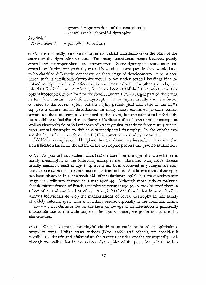
Sex-linked ~-chron1oso~al
grouped pigmentations of the central retina central areolar choroidal dystrophy
- juvenile retinoschisis
re II. It is not really possible to formulate a strict classification on the basis of the extent of the dystrophic process. Too many transitional forms between purely central and centroperipheral are encountered. Some dystrophies show an initial central localization but gradually extend beyond it; consequently they would have to be classified differently dependent on their stage of development. Also, a condition such as vitelliform dystrophy would come under several headings if it involved multiple perifoveallesions (as in rare cases it does). On other grounds, too, this classification must be refuted, for it has been established that many processes ophthalmoscopically confined to the fovea, involve a much larger part of the retina in functional terms. Vitelliform dystrophy, for example, usually shows a lesion confined to the foveal region, but the highly pathological LjD-ratio of the EOG suggests a diffuse retinal disturbance. In many cases, se..'<-linked juvenile retinoschisis is ophthalmoscopically confined to the fovea, but the subnormal ERG indicates a diffuse retinal disturbance. Stargardt's disease often shows ophthalmoscopic as well as electrophysiological evidence of a very gradual transition from purely central tapetoretinal dystrophy to diffuse centroperipheral dystrophy. In the ophthalmoscopically purely central form, the EOG is sometimes already subnormal.
Additional examples could be given, but the above may be sufficient to show that a classification based on the extent of the dystrophic process can give no satisfaction.
re III. As pointed out earlier, classification based on the age of manifestation is hardly meaningful, as the following examples may illustrate. Stargardt's disease usually manifests itself at age 8-14, but it has been observed in younger subjects, and in some cases the onset has been much later in life. Vitelliform foveal dystrophy has been observed in a one-week-old infant (Barkman 1961), but we ourselves saw originate vitelliform changes in a man aged 44· Although most authors maintain that dominant drusen of Bruch's membrane occur at age 30-40, we observed them in a boy of I z and another boy of 14· Also, it has been found that in many families various individuals develop the manifestations of foveal dystrophy in that family at widely different ages. This is a striking feature especially in the dominant forms.
Since a strict classification on the basis of the age of manifestation is practically impossible due to the wide range of the ages of onset, we prefer not to use this classification.
re IV. We believe that a meaningful classification could be based on ophthalmoscopic features. Unlike many authors (Blodi 1966; and others), we consider it possible to identify and differentiate the various entities ophthalmoscopically. Although we realize that in the various dystrophies of the posterior pole there is a
37

continuous evolution of the ophthalmoscopic picture, this does not usually prohibit identification of the specific entity. However, this evolution is indeed an obstacle to any attempt at classification on the basis of the foveal appearance. This obstacle might be overcome by selecting a characteristic fundus picture for each dystrophy, but this would make the classification rather artificial. It is relevant in this context to mention the great importance of a follow-up covering several years, which makes it possible to study the total dynamics of the evolution of the clinical picture. This has been illustrated by such studies as that of Rosehr (1954). In one of Stargardt's patients, the initial purely central dystrophy was found 5o years later to have become an extensive diffuse centroperipheral tapetoretinal dystrophy.
Besides such follow-up studies, an investigation of patients of all age groups is also an effective method to gain an impression of the evolution of the various dystrophies. Evidently our understanding of this subject will increase with an increasing number of patients observed.
re V. It might be possible to make a classification based on the results of the various retinal function tests used in the various dystrophies. Such a classification, however, would be too complicated to be very useful.
We intend to present a diagram in which the results of various tests in the various dystrophies are arranged for convenient reading (see page 428).
re VI. From a clinical point of view, a classification based on the retinal layers primary and mainly involved would seem to be the most practical and meaningful solution (Deutman 1970 ). Such a classification makes it possible to understand and predict in rough outline the clinical course of the various dystrophies. For example, a dystrophy primarily confined to the pigment epithelium (e.g. fundus flavimaculatus) is not likely to produce soon changes in the neuroepithelium or the choriocapillaris. Likewise, drusen of Bruch's membrane can be expected to have an unfavourable effect on the retinal neuroepithelium only in later stages.
A problem in formulating this type of classification is the fact that little is known o£ the histology of the posterior pole dystrophies. Exceedingly scanty histological data are available on young patients, in whom senile retinal and choroidal changes are of course not yet present. Histological specimens from older patients often show senile and autolytic postmortem changes, which impair assessment.
With the more sophisticated techniques of retinal electrophysiology and fluorescein angiography, however, localization of retinal changes has become less difficult. It is therefore that we have used these modern facilities in order to ensure a firm foundation for the classification we prefer: that on the basis of the localization of the changes in the various rerinalla yers.
The various components of the ERG and EOG have become more important in localization as our understanding of these techniques has improved (Goutas 1968). Moreover, fluorescein angiography affords excellent possibilities for the differentiation of changes of the pigment epithelium, Bruch's membrane, the choriocapillaris

CUTE:R PI..E:XlF'CRM t.AYE:R
CUTE:R NUCI..~AR I..AY~R
E:XTE:RNAt. l-IMITING M~MBRANE:
RODS AND CCN~S
PIC:ME:NT ~PITH~I..IUM
BRUCH~ ME:MSRAN~ ---::~~~~~~~~~~~~~t:~~~t:~~~~~;t~~~
Nerve :fibre layer Neuroepithelium
Pigment epithelium
Bruch's membrane
Choroid
Fig. 18.
se."\:-linked juvenile retinoschisis Stargardt's disease dominant progressive foveal dystrophy progressive cone dystrophy central retinopathia pigmentosa vitelliform dystrophy of the fovea fundus B.avimaculatus reticular dystrophy of the retinal pigment epithelium butterfly-shaped pigment dystrophy of the fovea grouped pigmentation of the foveal area dominant drusen of Bruch's membrane (hyaline dystrophies) pseudo-infl.a.mmatory foveal dystrophy (Sorsby) central areolar choroidal dystrophy
and the choroid in relatively early stages. In addition, information on the probable localization-in-depth of retinal lesions can be obtained with the aid of photographs made with monochromatic light (Krill et aL 1966) and photographs made on orthochromatic and panchromatic film (Craandijk and Aan de Kerk 1969).
Combination of these data with the other clinical findings facilitates classification in this manner. It should be borne in mind, however~ that with increasing age the degenerative changes secondarily manifest themselves in the more superficial as well as in the deeper layers of the posterior pole. For example, the terminal stage of a condition of evidently tapetoretinal onset, such as Stargardt's disease, often shows extensive atrophy of the choriocapillaris and the choroid. In such a case, clliferential diagnosis from central areolar choroidal dystrophy is impossible unless the history of the case is known.
Our purpose in designing our own classification was not only to facilitate identifi-
39

cation and differentiation of the many clinical entities, but also to provide a frame in which newly discovered entities could be readily accommodated (figure I 8). For we are convinced that several as yet unknown dystrophies will be discovered in future or will arise from new mutations.
Of course we are not sure that the primary enzymatic disturbances are indeed localized in the layers indicated in our diagram of structures primarily and mainly involved. It looks as if many of the enzymatic disturbances which give rise to dystrophies, will have to be localized in the pigment epithelium, the "heart and soul of the retina" (Krill I969). Ophthalmoscopic, electrophysiological and fluoresceinangiographic findings seem to warrant this conclusion. It is highly probable that, apart from the dystrophies shown under the heading pigment epithelium in our classification, the dominant drusen of Bruch's membrane and the choroidere.mia or tapetochoroidal dystrophy (Goedbloed I942; Pameyer eta!. I96o; Kurstjens I965), and possibly also central areolar choroidal dystrophy are likewise primarily dystrophies of the pigment epithelium in which the deeper retinal layers are quickly affected.
Stargardt's disease might be a primary dystrophy of the pigment epithelium in which the more superficial localized photoreceptors are involved after a short time. This is suggested by the pigment displacements and the perifoveal deep-seated white spots. The fact that the EOG becomes subnormal before the ERG does when the peripheral retina is affected, also points in this direction.
In initial stages of retinopathia pigmentosa, too, the EOG is often seen to become subnormal before the ERG does (Arden I962; Arden and Fojas I962; Arden and Kolb I 964; Imaizumi et a!. I 96 5 ). However, Gouras and Carr (I 964) found dark adaptation changes and scotopic ERG changes as the first pronounced abnormalities in the early stages of retinopathia pigmentosa. On this basis they localized the primary defect somewhere in the receptor system, affecting the rods more than the cones.
There may well be several forms of retinopathia pigmentosa, with different sites of primary involvement. The results obtained by De Haas (I970) also point in this direction. Arden and Kolb (I964) suggested that there are two ophthalmoscopically indistinguishable forms of this condition, one of which starts in the pigment epithelium while the other begins in the rods.
Discrepancies between results can also be caused by differences in techniques used and by differences in material studied.
Moreover, there are probably some dystrophies in which the photoreceptors are primarily affected, e.g. dominant progressive central tapetoretinal dystrophy (see page I72) and progressive cone dystrophy (see page I8I). In these cases, there are no or only insignificant ophthalmoscopic changes in the presence of extensive abnormalities of the photoreceptors. Likewise sex-linked juvenile retinoschisis (see page 48) shows from the onset a subnormal ERG in combination with a normal EOG; and this suggests an intact pigment epithelium. This is confirmed by fluorescein angiography and histological examination.

It can be concluded that the primary defect is localized in the pigment epithelium in many, but certainly not in all, dystrophies of the central retina. In any case, we cannot agree with a statement made by Elwyn (I 9 55) that: "The pigment epithelium of the retina is not subject to degenerations on a hereditary basis". Many arguments can be advanced against this statement. We need only point out that Klien and Krill (r967) found dystrophy of the pigment epithelium as the sole histological abnormality in a case of fundus flavimaculatus. The other dystrophies we have described as starting in the pigment epithelium, can hardly have any primary localization other than the pigment epithelium. This will be further discussed in the various pertinent chapters.
REFERENCES
ALBOUY, J.: Un nouveau syndrome: dystrophic maculaire et p6rimaculaire au cours de la surdimutit6 congCnitalc. Thesis, Toulouse (r96o).
ALKIO, v. v.: Heredo-degeneratio maculae cent:ralis retinae bei 4 Geschwistcm. Acta Ophthal. r: 27-;8 (1923). AMALRIC, M.P.: Nouveau type de d6g6n6rescences tap6to-r6tinienne au cours de la surdi-mutit6. Bull. Soc.
fran~. Ophtal. 73:195-zir (r96o). AMALRIC, M.P. and BESSOU, P.: Nouvelle contribution 3. 1'6tude des dCg6n6rescences tap6to-r6tin.iennes au
cours de la surdi-mutit6, Bull. Soc. Ophtal. France 62:459-461 (1962). AMALRIC, M, P., FERRER, J., RINS, ::"!., BESSOU, P. and LESCURE, F.: Nouvelles contributions i l'Ctude de la
dCgCnCresccnce tapCto-rCtinienne dans la surdi~mutitC. Bull. Soc. frans:. Ophtal. 76: roo~ro7 (196;). AMALRIC, M. P. and BEssou, P.: Nouvelles preuvcs de la transmission hL.-rCditairc des dCgCnCrescences rCti
niennes "i minima" assodCcs ala surdi-mutit6. Rev. Oto neuro-ophtal. ;6: 109-111 (1964). ARDE~, G. B.: Alterations in the standing potential of the eye associated with retinal disease. Trans. Ophthal.
Soc. U.K. 8z:6;-7z (196z). ARDEN, G. B. and BARRADA, A.: Analysis of the electro-oculograms of a series of normal subjects. Brit. J.
Ophthal. 46'468-482 (x96a). ARDEN, G. B., BARRADA, A. and KELSEY, J. H.: New clinical test of retinal function based upon the standing
potential of the eye. Brit. J. Ophthal. 46:449-467 (196z). ARDE::-:T, G. B. and FOJAS, M. R.: Electrophysiological abnormalities in pigmentary degenerations of the retina.
Arch. Ophthal. 68:ro5-rz5 (r96z). ARDEN, G. B. and KOLB, H.: Electrophysiological investigations in retinal metabolic disease: their range and
application. E::...'P. Eye Res. ;:;;4-347 (1964). ARNOULD, G., DUREl.l'X, J. and HAR'I'EM:ANN, P.: Un cas de lacunc cdniennc congCnitalc avec manifestations
neurologiques (syndrome c6r6bcllo-spasmodique, Cpilepsie, dCE.cience mentale) et ophtalmologique (d6g6n6rcscencc maculaire). Rev, Ncurol. 9;:8;8-84; (1955).
BABEL,;. and FARPOUR, H.: L'origine g6n6tique des fossettcs colobomateuses du nerf optique. J.d. GCnCt. Hum. 16:187-198 (1967).
BAILLIART, J.P.: Les affections de la macula. Masson, Paris, 1961. BARKMA..'f, Y.: A clinical study of a central TRD. Acta Ophthal. ;9 :66;~671 (1961). BASSEN, F. A. and KORNZWEIG, A. L.: Malformation of the erythrocytes in a case of atypical retinitis pig
mentosa. Blood 5: ;8r-;87 (1950). BATTEN, F. E.: Cerebral degeneration with symmetrical changes in the macula in two members of a family.
Trans. Ophthal. Soc. U.K. z;:;86-;9o (r9o;). BAT'I'EN, R. D.: Two brothers with symmetrical disease of the maculae, commencing at the age of fourteen.
Trans. Ophthal. Soc. U.K. 17:48-49 (1897). BEGAUX, c., DECOCK, G. and BOGAERT, L. vAN: Sur une souche de myotonic dystrophiquc (Steinert), dont
au moins un des sujets atteints est porteur d'une dCgCnCrescence tap6to~r.Ctinienne atypique. J. GCnCt. hum. 1:91-101 (1952).
41

Bi::GAUX, c. and DECOCK, G.: La d6g6n6rescence tap6to-r6cinienne de la dystrophic myotonique. In ProblCmes actuels d'Ophtalmologie~ vol. I, p. 551-558. Karger, Basel, 1957.
BEHR, c.: Die Heredodegeneration der Makula. Klin. Mbl. Augenheilk. 6 5 :46 5-50 5 ( 1920 ). BEHR, c.: Die Anatomic der senilen Macula (die senile Form der macularen Heredodcgeneration). Klin.
Mbl. Augenheilk. 67: 551:-564 (19zi). BEHR, c.: Ein weiterer Beitrag zur Anatomic und zur Pathogenese dcr scheibenformigen Degeneration am
hinteren Augenpol. Z. Augcnheilk. 75:n6-z36 (1931). BERGSTEDT, M., JOI-I.A."fSSON, S, and MULLER, R.: Hereditary SpaStic ataxia with central retinal degeneration
and vestibular impairment. Neurology IZ: IZ4-I32 (1962). BESSIERE, E., BOUCHARD, J. and CHABOT, J.: L'6lectrorCtinographie dans les d6g6n6::escences tap6to-reti
niennes. Arch. Ophtal. 22:677-698 (1962). BEST, F.: Ueber cine hereditiire Maculaaffektion. Beitriigc zur Vererbungslehrc. Z. Augenheilk. 13: I99-2IZ
(1905). BIELSCHOWSKY, M.: Ueber spiitinfantile familii:i.re amaurotische Idiotic mit Kleinhirn symptomcn. (7. Jahres
vers. Ges. dtsch. Nerveciirzt, L. Brcslau, 29-9-1913). Dtsch. Z. Nervenheilk. 50:7-29 (1914). BIETTI, G. B.: Su alcune forme atipiche o rare di degcnerw.Jone rctinica (degenerazioni tappeto-retiniche
c quadri morbosi similari). Boll. Ocul. 16: 1159-1244 (1937). BLODI, F. c.: The pathology of central TRD (hereditary macular degeneration). Trans. amer. Acad.
Ophthal. Otolaryng. 70: I047-1053 (1966). BOCK, J.: Zur Klinik und Anatomic dcr gef'iss:i.hnlichen Streifen im Augcnhintergrund. Z. Augcnhcilk.
95! I-50 (I938). BOGAERT, L. VAN: D'une atrophic d:r6belleuse familiale avec dystrophic maculaire (type Stargardt) et ano
malies cong6nitales de la pigmentation cutan6e du type de l'incontinentia pigmenti. In Probl. actuels d'ophtalmologie, vol. I, p. 540-550. Karger, Basel, 1957·
BRALEY, A. E.: Dystrophy of the macula. Amer.]. Ophthal. 61: I-24 (1966). BROWN, A. L.: Familial rosette figure of the macula. Amer. J. Ophthal. I I : I 90 (I928). BURIAN, H. M. and BURNS, c. A.: Ocular changes in myotonic dystrophy. Trans. amer. Ophthal. Soc. 64:
25D-273 (1966). BUZZI, F.: Nuove sperienze fatte sull'occhio umano. Opuscoli scetti sulle scienzc e sullc atti 5:87 (I782),
ed. Giuseppe MarcHi, Milano. CARPE:-ITER, s. and SCHUMACHER, G. A.: Familial infantile cerebellar atrophy associated with retinal degener
ation. Arch. Ncurol. I4:8z-94 (1966). CHIEVITZ, J. H.: Die Area und Fovea centralis retinae bcim menschlichen Foetus. Intern. Mschr. Anat.
Physiol. 4:201 (1887). CLAUSEN, w.: Hcrcdo-degeneration dcr Makula. Kl. Mbl. Augenhcilk. 67:117-118 (19ZI). CLAUSEN, w.: Typischcs, beiderseitiges hereditiires Makula-kolobom. Klin. Mbl. Augenheilk. 67:116 (I921). CLAUSE~, w.: Zur Frage der Vercrbung der Makulakolobomc. Klin. Mbl. Augenhcilk. 81:385 (1928). COGAN, D. G.: Hcredodcgeneration of the retina. In Ncuro-Ophthalmology, vol. 2, p. 44-61. Mosby,
St. Louis, 1965. COGAN, D. G., KU\VABARA, F., MOSER, H. and HAZARD, G. W.: Retinopathy in a case of Farber'S lipogranulo
matosis. Arch. Ophthal. 75:752-757 (I966). CRAANDIJK, A. and .AA."f DE KERK, A. L.: Retinal photography using panchromatic and orthochromatic films.
Bdt. J. Ophthal. 53'568-573 (1969). CRAANDIJK, A.: Fluorescence angiography in central retinal vein occlusion. A pilot study of the therapeutic
effects of streptokinase and hcpatin. In Perspectives in Ophthalmology, vol. 1:, p. 57-64- Excerpta Medica Foundation, Amsterdam, I968.
CRAA~DIJK, A. and HOUTSMULLER, A. J.: Hypo and A-bCta lipoproteinaemia. In Perspectives in Ophthalmology, vol. ll pp. 96-1o2. Excerpta Medica Foundation, Amsterdam, I970.
DAVENPORT, R. c.: Bilateral macular coloboma in mother and son. Proc. Roy. Soc. Mcd. U.K. 2I!I09 (1927). Ref.: Klin. Mbl. Augenheilk. 79:851 (1927).
DEUTMA.-...r, A. F.: Hereditary dystrophies of the central retina and choroid. Perspectives in Ophthalm. vol. II pp. 57-79. Excerpta Medica Foundation, Amsterdam I970.
DOUGLAS, A. A., w AHEED, I. and WYSE, c. T,: Progressive bifocal chorioretinal atrophy. A rare familial disease of the eyes. Brit. J. Ophthal. p:742-751 (I968).
DOYNE, R. w.: Peculiar condition of choroiditis occurring in several members of the same family. Trans. Ophthal. Soc. U.K. 19072 (1899).
DOYNE, R. w.: A note on family chorioiditis. Trans. Ophthal. Soc. U.K. 30:93 (I9IO).
42

oOYNE, R. w.: Discussion de la communication de Mould. Familial choroiditis. Trans. Ophthal. Soc. U.K. ;o: IS9-I90 (I9Io).
DUJARDL"<: Rc':tinite pigmcntairc anormale chez deu.." frCres jumeaux. Clin. Ophtal. Io: IZ5-IZ7 (1904). DUKE-ELDER, s.: System of Ophthalmology, val. II. The anatomy of the visual system. Kimpton, London,
I961. DUKE-ELDER, s.: System of Ophthalmology. Vol. X. Diseases of the retina. Kimpton, London, 1967. EISLER, P.: Die Anatomic des menschlichcn Auges. Kurzes Handbuch der Ophthalmologic 1. Von Schieck
und BrUckner, Berlin, I9;o. ELWY;:.;r, H.: Heredodcgenerations and hercdoconstitutional defects of the retina. Arch. Ophthal. 55 :619-
633 (1955)· EVANS, P. J.: Familial macular colobomata. Brit. J. Ophthal. zr: 505-506 (1957). FABRY: Angiokeratema corporis di:ffusum. Arch. Derm. Syph. (Wicn) 45: I87 (r89S). FALLS, H. F.: The role of the sex chromosome in hereditary ocular pathology. Trans. amer. Ophthal. Soc.
50:435-467 (1952). FALLS, H. F.: A classification and clinical description of hereditary macular lesions. Trans. amer. Acad.
Ophthal. Otolaryng. 70: ro;4-1o46 (I966). FALLS, H. F.: Detection of the carrier state of genetically determined eye diseases. In Congenital anomalies
of the eye. Mosby, St. Louis, 1968. FARBER, s., COHE."<, J. and UZMAN, L. L.: Lipogranulomatosis: A new lipo-g:lycoprotein storage disease.
Mt Sinai Hasp. Bull. z4:Sx6 (I957). FAVRE, M.: A propos de deu." cas de dc':g6n6rescence hyaloid6or6tinienne. Ophthalmologica 155:604-609
(1958). FAVRE, M.: Discussion to A. Ricci. Ophthalmologica x;9:54z (I96o). FAVRE, M.: D6g6n6rcscence "'myopique" du corps vitr6 ct emmCtropie. Ophthalmologica I4I: ;57-;6z
(1961). FLEISCHER, :s.: Zur V ererbung seniler Makulavera.nderungen insbesondere der ScheibenfOrmigen Entartung
der Nethautmitte. Klin. Mbl. Augenheilk. IIO: I45-149 (I944). FOSTER, J. B. and INGRAM, T. T. s.: Familial cerebra-macular degeneration and ataxia. J. Neural. Neurosurg.
Psychiat. 25:63-68 (1962). FRANCESCHE"ITI, A., FRA."<<,;:01S, J. and BABEL, J·; Les h6r6do-d6g6n6rescenccs chorio-r6tiniennes. Masson,
Paris, I96;. FRANCESCHETII, A., DIETERLE, P., AMMANN, F. and MARTY, F.; Une nouvelle forme de fundus albipunctatus
cum hemeralopic. Ophthalmologica 145 :405-4I0 (I96;). FRAN<,;:OIS, J.: Nouvelle contribution a 1'6tude des h6r6do-d6g6n6resccnces tap&o-r6tinienncs p6riph6riques
et centrales. Acta XVI Concilium Ophthal. Brit. 498-506 (1950). FRANyoxs, J. and DEWEER, J·: D6gCn6rescence maculaire sc':nile et h6:rCdit6. Ann. Ocul. xS5: I;6-I 54 (1952). FRAN90IS, J.: Heredity in ophthalmology. Mosby, St. Louis, 1961. FRAN<,;:OIS, J., HAUSTR.Al'E-GOSSET, F. and DQ;:\I"CK, D.: Macular dystrophy in deafmutism. Acta genet. med.
gemellog. 16:63-70 (1967). FRANt;01S, J., HAUSTR.Al'E-GOSSET, F. and DONCK, D.: Dystrophic maculaire Ct surdi-mutit6. Ann. Ocul. 200:
890-899 (1967)· FRAN<,;:01S, J.: L'h6:rt:dit6 des dc':g6n6rescences maculaires s6niles. Arch. Opht. (Paris) 29:S99-90z (1969). FRIEDMA."m, A. I.: Serial analysis of changes in visual field defects, employing a new instrument to determine
the activity of diseases involving the visual pathways. Ophthalmologica 152: 1-rz (I966). FROMENT, J., BONNET, P. and COLR.AT, A.: H6r6do-d6g6nc':rations retinicnnes et spino-c6r<!bclleuses, variantes
ophtalmoscopiques et neurologiques pr6sent6es par ; g6n6rations successives. J. M6d. Lyon IS: I 55-x6z (1937). .
FUCHS, E.: Lehrbuch der Augenheilkunde, I5C Auflagc. Franz Deuticke, Leipzig und Wien, 19z6. GASS, J. D. M.: Pathogenesis of disciform detachment of the neuroepithelium. II. Idiopathic central serous
choroidopathy. Amer. J. Ophthal. 6;:5S7-615 (1967). GAUCHER, P. CH. E.: De I' cpitheliome primitif de la rate, hypertrophic idiopathiquc de I a rate sans leucCmie.
Thesis Paris, ISS z. GILBERT, W. R., LAWTON SMITH, J. and NYHAN, W. L.; The SjOgren-Larsson syndrome. Arch. Ophthal. So;
308-316 (1968). GOEDBLOED, J.: Mode of inheritance in choroidcremia. Ophthalmologica 104: ;oS-;15 (I94z). GOURAS, P. and CARR, R. E.: Electrophysiological studies in early retinitis pigmentosa. Arch. Ophthal. 7z:
104-110 (1964).
43

GOURAS, P.: Relationship of the electro-oculogram to the electroretinogram. In The clinical value of electroretinography; Proc. 5th ISCERG Symp. Ghent, 1966, pp. 66-73. Karger, Basel, I968.
GOWERS, w. R.: A lecture on abiotrophy. Lancet 1: roo3-Ioo7 (r9oz). HAAS,;.: Ueber das Zusammenvorkommen von Veriinderungcn der Retina und Chorioidea. Arch. Augen
heilk.;7:;45·348 (rS98). HAAS, E. B. H. DE: Heterozygous manifestations in various types of tapeto-retinal dystrophy. In Perspectives
In Ophthalmology, vol. II, p. 56. Excerpta Medica Foundation, Amsterdam, 1970. I-lAAS, E. B. H. DE, LITH, G. H. M, VAl':', RIJNDERS, J., Ri.JM:KE, A, M. L. and VOLMER, CH.: Usher's syndrome with
special reference to heterozygous manifestations. Doc. Ophthal. 28: I66-r9o (I 970 ). HAGEDOOR:-., A.: Angioid streaks. Arch. OphthaJ. 2I :746-774 (1939). HAGEDOORN, A.: .Angioid streaks. Arch. Ophthal. 21:935-965 (1939). HAGEDOORN, A. and BOSCH, C. VAN DEN: A double-projection campimeter. Amer. J. Ophthal. 40:89I-893
(r955). HALBERTSMA, K.: Over erfelijkc ontaarding van de gele vlek gepaard met kleurcnblindheid. Ned. T. Ge
neesk. 71: 2056-2o68 (I927). HALBERTSMA, K.: Ueber einige erbliche fami!Hire Augenerkrankungen. Ueber erbliche Entartung des gelben
Fleckes vereinigt mit Farbenblindheit. Klin. Mbl. Augenheilk. So: 794-801 (I928). HALSEY, J· H., SCO'I'T, TH. R. and FARMER, TH. w.: Adult hereditary cerebelloretinal degeneration. Neuro
logy 17:S7·9o (r967). HAVENER, w. H.: Cerebellar-macular abiotrophy. Arch. Ophthal. 45 :40-43 (1951). HENKES, H. E., DENIER VAl": DER GON, J· ]., MARLE, G, W. VAN and SCHREINEMACHERS, H. P,; Electro-oculo
graphy. A semi-automatic recording procedure. Brit. ]. Ophthal. 52: I 22- r 26 ( r 968). HOU'I'SMULLER, A.;.: Verhandlungen des II. Intemationalen Gespr3.che Uber Angiologie 1962. Angiologie
2! :276-278 (!96;). HUTCHINSON, J· and TAY, w.: Symmetrical central chorio-rctinal disease occurring in senile persons. Roy.
Lond. Ophthal. Hosp. Rep. 8:23 r-244 (1875). IMAIZUMI, K., TAKAHASHI, F., KAMEl, M., TOYAMA, F. and HORIE, E,: Relationship between the ERG and the
EOG in retinal pigmentary degeneration. In Retinal degenerations, ERG and optic pathways; Proc. of the 4th ISCERG Syrup., Japan 1965, p. 245-266. University of Tokyo, Tokyo, I965.
JA..'ISKY, ;.: Ueber einen noch nicht beschriebencn Fall der familiiiren amaurotischen Idiotic mit Hypoplasie des Kleinhirns. Z. Erforsch. jugendl. Schwachsinns 3:86 (I9IO).
JEXDRASSIK, F.: In Lewandowsky: Handbuch der Neurologic. Springer, Berlin, 1911. JONKERS, G. H.: Central serous retinopathy in a monozygotic twin pair. Acta Genet. med. et Gemcll. 3:
438-44I (1960). JUNGE,;.: Ocular changes in dystrophia myotonica, paramyotonia and myotonia congenita. Thesis. Junk,
The Hague, I966. KLIEN, B. A.: Angioid streaks. Amer. J. Ophthal. 30:955-968 (1947). KLIEN, B. A.: Some aspects of classification and differential diagnosis of senile macular degeneration. Amer.
]. Ophthal. 58:9'7-939 (x964). KLIEN, B. A. and KRILL, A. E.: Fundus Ravimaculatus: clinical, functional and histopathological observations.
Amer. ]. Ophthal. 64: I-25 (1967). KORNZWElG, A. L., FELDSTEIN, M. and SCHJ'o:'ElDER, J.: The eye in old age: IV. Ocular survey of over one
thousand aged persons with special reference to normal and disturbed visual function. Amer. J. Ophthal. 44:29-37 (x957).
KRANENBURG, E. w.: Crater like holes in the optic disc and central serous retinopathy. Thesi :, Amsterdam. Ellerman Harms, I959·
KRILL, A. E.: Personal communication. 1969. KRILL, A. E.: The electroretinographic and electrooculographic findings in patients with macular lesions.
Trans. amer. A cad. Ophthal. Otolaryng. 70: xo63-1083 (1966). KUFS, H.: Ueber cine Spatform dcr amaurotischen Idiotic und ihre heredo-familiiiren Grundlagcn. Z. ges.
Neurol. Psychiat. 95:169-188 (1925). KURSTJENS, J. H.: Choroideremia and gyrate atrophy of the choroid and retina. Thesis. Junk, 's-Graven
hage, 1965. LANG, w.: Central choroiditis, with disseminated patches in remainder of fundus. Trans. Ophthal. Soc. U.K.
5: I4D-l4I (188~). LEBER, TI-X.: Die Krankheiten der Netzhaut, Graefe-Saemisch-Hess Handbuch ges. Augenheilk. Engelmann,
Leipzig, I916.
44

LEDIC, P. and BOGAERT, L. vAN: HetCdo-dCgCnCrcscence cb'Cbelleuse et spastique avec dfgCnCresccncc maculaire. J. Genet. hum. 9: qo-157 (r96o).
LIEBRECHT: Retinitis puncatata albcscens. Klin. Mbl. Augenheilk. 55: 169-176 (1895). LITH, G. H. M. VAN and BALIK,;.: Variability of the electro-oculogram. To be published in Acta Ophthal
mologica. LITH, G. H. M. vAN and HENKES, H. E.: The local electric response of the central retinal area. In Advances in
electrophysiology and -pathology of the visual system; Proc. 6th ISCERG Symp. Erfurt, 1967. Thieme, Leipzig, 1968.
LOUIS-BAR, D. and PIROT, G.: Sur unc paraplCgie spasmodiquc avec dCg6nCresccncc maculaire chez deux freres. Ophthalmologica 109:52-45 (1945).
MAGERS, J·: Ueber hercditii.re Sehnervenatrophie und hereditii.re chorioiditis. Thesis, Jena, 1899· MAG1STRETTI, A.: Su una raro manifesuzionc oculare del morbo di Steinert. Boll. Ocul. 34: 617-629 (195 5). MARCo, P. DE: Dcgenerazione retinica oftalmoplegia associate a credo atassia cerebellare. Riv. Pat. ncrv.
ment. 89: 54-46 (1968). MAYOU, M. s.: Cerebral degeneration with symmetrical changes in the maculae in three members of a family.
Trans. Ophthal. Soc. U.K. 24:142-145 (1904). MORAx, v.: Ref.: Waardenburg P. ]., Genetics and Ophthalmology Vol. II, p. 1725. Van Gorcum, Assen,
I96J. MULLER, H.: Bemerkungen iiber den Bau und die Function der Retina. Verhdl. phys.-med. Ges. Wiirzburg
3:336 (1852). NIEMA.."fN, A.: Ein unbckanntes Krank.heitsbild. Jahrb. Kinderheilk. 79: 1-10 (1914). NORMA."!, R. M. and WOOD, N,: Congenital form of amaurotic familial idiocy. J. Neural. Psychiat. 4:175-190
(I94I). NOVOTNY, H. R. and AL vxs, D. L. : A method of photographing fluorescence in circulating blood in the human
retina. Circulation 24:82-86 (1961). OATMAN, E. L.: Maculo cerebral degeneration (familial). Amcr. J. Med. Sci. 142:221-235 (x9n). o'BRIEN, J. s.: Five gangliosidoses. Lancet II:So5 (1969). OOSTERHms, J. A. and LAM:MENS, A. J. ;.: Fluorescein photography of the ocular fundus. Ophthalmologica
149:210-220 (1965). PAMEYER, J. K., WAARDEl':'BURG, P. J. and HENKES, H. E.: Choroidercmia. Brit.]. Ophthal. 44:724-738 (1960). PHILLIPS, C. I. and GRIFFITHS, D. LLOYD: Macular coloboma and skeletal abnormality. Brit. J. Ophthal.
53=546-549 (1969)-PICK, L.: Zur pathologischen Anatomic des Morbus Gaucher. Mcd. Klin. r8: qo8-1409 (I 922). POLYAK, s. L.: The retina. The University of Chicago Press, Chicago, 1941. REDSLOB, E.: Anatomic macroscopique et microscopique des diffCrcntcs panics du globe oculaire. La
retine. In TraitC d'ophtalmologie. Vol. r, p. ;8z-;88. Masson, Paris, 1959. REDSLOB, E.: Lt=sions maculaires chez les jeunes. Ann. Ocul. 176:758-744 (1959). REFSUM, s.: Hercdopathia atactica polyneuritifonnis, a familial syndrome not previously described. Nord.
Med. 28:2682-2686 (1945). Acta Psychiat. scand. Suppl. 58, r-103 (1946). RICCI, A.: Clinique et tranSmission gCnCtique des diffCrentes formes de dfg6n~rescences vitrCo-rl.-tinienncs.
Ophthalmologica 139: 358-345 (r96o). Bull. Soc. Ophtal. France 6x: 6r8-66z (r96r). RIEGER, H.: Demonstrationen. Z. Augenheilk. 55: 546-548 (1925). RIEGER, H.: Ein Beitrag zur Kasuistik dcr tapeto-retinalen Degeneration (Hcredodcgcncradon der Macula
mit Beteiligung der Peripherie bei 3 BrUder). Z. Augenhcilk. 57:429-463 (1925). ROCHON-DUVIGNEAUD, A.: Anatomic de l'apparcil ncrveu.'C sensoricl de la vision (retine, ncr£ optique, centres
optiques). Encycl. Fran<;. d'Ophtal. I: 532-635 (1905). ROCHON-DUV1GNEAUD, A.: Recherches sur la fovea de la rl.'tine humaine ct paniculi2rcment sur le bouquet
des cOnes centrau.x. Arch. Anat. Microsc. II: 515-342 (1907). ROSEHR, K.: Ueber den weiteren Verlauf der von Stargardt und Bchr beschriebenen famililiren Degeneration
dcr Makula. Klin. Mbl. Augenheilk. 124:171-179 (1954). R6TTH, A. VOl':': Infantile Heredodegeneration der Maculagegend in Form von Retinitis pigmentosa in versa.
Ber. dtsch. Ophthal. Ges. 48:580-584 (1950). SACHS, B.: On arrested cerebral development with special reference to its cortical pathology. J. Ncrv. Ment.
Dis. 14:541-555 (r887). SANDOZ, Y. L.: Beidseitiger histologischer Befund bei senilem Maculapseudotumor, der zu einer neucn Auf
fassung dieser Altersvcriinderung Iti.hrt. Gracfes Arch. Ophthal. 140:724-747 (1959). SCHOTT, K.: Ueber d.as sogenannte Kolobom der Makula. Klin. Mbl. Augenhcilk. 67:415-424 (1921).
45

SJOGREN, T.: Klinische und crbbiologische Untersuchungen iiber die Heredo~ataxien. Munksgaard, Copenhagen, 1945·
SJOGREN, T.: Oligophrenia combined with congenital ichthyosiform erythrodermia spastic syndrome and macular-retinal degeneration. Acta Genet. Statist. Med. 6:So-91 (1956).
SJOGREN, T. and LARSSON, T.: Oligophrenia in combination with congenital ichthyosis and spastic disorders. Acta Psychiat. Neural. Scand. 52, suppl. II5, 1957.
SOEMMERRING, s. TH.: De foramine centrali limbo luteo cincto retinae humanae. Comment. Soc. reg. Sci. 13:; (1795-1798). Published 1799·
SORSBY, A.: Diseases of the retina. I. Retinal abiotrophy. Trans. Ophthal. Soc. U.K. 54: r6o-175 (1954). SORSBY, A.: Congenital coloboma of the macula. Brit.]. Ophthal. 19:65-90 (1955). SORSBY, A,: The dystrophies of the macula. Brit.]. Ophthal. 24:469-553 (1940). Sl?IELMEYER, w.: Ueber familifu:e amaurotische Idiotieen. Arch. Psychiat. Nervenkr. 40: 1058 (1905). STADLIN, w. and BOGAERT, L. VAN: Alt6rations chorio-r6tiniennes chez demc soeurs atteintes d'het6do-
ata.-xie du type Friedreich. Acta Neural. Psychiat. Belg. 49:705-750 (1949). STARGARDT, K.: Ueber familiare, progressive Degeneration in der Makulagegend des Auges. Graefes Arch.
Ophthal. 7" 554-5 50 (>909)· STARGARDT, K.: Ueber familifu:e, progressive Degeneration in der Makulagegend des Auges. Z. Augen
heilk. 50:95-II5 (I91;). STARGARDT, K.: Zur Kasuistik der ''familifu:en, progressiven Degeneration in der Makulagegend des Auges".
z. Augenheilk. ;5:249:-255 (1916). STARGARDT, K.: Ueber famili:i.re Degeneration in der Makulagegend des Auges, mit und ohne psychische
StOrungen. Arch. Psychiat. Nervenkr. 58:852-887 (19I7). STARGAR.DT, K.: Ein Fall von familifu:cr progressiver Makuladegeneration. Klin. Mbl. Augenheilk. 75:
'-46 (>925)· STEL'T.ERT, H.: Myopathologische Beitrii.ge. I. Ueber das klinische und anatomische Bild des Muskelschwunds
der Myotoniker. Dtsch. Zschr. Nervenheilk. 57:58-Io4(I909). STOCK, w.: Ueber cine bis jetzt noch nicht beschriebene Form derfamiliiir auftretenden Netzhautdegeneration
bei gleichzeitigen VerblOdung und Uber typische Pigmentdegeneration der Netzhaut. Klin. Mbl. Augenheilk. 46:225-244 (19oS).
STREIFF, E. B. and BABEL, J.: La senescence de la rd:ine. ProgrCs en ophtalmologie I5: 1-75 (I965). TAY, w.: Symmetrical changes in the region of the yellow spot in each eye of an infant. Trans. Ophthal.
Soc. U.K. I: 55-57 (1881). THIEL, R.: Atlas der Augenkrankheiten. Stuttgart, Thieme 194S. TILLE, H.: U:sions de la macula lutea chez !'enfant et l'adolescent dites "maladie de Stargardt". Thesis.
Maloine, Paris, I 929. TILLE, H.: D6g616rescences maculaires familiales de cause inconnue, Bull. Soc. ophtal. Paris, IS: 548 (I956).
ReL Ann. Ocul. >73:823 (>936). TREACHER COLLINS, E.: Diseases of the retina. Abiotrophy of the retinal neuroepithelium or "retinitis pig
mentosa". Trans. Ophthal. Soc. U.K. 59:165·195 (I9I9). VELZEBOER, c. M. J.: Hereditary degenerative macular lesions with inflammatory signs and late visual
disturbance. Brit.]. Ophthal. 47:64I-65o (196;). VERDUIN, J?. c.: Crater-like holes, local retinal degeneration and heredity. Ophthalmologica I49: 226 (I965). VERREY, F.: Myotonic dystrophy associated with degeneration of the macula. Ophthalmologica II4: z8I-
284 (>947)· VOGT, A.: Zur Pathologic und pathologischen Anatomic der vcrschiedenen Idioticformen. Mschr. Psychiat.
Neural. 18:I6I~171 (I905). voGT, A.: Ophthalmoskopische Untersuchungen der Macula lutca im rotfreien Licht. Klin. Mbl. Augcn
heilk. 66:521-565 (I9ZI). VOGT, A.: Die Nervenfaserzeichnung der menschlichen Netzhaut im rotfreien Licht. Klin. Mbl. Augenheilk.
66:718-730 (192I). VOGT, A.: Die Reflexion der Net7..hautvorderfiache im rotfreien Licht. Prfu:etinale (supcrfizielle retinale)
Faltchenbildung. Klin. Mbl. Augenheilk. 66:S58-859 (1921). VOGT, A.: Die Vererbung von Altersmcrkmalen des Auges. Senile Determination des Keimplasmas nach
Beobachtung an 52 Paaren 55 bis 81 jiih.riger Zwillingen. Schweiz. Med. Wschr. 65:25 (1935). WAARDENBURG, P. J.: Ueber familHir-erbliche Fiillevon seniler Makuladegeneration. Genetica IS: 5S-46(19;6). WAARDE.'ffiURG,P.J.: Waameming bij bet nageslacht van een ten opzichte van den erfelijken nystagmus
discordanteeneiigtweelingpaar. Ned. T. Geneesk. 82:45IZ(1958).

WAARDENBURG, P. J.: Hypoplasia of the central fovea. I I rth Meeting Nethcrl. Ophthal. Soc., Amsterdam, 15-1Z-I946. Ophthalmologica 115: II4 (1948).
WAARDENBURG, P. J.: Twin research in ophthalmology. Docum. Ophthal. vol. 4, p. 154-199. Junk, 's-Gravenhage, 1950.
WAARDENBURG, P. J·: On two female identical twin pairs concordant for senile macular degeneration. Acta XVIII Int. Congr. Ophthal. Brussels, !:446-447 (1958).
W AARDENBURG, P. J., FRANCESCHETTI, A. and KLEIN, D.: Genetics and Ophthalmology. Vol. !I. Van Gorcum, Assen, Thomas, Springfield, Ill. 1963.
WAARDENBURG, P. J., Personal communication, I968. WALD, G.: Human vision and the spectrum. Science ror:655-658 (1945). Ref.: Duke-Elder, S. System of
Ophthalmology. Vol. II. The anatomy of the visual system. Kimpton, London, 1961. WALSH, F. B.: Clinical neuro-ophthalmology. 2nd ed. p. 670:-671. Williams and Wilkins, Baltimore, 1957. WALSH, F. M, R.: Clinical neuro-ophthalmology, p. 741. Williams and Wilkins, Baltimore, I947· WEINER, L. P., KONIGSMARK, B. w., STOLL, J. and MAGLADERY, J• W.: Hereditary olivOpOntocerebcllar atrophy
with retinal degeneration. Arch. Neural. r6:364-376 (I967). WINKELMAN, J· E.: Angioid streaks. Ophthalmologica n5:84-88 (1948). YA.\1ADA, E.: Some structural features of the fovea centralis in the human retina. Arch. Ophthal. 82: I 51-
'59 (r969).
47

II
Sex-linked juvenile retinoschisis
I. INTRODUCTION
Sex-linked juvenile retinoschisis belongs in the group of vitreoretinal dystrophies which also includes Wagner's vitreoretinal dystrophy and Goldmann-Favre vitreotapetoretinal dystrophy. Although initially it was considered to be a relatively rare abnormality, it has recently become apparent that it is a fairly common condition. In the course of our study we have been able to confirm this.
According to Franceschetti et a!. (r963), only about roo case reports could be found in the literature; but a study by Vainio-Mattila eta!. (r969) in Finland revealed over roo patients in one large family alone.
We ourselves found 48 individuals with this affection, although we made no specific effort to collect as many cases as we could.
In many cases the correct diagnosis is probably not made due to the very great differences in expression of this condition. Not infrequently it is diagnosed as "heredodegeneration of the macula" (Korchmiros et a!. r 96 5) or Stargardt' s disease (Peralta and Santori 1967); others (Barut 1955) have diagnosed it as periphlebitis. Falls (r966) emphasized the deceptive similarity between juvenile retinoschisis and Eales' disease. All these different diagnoses are not so surprising because, on the one hand a foveal retinoschisis is the only abnormality in some 50% of cases (Jager 195 3; Ricci r 96o, r 96 r ), while on the other hand vascular sheaths and vitreous opacities are quite common in the more severe forms.
Haas (r898) was the first to describe cases of X-chromosomal juvenile retinoschisis. He observed two brothers with this condition, and his publication includes beautiful drawings of the foveal abnormality which characterizes juvenile retinoschisis and occurs in virtually all patients. The disease has been described in the literature under a wide variety of names.
Haas (r898) described it as "Veriinderungen der Retina und Chorioidea", and Thomson (1932) reported on a "neuroretinal disease in males". Anderson (1932) described a z-year-old boy who suffered from "anterior dialysis of the young", which

may have been juvenile retinoschisis. Pagenstecher (I 9 I 3) described the first family with juvenile retinoschisis (in which an X-chromosomal pattern of transmission was demonstrable, as in the families described later) in a paper entitled: "Ueber eine unter dem Bilde der Netzhautablosung verlaufende, erbliche Erkrankung der Retina".
The term retinoschisis, introduced for this disorder by Jager (I95 3) is generally accepted to date in the designation sex-linked juvenile retinoschisis or idiopathic hereditary retinoschisis (in young men) (Jager, I95 3; Kleinert I95 3; Ricci I96o, I96I; Gieser and Falls I96I; Forsius eta!. I96z, I963; Sarin et al. I964; Isbey and Beuerman I964; Cibis I965; Boudon and Sole I965; Sabates I966; Spencer I966; Schepens I966; Brockhurst I966; Falls I966; Bengtsson and Linder I967; Goodman I968; Yanoff eta!. I968; Donaldson I968; Lisch I968; Stepanik I969; Guyot-Sionnest I969; Vainnio-Mattila eta!. I969; Harris 1969; Pischel I969; Yazawa and Nakajima I969; Ewing and Ives I969 and Hauschild et al. I97o).
A few authors use the term "congenital cystic retinal detachment" or "inherited retinal detachment" (Cibis I94o; Juler I948; Magnus I95 I; Sorsby I9jl, I95 5, I967; Levy I9)2).
"Vitreous veils" (Condon and Somerville-Large I 9 55 ; Goodman et a!. I 96 5) and "congenital vascular veils (in the vitreous)" (Mann and McRay I938; Sheehan I952; McRay I954; Scorciarini-Coppola eta!. I958; Balian and Falls I96o; Keith I966) are likewise designations often used for this syndrome, and "cystic disease of the retina" (Juler I95 I; Pischel I963) is occasionally used also. Trevor Roper (I95o) described a man aged z8 with a "congenital retinal fold in association with pseudopapillitis", who is now generally accepted as having suffered from sex-linked juvenile retinoschlsis.
We prefer the term sex-linked or X-linked juvenile retinoschisis, because this condition has an X-chromosomal pattern of transmission, occurs very early in life (probably it is ophthalmoscopically present at birth), and histological studies have disclosed cleavage of the retina at the level of the nerve fibre layer (Y anoff eta!. I 968; Manschot I969). Since vascular or avascular vitreous veils and cysts are by no means always seen in these cases, we reject designations which emphasize these phenomena.
Since true detachment of the retina is rare in this condition (Schepens I 966), we also reject the term "congenital retinal detachment" (Sorsby et al. I 9 5 I ; Sorsby I 9 5 5, I967)·
Virtually all cases (98-Ioo%) involve foveal retinoschisis, the characteristics of which have been described clearly and in detail by several authors (Haas I 898; Lisch I937; Jager I953; Kleinert I953)· This foveal retinoschisis is pathognomonic for the syndrome, and thus constitutes the principal symptom of this relatively common condition.
I have never observed peripheral retinoschisis without foveal retinoschisis; it is my impression that it is non-existent, although such authors as Sorsby eta!. (I95 I), Condon and Somerville Large (I 9 55) and Gieser and Falls ( 196 I) have reported it.
49

In most cases, however, vision was greatly diminished, and this could hardly have any cause other than foveal changes. In elderly patients the characteristic foveal pattern disappears, and in some cases the foveal changes are not readily observable. This may e>cplain the fact that some reports describe a normal fovea.
2. GENERAL CLINICAL PICTURE IN THE PATIENTS
The syndrome is found virtually only in males. It is probably present since birth, and very young boys can be encountered as patients. They often do not visit an ophthalmologist until vision proves to be insufficient at school, and the condition is therefore frequently seen in age group 4-8.
We saw two boys with this condition as early as age ;, and juvenile retinoschisis has been observed even earlier in life. Pischel (r96;, 1969) saw three brothers aged z years, I year and three months, respectively; Hartis (r969) saw an infant aged rG months.
Also, these patients are often first seen when vitreous haemorrhage has occurred as a result of rupture of a retinal vesseL
The condition takes a slowly progressive course and can therefore be interpreted as dystrophy on the basis of dysplasia. Complaints about poor vision can long remain absent, and some patients will therefore not be recognized as such until later in life.
Strabismus (8 patients) and nystagmus (4 patients) are frequently seen in this syndrome; they are best interpreted in the light of the foveal changes and the nearly always present hypermetropia with astigmatism.
Anterior segment and lens are usually quite normal, although occasional reports mention posterior cortical cataract and slight opacities of the posterior capsule (Balian and Falls 196o).
We observed an opacity of the posterior capsule in one eye, although generally the abnormalities are symmetricaL At times, there are marked interfamilial differences in clinical features, but on the other hand there are usually close intrafamiliar similarities of the manifestations.
3· FUNDUS (OPHTHALMOSCOPIC FEATURES)
In this condition in particular, the fundus is best assessed by means of contact lens and slit-lamp examination, for both the vitreous and the retina can show extensive changes which can be properly judged only by binocular e."<amination. The syndrome of sex-linked juvenile retinoschisis comprises many symptoms, some of which are often absent. In the severe cases, however, the full multitude of symptoms may be encountered; but the expression of the gene is often so low, or the process so little advanced, that only foveal retinoschisis is present. However, this foveal retinoschisis is the characteristic symptom of sex-linked juvenile retinoschisis, and is
50

Fig. zab. Pathognomonic bilateral symmetrical picture of foveal .retinoschisis in an S-year-old boy with X-linked juvenile .retinoschisis. Note the wheel-like configuration with the radiate spoke-like striation. In this case the discs,
vessels and retinal peripheries arc quite normal.
51

Fig. 2. Foveal retinoschisis in a I?-ycar-old boy. In this case the cystoid structure is well discemiblc (Fam. Pie).
present in virtually all cases (Falls (r966) mentioned 98% of cases; our impression is that roo% would be correct).
This symptom is the sole pathological finding in some 5o% of cases. Only in Goldmann-Favre vitreotapetoretinal dystrophy has a similar foveal involvement been described (Franceschetti et aL r963), but according to Ricci (r96r) the fovea shows no true cystic degeneration in Goldmann-Favre disease. Dependent on the expressivity of the pathological gene and the evolution of the syndrome, we find the following characteristics:
r. Foveal retinoschisis (figures I and z). This consists of an optically empty zone, delimited by two retinal layers of which the more superficial one is very thin. This layer shows a typical radial plication which, we believe, is not determined by the course of the nerve fibres in the foveal area (Lisch r968) but by small plicae in the internal limiting membrane, resulting from the presence of a cystoid structure in the foveal centre (figure 3).
These sometimes subtle radial folds (figures 4 and 5) can be easily overlooked at ophthalmoscopy but, if seen, are unmistakable. They are always bilateral and nearly always symmetrical (figures r, 5 and 6). They are more clearly visible at narrowbeam than at broad-beam ophthalmoscopy, and more readily identified in red-free than in ordinary white light. Round microcysts are often seen in the perifoveal area (figure 6 and 7).
With increasing age, the characteristic radial folds in the foveal area disappear, and a fovea of non-specific atrophic appearance remains. We observed this in 6
52

patients over 40 years of age. The remaining patients showed the characteristic radial plication, except in two eyes in which general retinoschisis had involved the foveal area.
z. Silver-grey, glistening, spotty areas scattered over the retina (figure 8 and I 3). We found this likewise characteristic feature in all our cases, and we agree with Bengtsson and Linder (I967) that these areas are somewhat reminiscent of Berlin oedema. They probably represent tbe first visible pathological changes of the retinal periphery.
3. Greyish-white arborescent and dendritiform structures are fairly often observed on the anterior aspect of the retina (figure 9), usually in tbe lower temporal quadrant. They are assumed to be of vascular origin (Ewing and Ives I969).
4· Perivascular silver-grey cuffs are also often visible in the fundus. They closely resemble vascular sheaths and not infrequently lead to a diagnosis of periphlebitis (figures 9 and I 5).
5. True retinoschisis in the retinal periphery (figure IO and rr) is usually found in the lower temporal quadrant; it is sometimes not observed well until an exhaustive examination is made of tbe extreme periphery. We found this in about 50% of
Fig. J. The fovea in X-linked juvenile retinoschisis as seen in binocular slitlamp examination.
53

patients, and always bilaterally. The lower temporal quadrant may be predisposed to the more e>.'tensive changes because tbe optic cup closes in tbe lower temporal quadrant io the development of the eye. The superficial retinoschisis layer is very thin and may show round or oval-shaped defects.
Fig. 4a-b. Delicate radiate folds in the right fovea of a 9-ycar-old boy with hardly discernible microcysts pcri
foveally.
54

6. Veilr in the vitreous, with or JVithout retinal vessels enclosed (figures 12, 13, 14) were found in less than so% of our patients. The vessels in the veils were usually veins. However, like Balian and Falls (196o) we also observed some arterioles. The
Fig. Ja~b. Tiny radiate folds and small microcysts pcrifovcally in another boy with X-linked juvenile retinoschisis. It is obvious that these changes may be missed in
a superficial e.xamination.
55

vessels enter the vitreous from the retinal vessels, while other branches of the retinal vessels continue in the retina.
The veils may be attached to the disc (Forsius et al. 1963), as we observed in several cases (figure r 5 ). In some cases the vitreous veils are localized in front of
Fig. Oa-b. Clearly visible microcysts in foveal retinoschisis in an x8-ycar-old male.

Fig. 1· Foveal retinoschisis surrounded by a silver greyish retina in a 32-year-old male. The glistening retina is some
what reminiscent of Berlin oedema.
Fig. S. Silvery gloss of the retina in X-linked juvenile rctinoschisis. There is a small preretinal avascular veil. Some of the vessels have a corkscrew appearance.
57

Fig. !J. Characteristic white lattice~like dendritic markings in the midperiphery of a 17~year-old boy (for foveal area sec fig. z) (Faro. Pie). Note the drumstick appearance of some of the retinal vessels and the vascular sheathing due to perivascular and intraretinal opalescence, suggesting gliosis.
Fig. ro. Retinoschisis in the inferior temporal quadrant of a zo~yeat-old male, showing corkscrew arterioles, latticelike arborescent markings and vascular sheathing. There is
a denuded retinal area at the bottom left.

Fig. I I. Translucent veil-like membranes and :a denuded retinal area, having an atrophic :appearance.
Fig. I2. Vascular veils and many other characteristics of X-linked juvenile rctinoschisis in a zo-year-old male (Fam.Wil).
59

Fig. IJ. Translucent preretinal veils in the surrounding of foveal retinoschisis with pcrifovcal microcysts.
Fig. I 4· Avascular veil in X-linkcd juvenile rctinoschisis.
Go

Fig. I f. Retinoschisis endangering the foveal area. There is marked vascular sheathing and the margins of the disc are blurred (Fam. Hel).
the posterior pole and conceal the characteristic foveal changes. As a rule, however, they are to be found in the periphery.
7· A psettdopapillitis pictNre, probably caused by glial tissue which spreads beyond the edges of the disc, has been observed in some cases (Trevor Roper 1950; Magnus 195 I; Levy 1952). We observed this in, among other cases, patient VI-5, fam. Ra (figure x6). In advanced cases the disc is nearly always pale, especially on the temporal side, while the retinal vessels usually retain their normal calibre.
8. Pigmentations and greyish-white spots suggestive of a healed chorioretinitis can be observed throughout the retina (Levy 1952; Sabates 1966). In advanced cases, extensive ( chorio )retinal atrophy becomes manifest in the areas of retinoschisis, where the retina has been stripped of part of the nerve fibre layer (figure n). In three of our patients a pigment line traversed the foveal area obliquely. This line probably demarcates the boundary between the normal retina and the retinoschisis. In these eyes visual acuity was very low (light perception and xj6o) and there was strabismus or nystagmus.
9· Anterior vitreo11.r detachment (Kleinert 1953; Balian and Falls 196o; Sarin et al.
61

I964; Sabates I966; Lisch I968; Boudon and Sole I965; Guyot-Sionnest I969) and J)lneresis of the vitreozts can be found also. But this is not seen in cases in which only foveal retinoschisis is present; in my experience, it occurs only in cases in which pronounced retinoschisis exists.
The vitreous can also show vacuoles and punctiform opacities. We observed an apparently quite normal vitreous in I4 cases, while Bengtsson and Linder (I967) and Ricci (I96o, I96I) described a normal vitreous in all their cases.
IO. A common complication is haemorrhage in the vitreous due to rupture of retinal vessels in areas with retinoschisis. The reduced vision resulting from vitreous haemorrhage is often what prompts the patient to consult an ophthalmologist.
I 1. Trzte retinal detachment is rare[y joztnd (Schepens I 966). Like Bengtsson and Linder (I967), we saw it in none of our patients. Ricci (I96o, I96I) described retinal detachment in no fewer than 4 of his I z personal observations. In dubious cases the presence of pigment in the vitreous can be an important aid in differential diagnosis between retinoschisis and true detachment of the retina (Eisner I967). Furthermore differentiation may be made using the "white with pressure test". In retinoschisis scleral indentation results in blanching deep to the retinal vessels, whereas this phenomenon occurs at the vessel level in retinal detachment.
Fig. r6. Pseudo-papillitis picture in a 14-year-old boy with X-linked juvenile retinoschisis.
6z

4· REFRACTION
Refraction of the eyes in patients with juvenile retinoschisis is usually emmetropic or hypermetropic, and astigmatism is common in these cases. Four of our patients were emmetropes, and myopia was found in only one of our cases (Fam. vdBr). Myopia is sporadically described; it was reported by Guyot-Sionnest (1969) and Harris (1969).
5· VISUAL ACUITY
Vision can be virtually normal, even in the presence of foveal retinoschlsis; no prediction as to vision can therefore be made on the basis of the fundal features. We saw young patients with a vision of 5j6o, but also one with 8/ro, without any ophthalmoscopically demonstrable difference.
Visual acuity generally ranges between light perception and 9/ro, and a vision of z(ro-4(1o is often found. Vision gradually diminishes with increasing age, and in nearly all cases is likely to end at something like rjro. Complete blindness is an exception. We saw an 85-year-old man with this affection (III-z, fam. Kru), who could still move about his home with comparative ease. His vision was OD zj6o, OS 3/6o.
Neof"'l: J.t:!eK.
Dc:vo\- 3-_11-'61
-c
Fig. IJ. Characteristic visllill O.eld defects in X-linked juvenile rctinoschisis. Relative central scotoma and restriction in the superior nasal quadrant.

O.A. 10 4.5~::
cc···•J ljFF -1 !
1
I •,c
50 ss
Fig. x3. Delayed dark adaptation in X-linked juvenile retinoschisis. Both the photopic and the scotopic components are disturbed.
6. VISUAL FIELDS
The visual fields in patients with juvenile retinoschisis nearly always show a relative central scotoma, and in severe cases there may even be absolute scotoma. Reduction of the peripheral visual field is frequently encountered_ Since the retinoschisis usually involves the lower temporal quadrant, the visual field reduction is often observed in the upper nasal quadrant (figure 17); however, visual field reductions can be found in the other quadrants as welL
We found limitation of the visual field in zz of 38 eyes examined under this heading_ It largely corresponded to the ophthalmoscopically observed areas of retinoschisis_ Furthermore there is often a concentric constriction of the isopters.
7· COLOUR VISION
Several authors reported intact colour vision in sex-linked juvenile retinoschisis (Lisch 1937, 1968; Levy 1952; Ricci r96r); but Forsius et aL (r963) and Gieser and Falls (r96r) observed red-green dyschromatopsia in one of their cases, and Falls (1966) and Vainio-Mattila et aL (r969) likewise reported dyschromatopsias.
In 7 of our rz patients examined for colour vision, we saw red-green dyschromatopsia with slightly reduced red sensitivity (more pronounced with diminishing visual acuity).
8. DARK ADAPTATION
Dark adaptation in juvenile retinoschisis has been described as normal by some authors (Ricci 196o; Bengtsson and Linder 1967; Ponte 1967), but slightly disturbed dark adaptation curves have been reported by Lisch (1937), Gieser and Falls (r96r), Forsius et aL (r963) and Vainio-Mattila et aL (r969).

We obtained a normal dark adaptation curve in only 5 of the 14 subjects e:,amined; in 9, the curve was evidently delayed for cones and rods and took a course which was 0.5-1 log unit above the normal (figure 18).
9· ELECTRORETINOGRAPHY
Such ERG records as were obtained were invariably disturbed (Ricci 196o; Forsius eta!. 1963; Ponte 1967; Bengtsson and Linder 1967; Harris 1969; Vainio-Mattila eta!. 1969); a subnormal b-wave was always seen.
In 26 patients we recorded a subnormal b-wave, which was more disturbed scotopically than photopical!y. In no case was a normal ERG recorded. The photopic a-waves were at the lower limit of normal (fig. 19); this indicates that the photoreceptors themselves were not primarily affected. In patients over 40 and also in more severe cases earlier in life, no scotopic ERG could be obtained. In this affection, therefore, the retinal dysfunction showed unmistakable progression.
Fig. 19. ERG in a 6o-year-old patient with X-linked juvenile rctinoschisis. The scotopic b-waves are absent (not shown here). The photopic a-waves arc normal and
the photopic b-waves are definitely subnormal.
Disorders in the blood supply may be the cause of the ERG changes in the superficial retinal layers. The cleavage in the nerve fibre layer can severely obstruct the blood flow. In cases in which ophthalmoscopy shows only foveal involvement, while the visual fields are still intact, a subnormal ERG is already obtained. This can be assumed to mean that a circulatory disturbance already exists; but it is also conceivable that, independent of the schisis, there is dysfunction of the bipolar cells (where the ERG b-waves are produced). Another possibility is retrograde disturbance of the bipolar cells by the retinoschisis. In that case, however, a subnormal ERG could be expected also in Tay-Sachs disease, in which the ganglion cells are severely affected; but in this disease the ERG is entirely normal.
Local ERG and VER
The local ERG of the fovea was subnormal and, in patients with better vision (> o.5) in whom it produced a response, it showed a typical broad wave (page 435). The VER largely parallelled the foveal ERG findings.

Oscillatory potentials
In the few cases in which they were elicited, the OP's were decidedly subnormaL
IO. ELECTRO-OCULOGRAPHY
So far, no BOG data have been published on patients with juvenile retinoschisis. We studied the BOG in z I patients with juvenile retinoschisis. In three patients who were over 40 and had a hardly recordable ERG, we found likewise a subnormal BOG in which not only the LJD-ratio but the standing potential itself was highly pathologicaL In 3 other patients there were slightly subnormal values .
. In the remaining I 5 patients the BOG was entirely normaL This is an unusual finding, because it gives us the rare combination of a normal EOG and a subnormal ERG b-wave. This is the opposite of what can be observed in vitelliform foveal dystrophy, in which a highly pathological BOG is accompanied by a normal ERG (a- and b-wave). The normal BOG is suggestive of intactness of the deeper retinal structures such as photoreceptors, pigment epithelium, Bruch's membrane and choriocapillaris. This has been corroborated by the histological findings which Yanoff et aL (I968) found in the eye of a 4-year-old boy.
Only in longstanding cases did we find a subnormal BOG, and Manschot (I969) indeed found marked disturbances of the pigment epithelium in the posterior pole in a 6o-year-old man. As shown by the fact that ERG records can hardly be obtained in these longstanding cases, if at all, the photoreceptors probably have suffered badly; and this, too, can give rise to a pathological L/D-ratio.
I I. PHOTOGRAPHY
Orthochromatic films give ptints on which foveal tetinoschisis is fairly well visible (figure zoab). However, the foveal retinoschisis is of cystoid structure, and optimal reproduction therefore requires stereophotography.
Prints from panchromatic films show no changes, and this also suggests that the deeper retinal structures are intact (figure zoe).
12. FLUORESCEIN ANGIOGRAPHY
At fluorescein angiography of the posterior pole in an IS-year-old patient with juvenile retinoschisis (figure 2 I), the fovea showed no changes; this has been confirmed by others (Harris 1969; Vainio-Mattila et aL 1969). This warrants the conclusion that the pigment epithelium and the deeper layers such as Bruch's membrane and choriocapillaris are probably free from pathological changes.
Electrophysiological, photographic and fluorescein-angiographic as well as histological findings (Yanoff eta!. I968) show that this condition has its primary onset in superficial retinal layers, while the deeper retinal structures are still intact. Only in advanced stages there is indication of atrophic pigment epithelium in the central retinal area (Krause et aL 1970 ).
66

Fig. 20a-b. Superficial retinal striae in the fovea filmed on orthochromatic film. Right and left posterior pole of
III-23, fam. Br.
13. CARRIERS
The carriers of the pathological gene of se.'C-linked juvenile rerinoschisis are in any case the patients' mothers. I believe that these carriers cannot be distinguished from normal subjects either ophthalmoscopically or electrophysiologically, or in any

Fig. 2oc. The same striae filmed on panchromatic film. The pathological striation is not visible on this material.
Compare Fig. 2oa-b.
other way. We have made an exhaustive study of several of these carriers (I9 in total), without finding any abnormality. Retinal periphery, fovea, disc, retinal vessels and vitreous were quite normal, as were colour vision, visual fields, dark adaptation, ERG, local foveal ERG, OP and EOG.
Some authors, however, have reported slight abnormalities found in carriers. Gieser and Falls (I 96 I) found a unilateral macular cyst and a bilateral relative
central scotoma in a carrier. $abates (I 966) observed retinal changes of the type encountered in senile retino
schisis in two carders, aged 41 and 4z., respectively. Stepanik (I969) found slight retinoschisis in the retinal periphery in one carrier,
involving only one eye. Vainio-Mattila eta!. (I969) and Ewing and Ives (I969), like we ourselves, failed
to find abnormalities in carriers they submitted to exhaustive ophthalmoscopic examination. In a few older carriers they did find macular degeneration or peripheral retinoschisis, but because these changes are quite common in elderly people in general, they need not necessarily indicate a special predisposition of the carriers.
At this time, therefore, there is unfortunately as yet no method to demonstrate carriers before they have produced a son with sex-linked juvenile retinoschisis.
The literature includes only one description of a woman with X-chromosomal juvenile retinoschisis (Forsius eta!. 1962, 1963). This woman proved to be homozygotic for the pathological gene due to consanguineousness of her parents: father patient and mother carrier.
68

Fig. 21a-b. Conventional and fluorescence photograph of foveal retinoschisis in a 19-ycar-old boy (Faro. Wes.).
The fluorescein pattern is normal.
14. HISTOLOGICAL FINDINGS
Yanoff et aL (1968) were the first to make a histological investigation in a welldocumented case of sex-linked juvenile retinoschisis. The eye of a 4-year-old boy was enucleated on suspicion of retinoblastoma, although the contralateral eye

showed unequivocal signs of retinoschisis, which were also found in his brother. The retina was found to show a cleavage at the level of the nerve fibre layer. The internal
limiting membrane and part of the innermost retinal tissue were detached from the retina in large portions of the eye, but mainly in the lower temporal quadrant. "Special stains revealed no acid mucopolysaccharide within the areas of retinoschisis. A few of the sections showed a well-delineated hole in the outer layer of the split retina just posterior to the ora serrata. Erythrocytes and hemosiderin-laden macrophages were present in the sub retinal space. Numerous tiny nodules of hyperplasia of the underlying retinal pigment epithelium and one large nodule associated with abundant PAS-positive material were present."
Thus we find that, in this affection, the cleavage is localized in much more superficial layers of the retina than in senile retinoschisis, where it affects the outer plexiform and adjacent nuclear layers.
Correctly in our opinion, Yanoff eta!. (r968) prefer the term retinoschisis to the designation "congenital vascular veils" which Keith ( r 966) attempted to re-introduce. True retinoschisis is in fact involved, and our experience indicates that vascular veils are by no means always present in this condition.
Unlike juvenile retinoschisis, senile retinoschisis proves to be a slow-growing
Fig. 22. Microscopic section of macular region showing atrophy of pigment epithelium.

Fig. 23. Section of paramacular region showing folds in thickened internal limiting membrane covered by unfolded postetior hyaloid surface.
cyst which develops as a result of abnormal intraretinal secretion and accumulation of a hyaluronidase-susceptible acid mucopolysaccharide (Zimmerman and Spencer 196o; Hogan and Zimmerman 1962).
Yanoff et al. (r968) supposed that the basic pathology in juvenile retinoschisis consists of a hereditary disturbance of the innermost retinal layers.
The primary disorder might well be localized in the inner core of the cytoplasm of the Muller's cells. This defect would result in detachment of the internal limiting membrane with its adherent inner cores of the MUller's cells, from the remainder of the retina. The fact, that Yamada (1969) found many MUller's cells at the fovea combined with the phenomenon, that a foveal retinoschisis is always present supports this assumption.
Manschot (r969) had occasion to make a histological e.'l:amination of the eyes of a man who died at age Go, who had been a member of the family investigated by Jager (195 3), and who had had poor vision all his life. These eyes (unfortunately never submitted to ophthalmological examination) were enucleated 5 hours after death. Manschot's findings and conclusions were as follows:
"The sections through the macular (figure 22) and paramacular (figure 23, 24) region revealed a folded, thickened internal limiting membrane, degeneration of the nerve fibre layer - which seems to be the first stage of retinoschisis - and atrophy and proliferation of the pigment epithelium. The posterior hyaloid membrane
7'

- recognizable by the presence of vitreous cells - had separated from the internal limiting membrane. More peripherally, the degeneration of the nerve fibre layer was more advanced; in fact the nerve fibres had almost completely disappeared over large areas. At some places, the almost detached internal limiting membrane was kept in position by only a few pillars composed of compressed a:mnal extensions of the photoreceptor cells together with the compressed cytoplasm of Muller's cells. (figure 2 5 ). Elsewhere an actual superficial splitting in a level above the retinal vessels had occurred. A rupture in the detached internal limiting membrane was met with in a series of consecutive serial sections. The external retinal layers and the pigment epithelium peripheral of the perimacular region and also the choroid showed no abnormalities. The histopathology of these two eyes confirms the finding by Yanoff, Rahn and Zimmerman (r968) that in hereditary sex-linked retinoschisis- as in nonfamilial infantile retinoschisis - the splitting of the retina occurs in the nerve fibre layer and not in the deeper retinal layers as is found in idiopathic and secondary retinoschisis in adults. It was believed by these authors that the superficial retinoschisis is probably due to an inherited defect in the innermost portion of the cytoplasm of Muller's cells by which the internal limiting membrane and attached parts of Muller's cells split away from the rest of the retina. The findings of severe degeneration and atrophy of nerve fibres in both the central and peripheral parts of the
Fig. 24. Section of paramacular region showing a thickened internal limiting membrane with one large and numerous small folds. Beginning degeneration of nerve fibre layer. Irregular pigment epithelium.

Fig. 2f. Section of equatorial region: severe atrophy of nerve fibre layer.
retina in the present microscopic study seems to indicate that an inherited defect may also be located in the retinal nerve fibres. The optic atrophy which has been encountered in a large number of clinically studied cases seems in favour of this view."
However, it is possible that the nerve fibre and optic atrophy nearly always found in advanced cases, may be secondary to defects in Muller's cells.
15. PATHOGENESIS
The evolution of this condition is probably as follows. A primary defect in the nerve fibre layer or its supportive tissue results in retinoschisis. Tlus retinoschisis causes posterior vitreous body detachment, and traction from the vitreous can gradually lead to ruptures and defects in the separated superficial retinal structures. Next, veils come to float in the vitreous which do or do not enclose retinal vessels (figures 12, r 3 and q). Ruptures of retinal vessels caused by traction from the vitreous, can give rise to vitreous haemorrhages. The denuded retinal areas begin to assume an atrophic appearance, and extensive retinal atrophy can occur. In some cases there may next be defects also in the outer retinal layer, and true retinal detachment can then result. The vitreous ultimately shows vacuolation, syneresis and fibrillar degeneration. This vitreous degeneration can be a primary development; but it can also result from the retinoschisis. An argument in favour of the latter possibility is the fact that we found a normal vitreous in cases with only foveal retinoschisis. It is still uncertain whether the ERG changes are a result of the retinoschisis or con-
73

stitute a primary disorder in the bipolar cells. We can fully agree with the views of Juler (I947), Jager (I95 5) and Bengtsson and Linder (I967), and others, that the primary pathological structures are to be found in the inner retinal layers. We disagree with the views of Mann and McRay (I958) and Keith (I966), who opt in favour of a primary vitreous defect and a secondary retinal disturbance. The suggestion offered by Scorciarini-Coppola et a!. (I958) - that intraocular haemorrhages could cause proliferative retinitis - can be refuted, we believe, especially since histological findings are now available.
Juler (I95 I) suggested an inflammatory, possibly syphilitic cause for a few of his cases; Arruga and Weve (quoted by Juler I95I) likewise concluded that an early inflammation or vascular change rather than a developmental disorder may have been involved. Juler's hypothesis (in I947) that there was a "cystic change in the layers of the retina, having a developmental basis, with thinning and ruptures of the inner linings of the cysf', was much more in accordance with current views.
r6. MODE OF TRANSMISSION
Sex-linked juvenile retinoschisis shows an X-chromosomal recessive mode of transmission. Male patients transmit the pathological gene to all their apparendy normal daughters, while their sons are entirely normal. The daughters, indistinguishable from normal subjects, produce sons who are affected in 5o% of cases, and daughters who, in 50% of cases, carry the gene like their mothers.
Although carriers are generally not affected, it is possible that later in life they show a predisposition to macular degeneration and peripheral retinoschisis. In view of the Lyon hypothesis of random inactivation of one of the two X-chromosomes, one might e."pect mild abnormalities in the carriers (Krill I969), but this proves so far not to be true.
Linkage
The locus of the retinoschisis is currently a candidate for measurable linkage to the locus of the Xg blood group. The six families that provide linkage information indicate that these loci are not lying very close together (Eriksson et al. I 967; V ainioMattila et a!. I969). In two retinoschisis families in which deuteranomalopia occurred, the loci likewise did not seem to be closely spaced (Vainio-Mattila eta!. I969).
Sex ratio
There are indications that the secondary sex ratio is high (Eriksson et a!. I 967; Vainio-Mattila eta!. I969). Schepens (I966) had also noticed that children of retinoschisis carriers seemed to include more boys than girls. In the families we investigated, the carriers likewise showed a predilection for male children (69 males, 49 females).
74

I7. GENERAL PHYSICAL AND LABORATORY FINDINGS
General physical examination and laboratory studies disclosed no abnormalities in those of our patients in whom they were carried out.
Some authors have suggested shrinkage of the vitreous as a result of intraocular haemorrhage as a possible cause of this condition.
Scorciarini-Coppola (I 9 58) found hypoprothrombinaemia and factor VII deficiency. Balian and Falls (I96o) described a thrombocytopathy in one family. Hauschild et a!. (I 970) found in one of their 3 patients a mild thrombocytopenia and a slightly prolonged bleeding time. Gieser and Falls (I96I) had exhaustive clotting studies made in five of their cases: "Capillary fragility, Ivy bleeding time, whole blood and silicone clotting times, clot retraction, platelet count, platelet thromboplastic activity, one-stage prothrombin time, serum prothrombin activity, thromboplastin generation test, factors V and VII estimation and thrombelastography. There were no consistent significant abnormalities present." Kahan eta!. (I963) found symptoms of thalassaemia minor in a boy and those of acanthocytosis in his twin-brother. Falls (I966) described acanthocytosis in two patients with juvenile retinoschisis. In three patients examined in this respect, we found no abnormalities of the red corpuscles.
As can be e.'<pected in a condition which, so far as we know, involves only the eyes, the chromosome pattern was entirely normal (Korchmiros I 96 5 ; Bengtsson and Linder 1967).
I8. ASSOCIATED CONDITIONS
In one of our patients (III-23, fam. Br) we diagnosed glaucoma at a very early age. Gonioscopy revealed that the iris was inserted too anteriorly.
Adequate regulation of the ocular pressure was ensured by means of filtering operations. A younger brother of this patient, likewise suffering from juvenile retinoschlsis, showed a normal iridocomeal angle and a normal ocular pressure.
Sabates (I966) also described abnormalities of the irido-comeal angle, but no glaucoma. His most frequent finding was a delicate membrane extending from the iridal root to the line of Schwalbe, while fairly large dilated vessels were visible in the iridocomea1 angle.
Otherwise I have been unable to find associated conditions either in the literature or in personal observations.
I9. DIFFERENTIAL DIAGNOSIS
We believe that sex-linked juvenile retinoschisis is a common condition. It is probably the most common form of "juvenile macular degeneration". The fact that the condition is still often overlooked or misdiagnosed has led to the impression of rarity. We have noticed this in several of our patients and others have reported a similar experience (Bengtsson and Linder I967; Vainio-Mattila 1969). The condition is often diagnosed as heredodegeneration of the macula or retinal periphlebitis; other
75

diagnoses given are bilateral amblyopia of unknown origin, and retinal dysfunction. The cases described by Barut (r95 5: periphlebite retinienne), Oksala (r95 3: juvenile macular dystrophy), Korchmiros et a!. (r965: heredodegeneration of the fovea centralis), Peralta and Santori (r967: Stargardt's disease), Sebestyen et a!. (r967: juvenile degenerative ablatio retinae), Amalric (r968: nouveau type de plis retiniens congenitaux) are reminiscent of juvenile retinoschisis; and Planche XX fig. z in the book "Les Leredo-degenerescences chorioretiniennes" (Franceschetti eta!. r963) is more reminiscent of sex-linked juvenile retinoschisis than of Stargardt's disease.
The cases described by Bartels (1933), Jancke (1935) and Goodside (r96o) are sometimes classified as juvenile retinoschisis, but I doubt the correctness of this classification, part! y because these patients showed no cystoid foveal affection and because no se."\:-linked transmission was demonstrated.
Since juvenile retinoschisis shows substantial differences in expressivity, the differential diagnosis should encompass a great many, sometimes widely disparate diseases. Differentiation should consider the following conditions, dependent on the expressivity of the pathological gene and the evolution of the clinical picture.
r. Stargardt's disease. This is likewise frequently found in several children in the same family; however, girls are affected as frequently as boys. The transmission is autoson1al recessive, and the fovea does not show the characteristic radial and cystoid features of juvenile retinoschisis, but instead an atrophic aspect. Patients with Stargardt's disease have always had good vision prior to manifestation of the ocular abnormality, whereas patients with juvenile retinoschisis have diminished vision from birth.
2. Eales' disease (retinal periphlebitis). Vitreous haemorrhages and opacities as well as vascular sheaths and retinitis proliferans are seen in this disease. Since this disease if often found in young males and is bilateral, moreover, differential diagnosis is sometimes not easy. But foveal abnormalities are rare in the initial stages and, if present, they do not show the radial plication which characterizes foveal retinoschisis (Rosselet-Huguenin 1970).
3· Cicatrices of chorioretinitis. The pigmentations and greyish-white foci sometimes found in juvenile retinoschisis can be very reminiscent of a former chorioretinitis.
4· Wagner's vitreoretinal tfystrophy. This condition, first described by Wagner in 1938, has an autosomal dominant mode of transmission. In these cases vision is often initially good, and the posterior pole normal at ophthalmoscopy. The vitreous shows marked syneresis and as a rule is optically empty, while non-vascularized preretinal white-grey bands are usually visible at the level of the equator. No vascular veils are seen. Finally, complicated cataract occurs. Dark adaptation is undisturbed and the ERG is subnormal.
5. Vitreotapetoretinal tfystrophy of Goldltlann-Favre. This condition, first described by Goldmann and Favre in r 9 57, has an autosomal recessive mode of transmission. The vitreous is degenerative, and no veils with vessels are observed in it. Central and peripheral retinoschisis occurs, and lattice-like degeneration as well as

preretinal glial strands are often present in the retinal periphery, which also shows trabecular pigmentations (Mac Vicar and Wilbrandt, 1970). All this is accompanied by progressive impairment of retinal function, resulting in severe night-blindness and an unrecordable ERG. Complicated cataract finally results, and the progression of the dystrophic process ultimately leads to blindness.
6. Ablatio falcijormis. This condition, in which a retinal fold extends from the disc~ usually in temporal direction~ has an autosomal recessive mode of transmission. No vitreous veils are observed.
7· Inferior dialysis of the young (Sabates 1966; Schepens 1966) or juvenile retinal cyst (Falls 1966) usually entails bilateral retinal detachment in the lower temporal quadrant. According to Schepens (1966), many cases of so-called cysts which later produce dialysis are erroneous diagnoses of an early retinal detachment with a dialysis already present but initially overlooked.
8. Senile retinoschisis. This is a retinoschisis at the level of the outer plexiform retinal layer in elderly individuals, caused by intraretinal cystic degeneration of the peripheral retina. In none of these cases will the characteristic foveal retinoschisis with radial plication be encountered.
9· Retinal detachment. In the retinal detachment usually observed, none of the symptoms characteristic of juvenile retinoschisis is present.
I o. In severe cases with extensive vitreous opacities, differentiation from gliomas and psetldogliomas is reqnired (Pajcis 1950; Y an off et al. 1968).
r I. Autosomal juvenile retinoschisis. In this rare disorder females are affected, too, and there is no pathognomonic foveal retinoschisis (Cibis 196 5 ).
The "hereditary vitreoretinal degeneration and night-blindness" recently reported by Feiler-Ofry et al. (1966), and the "familial e:·;:udative vitreoretinopathy" which Criswick and Schepens (1969) described, somewhat resemble sex-linked juvenile retinoschisis, and adequate, clear differentiation of these syndromes is desirable. At the present time the syndromes, so recently described, are not readily classifiable.
The last differential diagnosis we mention concerns the chorio-retinal folds which can occur upon retrobulbar pressure on the eyeball and in association with thyroid dysfunction; radial plicae in the fovea also occur in many of these cases (figure z6). Sickle cell ocular disease (Goodman, 1968), too, can occasionally present symptoms and signs reminiscent of X-linked juvenile retinoschisis.
2.0. THERAPY
There is as yet no agreement about the treatment of choice in sex-linked juvenile retinoschisis. In fact no therapy seems be very successful. All the techni<lues employed against retinal detachment have been used in this condition, and photocoagulation in particular has been frequently resorted to (Pischel 1963, 1969; Utermann 1964; Tornquist 1964; Okun and Cibis r964; Cibis 1965; Colyear 1965; Harris 1968, 1969; Johnson 1969; Brockhurst 1970).
These authors advocate photocoagulation, by which the retinoschisis is arrested
77

Fig . .:.6a-b. Conventional and fluorescence photograph of chorioretinal folds in a ;5-year-old female, presumably suffering from an euthyroid form of Graves' disease.
with the aid of coagulates on the central side. The results of this therapy are described as very satisfactory. Okun and Cibis (r964) have advocated coagulation of the entire surface in the case of a cyst, because complete and permanent collapse of the retinal cyst usually followed.

Brockhurst (r966) is afraid that photocoagulation can in fact cause '"""acerbation of the retinoschisis; experience reported by Witmer (r965), Liesenhoff (r969) and Jungschaffer (r969) also points in this direction. Of course the luminance used and the duration of photocoagulation play an important role in this respect. Doses should be determined with prudence because overdosage is bound to produce disastrous results. Witmer (1965) has advocated diathermy, followed if necessary by puncture of the intraretinal cyst. Like sclerectomy and various buckling procedures, this method is used by various authors (Pischel 1963, 1969; Harris 1969).
Schepens (1966) advised that surgery be resorted to only if true retinal detachment has occurred, but Pischel (1969) had the impression after treating seven patients that early intervention offers the best chance of success. However, spontaneous improvement has also been observed (Schepens 1966). In only one of our patients (IV-3, fam. Bel) did we resort to surgery because the fovea was seriously endangered by retinoschisis (figure I 5 ). Sclerectomy with pucture of the intraretinal cyst was performed. The success of this operation in regard to the visual field will have to become apparent in future. Vision was already poor due to the foveal retinoschisis. It should always be borne in mind that, in the vast majority of cases, the ever-present foveal retinoschisis will make it impossible to obtain an ultimate visual acuity much better than rfro. In summary, we tend to advise surgical intervention in any case: r. if true retinal detachment exists; z. if the posterior pole is endangered by retinoschisis from the periphery; 3. if the retinoschisis shows so marked a progression that it exceeds the equator. Regular ophthalmoscopic and perimetric examinations are consequently important in all these cases, especially since the condition takes a very slow, gradually progressive course. The frequently seen defects in the inner retinal layer constitute no therapeutic indication. Only defects in the outer layer of the retina (rarely observed in this condition) require surgical intervention lest retinal detachment results.
21. FUTURE
It will be of importance to develop methods for the detection of carriers, and efforts will have to be made to identify the exact cause of the developmental defect underlying this condition.
In addition, it will be interesting to establish the extent to which sex-linked juvenile retinoschisis and sex-linked hemeralopia are related. Both conditions have an X-chromosomal mode of transmission; both are already present at birth, and involve disturbances in the cone- as well as in the rod-systems. In both conditions retinal function tests disclose diminished visual acuity, photopic as well as scotopic disorders of dark adaptation, a photopically and scotopically subnormal ERG, and a normal EOG. Both are characterized by diminished sensitivity to red. Both conditions show changes which seem to be localized on the innerside of the pigment epithelium.
79

22. CASE HISTORIES
The description of the case histories is restricted as much as possible to pathological findings. Normal findings usually are not mentioned.
The letters and numbers in brackets refer to the names and the dates of birth. This facilitates follow-up examinations. (for example: JJ-o8.o6.14 means: Jan Jansen, born qth June 1908).
r. Fam.. vBaa
66 00 u IJ()
IJO
[i!J@
IJQ
C)Q
80
Ell Ell
l'l€>
!13®
,6'.6 0
PvB (6;.o;.18) Poor vision since early childhood. The maternal grandfather and a maternal uncle are reported to have bad visual acuity in both eyes since many years. 1969: VOD S+3.50 5/2o; VOS S+4 rj3o. There is a concomitant convergent strabismus of OS. Refracting 111edia: Normal anterior segment of the eye. The vitreous body shows fine opacities and some fibrillary degeneration. F1mdi: Foveal rctinoschisis. In OS is a pigmented line visible in the posterior pole. In the infctior quadrants vascular veils arc present. Oval and round defects are in these vascularized veils.
Stltllltlary: A 4-ycar-old boy with X-linked juvenile retinoschisis. He has had a convergent strabismus since early childhood. In all probability the father and one of the brothers of this patient's mother are suffering from this condition too. The mother, being a carrier, has quite normal visual acuity, media and fundi.
So

2. Pam. Bak
l-2 ( BR) VODS xofxo, emmetropic. Rifracting media and fundi: Normal.
1 1 2 J 4
1961
II-; ( NCB-6I.09.27) Poor vision was noted in early childhood. I967: VOD C+xxz.5o 1.5/xo; VOS C+xX35° 1.5/ro. Vitreous botfy: Posterior vitreous detachment. Fibrillary degeneration and fine white opacities. Fundi: Foveal retinoschisis. Peripheral retinoschisis infratemporally. Some retinal veils in which veins are visible. Visual field.r: Limitation in the superior nasal quadrant. Reduced central sensitivity. Colour vision: Slight red-green dyschromatopsia (HRR). Dark adaptation: Phot. curves almost normal. Scot. curves 0.5 log.U. too high. ERG: I967: Scot. b-waves OD 5511V; OS 65pV.
Phot. b-waves OD 75ttV; OS 70pV. I968: Scot. b-waves OD 8511V; OS 9o11V.
Phot. b-waves OD 6o;N; OS 65pV. BOG: Unreliable because of bad fixation.
Sumn1ary: A 6-year-old boy, suffering from sex-linked juvenile retinoschisis. The mother, carrier of the pathological gene, has normal eyes.
3· Faro. dB
n
III-2 (WK) Poor vision was noted in cady childhood. Visited many ophthalmologists. No definite diagnosis has been made so far: I969: VOD C+4.25 X 185° z.fxo; VOS C+3 X 175° xjxo. Vitmm.s body: Posterior vitreous detachment. Fibrillary degeneration. Ftmdi: Atrophic changes in the foveal area. No stellate folds and no cystoid structure in the fovea. Vascularized retinal veils in the inferior temporal periphery. These veils project into the vitteous and carry veins from the retinal vasculature. The retina as a whole has a silver-grey glistening appearance.
Ill-J (WAdB-29.02.o8)This man has always had poor visual acuity in both eyes. Asks, whether he will become blind. No definite diagnosis has been made. I969: VOD S+; xf6o; VOS S+;.so xjro. Vitreotf.f botfy: Fibrillary degeneration and posterior vitreous detachment. Fundi: Atrophic changes in the foveal area. Absent foveal and foveolar reflexes. No stellate folds and no cystoid structure visible in the fovea. In the right eye is a pigmented line, e::-..-tending from the disc across the foveal area towards the periphery. The temporal side of the disc shows some pallor. The periphery has a silver-grey glistening appearance. Vascular veils are present infratemporally. Visual fields: Limitation in the superior nasal quadrant. Colour vision: Red-green dyschromatopsia (HRR). Decreased sensitivity to red (anomaloscope). Dark adaptation: Phot. and scot. curves z./3 log.U. too high. ERG: Uruecordable.
8r

F-ERG: Unrecordable. VER: Absent. BOG: Very low standing potential, which shows a very small rise during light adaptation.
Summary: Two cousins, who always had poor visual acuity in both eyes. No definite diagnosis had been made before the examination, although both individuals, being over 40 years of age, had visited many ophthalmologists. The diagnosis is X-linked juvenile retinoschisis. Vascular veils are present. The foveal retinoschisis has changed into foveal atrophy.
4· Pam. Br
' '
rn 1 ~ l
1-r (CMAEvE-90.0J.2f) This man has always had poor visual acuity in both eyes. Died 50 years ago.
Il-J (CZvE-2J.02.o7) VODS xoj1o, emmetropic. Refracting media and flmdi: Normal. ERG: Scot. b-waves OD 25op.V; OS 235ttV.
Phot. b-waves OD nottV; OS xzop.V. BOG: O:Q z.1o; OS 2.15.
Il-6 (jjB,E-2p2.r9) VODS Iojro. Refracting 111edia and flmdi: Normal.
Ill-S (]CV-p.ro.2S} Poor vision since early childhood. Slight complaints about vision in dark. No complaints about coloill' vision. r96S: VOD S+o.5o=C+x.5oX 30° 4jxo; VOS S+o.5o=C+x.5oX 150° zjxo. Refracting medi'a: Slight opacities in the lens of the left eye. Vitreott.r body: Fibrillary degeneration and syneresis. F1mdi: Foveal retinoschlsis, consisting of a cystoid Structuxe with a typical radiate folding of the superficial layer (:fig. 7). Retinoschisis in the inferior part of the retina (fig. 8, 13). Refuses further examination.
Ill-r2 (LDG-42.I2.04) Is reported to have poor vision since early childhood. Refuses to come for examination.
lll-IJ (CMAG-4J.Oj.2J.) I964: VOD 4/6o, emmetropic; VOS 2/1o, emmctropic. Old vitreous haemorrhage in the right eye. Vitreott.r body: Posterior vitreous detachment. F11ndi: Foveal retinoschisis. Discs, vessels and retinal periphery are normal. I970: VOD 4j6o, emmetropic; VOS S+o.5o=C-o.75 X 6o0 5(10.
Sz

Ill-20 (CFZ-JI.OI.27) This boy had never visual complaints. r969: VOD S+o.so=C+o.sox xSo" 7/ro; VOS S+o.so=C+o.sox xSo" 7/Io. Vitreous borfy: Normal. F11ndi: Foveal retinoschisis (fig. 6). Silver-grey glistening retina. Co!o11rvision: Slight red-green dyschromatopsia (HRR). Slightly decreased sensitivity to red(anomaloscope). Visual fields: Decreased central sensitivity. Normal peripheral limitations. Dark adaptation: Phot. and scot. curve about I log. U. too high. ERG: Scot. b-wavcs OD ns.uV; OS IZO p.V.
Phot. a-waves OD 35p.V; OS 4op.V. Phot. b-waves OD 6o.uV; OS 55t~V.
F-ERG: Subnormal. Curve broader than normal. VER: Present. BOG: ODS z. IO.
111-23 ( ABr-48.o2.10) Poor vision since early childhood. I9J6: VOD S+o.so=C+x X r8o0 rfxo; VOS C+r X r8o0 rfro. Refracting 111edia and vitreous borfy: Normal. Fundi: "Absent foveal reflexes. No distinct abnormalities". IJJS: VODS I/IO. Vi.rnal fields: Concentric limitation of sensitivity. No peripheral suppression. Colour vision: Red-green dyschromatopsia (HRR). Decreased sensitivity to red (anomaloscope). Dark adaptation: Curve slightly too high. ERG: Scot. b-waves subnormal.
Phot. b-waves slightly subnormal. 1966: VODS s/6o. The tension of both eyes is elevated. An Elliot trephining is performed on the right eye. After the operation the eye pressure of OD is normalized. r968: VODS s/6o. The tension of OD is normal, the tension of OS is still too high. Gonioscopy: Narrow angle, which is closed on some spots. The irisroots are inserted too far forward. The ciliary band is not visible. Vitreous borfy: Normal. Colo!fr vision: Unchanged. Visual fields: Unchanged. Dark adaptation: The curve is I·5 -2log.U. too high. ERG: Scot. b-waves ODS unrecordable.
Phot. b-waves OD 7ot~V; OS 75.uV. F-ERG: Subnormal. BOG' OD 2.56; OS 2.54. Photography: Orthochromatic films give far better results than do panchromatic graphic films. This suggests a superficial localization of the main abnormalities in X-linked juvenile retinoschisis (fig. 20 ). r969: A filtering procedure according to Scheie is performed in the left eye. Postoperatively the tension is normalized.
111-26 (FBr-60.04.I3) This boy resembles his brother, ill-23 strikingly. Both individuals are blond and have blue eyes, and they have the same character and physique. 1968: VOD 2/Io, emmetropic; VOS 2/10, emmetropic. Refracting tmdia and vitreotJS borfy: Normal. Ftmdi: Foveal retinoschisis (fig. 4). The foveal and foveolar reflexes are absent. The disc of the right eye has a strange recess. Vessels and retinal periphery are normal. Vist(a/ fields: Decreased central sensitivity. Normal peripheral limitations. Colo11r vision: Slight red-green dyschromatopsia (HRR). Decreased sensitivity tO red. Dark adaptation: Phot. almost normal, scot. c. slog. U. too high. ERG: Scot. b-waves OD 65t.N; OS 75ttV.
Phot. b-waves OD 7o"'V; OS 7op.V. BOG: OD z.r5; OS z.;o. Photography: Orthochromatic .films give good details of the :fine stellate folds radiating from the foveola toward the perifovcal area. Panchromatic films give far fewer details, often suggesting a normal posterior pole. Gonio.rcopy: Normal.

Summary: X-linked juvenile recinoschisis in at least 5 individuals belonging to one family. Foveal retinoschisis is present in all these 5 patients. Although the foveal alterations differ hardly at all ophthalmoscopically, the differences in visual acuity are remarkable (compare III-zo and III-.z;). Peripheral retinoschisis was found in only one of these 5 cases.
5· Fam. vdBr
n
11-2 (HPJ Poor visual acuity since early childhood.
IV-r (vB) Reported to have had poor visual acuity since early childhood.
IV-2 ( HD-46.r 2.r J) Poor visual acuity since early childhood. Foveal and peripheral retinoschisis infundo.
IV-J (DvdB-Jj.Oj.Ol) rg66: VOD S-;.50 xofxo; VOS S-0.50 zfxo. r968: VOD S-5 8fxo; VOS S-o.5o .zfxo. I970: VOD S-5 zjxo; VOS S-r .zfxo. Media: Fibrillary degeneration of the vitreous body. Syneresis and some optically empty structures. F11ndi: Foveal retinoschisis on both sides. Peripheral retinoschisis and greyish reflexes in the inferior part of both fundi. Visual folds: Decreased central sensitivity. Restriction on the upper side of both fields.
IV-6 (WvR-J2.0j.II) Has long been known as a patient with retinal detachment or retinoschisis and macular degeneration. Fttndi: Foveal and peripheral retinoschisis.
SmNmary: Pedigree with 5 affected individuals, suffering from X-linked juvenile retinoschisis. It is of interest to note that IV-5 is myopic in contrast to the hypermetropia which is nearly always found in this syndrome.
6. Fam. E-E
n
I-; {AE-3o.04.IJ} Was the only child. Has always had poor visual acuity in both eyes.
11-2 (CAvdE-2o.OJ.o3} VODS S-o.25 xofro. Refracting media and fttndi: Normal.

ERG: Scot. b-waves OD 290f.l.V; OS 315.uV. Phot. b-waves OD ro5.uV; OS rz.5.uV.
III-2 (WHvdE-JJ.04-JJ) I96o: VOD S+;=C+t.50X r8o0 ;fro; vos S+z.=C+0-50X 180° 4/Io. Vitreous bot!y: Normal. Ftmdi: Foveal retinoschisis. Silver-greyish glistening retina. Visual fields: Decreased central sensitivity. Colour vision: Normal. Dark adaptation: Curve 0.5 log.U. too high. ERG: Scot. b-waves OD 6opV; OS 7o.uV.
Phot. b-waves OD 55.uV; OS 7o.uV. Systentit examination: Normal. I967: VOD S+3·5o=C+r X I75° z./ro; VOS S+z.=C+o.5oX rsoo 4/Io. Media, fundi, visual fields and colour vision: Unchanged. Dark adaptation: Curve 0.5 log.U. too high. ERG: Scot. b-waves OD II5t~V.; OS roo.uV.
Phot. b-waves OD 7o.uV.; OS 65.uV. BOG: OD 3-45; OS z.6o.
Ill-4 (AvdE-!7·'1·21) z96o: Skiascopy: ODS +6. Refracting media: Normal. Fundi: Foveal retinoschisis. Parafoveally there ru:e some white-greyish areas with pigmentary changes. There is no retinoschisis present. ERG: Scot. b-waves OD 7z..uV; OS 55.uV.
Phot. b-wavcs OD 69.uV; OS 54t~V. Systematic examination: Normal. r967: VODS S+6 5(6o. Refracting media and fundi: Unchanged. Vistta! fields: Decreased central sensitivity. Normal peripheral limitations. Dark adaptation: Curve ends r log.U. too high. ERG: Scot. b-waves OD 65.uV; OS 7o.uV.
Phot. b-waves OD 501~V; OS 4o.uV. BOG: OD 1.95; OS z..oo.
Summary: Two brothers with X-linkcd juvenile retinoschisis. The younger of the brothers shows greyish retinal scars, resembling scars of a choroiditis.
7· Faro. Go
Il-2 (NGV-02 .... ) VODS rofxo, emmetropic. Refracting media and fundi: Normal.

111-2 ( WCG-23.0J.II} Poor vision since early childhood. Many times operated on the left eye. In both eyes nystagmus with horizontal and rotatory components. VOD rf5oo; VOS z/6o. Refracting 111edia: OD normal. OS some after-cataract. F11ndi: In the posterior pole of OD a white-greyish patch. resembling the scar of a choroiditis. The right disc is too pale. In the midpheriphery distinct rctinoschisis. The fundus of the left eye is not very well to judge because of the after-cataract.
111-J ( MGvH-JI.02.II) This patient is suffering from a dominantly inherited rctinopathia pigmentosa. Her father has been affected, while twO of her brothers (III-rz, 13) and three of her children (IV-1,2,4) arc affected too. She met her husband in an institute for visually handicapped individuals. All affected individuals have discs with wa."\:-like yellowish appearance, while the retinal vessels arc e::-.:tremely attenuated.
111-4 {]FG-29.07·09) Bad visual acuity since early childhood. Refracting 111edia: Normal. Fundi: Atrophic changes in the foveal area. The discs arc slightly too pale.
111-8 {]AvdHG-4r.II.or) VODS 10(10. Media andftmdi: Normal. ERG: Scot. b-waves OD 445.uV; OS 3701LV.
Phot. b-waves OD no.uV; OS II511V. EOG: ODS r.So.
1V-rJ (JvdH-6r.II.I2} VOD 5j3o; VOS C+1 X 1oo0 5/30. Vitreous bocfy: Fibrillary degeneration and fine white opacities. Posterior vitreous detachment. Fundi: Foveal retinoschisis. Vascular veils in the inferior part of the vitreous body. Some white preretinal strands and dendritic structures. Colottr v<sion: Slight red-green dyschromatopsia (HRR). ERG: Scot. and Phot. b-waves definitely subnormal.
1V-q ( MvdH-64.02.20) Nystagmus of the left eye. r968: VOD 5(15; VOS searching movements. Vitreotts bocfy: Posterior vitreous detachment. Fundi: Foveal retinoschisis in OD. A darkly pigmented line runs over the fovea of the left eye. Greyish preretinal strands and vascularized veils are visible in the inferior part of both fundi. ERG: Scot. b-waves OD Got-tV; OS 5o,u.V.
Phot. b-waves OD Sofi.V; OS 651-N.
IV-r; ( BvdH-6;.II.II} First examination on x8-x.z-x967. F1mdi: Foveal retinoschisis in both eyes. Discs, vessels and retinal periphery are normal ophthalmoscopically.
IV-r8 (GCvD-62.II.07) Is reported to have bad visual acuity since early childhood.
SN/l/11/ary: X-linked juvenile retinoschisis in 5 individuals, belonging to one family. Three brothers (IV-13, 14 and 15) show the pathognomonic foveal retinoschisis, while the abnormalities in III-.z and 4 are more difficult to interpret. This illustrates the importance of family examination, whenever the diagnosis renders difficulties. A sad circumstance, worthy of mention, is the marriage between an affected man and a wife, suffering from a primary pigmentary retinopathy. IV-3 is the only child out of this marriage with normal eyes. However, since her father is suffering from X-linked juvenile retinoschisis, she is a carrier of this disease.
8. Faro. Hel. 11-2 ( R-9r) Has always had poor visual acuity in both eyes. Refracting 111edia: Senile cataract. Fundi: Vascular sheathing and vascularized veils. Coarse pigmentations in the retinal periphery. Discs pale temporally. Vessels slightly attenuated.
86

III-r ( AHR) VODS rofro. Media and ftmdi: Normal.
IV-J (JH-47·o8.27) r966: Operated elsewhere because of "retinal detachment". r969: VOD C-1.5oX 5° ;/ro; VOS S+z..5o o.5/6o. Vitreous bocfy: Posterior vitreous detachment and syneresis. Fine white opacities and fibrillary degeneration. Fundi: OD: Foveal retinoschisis. Supratemporally old pigmented coagulations. In the inferior part a large cystoid structure of retinoschisis with two superficial defects. This cystlike structure c::-.."tends as far as the fovea. (fig. 15).
OS: EA'tensive areas of rctinoschisis with arcade-like dehiscences and vascularized veils. Vima! ftc/d.;: OD: Strong defects in the superior part, particularly supranasally.
OS: A small remnant temporally. Dark adaptation: Curve I log. U. too high. ERG: Unreliable. F-ERG: OD subnormal. OS: no fixation possible. VER: Subnormal. BOG: OD z.oo; OS 2.40.
Therapy: An encircling procedure with sclera-donor-tissue is done in the right eye. The retina around the area of retinoschisis is treated with photocoagulations.
ll-4 ( HH); ll-6 (]H); 11-S ( BH) and 11-9 (SH) all suffer from a serious type of SC.'<-linked juvenile retinoschisis, characterized by large areas of retinoschisis and large visual field defects.
' '
'
m
Su;;nttary: Five brothers showing a serious type of X-linked juvenile retinoschisis. The right eye of one of these brothers was operated upon, because the posterior pole was endangered by a large area of retinoschisis.
9· Fam.Jac ' '
J 2 • J
m
I-I Reported to have had poor visual acuity all hi-. life.
II-; (NA]O} VODS rofro, emmetropic. Refracting media and ftmdi: Normal.

III-I (WJ-#.IO.If) Bad visual acuity since early childhood. I 9 J 4: Diagnosis elsewhere "Periphlebitis retinae with retinitis proliferans". Concomitant convergent squint of the right eye. VOD S+s=C+:. x xsoo :.f6o; VOS S+7·5o=C+r.5oX xSoo :.j10. Refracting media: White opacities in the anterior and posterior cortex. F1111di: Pre-retinal whitish strands before the posterior pole of OD. Pigment-clumping in the inferior retinal periphery. In the left eye remnants of a vitreous haemorrhage and white strands in the inferior part of the retina. Vascular sheathing infratemporally.
III-4 (W}-4J-04.I8} Poor visual acuity since early childhood. I9f7: VOD S+6=C+o·75 X40° ;/xo; VOS S+;=C+x X x;oo ;/xo. Refracting media: Normal. Fundi: Fovealretinoschisis. Silver-greyish reBe.'<es in the retinal periphery. Visual fields: Decreased cent1al sensitivity. Dark adaptation: Curve o.5 log.U. too high. ERG: Scot. b-wavcs OD II5tN; OS I35ttV.
Phot. b-·waves OD 105r~V; OS I051·N. EOG, ODS z.oo. I969: VOD S+;.:.s=C+o.soX40° ;fxo; VOS S+;=C+I X x;o0 ;/xo. Rcfrc.cting 111edia: Normal. Normal vitreous body. Ftmdi: Foveal retinoschiJis consisting of a cystoid structure with a fine radiate folding of the superficial layer. Silver-greyish glistening areas in the retinal periphery. No peripheral retinoschisis visible. Visual fields: Unchanged. Colour vision: Decreased sensitivity to red (anomaloscopc). Slight red-green dyschromatopsia (HRR). Dark adaptation: Phot. and scot. part of the curve I log.U. too high. ERG: Scot. b-waves OD I05t~V; OS II5 p.V.
Phot. b-waves OD 6511-V; OS Ss.uV. F-ERG: Subnormal. VER: Slightly subnormal. BOG, OD r.Sz; OS r.S6. I9JO: Condition unchanged. ERG: Scot. b-waves OD xn.uV; OS x64.uV.
Phot. b-waves OD 4o.uV; OS 67p.V. Phot.a-waves OD u,uV; OS ;6tN.
Strttlltlary: X-linked juvenile retinoschisis in two cousins. There is a remarkable difference in e.'Pression in these two cases. III-4 demonstrates a foveal retinoschisis and some silver-greyish glistening retinal areas, while III-I shows an e::-..1:ensive type of this complex syndrome. In III-x differential diagnosis has to be made with periphlebitis retinae.
:xo. Pam. d.Kn.
•
I-3 Reported to have had bad vjsual acuity all his life.
88

III-2 (]AB-oo.oS.o6) Bad visual acuity since early childhood. 1969: VOD S+x.5o 5/6o; VOS S+r=C+o.5ox rSo0 rfro. Reads D=o.So with addition S+5· Refracting media: OD senile cataract; OS incipient senile cataract. Vitreous botfy: Fibrillary degeneration, syneresis and posterior vitreous detachment. F/.tndi: Atrophic changes in the posterior pole. Drusen of Bruch's membrane are present. The discs are pale and the vessels arc slightly attenuated. The foveae show some stellate folds. Infratcmporally retinoschisis and vascular veils. Colour vision: Medium red-green dyschromatopsia and medium blue-yellow dyschromatopsia (HRR). Decreased sensitivity tO red (anomaloscope). ERG: Scot. b-waves ODS unrccordable.
Phot.a-wavcs OD :!2.ttV; OS 30ttV. Phot. b-waves OD 30ttV; OS 5oflV.
BOG: ODS 1.40 (not quite reliable). The standing potential itself is lower than normal.
III-4 (GB-19.04.29) Has always had poor visual acuity in both eyes. 1969: OS: Concomitant divergent squint. VOD S+5=C+3-50XI8o0 zfro; VOS S+5=C+sxr8oo r/soo, Reads D=o.So with addition S+4· Refracting media: Some lental opacities. Fibrillary degeneration and posterior detachment of the vitreous body. Fundi: Atrophic changes in the posterior pole. Particularly infra.tempora.lly vascular veils. Discs are too pale on the temporal side, while the vessels are slightly attenuated, Colour vision: Mild red-green dyschromatopsia (HRR). Decreased sensitivity to red (anomaloscope). ERG: Scot. b-waves ODS unrecordable.
Phot.a-waves OD I4ttV; OS 30f.lV. Phot. b-waves OD s6ttV; OS pf.lV.
BOG: ODS 1.40. The light-insensitive part of the standing potential is lower than normal.
lll-5 (]HdKB-24) VODS ro/ro, emroetropic. Refracting n;edia and fundi: Normal.
IV-4 (]AdK-4S.or.o;) Bad visual acuity since early childhood. Concomitant divergent squint of OS. 1961: VOD S-r=C-:!X90° 3/Io; vos S-r=C-2X90° 4/6o. Fundi: Cystoid foveal structure. In the inferior temporal periphery rctinosehisis and pigmentations. Visual fields: Restriction in the superior nasal periphery. Relative central scotoma (fig. 17). Dark adaptation: Curve I log.U. too high. (scot and phot.) (fig. rS). ERG: Scot. b-waves ODS unrecordable.
Phot. b-waves ODS 40ttV,
BOG: OD r.So; OS r.7o. 1969: VOD C+I X II0° 2/ro; VOS S-o.5o=C+r.50X I05° 2j6o. Refracting 111edia: Fibrillary degeneration of the vitreous body. Some parts of the vitreous arc optically empty. Ftmdi: Foveal retinoschisis with fine stellate folds. Some vascular sheathing around the disc. Infratemporally vascular veils with veins, originating in the retina (fig. II). Visual fields: On both sides restriction supranasally up to 30 degrees from the centre. Decreased central sensitivity and relative central scotomata. Dark adaptation: The curve is I log.U. too high. ERG: Scot. b-waves ODS unrecordable.
Phot. b-waves ODS 30f.lV, F-ERG: Subnormal. VER: Subnormal. BOG: OD z.z6; OS '·74·
IV-6 (LCdK-JJ.I0.2J) Bad visual acuity since early childhood. Had nystagmus, which disappeared spontaneously. 1961: VOD S+9.50 1.5/ro; VOS S+S=C+1X9oo 6/6o. Fundi: Cystoid foveal alterations with superficially located radial folds. In the inferior temporal periphery retinoschisis and pigment alterations.
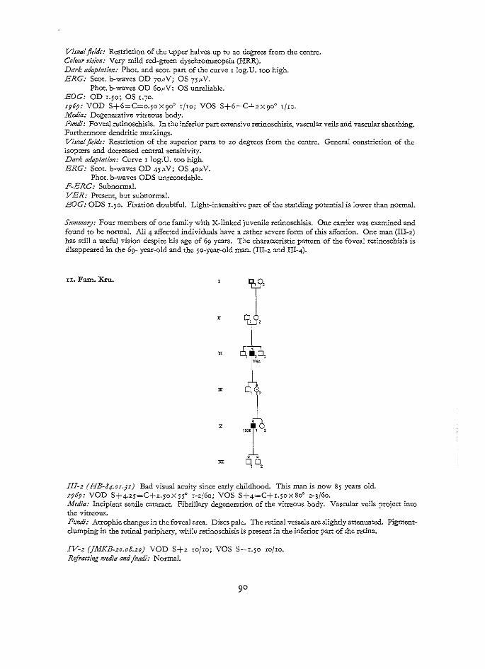
Visual fields: Restriction of the upper halves up to 20 degrees from the centre. Colour vision: Very mild red-green dyschromatopsia (HRR). Dark adaptation: Phot. and scot. part of the curve r log.U. too high. ERG: Scot. b-waves OD 7o.uV; OS 75ttV.
Phot. b-waves OD 6o.uV; OS unreliable. BOG: OD 1.50; OS r.7o. r969: VOD S+6=C=o.5o X 90° rfro; VOS S+6=C+.:: X 90° rjro. Media: Degenerative vitreous body. Fundi: Foveal rctinoschisis. In the inferior part C.'\.'tensive retinoschisis, vascular veils and vascular sheathing. Furthermore dendritic markings. Vis1Ja! fields: Restriction of the superior parts to zo degrees from the centre. General constriction of the isopters and decreased central sensitivity. Dark adaptation: Curve I log.U. too high. ERG: Scot. b-waves OD 45ttV; OS 4o.uV.
Phot. b-waves ODS uruecordable. F-ERG: Subnormal. VER: Present, but subnormal. BOG: ODS 1.50. Fixation doubtful. Light-insensitive part of the standing potential is lower than normal.
StJmmary: Four members of one family with X-linked juvenile retinoschisis. One carrier was examined and found to be normal. All 4 affected individuals have a rather severe form of this affection. One man (III-.::) has still a useful vision despite his age of 69 years. The characteristic pattern of the foveal retinoschisis is disappeared in the 69- year-old and the 5o-year-old man. (ID-2 and III-4).
xx. Fam. Kru.
m
y
Ill-2 (HB-84.0r.p} Bad visual acuity since early childhood. This man is now 85 years old. r969: VOD S+4.25=C+z.5oX55o r-zj6o; VOS S+4=C+r.5ox8oo 2-;(6o. Media: Incipient senile cataract. Fibrillary degeneration of the vitreous body. Vascular veils project into the vitreous. Fundi: Atrophic changes in the foveal area. Discs pale. The retinal vessels arc slightly attenuated. Pigmentclumping in the retinal periphery, while retinoschisis is present in the inferior part of the retina.
IV-2 (]MKB-2o.o8.2o) VOD S+z ro/ro; VOS S+1.50 ro/ro. Refracting H!edia and fundi: Normal.

V-I (]K-j8.II.2j) z962: VOD S-o.5o=C+.2 X x8o0 4{10; VOS C+zx r8o0 3(10. zg6g: VOD C+I.50XI80° zfxo; VOS C+zxr8o0 5j6o. Is complaining of metamorphopsia. This is demonstrated with the help of the Amsler test. Vitreotts borfy: Syneresis and fibrillary degeneration. Fine white opacities throughout the vitreous. Vascular veils infratemporally. F#ndi: Normal re:fle.xes are absent. Pathognomonic cystoid structure with superficial stellate folds in both foveae. The discs arc slightly too pale on the temporal side. The retinal vessels have a normal calibre. In the inferior temporal periphery rctinoschisis, vascular veils, vascular sheathing and dendritic markings arc present. Vis:Ja! fields: Decreased central sensitivity. Restriction supranasally. Colour vision: Very mild redMgrecn and blueMyellow dyschromatopsia (HRR). Decreased sensitivity to red (anomaloscope). Tritan axis in the right eye (Farnsworth D-15). No specific axis in the left eye (Farnsworth D-Ij)· ERG' Scot. b-waves OD 55r1V;
Phot. b-waves OD 4S11V; Phot. a-waves OD 1S.uV;
BOG: OD 1.76; OS 1.71. OP: Subnormal.
OS 69pV. OS 63pV. OS 26t1V.
Sumn1ary: X-linked juvenile retinoschisis in a Ss-year old man and his 31-year old grandson. The carrier of the pathological gene has normal eyes. The 85-year old man is able to move around in his house and has useful sight. This might be a consolation for other individuals suffering from this affection.
12. Fam. Ma.
111-2 {W]M-46.IO.IJ) This patient got a bullet in his left eye in 1965. The traumatic cataract has been removed. I968: VOD S/xo, cmmetropic; VOS rf6o S+12 3(10. Refracting media: OD normal. OS corneal scar and aphakia. Fundi: Foveal retinoschisis (fig. 5). Silver-greyish glistening patches in the retinal periphery. Visual fields: Decreased central sensitivity. Normal peripheral limitations. Dark adaptation: Curve 1 log.U. too high, pbot. and scot. ERG: Scot. b-waves OD uof.IV; OS 135.uV.
Pbot. b-waves OD 5511V; OS 55.uV. BOG: ODS 2.oo.
S#mmary: A patient with X-linked juvenile retinoschisis, showing the pathognomonic foveal alterations of this condition. The visual acuity of Sfxo of the right eye is remarkably good.
I3· Fam. Pie 11-2 { P F) Born in Indonesia. Like her husband of Indonesian origin. VODS IO/to. Media and fundi: Normal.
111-zUP-J2.0J.o6) Bad visual acuity since early childhood. VODS S+ I 3/IO. Vitreous body: Posterior vitreous detachment. Fibrillary degeneration and optically empty structures. Tiny white opacities all over the vitreous.
9'

Fundi: Foveal retinoschisis (fig. 2). The discs show a temporal crescent. The vessels are normal. Infratemporally preretinal whitish dendritic structures and vascular sheathing (fig. 9). Rctinoschisis in the temporal inferior periphery. Vimalfie!ds: Supranasally restriction to 20 degrees from the centre. Decreased central sensitivity. Dark adaptation: Normal. ERG: Scot. b-waves OD 95.uV; OS 85ttV,
Phot. b-waves OD So.uV; OS 6o1~V. EQG, OD qo; OS I-75· Blood: No acanthocytosis. Normal.
111-z (FP-JJ.o6.28) VOD C+o.50X xSo0 2/IO; vos C+o.50X 180° 5/Io. Vitreous botfy: Posterior vitreous detachment. Degenerative changes. Fundi: Foveal retinoschisis. Discs and vessels are normal. In the far inferior temporal periphery preretinal whitish dendritic structures and some retinoschisis. Colour vision: Normal (HRR). Visual fields: Concentric constriction, more in the superior parts than in the inferior parts. ERG: Scot. b-waves OD uo.uV; OS roottV.
Phot. b-wavcs OD 7o.uV; OS 7o.uV. BOG: OD 2.oo; OS 1.50.
111-j (GP·JJ.Ol.I7} VOD C+r X r65° 5/ro; VOS S+r.5o 2/ro. Vitreous body: Posterior vitreous detachment and degenerative changes. Ftmdi: Foveal retinoschisis. lnfratemporally vasculn veils and dendritic markings. Colour vision: Normal. ERG: Scot. b-waves OD 1701-'V; OS I5DttV.
Phot. b-waves OD 90;tV; OS 901-'V.
111-6 ( MP-6r.r0.2J) VOD S+o.5o=C+o.75XSo0 ro/ro; VOS S+o-5o=C+o·75XI00° 5/ro. VitreoHs botfy: Posterior vitreous detachment and degenerative changes. Ftmdi: Foveal retinoschisis. In the far inferior temporal periphery dendritic structures and some retinoschisis. Blood: Normal, no acanthocytosis.
Summary: All 4 sons of an Indonesian married couple are suffering from X-linked juvenile retinoschisis. The 3 daughters are normal. The patients demonstrate the charactetistic foveal retinoschisis, while infratemporally retinoschisis and white dendritic markings are present. The interindividual ophthalmoscopic patterns in the family-members are very much alike, indicating that there are only slight differences in C..'-"Pression in this family.
q. Fam. Ra. lll-r,2 en J. Reported to have had bad visual acuity all their life.
IV-7 en V-2. Carriers. They have normal media and fundi.

V-4 (}dC-J4.0J.07) Bad visual acuity since early childhood. Concomitant convergent squint of OS. 1968: VOD S+z zfro; VOS S+z.5o s/6o. Media: Normal. Fundi: Foveal retinoschisis. Patchy silver-greyish glistening reflexes in some parts of the retina, particularly infratemporally.
VI-! ( RR-;;.o.2.28) Has always had bad visual acuity. 1960: Diagnosis elsewhere: "persistent hyperplastic primary vitreous". VOD zjiO; VOS zj6o. 1968: VOD S+5.25=C+rx9oo zjro; VOS S+5.25=C+rX9oo zj6o. Refracting media: Slight opacities in the posterior cortex of OD. Cataract of the posterior cortex of OS. Fibrillary degeneration and syneresis of the vitreous body, in which fine white opacities are present. Posterior vitreous detachment. In both eyes big vascularized veils with arcade-like dehiscences are present. Fundi: Foveal retinoschisis. Extensive retinoschisis infratemporally. Retinal arteries and veins project from the retina into the vitreous in translucent veil-like membranes (fig. 14). Some of the veils insert on the disc. The disc of the left eye has a pseudopapillitis picture (fig. 16). Visual fields: Both visual fields arc con.£ned to 25 degrees from the centre on the nasal side. There is a decreased central sensitivity. Dark adaptation: The curve is 0.5 log.U. too high. Phot. and scot. ERG: Scot. b-waves OD 9opV; OS 651-N. Phot. b-waves OD 6pN; OS 45,uV. BOG: OD 1.85; OS 1.70. 0 P: Subnormal. Systemic examination: Normal. 1969: Vitreous haemorrhage occurs suddenly in OS. This is cleared up in 5 weeks without any therapy.
m
1 0
~~ ' ' ' 195~
Summary: Two members of a family with X-linked juvenile retinoschisis. There is a striking difference in c,._-pression between the tvm patients. Foveal retinoschisis is the only abnormality found in V-4, while VI-5 demonstrates the whole range of abnormalities, which may be found in this comple.-x syndrome. Two carriers, e.'<fl.mined ophthalmologically are fully normal.
x5. Pam. Si 1-z Reported to have had bad visual acuity all his life.
li-z (]SvD-17.02 .... ) VODS rojxo. Refructing medic.. and ftmdi: Normal.
93

II-6 (]WvD-2J.I0.2J) VODS xojxo. Refracting media and ftmdi: Normal.
li-S (EOvD-26.o6.rr) VODS xojro. Refracting media and/~~tzdi: Normal.
Ill-2 (FS-Jo.o6.24) Bad visual acuity since early childhood. r969: VOD 3/Io, emmetropic; VOS 4/ro, emmetropic. Media: Normal. FHndi: Foveal retinoschisis. No other abnormalities. ERG: Scot. b-waves OD 501~V; OS 7ot~V.
Phot. b-waves OD 65.uV; OS 50t~V. BOG' OD 2.4o; OS 2.36.
111-ro (WW-J7.07.or) I967: VOD S+r=C+o.5o X I0° 4/Io; VOS C+1.25 X I70° 4/Io. Media: Normal. Fundi: Foveal rctinoschisis. Vi.rw.! fields: Decreased central sensitivity. ERG: Scot. b-waves and phot. b-waves subnormal. BOG' OD >.>8; OS •·•3·
Jl[-IO (JV.O. J J.02.09) r967: VOD C+o.50X 75° 4/ro; VOS S+o.5o=C+o.5ox I20° 6/xo. Media: Norma.l. Fundi: Foveal retinoschisis. Preretinal strands in the inferior parts. Visual fields: Restricted supranasally. Decreased central sensitivity. ERG: Scot. b-waves and phot. b-waves: Subnormal.
III-I? (EvO- J6.ro.2J) Concomitant convergent squint of OD. I967: von S+z.5o=C+I.50XI00° rjxo; vos S+r.5o=C+o.50X90° 7/Io. Media: Normal. Fundi: Foveal rctinoschisis. Retinoschisis infratempo:rally. Visual fields: Restriction supranasally. Decreased central sensitivity and concentric impairment. ERG: Scot. b-waves and phot. b-waves: Subnormal.
' 1917
• t ' ' " t922 192~
Olio S91011121~1,151617
1957 1950
Sunmtary: X-linkcd juvenile rctinoschisis in four boys, belonging to the same family. All affected individuals have a foveal retinoschisis, while only one of them (III-I 7) has a peripheral retinoschisis too. Three carriers were examined and found to be normal.
r6. Pam. v.Sol 1-r ( P R-r J.OI.II} Bad visual acuity since early childhood. Had a connatal cataract of the right eye. I94J.' Discission of the cataract ofOD. I967: Complains of bad visual acuity, particularly in darkness. VOD S+5 light perception; VOS S+5 rj6o.
94

There is a concomitant divergent squint of OD and nystagmus of both eyes. 1\1cdia: OD: Aphakia with some after-cataract. OS: Immature cataract. Fundi: OD: Pigmentations and depigmentations with preretinal glious alterations. Atrophic fovea. OS: VascuJar sheathing and patches resembling an old chorioretinitis. Atrophic foveal alterations. VER: OD: Absent. OS: Very slight responses.
II-I ('SR) VODS rofw. Media and jtmdi: Normal.
Ill-1 { .&S-6.2.07.II) 1969: VOD S+x.5o Sfxo; VOS S+x.5o 4/Io. 1970: VOD S+x.5o ;Jxo; VOS S+x.5o ;jxo. Media: Normal. Fundi: Foveal retinoschisis (£g. xab). Silver-greyish glistening reflexes in some parts of the retinal periphery Co/om· vision: Normal (HRR).
1915
m i 1~52
SUIIJnJary: X-link.ed juvenile retinoschisis in a 54-year-old man and his grandson. The grandson has a foveal retinoschisis, the 54-year-old man, however, has severe lcnto-vitreo-retinal changes. The carrier has normal eyes.
r7.Fam. Wes
I-2 ( HWB) VODS ro(ro. Media and fundi: Normal.
' X ' ' 1951
11-z {HW-p.z2.27) Since some years bad visual acuity with both eyes. No family-members with bad visual acuity are known. 1966: VODS S+o.5o 4/to. Diagnosis: "Hercdodegcncration of the macula". Syste11n"c examination: Normal. I967: VODS S+o.5o 4/Io. Amsler test: Normal. Media: Normal. Ftmdi: Foveal retinoschisis (£g. 2.1). In the far inferior temporal periphery there are some greyish-white opalescent structures and pigmentations. The whole retina has patchy metallic glistening reflexes. Vis/#11 fields: Slightly decreased central sensitivity. Normal peripheries. Colour vision: Normal (HRR, anomaloscope, Farnsworth D 15). Dark adaptation: The curve is z.j; log.U. too high. ERG: Scot. b-waves OD IoopV; OS I05p.V.
Phot. b-waves OD 6a,~V; OS 50p.V. F-ERG: Subnormal.
95

EOG: OD 3-32; OS 2.91. I963: VODS S+0-50 4/IO. No changes. Flltoresccin angiography: Normal angiogram (fig. 21).
Stm11t1ary: A boy with X-linked juvenile retinoschisis. The pathognomonic foveal rctinoschisis presents the diagnosis, although no history of bad vision is reported in this family.
IS. Fam. Wil
l-2 ( MvG-82.0I.Oj} Is reported to have bad visual acuity since early childhood.
11-z (WWvG-r6.oJ.jO) VOD S+r.75=C+o.50X r6o0 rofxo; VOS S+z.=C+o.25 X45° rojro. Media and fundi: Normal.
lll-2 (}W-49-0S.ro) Bad visual acuity since early childhood. Concomitant divergent squint of the right eye. VOD S+4=C+o.75X125° r/ro; VOS S+6.5o=C+o.5ox;oo z.Jro. Media: Slight opacities in both posterior cortices. Posterior vitreous detachment and fibrillary degeneration of the vitreous body. Fundi: Foveal retinoschisis. Temporally a distinct retinoschisis with vascular veils and preretinal strands. Furthermore white dendritic markings and vascular sheathing (:fig. ro,12). The discs have a pseudopapillitis picture. Visttal fte!d.r: Restriction nasally. Supranasally there is more restriction than infranasally. Dark adaptation: Almost normal curve. ERG: Scot. b-waves OD 95p.V; OS 1551tV.
Phot. b-waves OD 9op.V; OS 75t~V. BOG: OD r.2o; OS x.85. The value of OD is unreliable because of the squint.
StrHNnary: A boy, suffering from a severe form of X-linked juvenile retinoschisis. Nearly all possible ophthalmoscopic alterations of X-linked juvenile retinoschisis are to be found in this patient.
REFERENCES
AMALR1C, P.: Un nouveau type de plis rCtiniens congbnitaux. Arch. Ophtal. 28: 507-512 (1968). ANDERSON, J. R..: Anterior dialysis of retina: disinsertion or avulsion at ora scrrata. Brit. J. Ophthal, r6:
641-67o; 705-727 (1932). BALIAN, J· v. and FALLS, H. F.: Congenital vascular veils in the vitreous. Arch. Ophthal. 63:92-101 (r96o). BARTELS, M.: Ueber die Entstehung von NetzhautablOsungen. Klin. Mbl. Augcnheilk. 91:437-450 (1933). BARUT, c.: P6riphU:bitc r6tiniennc associ6e a des l6sions maculaires constat6e chez deux frt:res ct 6voluant
depuis l'enfance. Bull. Soc. Ophtal. France 55: r85-188 (195 5). BE:-.TGTSSON, B. and LINDER, B.: Sex-linked hereditary juvenile retinoschisis (presentation of two affected
families). Acta Ophthal. 45 :4II-422 (1967).

BOUDON, :M. and SOLE, P.: Rt:cinoschisis idiopathique des jeunes gens. Bull. Soc. Ophtal. France 65: 5 27-5; I (1965).
BR.OCKHUR.ST, R. ;.: Management of :retinoschisis. In Retinal diseases. p. r89-191. Lea and Febiger, Philadelphia, 1966.
cmrs, P. A.: Doppelseitige NetzhautablOsung bei einem eineiigen Zwillingspaar. Ber. dtsch. Ophthal. Ges. 53:251-256 (1940). In Zbl. ges. Ophthal. 45:644(1940).
cmrs, P, A.: Hereditary hyaloideoretinopathies with retinoschisis. In Vitreoretinal pathology and surgc.ry in retinal detachment, p. 87-95. Mosby, St. Louis, 1965.
crBrs, P. A.: Errors in the management of retinal detachments. In Mod. Probl. Ophthal. vol. 3: 285-;os. Karger, Basel, 1965.
cmrs, P. A.: Vitr6 et d6collcmcnt de r6tine. Arch. Ophtal. 25 :6z7; 637 (1965). cmrs, P. A.: Retinoschisis-retinal cysts. Trans. Amer. Ophthal. Soc. 63:417-453 (1965). CIBIS, P. A.: Recent development in the treatment of retinoschisis and idiopathic retinal detachment. Amer.
J. Ophthal. 6z:581-58a (1966). COL YEAR, B. H.: Diagnostic diff6rentiel du r6tinoschisis. Arch. Ophtal. 25:631 (1965). COLYEAR, B. H.: Traitement par la photo-coagulation du r6tinoschisis en "dOme". Arch. Ophtal. 25:637
(1965). CONDON, R. A. and SOMERVILLE-LARGE, L. B.: Vitreous veils and associated conditions in five brothers.
Trans. Ophthal. Soc. U.K. 75: 709-7!! (1955). CRISWICK, V, G. and SCHEPENS, C. L,: Familial e'-.-udative vitreoretinopathy. Amer. J. Ophthal. 68:578-594
(1969). DONALDSON, D. D.: Juvenile retinoschisis. Arch. Ophthal. So: 524 (1968). EISNER, G.: Traumatische Veriinderungen der pars plana corporis ciliaris. Ophthalmologica 154:;u-317
(1967)· ERIKSSON, A. W., VAINIQ-MA'I"''ILA, B. A., KRAUSE, U. e.a.: Secondary sex ratio in families with X-chromosomal
disorders. Hereditas 57:37;-381 (1967). ERIKSSON, A. w., FELLM:AN, ;., VADJIO-MATTILA, B. A. e.a.: Studies of X-link.ed retinoschisis in Finland. In
Progress in ncuro~ophthalmology. Vol. II, p. 36o-362. Proc. Sec. Int. Congress of Neurogen. and Neuro-Ophth. Montreal, 1967. Excerpta Medica, .Amsterdam, 1969.
EWm-G, c. c. and IVES, E.;.: Juvenile hereditary retinoschisis. Trans. OphthaL Soc. U.K. 89:.29-39 (1969). FALLS, H. F.: Retinoschisis. In Retinal diseases, p. 182-18;. Lea and Febiger, Philadelphia, 1966. FAVRE, M. and GOLDMANN, H.: Zur Genese der hinteren GlaskOrperabhebung. Ophthalmologica 132:87-
97 (1957)· FAVRE, M.: A propos de deu."\: cas de d6g6n6rescence hyaloid6o-rCtinienne. Ophthalmologica r 3 5 : 604-609
(1958). FEILER-OFRY, v. ADAM:, A., and REGENBOGEN, L. e,a.: Hereditary vitreoretinal degeneration and night blind
ness. Amer. J. Ophthal. 67: 55 3-5 58 (1969). FORSIUS, H., VAI='!lO-MATTILA, B, A. and ERIKSSO::-<, A. W.: X-linked hereditary retinoschisis. Brit. J. Ophthal.
46:678-681 (1962). FORSIUS, H., ERIKSSON, A. W. and VArNIO~MATTILA, B, A.: Geschlechtsgebundene erbliche Retinoschisis in
zwei Familien in Finnland. Klin. Mbl. Augenheilk. 14;:8o6-8I6 (1963). FRANCESCHETTI, A., FRANS'=OIS, ;. and BABEL,;.: Les h6r6dodCg6nCrescences choriorCtiniennes. Masson et
Cie, Paris, 1963. GIESER, E. P. and FALLS, H. F.: Hereditary retinoschisis. Amer. J. Qphthal. 51: 1193-2000 (1961). GOODMAN, G., RIPPS, H. and SIEGEL, r. M.: Sex~ linked ocular disorders: trait e'-.-pressivity in males and carrier
females. Arch. Ophthal. 7;:;87-398 (1965). GOODMAN, G.: Sex-linked juvenile retinoschisis. In Congenital anomalies of the eye, p . .284-286. Mosby,
St. Louis, 1968. GOODSIDE, v.: Congenital vitreous veil. Arch. Ophthal. 6;: 682-686 (196o). GUYOT-SIONNEST, Mme.: A propos d'une famille atteinte de r6tinoschisis idiopathique rCcessif liC au sexe.
Ann. Ocul. 202: 573-598 (1969). HAAS,;.: Ueber das Zusammenvorkommen von Verii.nderungen der Retina und Chorioidea. Arch. Augen
heilk. 37:34;-;48 (r898). HARRIS, G. s.: Retinoschisis: pathogenesis and treatment. Canad. J, Ophthal. 3:; 12-317 (1968). HARRIS, G. s.: Juvenile sex-linked retinoschisis. In Mod. Probl. Ophthal. vol. 8, p. 363-370. Karger,
Basel, 1969. HAUSCHILD, E., HILLER, H. and HEYDENREICH, A.: Beitrag zur Krankheitsbild dcr vitreo-retinalcn Degene
ration. Klin. Mbl. Augenheilk. 156:6z.8-643 (1970).
97

HOGA.."<, M. ;. and ZIMMERMAN, L. E.: Ophthalmic pathology, ed. 2, pp. 5 50-554· W. B. Saunders, Philadelphia, 1962.
ISBEY JR., E. K., BEUER..V!AN, V, A. and ASHEVILLE, N. C.; Hereditary retinoschisis with retinal detachment. Sth. Med. ]. (Bghmn. Ala) 57:442-445 (1964). In Exc. Med. Sect. XII. I9"3o-I;I (1965).
JAGER, G. M.: A hereditary affection of the retina. Ophthalmologica rz5 =470-471 (I955). JAGER, G. M,: A hereditary retinal disease. Trans. Ophthal. Soc. U.K. 75: 6r7-6I9 (195 3). JANCKE, G.: Echte Netzhautcysten und Netzhautl6sung beim Jugendlichen. Klin. Mbl. Augenheilk. 95:
145-148 (1935)· JOH::-;rSON: In Retinal detachment in juveniles. Colloque, Cambridge, 1968. Mod. Probl. Ophthal. vol. 8:
407. Karger, Basel, I969. JULER, F.: Unusual form of retinal detachment (cystic?) in children. Trans. Ophthal. Soc. U.K. 67:83-96
(1948). JULER, F.: Further report on cystic disease of the retina in children. Trans. Ophthal. Soc. U.K. 71: 199-zo5
(195 I). ;mrGSCHAFFER! In Retinal detachment in juveniles. Colloque, Cambridge, 1968. Mod. Probl. Ophthal.
vol. 8:407. Karger, Basel, 1969. KM·rAN, A., KAHA::-;r, I. L. and BENKO, A.: Erythrocytic anomalies in hereditary vitreo-retinal degeneration.
Brit. J. Ophthal. 47:620-651 (1965). KEITH, c. G.: Retinal cysts and retinoschisis. Brit. J. Ophthal. 50:617-6:::.8 (1966). KLEI::-;rERT, H.: Eine recessiv-geschlechtsgebundene Form der idiopathischen Netzhautspaltung bei nicht
myopen Jugendlichen. Graefes Arch. Ophthal. I54:295-505 (1955). KORCI-IMARos, r., NADRAI, A., SCHOLER, D. e.a.: Contribution to the heredodegeneration of the fovea centralis.
Acta Fac. Med. Univ. Bruncn. 25!153-156 (r965). KRILL, A. E.: X-chromosomal-linked diseases affecting the eye: status of the heterozygote female. Trans.
Amer. Ophthal. Soc. 67:555-608 (I969). LEVY,;.: Inherited retinal detachment. Brit. J. Ophthal. 56:6:::.6-655 (I952). LIESENHOFF: In Retinal detachment in juveniles. Colloque, Cambridge, 1968. Mod. Probl. Ophthal. 8:407.
Karger, Basel, I969. LISCH, K.: Seltene Formen von tapeto-retinaler Degeneration. Klin. Mbl. Augenheilk. 98:498-504 (1957). LISCH, K.: Idiopathische hereditiire Retinoschisis. Klin. Mbl. Augenheilk. 153!204-2IO (1968). MACRAE, A.: Congenital vascular veils in the vitreous. Trans. Ophthal. Soc. U.K. 74: r87-zo6 (I954). MACVICAR, J. E. and WILBRANDT, H. R.: Hereditary retinoschisis and early hemeralopia. Arch. Ophthal. 83:
629-6;9 (1970). MAGNUS,;. A.: Case of retinal detachment in a child (cystic? congenital?). Trans. Ophthal. Soc. U.K. 7I!
728-730 (195I). MANN, I. and MACRAE, A.: Congenital vascular veils in the vitreous. Brit. J. Ophthal. 22: I-10 (1958). MANSCHOT, w. A.: Personal communication, I969. OKSALA, A.: Juvenile macular dystrophy. Acta Ophthal. ;)I: 385-594 (I953)· OKUN,_ E. and cr:srs, P. A.: The role of photocoagulation in the management of retinoschisis. Arch. Ophthal.
72:3o9-5I4 (I964). PAGENSTECHER, H. E.: Ueber cine unter dem Bilde der Netzhauta.bl6sung verlaufende, erbliche Erkrankung
der Retina. Graefes Arch. Ophthal. 86:457-462 (I9I5)· PAJTAS, ;.: Cas de pseudogliome familial h6rt:ditaire dans trois g6nCrations (Retinitis exsudativa Coats).
Ophthalmologica Izo:41 I-4I5 (1950). PERALTA, s. and SANTORI, M,: Degenerazione maculare centrale tipo Stargardt associata ad alt~ioni vitrcali
congenitc. Ann. Ottal. 95::::.57-242 (r967). PISCHEL, D. K.: Surgical treatment of retinal cysts. Amer. J. Ophthal. 56: I-I6 (1965). PISCHEL, D. K.: Three brothers with juvenile retinoschisis. In Mod. Probl. Ophthal. vol. 8: 38I-589. Karger,
Basel, I 969. PONTE, F.: Contribute clinico allo studio delle eredo-degenerazioni microcystichc della retina. Degenerazioni
vitreoretiniche, Boll. Ocul. 46:79-II5 (1967). RICCI, A.: Clinique et transmission gCnCtique des diff&rentes formes de d6gCn6rescences vitreo-rctiniennes.
Ophthalmologica 159:338-343 (I96o). RICCI, A.: Oinique et transmission hCrt!ditaire des dt:gt:n6rescences vittt!o-rCtiniennes. Bull. Soc. Ophtal.
France 6I :6I8-66z (I961). RICHARDS,;. s. F. and HARRIS, c. s.: Retinoschisis treated by photOcoagulation. A pathological case report.
Canad. J. Ophthal. 2:I99-202 (I967).

RIEGER, H.: Ueber zwei beroerkenswerte Bille von beidseitiger, idiopatbischer Net7.hautabhebung bei jugendlichen Hypermetropen. K.lin. Mbl. Augenheilk. uo:86-88 (1944).
SABATES, F.: Juvenile retinoschisis. Amer. J. Ophthal. 62:68;,-688 (1966). SARIN, L. K., GREEN, W. R. and DAILEY, E. G.: Juvenile retinoschisis, Amer, J. Ophthal. 57:79;5-796 (1964). SCI-IEPENS, c. L.: Summation and discussion. Retinoschisis. In Retinal diseases, p. 19I-I94· Lea and Febiger,
Philadelphia, 1966. scoRCIARINI-COPPOLA, A., ORLANDO, E. e.a.: Vascular and coagulatory changes found in so-called congenital
vascular veils of the vitreous body. Boll. Ocul. n: 210-224 (I958). SEBEsrrBN, ;., STRENGER, J and szAB6, G.: Ueber die juvenile degenerative Ablatio retinae, Klin. Mbl.
Augenheilk. I50:8;1:84o (r967). SI-IEEHA::-.T, B.: Congenital vascular veils in the vitreous. Trans. Ophtbal. Soc. U.K. 72: 6z;-628 (1952). SORSBY, A., KLEIN, M., I-IURNDALL GANN, J. e.a.: Unusual retinal detachment, possibly se.x-linkcd. Brit. J.
Ophthal. 55: r-10 (1951). SORSBY, A.: Congenital cystic sex-linked retinal detachment. In Modem trends in ophthalmology. ;rd ser.
p. 197-199· Butterworth, London, 1955· soRSBY, A.: Se.."X-linked congenital cystic retinal detachment. In Modem trends in ophthalmology. 4th ser.
p. 108-IIO. Butterworth, London, I967. SPENCER, w. 1-1.: Pathology of retinoschisis. In Retinal diseases p. I85-189. Lea and Febiger,Philadclphia,
1966. STEPA::-.TIK, J.: Beidseitige idiopathische Retinoschisis bei einem 4 Jahre altcn Knaben. Klin. Mbl. Augen
heilk. '55' 55-56 ('969). THOMSON', E.: Memorandum regarding a family in which neuroretinal disease of an unusual kind occurred
only in the males. Brit. J. Ophthal. r6:681-686 (19;p.). TORNQUIST, R.: Diagnostic and prognostic problems of retinoschisis. Acta Ophthal. 42 :4;8-443 (1964). TREVOR-ROPER, P. D.: Congenital retinal fold in association with pseudopapillitis. Proc. Roy. Soc. Med.
4;5: IOII (1950). UTE&\!A.-..rn, o.: Beitrag zur Klinik und Behandlung der Rctinoschisis. Klin. Mbl. Augenheilk. 144: ;84-
393 ('964)-VAINIO-MAT'I'ILA, B. A., ERIKSSON, A. w. and FORSIUS, H.: X-chromosomal recessive retinoschisis in the region
of Pori. Acta Ophthal. 47: II35-ll48 (1969). wAGNER, H.: Ein bisher unbekanntes Erbleiden des Auges (Degeneratio hyaloideo-retinalis hereditaria),
beobachtet im Kanton ZUrich. K.lin. Mbl. Augenheilk. too: 840-857 (1938). WITMER, R.: Traitement des r6tinoschisis. Arch. Ophtal. Z-5: 637 (1965). YAMADA, E.: Some structural features of the fovea centntlis in the human retina. Arch. Ophthal. 82: 15 r-159
('969)-YAN'OFF, M., RAHN, E. H. and ZIMMERMAN, L, E.: HistOpathology of juvenile rctinoschisis. Arch. Ophthal.
79'49-55 ('968). YAZAWA, K. and NAKAJIMA, A.: Hereditary juvenile retinoschisis. Ann. Ophthal. (Chic.) I :;o-53 (1969). ZIMMERMAN, L. E. and SPENCER, w. 1-1.: The pathologic anatomy of retinoschisis, with a report of two cases
diagnosed clinically as malignant melanoma. Arch. Ophthal. 6;: ro-19 (1960).
ADDENDUM
BROCKHURST, R. J.: Photocoagulation in congenital rctinoschisis. Arch. Ophthal. 84: I 58-165 ( r 970). GOODMAN, G.: Sickle cell ocular disease. In Congenital anomalies of the eye p. Z-47-t-71. Mosby, St. Louis,
1968. KRAUSE, U., VAINIO-MATTILA, B. A., ERIKSSON, A. W. e.a.: Fluorescein angiogtaphic studies on X-chromosomal
retinoschisis. Acta Ophthal. 48:794-807 (1970). MAN'SCHOT, w. A.: Erfelijke aandoeningen met netvliesloslating. Ned. T. Geneesk. n;:zz6o (r969). ROSSELET-HUGUENIN, G.: Eales disease. Maladie ou Syndrome. Thesis, Lausanne. Progrt:s Ophtal. z.;,: Z-73-
;,ro (1970).
99

III
StargardtJ s disease
I. INTRODUCTION
Stargardt's disease is a bilateral, symmetrical, progressive affection which, at least in the initial stage, is confined to the foveal area; its occurrence is familial and usually at an early age, and it ultimately leads to loss of central vision.
In 1909, Stargardt described two families (H and N) whlch, in age group 12-zo, included seven individuals with progressive degeneration of the macular region. The patients' parents llitd good visual acuity and normal eyes, and were not consanguineous. The patients had had good vision in early childhood, but between age 8 and age I 5 developed gradual, progressive loss of central vision. In the posterior pole there were slight pigment cllitnges with yellow-grey spots, resulting in a fairly well-defined, grubby atrophic central focus of horizontal oval shape. In some patients a broad ring of numerous white-yellow spots developed around thls focus (diameter r-z pd), while the retinal vessels and retinal periphery remained quite normal. The optic discs were somewhat too pale on the temporal side.
However, patients with conditions closely resembling that described by Stargardt in 1909, had been known earlier. Leber (1874) llitd described a 7-year-old girl with progressive degeneration of the retinal centre, to whlch slight changes in the retinal periphery were later added (Stargardt, 1909).
The brother and sister with central and pericentral changes described by Lang (r885) may also have suffered for some time from Stargardt's disease.
R. D. Batten (1897) described two brothers who at age 14 developed a symmetrical macular affection characterized by finely mottled pigment displacements. It was specifically mentioned that the elder brother (age zr) showed not only more marked central pigment disturbances than hls younger brohter (age 14), but unlike the latter also showed pigment displacement in the retinal periphery.
The two brothers described by Magers (1899) were very probably patients with long-standing Stargardt's disease, because the posterior pole showed pronounced pigmentations and choroidal atrophy.
100

Dujardin (1904) described two brothers and Jackson (1905) reported on two sisters - all chlldren of consanguineous parents.
The literature also comprises the solitary case described by Valude (1906) and two sisters described by Steindorff (1906). Baumgarten (1907) observed a 13-year-old girl with central and peripheral fundal changes, and Jennings (1909) made mention of three children with progressive posterior pole dystrophy in a consanguineous marriage.
After 1909 Stargardt continued to publish reports on thls progressive posterior pole dystrophy in young patients. In 1913 he described two brothers aged 45 and 38, and their 43-year-old sister, whose vision had been poor for years. In these patients he found peripheral retioal changes besides a marked central affection. Stargardt himself described the picture as a retinitis pigmentosa taking an inversed course (retinopathia pigmentosa in versa): in classical retinopathia pigmentosa the equatorial retinal areas are first affected~ in contrast to the course seen in his cases. In 1916 he added to his series the case of a 3 5-year-old man whose vision had been poor since age u; and in 1917 and 1925 he again described patients with the same affection (Stargardt 1917, 1925).
In 1917, Stargardt published a classification of central tapetoretioal dystrophies in which he strictly differentiated between the progressive dystrophies of the central retioa without dementia, and those with dementia: the so-called maculo-cerebral dystrophies (lipidoses) which several other authors have also described (F. E. Batten 1903; Mayou 1904; Stephenson 1904; Spielmeyer 1905; Vogt 1905; Stock 1908; Oatman I 9 I I ; Beach I 926, and others).
None of the families described included individuals with a purely retioal affection as well as patients with a maculo-cerebral condition. From this fact Stargardt rightly concluded that the progressive posterior pole dystrophy he had described, and the maculocerebral dystrophy, are totally different entities. Only Alkio (1923) found concomitance of Stargardt's disease and Tay-Sachs disease in the same family (an incidental finding, in our opinion). A boy with possibly Tay-Sachs disease or Spielmeyer-V ogt disease had a father with normal eyes, but this father had three brothers and one sister suffering from Stargardt's disease.
Since Stargardt's perspicuous studies, publications on this affection have contioued to appear: Lutz (1911): 4 children in I family; Stirling (1912): I case; Darier (1914): 5 children in 2 families; Maewsky (1914): I case with suggestion of peripheral retinal changes; Pusey (1915): 5 children in I family; Feingold (1916): 3 children in a consanguineous marriage.
Many publications have followed in subsequent decades. The observations of the various authors are not always the same; purely central, but also centroperipheral affections as well as transitions between these two types have been described.
For the sake of simplicity we present in table I a survey of publications which, so far as we could establish, concern themselves with Stargardt's disease.
!01

a. I920-I!J!O
1920
1921
1922
1923 1924 1925 1926 1927 1928 1929
Table I
Chance Becker; Oguchi and Yano Nakajima (quoted by Waardenburg et al. 1965) Alkio; Lewina; Takeuchi and Horiguchi Cavara; Firjukowa; Morelli Bollack; Rieger Lagrange and Peron; Stcyn; Urbanek Vom Hofc Blank; Knapp; Morelli Till6
b. I9JO-I940
1930 Pillat, Schiff-Wertheimer and TillC 1931 Letchworth; Rossi; Von ROtth 1932 Accardi; Waardcnburg 1933 Braun 1934 Danielson 1935 Borsellino; Caocci; Ibuki; Neamc 1936 Bumier; Klien; TillC 1957 Muromoto 1938 Alvaro; Ogata 1939 Wakayama
c. I940-19JO
d.
1940 Danis; MacRay; O'Rourke; Sorsby 1941 Franccschetti and Klien; Hallermann; Sorsby 1942 Crawford; O'Brien and Roper 1943 Lloyd 1944 Neame 1945 Missi:roli 1946 Bonnet; Folk; Gartner; MacRay; Renard 1947 Feldman; Moffat; Ryerson; Sukumlyn 1948 Agatston 1949 Brognoli; Redslob
I9J0-1960
1950 l95l l952 I955 l954 I955 1956 I957 I958 I959
Mortelmans; Peiker Biro; DiPrima; Scuderi and Siliato; Sun Danic; Klimkova and Velick:Y; Peterson; Szek.lcr Kaplan; Perron et al. Anastasi and Bellavia; Bernheim ct al.; Roschr; Samuels Bonamour; Friemann; Landolt; Mylius F:rans:ois et al.; Gaba; SchOnfeldcr BessiCre et al.; Van Bogaert; Hong; Ohrt Bessiete and Chabot; Kozlowski; Spallino Bozzoni; Tiberi and Cuccagna
C. I960-I970
I96o 196X 1962
Cox; LtD::; Vancca and Tudor Cox; Etzine; Straub Bessiete et al.; Calmcttes et al.; Chen and Lin; Frans:ois et al.; Gructzner Biesheuvel; Dalgleish and Naylor
!02

1964 Korozwcig 1965 Frans;ois and De Rouck; Norton ct al. 1966 Blodi; Krill; Rucdemann 1967 Shcrshcvskaya; Yasukura ct al. 1968 Erslcr and Jaczynowska; Bengisu et al. 1969 BcssiCrc ct al.; Frans:ois and De Lacy 1970 Soler Sala; Merin and Landau.
This survey lists those dystrophies that had or can be suspected to have had a recessive mode of transmission. Since the literature also comprises reports on dominant progressive foveal dystrophies, several authors have described these affections as the dominant form of Stargardt's disease (Franceschetti et al. 1963; Duke-Elder 1967); but because Stargardt himself described a recessive affection, we consider it desirable to confine the designation Stargardt's disease to the recessive form and to give the dominant foveal dystrophy which resembles it a separate place under the heading dominant progressive central tapetoretinal dystrophy or dominant progressive foveal dystrophy.
The disease must be strictly differentiated from dominant progressive cone dystrophy and dominant vitelliform dystrophy, which on occasion are mistakenly recorded as dominant form of Stargardt's disease (Bruna 195 r; Franceschetti et al. 1963; Duke-Elder 1967).- The central tapetoretinal dystrophies in which peripheral retinal changes develop after some time, constitute a problem in a way. There is still considerable confusion in this respect as a result of the numerous different names by which these centroperipheral tapetoretinal dystrophies are known. As early as 1913, Stargardt himself described three individuals in one family who showed such a centroperipheral tapetoretinal dystrophy (fam. S). In our opinion there can be no convincing objection to the use of the term Stargardt's disease with reference to these diffuse retinal affections. Franceschetti, Fran,ois and Babel (1963) described this form as Stargardt's disease with peripheral involvement ("maladie de Stargardt avec participation peripherique"), and we agree with this designation.
Other terms used are: centroperipheral tapetoretinal dystrophy (TRD) (Waardenburg eta!. 1963); macular form of diffuse TRD (Gruetzner 196z); mh:ed TRD (Bessiere et al. r96z); diffuse TRD (Duke-Elder r967); and central retinitis pigmentosa (Lux I 960 ).
We believe that many cases described as "central (inverse) tapetoretinal dystrophy" (Franceschetti, Fran,ois and Babel 1963; Duke-Elder 1967) should also be classified in this group. The "progressive rod-cone degeneration" described by Berson et a!. (r968) is probably the same affection. This form- in which the fovea first becomes dystrophic, while peripheral changes occur later - might be called centrifugal TRD or centrally started TRD.
Occasionally, however, it may be impossible to establish whether the dystrophic process in fact started in the central retina. Perron et a!. (195 3), for e..'l:ample, described a patient in whom retinal centre and periphery were simultaneously involved; and our own case material includes patients of whom we do not know with certainty whether the dystrophy started centrally or was diffuse from the onset (fam. Bol and

Hu). Be this as it may, it is a fact that the literature and our own case material both include many examples in which an initially purely central TRD was later associated with peripheral retinal changes.
Rieger (I 92 5) described two brothers with central and peripheral retinal changes, while a third brother showed a normal posterior pole and only peripheral changes. So far as we know, there have been no other observations of this kind.
Bonnet (I946), Gruetzner (I96z) and Shapira (quoted by Perron eta!. 1953) hold that a strict distinction must be made between Stargardt's disease, which remains purely central, and TRD which starts at the centre and extends towards the periphery. In our opinion this is impossible, because all transitions between purely central and diffuse TRD have been described. There are purely foveal forms besides affections which involve not only the fovea but also the pericentral or even the entire peripheral retina. If observations are limited to a single period in the patient's life, the impression may be gained that there are clearly distinct purely central and centroperipheral forms. Unfortunately, many reports cover only a single period of observation. But when long-term follow-ups have been made it is qnite clear that in many cases an originally purely central TRD develops into a centroperipheral TRD (Leber I874; Waardenburg I932, I963; Wakayama I939; Brognoli I949; Rosehr I954; Gaba I956, and others). It has also been frequently found that, in a given family, older brothres or sisters had already developed a centroperipheral TRD, while younger brothers or sisters still showed a purely central TRD (Batten I897; Becker I92I; Oguchi and Yano I92I; Von Rotth I93'; Wakayama I939; Etzine I96I, and others). The reverse situation has never been described. This means, we believe, that the eyes of older brothers or sisters generally indicate how the eyes of younger brothers or sisters will ultimately become. In only one family (fam. Me) we found the rare situation, that the youngest had Stargardt's disease with peripheral involvement, while z older siblings had a pure central form of Stargardt's disease.
A beautiful example of an unsuspected development has been given by Rosehr (I 954). In I 95 3 - 50 years after Stargardt first examined Dorothea H-it was found that an affection initially confined to the fovea had led to diffuse retinal changes: pigment proliferations and drusen-like structures; and the posterior pole likewise showed marked changes. The younger sister, Marie H, did show posterior pole changes but the retinal periphery was still virtually normaL These findings would seem to contradict most other data in the literature and our own observations, which show that affected children in the same family are likely to show the same development.
Stargardt's own studies already yielded arguments against the widely accepted view that the affection which bears his name would be confined to the fovea. The S family in which in I 9 I 3 he described a centroperipheral TRD provides such an argument, and the fundal pattern in Paul H - one of his first patients - already showed numerous white sharkfin-shaped spots around the papillofoveal area. In cases in which many white spots are visible around the dystrophic fovea, differential diagnosis from fundus flavimaculatus is e.'l:ceedingly diflicnlt. M ylius (I 9 55) also
I04
pointed out that Stargardt's disease is not strictly limited to the foveal area. Today, many ophthalmologists confronted with the Paul H originally described by Stargardt, would diagnose a case of fundus f!avimaculatus!
The same applies to the patients described by Hallermann (I94I), Agatston (I948), Friemann (I95 5) and Mylius (I95 5). On the other hand, some of the cases offundus f!avimaculatus described by such authors as Franceschetti and Fran,ois (I965), Carr (I965), Amalric et al. (I967) and Scialfa (I964) might well be classified as Stargardt's disease. Cases 2 and 5 in Carr's study (I 96 5) in fact do not resemble a fundus f!avimaculatus strikingly but are more suggestive in my opinion of a centrally started TRD.
In the terminal stage of Stargardt's disease, the posterior pole often shows the features of extensive choroidal atrophy with or without pigmentations, and the retinal periphery can show features ranging from a normal fundus through advanced retinopathia pigmentosa. It can be deduced from this that in advanced stages it is not always possible to establish a reliable diagnosis on the basis of the ophthalmoscopic findings if one is not familiar with the initial stages of the disease.
Unlike many authors, we agree with Stargardt and Tille (1929) that Stargardt's disease is an ophthalmoscopically well-recognizable entity. Additional data on mode of transmission and retinal functions can verify the diagnosis. The progressive loss of vision is an important characteristic. The age of onset is much less characteristic and varies widely. The entity named after Stargardt might be roughly divided as follows: r. pure! y central TRD ; 2. central and peri central TRD (sometimes indistinguishable from fundus f!avi
maculatus with foveal dystrophy); 3. centroperipheral TRD:
a. with an intact visual field (pigmentations and depigmentations, virtually normal calibre of fundal vessels);
b. with visual field defects (bone trabeculae and constricted fundal vessels). In this respect we should bear in mind the evolutional character of this affection, which can cause the picture to change substantially in the course of the years and may present all forms and transitions of I, 2 and 3 in succession. Ultimately a sharply defined focus of choroidal atrophy, indistinguishable from central choroidal atrophy, may develop in the posterior pole.
2. GENERAL CLINICAL PICTURE TN THE PATIENTS
The two principal features of the clinical picture of Stargardt's disease are the changes in visual acuity and the fundal features.
The patients usually report between age 6 and age zo with bilateral gradual diminution of vision, although previously visual acuity has been entirely normal. In some cases the first symptoms do not occur until age 30-40.
There is often mild myopia and patients complain of photophobia, vision usually being optimal in twilight. There is no male or female predominance, and several
I05

Fig. Io. Initial stage of Stargardt's disease. Disturbed foveal and foveolar reflexes. Fovea looks as if covered with varnish (snail's slime). Some unobtrusive yellowish-white
.flecks in the foveal area. (Fam. Oe).
Fig. rb. Fluorescein angiography reveals not only defects in the retinal pigment epithelium at the site of the fovea, but also small round defects in the pigment epithelium in the
perifovcal area.
ro6

children in the same family are often affected. Consanguineousness of parents of patients with Stargardt's disease has a hlgher incidence than normaL
A familiar story is the one about the schoolchild who has always been seated at the back of the classroom but must move to the front because no spectacles are effective.
In some cases there is considerable diminution of vision although no ophthalmoscopic changes exist. Neurasthenia or even hysteria is often suggested in such cases, and some ophthalmologist may even so far forget themselves as to get angry with the patients (always a mistake). The reverse situation - still normal visual acuity in association with slight foveal changes - can also be encountered. An important point is that the loss of visual acuity is nearly always symmetrical, although there are exceptions to this rule. We have seen patients with vision rjro in one and vision 9/ro in the other eye (fam. Kat), but in these cases the ophthalmoscopic changes were already symmetrical. The fact that ophthalmoscopy discloses an almost photographically exact symmetry of the two eyes can suggest a hereditary origin at an early stage.
Vision gradually diminishes to rfro; in many cases it takes many years to reach this low value, but occasionally one may see very rapid deterioration of vision (Lutz r9u). Peripheral retinal dystrophy comes on insidiously in many cases, and in these cases vision can be even worse, although true blindness is a rarity.
3· FUNDUS (OPHTHALMOSCOPIC FINDINGS)
In the initial stage there are no or hardly any ophthalmoscopic changes. The first signs are disappearance of the foveolar reflex (fig. r), followed after some time by disappearance of the foveal margin reflex (fig. z). Changes in the pigment epithelium become visible in the form of grey, yellowish and brown spots, and the fovea shows a granulated appearance (figs. z, 3). The fovea also gives an impression of being covered by varnish or snail-slime (fig. 1-4). Sometimes there is an exceedingly delicate plication of the internal limiting membrane, radiating in all directions (fig. 5 ). This radial aspect entails a risk of confusion with sex-linked juvenile retinoschisis, in which the fovea shows a characteristic cystoid alteration with clearly visible superficially localized radial plicae (page 5 r). These striae, however, are usually less delicate than the very fine plicae seen in Stargardt's disease. As in Stargardt's original cases, one frequently observes the occurrence of whitish-yellow, ill-defined perifoveal spots, localized beneath the vessels and probably in the pigment epithelium of the retina (fig. 6). The pigmentations in the foveal area vary markedly; thick "soot-flakes" can appear at the site of what used to be the fovea (fig. 7).
Finally there appears a horizontal oval of atrophic pigment epithelium which usually measures z pd in width and I. 5 pd in height (fig. 8). As the process advances, the focus can increase to as much as 5 pd. This more or less sharply defined focus of atrophic pigment epithelium has a greyish, grubby appearance and shows glistening

metallic reflexes when illuminated. The American literature describes this as "beaten bronze atrophy", and this is a very precise description. The glistening reflexes might be caused by hyalinization of Bruch's membrane.
While the central focus is extending, new white spots often occur which ultimately
Fig. 2a-b. Statgardt's disease in a 24-year-old male. Visual complaints started six months previously. Tiny ill-defined whitish flecks at the exact site of the foveal margin. The
normal foveal reflc.-..;:es have disappeared. (Pam. Wu).
ro8

form a broad ring around the central retina or even around the disc and central retina (figs 6a, 9). In such cases differential diagnosis from fundus flavimaculatus constitutes a problem, but in view of Stargardt's original publications on this picture we prefer the designation Stargardt's disease.
Fig. Ja~b . .A.nother case of Stargardt's dlseasc. Symmetrical atrophic patches, which measure approximately one disc diameter arc the only abnormalities at ophthalmoscopy. There is a fine granular aspect in the foveal area. (Pam. Hor).

Fig. 4a. Whitish flecks in the foveal area, which has a slightly swollen aspect. (Fam. Oe).
Fig. 4b. Fluorescein angiography reveals defects in the retinal pigment epithelium, mainly in the foveal area, but
also in the perifoveal area.
IIO

Fig. ;. Delicate radiate striation of the internal limiting membrane in Stargardt's disease with peripheral involvement (Fam. Bol). There is extensive atrophy of the pigment epithelium and the choriocapillaris. Some of the larger
choroidal vessels arc visible.
The atrophic process extends not only in width but often also in depth. In such cases the choroid becomes visible due to atrophy of the pigment layer, and after many years it assumes an atrophic appearance (figs. 9, Io). The ultimate result is an atrophic posterior pole, which can assume the features of central choroidal atrophy (fig. II).
A striking feature is the symmetrical aspect of the posterior pole through all stages of development (figs. z, 3, 6, 7, 8 and Iz). Only very occasionally are the fundal features less symmetrical (fig. I 3 ). It is questionable whether the cases described in the literature as unilateral Stargardt's disease can in fact be counted as such. Our material includes not a single case of unilateral Stargardt's disease. In recessive conditions the expression generally seems to be more constant than in dominant conditions, which often show marked differences in expression; this can be observed in such affections as Waardenburg's syndrome, Rieger's syndrome, vitelliform foveal dystrophy and dominant drusen of Bruch's membrane.
The disc, retinal vessels and peripheral retina ate normal in the early stages, and remain normal if the dystrophic process is confined to the posterior pole.
Very often, however, an initially purely foveal dystrophy is followed by a perifoveal central affection, characterized by whitish-yellow round or sharkfin-shaped spots, or by true peripheral dystrophy. We observed this in many cases (fam. vB, ~~~B~~~~~~~~~~~~~~
III

Fig. 6a~b. Symmetrical areas of beaten bronze atrophy surrounded by a crown of fuzzy whitish flecks (Fam. Krij). These flecks are also on the nasal side of the disc. Differential diagnosis with fundus fiavimaculatus bas to be made.
112

Fig. Oc. A horizontal oval of atrophic pigment epithelium surrounded by a broad band of ill-defined flecks. F1uores
cencc photograph of the fundus depicted in fig. 6a.
Sto, fam. vdZ). We found central TRD in the families Hor, Ja, Kat, KlW, Krij, Oe, Wil, Wu and Zo.
We are not sure, however, that some slight affection of the periphery has already occurred in some families. Fam. vW showed an unmistakable transition from a central to a centroperipheral dystrophy; transitional forms can be observed also in the families vB, Har, Krij and vdZ.
When affected, the retinal periphery shows round~ granular black pigmentations, often surrounded by areas of depigmentation (figs. I4, I 5, I 6). In cases showing these centroperiphcral dystrophies the horizontal oval central area of "beaten bronze atrophy" nearly always loses its well-defined features (figs. 7, I2, I7)· In a few cases, bone trabeculae even appear in the retinal periphery (fig. I8a), while the disc assumes a wax-like pallor and the retinal arteries become very slender (fam. Kni) (fig. I8b). But the terminal stage of centroperipheral TRD can also be characterized by circumscribed areas of choriorerinal atrophy, which are most pronounced in the posterior pole (fig. II, 19).
Examples in the literature are the cases reported by Leber (I874), Waardenburg (I932, I963), Wakayama (I939), Brognoli (I949), Perron et al. (I953), Rosehr I954) and Gaba (I956). These authors observed transitions from purely central to centroperipheral dystrophies. And many authors reported already developed centroperipheral dystrophies (Batten I 897; Baumgarten I 907; Stargardt I 9 I 3 ; Maewski I9I4; Becker I92I; Oguchi and Yano I92I; Takeuchi and Horiguchi I923; Firjukowa I924; Rieger I925; Urbanek I926; Morelli I928; Schiff-Wertheimer and

Fig. 7a-b. Atrophic pigment epithelium and choriocapillaris in the posterior poles of a patient with Stargardt's disease with peripheral involvement (ccntroperipheral tapetoretinal dystrophy). Many ill-defined whitish flecks surround the heavily pigmented atrophic central zone (Pam. dBr).

Fig. Sa~b. Symmetrical atrophic lesions with granular pigmentation restricted exactly to the area of the fovea centralis. Some whitish flecks are at the site of the foveal margin.
(Fam. Ja).
Ilj

Fig. 9. A central patch of atrophic pigment epithelium and choriocapillaris surrounded by tiny flecks and larger fundus flavimaculatus-llke lesions, also on the nasal side of the disc.
Fig. IO. There is distinct atrophy of the choroid, choriocapillaris and pigment epithelium in this individual with Stargardt's disease with peripheral involvement. Differential diagnosis with incipient areolar choroidal dystrophy
is almost impossible (Fam. vB).
n6

Fig. II. Extensive choriorctinal atrophy of the posterior pole in a 56-year-old female with ccntroperipheral tapetoretinal dystrophy (Fam. Kni). This pictu::e is undistinguish-
able from that of central areolar choroidal atrophy.
Tille I93o; Von Rotth I93 I; Rossi I93 I; Danielson I934; Ibuki I93 5; Klien I936; Tille I936; Barsotti I9J7; Muramoto I9J7; Ogata I938; Sorsby I940, I94I; Offret and Bregeat I94z; Feldman I947; Ryerson I947; Brognoli I949; Fran~ois I949; Biro I95I; Danic I952; Perron et aL I953; L. Samuels I954; Anastasi and Bellavia I 9 54; Bessiere et aL I 9 57; Kozlowski I 9 58; Lux I 96o; Etzine I 96 I ; Bessiere et aL I96z; Gruetzner I96z; Biesheuvel I963; Shershevskaya I967; Ersler and Jaczynowska I968; Sicault I968, and others).
4· REFRACTION
The majority of cases showed low myopia with or without astigmatism; this was in marked contrast to vitelliform dystrophy, in which hypermetropia with or without astigmatism was practically the only abnormality of refraction observed.
5· VISUAL ACUI'TY
Central vision is ultimately lost, although it may be many years before this happens. Some authors have described fairly rapid loss of central vision (Lutz I9I I). Our own experience indicates that this is fairly rare. In two sisters, however, we saw visual acuity diminish in two years from 8/Io to I/Ic (fam. vW). In many cases vision diminishes gradually and symmetrically; in other cases vision may be as low
II7

Fig. I2a-b. The posterior poles in Surgardt's disease with peripheral involvement showing centrally ill-defined atrophic lesions surrounded by whitish flecks (Fam. dBr).
II8

Fig. I Ja-b. Asymmetrical lesions in Stargardt's disease. In both eyes perifoveal whitish spots are visible in this
33-year-old male (Pam. Zo).
II9

F~g. I4
-- .....,_
Fig. If
F~g. I 4-r J-I6. Pathognomonic lesions of the retinal periphery in 3 different individuals with Stargardt's disease with peripheral involvement (Faro. vB; dBr; Bol). Many small round pigmented spots often surrounded by whitish areas.
The retinal vessels arc only slightly attenuated.
IZO

Fig. z6
Fig. I ;a. Another patient with centroperipheral tapetoretinal dystrophy. The foveal lesions are rather ill-defined.
For left eye see fig. r7b. (Faro. Sto).
!2!

Fig. I 7b. Another patient with centroperipheml tapetoretinal dystrophy. The foveal lesions are rather ill-defined
(Fam. Sto). For right eye sec fig. 17a.
Fig. I Jc. Fluorescein angiography reveals atrophy of the pigment epithelium, choriocapillaris and partly of the chorojd. There is no leakage of fluorescein, indicating a
normal Bruch?s membrane.
IZZ

Fig. I Sa. Bone corpuscles in the perifoveal area, in the midand far periphery of a 2.2.-year-old patient with centro
peripheral tapetoretinal dystrophy (Fam. Kni).
Fig. z8b. The same patient showing diffuse chorioretinal atrophy in the posterior pole. This dystrophy started with foveal changes in an otherwise normal retina. The discs
are pale and the arterioles are attenuated.

Fig. 19. Composite photograph of the fundus of a 56-year-old female (see fig. I I) with centroperipheral tapetoretinal dystrophy (Fam. Kni). Note the patches of choroidal atrophy.
as 5j6o in one eye while in the contralateral eye it is still 9/10 (families Kat, KlW and Oe).
In these cases, however, the ophthalmoscopic features are nevertheless virtually symmetrical. Vision finally diminishes to rjro or 5/Go. Only if the retinal periphery becomes involved in the dystrophic process can vision diminish further to values between r jGo and 5/Go (fam. Kni).
In dubious cases, when Stargardt's disease is suspected and the fovea shows no pronounced changes, vision can be determined with a neutral density filter. This amblyopia test indicates whether the loss of vision has a functional or an organic cause. True functional amblyopia (unlike organic abnormalities as for instance Stargardt's disease) is not associated with diminution of vision. Bessiere et al. (r969) used a similar procedure in such cases. They determined the "acuite visuelle dynamique" (determining visual acuity at low luminances). In 5 patients this function test was the first to be disturbed.
6. VISUAL FIELDS
The peripheral limits of the visual fields remain normal as long as the retinal periphery is unaffected. Once it is affected, some slight concentric limitation of the isopters usually occurs. A relative central scotoma is soon found, although fi.."<ation problems may make it possible to record only a diminished central sensitivity. This
124

relative scotoma develops rapidly for red, and later for green. The central scotoma corresponds to the size of the central atrophic focus, and rarely exceeds 20 degrees. The ultimate result is an absolute central scotoma with sensitivity gradually increasing towards the periphery, and associated with eccentric fi."'(ation.
It is remarkable that the peripheral visual field is so little affected in most centroperipheral forms of Stargardt's disease. Ultimate visual field defects occur only in those forms which show the presence of true trabeculae, wax-like pallor of the disc and very constricted vessels.
Bessiere et aL (I969) found static perimetry to be very valuable in the initial stages of Stargardt's disease in which a diagnosis has not yet been established with certainty. In more advanced stages, the curve obtained was usually a transitional form of the curves usually seen in purely retinal and purely opticoneural affections. In affections of the optic nerve there are often spiked curve-segments, whereas in retinal affections the curves have a more gradual course. In vie\N of their findings, Bessiere et aL (I969) maintained that in Stargardt's disease both the photoreceptors and the intraretinal nerve fibres are affected.
In our opinion the nerve fibres are affected only secondarily because in advances stages there is always temporal pallor of the disc, which is never observed in early stages.
7· COLOUR VISION
Detailed studies of colour vision have been made in Stargardt patients (Kahan and Sipos I95 I; Hong I957; Cox I96o, I96I; Gruetzner I96I, I962; Verriest I964). Our findings corroborate the results obtained by these authors. In all cases we found acquired red-green dyschromatopsia with diminished red sensitivity, of the type encountered in congenital protanomaly or protanopia (acquired red-green dyschromatopsia type I, V erriest I 964). In the centroperipheral dystrophies we also found a tritan disturbance.
Even earlier case reports described a central scotoma for red and green objects (Leber I877; Batten I897; Baumgarten I907; Stargardt I909, I9I3, I9I6, I9I7; Behr I92o; Morelli I924). It was initially thought that the dyschromatopsia occurred as a separate congenital anomaly linked to the foveal dystrophy, and not as a consequence of the dystrophy (Behr I9zo). Clausen (I92I) and Rieger (I925), however, demonstrated that the dyschromatopsia is acquired as a result of an affection of the photoreceptors in the posterior pole. They noticed that patients whose colour vision had been normal, developed dyschromatopsia with the progress of the foveal dystrophy. Nevertheless, congenital disorders of colour vision can indeed be incidental findings in individuals with foveal dystrophy, for about 8% of all males and o. 5% of all females suffer from congenital colour blindness (Krill I 968). In our opinion the cases of Halbertsma (I928) and possibly those of Landolt (I95 5) come under this heading.
According to Gruetzner (I96I), the degree of acquired dyschromatopsia in Stargardt's disease is largely independent of the remaining visual acuity, the ophthal-
I25

moscopic findings and the duration of the affection. Landolt (I 9 55), Gruetzner (I 96 r) and Verriest (r964) maintained that colour vision is affected very early, sometimes while visual acuity is still quite normaL But Cox (r96o, I96I) believed that visual acuity is nearly always affected when an acquired dyschromatopsia is found. We never saw Stargardt patients with normal visual acuity, and can therefore form no opinion on this point. Bessiere eta!. (I969), however, found normal colour vision in patients with normal or slightly diminished visual acuity. Of course it is largely dependent on the sensitivity of the methods used whether dimiuished visual acuity or diminished colour vision is first demonstrated. It is my impression that, in hereditary dystrophies of the posterior pole in general, disorders of colour vision occur only when visual acuity is already diminished.
It has been demonstrated with the aid of the anomaloscope (Stargardt 1925; Rieger I925; Franceschetti I93o; Franceschetti and Klein 194I, 1947; Kah:in and Sipos I95I; Landolt I95 5; Franc;ois eta!. 1956, I957, r96r, 1962; Hong I957; Cox r96o, I96I; Gruetzner I96r; Verriest I964) that red sensitivity is so diminished as to produce results resembling those obtained in the case of protanomaly. Later, there are resnlts such as those obtained in protanopia, and finally an acquired achromatopsia occurs in the central visual field (Gruetzner I96I).
Thus we find a gradual transition from the trichromatic stage (acquired protanomaly) through the dichromatic stage (acquired protanopia) to the monochromatic stage (acquired achromatopsia).
The dichromatic stage is often found in association with a visual acuity of I j ro, and the monochromatic stage corresponds with an acuity lower than 5/Ioo (V erriest I964). Red-green dyschromatopsia is often found as far as 30 degrees from the retinal centre, and the retinal periphery usually retains normal colour vision. In all our patients we found an unmistakable diminution of red sensitivity with the anomaloscope.
The HRR test often demonstrates mild or medium red-green dyschromatopsia, which increases with dimiuishing visual acuity. In a few cases in which the retinal periphery was involved in the dystrophic process, we also found blue-yellov; dyschromatopsia (families dBr, Bol, dG and Sto ).
The Farnsworth panel D-IJ and the FarnsJvorth Ioo htte test reveal no abnormality in the early stages; in more advanced stages of Stargardt's disease a red-green a.-xis (and several times also a tritan aods) has been found. However, unclassifiable results have been reported also. Verriest (r964) maintained that a correlation e.'Cists between visual acuity and the total numeral index of the roo hue test. None of the Stargardt patients adequately perceived the Farnsworth tritan plate. The blue square was never seen. However, the selective invisibility of the blue square is of no great diagnostic value because it is encountered in red-green as well as in blue-yellow dyschromatopsias (Franceschetti eta!. 1963)-
The ma.'Cimum of the spectral luminosity curve shows a progressive displacement to the shorter wavelengths (Hong 1957; Cox I96o; Gruetzner r96I; Franc;ois eta!. I96z).
Iz6

.
1 j
'
10"" I
10~ '' --==-"1:;:.
I
I ' ! I
' ' ' '
r=:= 1
'
6"
' ' '·· ....
Fig. 20. Representative dark adaptation curve in a patient with Stargardt's disease with peripheral involvement (Pam. dG). Both photopic and scotopic components are delayed (approximately I log U).
In Stargardt's disease, the maximum hue discrimination is no longer at the orange hues, and in centroperipheral dystrophies the blue-green hues are like..,ise perceived less well (Gruetzner I962).
The impression is that in this type I red-green dyschromatopsia there is a gradual diminution of sensitivity and then toral destruction of all foveal cones; the function of the cones containing the fundamental red is affected first and most intensively (Gruetzner I 96 I, I962; Verriest I 964). In early stages this could result from selective affection of the red-sensitive cones, but it is more likely due to the fact that the retinal centre largely consists of red-sensitive cones.
It is a conspicuous fact that the patients are often unaware of their disturbed colour vision, although in some cases the patients rliemselves had already noticed rliat they saw the colour red less well.
The findings warrant the conclusion that the most characteristic disturbance of colour vision in Stargardt's disease is a diminished red sensitivity which, via a protanomalous and a protanopic stage, can ultimately lead to acltromatopsia in the centre of the visual field.
8. DARK ADAPTATION
Dark adaptation is quite normal in Stargardt's disease if rlie dystrophic process is confined to the fovea or rlie posterior pole. But when the retinal periphery is involved in rlie dystrophic process, there is gradually increasing delay in dark adaptation for borli systems. Both rlie photopic and the scotopic segment of rlie dark adaptation curve are disturbed in such cases (fig. zo). This delay rarely exceeds I.j log. U. Only in cases in which true retinopathia pigmentosa features develop can dark adaptation diminish furrlier.
127

The dark adaptation test is a sensitive indicator in demonstrating diffuse involvement of the retina in centroperipheral TRD; it usually indicates involvement of the retinal periphery in early stages.
9· ELECTRORETINOGRAPHY
The routine electroretinogram (ERG) is quite normal in the purely central form of Stargardt's disease; and this is understandable because the ERG shows the overall response of the entire retina. Jacobson eta!. (r96o) found a normal ERG in monkeys in which they had photocoagulated the maculae; and Ponte (r96r) described a normal ERG in patients with a solar retinopathy.
Subnormal ERG features in central retinal affections warrant the conclusion that a larger part of the retina is involved than ophthalmoscopic findings suggest. In our patients with only central ophthalmoscopic changes, we found an entirely normal scotopic and photopic ERG (families vB, Dr, Har, Hor, Krij, Oe, Wil, Wu and Zo).
In the case of perifoveal and peripheral retinal involvement, however, gradually diminishing responses are obtained, both in the scotopic and the photopic system. The designation cone-rod dystrophy is justifiable in these cases (Berson eta!. 1968).
It is our experience that the ERG findings generally parallel the ophthalmoscopic changes. When the retinal periphery was ophthalmoscopically entirely normal, we nearly always obtained a normal ERG. We were unable to corroborate the observation of Franceschetti eta!. (195 5), who obtained a highly pathological ERG in a patient in whom only foveal changes were seen. However, we did always obtain a decidedly subnormal ERG in association with only foveal ophthalmoscopic changes in cases of sex-linked juvenile retinoschisis. This raises the question whether the patient described by Franceschetti eta!. (195 5)- a 23-year-old man with a perifoveal reflexring showing delicate radial striation- was not in fact suffering from sex-linked juvenile retinoschisis.
Many other authors have described a normal scotopic ERG in the purely central form of Stargardt's disease (Karpe 1945, 1958; Dollfus eta!. 19p; Fran~ois 1952; Franceschetti eta!. 195 5; Wadensten 1956; Bessiere eta!. 195 7; Fran~ois eta!. 195 8, 1962, 1968; Straub 1961); and the photopic ERG is usually normal also (Franceschetti et a!. r 963).
However, a subnormal ERG was obtained several times upon red light stimulation (Jacobson eta!. 1956; Bessiere and Chabot 1958; Jayle eta!. 1959; Toufic 1959; Ruedemann and Noell 1961; Bessiere eta!. 1962, 1969; Krill 1966).
The CFF (critical frequency of flicker fusion) is generally normal also (Iser and Goodman 1956; Fran~ois eta!. 1956; Wadensten 1956; Heck 1957; Henkes 1958).
Jaeger et a!. (r96o) found with the help of the photopic ERG, that the spectral sensitivity curve may show displacement to the shorter wavelengths.
Noell (r96o), Ruedemann and Noell (r96r) and Ruedemann (1966) focused attention on the fact that, in many cases in which the central retina is involved, a
!28

normal ERG is obtained because the technique used is not sufficiently sophisticated. In every patient with a circumscribed lesion of I disc diameter or more, Noell (196o) found several changes in the a-wave, in flicker response and in red light response.
We obtained photopically as well as scotopically subnormal, electro-negative or even unobtainable ERG's in the centroperipheral dystrophies (families Boe, Bol, dBr, dG, Hu, Kni, Ste and Sto), confirming the findings of Perron et al. (1953), Franqois et al. (1956), Bessiere et al. (1958), Biesheuvel (1963), Ruedemann (1966), Berson et al. (1968) and Sicault (1968).
Foveal electroretinogram and visttal!J evoked responses
The foveal ERG (F-ERG) was distinctly subnormal in all cases, including those in which visual acuity was still fair (II-3, fam. Wu). The VER were likewise subnormal in all cases. Bankes (1967) also reported a subnormal local ERG of the fovea.
These techniques are very important for an early diagnosis, especially in cases in which ophthalmoscopy shows no or hardly any changes as yet, and in which optic nerve lesions or functional disorders must be ruled out.
Oscillatory potentials
The OP were normal in dystrophies which were ophthalmoscopically confined to the retinal centre. The OP showed changes at an early stage in cases of peripheral involvement.
10. ELECTRO-OCULOGRAPHY
Like the ERG, the LPJDT-ratio of the EOG was quite normal in the purely central forms of Stargardt's disease (families Dr, Hor, Ja, Krij and Oe); but when the retinal periphery was involved the EOG soon became subnormal; in our e.-,perience, the EOG is the most sensitive objective indicator of diffuse retinal dysfunction in this disease.
In fact the EOG was already slightly subnormal in a few cases in which hardly any ophthalmoscopic changes were seen as yet outside the foveal area (families vW, Wu, Zo and vdZ).
In centroperipheral dystrophies the EOG is very pathological, and the standing potential shows no rise whatever in light adaptation in cases characterized by trabeculae in the retinal periphery, wa.'<-like pallor of the disc and constricted arteries.
In no patient did we find the combination of a normal EOG with a subnormal ERG. This confirms the findings of Franqois et al. (1968, 1969), who obtained a subnormal EOG whenever the ERG was subnormal. In many cases, however, we did find a subnormal EOG in combination with a normal ERG (families vB, Har, Wu, vW, Zo and vdZ).
A subnormal EOG and subnormal ERG were always found in families with centroperipheral dystrophy (families Boe, Bol, dBr, dG, Hu, Kni, Ste and Sto).
129

Fig. 2ra~b. Conventional and fluorescence photograph of Stargardt's disease in a 41-yea.r-old female, showing the pathognomonic central area of beaten bronze atrophy surrounded by whitish flecks. There are distinct defects in the
retinal pigment epithelum at the site of the fovea.
I )0

In conclusion, it can be stated that the EOG is generally the first of the overall retinal function tests to become subnormal in cases of Stargardt's disease in which the retinal periphery becomes involved in the dystrophic process. This is suggestive of a primary affection of the pigment epithelium (see page 40).
For differential diagnosis from vitelliform dystrophy of the fovea it is important to know that the low BOG-ratios observed in vitelliform foveal dystrophy are seen only in the terminal stages of ophthalmoscopically centroperipheral dystrophies.
II. PHOTOGRAPHY
The areas of beaten bronze atrophy in the posterior pole are very clearly visible in photographs on orthochromatic graphic film (fig. 6). The photographs on panchromatic graphic film, however, show the changes at the level of the pigment epithelium, e.g. the peripheral pigmentations, in better detail (fig. I Sa).
!2. FLUORESCEIN ANGIOGRAPHY
Fluorescence in the centre of the central retinal focus is already visible in the arterial phase, and becomes more intensive in the venous phase (fig. I, 4, 6c). The fluorescence persists for a fairly long time but no leakage of fluorescein occurs, and the fluorescence pattern therefore suggests defects of the pigment layer while Bruch's membrane is intact. The afterfluorescence is probably explained by the fact that the pigment epithelium defects give a view of the fluorescein normally oozing from the smaller vessels of the choriocapillaris. The size of the fluorescent area depends on the size of the defect in the pigment epithelium.
Very often there is an annular pattern of numerous fluorescent spots around the central fluorescent focus (fig. 6c, 2Ib, zzb). We do not share Amalric's view (I966) that Bruch's membrane has a hyperpermeability to fluorescein; for no sign of fluorescein leakage is observed.
Fluorescein angiograms of Stargardt's disease have been published by sev~ral authors (Nortonetal. I965; Amalric I966, I967; Keneyeres and Slezak I967; Babel and Farpour I967; Jiitte and Lemke I968; Fran,ois and De Laey I969; and others).
13. CARRIERS
The parents of patients with Stargardt's disease show quite normal ophthalmoscopic findings. The parents can be regarded as "sure heterozygotes" for the pathological gene, and are therefore carriers of this gene.
Several parents of patients with Stargardt's disease were submitted to comprehensive investigation, including various electrophysiological studies (families Hor, Sto and vW).
All carriers proved to yield entirely normal ophthalmoscopic and electrophysiological findings. EOG, ERG, F-ERG and OP were without abnormalities. This is

Fig. 22a-b. Conventional and fluorescence photograph of Stargardt's disease in another female showing the characteristic pattern of athrophic pigment epithelium in the foveal
area.
I)Z

not too surprising in view of the fact that in the patients themselves the F-ERG is initially the only evidently disturbed electrophysiological function test.
However, Merin and Landau ( 1970) shortly reported about abnormal findings in relatives of patients with Stargardt's disease.
I4. HISTOLOGICAL FINDINGS
Histological studies on Stargardt's disease have been few and far between. The studies published by Harms (1904) and Behr (r9zr) may concern Stargardt patients, but this is by no means certain.
Klien (r95o) had occasion to make a histological examination of a Stargardt eye which had been removed in an exenteration for infiltrative squamous-cell carcinoma; Paufique and Hervouet (r963) examined an eye enucleated from a 35-year-old Stargardt patient in view of a peripheral malignant melanoma of the choroid.
Vail and Shoch (r965) studied the eyes of a 78-year-old woman with dominant progressive foveal dystrophy; Brihaye-Van Geertruyden (196z) had occasion to make a histological study of an eye from a patient with spinocerebellar ata:da and progressive foveal dystrophy, who had died at age 3 r.
The histological findings reported are generally in agreement. Blodi ( r 966) has summarized them as follows. "The hallmark is a complete disappearance of the visual elements in the macular area. The macular cones are gone and so are the cones and rods in the perimacular region. The pigment epithelium has also disappeared in this area and only a few degenerated nuclei are visible between Bruch's membrane and the external limiting membrane. Farther out, towards the periphery, a layer of vascularized connective tissue may lie between Bruch's membrane and the atrophic t."<temallayers of the retina. The extent of the atrophy seems to increase with age, so that in old patients an area much larger than the macula is involved. Secondary degenerative or proliferative changes may occur. The inner layers of the retina may show a cystoid degeneration and there may be deposition of calcium. The proliferative changes occur mainly in the pigment epithelium. They are usually spotty, circumscribed and occasionally quite subtle and mild. Bruch's membrane and choroid, especially the choriocapillaris, show no pathologic changes of note."
In our opinion, atrophic changes of the choroid can certainly be found in terminal stages. At least this was clinically quite evident in some of our patients (families v B, Kni and vdZ).
15. PATHOGENESIS
The basic pathological process seems to be localized in the photoreceptors of the central retina. This is suggested by the fact that diminution of vision is frequently observed in the initial stages in the absence of pronounced ophthalmoscopic changes.
On the other hand, the often perifoveal yellow-white spots, the pathological fluorescein angiogram, and the often subnormal EOG in processes extending
I))

towards the periphery, in combination with a normal ERG and normal dark adaptation, are suggestive of a localization of the primary pathological agent in the pigment epithelium.
For the time being, therefore, the designation central TRD would seem to be well-chosen. The question whether the cells of the pigment epithelium or the photoreceptors are first affected, must remain unanswered. These cases may immediately develop a combined dystrophy of both retinal structures on the basis of an enzymatic disorder.
16. MODE OF TRANSMISSION
The mode of transmission of Stargardt's disease is autosomal recessive. There have been reports on progressive foveal dystrophies with autosomal dominant transmission, and these have been interpreted as dominant forms of Stargardt's disease. These forms will be discussed in a separate chapter because no dominant transmission has been observed in Stargardt's original patients.
The autosomal recessive mode of transmission is plausible for the following reasons. r. There are often several Stargardt siblings in one family. z. The parents of patients are ophthalmologically quite normal and often con-
sanguineous. 3. Males and females are about equally affected. 4· There are generally more normal subjects than patients in each sibship. We found parental consanguineousness in five families (Bol, Bar, Hor, Kni and Krij). In fam. Kni, with centroperipheral TRD, the mother and 7 of the 1z children are affected. This is probable an example of pseudo-dominance, for the mother married a relative. The mother is probably homozygote and the father heterozygote for the pathological gene. , The fam. vB, in which 3 brothers in one family married 3 sisters in another family,
is likewise interesting. Two of the marriages produced Stargardt children. The four parents of these children would appear to be all heterozygotes for the pathological gene that causes Stargardt's disease.
In the literature we find numerous descriptions of families with parental consanguineousness (Dujardin 1904; Jackson 1905; Jennings 1909; Feingold 1916; Behr 19zo; Oguchi and Yano 19z1; Nakajima 19zz (quoted by Waardenburg et al. 1963); Maeda 1925 (quoted by Waardenburg et al. 1963); Rieger 1925; Togano 1930 (quoted by Waardenburg et al. 1963); Caocci 1935; Ibuki 1935; Neame 1935; Wagner 1935; Burnier 1936; Ishida 1936; Alvaro 1938; Ogata 1938; MacRay 1940, 1946; Sorsby 1940, 1941; Gartner 1946; Agatston 1948; Perron eta!. 195 3; Anastasi and Bellavia 1954; Franc;ois and Verriest 1956; Tiberi and Cuccagna 1959; Et>ine 1961; Franc;ois eta!. 196z; Yasukura eta!. 1967).
A great many of the families described in the literature include more than one affected child, as do many of the families we studied. Nevertheless, sporadic cases
134

are encountered also, and this need not be surprising because, fortunately, only 25% of children of heterozygotes for the pathological gene are affected.
In the majority of our families we found more than one affected child (see Case histories). Affected twins have been described by Dujardin (1904), Nettleship (1908), Neame (1944), and others.
17. GENERAL PHYSICAL AND LABORATORY FINDINGS
The literature reports no abnormalities regularly found in Stargardt's disease. In our patients, extensive physical and laboratory e..xaminations disclosed no abnormalities. Neurological examination also produced negative results.
18. ASSOCIATED CONDITIONS
The literature has described many affections as associated with Stargardt's disease. It is exceedingly difficult to estimate the merits of all these publications; it is not certain that the term Stargardt's disease is always correctly used.
We therefore discuss the combination of progressive foveal dystrophies with affections elsewhere in the organism. Progressive foveal dystrophies accompanied by peripheral retinal changes have already been discussed in detail. The lipidoses (maculocerebral dystrophies) will be left undiscussed here because they are quite different conditions which can be clearly distinguished from Stargardt's disease.
We believe that Stargardt's disease is a purely retinal affection which normally occurs without any associated abnormality, and is only sporadically accompanied by an affection elsewhere in the organism.
No concomitant abnormality was found in any of our patients. In the literature, we found the following conditions mentioned in association with progressive foveal dystrophy.
1. Neurological changes. There are spinocerebellar and cerebellar spastic disorders in which foveal dystrophies or centroperipheral TRD occur (Froment et al. 1937, 1938; Sjogren 1943; Walsh 1947, 1957; Havener 1951; Amould eta!. 195 5; Van Bogaert 1957; Ledic and Van Bogaert r96o; Foster and Ingram 1962; Bergstedt eta!. 1962; Bessiereetal. 1962; Carpenter and Schumacher 1966; Weiner eta!. 1967; Halsey eta!. 1967; De Marco 1968). In view of their progressive nature these foveal affections have been described as Stargardt's disease (Van Bogaert 1957; Ledic and Van Bogaert 196o; Bessiere et al. 1962).
In other publications, however, the fovea shows none of the characteristic features of Stargardt's disease (Stadlin and Van Bogaert 1949; Weiner et al. 1967). The manifestations of the spino-cerebello-retinal dystrophies must probably be ascribed to one or several pleiotropic genes.
Franceschetti and Klein (1941, 1947) found 21 patients with Friedreich's ata.ocia and 4 with Stargardt's disease in one large family (Glaser). No individual showed
135

both conditions. These authors suggested that in this family there was a pleiotropy with a phenotype alternating between Friedreich's ata.xia and Stargardt's disease. They interpreted Friedreich's ata.xia and Stargardt's disease as equivalents. In my opinion it is highly probable that two different pathological genes prevailed in this family.
Louis-Bar and Pirot (I945) observed centroperipheral TRD in paraplegic patients, and Jequier and Streiff (I947) described patients with foveal dystrophy in a family in which other members showed paraplegia.
Other conditions described in Stargardt patients are hyperkinesia (Friemann I95 5), statokinetic tremors with increased refle."es (Vancea and Tudor I96o), absence of patellar reflexes (Dejean et a!. I 942) and abnormalities of the sella turcica (Klimkova-Deutschova and Velicky I952; Schonfelder I956).
2. Endocrine disorders. There are reports on individuals with foveal dystrophy and abortive forms of the Laurence-Moon-Biedl-Bardet syndrome (Barsotti I939; Klimkova-Deutschova and Velicky I952; Albrectsen and Svendsen (I956).
Redslob (I940, I946) and Rau (I947) described hypothyroidism in association with progressive foveal changes which cannot be identified with certainty as Stargardt's disease.
3· Hearing defects. Morelli (I9z8) described a family with hypacusis which started at the same time as progressive foveal changes. Cogan (I945, I948) and Rau (I947) described progressive loss of hearing in association with progressive foveal dystrophy, and Scuderi and Siliato (I952) observed connatal hypacusis.
4· Disturbed hairgrmvth has been described in association with progressive foveal dystrophy (Wagner I935; Albrectsen and Svendsen I956; Yasukura et al. I967).
5. Other ocular affections. Stargardt-type progressive foveal dystrophy has been described in association with Fuchs' heterochromia (Fran>ois and Mastilovic I96I; Fran>ois et al. I962), corneal dystrophy (Walsh I962; Kornzweig I964), and keratoconus (Bunge I936; Malik eta!. I966).
De Rosa (I924) and Borsellino (I935) described foveal dystrophy in association with connatal microphthalmos.
6. Other affections. There are reports on progressive bilateral foveal dystrophies in association with ovalocytosis (Scuderi and Siliato I95 I), Duhring-Brocq disease (Bonamour I95 5), gangrenous manifestations (Bernheim et al. I954), nephrocalcinosis (Calmettes eta!. I962), occlusion of the central retinal vein (Bonnet and Hngonnier I947), melanoblastoma (Paufique and Hervouet I963), and craniopharyngeoma (Pauli que et a!. I 9 55) and syndrome of the first branchial arch (Di Tizio and Melchionda I965). Fran>ois and De Rouck (I965) found slightly pathological BEG-changes in II of 26 examined individnals with Stargardt's disease.

I9· DIFFERENTIAL DIAGNOSIS
The differential diagnosis of Stargardt's disease is not always easy, although after our study we are convinced that its ophthalmoscopic features are sufficiently characteristic to warrant a definite diagnosis. The bilaterality and symmetry of this condition of course play an important role in differential diagnosis, which should consider the following possibilities.
r. Spielmeyer- Vogt disease. In this condition, foveal degeneration can be the first conspicuous symptom. If the cerebral manifestations of this condition are absent, then differential diagnosis from Stargardt's disease can be difficult. In SpielmeyerV ogt disease, too, one often obse:rves peripheral retinal changes, constricted vessels and pale discs. The fovea (page zo) does not show the perifoveal yellow-white spots that more or less characterize the Stargardt patient.
The ERG and EOG in Spielmeyer-Vogt patients are disturbed earlier than in Star!!:ardt patients, in whom the retinal periphery becomes dystrophic. The parents of children with Spielmeyer-Vogt disease show ophthalmoscopically normal retinae and quite normal retinal function tests (personal observations).
z. Dominant progressive foveal dystrop!Jy. This dominant affection can closely resemble Stargardt's disease. However, it tends to occur at a later age and to progress less rapidly. The mode of transmission is so far the principal factor on the basis of "'hich these conditions can be differentiated. The difference in mode of transmission, however, does not necessarily mean that these are two different entities.
3· Progressive cone dystrop/Jy. So far as we know, this condition likewise has an autosomal dominant mode of transmission. Its diagnosis is established on the basis of the extreme photophobia, acquired total achromatopsia and a decidedly subnormal photopic ERG with virtually normal scotopic ERG. In the initial stages the slight foveal alterations may impede differential diagnosis from Stargardt's disease.
4· Sex-linked juvenile retinoschisis. This condition occurs only in males and shows a pathognomonic cystoid foveal alteration with radial plicae. The ERG is very subnormal even while the retinal periphery is apparently normal; in our experience, this is the principal distinction because Stargardt patients do not show a subnormal ERG until the retinal periphery starts to show ophthalmoscopic changes.
In its more extensive form, sex-linked juvenile retinoschisis is characterized by so many changes that differential diagnosis poses no problem.
5. Vitel!iform dystrophy of the fovea. This condition is readlly differentiated from Stargardt's disease on the basis of the ophthalmoscopic findings; moreover its mode of transmission is dominant. Occasionally, an atrophic stage of vitelliform foveal dystrophy might theoretically be confused with Stargardt's disease. But the diagnosis
137

can be established on the basis of the EOG, which is markedly pathological in all stages of vitelliform dystrophy, and if necessary by a family study.
6. Chloroquine retinopatf?y. In this condition the fovea develops a so-called hull's eye (fig. z3a) which somewhat resembles the foveal dystrophy in Stargardt's disease. The fluorescein angiograms in both conditions are also more or less alike (fig. z3b). History and family history are important in differential diagnosis. The ultimate picture in chloroquine retinopathy is one of delicate pigment changes in the retinal periphery, constricted vessels and pale discs, with subnormal EOG and ERG. However, dark adaptation often remains unaffected while EOG and ERG are already pathological (Gouras and Gunkel 1963; Potts 1966).
7· Phenothiazine retinopatf?y. In this condition the fovea shows no bull's-eye but delicate punctiform pigment changes; otherwise the features closely resemble those of chloroquine retinopathy.
8. Affections of the optic nerve. A wide variety of bilateral optic nerve affections can give difficulties of differential diagnosis from the early stage of Stargardt's disease, when the foveal features are still almost normal. Possibilities always to be taken into account are Leber's optic atrophy and the autosomal recessive and dominant atrophies of the optic nerve.
An important aid in differential diagnosis is the F-ERG with the simultaneously obtained VER. A subnormal F-ERG indicates a foveal involvement, but a normal F-ERG with subnormal VER is suggestive of an affection of the optic nerve.
In most cases, however, the ophthalmoscopic changes will establish the diagnosis.
20. THERAPY
There is no therapy, and this is why it is important to prevent consanguineous marriages whenever possible, especially in families in which Stargardt's disease is already present.
2!. FUTURE
Like the other dystrophies of the central retina and choroid, Statgardt's disease would seem to be a congenital metabolic disorder. Histochemical studies will be required to demonstrate the primary disturbance in enzymatic function.
It would be a great advance if carriers could be identified before they can produce children with this disease. Stargardt patients are often quite capable of leading a happy life, but the visual handicap is nevertheless such that there are few occupations or professions in which the patients can hold their own.

Fig. 2Ja. Chloroquine retinopathy in a man in his forties showing the hull's eye picture.
Fig. 2Jb. Fluorescein angiography shows a well defined hori7..ontal oval of atrophic pigment epithelium. There are
no whitish :flecks perifoveally.
1 39

22. CASE HISTORIES
r. Fam. Boe 11-2 (]MAB-41.0I.27) Bad visual acuity since 1950. I9J9: VODS 5{6o, emmetropic. Refracting media: Normal. Fundi: Sharply defined atrophic area in the posterior pole of both eyes. Discs slightly pale on the temporal side. Retinal periphery and retinal vessels normal. VisM! fte/d.r: Central scotomata. Dark adaptation: Curve 0.5 log U. too high (phot. and scot.). ERG: Scot. b-wavcs OD :.oopV; OS 2.0511V.
Phot. b-waves OD SopV; OS 7511V. r964: Visual acuity is diminishing. VODS 1j6o. Fundi: Atrophic areas in the posterior pole with glistening reflexes. Retinal vessels slightly attenuated. Retinal periphery shows pigmentations and depigmcntations. No real bone corpuscles are present. Vi mal field.;: Large central scotomata. Concentric impairment of sensitivity. Colour vision: Red-green dyschromatopsia (HRR). Dark adaptation: 0.5 log. U. delayed in the phot. and scot. part of the curve. ERG: Scot. b-waves OD r;opV; OS 1451N.
Phot. b-waves OD 2511V; OS 3511V. BOG: OD r.44; OS x.4o. r91i6: VODS r/6o. Patient complains of decreasing vision in darkness. F11ndi: Centroperipheral dystrophy.
Il-3 ( HB-48.09.21) Bad visual acuity since the age of 9· I9J9: VODS S+1 1{10. Fundi: A horizontal-oval area of tapetoretinal atrophy in the centre of the retina. Glistening refle.xes from this area of beaten bronze atrophy, which measures I disc diameter. Discs: Slight! y too pale on the temporal side. Normal retinal vessels and retinal periphery. VisMI ftcld.r: Small central scotoma. Dark adaptation: The curve is 1 log. U. too high for both systems. ERG: Scot. b-waves OD 215,uV; OS 235.uV.
Phot. b-wavcs OD 6o,uV; OS 65pV. r96r: VODS 5{6o, emrnetropic. Fmuli: Posterior poles: Beaten bronze atrophy. Discs: Slightly too pale on the temporal side. Vessels: Normal. Retinal periphery: pigment-clumping along the retinal vessels. Colo11r vision: Mild red-green dyschromatopsia. ERG: Scot. b-waves OD r6ot1V; OS aor1V.
Phot. b-waves OD SottV; OS 85ttV,
Summary: Two children from a non-consanguineous marriage with a centroperipheral tapetoretinal dystrophy (Stargardt's disease with peripheral involvement). Initially the diagnosis was Stargardt's disease (the pure form being without peripheral involvement). Follow-up studies revealed the spreading of the dystrophic process to the retinal periphery. The sister (II-z) suffers from an advanced form of diffuse tapetoretinal dystrophy, while the brother in 1961 only had central tapetoretinal changes. There is no doubt, that the sister demonstrates the future development of the fundi of her brother (II-;).
2. Fam. Bol Vl-3 ( PB-;r.oi.IJ) Bad visual acuity since the age of S. I964: VOD ;/6o, emrnetropic. VOS 5/6o, emmetropic. F~tndi: Central tapetoretinal dystrophy. Discs and vessels arc normal. Pigmentary dumping and areas of depigmentation in the retinal periphery. Vi.r11af fields: Central scotoma. (OD: 10 degrees. OS: 15 degrees.) Dark adaptation: Curve 1 log. U. too high for both systems.

ERG: Scot. b-waves OD soflV; OS 7oflV. Phot. b-waves OD 6oflV; OS 6spV.
BOG: ODS x.oo. 1966.- VOD 3/6o; VOS j/6o. Fundi: Ill-defined area of tapetoretinal atrophy in the centre of the posterior pole. Fine pigmentations, depigmcntations and glistening refl.c.-.::cs in an area of z disc diameters. A delicate radiate folding of the internal limiting membrane. (fig. 5). Discs slightly too pale on the temporal side. Retinal vessels are normal. In the retinal mid-periphery greyish-black clumps of pigment, often surrounded by a white halo (fig. r6). Visual fields: Central scotoma of 15 degrees. Concentric limitation of sensitivity. Colo11r vision: Red-green dyschromatopsia and a very mild blue-yellow dyschromatopsia (HRR). Dark adaptation: The curve is I log. U. too high (phot. and scot.). ERG: Scot. b-waves OD 75flV; OS 84pV.
Phot. b-waves OD I7flV; OS ZZflV. BOG.- ODS r.oo.
Vl-5 (LB-J4.0I.I9) Bad visual acuity since the age of S. VODS s/6o, emmctropic. Ft-tndi: Essentially the same pattern as demonstrated by his brother VI-3. Centroperipheral tapetoretinal dystrophy with uneven distribution of pigment throughout the fundus. Visual fields, colo11r vision and dark adaptation: Similar to VI-3. ERG: Scot. b-waves OD 8oflV; OS 8t.f.!V.
Phot. b-waves ODS IoflV. BOG: ODS x.oo.
Vl-6 ( AB-JJ.04.16) No visual complaints. VODS II/Io. Fttndi: Normal. Visual fields, colottr vision and dark adaptation: Normal. ERG: Scot. b-waves OD z6of.!V; OS :.t.oflV.
Phot. b-waves OD zzo.uV; OS 95.uV. BOG: OD x.88; OS z.o8.
m
SurHmary: Two children with an initially central started tapetoretinal dystrophy from a consanguineous marriage. Both boys had their first visual complaints at the age of 8,

3· Faro. dBr
' ' JI-2 (PHdB-33-04.20) Poor visual acuity since the age of 14. 19J6: VODS xfro, emmetropic.
' .
Visual fields: Central scotoma. Concentric impairment of sensitivity. Dark adaptation: The curve is I log U. too high for both systems. ERG: Normal. I9f8' VOD 5/6o; VOS 6f6o. Fundi: Absent foveal and foveolar reflexes. Brownish pigmentary disturbances in the centre of the retina. Diffuse disturbance of the normal pigmentation. Numerous yellowish-white spots are present in the midperiphery. The retinal vessels are normal and the discs are slightly too pale on the temporal side (fig. 7). VimalfieltU: Central scotoma. Concentrically decreased s~nsitivity. Colour vision: Red-green dyschromatopsia and decreased sensitivity to red (HRR and anomaloscope). Dark adaptation: The curve is r log. U. too high for both systems. I96J: Concomitant divergent strabismus of OD. Fixation slightly temporal of the disc. F1111di: No striking changes~ as compared to 1958. Colour uision: The HRR-test reveals also a mild blue-yellow dyschromatopsia. r968' VODS 4/6o. F1111di: Clumping of pigment in the foveal area, which shows atrophy of the pigment epithelium. Yellowishwhite dots throughout the fundus and some greyish pigmentations. Discs too pale on the temporal side. The retinal arteries are slightly attenuated. Visttal fields: Central scotoma. Concentric impairment of sensitivity. Normal peripheral limitations. Colour vision: Mild red-green and very mild blue-yellow dyschromatopsia (HRR). Decreased sensitivity to red (anornaloscope). Dark adaptation: Curve 1.5 log U. too high for both systems. ERG: Scot. b-waves OD 150/'V; OS I75f-lV.
Phot. b-wavcs OD SopV; OS I25t~V. BOG: Unreliable results because of bad fixation.
II-; ( PCdB-;6.o8.r6) Poor visual acuity since the age of 12. Better vision at dusk than in daytime. I9J9: VODS S-2.50 6J6o. Fundi: Atrophic foveal area. Normal periphery, vessels and discs. VisttalfieltU: Central scotoma of 15 degrees. Normal periphery. Colour vision: Medium red-green dyschromatopsia and mild blue-yellow dyschromatopsia (HRR). Decreased sensitivity to red (anomaloscope). Dark adaptation: Normal. I967' VOD Jf6o; VOS 4/6o. Ftmdi: Atrophic foveal areas of 3 disc diameters. Glistening reflexes and clumping of pigment in the posterior pole (fig. 12). Normal vessels and discs. Greyish pigmentations surrounded by a whitish halo in the mid-periphery (fig. 15). Visual fields and colour uision: Unchanged. Dark adaptation: Curve 0.5 log U. too high for both systems. ERG: Scot. b-waves OD 85pV; OS 45ttV.
Phot. b-waves OD 4o;~V; OS 70ttV. EQG, OD r.38; OS r.36.
Il-J { HdB-42.oi.27} I9J3: Poor visual acuity since some years. VODS: S-4 xjxo.

Fundi: Fine pigmentary changes in the foveal area. Normal disc> vessels and periphery. Visual fields: Decreased central sensitivity. Colour vision: Decreased sensitivity to red (anomaloscope). Dark adaptation: Curve 0.5 log. U. too high for both systems. ERG: Scot. b-waves OD 2.I4.uV; OS 352..uV.
Phot. b-waves OD 9z.pV; OS 94,uV. I968' VOD zf6o; VOS ;(6o. Fundi: Atrophic changes with pigmentary disturbances and yellowish glistening reflc.xes in the foveal area. Disc slightly too pale on the temporal side. Vessels slightly attenuated. In the periphery greyish-black pigmentations surrounded by a white halo (fig. 15). Visual fields: Central scotoma. Concentric impairment of sensitivity. Normal peripheral limitations. Colour vision: Mild red-green dyschromatopsia (HRR). Decreased sensitivity to red (anomaloscope). Dark adaptation: Curve I log. U. too high for both systems. ERG: Scot. b-waves OD 95.uV; OS xz.o.uV.
Phot. b-waves OD 55,uV; OS 6opV. BOG: OD 1.5o; OS 1.72..
Srm1mary: One man and two of his sisters from a non-consanguineous marriage, suffering from an initially central diffuse tapetoretinal dystrophy. Bone corpuscle figures and pigmentation along the sheaths of the vessels are absent. The dark adaptation, ERG and EOG illustrate the peripheral involvement of the eyes in this centroperipheral tapetoretinal dystrophy.
4· Farn. vB
m
111-2 ( ASvB-27.0J.2S) Poor visual acuity since the age of 18. Increased impairment of vision after the birth of her second child in 1949· I949." von S+x 4/Io; vos S+x 4/IO. Fundi: Bilateral symmetrical pigment clumping and yellowish dots in a horizontally ovoid zone, slightly larger than 1 disc diameter. The pigment epithelium in the foveal area is atrophic. Discs, vessels and retinal periphery are normal. TberaJ.ry: Vitamine B complex. I9f6: A rather rapid loss of central visual acuity has occurred. She is told by an ophthalmologist that her childtcn will have the same affection. VODS I/IO. I967: Gradually visual acuity diminishes. VODS ;(6o. Fundi: Normal foveal reflexes are absent. "Beaten bronze" retinal atrophy in the foveal area. The choroid has an atrophic aspect too (fig. 1o). Glistening reflexes and :fine pigmentary disturbances are present in the posterior pole. The discs are slightly too pale temporally. The vessels are normal. Small greyish-black and yellowish-white dots are scattered throughout the retinal periphery (fig. 14). Vis1J.al fields: Central scotoma of xo-15 degrees.
143

Dark adaptation: The curve is I log. U. too high for both systems. ERG: Scot. b-waves OD zoo.uV; OS 22o.uV.
Phot. b-waves OD I5o.uV; OS r6ottV. BOG: Unreliable because of bad fi:-.::ation. The L/D-ratio seems to be too low.
!ll-J (jvB-2g.o8.o7) z967: Stargardt's disease with peripheral involvement. ERG: Normal. BOG: Subnormal (Made elsewhere).
111-4 ( M]WCvdMvB-p.o2.20) Poor visual acuity since the age of r8. Impairment of vision in pregnancy and shortly after deliveries. I9J4." VOD r/xo; VOS 2/10, emmetropic. I960: VOD 5/6o; vos r/Io. Ig6;' VOD 4/6o; VOS 5/6o. Fundi: Atrophic foveal area-s with fine pigmentary changes and many perifoveal yellowi-sh-white dots. Spotty retinal periphery with pigmentations and depigmentations. Visual fields: Relative central scotoma. Normal peripheral limitations. ERG: Scot. and Phot. subnormal(made elsewhere).
1Il-6 ( BvB-p.OJ.p) Poor visual acuity since the age of 17. z96o: VOD S-5.50 5/Io; VOS S-5 4/IO. Fundi: Brownish atrophic foveal area, surrounded by many yellowish-white spots. Ig68' VOD S-5 5j6o; VOS S-4.50 5j6o. Refracting ;nedia: Cataracta coronaria. F1mdi: Horizontally ovoid zone of beaten bronze atrophy. Nh.ny perifoveal whitish spots. Discs slightly too pale on the temporal side. Retinal vessels and periphery are normal. Visual fields: Absolute central scotoma of 5 degrees. Colour vision: Red-green dyschromatopsia (HRR). Decreased red sensitivity (anomaloscope). ERG: Scot. b-waves OD 195.uV; OS 175.UV.
Phot. b-waves OD IZOt.N; OS I25.UV. BOG: OD 1.29; OS 1.42.
111-9 ( AEvGvB-J6.o4.26) Poor visual acuity since the age of 2r. z96o: VOD r/xo; VOS 5/10. Emmetropic. I96J: VOD Ijro; vos 5/Go. F11ndi: Horizontally ovoid zone of beaten bronze atrophy, surrounded by many yellowish-white spots, localized perifoveally (fig. 9). The posterior pole resembles the picture described in fundus flavimaculatus. Disc slightly too pale temporally. Vessels and periphery normal. Visual fields: Central scotoma of 20-50 degrees. Colour vision: Mild red-green dyschromatopsia (HRR). Decreased red-sensitivity (anomaloscopc). Red is seen as grey or black in low illumination. Dark adaptation: Normal. ERG: Scot. b-waves OD :z.3-8.uV; OS 3-ll.uV.
Phot. b-waves OD S5p:V; OS u:z..uV. BOG: OD r.78; OS z.I8.
Summary: Three brother-s married three sisters from another family. There is no consanguinity. The gene of Stargardt"s disease occurs in both families (in II-z,7 and in II-12,17). The patients are suffering from Stargardt's disease with gradual involvement of the retinal periphery. In III-6 the EOG is already pathological, whereas the retinal periphery is still normal ophthalmoscopically. III-2,3 have peripheral retinal alterations, subnormal EOG~s and still normal ERG•s. The EOG appeats to be more sensitive in this affection as regards detecting diffuse retinal abnormalities. The picture of the fundus of TII-9 resembles closely the pictures described in fundus flavimaculatus. This patient demonstrates, that strict separation of Stargardt's disease from fundus flavimaculatus with foveal involvement is often difficult, if not impossible. This pedigree resembles the pedigree described by Gruetzner in 1962. Gruet7.ner described 2 brothers, who
144

married 2 sisters from another family. Four of the 9 children from these 2 marriages developed centroperipheral dystrophy (Stargardt's disease with peripheral involvement).
5· Fam. Dr. FD- ( f9.0I.OJ) Poor visual acuity since 1966. The parents arc not consanguineous. I967: VODS 2jro, emmetropic. Fundi: A horizontally ovoid zone of retinal atrophy in the foveal area. Yellowish-white dots surround this ru:ea of beaten bronze atrophy. Disc, vessels and periphery are normal. Visual fields: Central scotoma. Dark adaptation: Normal. ERG: Scot. b-waves OD 240;AV; OS 245.uV.
Phot. b-waves OD rro.uV; OS II5p.V. BOG: OD 2.)6; OS 2.09.
Summary: An 8-yca.r-old girl from a non-consanguineous marriage. She had normal visual acuity until the age of 7· Then visually acuity decreased gradually to z./ro. This is a classical example of Stargardt's disease.
6.Fam. dG
I-I and l-2 are not consanguineous.
X ' ' 1947
II-I (SdC-4J·o6.I6) Poor visual acuity since the age of 8. I9JJ: VOD 5/6o; VOS rjro, emmetropic. Fundi: Slight pigmentary disturbances in the foveal area. Disc too pale on the temporal side. Vessels and periphery are normal. Visual fields: Decreased central sensitivity. Colour vision: Decreased sensitivity tO red (anomaloscopc). Dark adaptation: Curve slightly too high for both systems. ERG: Scot. and phot. subnormal. Since the photopic ERG is affected more than the scotopic ERG one thinks of incomplete achromatopsia. I9f8: VOD S-z 4/6o; VOS S-3.25 r/ro. Fundi: Slight foveal alterations. No normal foveal reflexes. Colour vision: Red-greed dyschromatopsia(HRR). Decreased sensitivity to red (anomaloscope). Dark adaptation: Unchanged. ERG: Scot. b-waves OD 142.uV; OS r6o.uV.
Phot. b-waves OD 45;..V; OS 4o.uV. I9f9: VOD 4j6o; VOS rfro. Fundi: Unchanged. Visual fields and dark adaptation: Unchanged. Colour vision: Strong red-green dyschromatopsia (HRR). Protan axis (Farnsworth D 15). Decreased sensitivity to red (anomaloscope). ERG: Scot. b-waves OD 175.uV; OS 2051-N.
Phot. b-waves OD 4o.uV; OS 30P.V. CFF: 72Hz. I96o: VOD 4j6o; VOS rjro. Fundi: No essential changes. Some fine pigmentations throughout the periphery. Visual fields: Central scotoma. Colour vision: Marked red-green dyschromatopsia and mild blue-yellow dyschromatopsia (HRR). Protan and tritan a.-cis (Farnsworth D 15).
145

Dark adaptation: Curve r log. U. too high for both systems. ERG: Scot. b-waves OD z.o51·N; OS r6op.V.
Phot. b-waves OD 4o.uV; OS ;o,uV. CFF, 47Hz. I96" VOD zf6o; VOS 5/Go. Ftmdi: A horizontally ovoid zone, slightly larger than:::. disc diameter, of retinal atrophy, :fine pigmentations and yellowish glisteningrefie."<eS. Disc slightly too pale temporally. Vessels normal. In the retinal periphery whitish and greyish-black dots. ERG: Scot. b-waves OD 9o.uV; OS ro5,uV.
Phot. b-waves OD 6o,uV; OS 35,uV. BOG: Subnormal. I96J' VOD zf6o; VOS 3/Go. ERG: Scot. b-waves OD So,u V; OS 90,u V; Phot. b--waves ODS uruecordable. BOG: ODS r.oo. 1967: VOD S-7 ;/6o; VOS S-6 ;j6o. Fundi: Atrophic posterior pole. Disc too pale temporally. Vessels slightly attenuated. Pigmentary disturbances throughout the retinal periphery. Visual fields: Central scotoma. Concentric impairment of sensitivity. Normal peripheral limitations. Dark adaptation: The curve is 1.5 log. U. too high. ERG: Scot. b-waves OD 1451-N; OS 1351-..V.
Phot. b-waves ODS unrecordable. F-ERG: Subnormal. VER: Subnormal.
ll-2 ( AdG-J2.o6.2J) No visual complaints. 1967: VOD S-6 II/ro; VOS S-7 n/ro. Fundi: Normal. Visual fields, colour vision and dark adaptation: Normal. ERG: Scot. b-waves OD ;4o,u.V; OS z7o,uV.
Phot. b-waves OD 8of.'V; OS 7o,uV. F-ERG, Normal. VER: Normal. BOG: OD z.oo; OS z.4o.
Summary: A boy with Stargardt's disease and peripheral involvement. The dystrophy started in the centre of the retina and spread gradually over the whole retina during the follow-up of ro years. This is a good example of a centrally started diffuse tapetoretinal dystrophy. The sister of the patient has normal eyes ophthalmoscopically and normal retinal functions.
7· Fam. Ha.r. V-1 ( BH-J2.0j.24) Born with a hare-lip. January 1962: First ophthalmological consultation. VOD S-z zfxo; VOS S-z 5/Io. May 1962: VOD xfxo; VOS ;/xo. December 1962: VOD xfxo; VOS ;f6o. Ftmdi: Fine pigmentary disturbances in the foveal area. Disc, vessels and periphery arc normal. Visual fields: Decreased central sensitivity. Colour vision: Red-green dyschromatopsia (HRR). Dark adaptation: Curve 0.5 log. U. too high for both systems. ERG: Scot. b-waves OD .zo5t~V; OS zoop.V.
Phot. b-waves OD 6of.'V; OS 5o,u.V. BOG: OD 1.9o; OS 1.70. 1966: VODS ;/xo. Fundi: Absent foveal reflexes. Glistening reflexes and pigmentary disturbances in the foveal area. Disc and vessels are normal. Pigmentations and depigmentations throughout the retinal periphery. Visual fields: Central scotoma of Io degrees.

Dark adaptation: The curve is 2/; log U. too high for both systems. ERG: Scot. b-waves ODS I55JIV.
Phot. b-waves OD 45.uV; OS 4o.uV. BOG: OD 1.18; OS x.x;.
V-2 {]PH-J4.04.IJ) Also has a hare-lip. VODS xofxo. Fundi: Normal. Visual fields, to/our vision, dark adaptation, ERG and BOG are normal.
V-J (CMH-JO.or.oJ) Also has a hare-lip. IJ66: VODS !.5/IO. Fundi: Pigmentary disturbances in the foveal area, Disc and vessels are normal. Some pigmentary disturbances in the retinal periphery. Visual fields: Relative central scotoma of 8 degrees. Dark adaptation: Normal. ERG: Scot. b-waves OD 255.uV; OS 24o.uV.
Phot. b-waves OD 15ot~V; OS 95.uV. BOG: OD x.5o; OS r.7o.
m
Sunmtary: An initially central progressive diffuse tapetoretinal dystrophy in a boy and his sister from a consanguineous marriage. V-1 had only foveal alterations in 1962. In 1966, however, the retinal peripl:f.ery was clearly involved in the dystrophic process. V-; shows slight peripheral alterations and a mildly subnormal EOG, indicating the initial stages of the peripheral dystrophy.
S. Fam. Hor. IV-I ( HH-o4.oS.r9) Sure heterozygote for the pathological gene. Fundi: Normal. OP: Normal.
V-2 (NVH-.JJ.OJ.IJ) Poor visual acuity since 1960. IJ03: VODS S-x 5j6o. Fundi: Fine pigmentary disturbances, yellowish refie.'<es and retinal atrophy in an almost round area, localized at the site of the fovea. Disc, vessels and periphery are normal. Colour vision: Red-green dyschromatopsia (HRR). Tritan axis (Farnsworth D 15).
V-4 (HH-4o.o6.;o) r964: Since 6 months visual complaints. Vision at dusk is better than in daytime. VODS S-1.25 rj1o.
'47

Fundi: Essentially similar to the fundi of V-z (6g. 3). No normal foveal rcfle.-.;:es. Disc slightly too pale temporally. Visual fields: Decreased central sensitivity. Dark adaptation: Normal. ERG: Scot. b-waves OD ::::.Sot-tV; OS z5opV.
Phot. b-waves OD 75t-tV; OS 7ot-tV. BOG: OD z.oo; OS .z..ox. r967' VODS 5/6o. Fundi: Atrophic foveal area of about r disc diameter. The atrophic area has been slightly enlarged since 1964. Fixation on the temporal side of the damaged fovea. Normal vessels and periphery. Disc slightly too pale temporally. Colour vision: Red-green dyschromatopsia (HRR). Decreased sensitivity to red (anomaloscope). ERG: Scot. b-waves OD 2:.451N; OS r85ttV.
Phot. b-waves OD xoo.uV; OS 55.uV.
' '
ok 1BS1
m .
' ' ' ' ' ' 1B79 1B9l ""
"'
" Summary: Two children from a consanguineous marriage with Stargardt's disease. The dystrophy is limited to the fovea in both individuals. There are no perifoveal yellowish dotS. The retinal function tests indicate a normal retinal periphery. It is important to examine these individuals later to establish whether there is a tendency to develop a centroperipheral dystrophy.
9· Fam.Ja ( AMJE-44-0J.OJ) Patient is an adopted child. Family-examination is not possible. Decreased visual acuity since the age of x6. I96J: VOD S-x=C-rxx8o0 rfro; VOS S-r=C-rxr8o0 rjxo. F~~t~di: Horizontally ovoid, sharply defined area of atrophic pigment epithelium in both foveae. Disc slightly too pale temporally. Visual fields: Decreased central sensitivity. Colour vision: Mild red-green dyschromatopsia (HRR). Dark adaptation: Normal. ERG: Scot. b-waves OD zrs.uV; OS 3o5.uV.
Phot. b-waves OD 951-N; OS 95pV. BOG, OD ,.79 ; OS •.86. I96S' VODS 4/6o. Ftmdi: The central atrophic area is surrounded by small yellowish flecks (fig. 8). The retinal periphery

demonstrates a mild granular pigmentary disturbance. The disc is slightly too pale temporally. The retinal vessels are normal. ViSual fields: Central scotoma of 5~1o degrees. Colour vision: Decreased sensitivity to red (anomaloscope). Mild red-green dyschromatopsia (HRR). Dark adaptation: The curve is 0.5 log. U. too high for both systems. ERG: Scot. b-waves OD 195.uV; OS 275.uV.
Phot. b-waves OD So.uV; OS 9o.uV. BOG: OD 1.94; OS z..;z..
Smmt1ary: Stargardt's disease in a female patient with no known relatives. Ophthalmoscopically there are slight peripheral alterations, while ERG and EOG are still normal. The dark adaptation is mildly disturbed.
:~o.Fam. Hu 11-r (PH-JJ.IO.:JO) rg63: Poor visual acuity since one year. Elsewhere the diagnosis of braintumour or lipidosis has been mentioned. VOD '(6o; VOS 5(6o. Both eyes have a nystagmus. Fundi: Atrophic foveal area with glistening reflexes. Disc too pale temporally. The vessels are normal. The retinal periphery demonstrates pigmentary disturbances. Visual fields: Large central scotoma of z.6 degrees. Dark adaptation: The curve is x log. U. too high for both systems. ERG: Scot. b-waves OD IZ5ttV; OS xoor1V.
Phot. b-waves OD 6ot1V; OS no responses recorded. BOG: OD x.xS; OS x.os. Systemic exanlination: Extensive general physical and laboratory examinations revealed no abnormalities. Neurological examination was normal, too.
Summary: An rr-year-old boy from a non-consanguineous marriage suffering from centroperipheral tapetoretinal dystrophy. The most severe abnormalities are found in the centre of the retina, indicating that this dystrophy has started in all probability in the foveal area.
:u. Fam. Kat
m
11-2 (L]RK-IJ-02-0J) 1946: VOD S-x rojro; VOS S-1.25 rofxo. Refracting media am:ljundi: Nonnal. 1916: VOD S-t.z.5 7/ro; VOS S-r.z.5 5{6o.
'" '! '
F1mdi: Pigmentary disturbances at the fovea. The disc of the left eye is slightly too pale. rg66: VODS S-r.z.5 rjro. Fundi: Atrophic area of "beaten bronze atrophy" in the centre of the posterior pole. Some ill defined yellowish flecks around the zone of atrophy. Disc too pale on the temporal side. Vessels and periphery are normal. Systenlic examination: Normal.
'49

11-4 { BCWK-2J.12.oS) Has always had normal visual acuity. I969: VOD S-o.5o 9/Io; VOS S-2. 5/6o. Fundi: Essentially the same abnormalities as in IT-z. It is remarkable that the ophthalmoscopic lesions are so symmetrical, despite the difference in visual acuity of both eyes.
Summary: Two sisters showing Stargardt's disease at a relatively late age. The parents of the patients are in all probability non-consanguineous. There are 3 interesting features: I. In both patients decrease of visual acuity started at the age of 40. z. The a-symmetrical impairment of visual acuity. (In both patients the left eye was the first to be affected). 3· Ophthalmoscopically there are no clear differences between both eyes at the stage in which the visual acuities differ widely.
12.Fam.KIW (MKLWvP-28.02.09) Poor visual acuity in OD since one year. Family-history negative. I969: VOD ;>/6o; VOS 7/lO. ODS are emmetropic. Fmuli: Atrophic foveal area, surrounded by yellowish-white dots (fig. na). Striking symmetrical lesions. Visual fields: OD central scotoma; OS decreased central sensitivity. Co/om· vision: OD mild red-green dyschromatopsia (HRR). Decreased sensitivity to red (anomaloscopc). OS normal. Dark adaptation: Normal. ERG: Normal scotopic and photopic b-waves. F-ERG: Rudimentary responses. VER: Rudimentary responses. BOG: OD 1.46; OS I.J+ F!ttorescein angiography: This reveals a flecked pattern of atrophic pigment epithelium. There is no leakage of fluorescein (fig. :zxb). Systemic examinati(}t1: Normal.
Summary: A 41-year-old patient with Stargardt's disease. Before the age of 40 there were no visual complaints. Despite the symmetrical appearance of both fundi, the visual acuity differs considerably. Usually the impairment of visual acuity is rather similar in both eyes. The subnormal EOG indicates a diffuse disturbance of retinal function, despite the normal ophthalmoscopic picture of the retinal periphery.
n.Fam.Kni.
m
/J/-2 (JVK-3J.OI.o8) IJJ4' VOD 5(5; VOS 5/ro. Refracting media and fttndi: Normal.
91009
'•
IjO

IV-> (JKV-Ij.OI.2j) r943: VODS S-x sfro. Fundi: Fine pigmentary disturbances in the foveal atea, The periphery shows a diffuse mottling of pigment. Systemic examination: Normal. rg6i: VOD S-r.so r/z.o; VOS S-z. 5/5o. Fttndi: Large area of choriorctinal atrophy in the posterior pole (fig. rr). Severe pigmentary disturbances. In the retinal periphery there are round patches of choriorctinal atrophy with and without pigmentations (fig. 19). Disc too pale temporally. The vessels are close to normal. Visual fields: Large central scotoma. Concentric impairment of sensitivity. Dark adaptation: The curve is 1.5 log U. too high for both systems. ERG: Unrecordablc. BOG: ODS I.OO.
IV-6 (]AV-2J.I2.II} Since childhood poor visual acuity in both eyes. Is reported to have the same affection as IV-2.
V-J (HK-J8.or.ro) Poor visual acuity since the age of 5· I943: VOD 5/Io; VOS 5/'5· 1948: VODS sf so. Atrophic foveal area. I9fi' VODS 5-I 5/50. 19r7: VODS 2-;j6o. 1960: Fundi: Narrow arteries. Pale disc. Vis 1m! fields: Central scotoma. Restricted in the periphery. 1964: Fundi: Narrow arteries. Pale disc. Atrophic foveal area and bone corpuscles in the retinal periphery. 1967: VOD I/15; VOS 1{50. 1968: VOD: light perception; VOS ;/;oo. Concomitant convergent strabismus of OS. Fundi: The picture closely resembles the end-stage of retinopathia pigmentosa. In addition to this the posterior pole is showing marked chorioretinal atrophy.
V-7 ( PK-41.0J.01) I9f2: VOD 5/JO; vos 5/20. Fundi: Dystrophic changes in the foveal area. I9f9: VODS 5/JO. Visual fields: Normal peripheral limitations. 1960: Fundi: Atrophic foveal area. Greyish-white and pigmented dots in the retinal periphery. Visual fields: Decreased central sensitivity. Restriction temporally. 1961: VOD S-o.so 5/5o; VOS S-x 5/;o. Fundi: Atrophic posterior pole. Some more albcscens-like as well as pigmented flecks in the retinal periphery. Disc and retinal vessels are normal.
V-8 (jvdBK-42.10.p) Some visual complaints after the delivery of her &stand second child. VODS: wfxo, emmetropic. Fundi: Normal. ERG: Normal.
V-10 ( HK-46.0J.28) Poor visual acuity since the age of 6. 19f2: VOD sfxo; vos 5/20. Fundi: The foveal reflexes arc not normal. 8-8-I9j6: VOD 4/Io; VOS 5/Jo. Fundi: Fine pigmentary changes in the foveal area. 12-9-19f6: VODS 5/50· 19f9: Fttndi: Atrophic foveal area. Fine pigmentary dots and whitish flecks in the retinal periphery. Visual fields: Restricted. Dark adaptation: Subjectively decreased. I967: VOD I/Jo; VOS Ijzo. I968: VOD I/6o; VOS 2j6o.
151

F1mdi: Atrophic foveal area with pigmentary changes and glistening refie.-.::es. Disc too pale. The vessels are slightly attenuated, particularly the arteries. Bone corpuscles are visible in the retinal periphery. Visual fields: Restricted, particularly on the nasal side.
V-II ( A]K-. .f.7.o3.jo) Visual acuity was normal at the age of 6. Poor visual acuity since the age of 8. I9J4' VOD S+x~C+zX90° 5/15; VOS S+x~C+xX90° 5/xo. Fundi: Normal. The fovea is normal at ophthalmoscopy. I9J!' VOD 5/50; VOS 5/zo. F11ndi: Dystrophic changes in the foveal area. I9J6' VODS 5/50· 1960: VODS 5/50. Concomitant divergent strabismus of OD. Ftmdi: Atrophic foveal area. Normal periphery. Vis:Ja! fields: Normal. Dark adaptation: Mild subjective hemeralopia. T96}." VODS 5/50. Fundi: There arc now clear pigmentary disturbances in the retinal periphery. I968' VOD xj6o; VOS 4/6o. FtJ.ndi: A horizontally ovoid zone of chorioretinal atrophy in the posterior pole. This area is not sharply defined and about 3 disc diameter in size. Around thio; atrophic zone there are fine pigmentations and depigmentations (fig. rSb). The retinal periphery demonstrates bone corpuscles along the vessels (fig. rSa). The disc is pale and the vessels arc attenuated. Visual fields: Marked defects. The nasal part is absent. Cololfr vision: Tritan axis (Farnsworth D 15). Dark adaptation: The curve is 2log. U. too high for both systems. ERG: Scot. b-waves OD 35ttV; OS 70ttV.
Phot. b-waves OD 50ttV; OS 651tV. BOG: Impossible because of the poor :6..-.::ation.
V-I2 (}K-49-0J.o6) Poor visual acuity since the age of ro. I9J8' VODS 5/5· Ftmdi: Normal. I96j: VODS S-0-50 5/30. 1968: VODS S-o.5o 6/Go. Eccentric :6..-.::ation. Fundi: Fine pigmentary changes on a glistening background in the posterior pole. Small whitish and black spots in the periphery. The disc is too pale on the temporal side. The vessels are close to normal.
V-13 ( AK-JI.02.0I} Poor visual acuity since the age of 12..
I9J8' VODS 5/5· Fundi: Normal. I96j: VODS 5/20. FI!Jtdi: Pigmentary changes and retinal atrophy in the fovea. 1968: VODS 2j1o. Fundi: Chorioretinal atrophy in the posterior pole. The disc is normal, while the retinal arteries are slightly attenuated. The retinal periphery shows whitish dots and small round greyish-black spots surrounded by a white halo. There are no bone corpuscles.
V-I4 (FK-J4.I0.2I) 196;: VODS S+z 5/10. FtJ.ndi: The foveal reflexes arc not clearly visible. 196;: VOD 1j2o; VOS 1/15. Fttndi: Atrophic and pigmentary changes in the foveal area. This is a rather sharply defined area. The retinal periphery shows a flecked appearance with pigmentations and dcpigmentations. The disc is too pale temporally. The vessels have a normal calibre. Vijlfl11 jicld.r: Central scotoma. Concentric impairment of sensitivity.
Sttmlllary: An interesting family, suffering from an initially central diffuse tapetoretinal dystrophy. The boys develop in the endstages a genuine retinopathia pigmentosa with pale discs, narrow vessels and bone
IjZ

corpuscles in the retinal periphery. The visual fields tend to disappear completely. The female patients keep their visual fields and demonstrate pigmentary disturbances in the retinal periphery, but no bone corpuscles. The discs are slightly too pale temporally and the arteries are slightly attenuated. In this family, as in Stargardt's family S (1915) there is "sex limitation". The male patients are more severely affected than are the female patients. A dominant mode of inheritance is suggested by the fact, that the mother and 7 of the 12 children ate affected. However, the mother (e.xamined) and the father (anamnestic) of IV-z were not affected. Furthermore, IV-z and IV-; have a consanguineous marriage, indicating the possibility, that IV-z is homozygous and IV-; is heterozygous for the same gene. In all probability,like in other families with a centroperipheral tapetoretinal dystrophy the mode of inheritance in this family is autosomally recessive. This is supported by the fact that IV-6, who is reported to have the same affection as has IV-.z, has 9 children with a good visual acuity. Consequently there is a pseudo-dominant mode of inheritance in this family. The follow-up of this family has been done carefully over many yeats and beautifully demonstrates the gradual development of an originally foveal dystrophy into a cemroperipheral tapetoretinal dystrophy.
l4· Fam. Krij
llt
Vll-2 (KDG-J4·'J.o6) IJJ6: Hospitalized with diagnosis "central chorio-retinitis". Fundi: Atrophic changes and pigmentary disturbances in the foveal area. In the retinal periphery there ate pigmentary disturbances too. Systemic examination: Normal. I9J3: VODS S-0.50 .z.(1o. Diagnosis: "Juvenile macular degeneration". z96o: VODS .zfro. Fmzdi': A hori7..ontally ovoid 7..one of retinal atrophy in the foveal atea. This atea measures .z disc diameters. The retinal periphery shows pigmentary disturbances and whitish spots.
VII-4 (CNG-J8.0J.2J) I970: VOD z.(1o, emmetropic; VOS S+o.5o 10(ro. Ftmdi: Symmetrical atrophic changes in the posterior pole of both eyes. Glistening reflexes and fine pigmentary disturbances. Perifoveally yellowish-white flecks. Normal retinal periphery, disc and vessels.
'53

VII-7 ( EMK-47·' J.26) I96J: Since one year visual complaints. VOD S-2.50 4(10; VOS S-3 rfro. Fundi: Fine symmetrical pigmentary changes in the foveal area. Vi.rua/ fields: Central scotoma of 7 degrees. Dark adaptation, ERG and BOG: Normal. Sy.rtcmic examination: Normal. r964: VOD 3/ro; VOS rfro. I96J: VOD rjro; VOS rfro. I967: VODS 5/6o. Fundi: Horizontally ovoid zone of "beaten bronze atrophy", surrounded by yellowish-white, ill-defined spots. These spots a:re also on the nasal side of the disc (fig. 6). The disc is slightly too pale temporally. The retinal periphery and the retinal vessels a:re normal. Vi.rrtal fields: Central scotoma of 12. degrees. Colour vision: Decreased sensitivity to red (anomaloscope). Dark adaptation: Normal. ERG: Scot. b-waves ODS z.z.ottV; Phot. b-wavcs OD 15ottV; OS II5ttV, BOG: OD r.9o; OS 1.78. F-ERG: Subnonnal. VER: Subnormal. Fluorescein angiography: Fluorescence in the arterial phase in the central atrophic area. This fluorescence increases slightly in the venous phase. There is a broad band of fluorescing areas aroung this central area of atrophic retinal pigment epithelium. No fluorescein-leakage is visible (fig. 6c).
Srm11t1ary: A girl from a consanguineous marriage, with Stargardt's disease. The dystrophic process is limited to the posterior pole of the eye. Differential diagnosis from fundus flavimaculatus is difficult due to the many whitish flecks. In Stargardt's own cases (1909) there were patients with these flecks too, and therefore we make the diagnosis of Sta:tgardt's disease. VII-rand VII-2. ,who have a distant relationship with VII-4 also suffer from Stargardt's disease. VII-r has a centropedpheral tapetoretinal dystrophy, while VII-:. has so far had a pure central form of Stargardt's disease. This suggests, that the pure central type of Stargardt's disease and the centro-peripheral type (Sta:tgardt's disease with peripheral involvement) are different C.'\.-p:tessions of one disease, caused by one gene.
x5. Fam. Me
m
I' V'
.,.,l,.,o, 's:l 'SS 'S5 'S9 '50
li-t ,2 The parents of II-r are reported to be consanguineous. VODS rofro. Refracting media andfwuli: Normal.
III-I (LM-JJ.OJ.o8} 1970: VOD S-o.5o 8/ro; VOS S-0.75 8/ro. Refracting T!Jedia: Normal. Ftmdi: Mild atrophic changes in the centre of the fovea. The fovea has a greyish aspect, not unlike oedema. Perifovcally there are many yellowish flecks, arranged in a garland-pattern. The disc, vessels and periphery are normal.
154

Ill-2 (}PM-JJ.op2) rg;o: VOD S-2 6jro; vos s-.5 14/IO. Refracting media: Normal. Fundi: The fovea has an aspect, not unlike oedema. It looks greyish and as if being varnished or covered with snail's slime. Perifoveally there are yellowish flecks, located beneath the vessels. The disc, vessels and retinal periphery arc normal.
III-4 ( MM- J8.02.II) Impairment of visual acuity during the last 2 yeats. 8-ro-rgOg: VODS 9/ro. 2-;-rg;o: VOD rjro; VOS z.fro. IJ-J-I9JO: VODS 4{6o. Media: Normal. Fundi: Absent foveal reflexes. Darkly pigmented flecks in the foveal area. The disc and vessels are normal. In the retinal periphery there are many pigmentary disturbances. Greyish-black spots, surrounded by whitish halo's arc spread diffusely thtough the retina.
StJlJ!lltary: A very interesting family. Three children suffer from Stargardt's disease. The youngest has a centtoperipheral tapetoretinal dystrophy (Stargardt's disease with peripheral involvement), while the other z. children have a purely central tapetoretinal dystrophy. The finding of a pure central tapetoretinal dystrophy and a centtoperipheral tapetoretinal dystrophy in one and the same sibship (particularly the fact, that the youngest affected has the centroperipheral form) suggests that central TRD and ccntroperiphcral TRD are caused by the same genes and that consequently these different manifestations are due to difference in expression. This justifies the usc of the term "Stargardt's disease" in the centroperipheral tapetoretinal dystrophies (centrally started diffuse TRD's).
x6. Pam. Oe
ll-J (G0-24.I z.oS) Poor visual acuity since the age of 15. I9J6: VODS 5/Io, emmettopic. F~t~tdi: Fine pigmentary changes in the foveal area. z96o: VODS z.fxo. z967: VODS I/ro. F11ndi: A symmetrical area measuring I disc diameter of fine pigmentary changes, glistening reflc.'-::CS and mild atrophy of the retinal pigment epithelium. The disc is slightly too pale temporally, while the retinal vessels and retinal periphery are normal (fig. r). Visual fields: Relative central scotoma, increasing in intensity and size every year. Co!ottr vision: Mild red-green dyschromatopsia (HRR). Decreased sensitivity to red (anomaloscope). Dark adaptation: The curve was obtained several times. The results were always normal. ERG: Scot. b-waves OD z.6ot.N; OS 325r1V.
Phot. b-waves OD I55ttV; OS I20f.:V. Re-examination: Scot. b-waves OD z.S6f.:V; OS ;sr6.u;V.
Phot.a-waves OD 40ttV; OS 42f.:V. Phot. b-waves OD IOI.u;V; OS II6rtV.
BOG: OD z.S3; OS 3.11. OP: Normal. Fluorescein angiography: Slightly pathological fluorescein pattern in the arterial phase in the centre of the
Ijj

fovea. This fluorescence increases in the venous phase. Mter-fiuorescence is visible during z minutes. Above the foveal area, there are some small defects in the retinal pigment epithelium as well.
11-J ( AS0-2S.r2.42) Poor visual acuity since the age of 12..
I96o: VOD C+o.50X9o0 7/ro; VOS C+o.5oX r8o0 4/6o. I96J: VOD z(ro, decreasing to 3(6o. Refracting media: Small maculae, located centrally in the cornea. Ftmdi: Symmetrical horizontally ovoid zone of atrophic pigment epithelium in the foveal area. The area measures 1.5 disc-diameter. The disci'> slightly too pale temporally, while the retinal periphery and vessels are normal (fig. 4). Vistld! fields: Relative central scotoma. Colour vision: Mild red-green dyschromatopsia (HRR). Decreased sensitivity to red (anomaloscope). Dark adaptation: Normal. ERG: Nonnal. EGG.- OD z.6o; OS z.40. Systemic examination: Normal. r9tf7.- VODS 4(6o. Fundi: Horizontally ovoid zone of "beaten bronze atrophy". The fove:;~l reflexes have disappeared. Visual fields: Central scotoma of ro- I 5 degrees. Colotrr vision: Red-green dyschromatopsia (HRR). Decreased sensitivity to red (anomaloscope), Dark adaptation: Normal. ERG: Scot. b-waves OD 385,uV; OS 41o.uV.
Phot. b-waves ODS 105 p. V. F-ERG: Subnormal. VER: Subnormal. OP: Normal. BOG: Unreliable. Fluorescein angiograpi!J!: Fluorescein pattern indicating defects in the retinal pigment epithelium. Pathological fluorescence in the arterial phase, increasing in the venous phase in the foveal area. Mter-fluorescence is visible during 5 minutes. Under the fovea there is a very small fluorescent area, indicating a pigment epithelium defect (fig. 4b).
Stmnttary: Two sisters from a non-consanguineous marriage, with Stargardt's disease. The dystrophy is limited to the foveal area in these 2 cases. This is reflected by the normal dark adaptation, ERG and EOG. No consanguinity was found, although the ancestors were traced back through 5 generations.
17. Fam. dRo
m
1V-4 ( BvdL) Poor visual acuity since the age of 7· At the age of 7 gradual impairment of visual acuity started. Media: Normal.

Fundi: Pale discs and attenuated vessels. Strong pigmentations in the posterior pole. Diffuse chorioretinal atrophy and some pigment dots in the retinal peripheries.
IV-7 ( MvdL) Since the age of 7 gradually developing visual impairment. Approximately the same findings as in her brother.
V-I (MFEdR-JS.o8.o8) 1970: Visual impairment since some months. March: VOD C-o.so X !80° 5/Io; vos C-0·75 X !65° 7/IO. Diagnosis: Neuritis optica (?). Treatment: Prednisone and Vitamine B preparations. No results. April' VOD lfxo; VOS 7/Io. }1t11e: VOD 2/10; VOS 6/ro. july' VOD xfxo; VOS 5/Io. Media: Normal. Fundi: Small atrophic area in the centre of a slightly swollen appearing fovea. Many yellowish flecks surround the foveal area in a garland pattern. The discs are slightly too pale on the temporal side. The vessels and retinal peripheries are normal. Systemic examination: No abnormalities. The neurologist reported everything to be normal.
S1ttt1111ary: The proband is a 12 year-old-girl with Stargardt's disease. Visual impairment proceeds quickly. Because of the slight foveal alterations differential diagnosis from abnonnalities of the optic nerve is necessary. The perifovcal yellow flecks strongly support the diagnosis of Stargardt's disease. The other affected members of this pedigree show Stargardt's disease with peripheral involvement (centroperipheral tapetoretinal dystrophy).
xS. Fam. Ste (J]HS-37.01.02) Poor visual acuity since 1950. Her parents have always had a normal visual acuity. Her brother has the same affection. 1952: VOD 2{ro; VOS 4j6o. F1t11di: Atrophic changes in the foveal area. 1962: VOD S-o.5o=C-.z.x xoo xj6o; VOS S-o.5o=C-3 x 5° 4j6o. Refracting 111edia: Normal. Fundi: Symmetrical atrophic changes in the posterior pole. Many pigmentary disturbances. In the retinal periphery pigment clumping and whitish flecks. Disc too pale on the temporal side. Vi.r1tt1l fteld.r: Central scotoma. Normal periphery. Dark adaptation: The curve is I log. U. too high for both systems. ERG: Scot. b-waves OD 701-lV; OS 85/.f.V.
Phot. b-waves uru:ecordable.
(}S) I9JJ: VODS xj6o. Ftmdi: Centroperipheral tapetoretinal dystrophy (Stargardt's diseae with peripheral involvement).
Summary: A brother and sister with Stargardt's disease with peripheral involvement.
19. Fam. Sto [-I {SS-IJ.OJ.IJ) VODS 10/ro, emmetropic. F1mdi: Normal. ERG: Scot. b-waves OD 22ol-lV; OS z4of.lV.
Phot. b-waves OD xoo.uV; OS IIO/.f.V. EQG, OD qS; OS x.86.
l-2 ( AASM-I4.o;.u) VODS S+2.5o ujro. Fundi: Normal.
I 57

ERG: Scot. b~waves OD 24o.uV; OS 245.uV. Phot. b-waves OD xoo.uV; OS 90,uV.
BOG: OD z.oo; OS z.66.
11-I ( EvdHS-J8.07.:l6) Poor visual acuity since the age of 12.. Impairment of visual acuity particularly after deliveries. I9JJ: VOD S-1.50 z/ro; VOS S-1.50 4/Io. Fundi: Fine pigmentary dis'tW'bances in the foveal area. Some whitish and greyish spots in the retinal periphery. ERG: Scot. b-waves ODS 35o.uV. Systemic examination: Normal. I9J4." VODS I.5/IO. ERG: Subnormal. r96" VODS 4/6o. Fundi: A 1.5 disc diameter area of d.iifuse pigmentations and yellowish reflexes. Pigmentary dots and whitish flecks arc scattered throughout the retinal periphery. The disc is too pale temporally whereas the retinal vessels are normal. Visual fields: Relative scotoma of ;o degrees.
Absolute scotoma of Io degrees. Dark adaptation: The curve is I log. U. too high for both systems. ERG: Scot. b-waves OD 125 tJ.V; OS x;5.uV.
Phot. CFF~curve is subnormal. EQG, ODS qo. z967: VOD ;f6o; VOS z/6o. At dusk, visual acuity is best. Gradual impairment of visual functions subjectively. Complains of nightblindness. F11ndi: An c::-.."tensive area of atrophic pigment epithelium, pigmentations, and glistening reflexes in the posterior pole. Greyish-black and whitish flecks all over the retinal periphery. The disc is slightly too pale on the temporal side. The arteries are slightly attenuated. It was not possible to do a renewed retinal function study.
ll-.2 (RS-44·I2.IJ} VODS ujxo. Fmuli: Normal. ERG: Scot. b-waves OD z;o.uV; OS z9otJ.V.
Phot. b~waves OD 1z5.uV; OS I25tJ.V. BOG: OD z.24; OS 2.zo.
ll-J (C}S-48.oz.o8) Poor visual acuity since the age of 6. zg6z: VODS xfxo. Fundi: A I disc-diameter area of atrophic changes in the foveal area, Fine pigmentations and glistening rcfle."{es, Perifoveally many yellowish spots. Normal p:::riphery. The disc is slightly too pale temporally. Vi.rtt<JI fields: Central scotoma of 5 degrees. Colour vision: Red-green dyschromatopsia (HRR). Dark adaptation: The curve is I log U. too high for both systems. ERG: Scot. b~waves ODS x6otJ.V. CF F: Normal photopic activity. BOG' ODS 1.53. z968: VODS ;f6o. Reads D=o.S with hyperocular 4X. Fundi: Ccntroperipheral tapetoretinal dystrophy. Atrophic changes in the foveal area (:fig. 17). Many, whitish spots and some pigmentations in the retinal periphery. The disc is slightly too pale temporally, the vessels are normal. VisJJal ftc!d.r: Central scotoma of 20 degrees. Co!ottr vision: Medium red~green and mild blue~ yellow dyschromatopsia (HRR). Decreased sensitivity to red (anomaloscope). Protan axis (Farnsworth D 15). Dark adaptation: For both systems, the curve is I-5 log U. too high. ERG: Scot. b-waves OD 70t~V; OS 5o.uV.
Phot, b-waves OD 5o.uV; OS 6otJ.V. BOG: Unreliable.
158

'
SttmnJary: Initially central diffuse tapetoretinal dystrophy in a sister and brother from a non-consanguineous marriage. No consanguinity was found when the ancestors were searched as far back as 4 generations. The parents (I-r,z) had normal eyes and normal retinal function tests. The carriers of StaJ:"gardt's disease appear to be undetectable so far, unless their children have Stargardt's disease. The course of the retinal alterations in sister and brother runs parallel. The fundi of the sister indicate the future of the fundi of the brother. This family was described by Bicsheuvel (1963) in an article entitled "Central tapetoretinal degeneration with peripheral involvement".
20. Fam. TS (]MTS-46.u.o;) The family history reveals no visual defects. Visual impairment since 4 years. I970: VOD x/IO; VOS xfxo. EmmetrOpic. Media: Normal. Fundi: The fovea has the characteristic pattern of Stargardt's disease. Centrally in the fovea an area of atrophic retinal pigment epithelium. Around this atrophic zone a rather normal retina and around this almost normal zone a garland of whitish flecks in a horizontally ovoid pattern. The disc is slightly too pale on the temporal side. The vessels and periphery are normal (fig. 22a). Vistta! fields: Central scotoma. Normal periphery. Colour vision: Decreased red sensitivity (anomaloscope); Medium red-green dyschromatopsia (HRR); Pro tan and deutan defects (Farnsworth D-I 5 ). Dark adaptation: Normal. ERG: Normal. BOG: Normal. Fluorescein angiograpf?y: A target-shaped configuration, resembling the "hull's eye" of chloroquine retinopathy. There is pathological fluorescence due to defects in the pigment epithelium in the centre of the fovea and in an oval area around this centre. Between the fluorescing zones a normally appearing retinal area is found. There is no leakage of fluorescein. Without any doubt the fluorescing areas indicate defective pigment epithelium. In all probability Bruch's membrane is undamaged (fig. 22b).
Summary: A 24-year-old girl with foveae and visual acuities as characteristic for Stargardt's disease.
2r. Fam. Wil 111-J (}W-JJ.OI.22) r965: VODS C-o. 5o X x8o0 8/ro. I966: VODS !.5/IO. r968: VODS rfro. Rifracting media: Normal. Ftmdi: A horizontally ovoid zone of atrophy with glistening refle.'<es and fine pigmentary disturbances in the posterior pole. This zone is sw:rounded by many yellowish-white spots. In the retinal periphery fine pigmentary disturbances are visible. The disc is slightly too pale temporally. The retinal vessels are normal. Colour vision: Mild red-green dyschromatopsia (HRR). VistJ.al fields: Central scotoma. Dark adaptation: Normal. ERG: Normal. EOC: Normal.

n
m
S1i11m;ary: A girl, from a non-consanguineous marriage with Stargardt's disease. In 3 years' time the visual acuity decreased from 8/ro to 1.5/ro. Since the retinal periphery is showing mild pigmentary changes we may e:-.1'ect the development of a diffuse centropcriphcral dystrophy.
22.Fam. Wu II-2 (]GW) I969: VOD S-;.75=C-o.75X6oo 6/ro; VOS S-4.25 6/ro. Fundi: Atrophic changes in the foveal area.
II-; ( AjW-4J.OJ.I2) I96S: Decreasing visual acuity since 3-4 months. Difficulty in reading small print. Fixation-problems. VOD S-2.5o=C-x X 175° 6/ro; VOS S-2.5o=C-x x 5° 6/ro. FNndi: The foveal area has a somewhat greyish slightly swollen aspect, as if being varnished (fig. 2). Pcrifovcally there is a garland of yellowish-white fuzzy flecks, located beneath the vessels. Disc, vessels and retinal periphery are normal. Visual fields: Relative central scotoma of 5-10 degrees. ColoHr vision: Decreased sensitivity to red (anomaloscope). HRR and Farnsworth D15: Normal. Dark adaptation: Normal. ERG: Scot. b-waves OD 315f<V; OS 335f<V,
Phot. a-waves ODS 6of<V. Phot. b-waves OD 125f<V; OS I35f<V.
EGG: ODS q;. F-ERG: Subnormal. VER: Subnormal. r969: VODS xfro. Fundi: No important changes as compared to last year.
'
y m
: 2 ~ 194~
Summary: Two brothers from a non-consanguineous marriage, with Stargardt's disease. The impairment of visual acuity had a very rapid course. Ophthalmoscopically, the dystrophy is limited to the foveal area. Nevertheless the EOG of II-3 is already somewhat subnormal, indicating probably a more diffuse affection of the retina. In this family the age of onset of the manifestation of the dystrophy is over zo.
23.Fam. vW I-r,2 VODS rofro. Ftmdi: Normal. ERG and BOG: Normal.
x6o

ll~2 ( AvW~J6.or.Io} Poor visual acuity since the age of 2.4. October 1959: VOD C-.z.5oX r65° 8/ro; VOS S+o.5o=C-3 X 175° 8/ro. September IJ60: VOD 3/ro; VOS z/ro. Juli I Jill: VODS r~::.jro. Fundi: Atrophic changes in the foveal area. Systendc examination: Normal. 1963: VODS rjxo. Fundi: Atrophic changes in the foveal area, surrounded by a garland of yellowish flecks. Visual fields: Central scotoma of 8 degrees. Colour vision: Mild red~green dyschromatopsia (HRR). Dark adaptation: The curve is 0.5 log. U. too high for both systems. ERG: Scot. b~wavcs OD 2301tV; OS ::.or ~tV.
Phot. b-waves OD 8r.uV; OS 56.uV. 1967: VODS rjro. Fundi: Atrophic fovea. Normal retinal periphery, disc and vessels. Colour vision: Decreased sensitivity to red (anomaloscope). Dark adaptation: The curve is z/3 log. U. too high for both systems. ERG: Scot. b-waves OD po.uV; OS 33o.uV.
Phot. b~waves OD roo.uV; OS 95.uV. BOG: OD r.64; OS 1.57·
Il-4 ( PMBvW~JJ.OJ.04) Poor visual acuity since 196r. IJ6J: VOD S-0.75 4j6o; VOS S-t=C-o.5oX150° 4j6o. F1mdi: A horizontally ovoid zone of beaten bronze atrophy surrounded by many whitish flecks. Pigmentary disturbances centrally. Visual fields: Central scotoma of 7 degrees. ColoNr vision: Mild red-green dyschromatopsia (HRR). Dark adaptation: ::.j3 log. U. higher than normal for both systems. ERG: Scot. b-waves OD zror.N; OS 2I5ttV.
Phot. b-,vaves OD 85f'V; OS s6;tV. BOG: ODS r.so. r967: VODS 4/6o. Fundi: Atrophic changes in the foveal area with severe pigmentary disturbances, particularly in OD. Vistta! fields: Central scotoma of ro degrees. Colour vision: Decreased sensitivity to red (anomaloscope). Dark adaptation: The curve is I log. U. too high for both systems. ERG: Scot. b-waves OD 17o.,V; OS 2IOttV.
Phot. b-waves OD ns.uV; OS ns.uV. EGG: OD L66; OS 1.44.
II-f Died of leukaemia at the age of 4·
II-6 (HvW-48.u.or) I Jill: VODS 5/IO. Fundi: The foveal reflexes are disturbed. 1963: VODS zjro. F11ndi: Pigmentary changes in the foveal area. 1966: VODS 1-2./ro. Fundi: Atrophic glistening changes in the foveal area, surrounded by a garland of yellowish-white spots. Fine pigmentary disturbances in the centre of the atrophic foveal area. ]!tne 1966: Died of encephalitis.
¥, rro:b 1 2 ~ 4 5 G
19JG 193~ 19~8
161

Summary: Starga:rdt's disease in 3 children, born out of a non~consanguineous marriage. No consanguinity was found as far back as 4 generations. The retinal function tests indicate a disturbance of the diffuse (overall) retinal function. Nevertheless, ophthalmoscopically the :retinal periphery is normal. In all probability the patients will develop a centroperipheral tapetoretinal dystrophy. We can regard this stage as a transitional stage between a pure central and a centroperipheral dystrophy.
24.Fam. Zo
'
m
' '
' '
1 00
:l 4 5 G 1%S 19G7
II~; ( HZ-J4.02.o2) Myopia gravior with chorioretinal atrophy in the posterior pole of OS. The posterior pole of OD is normal.
ll-4 (GZ-;6.0J.I4) r967: Poor visual acuity since I year. Treated some months ago for "toxoplasmosis" (Sabin-Feldman reaction I :uS positive; CBR negative). VOD 4/Io; VOS 3/10. Emmet:ropic. Fundi: OD: A I disc~diameter area of fine pigmentary alterations, surrounded by yellowish flecks in the posterior pole (fig. 13a). OS: Two round punched out areas of atrophic pigment epithelium in the foveal area (fig. 13 b). Discs, vessels and retinal periphery arc normal. Vimalftcld.r: Central scotoma (zo degrees in OD; xo degrees in OS). Colour vi.rion: Mild red-green dyschromatopsia (HRR). Decreased sensitivity to red (anomaloscope). Tritan a.-..::is in OD (Farnsworth DI5). Dark adaptation: Normal. ERG: Scot. b-waves OD 35511V; OS 4o5.uV.
Phot. b-waves OD n5.uV; OS II5pV. F-ERG: Subnormal. VER: Subnormal. BOG' OD x.6o; OS L57· F!Hore.rcein angiography: Patient collapses. r968: VOD zfxo; VOS 3/10.
SunutJa!y: Stargardt's disease with first manifestations in a 3o-year-old man. In this case the retinal lesions arc not quite symmetrical, in contrast to most patients with Stargardfs disease. This man was treated elsewhere for toxoplasmosis despite a negative CBR. The subnormal EOG indicates a diffuse functional disturbance of the retina, although the retinal periphery is normal, ophthalmoscopically. It is quite possible that a diffuse centroperipheral dystrophy will be visible later.
25. Faro. vdZ ( fvdZS~24.I 2.24) I9J3: VOD S-r=C+o.75 X9oo 6/ro; VOS S-o.5o=C+o.75 X9oo 6/xo. r96o: VOD 6/xo; VOS zjxo. Fundi: Brownish pigmentary disturbances and atrophic changes in the foveal area. Normal retinal periphery. ERG: Scot. b-waves OD 35opV; OS 38opV.
Phot. b~waves OD I0511V; OS 13o.uV. Systemic exann'nation: Normal. I96J: VOD 6/xo; VOS zfxo.

Fundi: Atrophic changes in the posterior pole. Colour vision: Mild red-green dyschromatopsia (HRR). ERG: Scot. b-waves OD 295.aV; OS 195.aV.
Phot. b-waves OD 9o.aV; OS 9o.aV. BOG: Normal. z968' VOD 6/ro; VOS 2jro. Fundi: Sharply defined atrophic patches in the posterior pole of the eye. The choroid has an atrophic aspect, too. In the retinal periphery there arc pigmentary disturbances and whitish flecks. ERG: Scot. b-waves OD 205.aV; OS 215f.IV.
Phot. b-waves OD 95JtV; OS 9o.aV.
BOG: OD x.5o; OS !.45·
Summary: A lady in her forties with atrophic dystrophy of both foveae, The differential diagnosis is between Stargardt's disease and incipient central areolar choroidal atrophy. The retinal periphery is affected, too. This is reflected by the subnormal EOG. The members of her family are reported to have normal eyes. In all probability, this is a case of Stargardt's disease with peripheral involvement.
REFERENCES
ACCARDI, v.: Un caso di dcgene:razione progressiva della macula e dci suoi dintomi. Boll. Ocul. 11 :454~
452 (1932). AGATSTON, H.: Retinitis albescens with macular involvement. Amer. J. Ophthal. 32:275 (1949). ALBRECTSBN, B. and SVENDSEN, I. B.: Hypotlichosis, syndactyly and retinal degeneration in two siblings.
Acta derm.-venereol. 36: 96-IOI (1956). ALKIO, v. v.: Heredodegeneratio maculae centralis retinae bei 4 Geschwistem. Acta Ophthal. I: 27-38 (1923). ALVARO, M. E.: :Macula-degeneration nach Stargardt. Arqu. Clin. Oftal. 5:n9-I5o (1958). Ref. Zbl. ges.
Ophthal. 45:135 (1959). AMALRIC, P.: L'Intcret de l'angiographie fluoresd:inique dans !'etude des affections maculaires. Bull. Soc.
beige Ophtal. 144:987-1002 (1966). AMALRIC, p,: Quelques indications nouvelles de l'angiog:raphie fluoresceinique. Ann. Ocul. 200: 129-I62
(I967)· AMALRIC, P,, KM:EN'T, H. and REMKY, H.: Fundus flavimaculatus. Klin, Mbl. Augcnheilk. 150:625-656 (1967). ANASTASI, G. and BELLAVIA, M.: Su alcune alterazioni abiotrofiche del fondo oculare. G. ital. oftal. 7: ll-43
(I954). AR!"!OULD, G., DUREUX, J. and HARTEMANN, P.: Un cas de lacune crftnienne congCnitalc avec manifestations
new:ologiques (syndrome cb:ebello-spasmodique, epilepsie, deficience mentale) et ophtalmologique (dCg6:1&rescence maculaire). Rev. new:ol. 95: 858·845 (1955).
BABEL, J• and FARPOUR, H.: Les lesions de la choroide dans les beredo-deg6:16rescences i la lumit:J:e de la :fl.uoro-retinographie. Ophthalmologica I 56: 305-3 I 2 ( 1967).
BANKES, J. L. K.: The foveal electro-retinogram. Trans. Ophthal. Soc. U.K. 67: 249-262 ( 1967). BATTE."f, F. E.: Cerebral degeneration with symmetrical changes in the macula in two members of a family.
Trans. Ophthal. Soc. U.K. 25:386-390 (1905). BATTE!'!, R.n.: Two brothers with symmetrical disease of the maculae, commencing at the age of fourteen.
Trans. Ophthal. Soc. U.K. I7:48-49 (I897). BAUMGARTEN, M,: Zur Kenntniss der Retinitis Pigmentosa und ihre Komplikationen mit Glaukom und
Makulaverii.ndcrungen. Thesis Jena, 1907. BEACH, s. J·: Familial macule-cerebral degeneration in halfbrother and sister. Trans. Arner. Ophthal. Soc.
24:215-222 (1926). BECKER, F.: ZwOlfFiille doppelseitiger Degeneration der Makula lutea. Klin. Mbl. Augenheilk. 66:700-706
(I92I). BEHR., c.: Die Heredodegcneration der Makula. K.lin. Mbl. Augenheilk. 65:465-505 (1920). BEHR, c.: Die Anatomic der senilen Macula (die senile Form der macularen Heredo-degencration). Klin.
Mbl. Augcnhcilk. 67:551-564 (1921).

BENGISU, 0., iDIL, M. K., UGGUR, A., AGOPYAN, M. and 6ZERDEN, T.: Four cases of the Stargardt type of macular dystrophy. Ti.irk. Oftal. Cern. Yill. 53-55, 1966-1967 (1968).
BERGSTEDT, M., JOHANSSON, s. and ML'LLER, R.: Hereditary spastic ataxia with central retinal degeneration and vestibular impairment. Neurology 12: 1.2.4-132 (1962).
BERNHEIM, M., WERTHEIMER, P., FRA..'<<;OIS, R. e.a.: Manifestations gangrt.-neuscs rCcidivantes des e:>."tr<!mites, Presse MCd. 62:251-252 (1954).
BERSON, E. L., GOURAS, P. and GU:\I'KEL, R. D.: Progressive cone-rod degeneration. Arch. Ophthal. 8o: 68-76 (x968).
BESSI£RE, E., CHABOT, J., DUTERTRE, F. and MIRA:\I'DE, R.: Lesions macuJaires diffCrenci6es par l'eJectrorCtinographie et l'adaptomctrie. Bull. Soc. Ophtal. France 57:715-718 (1957).
BESSIERE, E., CHABOT, J., DUTERTRE F, e.a.; UtiJisations sCmiologiques de l'adaptom6trie et de l'Clectrort.'tinographie. Bull. Soc. Ophtal. France 57:706-714 (1957).
BESS1ERE, E. and CHABOT,;.: Observation de deux cas de maladie de Stargardt d'apparition tardive. Bull. Soc. Ophtal. France 58:761-766 (1958).
BESS1£RE, E., BOUCHARD, J· and CHABOT, J.: L't:JectrorCtinographie dans les d6gCnbescences tapCto-rc!tiniennes. Arch. Ophtal. 22:677-698 (1962).
BESS1ERE, E., VERlN, PH., ROUGER-HOUSS1N, J. and LE REBELLER, M. J.: Evaluation de la fonction visuclle dans la maladie de Stargardt. Deductions physiopathologiques. Arch. Ophtal. 29:463-477 (1969).
BIESHEUVEL, K.: Central tapeto-retinal degeneration with peripheral involvement. Ophthalmologica 146:
238-241 (1963)· BIR6, I.: Beitrage zu den ophthalmologischen Beziehungen der Blutsvcrwandtenlehen. Klin. Mbl. Augcn
heilk. II9:585-596 (195I). BLANK: Beiderscitige Makulaveriinderungen bei drei Geschwistem. Z. Augenheilk. 64:388-389 (1928). BLODI, F. c.: The pathology of central TRD (hereditary macular degeneration). Trans. Amer. Acad.
Ophthal. Otolaryng. 70: 1047-105 3 (1966). BOGAERT, L. VAN: D'unc atrophic cCrCbdleuse familiale avec dystrophic maculairc (type Stargardt) et
anomalies cong6nitales de la pigmentation cutanCe du type de \'incontinentia pigmenti. In Problt:mes actuels d'ophtalmologie 47:540-550 (1957).
BOLLACK, ;.: DCgCnCrescence familiale progressive de la macula. Bull. Soc. Ophtal. Paris, 77-79 (1925). BONAMOUR, M.G.: Chorio-rCtinite maculaire et maladie de Duhring-Brocq. Bull. Soc. Ophtal. France 55:
445-446 (I9jj). BONNET, P.: Maladie de Stargardt. In Renard, G. Les aspects pathologiques du fond de l'oeil dans les affec
tions de la rctine. I. p. Sz-85. Masson, Paris, 1946. BONN"ET, P. and HUGONN"1ER, R.: DCgCnCrescence maculaire familiale (maladie de Stargardt) avec thrombose de
la veine centrale de la rCtine. Ann. Ocul. 180:489 (1947). J. Ophtal. 6-7:22-23, (1947-1948). BORSELLINO, G.: Microftalmo bilaterale familiare associate a degenerazione maculare congenitale. Rass. ita!.
Ottal. 4: 18I-195 (1935). BORSOTTI, I.: Considerazione su di un caso di degenerazione tapeto-retinale con sede pedferica e central
(retinite pigrnentosa atipica). Rass. ital. Ottal. 6: 30-61 (1937). BORSOTTI, x.: Famiglia con degencrazione maculare e polidattilia. (Sindrome di Laurence-Moon-Bardet
Biedl frusta). Atti Congr. Soc. Oftal. ital. 121-133 (19;9). Ref. Zbl. gcs. Ophthal. 46:460 (1941). BOZZONI, F.: Contribute alia conoscenza delle discromatopsia acquisite. Boll. Ocul. 38:297-308 (I959). BRAUN, a.: Beitrag zur Frage des rnaculocerebralen Typus der Heredodegeneration der Macula lutea. Kiin.
Mbl. Augenheilk. 91:182-186 (1933). BRII-IAYE-VA."< GEERTRt.lYD&"<, M.: Discussion. Bull. MCm. Soc. fran~. Ophtal. 75:291 (1962). BROGNOLI, c.: Sull'insolita evoluzione del quadro oftalmoscopico di due fratelli affetti da degenerazione
della maculae dei suoi contorni (Stargardt). Boll. Ocul. 28:410-423 (1949). Ref. Exc. Med. Sect. XII. 4=1523 (1950).
BRUNA, F.: Si du una rara forma di degenerazione tapeto-rctinica della regione maculare. Boll. Ocul. 5o: 596-610 (195I). Ref. Zbl. gcs. Ophthal. 58:76 (I952).
BUNGE, E.: Totale Farbenblindheit, Hemeralopic, tapetoretinale Degeneration bei zwei Geschwistern. Klin . .Mbl. Augenheilk. 47:243-251 (I936).
BURN1ER, P.: Familial progressive macular degeneration. Arq. Inst. P. Burnier 4:41-49 (1935). Ref. Amer. J. Ophthal. '9'7;4 (x936).
CAL:METTES, L., SOREL, R., m::onATI, F. and SARAux, H.: Maladie de Stargardt et nCphrocalcinose familiale. Bull. Soc. frans;. Ophtal. 75:277-284 (1962).
CAOCCr, c.: Contribute alia conosce07..a della degenerazione famigliare della macula (Sindrome di Stargardt). Boll. Ocul. 14:717-755 (1:935).

SJOGREN, T.: Klinische und erbbiologische Untersuchungen iiber die Heredo-ata.xien. Munksgaard, Copenhagen, 1945·
SOLER SALA, ;. M.: Sta.rgardt's disease. Int. Congress Series no . .:o2, p. E 68, no. 205. XXI Int. Congress of Ophthal. Mexico, S-14 March 1970. Ex. Medica Found., Amsterdam, 1970.
SORSBY, A.: Retinitis pigmentosa with macular dystrophy, report of a familial group. In The dystrophies of the macula. Brit. J. Ophthal. 24:469-555 (1940).
SORSBY, A.: Retinitis pigmentosa with macular dystrophy. Report of a familial group. Brit. J. Ophthal. 2 5 :
524-526 (1941). SPALLINO, A.: Contribute alla conoscew..a delle degenerazioni familiari della macula lutea. Atti Soc. Oftal.
Lombarda 15:177-185 (1958). SPIELMEYER, w.: Ueber familiiire amaurotische Idiotieen. Arch. Psychiat. Nervcnkr. 40: 1058 (1905). STADLIN, w. and VAN BOGAERT, L.: Alterations choriorCtiniennes chez deu.x soeurs atteintes d'h6r6do-ataxie
du type Friedreich. Acta Neurol. Psychiatr, bdg. 49:705-750 (1949). STARGARDT, K.: Ueber familiiire, progressive Degeneration in der Makulagegend des Auges. Graefes Arch.
Ophthal. 71!534-550 (1909). STARGARDT, K.: Ueber familifu:e, progressive Degeneration in der Makulagegend des Auges. Z. Augenheilk.
50!95-ll5 (1915)· STARGARDT, K.: Zur Kasuistik der "familiiiren, progressiven Degeneration in der Makulagegend des Auges".
Z. Augenheilk. 55:249-255 (1916). STARGARDT, x.: Ober familiire Degeneration in der Makulagegend des Auges, mit und ohne psychische
StOmngen. Arch. Psychiat. Nervenkr. 58:852-887 (1917). STARGARDT, K.: Ein Fall von familiiirer progressiver Makuladegeneration. Klin. Mbl. Augenheilk. 75:246
(I92 5l· STEINDORFF: Zwei Schwestem mit doppelseitiger Optikusatrophie und chorioiditischen Veriinderungen in
der Makula. Klin. Mbl. Augenheilk. 44:555 (1906). STEPHENSON, s.: Diseases of the retina and choroid. Trans. Ophthal. Soc. U.K. 24:144-145 (1904). STEYN, J· s.: Familial juvenile degeneration of the macula. Brit.]. Ophthal. xo: 591-417 (1926). STIRLING, A. w.: A form of family chorioiditis. Ophthalmoscope 10: 141 (1912). Ref . .A.rch. Augenheilk.
70! 1912 (1912). STOCK, w.: Ueber eine bis jetzt noch nicht beschriebene Form der familiar auftretenden Netzhautdegeneration
bei gleichzeitiger VerblOdung und iiber typische Pigmentdegcneration der Netzhaut. Klin. Mbl. Augenheilk. 46:225-244(1908).
STRAUB, w.: Das Elektrorctinogramm; c.xpcrimcntelle und klinische Beobachtungen. Beih. Klin. Mbl. Augenheilk. Heft 56. Enke Verlag, Stuttgart, 1961.
SUKUMLYN, s. w.: Macular and choroidal degeneration. Amcr. ]. Ophthal. 50: II79-n8o (1947). SUN, s. F.: Heredodegeneration with report of case of infantile type without cerebral symptom. Clin. ] .
Ophthal. 2:20-25 (1951). Clin. Med.]. 69:440-444(1951). Ref. Amer. ]. hum. Genet, 4:590 (1952). SZEKLER, R.: Heredodegeneration of macula lutea. Med. Arh. (Sarajevo) 6:79-85 (1952). Ref. Amcr. ].
hum. Genet. 5:409 (1953). TAKEUCHI, M. and HORIGUCHI, T,: Macular degeneration and peripheral pigmentdystrophy of the retina.
1925. Cited by Waardenburg et al. 1965. TIBERT, G. F. and cuccAGNA, F.: Degenerazione eredo-familiare della maculae sclerosi coroideale centrale.
Giorn. ital. oftal. 12:25-44 (1959). Tru.E, H.: Lesions de la macula lutCa chez renfant et l'adolescent dites '"maladie de Stargardt". Thesis Paris.
Maloine, Paris, 1929. TILLB, H.: D6gl:n6rescenccs maculaires familialcs de cause inconnuc. Bull. Soc. Ophtal. Paris 18:548-557
(1936). Ref. Ann. Ocul. I73o8z; (1936). TOUFIC, D.: L'i:lectroretinographie clinique de la r6gion maculaire. Thesis Bordeau.x, 1959. TRIMARCHI, F., CARELLA, G. and ASPER!, G.: Coesistenza familiarc di malattia di Stargardt e di degenerazione
pigmentaria della retina. Ann. Ottal. 94:787-792 (1968). URBANEK,;.: Demonstration cines Falles von tapetoretinaler Makula-degenration. Z. Augenheilk. 59!520-
32.:!. (1926). VAIL, D. and SHOCH, D.: Hereditary degeneration of the macula. Trans. Amer. Ophthal. Soc. 65:51-65
('965). VALUDE, E.: Doppelseitige angeborencn Retinitis und Pigmentierung in der Makulagegend. Ref. Klin.
Mbl. Augenheilk. 44:426 (1906). VALUDE, E. and SCH1FF-\VERTHEIMER, s.: Double chorio-r6tinite maculaire juvenile et non familiale. Bull.
Soc. Ophtal., Paris, 287-290 (1925).

OHRT, v.: Gyrate atrophy of choroid and retina and inverse retinitis pigmentosa in the same patient. Acta Ophthal. 35:26~31 (1957).
o'ROURKE, D. H.: Juvenile macular degeneration. Colorado Ophthal. Soc., 1940. Ref. Amer.]. Ophthal. 24:702 (1941).
PAUFIQUE, L., ROt:GIER, J. and MAGNARD, r.: DCg&nCrescence maculaire associCc a l'hbmianopsic bitemporale au cours d'un cd.nio-pharyngiomc. Revue d'O.N.O. 27: 57~ 59 (1955).
PAUFIQUE, L. and HERVOUET, F.: Anatomic pathologique d'un cas de maladie de Stargardt. Bull. Soc. frans;. Ophtal. 76:1o8-114(1963).
PEIKER, F.: Zentrale Augenhintergrundsveriinderungen bei Jugendlichen. Dtsch. Gcsundh. Wes. 1418~
J422 (1950). PERRON, R., TOGNET, J. and MOU-'!IER, J.: LCsions dCgetll:ratives maculaires et rl:tinites pigmentaires. Bull.
Soc. Ophtal. France 53:385-388 (1953). PESME, P. and VERGER, P.: Forme juvCnile de l'idiotie amaurotique familiale avec dCg<!nCrescence maculaire
du type Tay-Sachs. Arch. Ophtal. 7:406 (1947). PETERSON, B. H.: Hereditary macular degeneration. Color. Ophthal. Soc. 19-4~1951· Ref. Amer. J. Ophthal.
35:1507-1508 (I9JZ). PILLAT, A.: Tapeto-retinal degeneration of the central fundus region. Amer. J. Ophthal. 13: l-l2 (1930). POXTE, F.: Electroretinography in solar macular injury. First Symp. of ISCERG, Stockholm 1961. Acta
Ophthal. suppl. 70, 238~244 (1962). POTTS, A.M.: Drug induced macular disease. Trans. Amer. Acad. Ophthal. and Otolaryng. 70:1054-1057
('966). PUSEY, B.: Family degeneration of the macula lutca, with a suggestion as to its cause. Trans. Amer. Ophthal.
Soc. 14:364(I9I5). Ref. Klin. Mbl. Augenheilk. 56:3II (I916). RAU, L.: Hypothyroidism with macular abiotrophy and inner ear deafness. Proc. roy. Soc. Med. 40:468-
469 ('947). REDSLOB_, E.: LCsions maculaires d'origine endocrinienne. Bull. Ml:m. Soc. frans;. Ophtal. 59:25-32
('940·46). REDSLOB, E.: Dt:genCrescence pigmentaire de la ehoriorl:tinite a revolution paradoxale. Ann. Ocul. !82:
445·448 ('949)· REXARD, G.: Les aspects pathologiques du fond de 1' oeil dans les affections de la rCtine. Planche XXI, p.
98-103. Masson, Paris, 1946. RICCI, A., PSILAS, K. and TSACOPOULOS, M.: Le diagnostic des dCgCnCrescences maculaires par angiofluores~
cence est-il possible? Bull. Soc. frans;. Ophtal. 82:432-448 (1969). RIEGER, H.: Tapeto-retinale Degeneration. Z. Augenhcilk. 55: 346~348 (1925). RIEGER, H.: Ein Beitrag zur Kasuistik der tapetoretinalen Degeneration (Heredodegeneration der Macula
mit Beteiligung der Peripherie bei 3 Briidem). Z. Augenheilk. 57:429-463 (1925). ROSEHR, K.: Ueber den wcitercn Verlauf der von Stargardt und Behr beschriebenen familiiiren Degeneration
der .M:akula. Klin. Mbl. Augenheilk. 124: I7I-I79 (1954). ROSSI, v.: Degenerazioni maculari retinichc famigliari associate a calcificazioni della dura meninge e della
epifisi. Arch. Ottal. 38:97-I31 (193I). R6TTH, A. vo:-.:r: Infantile Heredodegeneration der Maculagegend in Form von retinitis pigmentosa inversa.
Ber. dtsch. Ophthal. Ges. 48:380-384 (I93o). Ref. Zbl. ges. Ophthal. 25:237 (193 r). RUEDEMANx JR., A. D. and XOELL, w. K.: The electroretinogram in central retinal degeneration. Trans.
Amer. Acad. Ophthal. Otolaryng. 7I: 576-594 (1961). RUEDEMANN, JR., A. D.: The ERG in hereditary central retinal degeneration or heredomacular disease
(central tapetoretinal degeneration). Amer. J. Ophthal. 6x :z9o-320 (1966). RYERSO='f, F. s.: Retinal atrophy with macular degeneration. Amer. J. Ophthal. 30: 1029 (1947). SAMUELS, L. s.: Heredodegeneration of the macula lutea in association with retinitis pigmentosa in three
members of one family. J. All~India ophthal. Soc. 2:82-83 (1954). SCHIFF-WERTHEIMER, s. and TILLE, H.: Lesions progressives bilatCrales de la macula des jcunes sujets (maladic
de Stargardt). Ann. Ocul.167:1~:r4(1930). SCH6NFELDER, T.: Ueber tapeto-retinale Degeneration. Arch. Kinderheilk. 15 3:69-80 (1956). SCIALFA, A.: Fundus flavirnaculatus. Giom. ita!. oftal. q: 341-35 5 (1964). SCUDERI, G. and SILIA TO, G.: La dcgcnerazione maculare familiare di Stargardt. Arch. Ottal. 55 : 99- I z6
(1951). Ref. Exc. Med. Sect. XII. 6:138 (1952). SHERSHEVSKAYA, s. F.: Clinical picture and biomicroscopy in some cases of juvenile dystrophy of macula
lutea. Vestn. Oftal. So:31~33 (r967). Ref. Zbl. ges. Ophthal. 98:575-576 (r967). SlCAULT, R.: DCgCnCresccnce maculaire juv<!nile. Clin. Ophtal. no. 2: I I3-IZ2 (1968).

LEDIC, P. and VAN BOGAERT, L.: Hfrt!do-dfgfnfrescence efrCbelleuse et spastique avec dfgfnfresccnce maculaire. ]. Genet. hum. 9: 140~157 (r96o).
LETCHWORTH, T. w.: Stargardt's disease. Proc. Royal Soc. Med. 2~: 69o (193 r). LEWINA, L. s.: Familiire hereditii:re Degeneration der Makula lutea. Russki Oftal. ]. 2:44-46 (1923). Ref.
Zbl. ges. Ophthal. I r: 294 (1924). LOUIS-BAR, D. and PIROT, G.: Sur une paraplegic spasmodique avec dfgfn6rescence maculaire chez deux
frt:res. Ophthalmologica 109:32-43 (1945). LUTZ, A.: 'Obcr eine Familie mit hereditlircr familifuer Chorio-retinitis. Klin . .:Mbl. Augenheilk. 49: 699~703
(19II). LUX, P.: Elekttoretinographische Befundc bei einer Familic mit Retinopathia pigmemosa centralis (sog.
inverser Typ). Ber. dtsch. Ophthal. Ges. 63: 322~326 (196o). MacRAE, A.: Two cnes of familial macular degeneration. Trans. Ophthal. Soc. U.K. 60:239 (1940). Ref.
Amer. J. Ophthal. 25: II::. (1941). MaCRAE, A.: Familial macular degeneration occurring at puberty. Trans. Ophthal. Soc. U.K. 66:6oo (1946). MAEWSKY: Abnorme retinitis pigmentosa. Sitz. ber. Ophthal. Ges. Odessa. Ref. Zbl.ges. Ophthal. 1:34I
(1914). MAGERS,;.: Dbcr hcreditfu:e Schnervenatrophie und hereditfu:e Chotioiditis. Thesis, Jena, 1899. MALIK, s. R. K., soon, G. c., GUPTA, A. N. and GUPTA, D. K.: Heredo-macular degeneration with congenital
keratoconus. Orient. Arch. Ophthal. 4: I77~18o (1966). MATSUI, M., KOH, K., ORISAKA, K. and MATSUMOTO, K.: Studies on the fluorescence fundus pictures of the
macular diseases. Part I: primary macular degenerations. Jap. ]. Clin. Ophthal. 23: 531~538 (I969). MAYOU, M, s.: Cerebral degeneration with symmetrical changes in the maculae in three members of? family.
Trans. Ophthal. Soc. U.K. 24:142-145 (1904). MERIN, s. and LA."'-TDAU, ;.: Abnormal findings in relatives of patients with juvenile hereditary macular
degeneration (Stargardt's disease). Ophthalmologica 161: I~ro (1970). MISSIROLLI, G.: Dcgenera7.lone famigliare ereditaria della macula e dei suoi dintomi con grande divcrsita
di repcrto oftalmoscopico da soggetto a soggctto. Boll. Ocul. 24:287-302 (1945). MOFFAT, P.M.: Familial macular degeneration, adolescent type. Proc. Roy. Soc. Med. 40:817 (1947). MORELLI, E.: Sulla degenerazione farnigliare ereditaria della macula. BolL Ocul. 3: 503-519; 391-412 (1924).
Ref. Klin. Mbl. Augenheilk. 73:277 (1924). MORELLI, E.: Sulle degenerazioni maculari a tipo farniliale. Boll. Ocul. 7:189~223 (1928). Ref. Zbl. ges.
Ophthal. 20:301 (1929). MORTELMAN'S, L.: D6g6n6rescence macula ire familial e. Ophthalmologica 120: r 57- I 67 (I 950 ). MUROMOTO, K.: Dbcr einen innigen Zusammenhang zwischen familiiirer progressiver Macula-degeneration
und Pigmentdegeneration der Netzhaut in einer Familie. Acta Soc. Ophthal. Jap. 41: 2168-2I8o (1937). MYLIUS, K.: Ueber Fundusveriinderungen auszcrhalb des Foveagebietes bci dcr Heredodegencration der
Macula (Behr). Klin. Mbl. Augenheilk. 126: 5 39-546 (I95 5). NEAME, H.: Two cases (brother and sister) of familiar macular degeneration in adolescence. Proc. Roy.
Soc. Mcd. 28: 526 (I935)· NEAME, H.: Familial macular degeneration in twins. Trans. Ophthal. Soc. U.K. 64:213~215 (1944). :\!ETTLESHIP, E.: On retinitis pigmentosa and allied diseases. Ophthal. Hosp. Rep. I7: I, I5 I, 333 (1908). NOELL, w. K.: Discussion to Jacobson,]. H. et al. (The role of the macula in the electroretinogram of
monkey and man). Amer. ]. Ophthal. 50:899-900 (r96o). NORTON, E. w. D., GAss, ;. D., LAWTON SMITH, J. e.a.: Diagnosis. Flurorescein in the study of macular disease.
Trans. Amcr. Acad. Ophthal. Otolaryng. 69:631~642 (1965). OATMAN, E. L.: Two cases, a brother and a f!ster, both of whom were affected with a progressive atrophy
of the retina, commencing at the macula and gradually spreading towards periphery. Arch. Ophthal. J6' 554-556 (1907)·
OATMA:-;r, E. L.: Maculocerebral degeneration(familial). Amcr. ]. Med. Sci. 142!221-235 (r9n). o'BRIEN, T, A. and ROPER, K. L.: Heredomacular degeneration. Two cases of adolescent type. Amer. ].
Ophthal. 25:67-69 (1942). OFFRET, G. and BREGEAT, P.: Aspects atypiques de d6genCrescence pigmentaire de la r6tine. Bull. Soc.
Ophtal. Paris, I9 Dec. (1942). OGA'l'A, N.: G'ber drei Fi:ille von hereditiirer progressiver Degeneration der Macula. Chuo~Ganka~Iho 50:
39-45 (1938). Ref. Zbl. ges. Ophthal. 4"674 (1938). OGUCHI, z. and YANO, F.: Ober zwci Fiille von Brtidern, wo ein Zusammenhang zwischen familiirer pro~
gressiver Entartung der Macula lutea und der Pigment-degeneration der Retina ersichtlich war. Nippon Gank. Zashi, march 1921. Ref. Klin. Mbl. Augenheilk. 68:271 (I92I).
r68

HENKES, H. E.: Clinical results of cone electroretinography. 18th Cone. Ophthal. Belgica. Acta I, p. 615-626. Imprimerie medicale et scientifiquc, Bru.-..;:cllcs, 1959.
HOFE, VOM: Demonstration zweier Faile von familiiirer progrcssiver Degeneration der Macula beiderscits. Sitz. ber. Ver. mittcldtsch. Augeniirztc. Ref. Z. Augcnhcilk. 65:188 (1927).
HONG, s.: Types of acquired colour vision defects. Arch. Ophthal. 58: 505-509 (1957). 1BUKI, R.: Ober cine Familie von familiiirer progressivcr Degeneration in der Maculagegend des Auges.
Acta Soc. ophthal. jap. ;8:2241-::52 (1955). Ref. Zbl. ges. Ophthal. 5;:6o2 (I9;5). ISER, G. and GOODMAN, G.: Clinical studies with flicker-electroretinography. Amer. J. Ophthal. 42:2:7-
258 (I956). ISHIDA, K.: Dber 2 Fiille von der progressiven Degeneration in dcr Maculagegend des Auges. Chuo-Ganka
Iho ;a: 51-57 (1958). Ref. Klin. Mbl. Augcnheilk. 102:452 (I9;9). JACKSON, E.: A type of degeneration involving the central zone of the choroid and retina. Trans. Amer.
Ophthal. Soc. Io:150(I905). JACOBSON, J. H., BASAR, A. and KOR:-<ZWEIG, A. L.: Spectro-differential electrOretinography in macular disease.
Amer. J. Ophthal. 42:199-205 (I956). JACOBSO=-<, ;. H., NAJAC, H. T., STEPHENS, G. e.a.: The role of the macula in the electroretinogram of monkey
and man. Amer. J. Ophthal. 50:889-899 (1960). JAEGER, w., LUX, P., GRDTZNER, P. c.a.: Subjektivc und objektive spektrale Hclligkcitsverteilung bei ange
borencn und erworbencn FarbensinnstOrungen. In Neurophysiologic und Psychophysik des visuellen Systems 199-2.08. Springer, Berlin, 1961.
JAYLE, G. E., BOYER, R. L. and CAMo, R. L.: L'Clectro-rCtinographie dynamique en ophtalmologie. Masson, Paris, 1959.
JENNINGS, J. E.: Symmetrical degeneration of the macula in three children of the same family. Ref. Amer. ]. Ophthru. z6:z96 (1909).
;EQUIER, M. and STREIFF, E. B.: Parapll:gie, dystrophic squclettiquc et d6g6nl:rescence tap6to-rt.-tinienne familiale. Arch. Klaus-Stift. Vererb. Forsch. 22: 129-I67 (1947).
JlrTTE, A. and LEM:KE, L.: lntravitalffubung am Augcnhintergrund mit Fluoreszein-Nattium. Biicherei des Augenarztes, Heft 49· Enke Verlag, Stuttgart, I968.
KAHAN, A. and SIP6S, M.: Die Feinstruktur der Maculagegend im Lichte der Funl.--tionspriifung von zentralen Tapeto-retinal-DegenerationsHillen. Graefes Arch. Ophthal. 15 I :476-499 (1951).
KAPLAN, M. M.: Heredomacular degeneration. Amer. J. Ophthal. ;6:989 (I9J5). KARPE, G.: The basis of clinical electroretinography. Acta Ophthal., suppl. 24. Munksgaard, Copenhagen,
1945· KARPE, G.: Indications for clinical electroretinography. Arch. Ophthal. 6o: 889-896 (1958). KENEYERES, P. and SLEZAK, H.: Bemerkenswertc Befunde nach intraveni:iser Fluoreszein-injektion bei
Degenerationen des hintercn Augenpoles. K.lin. Mbl. Augenheilk. 15 r: 641-648 (1967). KLIEN, B. A.: Juvenile hereditary degeneration of the macula. Amer. J. Ophthal. 19: 6u-6rz (1956). KLIEN, B. A.: The heredo-degeneration of the macula lutea. Amer. J. Ophthal. ;; : ;7I-;79 (I95o). KLIMKOVA-DEUTSCHovA, E. and VELICK)\ ;.: Stargardfs degeneration of the retina with neurological and
endocrine complications. Ophthalmologica 125: 162-I68 (I952). KNAPP, A.: Retinal degeneration in the macular region without cerebral symptoms. Trans. Amer. Ophthal.
Soc. 26: 155-r6r (1928). Arch. Ophthal. 1: ;u-:P4 (1929). KORNZWEIG, A. L.: Epithelial dystrophy of cornea associated with macular degeneration of the retina.
]. Pcdiat. Ophthal. I' 51-56 (1964). KOZLOWSKI, B.: On the familial degeneration of the macula of Stargardt's type. K.lin. oczna zS :49-59 (1958). KRILL, A. E.: The electro-retinographic and electro-oculographic findings in patients with macular lesions.
Trans. Amcr. Acad. Ophthal. Otolaryng. 70: 1o6;-1o8; (1966). KRILL, A. E.: Congenital color-vision defects. In Congenital anomalies of the eye, p. 422-4,I. Mosby,
St. Louis, r968. LAGRA."<GE, H. and PERON, ::-.:r.: FrCrcs atteints de l6sions progressives de la macula. Bull. Soc. Ophtal. France
26:120-124 (1926). LANDOLT, E.: Dber gleichartige, von der Sehschlirfc unabhiingige FarbensinnstOrung bei drei Geschwistcm
mit Maculadegeneration. Graefes Arch. Ophthal. 156: ;z;-;27 (I95 5). LANG, w.: Central choroiditis, with disseminated patches in remaindcr of fundus. Trans. Ophthal. Soc. U.K.
5: I40-141 (1885). LEBER, TH.: Die Krankheiten der Netzhaut und des Sehncrvcn. Graefe Saernisch Handbuch ges. Augcn
heilkunde r. ed. 5:521-1048 (I877). LEBER, TH.: Cited by Stargardt in Graefes Arch. Ophthal. 71:554-5 50 (1909).

FRA..'<CESCI-IETTI, A., FRAN<;ors, J. and BABEL,;.: Les h6r6do-d6g6n6tescences chorio-retiniennes. Masson, Paris, 196;.
FRANCESCI-IETTI, A. and FR.A..'<<;Ois, J·: Fundus flavimaculatus. Arch. Ophtal. 25: 505-550 (1965). FRA..'<<;01S, ;.: Contribution a 1'6tude des h6r6do-d6g6n6rescences tap6to-retiniennes. Ophthalmologica
IJ8:I-29 (1949). FRA!"f<;01S, J.: L'6lectto-r6tinographie dans les d6g6n6tescences tap6to-r6tiniennes p6tiph6riques et centrales.
Ann. Ocul. 185:842-856 (1952). FRAN<;OIS, J. and VERRIEST, G.: Le d6tection 2. }'aide des teStS de Farnsworth des dyschromatopsies acquises
dans les d6g6n6rescences tap6to-r6tiniennes. Bull. Soc. beige Ophtal. u;: 381-398 (1956). FRAN<;01S, J., VERRIEST, G. and DE ROUCK, A.: Les fonctions visuelles dans les d6g6n6rescences tap6to
retiniennes. Bibl. Ophthal. fasc. 43· Karger, Basel, 1956. FRAN<;ors, J. and VERRIEST, G.: Lcs dyschromatopsies acquises. Ann. Ocul. 190:713:746; 812-859; 893-
945 (1957). FRAN<;o1s, ;.: H6t6do-d6gfn6tescence de la macula. In L'her6dit6 en ophtalmologie, p. 545-568. Masson,
Paris, 1958. FRAN<;:oxs, ;. and MASTILOVIC, B.: L'h6t6rochromie de Fuchs associCc aux Mr6do-d6g6n6rescences chorio
rCtinienncs. Ann. Ocul. 194:385-396 (1961). FRA.'<<;OIS, J. and VER.R.IEST, G.: H6t6do-d6g6n6rescence maculaire juv6nilc avec atteinte pr6dominante de
la vision photopique. Bull. Soc. beige Ophtal. 128:273-280 (1961). FRAN<;OIS, J., VER.R.IEST, G. and DE ROUCK, A.: H6r6do-d6g6n6rescence maculaire juvenile avec atteinte pr6-
dominante de la vision photopique. Ann. Ocul. 195: II37-II91 (1962). FRA!"f<;ors, J. and DE ROUCK, A.: Electroretinography in the diagnosis of congenital blindness. In The clicical
value of electroretinography, ISCERG symp. Ghent, 1966, p. 45 r-472. Karger, Basel, (1968). FRA.-...:r<;OIS, J., DE ROUCK, A. and FERNANDEZ-SASSO, D.; Electroretinography and electro-oculography in
diseases of the posterior pole of the eye. In Adv. Ophthal. 21:132-163. Karger, Basel, 1969. FR.A..'<<;ors, J. and DE LAEY, J. ;.: D6g6n6resccnccs maculaires juvCniles ct fluoro-angiographie rt.-tinienne.
Bull. Soc. frans;. Ophtal. 82:4r7-43I (1969). FRAN<;ors, J. and DE LAEY, J. ;.: Langiographie fluorcsc6inique dans les d6g6n6rescences maculaires.
Arch. Ophtal. (Paris) 29:497-508 (1969). FRIEMANN, w.: Heredodegeneration der Makula mit Hyperkinesen. K.lin. Mbl. Augcnheilk. 126:460-469
(1955)· FROMENT, J., BONNET, P. and COLRAT, A.: H6rt:do-degenerations retinicnnes ct spino-c6r6bclleuses, variantes
ophtalmoscopiques et neurologi.ques prL-sentCes pat 3 g6n6racions successives. J. M6d. Lyon 18: 153-162 (1937).
FROMENT, J., BONl'-"ET, P. and COLRAT, A.: HCrCdo-dCgCnCrations rCtiniennes et spinoc6r6bclleuses. ]. M6d. Lyon 2o: I 53 (1938).
G:\BA, v.: Observation of the onset of macular degeneration. Cs. Ofthal. 12:432-43 5 (1956). Ref. Ophthal. Lit. 1o:5oo (1957).
GARTNER, s.: Macular degneration. Amer. J. Ophthal. 29:468-469 (1946). GOURAS, P. and GUNKEL, R., D.: The EOG in chloroquine and other retinopathies. Arch. Ophthal. 70:629-
639 (>96,). GRi)TZNER, P,: Typische erworbene FarbensinnstOrungen bei heredo-degenerativen Maculaleiden. Klin.
Mbl. Augcnheilk. r36: 582-583 (196o). GRfrrZNER, P.: Typical disturbances of colour sense in hcredo-degencrative affections of the macula. Graefes
Arch. Ophthal. 163:99-II6 (1961). GRfrrZNER, P.: Maculiire Form der diffusen tapeto-retinalen Degeneration; unter besonderer Beriicksichti
gung der dabei vorhandcnen FarbensinnstOrung. Graefes Arch. Ophthal. 165:227-245 (1962). HALBERTSMA, K.: Dbcr einige crbliche familii:ire Augenerkrankungen. Klin. Mbl. Augcnheilk. 80:794-801
(1928). HALLER.\1:AN!"f, w.: Zur Differential-Diagnose entziindlichcr und degenerativer Veriinderungen am hinteren
Augcnpol. Klin. Mbl. Augenheilk. ro6:;;7-339 (1941). HALSEY, J. H., SCOTT, TH. R. and FARMER, TH. w.: Adult heredimry ccrebello-retinal degeneration. Neurology
17:87-90 (1967). HARMS, c.: Anatomisches iiber die senile Macula-affektion. Klin. Mbl. Augenheilk. 42:448-46r (1904). HAVEN'ER, w. H.: Cerebellar-macular abiotrophy. Arch. Ophthal. 45:40-43 (1951). I-IECK,J.: The flicker-electroretinogram of the human eye. Acta Physiol. Scand. 39: 158-166 (1957). HENKES, H. E.: Differentiation and evaluation of rod and cone system responses in the human electro
retinogram. Ophthalmologica 135:138-146 (1958).
x66

CARPENTER, s. and SCHUMACHER, G. A.: Familial infantile cerebellar atrophy associated with retinal degeneration. Arch. Neural. 14:82-94 (1966).
CARR, R. E.: Fundus flavimaculatus. Arch. Ophthal. 74: 163-168 (1965). CAVARA, v.: Sulla degenerazione famigliare della macula e dei suoi dintorni. Boll. Ocul. 3: 128-164 (1924).
Ref. Z. Augcnheilk. 61:273 (19z4). CHANCE, B.: Symmetric macular degeneration in a brother and sister. Amer. J. Ophthal. 3:241-243 (1920). CHEN, c. w. and LIN, c. Y.: Heredofamilial macular degeoeration. Trans. Soc. Ophthal. Sin. I:68-72
(196z). CLAUSEN, w.: Heredo-degeneration der Makula. Klin. Mbl. Augenheilk. 67: 117-I18 (192I). COGA:-<, J·: Abiotrophy. South Ophthal. Soc. (case X), I: 53 (I945-48). cox, J·: Colour vision defects, acquired in diseases of the eye. Brit. J. Physiol. Opt. 17: 195-:n6 (1960). cox, J·: Colour vision defects acquired in diseases of the eye. IV. Analysis of results. Brit. J. Physic!. Opt.
18:3-32 (r96I). cox, J.: Colour vision defects acquired in diseases of the eye. V. Conclusions. Brit.]. Physiol. Opt. 18:
67-84 (1961). CRAWFORD, J. w.: Familial degeneration of the macula lutea. Amer. ]. Ophthal. 25:525-543 (1942). DALGLEISH, R. and NAYLOR, E.: Bilateral eccentric fi....;:ation with no ocular deviation in a case ofhcredomacular
degeneration. Brit. J. Ophthal. 47:II-13 (196;). DA:-<rc, M.: Beitrag zur Kenntnis der Atrophia gyrata retinae et chodoideae mit der juvenilen Heredode
generation der Macula. Srpski Aichiv So: 170-174 (1952). Ref. Zbl. ges. Ophthal. 59: r6o (1953). DA.J'.:'IELSON, R. w.: Familial macular degeneration without cerebral changes. Amer. ]. Ophthal. I7:1154-
1155 (1934). DARIER, A.: D.!g616rescence maculaire familiale progressive (Stargardt). Clin. Ophtal. zo:3-13 (1914).
Ophthalmoscope a: 149· DEJEAN, CH., FERRIE, J· and LAFO:K', R.: Maladie de Sta:rgardt atypique, associCe a un syndrome neurologique.
]. Ophtal. 1:14-20(1942). DE MARCO, P,: Degenerazione retinica e oftalmoplegia associata a credo atassia cerebellare. Riv. Pat. nerv.
ment. 89: 34-46 (1968). DE ROSA, G.: Degenemzione della macula c microfta.lmo familiari congeniti. Soc. ita!. oftal. Atti del Congr
p. 68 (1924). Ref. Klin. Mbl. Augenheilk. 75:788 (1925). DI PRIMA, J.: Un caso di degenerazionc progressive monoculare delle macula in un giovane soggctto.
Giom. ita!. Oftal. 4:220-225 (1951). DOLLFUS, M.A., KRAUT.HAMER, S. and CHALVAIGXAC A.: Sur un nouvel appareil d'Clectro-rCtinographie et
premiers rCsultats cliniques. Bull. Soc. Ophtal. France 5 I: 627-635 (1951). DUJARDI=-<: RCtinite pigmentaire anormale chez deux fri!res jumeaux. Clin. Ophtal. 1o: 125-127 (1904). DUKE~ ELDER, s.: System of Ophthalmology. Vol. X. Diseases of the retina. Kimpton, London, 1967. ERSLER, G. and JACZYNOWSKA, z.: An atypical case of Stargardt's disease. Klin. oczna ;8:375-;So (1968).
Ref. Ophthal. Lit. 22: 6z4 (1968). ETZINE, s.: Familial association of central and peripheral retinal degeneration. Ophthalmologica 142:389-
391 (196!). FEI:-<GOLD, M,: Progressive macular degeneration in three members of a family. Arch. Ophthal. 45: 5 ;; (1916). FELDMA."f, A. w.: Bilateral retinal and macular dystrophy. Amer. J. Ophthal. 30:212 (1947). FIRJUKOWA, E.: Ein Fall von familiiren Verfulderungen des Augengrundes. Russki Ophthal.]. ;:77-78
(1924). Ref. Zbl. ges. Ophthal. 14:45 (1925). FOLK, R. M.: Heredo-familial macular degeneration. Amer. ]. Ophthal. 29:I46o-146r (1946). FOSTER, J. B. and IJ>'GRAM, T. 'r. s.: Familial cerebra-macular degeneration and ataxia. J. Neural. Neurosurg.
Psychiat. 25:6;-68 (1962). FRA:-<CESCHETTI, A.: Zwci Schwestern mit friihzeitiger Makulaaffection und Protanomalic. In Schieck und
BrUckner. Kurzes Handbuch der Ophthalmologic. Bd. r. 758,759, 780~781 (I93o). FRA::-<CESCHETTI, A. and KLEIN, D.: Ueber das Vorkommen von hereditiirer juvenilen Maculadegeneration
(type Stargardt) bei zwei Geschwisterpaaren der Friedreich Sippe "'Glaser" mit kasuistischen Bcitrigen von Retinitis pigmentosa und dominanter labyrinthiirer SchwerhOrigkeit. Arch. Klaus-Stift. V ererb. Forsch. 16:469-493 (1941).
FRANCESCHETTI, A. and KLEX:N, D.: \'\leiterer Beitrag zur Frage der genetischen Beziehungen zwischen der Friedrcich 'sen Ata.xie und den verschiedenen Formen der tapeto-retinalen Degenerationen. Bull. Acad. Suisse med. z:;zr-330 (1947), Arch. Klaus-Stift. Vcrerb. Forsch. 22:93-121 (1947).
FRANCESCHETT1, A., DIETERLE, P, and GOSLICH, H.: Die Bedeutung der Elektroretinog:t:aphie ftir die Prognose der juvenilen Maculadegeneration. Ber. dtsch. Ophthal. Ges. 59:66-71 (1956).

VANCEA, J:>. and TUDOR, E.: D6gbn6rcscenee maculaire familiale progressive (maladie de Stargardt). Ann. Ocul. 193:914-920 (1960).
VERRIEST, G.: Les d.Ciiciences acquiscs de la discrimination chromatique. M6rnoires de l'AcadCmic Royale de MCdccine de Belgique. Tome IV. 1964.
VOGT, H.: Zur Pathologic und pathologischen Anatomic der verschiedenen Idiotic Form en. Mschr. Psychiat. Neurol. 18:r61-171 (1905); 18:31o-357 (1905).
WAARDENBURG, J:>. J.: Das menschliche Auge und seine Erbanlagen, p. ;63. Nijhoff, The Hague, 1932. WAARDENBURG, J:>. J.: Ober familiir-erblichc Fille von seniler Maculadcgencration. Genetica 18:38-46
(1936). WAARDENBURG, P. J·= Einige Bcobachtungen bci erblichen Augcnabweichungen. Klin. :Mbl. Augcnheilk.
IOI :924-925 (1938). WAARDENBURG, P. J., FRANCESCHETTI, A and KLEIN, D.! Genetics and ophthalmology. Van Gorcum, Assen.
Thomas, Springfield, 1963. WADENSTEN, L.: The use of flicker ERG in the human eye. Acta Ophthal. 34:311-340 (1956). WAGNER, H.: Makulaaffektion vergcscllschaftet mit Haarabnormaliriit vom Lanugotypus, bcidc vielleicht
angeboren beizwei Geschwistem. Graefes Arch. Ophthal. 134:74-81 (1935). WAKAYAM.A, Y.: Ueber die familiire progressive Maculadegcneration. Chuo-Ganka-Iho ;x:.z..z.-;; (1939).
Ref. Zbl. ges. Ophthal. 44: 7oo (1940). WALSH, F.».: Clinical neuro-ophthalmology, p. 741. Williams and Wilkins, Baltimore, 1947. WALSH, F. B.: Oinical neuro-ophthalmology; .z.nd ed. p. 67o-671. Williams and Wilkins, Baltimore, 1957· WALSH, F. B.: Clinical neuro-ophthalmology; ;rd cd. p. 8o5. Williams and Wilkins, Baltimore, 1962. WEINER, L. P., KONIGSMARK, B. W., STOLL, J. and MAGLADERY, J. W.: Hereditary olivopontocerebcllar atrophy
with retinal degeneration. Arch. Neural. 16:;64-376 (1967). WRIGHT, R. E.: Familial macular degeneration. Brit.]. Ophthal. 19:r6o-x65 (1935). YASUKURA, K., MIZOTA, s. and MABDA, K.: Macular degeneration in siblings with unusual hairgrowth. Jap.
J. din. Ophthal . .zr :495-500 (1967).
ADDENDUM
DI '!IZIO, A. and MELCHIONDA, c.: Stargardt's central macular degeneration associated with the syndrome of the first branchial arch. Ann. Ottal. 91: 1545-1557 (1965).
FRAN<.fOIS, J· and DE ROUCK, A.: Elcktrorctinografische und elekt:roenzephalographische Untersuchungen bei der Stargardtschen Erk.rankung. Klin. Mbl. Augenheilk. 147:654-662. (1965).
LLOYD, R. L.: The hereditary macular degenerations. Amer. J. Ophtal. .z6:499-5o8 (1943).
171

IV
Dominant progressive foveal dystrophy
I. INTRODUCTION
Dominant progressive foveal dystrophy closely resembles Stargardt's disease, and in fact is described as dominant Stargardt disease by several authors (Franceschetti et al. 1963; Duke-Elder 1967). Sorsby and Davey (195 5) and Sorsby (1957), however, differentiated this condition from recessive Stargardt's disease. We agree with this differentiation because all the cases described by Stargardt (1909, 1913, 1916, 1917, 1925) showed a recessive mode of transmission.
The clinical picture of this condition shows marked similarities to that of Stargardt's disease, and in this respect these affections can be compared with retinopathia pigmentosa, which shows as many as three modes of transmission. Dominant progressive foveal dystrophy is much less frequently seen than recessive Stargardt's disease; it tends less towards affection of the peripheral retina, occurs at a later age and generally takes a less progressive course (Sorsby and Davey 195 5, Sorsby 195 7).
We advocate strict differentiation of this dominant condition from recessive progressive foveal dystrophy, because it is only by sharp differentiation of the various forms that the singularly complicated group of the hereditary foveal dystrophies can to some extent be disentangled. Moreover, it is my impression that we may be dealing with two different entities, caused by different pathological genes.
Blue (1919) was the first to report on progressive foveal dystrophy occurring in more than one generation: a case involving a 43-year-old father and his 18-year-old daughter. Since a further family study was impossible, it is not certain that this dystrophy had an autosomal dominant mode of transmission.
Clausen (1921) described a "typische feinBeckige, auf die Makula beschrankte tapeto-retinale Degeneration" in a father and three of his children (the father's father being reported also to have had poor vision). Clausen also found acquired achromatopsia, and this means that a diagnosis of progressive cone dystrophy cannot be ruled out with certainty.
McQuarry (193 5) reported on four generations of the "Jones family''. The diag-

nosis had initially been hereditary optic nerve atrophy, and then retinitis pigmentosa, but was ultimately established as "macular degeneration with no affection of the cerebral region". Another report on dominant progressive foveal dystrophy was that by Gasteiger (I 9 3 6), who in three generations found slight pigment irregularities in the fovea, which developed into a large, dirty yellow focus.
In his study "The dystrophies of the macula", Sorsby (I94o) described the C family with progressive foveal dystrophy in two (anamnestically in three) generations.
Bonnet and Hugonnier (I943, I947) observed progressive foveal dystrophy in four generations, and Dollfus (I948) described progressive foveal dystrophy in five patients in two generations of a family with a history of foveal dystrophy through sL"'( generations.
Fran~ois (I949) saw a family with "central tapetoretinal degeneration" in three generations. K.odilinye (I 9 5 z) found atrophic lesions in the central retina in a mother and three of her children. In addition there were slight peripheral changes in the mother and one of the daughters.
Dominant progressive foveal dystrophies were described in I95 5 by Davis and Hollenhorst, Franceschetti et a!., Goslich, and Sorsby and Davey. The 24 patients found by Davis and Hollenhorst in five generations, may have suffered from dominant progressive cone dystrophy.
Goslich (I95 5) and Franceschetti et al. (I95 5) reported on five affected individuals in four generations of the same family with "Stargardt-type hereditary familial juvenile macular degeneration". Sorsby and Davey (I95 5) described three families in which dominant progressive foveal dystrophy occurred.
Vail and Shoch (I958, I965) presented a large family in which dominant progressive foveal dystrophy occurred, while Teterina (I97o) described a progressive hereditary dystrophy of the fovea, which resembled Stargardt's disease and occurred in five generations.
The possibility of pseudo-dominance should always be borne in mind. The Kni family we present on page I 50 includes an affected mother with seven affected children. The genealogical study disclosed that the father and mother were consanguineous, and the hereditary retinal affection is therefore probably autosomal recessive instead of dominant. The anamnestic data in this case also suggested a resessive condition.
Both Franceschetti eta!. (1963) and Duke-Elder (1967) have listed other authors of case reports on dominant progressive foveal dystrophies. However, in my opinion the authors in question described not always progressive foveal dystrophies under this heading but also instances of vitelliform dystrophy of the fovea.
2.. GENERAL CLINICAL PICTURE IN THE PATIENTS
The clinical picture closely resembles that of Stargardt's disease. The condition generally occurs a little later in life and its progression is less rapid than in Stargardt patients. The patients report diminished vision and sometimes diminution of colour
I73

discrimination. The bilaterality of the affection and otherwise unaffected health suggest a hereditary process.
3· FUNDUS (OPHTHALMOSCOPIC FEATURES)
In the early stages there is no or hardly any sign of foveal alteration, and even in later stages the fundal changes may be hardly perceptible (fam. Boo). The most common finding is disappearance of the normal foveal and foveolar reflexes (figs. I and z), and finely mottled pigment changes may be evident (Clausen I9ZI; Gasteiger I936; Dollfus I948; Goslich I955; Sorsby and Davey I955). In a few cases there is a focus of beaten bronze atrophy as often observed in Stargardt's disease (Gasteiger I936; Dollfus I948; Sorsby and Davey I955)- General atrophy of the central retina can ultimately occur (Goslich I95 5; Vail and Shoch I958).
Disc, retinal vessels and retinal periphery usually remain normal. Some temporal pallor of the disc can be observed in advanced cases, and slight peripheral retinal changes have been occasionally described (Kodilinye I 9 j2; Ponte and Scialfa I 96 5 ). In our cases absent foveal and foveolar reflexes were the sole ophthalmoscopic changes (fig. I, z), even in a man aged 54 (I-I, fam. Boo).
4· REFRACTION
B ypermetropia as well as myopia, with or without astigmatism, have been described. The number of reports is too small to warrant definite conclusions.
5· VISUAL ACUITY
As in Stargardt's disease, visual acuity finally diminishes to Ijio. The course of the loss of vision is slower, however, and it may take years before vision is so poor. Since the remainder of the retina is usually not involved, there is no further diminution of vision.
6. VISUAL FIELDS
There are no changes other than a central scotoma, whlch always increases in extent and depth with progression of the dystrophic process. The peripheral boundaries remain intact. The patients we examined showed a scotoma of 5 degrees.
7· COLOUR VISION
Colour vision seems to be subject to the same changes as those found in Stargardt's disease. In general, the few publications on this condition make very little mention of colour vision. In our three patients we found diminished red sensitivity. Sorsby and Davey (I955) also described red weakness as the most common abnormality. Vail and Shoch (I965) reported an early red-green dyschromatopsia followed ultimately by acquired achromatopsia. The families described as showing total acquired
I74

Fig. r. Irregular foveal reflexes and a slightly swollen appearance of the foveal area in a 2.8-year-old male with
dominantly inherited progressive foveal dystrophy.
Fig. 2. :Mild pathological changes in the posterior pole of a z.z.-year-old male with dominantly inherited progressive foveal dystrophy, consisting of irregular foveal refle.xes and
a slightly swollen appearance of the foveal area.
I7)

colour blindness (Clausen I92I: Davis and Hollenhorst I95 5; Steinmetz eta!. I956) may have been suffering from dominant progressive cone dystrophy (page I 8 I).
Vail and Shoch (I958) described an "almost complete absence of color sense" in their cases. It is difficult to conclude from the early publications whether the dominant foveai dystrophies described come under the heading of progressive cone dystrophy or under that of dominant progressive foveal dystrophy. The methods of examining colour vision are not always specified, and exact assessment of results is therefore impossible. Inability to read all the Ishihara test plates does not necessarily imply almost total colour blindness, as Vail and Shoch (I958) suggested.
8. DARK ADAPTATION
Dark adaptation is generally quite normal (Goslich I 9 55). Sorsby and Davey (I 9 55) described normal as well as slightly disturbed curves, indicating slight diminution of cone and rod sensitivity. We ourselves obtained entirely normal dark adaptation curves.
9· ELECTRORETINOGRAPHY
Dominant progressive foveal dystrophy has an entirely normal photopic and scotopic ERG (Goslich I955; Franceschetti eta!. I955)· If indeed there are forms with involvement of the retinal periphery, as mentioned by Kodilinye (I95 z), then a subnormal photopic as well as scotopic ERG can be expected. The patients we examined had a normal ERG.
The F-ERG of the fovea was highly subnormal in the three patients we examined. Since the ophthalmoscopic foveal changes were very slight, this finding was conclusive of the foveal dystrophy we suspected on the basis of visual fields, colour vision and ophthalmoscopy. The VER were likewise diminished, but still present.
10. ELECTRO-OCULOGRAPHY
In two of the three patients we examined the EOG was subnormal; the values in the third patient were borderline values. There may already have been a diffuse disturbance in the function of the pigment epithelium.
II. PHOTOGRAPHY
Our photographs show hardly any changes. The foveal reflexes are not pronounced, and the foveolar reflex is absent. The entire foveal area is somewhat greyish, and rich in reflexes.
12. FLUORESCEIN ANGIOGRAPHY
We have obtained no fluorescein angiograms, nor do we know of any published in the literature.

13. CARRIERS
Since the mode of transmission was regularly dominant in the dominant progressive foveal dystrophies so far reported, the e"-pression is probably already so strong that no real carriers occur.
14. HISTOLOGICAL FINDINGS
Vail and Shoch ( r 965) had occasion to have a histological study made of the eyes of one of their patients: a woman aged 78. The outer nuclear layer and the layer of rods and cones had disappeared completely, while the pigment epithelium showed pronounced pigment changes.
15· PATHOGENESIS
Like Stargardt's disease, dominant progressive foveal dystrophy is probably a primary dystrophy of the photoreceptors and pigment epithelium at the site of the fovea. The inconsiderable ophthalmoscopic changes are suggestive of a normal pigment epithelium, but the subnormal BOG (at least in our patients) in the presence of otherwise normal retinal function tests, indicates the possibility of a diffuse dysfunction of the pigment epithelium.
16. MODE OF TRANSMISSION
The mode of transmission of dominant progressive foveal dystrophy is regularly autosomal dominant (McQuarry 193 5; Bonnet and Hugonnier 1943; Dollfus 1948; Charbonneau 1954 (quoted by Francheschetti et al. r963); Davis and Hollenhorst 195 5; Goslich 195 5; Sorsby and Davey 195 5; Vail and Shoch 1958, r965).
17. GENERAL PHYSICAL AND LABORATORY FINDINGS
No general abnormalities were found in any of the patients submitted to comprehensive examination.
18. ASSOCIATED CONDITIONS
There have been no reports on conditions found associated with dominant progressive foveal dystrophy, incidentally or otherwise.
19. DIFFERENTIAL DIAGNOSIS
The disease must be differentiated from the other dystrophies described in this study. In addition to the conditions listed in the differential diagnosis of Stargardt's disease, the following entities should receive special attention:
I77

a. Stargardt's disease (recessive progressive foveal dystrophy, see page roo). Apart from the mode of transmission there are hardly any differences between the recessive and the dominant form of progressive foveal dystrophy (Sorsby and Davey 195 5).
b. Progressive cone t/ystrophy. This likewise dominant condition can pose great difficulties of differentiation, especially in early stages when there are no or hardly any ophthalmoscopic foveal c.hanges. Diagnostics of cone dystrophy are: extreme photophobia, rapidly developing achromatopsia, an unrecordable photopic and a normal scotopic ERG.
c. Vitelliform t/ystroplj' of the fovea. Its mode of transmission is irregularly dominant; its ophthalmoscopic features nearly always distinguish it from dominant progressive foveal dystrophy. In dubious cases the family history and the very pathological EOG can confirm a diagnosis of vitelliform dystrophy.
d. Acquired bilateral optic nerve atrophy of any aetiology is the last condition to be differentiated. The fundal changes in dominant progressive foveal dystrophy can be so slight that an ophthalmoscopic diagnosis cannot be made with certainty. In such cases the F-ERG, recorded simultaneously with the VER, provide the solution. A normal F-ERG with absent VER indicates involvement of the optic nerve; a subnormal F-ERG, however, suggests foveal involvement. This is a very valuable aid in deciding between foveal and optic nerve involvement in dubious cases.
20. THERAPY
There is no therapy.
21. FUTURE
It will be of paramount importance to trace more families with dominant progressive foveal dystrophy. Whenever such a family is detected, the retinal function should be investigated as fully as possible so that any possible differences between dominant and recessive progressive foveal dystrophy (Stargardt's disease) can be established. Improved differentiation from the likewise dominant progressive cone dystrophy is also required. Too few cases have so far been described in the literature.
Also, it is not inconceivable that there are several different domlnant progressive foveal dystrophies. Only careful e.xamination and computation of new cases can improve our understanding of these conditions.

22. CASE HISTORIES
x. Faro. Hoo 1-z,z Nothing is known about their visual acuity. Died a long time ago. There is no consanguinity.
II-I (]H-zO.oj.OS} Gradual impairment of visual acuity during the last years. z969: VODS r/xo, ernmetropic. Media: Normal. Fundi: Discs, vessels and retinal peripheries are normal. The foveal reflexes are disturbed. There are slight pigmentary alterations in the foveal area. Vi;ual fields: Central scotoma, approximately 5 degrees in size. Colour vision: Decreased red sensitivity (anomaloscope). Dark adaptation: Normal. ERG: Scot. b-waves OD zoz.p.V; OS ;;op.V.
Phot. b--waves OD 7op.V; OS IIO/tV. F-ERG: Subnormal. VER: Subnormal, but present. BOG: OD 1.76; OS x.So.
III-z (]H-4z.o6.27) Since some years and particulally in the last year visual impairment. z969: VOD S-0.50 7/Io; VOS S-0.75 z.jro. Medi'a: Normal. Fundi: Absent foveal reflexes and tiny pigmentary dh:turbances in the centre of the fovea (fig. r). Normal discs. vessels and retinal peripheries. Visual fields: Central scotoma of 5 degrees. Colour vision: Decreased red sensitivity (anomaloscope). Mild red-green dyschromatopsia (HRR). Dark adaptation: Normal. ERG: Scot. b-waves OD z.6op.V; OS z.65p.V.
Phot. b-waves OD rz.op.V; OS rz.ot~V. F-ERG: Subnormal. VER: Subnormal, but present. EQG, OD I.J4; OS I.28.
III-2 (CH-47·08.22) Impairment of visual acuity during the last few years. z969: VOD S-4.50 xjro; VOS S-5 rfxo. Fundi: Irregular, abnormal foveal reflexes. Slight pigmentary alterations in the foveal area (fig. :z.). Visual fields: Central scotoma of 5 degrees. Colour vision: Decreased red sensitivity (anornaloscope). Dark adaptation: Normal. ERG: Scot. b--waves OD z.SoJ<V; OS z.SottV. Phot. b-waves OD II5p.V; OS 130J<V. F-ERG: Subnormal. VER: Subnormal but present responses. BOG: OD 1.7o; OS 1.54.
n
m
v· I
~' ·;. . ' 1~,, 19,7
Summary: A father and two of his sons with progressive loss of visual acuity, without distinct ophthalmoscopic alterations. The only visible abnormalities consist of irregular or absent foveal reflexes and tiny pigment disturbances at the site of the fovea, The results of the retinal function tests indicate foveal dystrophy. The subnormal EOG reflects a more diffuse affection of the retina than foveal dy~trophy alone.
179

REFERENCES
BLUE, R.: Family degeneration of the macula lutca. J. Amcr. Med. Ass. 75: I528-I55 I (1919). BONNET, P. and HUGONNIER, R.: D6gCn6rcsccncc maculairc familiale progressive 6tudiCe sur quatrc g6n6-
rations d'une meme famillc. J. Ophtal. 2: n6-544 (1945). Ann. Ocul. ISO: 247 (I947)-CHARBONNEAU, R.: Ht:rt:do-d6gt:n6rescence de la macula (maladic de Stargardt). Trans. Canad. Ophthal.
Soc. 7: xo-15 (1954). Cited by Franccschctti et al., 1965. CLAUSEN, w.: Hcredo-dcgcneration der Makula. KJin. Mbl. Augenheilk. 67: 117-1I8 (1921). DAVIS, c. T. and HOLLENHORST, R. w.: Hereditary degeneration of the macula (occurring in five generations).
Amcr.]. Ophthal.J9'657-645 (r955). DOLLFUS, M. M. A.: Une famille de dt:gCni:rescence ri:tinienne maculaire. Bull. Soc. fran~. Ophtal. 6r: 545-548
(I948). DUKE-ELDER, s.: System of ophthalmology. Vol. X. Diseases of the retina. Kimpton, London, I 967. ELWYN, H.: Heredodegeneration and heredoconstitutional defects of the retina. Arch. Ophthal. 55:619-
633 (I955). FRANCESCI-IETT1, A., DIETERLE, P. and GOSLICH, H.: Die Bedeutung der EJektroretinographie f"Ur die Prognose
der juvenilen Maculadegeneration. Ber. dtsch. Ophthal. Ges. 59:66-71 (1955). FRANCESCHEITI, A., FRA.....:r<;ors, ;. and BABEL,;.: Les h6rb:lod6gc'!n6rescences chorior6tiniennes. Masson,
Paris, 1965. FRAN<;Ors, J.: Contribution a l'i:tude des hCrt:dodCgt:nCrescences tapi:to-rL>tiniennes. (dCg6nCrescence chorio
ri:tinienne familiale a MrCditt: dominante. Coexistence familiale de la dCgt:nCrescence pigmentaire de la r6tine ct de la maladie de Stargardt). Ophthalmologica I 18: 1-29 (1949).
GASTEIGER, H.: Zur familiii.ren Maculaentartung. Bcr. dtsch. Ophthai. Gcs. 51: 86-87 (I956). GASTEIGER, H.: Diskussion. Mitteilung von erblicher Makula-degeneration. 5 r. Tagung ophthal. Ges.
Heidelberg I956. Zbl. ges. Ophthal. 56:510 (1956). GOSLICH, H.: K.linische und elektrorctinographische Untersuchungen in drci Gencrationen von famili9.r
hereditiir juvcniler Makuladegeneration (Stargardt). Klin. Mbl. Augenheilk. 127:657-665 (195 5). KODILINYE, H. c.: Cases of familial macular dystrophy. Trans. ophthal. Soc. U.K. 71:695-698 (1952). In
Zbl. gcs. Ophthal. 590402 (r952). MCQUARRIE, M. D.: Two pedigrees of hereditary blindness in man. J. Genet. 50: I47-155 (1955). sORSBY, A.: The dystrophies of the macula. Brit. J. Ophthal. 24:469-535 (1940). SORSBY, A. and DAVEY, ;. B.: Dominant macular dystrophy. Brit. ]. Ophthal. 59:585-597 (1955). SORSBY, A.: Some dominantly inherited central fundus lesions. Acta Genet, Statist. Med. 7:292.-295 (1957). STARGARDT, K.: Ueber familiiire, progressive Degeneration in der Makulagegend des Auges. Graefcs Arch.
Ophthal. 7I: 554-550 (1909). STARGARDT, K.: Ueber familiare, progressive Degeneration in der Makulagegend des Auges. Z. Augenheilk.
50!95-115 (1915)· sTARGARDT, x.: Zur Kasuistik der "familiar-en, progressivcn Degeneration in der Makulagegend des Augcs".
Z. Augenheilk. 55:2.49-255 (I916). STARGARDT, K.: Ueber familiiire Degeneration in der Makulagegend des Auges, mit und ohne psychische
StOrungen. Arch. Psychiat. Nervenkr. 58: 852-887 (19I7). STARGARDT, K.: Ein Fall von familHirer progressiver Makuladegeneration. Klin. Mbl. Augenhcilk. 75:246
(I9 2 5l· STEINMETZ, R. D., OGLE, K. N. and RUCKER, c. w.: Some physiological considerations of hereditary macular
degenerations. Amer. J. Ophthal. 42:304-517 (1956). TETERINA, T. P.: Hereditary dystrophy of the macula Iutea traced in five generations. Vestn. Oftal. (Mosk.)
85:6o-64. Z. blatt. ges. Ophthal. Io4:155 (1970). VAIL, D. and SHOCH,D.: Hereditary degeneration of the macula. Trans. Amer. Ophthal. Soc. 56:58-68 (1958). VAIL, D. and sr-rocH, D.: Hereditary degeneration of the macula. Trans.Amer. Ophthal. Soc. 65:51-65 (1965).
ADDENDUM
PONTE, F. and SCIALFA, A.: Some uncommon symptoms of hereditary macular degeneration. Clinical investigation. G. ita!. Oftal. 17:269-290 (I965).
rSo

v
Progressive cone 4Jstrophy
I. INTRODUCTION
There are several known types of cone dysfunction, stationary as well as progressive. Goodman et al. (r963) distinguished the following so-called cone dysfunction syndromes.
A. Congmital colour vision defects 1vithout amblyopia Deuteranopia and protanopia (sex-linked recessive transmission), tritanopia (probably autosomal dominant transmission).
B. Complete colour blindness without amb[yopia Cone monochromatism. The mode of transmission remains to be established.
C. Congenital incomplete colour blindness with subnormal visual acuity Incomplete achromatopsia. Sex-linked recessive and possibly also autosomal recessive transmission.
D. Congenital complete colour blindness 1vith subnormal visual acuity Complete achromatopsia with amblyopia; rod monochromatism. Autosomal recessive transmission.
E. Progressive cone degenerations Mode of transmission as yet unknown.
F. Generalized cone-rod deficiencies, where symptoms relating to the cone clysfimctions doJJJinate In our opinion this heading covers the cases of centroperipheral tapetoretinal dystrophy (Stargardt's disease with peripheral involvement) (see page 103).
G. Gmeralized cone-rod deficiencies, where rod disorders dominate Retinopatbia pigmentosa is a good example of this category.
r8r

To these conditions we !llilde add sex-linked hemeralopia, which is often accompanied by myopia, as "congenital cone-rod deficiency where cones and rods seem equally affected".
The congenital forms are usually stationary, whereas the forms which become manifest at a later age, are progressive. The abovementioned "progressive cone degeneration", which we prefer to call progressive cone dystrophy, has so far been described in only a few reports. Steinmetz et al. (I956), using psychophysical test methods, demonstrated autosomal dominant progressive cone dystrophy in a family. Berson et al. (I968) described a father and son with progressive cone dystrophy, the paternal father and paternal grandmother having been affected also according to the history. ERG studies demonstrated a selective dysfunction of the cone system.
Other retinal dystrophies which seemed to involve selective cone dysfunction have been described by Fran~ois et aL (I956), Sloan and Brown (I96z) and Goodman et aL (I963, I966).
Fran~ois et aL (I956) presented the case of a I7-year-old male who showed all the clinical features of Stargardt's disease. Visual acuity and visual fields were more affected under photopic than under scotopic conditions, and the retinal centre totally lacked colour vision. It seems questionable whether this was indeed a case of progressive cone dystrophy, for colour vision was undisturbed in the retinal periphery.
Sloan and Brown (r96z) described five cases, including two brothers, with progressive Joss of vision and Joss of colour vision. Three of the five patients had relatives with similar disturbances.
Goodman et al. (I963) described a man aged 35 with a two-year history of visual complaints. Visual acuity was diminished; additional findings were achro!lliltopsia and absent photopic ERG in the presence of a normal scotopic ERG; these findings met the criteria of progressive cone dystrophy.
In I 966, Goodman et al. described a 4o-year-old man with pronounced loss of visual acuity and colour vision in the right eye, and a similar but less pronounced disturbance in the left eye. The changes had come gradually. The photophobia was such that patient always used sunglasses outdoors, and avoided sunlight whenever possible.
The "typical finely mottled tapetoretinal degeneration of the macula" described by Clausen (r9zi) in a father and three of his children, might likewise represent cone dystrophy: the patients showed total colour blindness although colour vision had previously been normal.
Davis and Hollenhorst (I 9 55) described a family with dominant progressive foveal dystrophy in which slight foveal changes were accompanied by extreme photophobia and !llilrked loss of vision and colour vision; this family may also have suffered from progressive cone dystrophy.
Among our own patients with foveal dystrophies we have found none with selective cone dystrophy, although in all these patients colour vision and photopic
!82

ERG were examined. Stationary congenital cone dysfunctions such as complete and incomplete achromatopsia, however, were observed fairly often; the diagnosis was verified on the basis of the ERG. This always showed normal scotopic and absent or decidedly subnormal photopic responses.
2. GENERAL CLINICAL PICTURE IN THE PATIENTS
Patients report to the ophthalmologist because their previously normal vision has diminished. Photophobia is a common complaint, and colour vision is much diminished. The photophobia is e.xactly like that seen in achromatopsia, and suggests diffuse involvement of the cones. Visual acuity is experienced as much better in twilight than in bright light (dayblindness). A history disclosing several similarly affected relatives is not uncommon (Steinmetz eta!. 1956; Sloan and Brown 1962; Berson et a!. I968). The age of manifestation in the cases so far described was between 6 and 5o years. A striking characteristic of the patients is that they nearly always wear sunglasses and avoid sunlight. They feel virtually blinded in bright sunlight.
3· FUNDUS (OPHTHALMOSCOPIC FEATURES)
In many cases the retina shows no abnormalities, and certainly not in ioltial stages. In some cases, however, there are slight foveal changes such as absent reflexes, slight depigmentations or slight granular pigmentations. The disc, retinal vessels and retinal periphery are usually normal, but the temporal part of the disc may show some pallor.
4· REFRACTION
The patients described by Berson eta!. (I968) showed myopia and astigmatism. In the other publications, refraction is hardly mentioned.
5· VISUAL ACUITY
Visual acuity is initially normal but in the early stages of the affection soon shows a diminution which continues down to values below r/Io. The 45-year-old man described by Berson eta!. (r968) had a vision of zj3oo ODS.
6. VISUAL FIELDS
Bilateral central scotomas develop, while the peripheral boundaries remain intact.
7· COLOUR VISION
Colour vision is totally lost after some time (Berson et al. 1968), so that the term acquired total achromatopsia applies. In early stages there may be a tritanomaly but also red-green dyschromatopsia (Steinmetz et a!. 1956). The process as a role develops gradually from normal colour vision to total achromatopsia.

8. DARK ADAPTATION
The dark adaptation test discloses monophasic curves with normal rod thresholds (Steinmetz et a!. 1956; Berson et a!. 1968). However, Sloan and Brown (1962) found a biphasic curve with abnormal cone and normal or virtually normal rod thresholds.
9· ELECTRORETINOGRAPHY
The ERG shows markedly diminished or absent cone responses, while the rod responses are normal (Sloan and Brown 196z; Goodman eta!. 1963; Berson eta!. '968).
TO. ELECTRO-OCULOGRAPHY
The EOG shows a normal Lp/Dt-ratio in progressive cone dystrophy. Both the standing potential itself and the LjD-ratio are normal (Berson eta!. 1968).
II. PHOTOGRAPHY
Photographs of progressive cone dystrophy were published by Goodman et al. (1966). Hardly any changes are discernible.
12. FLUORESCEIN ANGIOGRAPHY
So far as we know, fluorescein angiography has not been used in this condition. The inconsiderable ophthalmoscopic changes and the normal EOG indicate that a normal angiogram might be expected.
13. CARRIERS
Little is known about the mode of transmission of this condition, and nothing about possible carriers.
14. HISTOLOGICAL FINDINGS
There are no reports on histological findings in this condition, but it is beyond doubt that the dystrophy primarily and selectively involves the cones.
15. PATHOGENESIS
The hypothesis of an enzymatic disorder in the cones proper seems attractive.
r6. MODE OF TRANSMISSION
The mode of transmission of progressive cone dystrophy is probably autosomal dominant (Steinmetz eta!. 1956; Berson et al. 1968; Schmidt 1970). However, it is

possible that there are several modes of transmission (Sloan and Brown I 962; Goodman eta!. I963).
17. GENERAL PHYSICAL AND LABORATORY FINDINGS
General physical and laboratory findings have been reported as normal (Berson et a!. I968).
18. ASSOCIATED CONDITIONS
There is one report on association of progressive cone dystrophy with other affections. Bjork eta!. (I956) found several patients with hereditary cerebellar ataxia and an acquired cone dysfunction.
19. DIFFERENTIAL DIAGNOSIS
Progressive cone dystrophy must be differentiated from the following conditions.
a. Complete or incomplete achromatopsia. In these cases there is the same retinal dysfunction as in the terminal stages of progressive cone dystrophy, but the changes have been present since birth. Slight foveal changes may e."<ist, but normal foveae have also been described. Complete achromatopsia has an autosomal recessive mode of transmission; the transmission of incomplete achromatopsia can be autosomal recessive as well as sex-linked recessive.
b. Don;ittant pro6aressive foveal dytroplqy. In this condition only the fovea and its immediate surroundings are generally affected. The remaining retina usually shows quite normal functions of cones as well as rods (page I72).
c. Stargardt's disease. If in this recessive disease only the central retina is involved, then the cone and rod functions of the remainder of the retina are quite intact. If both the central and the peripheral retina are involved, then there is an affection of the cones and rods which in our opinion is identical to the "generalized cone-rod deficiencies, where symptoms relating to the cone dysfunctions dominate" (Goodman eta!. I963), and to "progressive cone-rod degeneration" (Berson eta!. I968).
d. Retinopathia pigmentosa. This differential diagnosis generally offers no difficulty. When dealing with the "sine pigmento" variant, the ERG can decide the diagnosis, at least in the early stages. The scotopic ERG is soon affected while the photopic ERG remains normal a little longer. The ophthalmoscopic features differ from those of progressive cone dystrophy in that there are pale discs, attenuated arteries and trabecular pigmentations to some extent.
e. Central retinopathia pigmentosa. This condition is characterized by the presence of perifoveal pigmentation of trabecular shape, and the cone function tests are long quite normal (page I 89).
I85

Fig. Ia. Chloroquine retinopathy in a 68-year-old female showing the pathognomonic hull's eye picture. This affection has to be differentiated from hereditary foveal
affections.
Fig. Ib. Fluorescein angiography reveals a horizontally ovoid zone of atrophic pigment epithelium, surrounding a
centrally located pigmented circular structure.
186

f. Drug-indttced retinopat~y. Chloroquine and phenothiazine derivatives can give rise to an extensive retinopathy; one of its characteristics is the presence of delicate central and peripheral retinal pigment changes (fig. r). In this retinopathy both the rod and the cone functions are affected after some time (Potts I 966). The scotopic and photopic ERG as well as the BOG are decidedly pathological in these cases. Dark adaptation, however, is generally normal until advanced stages.
g. Acquired bilateral optic nerve atrop~. Progressive loss of vision without distinct ophthalmoscopic changes can cause difficulties of differentiation. ERG and simultaneous recording ofF-ERG and VER can ascertain the diagnosis. In optic nerve atrophy, both scotopic and photopic ERG's are generally normaL The F-ERG is likewise normal, but the VER are absent.
Zweifach and Wolf (r968) found, that patients with cone dysfunction regularly demonstrated improvement in acuity with reduction in illumination in contrast to normals and patients with optic nerve or macular disease.
h. Non-hereditary acquired cone tfysfimction. This could be caused by drugs or toxins (Siegel and Smith 1967).
20. THERAPY
There is no therapy.
21. FUTURE
It is of importance that more families with this condition be described in the near future, so that the mode of transmission can be established and this condition, as a separate entity, can be more sharply differentiated from other, similar conditions.
22. CASE HISTORIES
We do not have own case histories.
REFERENCES
BERSON, E. n., GOURAS, P. and GUNKEL, R. D.: Progressive cone degeneration dominantly inherited. Arch. Ophthal. Soo77-8; (1968).
BJORK, A., LL'IDBLOM, v. and WADENS'I'EN, L.: Retinal degeneration in hereditary ataxia. ]. Neurol. Neurosurg. Psychiat. I9:I86-I93 (I956).
CLAUSEN, w.: Hercdo-degeneration der Makula. Klin. Mbl. Augcnheilk. 67: I I7--I I 8 ( 192. I). DAVIS, c. 'I'. and HOLLEN'HOR.S'I', R. w.: Hereditary degeneration of the macula (occurring in five generations).
Amcr. J. Ophthal. 3906;7-643 (1955). FRANQOIS, J., VERRIEST, G., DE R.OUCK, A. and HUMBLE'!', M.: D6g6nb:escence maculaire juvCnile avec atteinte
predominante de la vision photopique. Ophthalmologica I3l: 393-402 (195 6). GOODMAN, G., RIPPS, H. and SIEGEL, I. M.: Cone dysfunction syndromes. Arch. Ophthal. 70;2.14-231 (1963).

GOODMAN, G., RIPPS, H. and siEGEL, 1. M.: Progressive cone degeneration. In Clinical electroretinography; Proceedings of the 3rd ISCERG Symposium, Illinois, 1964, p. 363-372. Pergamon Press, Oxford, 1966.
POrrs, A.M.: Drug-induced macular disease. Trans. Amer. Acad. Ophthal. Otolaryng. 70:1054-I057(I966). SLOA."f, L. L. and BROWN, D. )·: Progressive retinal degeneration with selective involvement of the cone
mechanism. Amer. ]. Ophthal. 54:629-641 (1962). S'I'EINME'I'Z, R. D., OGLE, K. N. and RUCKER, c. w.: Some physiological considerations of hereditary macular
degeneration. Amer. J. Ophthal. 42:5o4-;l7(1956). ZWEIFACH, P. H. and WOLF, E.: Acquired cone dysfunction and other photopic system diseases. Arch.
Ophthal. 79or8-H (r96S).
ADDENDUM
SCHMID'!', B.: Paper presented at the 7th ISCERG Symp. Istanbul, 1969. Ref. Year Book of Ophthal. 1970, p. zoo. Year Book Med. Pub!., Chicago, 1970.
SIEGEL, I. M. and SMI'I'H, B. F.: Acquired cone dysfunction. Arch. Ophthal. n:S-13 (1967).
188

VI
Central (and peri central) retinopathia pigmentosa
(retinopathia pigmentosa inversa)
I. INTRODUCTION
Duke-Elder (I940, I967) has defined "central (inverse) pigmentary dystrophy" as: "A dystrophy in which the pigmentary disturbances, either as spiderlike clumps or scattered black dots, take the form of an island round the macula". Duke-Elder pointed out that these pigmentations are frequently accompanied by choroidal atrophy. No strict division into central and pericentral retinopathia pigmcntosa can be made. Duke-Elder defined the latter as a condition "showing a picture between the true central and the classical equatorial lesions, wherein a pigmented zone occurs immediately around the macula, often having good central vision".
Franceschetti et a!. (I 96 3) presented a similar description of this condition. They noted that the degenerative process of retinopathia pigmentosa can be confined to one eye (unilateral retinopathia pigmentosa) or can be localized in a sector of the retina (sector retinopathy) or in the posterior pole of the eye, the macula remaining or not remaining intact. In the latter case they speak of central, pericentral, or inverse retinopathia pigmentosa.
The literature comprises many controversial views on this condition, and it is quite evident that authors are by no means always speaking of the same process when they use one of the three abovementioned designations.
In I 948, Sorsby described retinopathia pigmentosa in versa as an "ill-defined entity"; today, z.z years later, we are still unable to make a much more positive statement concerning this condition.
We intend to use this designation with reference to such cases as fulfil the ciriteria set by Duke-Elder (I94o, I967) and Franceschetti et a!. (I963). Falls (I966) took a similar view of this aifection.
It should be stressed that we do not use this designation for central tapetoretinal dystrophies in which centroperipheral involvement is observed after some time (Stargardt's disease with involvement of the periphery); some authors in fact do use

Fig. I. Perifoveal bone corpuscles in the right eye of a z6~year-old male (Fam. Mel.).
these terms to refer to conditions which we call centroperipheral dystrophies (Von Rotth I93o; Lux I96I).
In Stargardt's disease, but also in vitelliform dystrophy of the fovea, extensive pigmentations can occur in the posterior pole; but in our opinion this does not warrant a description of these conditions as central retinopathla pigmentosa.
Central retinopathia pigmentosa means: changes like those of classical retinopathia pigmentosa, but confined to the centre of the retina. The fovea proper contains no vessels, and true trabeculae can therefore not occur at this site.
Retinopathia pigmentosa in versa means: retinopathia pigmentosa which takes a course that is the opposite of the course of classical peripheral retinopathia pigmentosa. Stargardt (I 9 I 3) used this term, for example, to indicate the inverse course of a retinopathia pigmentosa-like condition in his S family. In this family there was foveal dystwphy and also dystrophy of the peripheral retina, which had probably occurred later. The affections with these features have been discussed in detail in the chapter on Stargardt's disease: Stargardt's disease with involvement of the periphery, or centroperipheral TRD. This affection is also known as mi."ed TRD or "rod-cone dystrophy". It is characterized by an initial involvement of the retinal centre, later followed by involvement of the retinal periphery. In view of this development, the word "inversa" is very appropriate, even though a centroperipheral TRD with classical trabeculae, wax-like pallor of the discs and stenosed arteries is seldom observed. We saw it only in one family (fam. Kni, page I 50).

Since this picture has been discussed in detail in the Stargardt chapter, we need not dwell on it here.
In this chapter we coniine ourselves to conditions in which trabecula-like pigmentations are observed around or near the fovea. There are seYeral relevant publications (Kapuscinski I9o8; Wittmer I9I I; Lafon I9I 3; Pillat I93o; Cardello I949; Fran>ois eta!. I95 6; Francescbetti eta!. I963; Miglior et al. I969).
The following publications are probably also dealing with this entity: Danis (I924); Hine (I928), Renard (I946), Ohrt (I957) and Hommer (I969).
2. GENERAL CLINICAL PICTURE IN THE PATIENTS
Patients are often identified at routine ophthalmoscopic examination; in the early stages in particular, they are asymptomatic.
5· FUNDUS (OPHTHALMOSCOPIC FEATURES)
The fovea presents a normal appearance, with trabecula-like pigmentations surrounding it (fig. I, z). The retinal periphery, retinal vessels and disc are quite normal. The choroid may in some cases present an atrophic appearance, and pigmentations sometimes come very close to the foveal area (fig. zab).
4· REFRACTION
The e.'<:act data on this not very clearly defined entity are too scanty to warrant the conclusion that any abnormality of refraction can be described as characteristic of this condition.
5· VISUAL ACUITY
Visual acuity is generally normal in the initial stages, but can be markedly diminished in more advanced stages.
6. VISUAL FIELDS
The visual fields often show an annular scotoma around the foveal area, and the retinal periphery shows normal boundaries.
7· COLOUR VISION
Colour vision is normal in the initial stages, but after some time a blue-yellow dyschromatopsia develops which can be accompanied by diminished red sensitivity if the fovea proper becomes involved in the dystrophic process.
8. DARK ADAPTATION
Dark adaptation is either normal or shows a very slight delay.

9· ELECTRORETINOGRAPHY
The ERG is either normal or slightly subnormal (Falls 1966; Duke-Elder 1967; Miglior eta!. 1969).
Fig. 2a~b. Retinopathia pigmentosa~likc pigmentations and choroidal atrophy in the posterior poles of a 46~year~old
man (Fam. Zijl).

IO. ELECTRO-OCULOGRAPHY
The EOG is initially normal but after some times becomes subnormal (earlier than the ERG).
Fig. J. Conventional and fluorescence photograph of :l
patient with pigmented paravenous retinal degeneration (after Amalric).
193

II. PHOTOGRAPHY
Orthochromatic and panchromatic graphic films produce no very different prints in this condition.
12.. FLUORESCEIN ANGIOGRAPHY
Fluorescein angiography discloses an atrophic pigment epithelium, and shows black trabecular pigmentations clearly outlined in front of the fluorescent choroid.
I;. CARRIERS
The carriers (the parents of the affected individuals) are quite normaL
I4. HISTOLOGICAL FINDINGS
The histology is probably exactly the same as that of classical retinopathia pigmentosa (Duke-Elder 1967), but the process is more localized.
15. PATHOGENESIS
As in classical retinopathia pigmentosa, photoreceptors and pigment epithelium seem to be primarily affected.
I6. MODE OF TRANSMISSION
The mode of transmission of central (pericentral) retinopathia pigmentosa is presumed to be autosomal recessive (Francescbetti et al. 1963).
I7- GENERAL PHYSICAL AND LABORATORY FINDINGS
There are no characteristic findings of general physical and laboratory studies.
I8. ASSOCIATED CONDITIONS
There are no reports on conditions associated with this retinal abnormality.
I9· DIFFERENTIAL DIAGNOSIS
Central (pericentral) retinopathia pigmentosa is to be differentiated from the following conditions.
a. Classical retinopathia pigmentosa, in which the posterior pole of the eye remains intact for a long time.
'94

b. Sector-shaped retinopathia pigmentosa, in whlch only a sector of the retina shows trabecular pigmentations. (Franceschetti eta!. 1963; Krill eta!. 1970).
c. Pigmented paravenous retinal degeneration, in which pigmentations e.."'\:tend along the veins from the disc to the periphery (Brown, 1937; Weve, 1957; Amaltic and Schum 1968; Baquis 1968; Bonamour and Ravault, 1968). (fig. 3).
d. S targardt' s disease, in which pigmentations may occur in the atrophic foveal focus in a minority of cases; no true trabecular pigmentations are found in the posterior pole.
e. Other conditions in which marked pigmentations can occur in the posterior pole (chorioretinitis, vitelliform dystrophy, rubeolat retinopathy, etc.).
20. THERAPY
There is no effective therapy. None of the medications tried (hormonal or vitamin preparations and vasodilators) has been successful. Nor have surgical interventions such as placenta implantations been effective. The futility of the abovementioned therapies is explained by the fact that the primary pathological process is localized in the photoreceptors and the pigment epithelium, but not in the choroidal and retinal vasculature.
2!. FUTURE
A more unequivocal and better defined picture of thls condition is desirable. To ensure thls, reports on thls condition should carefully define what is understood by central or pericentral or inverse retinopathla pigmentosa. The literature has so far comprised few cases described with precision.
Extensive family studies and photographic and retinal function studies can help us establish whether we are dealing with a variant of (peripheral) retinopathla pigmentosa or rather with a separate entity.
22. CASE HISTORIES
I. Pam. Mel PAMM-43·07.IO Since some years poor vi~ual acuity in the right eye. The family is reported to have normal eyes. The parerts arc not consanguineous. r962: VOD o.5/6o; VOS ujro. Media: Normal. Ftmdi: Bone corpuscles in the perifoveal area. The choroid has an atrophic appearance in the posterior pole (fig. x). Vimoi jicldr: OD central scotoma. OS normal. Dark adaptation: Normal. ERG: Scot. b-waves OD r6o.uV; OS 21o_uV.
Phot. b-waves OD 65t~V; OS 9opV. 1}63: VOD rj6o; VOS rojro.
195

Fundi: In the perifovcal area of OD some bone corpuscles and an atrophic choroid. In the retinal peripheries of both eyes some bone corpuscles. Visual ftc/tit: OD central scotoma. OS annular scotoma around the fovea. Colour vision: OD unrecordable. OS normal. Dark adaptation: Normal. ERG: Scot. b-waves OD r5o.uV; OS 225.uV.
Phot.a-waves OD 35.uV; OS 55ttV. Phot. b-wavcs OD 5o.uV; OS 75.uV.
BOG: OD r.z;; OS 1.98.
Sut11mary: Pericentral retinopathia pigmentosa in a young man. Ophthalmoscopically there are distinct abnormalities in the right eye and slight abnormalities in the left eye. Particularly the visual fields indicate the bilateral affection, which seems to be unilateral on superficial examination.
z.Fam. Zijl
m
II-I ( ACZ-22.1 2.2;) VOD S+o.5o=C-o.25 X 8o0 u/xo; VOS S+o.5o 10/ro. Media: Normal. Fundi: Rather coarse pigmentations scattered over the fundus. The posterior poles show the most widespread pigmentations. Visual fteldt: Normal. Dark adaptation: Normal. ERG: Scot. b-waves OD 272.uV; OS z6o.uV.
Phot.a-waves OD ;S.uV; OS ;z.uV. Phot. b-wavcs OD SzttV; OS 74.uV.
BOG' OD l.4J; OS 1.44.
Il-2 (C}Z-24.0J.2I) Poor visual acuity since ro years. Concomitant divergent strabismus OD. VOD C-1 X90° o.5j6o; VOS C-x.5oX II0° zjro. Media: Normal. Frmdi: Large dumps of pigment in the perifovcal area. The choroid has an atrophic aspect (fig. zab).Discs and vessels are normal. The retinal peripheries show a granular pigmentation. ERG: Scot. b-waves OD 124ttV; OS uSttV,
Phot.a-waves OD rS.uV; OS 17.uV. Phot. b-waves OD 44ttV; OS 47.uV.
BOG: OD 1.41; OS 1.17.
Sut11111ary: A patient (li-z) with a pericentral retinopathia pigmentosa. His brother had no complaints, however, diffuse pigmentation was seen throughout the fundus and he had a clearly subnormal EOG. This might be a patient with a low e.-q>ression of the pathological gene.

REFERENCES
AMALRIC, P. and SCHUM, u.: Pigmentierte paravenOse Netz- und Aderhautatrophie. Klin. Mbl. Augenheilk. 155'770-775 (196&).
BAQUIS, c.: Un caso di dcgencrazione paravenosa pigmentata della retina associata a glaucoma ad angelo chiuso. Ann. Ottalm. e Clin. Ocul. 94~ 1o86-ro9S (1968).
BONAMOUR, G. and RAVAULT, M.: Pigmentary paravenous chorioretinal degeneration. Bull. Soc. Ophtal. Fr. 68:681-684 (1968).
BROWN, T. H.: Retino-choroiditis radiata. Brit. ]. Ophthal. 2.1:645-648 (1957). CARDELLO, G.: Due rari casi di degene:razione tapetoretinica definibile come "retinosi pigmentaria centrale".
Rass. ital. Ottal. 18:396-406 (1949). DANIS, P.: DCgCn6rescence pigmentaire de la macula. Bull. Soc. bclge Ophtal. 50:32.-33 (r9z4). DUKE-ELDER, s.: Textbook of ophthalmology; znd impr. Vol. III. Diseases of the inner eye. Kimpton,
London, 1940. DUKE-ELDER, s.: System of ophthalmology. Vol. X. Diseases of the retina. Kimpton, London, 1967. FALLS, H. F.: A classification and clinical description of hereditary macular lesions. Trans. amer. Acad.
Ophthal. Otolaryng. 70: 1034-1046 (1966). FRANCESCHETTI, A., FRANc;:oxs, J. and BABEL, J.: Les hCr6dod6g6n6resccnces cho:rio-rt.-tiniennes (d6g6n6res
cences tap6to-r6tiniennes). Masson, Paris, 1963. FRANc;:ors, J.: L'6lectro-r6tinographie dans les d6g6n6rescences tap6to-r6tiniennes p6riph6riques et centrales.
Ann. Ocul. 185:842-856 (1952). FRAN<}OIS, J., VERRIEST, G. and DE ROUCK, A.: Les fonctions visuelles dans les d6g6n6rescences tap6to-rb:i
niennes. Bibl. Ophthal., fasc. 43· Karger, Basel, 1956. HINE, M.: An atypical case of retinitis pigmentosa (with pedigree from 1791-I928). Trans. Ophthal. Soc. U.K.
48: 16o-166 (1928). Proc. Roy. Soc. Med. 2.1:410 (1927-I928); P:roc. Roy. Soc. Med. 17:14 (1924). HOMMER, K.: Das Elektroretinogramm bei der zentralen Retinitis pigmentosa. Graefcs Arch. Ophthal.
178:;o-4l (1969). KAPUSCINSKI, w.: Zur Kenntniss de:r Retinitis pigmentosa, besondcrs derjenigen ohne allgemeine Heme-
ralopic. Thesis Freiburg, 1908. KRILL, A. E., ARCHER, D. and MARTIN, D.: Sector retinitis pigmentosa. Amer. ]. Ophthal. 69:977-987 (1970). LAFON, CH.: Pigmentation annulairc de la rb:ine. Arch. Ophtal. 33:634-640 (191 3). LUX, P.: Elektroretinographische Befunde bei einer Familie mit zentraler Rctinopathia pigmentosa (sog.
inverser Typ). Ber. dtsch. Ophthal. Ges. 63:32.3-326 (1961). MIGLIOR, M., SPINELLI, D. and CASTELLANI, F.: La r6tinopathie pigmentaire p6ricentrale, Ann. Qcul. 2.02:
447-456 (1969). OHRT, v.: Gyrate atrophy of choroid and retina and inverse retinitis pigmentosa in the same patient. Acta
Ophthal.l5:z6-;1 (1957). PILLAT, A,: Tapeto-retinal degeneration of the central fundus .region. Amer. J. Ophthal. t 3:1-12 (1930). RENARD, G.: Les aspects pathologiques du fond de l'oeil dans lcs affections de Ia rb:ine. Planche XVI, fig. r.
Masson, Paris, I946. R6TTH, A. voN: Infantile Heredo-degeneration dcr Maculagegend in Form von Retinitis pigmentosa in versa.
Ber. dtsch. Ophthal. Ges. 48: 38o-384 (1930). SORSBY, A.: The classification of the unassodated dystrophies of the fundus. Trans. Ophthal. Soc. U.K. 68:
IO~-II3 (I948). STARGARDT, K.: Dber familiii.rc, progressive Degeneration in der Maculagegend des Auges. Z. Augenheilk.
30:95-II6 (1913). WEVE, H. J. M.: Dcgeneratio retinae paravenosa. Mod. Probl. Ophthal. (Karger, Basel) I: 664-670 (1957). WITTMER, H.: Ein Beitrag zur Kenntnis der anomalen Form en der Retinitis pigmentosa. Arch. Augcnheilk.
68:82-86 (I9II).
197

VII
Vitelliform dystrophy of the fovea
I. INTRODUCTION
It is only in recent years that vitelliform dystrophy of the fovea has been identified as a clearly separate entity. The many names given to this condition in the course of the years demonstrate this. In table I we present a review of the various designations used with reference to vitelliform dystrophy.
In 1883, Adams presented the first report on vitelliform dystrophy of the fovea in a paper entitled "Case showing peculiar changes in the macula". He found these peculiar changes in the eyes of a 37-year-old woman, who nevertheless had a vision of r.o OD and o.4 OS.
The first familial cases were described by Best in I 90 5 ; in 8 of the 59 members of a family he examined, he found foveal changes which consisted of red, round, sharply defined foci that closely resembled the cicatri.'t of central chorioretinitis. In 6 individuals the condition was bilateral, and in the remaining 2 it was unilateral. None of the cases described by Best showed the classical, perfectly vitelliform stage which was later to become known from such publications as that of Zanen and Rausin (1950). This was possibly due to the fact that the youngest of his patients was 9 years old, while the majority were over zo.
Best found many other ocular abnormalities in this family (the J family in Klein Linden, near Giessen). Of the 59 family members examined, 3 I showed one of the often hereditary abnormalities: marked hypermetropia, astigmatism, concomitant or convergent strabismus and amblyopia.
Further investigations of this family were later made by Vossius (1921), Weisel (1922) and Jung (1936), who identified 22 affected individuals among 300 members examined.
There are still authors who question the identity of the affection in this family (later also called Best's disease) with vitelliform dystrophy of the fovea (Fran~ois 1967, 1968; Zanen and Balsacq 1968), but we have no doubt that the family described by Best was in fact suffering from vitelliform dystrophy. The fact that Best did not

observe the classical vitelliform stage later described by such authors as Zanen and Rausin (I95o) does not alter our conviction. For it has been established that the vitelliform stage represents an early developmental phase of this dystrophy and, moreover, is by no means an obligate feature. We believe that many patients never show this stage.
In his exhaustive study on heredodegeneration of the macula, Behr (I92o) likewise described a family with vitelliform dystrophy of the fovea. This family M was later re-investigated by Friemann (I95 3). Behr's description clearly warrants the conclusion that vitelliform dystrophy was involved. He observed yellow round structures in the central fundus, with still fairly intact vision, in several generations with the characteristics of dominant transmission.
Roll (I 92 I) described a patient with a "circular plaque of what appears to be organized fibrous tissue in the macula of each eye"; this, too may have been a case of vitelliform dystrophy.
1883 Adams
Vossius Jung Huysmans
Table I
Peculiar changes in the macula A hereditary macular degeneration Best's familial macular degeneration Congenital macular degeneration EA"Udative central detachment of the retina (macular pscudocysts)
1940 Rochat Familial cystic macular degeneration 1946 Bonnet et al. Central serous chorioretinitis 1949 Berkley and Bussey Heredodegeneration of the macula 1950 Zanen and Rausin Kystc vitelliforme de la macula 1951 Friedenwald and Peculiar macular lesions with unaccountably
Maumenee good vision 1953 Pameyer Juvenile disciform macular degeneration 1956 Sorsby et al. Macular cysts 1958 Gregory Familial macular defectS 1961 ZanenandHcrmans Vitelliform discs 1961 Barkman Central tapetoretinal degeneration 1964 Braley and Spivey Hereditary vitelline macular degeneration 1965 Remky et al. Dominant autosomal macular degeneration
I 966 Krill et al. 1966 Falls
with cystic and vitclliform stages Hereditary vitelliruptive macular degeneration Best's disease
A selective review of various designations used with reference to vitelliform dystrophy of the fovea.
It is uncertain whether the z brothers with foveal dystrophy described by Steyn (I 926) belong under this heading. The father and his sister had a history of poor vision; however, they may have suffered from Stargardt's disease.
So far as can be established, the first report from South· America was that of Tiscornia (I926), who described a 6I-year-old woman and 3 of her children with
I99

greyish-yellow macular foci rerruruscem of chorioretinitis. This family, later reinvestigated by Darnel (1948), is probably suffering from vitelliform dystrophy.
Argai'iaraz and Esteban Androgue (1927) described a family with 3 patients, one of whom had a disc-sized yellow focus in each fovea; these are probably also cases of vitelliform dystrophy.
Fig. Ia-b. Right and left fovea of a boy with vitelliform dystrophy showing in both eyes the intact egg-yolk lesion
(Fam. Ko-K).
200

Fig. 2. Ruptured vitclliform disc with drusen~ like deposits in a 43-year-old man (Pam, PI).
Rieger (I929) described a girl whose left eye contained a structure resembling a vitelliform lesion.
In I 9 34, Mazzi found a bilateral oval-shaped foveal chorioretinitis focus with haemorrhages in a 5 I-year-old man and his 25-year-old son. In view of the illustrations and because vision was not seriously affected, we conclude that these were cases of vitelliform dystrophy.
The characteristic yellow focus which can be observed in the fovea in vitelliform dystrophy (fig. I) was described by Jess (I938) as "ein durch Nebel scheinender Sonnen ball".
Niccol (I938) described a boy of 4 and his sister of 3 years with bilateral round yellow-white non-excavated "developmental defects at the macula". Vision was undisturbed. The mother's foveae showed light-yellow spots beneath the retinal vessels. Both children were hypermetropic, and the boy had a mild degree of convergent strabismus. Niccol himself and subsequent authors described this condition as macular coloboma. We believe that these were undoubtedly cases of vitelliform dystrophy: they presented the appearance, and vision was not markedly disturbed (which rules out macular coloboma).
The distinction between vitelliform foveal dystrophy and central serous choroidapathy (Gass I967) was initially not at all clear, and the conditions were confused. For example, Streiff (I939) described "chorioretinitis centralis serosa" in a 27-year-old man with a bilateral vesiculiform foveal lesion which encompassed some sort of hypopyon. This was undoubtedly a case of vitelliform dystrophy.
20I

Galeazzi (I939) observed a man and 4 of his children with a symmetrical paramacular lesion which, according to his data, may have been vitelliform dystrophy.
In I940 three reports described vitelliform dystrophy by clliferent names: "exudative foveal dystrophy" (Sorsby ), "exudative central detachment of the retina (macular pseudocysts)" (Huysmans) and "cystic macular degeneration" (Rochat).
In his exhaustive study on "The dystrophies of the macula", Sorsby (I94o) described two families (M and N) with "exudative foveal dystrophy". TheM family included I4 affected members in 3 generations; in theN family a brother and sister were affected. These families almost certainly come under the heading of vitelliform dystrophy.
In The Netherlands Huysmans (I 940) described 7 affected siblings as suffering from "exudative central detachment of the retina (macular pseudocysts)", while Rochat (I94o) reported on 3 children of the same parents, who showed "cystic macular degeneration".
Renard (I946) in France observed z families (T and C) with vitelliform changes. The T family included 3 affected individuals in 2 generations, while the C family had 4 affected members in 3 generations.
Folk (I946) described 3 families, one of which might have shown the vitelliform entity. At the I 948 Chicago meeting of the American Academy of Ophthalmology and Otolaryngology, Riser et a!. (I948) presented fundus photographs of several members of an American family with foveal changes which were described as "an egg with the sunny side up".
Fig. J· Almost intact vitellifonn disc in an 8-year-old boy, son of the man whose fundus is depicted in £g. 2.
202

Berkley and Bussey (1949) described a family with 8 patients in 3 generations; special feature: 3 cases of unilateral vitelliform foveal dystrophy.
The early fifties were characterized by a flood of reports, including the publication of Zanen and Rausin (195o), which introduced the designation "vitelliformis". The name vitelliform macular degeneration (better: vitelliform foveal dystrophy) has since been used for all stages of this evolutive condition. The classical egg-yolk appearance, however, need not be present or have been present in all patients or families in order to warrant a diagnosis of vitelliform foveal dystrophy. Krill et al. (I 966) perfer the designation "vitelliruptive macular degeneration" to indicate the evolutive character of this dystrophy, and also because the name vitelliform refers to only one particular stage of this condition.
Argentina
Austria Belgium
C:;phoslovakia Eng/ami
France
Gcm1any
Hungary Italy
Japan the Netherlands
p,.u Sweden Snlitzerland United States of America
Table II
Tiscornia (192.6); Argailaraz and Anclrogu6 (192.7); Darnel (1948); Com::a-Meyer (1953); Garcia Nocito et al. (1964); Urrets-Zavalia and Moyano (1969). Rieger (192.9); Hruby (1956, 1967); Rieger (1969, 1970). Michiels and Delfosse (1949); Hrunbrcsin (195o); Zanen and Rausin (1950, 1951); Zancn (1953); Zanen and Lcmpercur (1954); Zanen and Hermans (196r); de Walschc (r96;); Fran~ois ct al. (1966, 1967, 1968); Zanen (1967); Zancn and Balsacq (1968); Frans;ois and de Lacy (1969). Pur (r964); Streicher (r967). Adams (188;); Roll (1921); Niccol (1938); Sorsby (1940); Sorsby et al. (1956); Gregory (1958); Sorsby and Wren (1960); Graham et al. (1964); Sorsby (1967); Rosen (1969). Bonnet(1938); Bonnet et al. (1946); Renard (1946); Belz(1948); Biscbler(1952); Hermann (1952); BCrard (1953); Renard et al. (196o); Gallet (1961); Calmettes and Deodati (1962); Nordmann and Eberhardt (1962); P. Frans;ois et al. (1963); Hermann and Vemin (1963); Moser (196;); Etienne et al. (1964); Pommier (1964); Bonarnour and Pommier (1965); Bronner et al. (1965); BCrard (1966); Bonnet et al. (1966); Lefranc (1966); BonamoUl' (1967); Le Hunsec and Pierre (1967); Dome (1970); P. Frans;ois et al. (1970). Best (1905); Bchr (1920); Vossius (1921); Weisel (1922); Blank (1928); Jung (1936); Jess (19;8); Jaeger (1951); Riehm (1952); Friemann (1953); Weber (r96o); HaimbOck (1962); Littann (1965); Remky et al. (1965); Dendcn (1966). Varga (r967): Bir6 (r968). Mazzi (1934); Galea.zzi (1939); Bruna (1951); Capalbi (1954); Montaldi and Nicodemi (1961); Capalbi and Salvi (1962); Pansini and Ligorio (r96z); Maggi (196;); Chinaglia and Perini (1964); Massimeo et al. (1964); Melodia and Tabacchi (1965); Spinelli and De Molfetta (r965); Belmonte (1966); Tota(1966); Gorgone (1967); Mazza et al. (1968); Palmieri and Ma:r..La (1968). Tsukahara (1968). Steyn (1926); Huysmans (1940); Rochat (1940, 1941); Weve 1944; Pameyer (195 ;, 1954); Dekking (195 5); Vclzebocr (196;); Deutman (1968, 1969). Ego-Aguirre Benvenuto (1959). Barkman (1961). Streiff(1939); Martenet (1967, 1968); Ricci et al. (1969). Riser et al. (1948); Berkley and Bussey (1949); Falls (1949); Bussey and Berkley (195o); Friedenwald and 1\faumcnee (1951); Falls (1952); McFarland (1955); Grimm and Tedford (196;); Braley and Spivey (1964); Braconnier (1965); Cogan (r965); Braley (1966); Hermann (1966); Krill et al. (1966); Braley (1968); Curry and Moorman (1968); Krill et al. (1968); Morse and MacLean (1968).

Fig. 4· Small multiple vitelliform lesions (deposits) ncar the margin of a large atrophic vitelliform lesion. This lesion resembles the figures shown by Braley and diagnosed as
polymorphic foveal dystrophy.
The many publications whlch have appeared since I 9 5o, will not be discussed in detail, but they will be mentioned at relevant places elsewhere in thls chapter and tabulated with the earlier publications in table II.
Although we have long been convinced, with many others, that vitelliform foveal dystrophy is a separate entity, there is still a surprising amount of confusion about thls condition. In the French literature in particular, the prevalent view is that retinopathla centralis serosa or chorioidopathla centralis serosa (Gass 1967) is closely related to vitelliform foveal dystrophy (Bonnet et a!. 1938, 1946, 1966; Bischler 1952; Nordmann and Eberhardt 1962; Le Hunsec and Pierre 1967; Martenet 1967; Urrets-Zavalia and Moyano 1969). Vitelliform foveal dystrophy (VFD) is consequently often described as "atypical retinopathla centralis serosa". Streiff (1939) and Riehm (1952) likewise described VFD as atypical retinopathla centralis serosa. In our opinion, however the two conditions should be regarded as separate entities, as demonstrated by our EOG findings, which were quite different in the two conditions. A detailed discussion of these findings will be presented later.
There is also often confusion with Stargardt's disease, but thls can also be differentitated in several ways. Bruna (1951) and Berard (1966) reported VFD in families with Stargardt's disease. These were undoubtedly families with VFD. There are no transitional or mi.'<ed forms, and we believe that coincidence of these two entirely different entities in the same family has never been really observed. Nor is it under-
204

Fig. Ja. Vitelliform cyst with pseudohypopyon in a Z7-year-old male (Fam. KlK) (after Pameyer).
Fig. Jb. The same eye 15 years later, showing a ruptured cyst with atrophic pigment epithelium centrally and multiple
vitelliform structures and deposits at the bottom.
20j

Fig. 6. Vitelliform cyst with deposits centrally and small multiple vitelliform lesions at the bottom.
Fig. 7a. A tiny yellowish fleck ncar the foveola and some small multiple vitclliform lesions above the fovea in a
44-year-old man (Pam. Vr).
zo6

Fig. 7b. One year later a real vitelliform disc is visible in the foveal area.
Fig. jc. Fluorescein angiography reveals minimal fluorescence centrally and less fluorescence than normal at the site
of the multiple vitellifonn lesions.

standable that Duke-Elder (r967) described a family with VFD while the caption of the excellent colour photographs read "Stargardt's disease"; this must have been an error in printing.
Another condition which closely resembles VFD is central chorioretinitis. Patients with VFD are still being submitted to extensive e..'aminations and treatments because an inflammation is believed to exist. In view of the ophthalmoscopic features this is understandable. But it is strange that even in evidently familial cases an inflammatory aetiology is often not ruled out, even after a negative general clinical examination (Ferrie r946; Jaeger 195 r; Riehm 195 2; Pillat 1962).
It is also remarkable that J. Fransois (r967, 1968) distinguished between VFD, "cystic macular degeneration" and "Best's macular heredodegeneration"; in our opinion there can be no doubt that all these designations refer to the same condition, and several other authors have reached the same conclusion (P. Fransois eta!. 1963; Braley and Spivey 1964; Remky eta!. 1965; Krill et aL 1966; Deutman 1969).
In r 966 Braley introduced what he believed to be a new syndrome, which in our opinion is an evident representative ofVFD. He called this condition "polymorphic macular degeneration" and Duke-Elder (r967) also described this "polymorphic macular degeneration" as a separate entity. We do not believe that this is in fact a new syndrome. The clinical features of VFD are known to be very variable. In one of our families (fam. Fl) we observed a father with fundus features (fig. 2) identical to the photographs which Braley published to illustrate his polymorphic macular degeneration; but one of this man's children showed a classical vitelliform disc ODS (fig. 3). We made a similar observation in another family (fam. Bl). The family described by Pameyer ( r 9 54) had likewise shown true vitelliform lesions in the past (fam. I<J K), but in 1968 there were extensive changes identical to what Braley introduced as a new entity (fig. 4-6).
The multiple vitel!iform lesions (fig. 4-7) so beautifully described by Littann (1965), and later by Denden (r966) and Deutman (r969), are nothing but variants of VFD in our opinion. They support the theory (based on EOG findings) that VFD involves a diffuse disturbance in the deeper retinal layers, probably in the pigment epithelium.
2. GENERAL CLINICAL PICTURE IN THE PATIENTS
A vitelliform lesion can be detected at ophthalmoscopic examination of a patient with quite normal visual acuity. It is a general fact that substantial ophthalmoscopic changes can be associated with normal vision, while on the other hand hardly discernible changes can accompany greatly diminished vision (e.g. in incipient Stargardt's disease).
Pronounced hypermetropia with or without astigmatism is a common finding, and convergent strabismus is also frequent; nystagmus, however, is rarely observed.
Some authors were convinced that the vitelliform lesion is always present at birth (Best 1905; Jung 1936; Falls, quoted by Grimm and Tedford 1963, and Braley 1966;
zo8
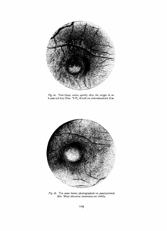
Fig. 8a. Vitellifonn lesion shortly after the ong:tn in an 8-year-old boy (Fam. T-T), filmed on orthochromatic film.
Fig. 8b. The same lesion photographed on panchromatic film. More abnormal structures are visible.

Fig. 9· Delicate alterations in the foveola of a 22-year-old female. Probably this is an initial stage of the vitclliform disc. Rather often we found these minimal lesions in family-members of patients suffering from vitelliform dystrophy. A pathological BOG L/D-ratio was always
found in these cases.
Braley 1966), and this is why this condition is still often counted among the congenital macular heredodegenerations. Since Barkman (1961) observed this foveal abnormality in an infant aged I week, while Braley (1968) made the same observation in an infant aged 2 weeks, it is indeed likely that the vitelliform structure can be present already at birth. Falls (quoted by Grimm and Tedford 1963, and Braley 1966) even held that an individual with normal fundi cannot later develop VFD. We have found that this is a fallacy, because many investigators have observed normal foveae to develop pathological changes later in life (Huysmans 194o; Hermann and Vernin 1963; Friedenwald and Maumenee 1951; Zanen and Hermans '961).
We personally observed a vitelliform disc in a 6-year-old boy (IV-Ij, fam. TT; fig. 8) and in a44-year-old man (V-7, fam. Vr; fig. 7), in eyes which had been normal or virtually normal. One year earlier the boy's foveae were entirely normal; but the man had already shown a few small multiple vitelliform lesions over the right fovea and a small white-yellow spot at the site of the foveola when examined a year earlier (fig. 7). In two women aged 43 and 32, respectively, who were known as carriers on the basis of EOG findings, we saw normal foveae develop pathological changes in the course of a year (III-z; III-8, fam. EW). It should be pointed out that these
2!0

Fig. IO. Partly ruptured vitelliform disc in a 35~year-old male. (Fam. B-E).
Fig. I I. Intravitelline haemorrhage in one of our patients (Fam. Pl). In all probability these haemorrhages are based
on ruptures in Bruch's membrane.
ZII

Fig. I 2a-b. Vitelliform lesions resembling scars of central choroiditis (Fam. V r). In this case a diagnosis of toxo
plasmosis was initially made.
212

Fig. I 2c. Fluorescein angiography of the left eye reveals a central defect of the pigment epithelium. This defect is not larger than is suggested by normal ophthalmoscopy.
Fig. I 2d. There is after-fluorescence (after the clearance of the fluorescein from the retinal vessels) but not leakage of
fluorescein. This suggests a normal Bruch's membrane.
2!3

Fig. I Ja-b. Vitelliform.lesions in a 15-year-old boy. In this case also a diagnosis of toxoplasmosis was made despite
normal serology (Fam. Vr).
2. !4

Fig. I 4· Delicate folding of the internal limiting membrane around a slightly prominent vitelliform disc (Fam. WA).
pathological changes were very slight: both women showed only a small yellowwhite spot at the site of the foveola (fig. 9).
The literature shows that the classical vitelliform lesion is most frequently observed between age 5 and age r 5, with a ma."'cimum at about age 6. Since most children start attendiog kindergarten at age 4 (vision is usually examined periodically at schools), this fact need not be conclusive of the age of onset. Even in the presence of a vitelliform lesion, vision is initially good, and the time of onset can therefore not be determined on the basis of visual acuity.
The possibility that a vite!liform lesion occurs in senescence cannot be ruled out, theoretically. This is why a classification of posterior pole dystrophies on the basis of the age of onset is untenable. Apart from ophthalmoscopic examination at normal visual acuity, the foveal abnormality can be detected also in a patient with diminished vision. More or less acute diminution of vision in patients with VFD can occur in several different ways: r. due to rupture of the vitelliform cyst, causing cyst contents to emerge and cause
damage to the photoreceptors. The consequences of a rupture are clearly visible in patient IV-r, fam. B-E (fig. ro); part of the vitelliform structure is still intact, while another part has ruptured;
2. due to intravitelline haemorrhage (fig. rr) (III-z, fam. PI); 3· without any ophthalmoscopically visible changes in the existing picture, e.g. in
V-r9 and V-zo, fam. Vr (fig. rz, r3). In these patients vision returned to the initial level after some time. At the out-
2Ij

patient clinic we also saw several patients (e.g. V-3, fam. W.A.) with acute irreversible changes in vision, without any changes other than the already observed vitelliform structures.
In most cases, therefore, the ophthalmoscopic picture gives no information on vision. This is quite apparent in patient V-3 fam. W.A. This young man showed more or less identical structures in both foveae but vision was IO/IO OD and I/IO OS.
Metamorphopsia can occur at the time of diminution of vision, but it is rarely as pronounced as that in chorioidiopathia centralis serosa. The same applies to hypermetropia.
It is worthy of note that one of our patients (V-7,fam. Vr) perceived light flashes in one of his eyes in the dark at the time of occurrence of the vitelliform disc. This patient was already known to show dubious changes in the posterior pole (fig. 7a), with a highly pathological EOG. About a year after the last examination at which no vitelliform structure had been visible in the fovea, the patient complained to me about light flashes in his left eye. To my surprise, I found a classical, intact vitelliform lesion in both foveae (fig. 7b ).
3· FUNDUS (OPHTHALMOSCOPIC FEATURES)
In most cases VFD is a bilateral abnormality, but unilateral cases have been frequently described, both in young people and in middle-aged indiviuals (Best 1905; Huysmans 194o; Berkley and Bussey 1949; Bruna 1951; Zanen and Rausin 1951; Capalbi 1954; Zanen and Hermans 1961; De Walsche 1963; Braley and Spivey 1964; Remky eta!. 1965; Krill eta!. 1966; Fran<;ois 1968; Deutman 1969).
De Walsche (r963) described a 56-year-old woman and Zanen and Hermans (r96r) an rr-year-old boy with unilateral VFD; this illustrates that unilateral changes can be found early as well as later in life.
There are many exact descriptions of the classical vitelliform structure. The fovea encompasses an egg-yellow (sometimes orange or pinkish red), round, slightly elevated structure surrounded by a somewhat darker border. The retinal vessels take an undisturbed course past the edge of this disc (the size of which is 0.5-3 optic disc diameters), which shows an unmistakable resemblance to the intact yolk of a fried egg or to a tinned peach half (fig. r).
V e:ry often this classical structure is not seen at the time of examination, and our impression is that in some cases this characteristic picture never occurs.
Our study of 98 ophthalmoscopically affected individuals shows that the pictures found range from an exceedingly slight change to a condition resembling the terminal stage of extensive chorioretinitis. Moreover, the carriers of the pathological gene can show a normal fovea, or a fovea with a not obviously pathological, more or less non-specific macrogranular appearance.
In several cases we found as the first sign of VFD a very small yellow-white spot (fig. I 5), developing into a small yellowish disc, sometimes exactly in the foveola
2!6

Fig. I ;a-b. Mild foveal alterations in a 48-year-old female suffering from vitelliform dystrophy (Fam. Ko-K). This individual is the mother of the boy whose fundi are
depicted in fig. r.
217

Fig. r6-q. Foveal changes in the "previtelliform stage". These lesions were found during examination of families of patients with true vitclliform lesions (Fam. W A; Fl and
Ko-K).
zr3

Fig. z8-I9. Foveal changes in the "previtellifonn stage ... These lesions were found during e.-mmination of families of patients with true vitellifonn lesions (Fam. W A; Fl and
K<rK).
219

but at other times eccentric (fig. I6, I7)- Tbis disc can become a vitelliform lesion, but in some cases several drusen-like or spotty configurations occur (fig. IS, I9)- In many cases, however, the egg-yolk lesion does make its appearance.
The age of occurrence of this vitelliform stage is usually between age 3 and age I 5, according to the literature and our personal observations. We observed the eggyolk in 9 patients whose respective ages were 6, 6, 7, 7, 9, 44, 19, 13, and 9 years (V-I3 fam. BE; IV-z fam. WE; IV-I5 fam. T-T; IV-I7 fam. Fl; IX-6 fam. Ko-K; V-7 fam. Vr; III-z fam. Bl; III-4 fam. Bl; III-6 fam. Bl).
Friedenwald and Maumenee (I95 I) observed the vitelliform stage in a woman aged 42 and in a middle-aged man.
The vitelliform lesion is not always found in the exact centre of the retina; tbis is quite apparent in the multiple vitelliform structures (Littann I965; Denden I966). Extrafoveally localized vitelliform structures were also described by Galeazzi (I939) and Calmettes and Deodati (I96z). Our patients V-7 fam. Vr and II-7 fam. KZ likewise showed eccentric vitel!iform changes (figs. 7 and zo).
The vitelliform lesion can be slightly more prominent than the surrounding pigment epithelium, but tbis causes only a slight prominence (rarely e.--<ceeding I dioptre) of the retina in front of it. As a result, a delicate radial pattern of streaks can occur around tbis structure (fig. I4, zi).
Binocular contact lens examination indicates the likelihood that the egg-yolk is localized in the pigment epithelium (fig. zz). The fact that the yellow of a fully developed, intact vitelliform disc is so clearly visible, indicates that the pigment epithelium cannot be entirely intact in front of it. The fact that vision is usually normal at tbis stage proves that het neuroepithelium is still intact. A dark border surrounds the disc and marks the boundary between egg-yolk and pigment epithelium. The yolk proper admits little light, but it is clearly visible that a yellowish, homogeneous substance, hardly prominent if at all, lies immediately beneath the retina, that is to say: probably in the pigment epithelium. No true cyst is visible at tbis stage, and tbis is why in the early stages of VFD we use the designation "vitelliform disc" (Zanen and Hermans I 96 I).
Optic disc, retinal vessels and retinal periphery are nearly always quite normal in patients with VFD. So far as can be established, the choroid shows no abnormality. Occasionally, several vitelliform structures can be found in the posterior pole (fig. 7, zo), and in a fair number of cases the fundal periphery shows a granular appearance wbich cannot be identified as a distinctly pathological change (fig. 23). In one patient (IV- I 9 fam. UT) we found peripheral areas of pigmentation and depigmentation (fig. 24). The classical vitelliform lesion is not irreversible but may persist more than a year before it begins to show distinct changes.
As a rule there is disintegration; the contents of the yolk show fragmentation, possibly as a result of syneresis of pigment epithelium cells tbat have become necrotic (fig. 25). The yolk may even disappear, whereupon the fovea resumes a virtually normal appearance (Zanen and Hermans I96I; Bischler I952). We ourselves observed complete disappearance of a vitelliform structure in two cases (II-7 fam. KZ;
220

Fig. 20a. Two vitelliform lesions in the eye of a female in her thirties (Fam. K-Z). At the fovea an atrophic lesion. On the superior nasal side of this lesion an intact vitelliform
disc.
Fig. 20b. Fluorescein angiography shows pathological fluorescence mainly at the site of the fovea. Some spots of increased fluorescence are visible at the site of the intact vitelliform disc. This suggests slight defects in the retinal
pigment epithelium.
22!

Fig. 21. Vitelliform lesion in an IS-year-old boy. Note the £nc folding of the internal limiting membrane. In the other eye this boy developed an anterior disinsertion of the
retina (Fam. UT).
V-1 3 faro. BE). The vitelliform abnormality can recur after such a regression (Zanen and Hermans 1961; Franceschetti et al. 1963).
Regression of the vitelliform lesion to an ophthalmoscopically normal fovea can be explained both by metabolic processes in the pigment epithelium and by absorption from the choriocapillaris. As a rule the vitelliform disc develops into a cyst, in which vitelliform remnants can still be observed (fig. z6). If the egg metaphor is to be continued, this stage can be described as "scrambled egg" stage. At the site of disappearance of the egg-yolk one often observes atrophic pigment epithelium with the choroid (redder than adjacent parts) shimmering through (fig. 27).
A different course is also possible. The contents of the yolk can become subject to syneresis, giving rise to a cyst with a fluid-level (fig. z8). Because of the striking resemblance to a hypopyon, this stage is sometimes called pseudo-hypopyon stage. There may also be pseudo-descemet mottling on the posterior aspect of the cyst's anterior wall, the rerina (Pameyer 1954).
Remky et a!. (1965) clearly demonstrated the liquid character of this pseudohypopyon by photographing a patient's fundus in different positions. This pseudohypopyon stage is most commonly encountered between age 30 and age 40 (Remky et al. 1965). The cyst can rupture, with proliferation of pigment epithelium as a result, which may produce marked pigmentations.
The resulting clinical picture (fig. 29) can be readily confused with a cicatrix of chorioretinitis centralis (fig. 30 ).
222

Fig. 22. Schematic drawing of six different stages of the rise and fall of the vitelliform disc (binocular slitlamp-cxamination).
a. normal fovea, pathological EOG b. minimal foveal lesion c. intact vitclliform disc d. ruptured vitelliforrn disc c. pseudo-hypopyon (vitellifonn cyst) f. atrophy of the cenwl retina.
223

Fig. 23. Fine mottling of pigment as is often seen in the retinal peripheries of patients with vitellifonn dystrophy
(Fam. WA) (panchromatic film).
Fig. 24· Area of pigmentation and depigmentation in the retinal periphery of a zx-ycar-old male with vitellifonn
dystrophy.
ZZ4

Fig. 25a. Intact vitelliform disc in a 6-year-old boy (Fam. E-W).
Fig. 2Jb. The same eye 6 years later. The disc is disintegrated (Fam. E-W).

Fig. 26. Vitelliform cyst in a 24-year-old male (Fam. Ko-K)·
Fig . .:q. Vitelliform cyst with yellowish-white deposits in a 52-year-old male (Fam. Fl).
zz6

Fig. 28. Pscudohypopyon-structw:c in a vitclliform cyst in the eye of a ;7-year-old female (Fam dB).
Fig. 29. Severe pigmentations at the bottom of a viteJliform lesion in a 52.-year-old male (Fam. Fl).

Fig. 30. Scar of central choroiditis in the foveal area. Note the small scar infratemporally from the fovea.
Fig. ji. Multiple deposits in a round area of atrophic pigment epithelium in a 41-year-old male suffering from
vitelliform dystrophy (Pam. Vr).
228
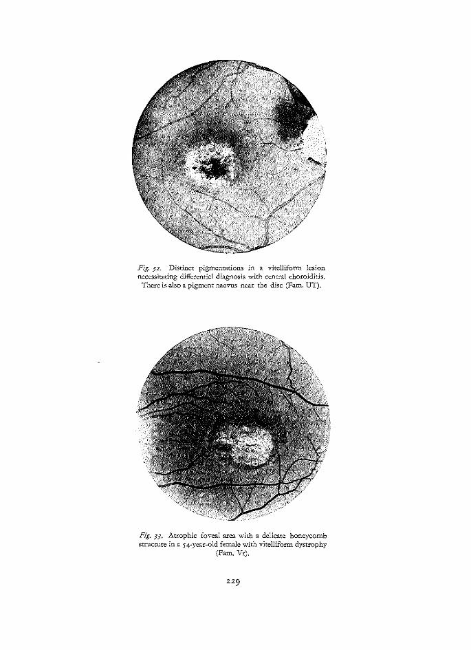
Fig. p. Distinct pigmentations in a vitelliform lesion necessitating differential diagnosis with central choroiditis.
There is also a pigment naevus ncar the disc (Fam. UT).
Fig. Jj. Atrophic foveal area with a delicate honeycomb structure in a 54-year-old female with vitelliform dystrophy
(Pam. Vr).

Fig. J4· Well defined area of retinal atrophy in both eyes of a 7;-year-old male with vitelliform dystrophy (Fam. UT).

Fig. ffJa~b. Vitclliform cysts with pseudo-hypopyon aspectS. Note the differences in size of the vitellifonn cysts in the
right and left eye (Fam. UT).

In a few cases there may be haemorrhages deep in the retina at the site of the vitelliform lesion (Mazzi I934; De Walsche I963; Braley I966). We ourselves observed this in only one case (III-z fam. PI; fig. n). These haematomas are absorbed in the course of a few weeks. We believe that the occurrence of haemorrhages points in the direction of a no longer quite intact Bruch's membrane and/or choriocapillaris. But in initial stages these haemorrhages are never seen, and on this basis a primary affection of the choriocapillaris and for Bruch's membrane can be practically ruled out.
The vitelliform lesion ultimately results in a circular area of atrophic pigment epithelium in which yellow lumps can become manifest (fig. p). There are sometimes fairly pronounced pigmentations (fig. 32), although in many cases these are absent (fig. 33). One of the patients in whom we saw this was a 74-year-old man (II-6 fam. UT), who showed a circumscribed area of pigmental epithelium atrophy and an atrophic choroid (fig. 34).
Many authors, including Frans;ois (I967) and Krill et aL (I966) maintain that, after disintegration, the foveal features are indistinguishable from those in other affections. Although we could confirm this in incidental cases, our study has convinced us that in most cases the foveal abnormality retains its typical characteristics in which the basic vitelliform process can still be clearly recognized up to a very advanced age. We observed this in several patients (II-6 fam. UT; III-8, IV-5,V-6 and V-9, fam. Vr). The circular central atrophy of the pigment epithelium remains a characteristic feature of the vitelliform dystrophy until advanced age (fig. 34).
In summary, and in a strictly schematic way, the evolution ofVFD can be described as follows: L normal fovea (but already a pathological EOG) (fig. 4I); z. previtelliform stage (fig. I 5); 3· vitelliform stage (fig. I, 42b); 4· "scrambled egg" stage (fig. z6, fig. 4za). 5. cyst stage (fig. 2 7); 6. pseudo-hypopyon stage (fig. 28, 3 5); 7· chorioretinal atrophy stage (fig. 33, 34). The development of VFD can skip several of these stages, and various transitional forms of course occur.
Although VFD is usually bilateral, a perfectly symmetrical fundus picture is rarely seen. There may be substantial differences between OD and OS, both ophthalmoscopically and in terms of visual acuity (figs. 3 5, 3 6).
4· REFRACTION
Pronounced hypermetropia with astigmatism is often found in patients as well as their relatives; convergent concomitant strabismus with amblyopia is also often observed. Practically all authors mention hypermetropia (Best I9o5; Behr I92o; Huysmans I94o; Sorsby I94o; Berkley and Bussey I949; Falls I949; Zanen and Rausin I95 I; Capalbi I954; Pameyer I954; McFarland I95 5; Sorsby et al. I95 6;

Fig. j6a-b. Vitclliform cysts with pseudo-hypopyon aspects. Note the differences in size of the vitelliform cysts in the
right and left eye.

Gregory I958; Chinaglia and Perini I964; Braley and Spivey I964; Melodia and Tabacchi I965; Spinelli and De Molfetta I965; Remky et al. I965; Hermann I966; Belmonte I 966; Krill et al. I 966; Gorgone I 967; Curry and Moorman I 968; and others).
Yet this hypermetropia is not an obligate finding. In one patient we found slight myopia (IV-z fam. EW). Bonnet et al. (I966) saw a girl who developed slight myopia; and Gorgone (I967) saw a myopic girl with a hypermetropic mother, both showing VFD. The pronounced hypermetropia usually found cannot be ascribed to any prominence of the fovea because this is rarely more than I dioptre.
There are no marked abnormalities of refraction of the type found in central serous choroidopathy. The commonly observed hypermetropia with strabismus makes it desirable to point out to future parents in these families that their children will have to be examined by the ophthalmologist at a very early age.
5· VISUAL ACUITY
Vision is initially normal or only slightly subnormal, even in the presence of a fully developed vitelliform disc. This often causes surprise. This good vision can only be explained by assuming that the neuroepithelium (i.e. the cones) are initially not involved in the pathological process. Moreover, the functions usually required for good vision, usually show no fundamental disturbances. In spite of the vitelliform structure, therefore, the pigment epithelium which is its site will continue to function fairly well, even at the site of the fovea. A primary disturbance of Bruch's membrane is more likely to give rise to exudates and haemorrhagic processes, as in the case of angioid streaks, Sorsby's pseudo-inflammatory dystrophy and Junius-Kuhnt disciform posterior pole degeneration; in that case there is leakage of fluorescein in fluorescein angiography. But this is never observed in VFD.
If very sensitive methods are used to determine vision, it is probably always possible to demonstrate at least a very slight diminution of vision. Using a photometer and different light intensities with different wavelengths, Zanen and Balsacq (I968) found an unmistakable diminution of foveal sensitivity in a patient with VFD whose visual acnity was still virtually normal. We were able to demonstrate the same in our of our patients (IV-z faro. EW) with the aid of the Friedman visual field analyser.
As determined by conventional routine methods now in use, visual acuity is in many cases still quite normal or only slightly subnormal in the initial stages; but when the contents of the vitelliform disc begin to disintegrate, vision is seen to diminish.
This diminution of vision can be abrupt (upon rupture of the vitelliform cyst). The contents of the vitelliform structure then emerge and damage the photoreceptors. The cause of diminished vision is sometimes quite apparent at ophthalmoscopy; on the other hand there are many cases without any change at the time of abrupt diminution of vision.
As we pointed out, haemorrhages in the vitel!iform structure can also cause abrupt

diminution of vision. It is impossible to predict whether the loss of vision will be reversible or irreversible, but it is conspicuous that loss of vision occurs when general resistance is low, e.g. during illness, after operation or parturition, and also in response to traumatic mental influences.
In the atrophic terminal stage, vision is often as low as 5 j6o to 2jro. Many authors report that substantial diminution of vision does not occur until a more advanced age, but we have seen considerably diminished vision at an earlier age (fam.Kl.K). We saw a patient under 20 whose vision was only 0.2 ODS (i!-r9 fam. V) and a boy of ro with vision rjro ODS (IV-zo fam. UT). Patients with non-specific lesions (yellow drusen-like alterations in the posterior pole) can retain good vision to a very advanced age because the neuroepithelium remains intact.
When the foveal cones are involved, variable patterns of eccentric fi:mtion develop. Hermann (r966) reported '~eccentric viewing" but no genuine eccentric fi~tion in a 7-year-old girl in whom a vitelliform structure developed in the course of 5 years to a lesion with degeneration and pigmented changes. In the long run, however, genuine eccentric fi.,ation does develop as damage of the photoreceptors increases (Graham et al. r964; personal observations).
6. VISUAL FIELDS
The peripheral boundaries of the visual fields are quite normal throughout all stages of this condition. The central visual field of course shows a diminution of sensitivity, which roughly parallels the loss of vision.
Scotomas occur, initially for red and later for green, followed by relative scotomas for white light; in serious cases an absolute central scotoma can occur. The size of the scotoma is dependent on the size of the vitelliform structure.
A paracentral scotoma is occasionally found in association with paracentral vitelliform structures of the kind sometimes seen in multiple vitelliform lesions.
7· COLOUR VISION
Diminution of visual acuity in VFD is generally accompanied by an acquired disturbance of colour vision. The patient is usually unaware of this, but some patients complain that the colour red is less well perceptible. There are no characteristic disturbances of colour vision. The findings obtained in this respect prove to be largely dependent on the methods used to determine colour vision. Central colour vision shows marked deterioration with increasingly disturbed vision. Krill et al. (r966) demonstrated this quite clearly with the Famsworth-Munsell roo-hue test.
A wide range of various disturbances of colour vision have been described, but undisturbed colour vision has likewise been reported fairly often (Falls 1949; Zanen and Rausin 195 r; Capalbi 1954; Sorsby and Wren r96o; Hermann and Veroin r963; Franceschetti, Fran~ois and Babel r963; Etienne et al. 1964; Hermann r966; Belmonte 1966; Le Hunsec and Pierre 1967; Tsukahara et al. 1968).

Others have reported a slightly diminished general colour sense without specific axis (anomalous trichromasia) (Falls 1949; Zanen and Lempereur 1954; Zanen and Hermans 1961; Verriest 1964).
Mild red-green dyschromatopsia has been described also (Zanen and Hermans 1961; Grimm and Tedtord 1963; Velzeboer 1963; Braley and Spivey 1964; Remky eta!. 1965; Fran>ois et aL 1966; Krill eta!. 1966).
Many authors reported anomaloscopic evidence of reduced red sensitivity (Bischler 1952; Sorsby eta!. 1956; Zanen and Hermans 1961; Velzeboer 1963; Krill et aL r 966). This corresponds to type II of acquired red-green dyschromatopsia (V eniest 1964). But Braley and Spivey (1964) described type I acquired red-green dyschromatopsia, and Melodia and Tabacchi ( 196 5) established mild deuteranomaly with the roo-hue test. Blue-yellow dyschromatopsia was found in a few cases (Spinelli and De Molfetta 1965; Krill et aL 1966), and blue-green dyschromatopsia was reported by others (Zanen and Rausin 1951; Velzeboer 1963; Melodia and Tabacchi 1965; Krill et aL 1966).
This enumeration shows that a wide variety of disturbances of colour vision can be found. In 23 of the 30 affected subjects we examined for colour vision, we found a more or less markedly reduced red sensitivity (anomaloscope). The HRR test disclosed mild-to-moderate red-green dyschromatopsia in 20 of the 30 individuals examined, and slight blue-yellow dyschromatopsia was observed in only one case. The decrease in red sensitivity was quite apparent at a vision of o. 5 or less. The Farnsworth panel D-1 5 test revealed a tritan disorder in 8 patients, while a red-green disorder was found in only 2 cases. All the patients with a tritan disorder had a vision of o.z or less. Some slight disturbance of colour vision without specific axis (anomalous trichromasia) was observed in 5 patients.
The effect of the vitelliform abnormality on colour vision is perfectly illustrated by the data on our patient IV-19 fam. UT, with a vitelliform lesion and loss of vision to rjro OD, and a normal posterior pole, and vision rojro OS (table III).
Table Ill
Vision Anomaloscope HRR test Farnsworth D~ I 5
OD I/IO diminished red mild R-G tritan a:·ds sensitivity dyschromatopsia
OS rojro normal normal normal
This case confirms the incorrectness of Behr' s original view that the colour vision disorders do not result from the VFD but occur as a concurrent hereditary abnormality.
Our conclusion is that with increasing loss of vision colour vision diminishes on the whole, and that the red receptors in particular are affected. With increasing loss of vision, a subsequent tritan disorder is not uncommon.

8. DARK ADAPTATION
Sorsby eta!. (1956) found a slight disturbance of dark adaptation in z members of the CNS family, but good dark adaptation in another member of this family. In their HN family- also with VFD -they found good dark adaptation. Braley and Spivey (r964) also described a slightly disturbed dark adaptation curve: "The rod or final phase of dark adaptation was normal in all our instances, both affiicted and normal. However, although the early mesopic plateau was not grossly abnormal, there was certainly a general prolongation of the Kohlrausch kink in those affected".
Unlike these authors, we found an entirely normal dark adaptation curve in 21 of 22 cases studied. In cases of repeated examination, too, we found no anomalies. When vision is poor the streak figure of the Goldmann-Weekers apparatus can be perceived too late, without dark adaptation being pathological.
This possibility should always be borne in mind; in dubious cases the curve should be plotted again by the integral method.
Other authors have also described normal dark adaptation curves (Remky et a!. 1965; Fran~ois eta!. 1966, 1967; Krill eta!. 1966; Tsukahara eta!. 1968).
9· ELECTRORETINOGRAPHY
The ERG is usually normal in VFD (Gallet r96r; Franceschetti, Fran~ois and Babel 1963; Braley and Spivey 1964; Chinaglia and Perini 1964; Krill eta!. 1966; Bonnet eta!. 1966; Denden 1966; Fran~ois 1967, 1968; Tsukahara eta!. 1968; Deutman 1969). The critical flicker fusion (CFF) is likewise normal (Braley and Spivey 1964; Fran~ois et a!. 1967, 1968).
Fran~ois eta!. (r967, 1968) did point out that the ERG values of the a- and b-waves were lower than normal, but they defined these values as decidedly within the limits of normal. Braley and Spivey (r964) likewise reported normal ERG's. The a-waves were normal, and failed to show the anomalous slope reported by Ruedemann and Noell (r96r). The following features were found to deviate from the normal. "By plotting the amplitude of the b-wave in microvolts against time of increasing dark adaptation at a constant stimulus intensity, it was found that most affected individuals exhibit a slow rise during the first r 2 minutes of dark adaptation. At 20 minutes the b-wave amplitude exhibits a very sharp elevation to a level equal to that of the normals. By conttast in the unaffected, the progress is rather rapid to the I 2 minute period. They then show proportionally very little increase in the zo minute stimulus."
We ourselves found normal scotopic and photopic ERG values in 23 individuals (page 432). The photopic a- and b-wave were both normal. In only one man of 77 years (III-8, fam. V) did we find a subnormal scotopic b-wave of the ERG, probably due to senile retinal changes.
Local ERG of the fovea and visual(y evoked responses
The F-ERG was recorded in 7 individuals. It was normal in association with good

vision, and subnormal when vision was poor (as was to be expected). There was no distinct correlation between vision and F-ERG response, which was probably in part determined by the poor ii.'-:ation in diminishing vision.
We generally found normal responses at a vision exceeding 4jro, and subnormal responses at lower values. The VER largely parallelled the F-ERG responses. They were normal at a vision exceeding 4/Io, and subnormal at lower values.
Biersdorf and Diller (I 969), who used a different technique of local foveal stimulation, found a normal foveal response ODS in a patient with VFD whose vision was 20j4o OD and zoj2o OS.
Oscillatocy potential.r
The literature comprises no reports on the OP in VFD. In 2 patients examined (IV-2 fam. EW; fam. Vr) we found quite normal OP's and we consequently believe that the OP as well as the ERG a- and b-waves are generally normal in VFD.
IO. ELECTRO-OCULOGRAPHY
Krill eta!. (r966) and Franc;ois eta!. (r966, 1967, 1968) obtained a distinctly abnormal EOG as the sole sign of disturbed retinal function.
The standing potential proper is somewhat low, or normal (Franc;ois 1966, 1967), but the LpJDt-ratio is clearly pathological. We have confirmed this in 40 cases from rz different families. In none of the patients did the Lp/Dt-ratio e.'-:ceed 1.50, and 1.65 can be regarded as the e.'<treme lower limit of normal.* Although the EOG can show marked variations in normal subjects (Kelsey 1967), it can be categorically stated that our cases ofVFD showed a highly pathological EOG, with in many cases complete absence of a light rise, causing the EOG to take a more or less flat course (fig. 37)·
This flat course of the EOG record is characteristic of VFD. The foveal changes were not very pronounced in 5 of the 40 patients e.'<amined. Like a patient described by Franc;ois et a!. (1967), our own patient with a uni-
lateral vitelliform abnormality (IV-19 fam. UT) also proved to show a highly pathological EOG in both eyes.
A new finding was that carriers of the pathological gene with ophthalmoscopically normal foveae proved to show a highly pathological EOG, like the patients (Deutman 1969). This means that the EOG is a useful aid in the diagnosis of carriers, and is therefore an indispensable method of investigation in genetic counselling. We detected 16 ophthalmoscopically normal carriers of the gene in 5 different families (families TT, K-Z, EW, dB and Vr); in z of these x6, slight ophthalmoscopic changes were later observed. If a patient were e.'<amined for it at birth it is highly probable that a pathological LpJDt-ratio of the EOG would be found.
* The other day we examined a new family with VFD. In this family, there were two affected individuals who showed the comparatively high L/D-ratio of x.6o.

IIIII l Ill I IIIII I 111111111
lllllllllllllllllllllllff s 10 15 20 25
O.A. LA.
00
115%
OS
120%
minutes
Fig. 5 7a. BOG bar figure in a normal individual (top) and in a patient with vitelliform dystrophy (bottom).
The cause of this pathological EOG is probably a metabolic dysfunction resulting from a disorder of absence of an enzyme in the retinal pigment epithelium.
The EOG should be regarded as a function test for the entire retina, and a pathological EOG therefore indicates an overall retinal dysfunction.
This means that it is an interesting and unexpected finding to encounter a highly pathological EOG in a condition in which as a rule only the central retina shows any ophthalmoscopic change. The occurrence of multiple vitelliform structures already suggested a di1fuse retinal disorder (Littann 1965; Denden 1966). The fact that it is usually only the fovea which shows changes, can be explained by the fact that the fovea has a quite special and highly di1ferentiated anatoruical and functional position in the retina. Apart from this, the fovea centralis is a locus minoris resistentiae also in many other affections, e.g. chloroquine retinopathy (Hobbs et aL 195 9; Gouras and Gunkel 1963; Butler 1966), phenothiazine retinopathy (Verrey 1956; Rintelen et aL 1957; Alkemade 1968; Henkes 1698; Boet 197o) and senile changes. The fact that the metabolism is probably higher at the site of the fovea centralis than else-

where in the retina may explain the frequent occurrence of foveal changes in an apparently normal retina. Potts ( r 966) explained the predisposition of the fovea to certain pathological changes on the basis of the vascular conditions in the choroid. This hypothesis is very attractive for cases in which defects in Bruchs' membrane occur, e.g. in central serous choroidopathy, but we believe that it is untenable for the hereditary dystrophies.
In our opinion, the pathological EOG in VFD cannot be explained solely by an affection of the central retina- a possibility considered by Fran<;ois et al. (r967) as well as by Morse and MacLean (r968). An important argument against this view arose from our own study which, among other things, disclosed that the EOG is normal in many other affections of the posterior pole of the eye.
In central serous choroidopathy, central choroiditis and cases of foveal changes on the basis of angioid streaks, we obtained a quite norrr.alEOG (fig. 37b). The EOG is repeatedly quite normal also in many seuile foveal degenerations as well as in exudative disciform and dry atrophic processes. However, senile degenerative changes may also give subnormal BOG's.
Another argument against the view that a pathological EOG might be due to the localization of the vitelliform abnormality in the central retina, lies in the fact that ophthalmoscopically normal carriers with no posterior pole affection, likewise show a highly pathological EOG.
LID ratio
3.00
2.50
2.10
1.85
1.65
1.50
1.00
0.50
0
. . . .. ..
central norous choroldopathy
central clloroldltl:l
..
angioid
&treaks
.... .
vltelllform
dystrophy
Fig. :J?b. EOG-L/D~ratio in individU:J.ls with central serous choroidophathy, central choroiditis, angioid straks and vitelliform dystrophy. The results in ro unsdected patients (2o eyes) per entity arc presented (every black point means
one eye).

'The fact that initially the visual acuity, visual fields and colour vision, and ultimately the dark adaptation and ERG remain normal warrants the conclusion that the retinal neuro-epithelium in VFD must be entirely intact, initially and later, except at the site of the fovea. 'The additional fact that the EOG is highly pathological from the onset in cases of VFD warrants the conclusion that this condition involves a diffuse dysfunction of the pigment epithelium or deeper layers. Since fluorescein angiography shows that the choroid and Bruch's membrane are quite normal, at least in initial stages, we can conclude that diffuse dysfunction of the pigment epithelium exists. This is consistent with the suggestion, based on many exhaustive investigations, that the vitelliform disc must be localized in the pigment epithelium.
II. PHOTOGRAPHY
Interesting findings were reported by Krill eta!. (1966), who used a fundus camera modified for monochromatic light. 'They used wavelengths of 450 miL (blue), 5 33 miL (green) and 675 miL (red) as light sources. "'The blue revealed mainly retinal details not beyond the level of the receptors (where most of the light was probably absorbed); the green light revealed details at a more external retina! level, probably back to the pigment epithelium. 'The red light penetrated receptors and pigment epithelium and allowed visualization of the choroid. Large deposits of melanin at any level absorbed the red light and appeared black in the photographs."
'The red light photographs disclosed the most extensive lesions, and with red light a more homogeneous affection was seen than with white light.
In our photographic study we used orthochromatic and panchromatic graphic films, which have maximum spectral sensitivity at 580 miL and 595 mfL, respectively (Craandijk and Aan de Kerk 1969).
As expected, panchromatic film showed the changes of pigment epithelium much better than orthochromatic film, not only because of higher sensitivity for the long wavelengths which penetrate deeper into the retina, but mainly as a result of better contrast: I. 'The choroidal vessels are best visualized on orthochromatic film, but the choroidal
pigment is more clearly visible on panchromatic film (fig. 38). z. Photographs made of a colour slide with orthochromatic and panchromatic film,
show e:~:act!y the same differences as do photographs made directly of the posterior pole of the eye.
By using this method, therefore, we were able to detect more indications of the localization of retinal changes in depth.
On panchromatic film the vitelliform structures are visible much more extensively than on orthochromatic film (fig. 38, 39), and the results obtained with this film are therefore comparable with those obtained with monochromatic light. Using panchromatic film, we obtained a result in VFD which was about the same as that obtained by Krill eta!. (1966) with red monochromatic light: visualization of the area

Fig. 3Ka-b. Intact vitclliform disc, on orthochxomatic and panchromatic :61m respectively (Fam. Ko-K).

Fig. 39a-b. Vitellifonn lesion, on orthochromatic and panchromatic film respectively. The latter gives more
details about the pathological structure (Fam. WA).

with the most extensive involvement. These findings argue in favour of a localization of the vitelliform disc in or next to the pigment epithelium.
IZ. FLUORESCEIN ANGIOGRAPHY
Several fluorescein-angiographic studies have been made of VFD. Martenet (I967) published an e.-<cellent photograph of an advanced stage of VFD in a Io-year-old boy, and a fluorescein angiogram of what we believe was a central serous choroidopathy, which she described as VFD.
She observed: 1. marked fluorescence at the time of ma.--dmum filling, suggestive of an extensive
defect in the pigment layer; z. increased permeability to fluorescein at the site of the abnormality, possibly due
to increased permeability of the choroidal vessels. The fluorescein pattern was characteristic of that of an old chorioretinitis focus, or that often seen some times after photocoagulation.
Tsukahara eta! (I968) assumed on the basis of their fluorescein study that VFD must primarily be a congenital lesion of Bruch's membrane. We cannot accept this, for in fresh vitelliform lesions the fluorescein pattern is quite normal, as shown by Curry and Moorman (I968) and others. They made a fluorescein study of a classical intact vitelliform lesion. Their photographs showed a normal fluorescein pattern. A patient with a vitelliform disc of somewhat longer standing and with ophthalmoscopic features suggestive of pigment layer atrophy, showed slightly pathological fluorescence. These authors maintained that the normal fluorescein pattern in intact vitelliform lesions indicates structural intactness of Bruch's membrane and the pigment epithelium; and we agree with them. Only a very slight defect in Bruch's membrane, with intact pigment epithelium, might possibly escape observation at fluorescein angiography; although it is likely that in that case, as in central serous choroidopathy, fluorescein leakage would occur.
Morse aro.d MacLean (I 968) as well as Krill eta!. (I968) and Franc;ois and De Laey (I 969) described a fluorescein pattern in VFD which was characteristic of pigment layer defects, without fluorescein leakage being observed. Fluorescein leakage would indicate defects in Bruch's membrane andjor disturbances in the permeability of the choroid. Urrets-Zavalia and Moyano (I969) emphasized a finding which we obtained also: that the fluorescence of the central lesion is intensified (but not increased) after disappearance of the fluorescein from the retinal vessels.
They ascribed this to "increased choriocapillary and pigmentary epithelium permeability", from which they concluded that VFD must be a specific form of central serous choroidopathy. We must reject this conclusion because central serous choroidopathy is a quite different entity (as demonstrated by the EOG), with an entirely different fluorescein pattern. In central serous choroidopathy the fluorescein angiogram shows very localized leakage of fluorescein in the subretinal space, probably as a result of increased permeability of the chorio-capillaris (Gass
244

Fig. 4oa-b. Fluorescein angiography in central serous choroidopathy, showing leakage of fluorescein through
Bruch's membrane.
245

1967) andjor possibly due to a defect in Bruch's membrane (fig. 40). This leakage of fluorescein can be identified by a fluorescent area which increases in size and intensity and persists aftet the normal background fluorescence disappears. However, no fluorescein leakage has ever been demonstrated in VFD. Rosen (1969) asctibed the after-fluorescence in VFD to fluorescein absorption by the cyst contents.
In our carriers and patients with VFD we carried out an extensive fluoresceinangiographic investigation, the findings of which can be summarized as follows.
In one carrier (IV-1 5 fam. Vr) we observed a quite normal fluorescein pattern, and this pattern is also normal or virtually normal in the presence of a fresh vitelliform disc (fig. 41). In one patient with a recently formed vitelliform disc (V-7 fam. Vr), a hardly discernible fluorescence was observed only in the centre of the disc (fig. 7). In another man with an intact vitelliform lesion there was no pathological fluDl~escence either (Pam. Can.) (fig. 42). In VFD of some longer standing (V-19 fam. Vr) the pigment layer had in part disappeared so that pathological fluorescence was observed (fig. 12). This started in the arterial phase and increased in the venous phase. After disappearance of fluorescein from the choroidal and retinal vessels, a small central fluorescent area remained which gradually diminished in size.
However, the intensity of fluorescence initially increased (fig. 12d). There was no increase in the size of the fluorescent zone, and consequently there was no leakage of fluorescein. Perhaps the contents of the vitelliform cyst absorbed some fluorescein through Bruch's membrane (with or without pathological changes), whereupon the fluorescein spread fo:rward through the "egg-yolk". The yellow-white cyst contents adjacent to Bruch's membrane could absorb fluorescein much as drusen may do (Rubinstein and Paton 1966). It is probable, however, that after-fluorescence is visible because one looks through the pigment layer at the fluorescein normally escaping from the smaller vessels in the choriocapillaris. An increase of contrast might explain the apparently increasing fluorescence of the centre of the vitelliform lesion.
We also obtained fluorescein photographs in another interesting case. A woman (II-7 fam. K-Z) with two vitelliform lesions in the posterior pole (fig. zo) was submitted to fluorescein angiography in 1967. The C-'<trafoveally localized, virtually intact vitelliform lesion which had recently appeared, showed some slight pathological fluorescence, unlike the older atrophic foveal focus which showed the fluorescence that characterizes C-'<tensive central pigment epithelium atrophy (fig. zo).
13. CARRIERS
Ophthalmoscopically normal individuals who carry the pathological gene, are regarded as carriers. These individuals are encountered in some families with irregular dominant transmission of VFD. When skipping of a generation occurs, one may assume that a cattier is involved. Sorsby (1967) thought that these carriers differ from normal subjects in that they show mictogranular pigmentation of the fovea and its surroundings.

Fig. 4:1a~b. Conventional and fluorescence photograph of the posterior pole of a carrier of vitelliform dystrophy
(Faro. Vr).
247

Fig. 4U.
Photograph of section through the centre of the fovea of the left eye of the 59-year-old female proband (V-49) of the pedigree (see top left) with 69 affected individuals, presented by Barkman (r961). The photograph had to be composed of t\VO pieces because of an artificial detachment of the retina. The fundus photograph is from the left eye of this woman and was taken in June, 1958 (see top right). Deterioration of vision started at the age of 2.8 and she visited an ophthalmologist for the first time at the age of 46. Vision right eye 0.4; left eye o.8. In both foveae "cystic degeneration", measuring one disc-diameter was noticed. She died in 1969, at the age of 59, from coronary thrombosis. Ry Andersen performed the histopathological examination: ,Histopathological examination shows that the choriocapillaris is fairly \veil preserved and Bruch's membrane too, apart from some PAS-positive flat masses of cuticular substance from the inner cuticular part of Bruch's membrane and a couple of larger drusen one of which containing a small capillary from the choriocapillaris in the macular area. Serial sections of the eye show, a spotty thinning and degeneration of the retinal pigment epithelium with fe\.V or no melanin granules in an area a little less than one dd*. The inner retinal layer which is artificially detached from the pigment epithelium shows a severe defect of the sensory cells which are lacking in the same area as the degeneration of the retinal pigment epitheli:um. The outer intermediary layer in an area about three dd in and around the macula shows a proteinaceous eosinophilic oedema in hemato:-..")'lin and eosin stain. Special stains show that this oedema does not contain acid or neutral mucopolysaccharides or phospholipids. Oxydized tannin azo and mercury bromphenol blue stain arc positive suggesting protein. It cannot be excluded that this oedema could be a remnant of the vitelliform yello\.v cyst-like materi:1l seen in the early stage of the disease. But most likely it is not specific." Ry Andersen and Barkman concluded from their findings: ,.It is impossible from this old material to state the primary site or pathogenesis. But we find it most likely that the pigment epithelium in some way plays a very important part in the pathogenesis of the disease being responsible for the drusen and particular masses in the inner part of Bruch's membrane and for the increasing damage of the neuro-epithelium". (Mtcr Ry Andersen and Barkman. to be published by Ry _A.nderscn, S. and Barkman Y. in Acta Ophthalmologica).
* dd = disc dlameter

Fig. 4xt
We have indeed observed this, but this cannot be described as a really abnormal condition. As soon as the fovea begins to show distinct alterations, one is dealing with a patient and not with a carrier.
We were able to identify our first carrier in fam. Vr (IV-r6 fam. V r): his father, his sister and two of his sons were suffering from VFD, while the two different mothers of his affected sons had quite normal eyes. The carrier showed no ophthalmoscopic peculiarities (fig. 4rab) and his visual acuity was normal. But the BOG study revealed that his BOG was as pathological as that in the patients. After this surprising finding, we used the BOG for large-scale screening in this family and other families witb VFD. This enabled us to identify r 6 carriers with certainty in 5 of these families.
Because the Lp/Dt ratio of the BOG is particularly low in carriers of the pathological gene ofVFD, we can be sure of the value of this study.
249

Fig. 42a. Slightly disintegrated vitclliform disc (Fam. Can).
Fig. 42b. Intact vitclliform disc. Note the tiny yellowishwhitish spots in the egg-yolk.
2j0

Fig. 42t. Fluorescein angiography demonstrates an almost normal fluorescence pattem in the intact vitelliform disc. The choroidal fluorescence pattern is less well visible at
the site of the egg-yolk.
An even more extensive investigation of the families known to us, would undoubtedly have identified many more carriers. We found carriers in the families dB, EW, KZ, TT and Vr.
Of the I 6 idenrified carriers, we were able to follow 6 through 2 years; 2 of these developed au unmistakable foveal abnormality which had not been previously present (III-z and III-8 fam. EW). Both women had quite normal foveae to begin with, but already a pathological EOG. At follow-up, both showed a small yellowwhite spot at the site of one of the foveolae (fig. 9, I 5).
Moreover, one patient with very slight perifoveal changes and a white-yellow spot in the right foveola which could have been easily overlooked, showed bilateral fresh vitelliform structures one year after an EOG study which gave evidently pathological results (V-7 fam. Vr) (fig. 7).
Our findings emphasize not only the value but also the necessity of EOG examination of ophthalmoscopically normal relatives of patients with VFD. With the exception of the EOG, all functions tests we carried out in carriers of VFD were normal. In carrier IV-I6 fam. Vr, the F-ERG, VER and OP were normal, as were colour vision, visual fields and dark adaptation.
Fluorescein angiography was likewise normal in this carrier, as was to be expected in view of the fact that even in fresh vitelliform abnormalities a normal fluorescein pattern is still found (Curry and Moorman I968) (fig. 4I).
251

14. HISTOLOGICAL FINDINGS
The histological features of the classical vitelliform stage or of VFD in young patients are unknown. McFarland (195 5) did report on the histology (assessed by Klien) of the fovea in an 87-year-old man with a late stage of VFD. At age 8 5, ophthalmoscopic examination of this man disclosed "generalized retinal atrophy in the macular area and extensive choroidal sclerosis. Interspersed among the sclerotic choroidal vessels, was an occasional round area resembling a moderate size druse". Vision was 5/zoo ODS without correction.
The histological report reads as follows. "The anterior segment is normal, the ciliary muscle is of the hyperopic type, the ciliary processes show senile hyalinization. The main pathologic changes are found in the macular retina. There is an extensive defect of the first neuron, its nuclei are absent over an area of z. disc diameters temporal to the nerve head. The pigment epithelium is missing over a less extensive area. In the foveal and adjacent region a glial membrane replaces the pigment and neuroephitelium. The pigment epithelium is present for about 1/z dd temporal to the disc but its cells are flat and irregular. They appear of similar shape on the other side of the macular defect before they assume normal appearance again.
Bruch's membrane is very obvious due to a bluish cast, which is deepest at the posterior pole region and becomes uneven towards the periphery. In the circumpapiilary and rr..acular zones there are numerous breaks in it. V. Kossa stain was faintly positive, indicating some calcification of Bruch's membrane. The choriocapillaris is completely absent in the posterior polar region and here the large choroidal vessels show marked sclerosis of the senile involutionary type, viz.: thickening of the media and adventitia but no hyalinization.
In the macula there are several bleb-like spaces between external limiting and Bruch's membrane which contains finely granular debris of a bluish color (calcium?). There is a partial atrophy of the temporal half of the optic nerve with some glial proliferation. The marked choroidal sclerosis, fragility and calcification of Bruch's membrane and the atrophy of the chorio-capillaris are senile changes and are coexisting with the primary macular retinal degeneration. Diagnosis: heredodegeneration of the macula lutea, infantile type, at age 87."*
15. PATHOGENESIS
The primary localization of the vitelliform disc is as yet uncertain because there are no known histological studies of the initial stages of VFD. There are indications, howe,-er, that a localization in the pigment epithelium is probable.
There are the following possibilities concerning the primary localization of the vitelliform structure.
I. In the nettroepithelium. This was initially assumed by Klien (1950), but the good
* Addendum: For the newest histological findings, see fig. 41c, page 2.48-249·
252

v1swn in many cases argues against it. The fact that, at least in the initial stages, colour vision and F-ERG are normal also suggests intactness of the photoreceptors at the site of the fovea.
Binocular contact lens examination likewise suggests a localization past the photoreceptors (fig. 22); and the normal ERG indicates that the neuroepithelium in its totality functions well.
z. Bet1veen neuroepithe!ittm and pigment epithelium. The appearance of the yellow disc is so clear that it may well be believed to lie in front of the pigment epithelium . .However, the margin of the vitelliform structure merges so gradually into the surrounding apparently normal pigment epithelium that a localization in front of the pigment epithelium seems unlikely. Moreover, such a localization could also more quickly lead to involvement of the photoreceptors. The fact that fluorescein angiography discloses defects of the pigment epithelium after some time, indicates that the vitelliform structure is probably localized more to the back.
3· In the pigment epithelium. The following factors argue in favour of localization of the vitelliform lesion in the pigment epithelium.
a. The clear visibility of the yellow of the disc shows that no normal pigment epithelium can lie in front of it. We observed a recently formed vitelliform structure in a 44-year-old patient G'-7 fam. Vr), in whom the colour of the disc was yellow at the centre but increasingly reddish-brown towards the periphery, as if a yellowish substance were advancing in the cells of the pigment epithelium, with IruL'<imum prominence at the centre.
b. The virtually normal fluorescein pattern in the fresh vitelliform stage (fig. 7, 42) indicates an intact structure of Bruch's membrane and an almost intact pigment epithelium. The fact that no pathological fluorescence is observed in the initial stages does not rule out a localization in the pigment epithelium. For K.lien and Krill (r967) demonstrated that some recently formed fundus flavimaculatus lesions localized in the pigment epithelium cells produced a normal fluorescein pattern. Only in advanced stages did pathological fluorescence appear.
Abnormalities of Bruch's membrane cause leakage of fluorescein from the choriocapillaris to the subretinal spaces, as is frequently observed in central serous choroidopathy, disciform macular degeneration and angioid streaks.
The yellow intracellular mass will ultimately cause pigment layer atrophy, giving rise to the characteristic fluorescence of defects in the pigment epithelium.
c. In the presence of a normal ERG and dark adaptation curve, the pathological LpjDt-ratio of the EOG suggests dysfunction of the pigment epithelium, particularly since fluorescein angiography demonstrates that the Bruch membrane and choriocapillaris are probably intact.
d. Histological examination of an eye with fundus flavimaculatus disclosed that the yellowish spots which characterize this condition consist of acid mucopolysaccharides lying on the anterior side of the cells of the pigment epithelium (Klien
253

and Krill 1967). Apart from the pigment epithelium, no structure of the eye showed pathological changes. Fluorescein angiography and retinal function tests produce virtually identical results in fundus flavimaculatus and VFD. In fundus flavimaculatus, too, fluorescein angiography has disclosed defects of the pigment epithelium; and in this condition the BOG is the only retinal function test which shows unmistakable pathological changes.
e. Panchromatic gxaphic film shows a much more extensive lesion in VFD than orthochromatic graphic film (fig. 38, 39). Our experience shows that panchromatic film gives a much more faithful picture of changes in the pigment epithelium than orthochromatic film (Craandijk and Aan de Kerk 1969).
f. Photography with monochromatic light discloses much more extensive changes in VFD at the longer wavelengths, which penetrate deeper into the deeper retinal layers (red light) than at the shorter wavelengths (blue light) (Krill eta!. 1966).
In view of the above arguments I have formed the opinion that the substance which causes visualization of the vitelliform structure is localized in the cells of the pigment epithelium, mainly on the anterior side, immediately next to the photoreceptors. This localization has also been suggested by other authors (McFarland 1955; Velzeboer 1963; Blodi 1966; Braley 1968; Curry and Moorman 1968; Krill et a!. 1968; Morse and MacLean 1968).
4· Between pigment epithelium and Bruch's membrane. The appearance of the fully developed vitelliform disc shows that this localization is highly improbable. The vitelliform structure would never be so clearly visible and yellow if it were localized behind an intact pigment layer. If the pigment epithelium were pushed aside by the structure, then a pathological fluorescein pattern could be expected because the eggyolk is not likely to screen the fluorescent choriocapillaris. However, this localization cannot be ruled out with certainty (Krill eta!. 1966; Falls 1966; Morse and MacLean 1968).
5. In Bruch's membrane. This localization is exceedingly improbable in view of the anatomy of Bruch's membrane. It might be the site of the primary defect that gives rise to the formation of the vitelliform disc, but in that case one would certainly expect a pathological fluorescein angiogram in the initial stages.
The round lesion which occurs in VFD is reminiscent of central serous chroidopathy and disciform macular degeneration, and on the basis of this round structure a small primary defect in Bruch's membrane seems an attractive hypothesis. However, fluorescein findings have not so far lent support to it. Several authors have suggested a primary defect in Bruch's membrane (Grimm and Tedford 1963; Braley and Spivey 1964; Krill eta!. 1966; Tsukahara eta!. 1968; Urrets-Zavalia and Moyano 1969; and others).
6. In the choriocapillaris or beflveen choriocapillaris and Bruch's membrane. Falls (1949) suggested that: "Slow acting mild sub-oxidation and deficient nutrition were presumed to produce the hyaline and colloid-like changes and glial infiltration". But
254

he added: "That the hypothesis of primary retinal transudation may be wrong, however, is suggested by the very early and severe visual loss seen in senile disciform degenerations".
As we pointed out, we regard a localization in the pigment epithelium as most plausible. It seems that the cells of the pigment epithelium secrete a viscous yellowish substance which accumulates in the cells and becomes visible on the anterior side. The nature of this substance has been the subject of many speculations. Renard et al. (I 96o) and Remky et a!. (I 96 5) thought of lipids, and studied the lipid pattern (without much success). Since an acid mucopolysaccharide has been found in the lesions in fundus f!avimaculatus (Klien and Krill I967), the presence of a similar substance in VFD is an attractive hypothesis. Both conditions show a normal fluorescein pattern in the initial stages and a subnormal EOG as the only pathological function test.
Through the metabolism in the cells of the pigment epithelium the yellow substance can be transformed and disappear; another possibility is that the yellow substance destroys the cells in the long run so that they become necrotic, rupture or show s; neresis; the result is a cyst with a "scrambled egg" appearance. Liquefaction of the yellowish contents of the cyst then produces the pseudo-hypopyon appearance, and proliferation of pigment epithelium can produce pigmentations when the "vitelliruptive" process has damaged the adjacent pigment epithelium. The fact that this process is nearly always confined to the fovea may well be ascribed to the higher metabolism at this site in the retina, although anatomical factors cannot be ruled out.
16. MODE OF TRANSMISSION
It is generally accepted that VFD has an autosomal dominant mode of transmission with diminished penetrance and highly variable expression. Our findings confirm this.
We found a regular autosomal dominant transmission in 3 or more generations in 5 families (families BE, PI, vM, UT and WA).
In 4 families there was irregular dominance in 3 generations as judged by the fundus picture (families EW, KZ, TT and Vr). If a pathological EOG were accepted as indicating penetrance of the pathological gene, then the mode of transmission in these families might be described as regular.
In 2 families we saw 2 affected generations (families Bland Ko-K). In the dB family a second affected generation was demonstrable with the aid of the EOG; in 2 families (KIK and PI) the family study (incomplete as it was) supplied ophthalmoscopic evidence on only one affected generation. EOG examination was unfortunately impossible in these families.
The figures for affected and unaffected members roughly corresponded. The 98 (initially 96) ophthalmoscopically affected individuals included 43 (initially 4I) women and 55 men; and 5 (initially 7) women and 9 men were identified as carriers
255

with the aid of the EOG. As could be expected in an autosomal dominant condition, the affected individuals showed no marked male or female predominance.
The literature comprises many reports on regular dominant transmission in 3 or more generations (Behr I92o; Sorsby I940: fam. M; Renard I946: fam. F; Friemann I953; McFarland I955; Sorsby eta!. I956; Sorsby and Wren I96o; Gallet I96I; Grimm and Tedford I963; Hermann and Vernin I963; Velzeboer I963; Braley and Spivey I964; Graham eta!. I964; Remky eta!. I965: fam. H, fam. F; Belmonte I966; Streicher I967).
There are also publications on irregular dominant transmission (Best I905; Vossius I92I; Weisel I922; Jung I936; Berkley and Bussey I949; Barkman I96I).
Several authors have described VFD in 2 generations (Tiscornia I926; Argai\araz and Androgue I927; Galeazzi I939; Renard I964: fam. C; Bruna I95I; Correa Meyer I953; Dekking I955; Sorsby eta!. I956; Gregory I958; Chinaglia and Perini I964; Melodia and Tabacchi I965; Remky eta!. I965: fam. KDH; Krill eta!. I966; Fran>ois eta!. I967; Gorgone I967; Le Hunsec and Pierre I967; Martenet I967; Zanen and Balsacq I968).
X-chromosomal tratzsmissiotz was suggested by Falls (I95 2) in one family. However, the mode of transmission in this family might also be irregular dominant. In our patients we could find no indications of sex-linked transmission.
Atttosomal recessive transmission was not considered ruled out until recently. Sorsby (I 940: fam. N) and Zanen and Hermans (I 96 I) described consanguineousness of ophthalmoscopically normal parents of patients with VFD. Many others (e.g. Hambresin I95o; Gallet I96I; P. Fran>ois eta!. I963) likewise found normal parents of ophthalmoscopically affected children.
In all our families in which skipping of a generation occurred, or in which parents of patients had ophthalmoscopically normal eyes, the carrier state could be demonstrated with the aid of the EOG (if we had occasion to record it). This EOG examination will probably demonstrate in future that the mode of transmission of VFD is always dominant.
17. GENERAL PHYSICAL AND LABORATORY FINDINGS
Patients with VFD are generally healthy individuals with a normal intelligence and no demonstrable abnormality. Such of our patients as were hospitalized because the nature of their condition was not yet known, were submitted to an extensive physical examination ( cf. Methods). None of them showed any significant abnormality.
A few authors, however, have described slight abnormalities. Renard et a!. (I 96o) reported relatively increased serum cholesterol values in a 5-year-old girl. Braley and Spivey (I964) found that the serum cholesterol values in their patients were rather high. Their comment on this finding was: "It is most likely due to a wellsupplied Iowa dietary intake".

Zanen and Hermans (I96I), Remky eta!. (I965) and ourselves found normal serum cholesterol values and a normal serum lipid pattern.
In the abovementioned girl, Renard et a!. (I 96o) found slight changes in the protein pattern: increased "'2-globulin and decreased y-globulin fraction. Braley and Spivey (I964), however, found a significantly increased ~2-globulin fraction in 3 patients with VFD. Hermann and Veroin (I963) found a normal protein and lipid pattern in 3 female patients; Belmonte (I966) and Denden (I966) found a normal protein pattern. We found normal values in all cases in which serum electrophoresis was carried out.
Koulischer (quoted by Zanen and Balsacq I968) made karyotype studies in 3 patients in one family and found abnormal satellites at chromosomes I7 and IS. We considered it very unlikely that chromosome abnormalities should be demonstraqle as constant findings in VFD. It seems very improbable that so localized an abnormality as VFD could be based on a visible chromosomal anomaly; for in the case of such gross abnormalities of the chromosomes one usually finds extensive symptom complexes and syndromes but, so far as we know, never very localized disorders.
r8. ASSOCIATED CONDITIONS
VFD generally occurs as an isolated abnormality. Some authors have described occurrence of Stargardt's disease and VFD in the same family (Bruna I95 I; Graham et al. I964; Berard I966; Duke-Elder I967). Others found central serous choroidopathy in a family in which VFD occurred (Bonnet I938; Le Hunsec and Pierre I967).
We believe that these reports are based on misinte1pretations of findings in families in which only VFD occurred. None of the families we studied showed foveal changes inconsistent with the picture of VFD. However, these misinterpretations are understandable because the very variable features of VFD have long been insufficiently known.
Maggi (I963) described a child in whom he observed Leber's congenital amaurosis with a unilateral round yellow foveal lesion of I disc diameter, which he diagnosed as VFD. We doubt this diagnosis because: the family history was negative, the lesion was unilateral and showed an atrophic aspect.
Similar yellow round foveal changes with atrophy of the pigment layer have been observed in the Sjogren-Larsson syndrome (Gilbert eta!. I968).
Hambresin (I 9 5o) observed VFD in a brother and sister, of whom the former had a harelip.
In z of our patients (IV-zo fam. UT; V-Io fam. WA), detachment of the retina occurred for no apparent reason at age ro and age 30, respectively. Diffuse dysfunction of the pigment epithelium may have played a role in this respect.
We also found what we believed to be an incidental association of sex-linked hemeralopia, often accompanied by myopia, and VFD in one family (fam. EW). Two women (II-5 and III-7), carriers of both pathological genes, each had z sons with sex-linked hemeralopia without VFD, while these women themselves showed un-
257

Fig. 43· Central serous choroidopathy in the left eye of a 4I-ycar-old male. Note the characteristic small white flecks
in the serous cyst.
mistakable foveal changes of a vitelliform type. At examination of the patients with sex-linked hemeralopia we noticed that differential diagnosis from the so-called Aland syndrome (Forsius and Eriksson 1964) was impossible. This is why we believe that the Aland syndrome is not a separate entity at all, but merely sex-linked hemeralopia ("hemeralopia-myopia syndrome") (see page 293). (Franceschetti eta!. 1963)·
Iri fam. KK, with pronounced parental consanguineousness, a child with Leber's congenital amaurosis was born. When examined this child showed no vitelliform changes.
19. DIFFERENTIAL DIAGNOSIS
The ophthalmoscopic features of VFD are characteristic in many cases, but they are not always pathognomonic. There are pictures which closely resemble that of VFD and may make it difficult to differentiate, especially if family study and EOG examination are impossible. VFD must be differentiated from the following conditions.
a. All other tjystrophies of the central retina and choroid mentioned in this study. Of these conditions, Stargardt's disease (page lIz) with its horizontal-oval focus of beaten bronze atrophy shows the closest similarity to an atrophic stage of VFD (fig. 33, 34). When the vitelliform lesion disintegrates, honeycomb patterns of
2j8

Fig. 44· "Juvenile disciform degeneration" of the posterior pole in the left eye of a 15-year-old boy. This could be a
case of histoplasmosis.
drusen-like structures can result (fig. z) which necessitate diflerentiation from dominant drusen of Bruch's membrane (page 372). In the case of multiple vitelliform structures, differentiation must be made also from fundus flavimaculatus and fundus albipunctatus.
b. Central serous choroidopathy (retinitis or retinopathia centralis serosa) (fig.43). This condition is usually unilateral and most commonly seen in males aged zo-40. No familial factors are generally demonstrable. Marked metamorphopsia, micropsia and hypermetropia can occur in this condition, and circumscribed detachment of the neuroepithelium is observed. The EOG is normal (fig. 37b), unlike that in VFD, and colour vision usually shows a blue-yellow defect.
Fluorescein angiography often discloses one or several small foci where fluorescein leakage is observed (fig. 40). This is probably due to increased capillary permeability andjor a defect in Bruch's membrane, through which serous fluid escapes.
c. Serous detachment of the retinal pigmmt epithelium. This presents itself as an elevated, oval-shaped or round, sharply defined, convex yellow-grey lesion which looks solid and is localized at the site of the fovea. This condition shows marked subjective and objective similarity to central serous choroidopathy. The most characteristic difference from the latter is found at fluorescein angiography. The entire area of detachment becomes fluorescent soon after occurrence of the choroidal fluorescence.
259

Fig. 4!· Strange acquired unilateral foveal affection in an 8-year-old boy. Despite negative systemic reactions toxo
cara canis is suspected.
Fig. 46. Fresh paracentral choroiditis on the temporal side of a small scar. Toxoplasmosis serology was positive in
this case.
z6o

Fig. 47· Scar of central choroiditis resembling a ruptured vitelliform elise.
Fig. 48. Two patches of connatal toxoplasmosis in the posterior polar area.
2.61

Fluorescein leaks into this area and remains present ro-30 minutes or longer (Gass eta!. 1966; Maumenee 1967).
d. Juvenile disciform macular degeneration. This condition, described by V erhoeff and Grosmann (1937) and others, has been given several different names and its clinical picture is ill-defined. It is known also as haemorrhagic disciform macular degeneration (Watzke and Snyder 1968) and multifocal inner choroiditis (Krill et a!. 1969). A positive histoplasmin skin test is often found, and the picture is therefore often ascribed to histoplasmosis-choroiditis (Woods and Wahlen r96o). The clinical picture shows diminished visual acuity in a patient aged r 5-5o, due to a parafoveal dark-grey or yellow swelling of the choroid and pigment epithelium, often with subrerinal haemorrhage (fig. 44). The retinal periphery frequently shows small cicatrices of chorioretinitis, while the vitreous is clear.
Most of these cases probably involve a reaction to focal inflammatory infiltrates of choroidal cells (Gass 1967). In view of the fair variability of clinical pictures, Gass (1967) suspected that several aetiological factors determine the focal choroiditis (fig. 45)·
e. Cmtral chorioretinitis. This condition is characterized by an ill-defined yellow (para)foveal focus (fig. 46), while inflammatory cells are often seen in the vitreous. The patient e:·;periences an abrupt diminution of visual acuity. Differentiation between this entity and juvenile disciform macular degeneration can be difficult. The cause has been described as toxoplasmosis (Friedmann and Knox 1969), onchocerciasis (Budden 1962), infection with toxocara canis (Wilder 1950; Duguid 1961) and many other infectious diseases (cf. Duke-Elder 1966).
f. A cicatrix ~f central chorioretinitis. The cicatri.x of a central chorioretinitis (fig. 47), regardless of its cause, can closely resemble VFD in a stage following disintegration of the egg-yolk (fig. 27). Congenital toxoplasmosis cicatrices, nearly always of central localization, are usually characterized by a horizontal oval of intensive pigmentations surrounding a central area of lighter colour (fig. 48) (Franqois 1956). That differential diagnosis can be difficult is demonstrated by Franqois (1956), who in his paper on "Toxoplasmose oculaire" presents a drawing of congenital toxoplasmosis (p. 1z6, fig. 3o), while the same drawing is presented in "Les heredodegenerescences chorioretiniennes" (Franceschetti et a!. 1963) as illustration of a vitelliform disc (Vol. I, p. 481, fig. 311).
g. Colobomas of the central retina may occasional resemble vitelliform lesions. Differentiation offers no difficulty in view of poor vision since birth, usually extensive changes and white appearance of the colobomas.
h. Disciform macular degeneration Qunius Kuhnt). This rarely occurs before age Go, but in some cases it may e.xactly copy the features of the vitelliform disc (fig. 49)
2.62

Fig. 49· Disciform macular degeneration resembling vitelliform dystrophy in a 85-year-old female (after Oosterhuis).
Fig. JO. Angioid streaks causing C)."U.dative reactions in the posterior pole.

Fig. Jiab. Undefined bilateral lesions in the foveae of a man in his forties (histoplasmosis?). Systemic c.'allllination
was negative. EOG was normal.

Fig. J2a-b. Pathognomonic lesions of solar retinopathy showing small defects at the she of the foveola. Fluorescein
angiography is normal in this condition.
z6s

(Tota 1966). Differentiation generally offers little difficulty. Fluorescein angiography discloses leakage of fluorescein, and the EOG is often normal; both findings argue against VFD.
i. Foveal changes in angioid streaks. Angioid streaks often involve disciform degenerations (fig. 5o) of the postelior pole which, if round and yellowish, can somewhat resemble vitelliform changes. The EOG is generally normal in angioid streaks (Van Balen and Houtsmuller 1965) (fig. nb).
j. Foveal haemorrhage. In a few patients we observed a unilateral pre- or intraretinal haemorrhage of unknown origin, at the exact site of the fovea. The ophthalmoscopic features seemed to suggest that a drop of blood had fallen on the fovea. After a few weeks only a round yellowish focus remained, which necessitated differentiation from VFD.
k. The Sjogren-Larsson .ryndrome can be associated with foveal changes reminiscent of VFD (Gilbert et al. 1968).
Unidentified change .• ·. In the course of our study we found a few patients with more or less round yellowish bilateral foveal changes which we could not identify as any of the conditions listed here (fig. j!). The EOG was always normal and family studies failed to supply any clues.
Further investigations are being made in an attempt to identify these processes. ("Multifocal inner choroiditis"?).
Last, but not least solar retinopatf!y has to be differentiated particularly when there is a negative history. The bilateral foveal alterations could lead to the diagnosis hereditary dystrophy (fig. 52). Except for the visual acuity, retinal function tests and fluorescein angiography are normal in this affection.
20. THERAPY
In our opinion there is no rational therapy against VFD. Many attempts to ensure improvement by general cortisone medication have failed to produce unmistakable success. Visual loss is so often followed by spontaneous improvement that it is exceedingly difficult to assess the value of any therapy. Cortisone was used by Barkman (1961), Braley and Spivey (1964) and others. Barkman (1961) reported visual improvement following steroid medication, and Braley and Spivey (I 964) reported that: "Steroid therapy may have beneficial effects". It is possible that, at the time of gross changes in the vitelliform structure (vitelliruptive stage), cortisone can delay the formation of cicatrizing processes. But it is questionable whether a lasting effect can be expected without continued medication; and long-term corticosteroid medication can hardly be resorted to in view of the many side effects of steroids.
z66

Braley (1966) mentioned that Watzke treated 3 patients by photocoagulation. His conclusions were: "Photocoagulation may be a hopeful procedure in certain cases, but it is dangerous when applied directly to the fovea. Care must be exercised, in any event, to avoid excessive treatment. When the area around the fovea is to be burned, it is better to treat too little than too much".
It is impossible to compare any therapeutic results whatever with cases left untreated, because spontaneous improvement is very common.
The value of any therapy seems singularly questionable at this time, and especially the value of photocoagulation, which entails a grave risk for the fovea.
In a few of our patients in whom cortisone medication was or was not combined with pyrimethamine (Daraprim), because central chorioretinitis was thought possible, no distinct therapeutic effect was demonstrable.
2!. FUTURE
Biochemical and histochemical research will have to determine the causative factor of this intriguing condition. The underlying affection is probably an enzymatic disorder or a defect in the cells of the pigment epithelium.
It will be of importance to make a search for micro-changes elsewhere in the body in the hope of lifting at least part of the veil which obscures vitelliform dystrophy of the fovea.
22. CASE HISTORIES
r.Fam.d.B
l-r ( FdB-04.07.24) VODS rofxo. Ftmdi: Normal. BOG: ODS 1.50.
l-2 ( MdB) VOD IOjxo; OS prosthesis (after trauma). Ftmd!a: Normal. EQG, OD 2.57.
Il-r (CMCvEdB-p.O:J.I9) I96J: Impairment of visual acuity. Hospitalized. VOD 3/to; VOS zfxo.
,Q,
Fttndi: "Atypical central serous choroidopathy with pseudohypopyon". Treatment: Prednisone orally and Complamin. No improvement. I967' VOD S+o·75 3/xo; VOS S+o.75 4/60. Reads D=o.S without glasses.
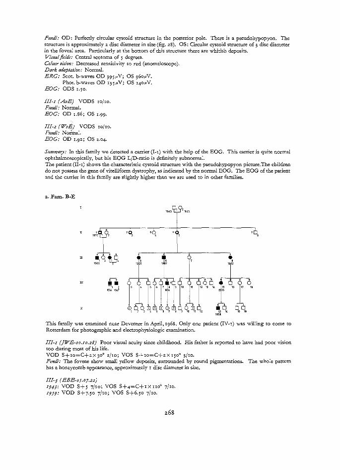
Fumli: OD: Perfectly circular cystoid structure in the posterior pole. There is a pseudohypopyon. The structure is approximately 2 disc diameter in size (fig. z8). OS: Circular cystoid structure of; disc diameter in the foveal area. Particularly at the bottom of this structure there are whitish deposits. Visual field!: Central scotoma of 5 degrees. Colour vision: Decreased sensitivity to red (anomaloscope). Dark adaptation: Normal. ERG: Scot. b-waves OD 395JLV; OS ;6o.uV.
Phot. b-waves OD r;-5.u-V; OS x4o,uV. BOG: ODS x.5o.
III-r (AvE) VODS ro/ro. Fundi: Normal. BOG: OD r.S6; OS t.99·
Ill-2 (WvE) VODS xojxo. Fundi: Noii.nal. BOG: OD x.9z; OS z.o4.
Su111mary: In this family we detected a carrier (I-r) with the help of the EOG. This carrier is quite normal ophthalmoscopically, but his EOG L/D-ratio is definitely subnormal. The patient (II-r) shows the characteristic cystoid structure with the pseudohypopyon picture. The children do not possess the gene of vitelliform dystrophy, as indicated by the normal EOG. The EOG of the patient and the carrier in this family are slightly higher than we are used to in other families.
'":! ,q\9, ; ' .
1905 1~ 1 12 ""' l •• 0 6 -, < SG7S91C 11 121J14 lS 16171%
W:l<\ 1947 . " I
6lfu v 10 ,, 12 1J 14 19SB ' "
This family was e.~ned near Deventer in April, 1968. Only one patient (IV-x) was willing to come to Rotterdam for photographic and electrophysiologic e. .... aminacion.
III-I (]WE-oo.zo.23) Poor visual acuity since childhood. His father is reported to have had poor vision too during most of his life. VOD S+ro=C+2x;oo z/ro; VOS S+ro=C+2X15oo ;/ro. Fundi: The foveae show small yellow deposits, surrounded by round pigmentations. The whole pattern has a honeycomb appearance, approximately r disc diameter in size.
lll-3 ( EBE-o;.o7.22) I94J: VOD S+ 5 7/Io; VOS S+4=C+r X II0° 7/IO. I9f9." VOD S+7-50 7/ro; VOS S+6.5o 7/Io.
z68

Fundi: Round yellowish spots in a circular area of atrophic pigment epithelium. This area is somewhat redder than the surrounding retina.
Ill-; (JHJS-o;.opo) Fundi: OD: Circular area of atrophic pigment epithelium surrounded by yellowish deposits. OS: Yellow deposits deep in the foveal atca.
III-6 (]WS-oJ.OJ.24) Poor visual acuity since the age of ro. Has glasses ODS +S. Fundi: Round area of 1.5 disc diameter in size in the foveal region with pigmentary disruption and yellowish bodies.
Ill-S (HPS-I2.I2.JO) VOD S+6.5o 4/6o; VOS S+6 4j6o. Fundi: A circulat atrophic area of approximately r disc diameter in size in the fovea. This area is surrounded by deep yellowish deposits. The left eye demonstrates also a dense pigment :reaction, resembling the scar of chorioretinitis. ·
IV-r (JHB-J4·'J·JI! I94J' VOD S+5 7/Io; VOS S+5 3/Io. Fundi: A picture in both foveae resembling a central serous choroidophathy. I9J2: VOD s/ro; VOS ;fro. Fundi: Cystoid structure in both foveae with a pscudohypopyon aspect. I 9 ;6: Pscudohypopyon has disappeared. r9o8: VOD S+5 3/xo; vos S+4-50 3/xo. Fixation: With the centre of the vitelliform lesion. Ftmdi: OD: A partly ruptured vitelliform disc (fig. ro). OS: A:n orange-yellow circular area of atrophic pigment epithelium in the fovea. In this area there ate many deposits of a yellowish material. Visualfieldr: Decreased central sensitivity in both eyes. Colour vision: Mild red-green dyschromatopsia (HRR). Decreased sensitivity to red (anomaloscope). Dark adaptation: Normal. ERG: Scot. b-wavcs OD ;o5pV; OS 225pV.
Phot. b-waves OD 1401-N; OS 1201-1V. BOG: OD I-40; OS I-45·
IV-2 (GWB-4J.I2.2J) I9JJ: VODS S+o.5o 8/ro. Ftmdi: Ruptured vitelliform disc with a scrambled-egg appearance. z967: Fundi: Circular area of atrophic pigment epithelium at the site of the fovea. Small round Yellowish deposits, approximately o.r disc diameter in size, are spread diffusely in this area.
IV-3 (JS-J4·'i·'9) Has glasses S+;.5o. Complains of decreased colour vision. Fttndi: In the foveal area a scrambled egg appearance with some pigmentary disruption. As in all other members of this family, the disc, retinal periphery and retinal vessels are perfectly normal.
IV-IJ (HJWP-JJ.OJ.IO) VOD S+z 8/ro; VOS S+z rj6o. Concomitant divergent strabismus of OS. Ftmdi: Delicate drusen-like yellowish bodies in the foveal area.
V-o ( Hf-o4.I2.0J) Fundi: The foveae are normal. The left eye shows a grouped pigmentation in the upper half of the fundus.
V-IJ ( BGW-;S.o7.24) r964: OD has an iris bicolor. VOD S+r 9/ro; VOS S+r 9/ro.
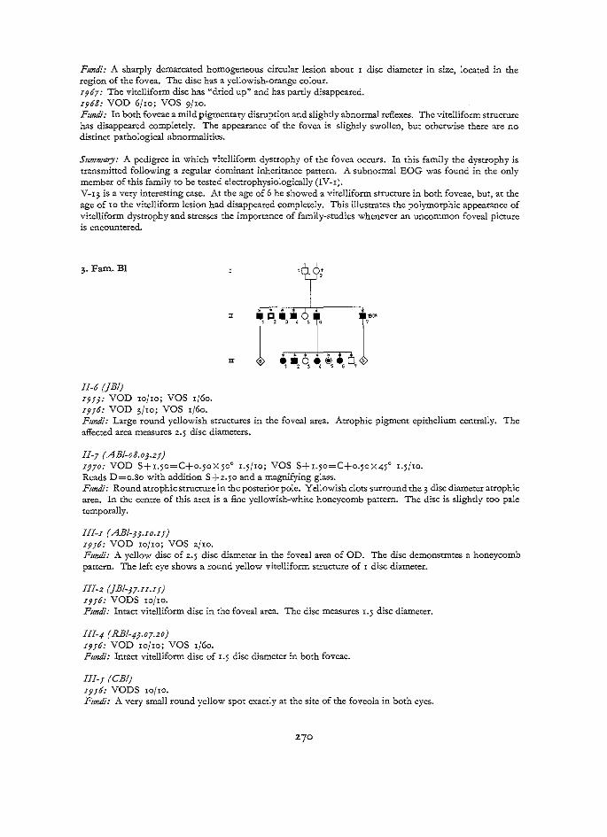
Fundi: A sharply demarcated homogeneous circular lesion about I disc diameter in size, located in the region of the fovea. The disc has a yellowish-orange colour. r967: The vitelliform disc has "dried up" and has partly disappeared. r968' VOD 6/to; VOS 9/Io. Fundi: In both foveae a mild pigmentary disruption and slightly abnormal reflexes. The vitelliform structure has disappeared completely. The appearance of the fovea is slightly swollen, but otherwise there are no distinct pathological abnormalities.
Sumn1ary: A pedigree in which vitelliform dystrophy of the fovea occurs. In this family the dystrophy is transmitted following a regular dominant inheritance pattern. A subnormal EOG '\vas found in the only member of this family to be tested electrophysiologically (IV-I). V-I 3 is a very interesting case. At the age of 6 he showed a vitellifonn structure in both foveae, but, at the age of Io the vitelliform lesion had disappeared completely. This illustrates the polymorphic appearance of vitelliform dystrophy and stresses the importance of family~studies whenever an uncommon foveal picture is encountered.
3· Fam. Bl
Il-6 (fBI) I9JJ: VOD Io/Io; VOS x/6o. I9f6' VOD 5/ro; VOS r/6o.
'0.0' ''(
Fundi: Large round yellowish structures in the foveal area. Atrophic pigment epithelium centrally. The affected area measures 2. 5 disc diameters.
II-7 ( ABI-oS.op;) I9JO: VOD S+x.5o=C+o.JoX 50° I.5/Io; VOS S+x.so=C+o.5oX45o I.5/Io. Reads D=o.So with addition S+2.50 and a magnifying glass. Fundi: Round atrophic structure in the posterior pole. Yellowish clots surround the ; disc diameter atrophic area. In the centre of this area is a fine yellowish-white honeycomb pattern. The disc is slightly too pale temporally.
III-I (ABI-jpO.IJ) I9J6: VOD xojro; VOS 2jro. Fundi: A yellow disc of 2.5 disc diameter in the foveal area of OD. The disc demonstrates a honeycomb pattern. The left eye shows a round yellow vitelliform structure of r disc diameter.
III-2 (}BI-Jpi.IJ) I9J6: VODS rojio. Fundi: Intact vitelliform disc in the foveal area. The disc measures I.5 disc diameter.
III-4 ( RBI-4f·'1·"') 19;6: VOD to/ro; VOS r/6o. Fundi: Intact vitelliform disc of I. 5 disc diameter in both foveae.
III-! (CBI) 1916: VODS tofro. Fmuli: A very small round yellow spot exactly at the site of the foveola in both eyes.

III-6 ( MBI-47.r 2.02)
I9f6: VOD 4{6o; VOS rojxo. Fundi: Classic, perfectly intact vitelliform disc in both eyes. In on :z.J; disc diameter, in OS I disc diameter.
Summary: A family with vitelliform dystrophy of the fovea, The pathological gene demonstrates high penetrancc and high c::-.:pressivity. In this family the inheritance pattern is that of an autosomally regular dominant. II-6 and II-7 show posterior pole lesions, such as they were described by Braley (1966) in a family with "polymorphic macular degeneration". III-:z.,4 and III-6 demonstrate the intact vitelliform lesion. From this observation it is concluded that Braley's polymorphic degeneration and vitelliform dystrophy are identical.
4· Fam. Can (CG-4j.o8.n) I9JO: Navigator of an Italian ship. Is corning from the vicinity of Genoa. Visual complaints about the right eye since 3 months. There is no strabismus. VOD S+I.Z5=C-z.Xx8o0 zjxo; VOS S+z.5o=C-t.XI0° xojxo. Fundi: A z. disc diameter vitelliform disc in the foveal area of OD. The disc is somewhat desintegrated. This is probably the cause of the decreased visual acuity (fig. 42a). The left eye demonstrates a beautiful 3 disc diameter vitelliform disc. It is perfectly intact (fig. 42b). Fluorescein angiograpf?y: OS: There is hardly any fluorescence visible in the foveal area, indicating that there are no large defects in the pigment epithelium. There is no leakage of fluorescein subretinally. This indicates a normal membrane of Bruch (fig. 42c).
Summary: Pathognomonic vitelliform lesions in an Italian navigating officer who visited Rotterdam by ship. The 27-ycar-old man had had visual complaints since 3 months. The opportunity to do fluorescein angiography in an intact vitellifonn disc was gratefully accepted. The results suggest a primary locali7..ation of the vitelliform lesion in the retinal pigment epithelium.
5· Pam. E-W
1B9S 1901 1SOS
i':" 0~ 11 121l 14151G1710 1920~
1950 1~~3 1959
II-I (HCKvtH-93.0J.II) Normal visual acuity until the age of 50. Is capable of reading papers. Refuses to come to the Eye Hospital. Fundi: A 0.5 disc diameter area of atrophic pigment epithelium in the centre of the fovea.
II- J ( MPvtH-o J.02.2f) 1958: VOD S+3 3/xo; VOS S+4.50 4/Io. Fundi: Pigmentary disruption in the foveal area. Vimal fields: Decreased central sensitivity. Colour vision: Normal. Dark adaptation: Normal.

ERG: Scot. b-waves OD zro.uV; OS zo5.uV. Phot. b-waves OD roo.uV; OS uo.uV.
I967: VOD S+j 5/Io; VOS S+pj 4/Io. Fundi: In the centre of the fovea a deposit of pigment, surrounded by a circular area of atrophic pigment epithelium. Visual fields: Decreased sensitivity centrally. Colour vision: Decreased sensitivity to red (anomaloscope). OD tritan axis; OS without characteristic a.xis (Fatnsworth D-15 ). Dark adaptation: Normal. ERG: Unsuccesful. BOG: OD r.o7; OS l.34·
lll-2 ( EK-J7·'4-24) 1967: VODS IX/ro. Fundi: Mild pigment mottling in the posterior pole. The foveal reflexes arc present but not quite normal. BOG: ODS r.oo. 1969: VODS II/ro. Fundi: A small yellowish-white deposit at the site of the foveola of the right eye. The left eye shows no pathological changes.
Ill-J (JB}E-u.o8.2I) 1967: VODS ufro. Fmuli: Normal. BOG: OD 2.3o; OS 2.ro.
Ill-? ( RMKW-24-IO.JO) 19J3: VOD 7/ro; VOS 8/ro. Fwuli: In the centre of the foveal area an atrophic area with fine pigmentary alterations. ViSttal fields: Normal. Colour vision: OD normal; OS Decreased sensitivity to red (anomaloscope). Dark adaptation: Normal. ERG: Normal. 1967: VOD 9/ro; VOS 9/ro. Emmetropic. Fundi: A yellowish cystoid structure of o.5 disc diameter. There is some pigmentary disturbance in this lesion. Visttalfie!ds: Slightly decreased central sensitivity. Normal periphery. Colour vision: Decreased red sensitivity (anomaloscope). Dark adaptation: Normal. ERG: Scot. b-waves OD 25o.uV; OS 195.uV.
Phot. b-waves OD roo,~V; OS 85t"V. BOG: ODS r.oo.
lll-8 ( KBW-26.04.28) 1960 en 1967: Fundi normal. 1969: VOD S+2 7/ro; VOS S+r.5o Sfxo. Fundi: Small round yellowish deposit in the centre of the fovea. Around the yellow foveola there are fine punctiform yellowish lesions. BOG: ODS r.oo.
lll-9 (AW-Jo.o6.jO) Brown hair, blue eyes. Poor visual acuity since early childhood. Nystagmoid movements with horizontal and rotatory components. 19J1: VOD C+r.5oX90° 6jro; VOS C+zX90° 6/w. 19J7: VOD 4/Io; VOS 3/Io. Fundi: No clear abnormalities. The fundi are quite blond, particularly around the disc. The vessels have a normal calibre. The foveae do not have normal reflexes and look hypoplastic. Vittta! fields: Concentric decreased sensitivity. Dark adaptation: The photopic part of the curve is 1 log. U. too high. The scotopic part of the curve is 2 log. U. tOO high.
272

I96S: VOD z.jro; VOS 5/ro. The iris demonstrates some atrophic spots in the infero-nasal parts. Fundi: Unchanged as compared to 1957· Visual fields: Normal peripheral limitations. Decreased sensitivity concentrically and particularly centrally. Colottr vision: Mild red-green dyschromatopsia. Dark adaptation: The curve starts I log. U. too high and ends 2.5 log. U. too high. ERG: Scot. b-waves OD 6r.uV; OS 7op.V.
Phot. b-waves ODS unrecordable. BOG: OD 1.55; OS r.5I. Unreliable because of the nystagmus.
Ill-Io (]W-4I.04.03) Blond hair, blue eyes. I9JO: VOD S-z=C-2X5° 5Jro; VOS S+r=C-5xx65o 5/Io. I9J3: VOD 3/ro; VOS 2jxo. Fundi: Blond fundi. No distinct abnormalities. Disc slightly pale temporally. The foveae have no normal refle."'es and look hypoplastic. Visual fields: Decreased sensitivity concentrically. Dark adaptation: The photopic part of the curve is I log. U. too high. The scotopic part ends 2.5 log. U. too high. ERG: Scot. b-waves 34p.V; OS 521-N.
Phot. b-waves unrecordable. z967: A delicate nystagmus latens. Slightly diaphanous irides. VOD 5/ro; VOS 2/10. Fttndi: The foveal reflexes arc irregular and broader than normal. Vimal fields: Constriction of the isopters. Colo1tr vision: Decreased sensitivity to red(anomaloscope). Dark adaptation: Phot. and scot. systems are disturbed. The curve ends 2.5-3log U. too high. ERG: Scot. b-waves OD 6ot~V; OS 45t~V.
Phot. b-waves OD 45tN; OS unrecordable. BOG: OD 1.40; OS x.69. Fixation unreliable.
IV-2 ( RBAE-;;.o6.o6) I96IC VOD 5/5; vos 5/'5· Fundi: In OD, a classic yellow vitelliform disc is visible (fig. 25a). In OS there is a red and yellowish coloured area of I disc diameter at the site of the fovea. There is no protrusion. Visual fields: Normal. Colottr vision: Normal. Dark adaptation: Normal. ERG: Scot. b-waves OD 305p.V; OS .z851~V.
Phot. b-waves OD I2011V; OS 125,uV. Systematic exantination: Normal. I967: VOD S-1.25 rojw; VOS S-1.25 rojro. Fundi: Sharply defined circular structure at the fovea, There is no distinct protrusion. There are in this circular area yellow deposits, surrounded by an atrophic pigment epithelium. This atrophic pigment epithelium is redder than the surrounding normal retina. The yellow deposits protrude slightly from the atrophic pigment epithelium (fig. z 5 b). Vimal fie!tls: Slightly decreased central sensitivity. Colour vision: Decreased sensitivity to red (anomaloscope). ERG: Scot. b-waves OD 2651-1V; OS z6op.V.
Phot. b·waves OD I2op.V; OS II5.uV. OP: Normal. BOG: OD 1.22; OS r.zx. r96S: VOD S-1.50 8/ro; VOS S-1.50 9/ro.
IV-IJ ( RK-48.08.2;) I9J6: VODS 5/20. I9J9: VOD S-x.5o=C-I.50XI80° 3/Io; VOS S-0.5o=C-o.50XI8o0 3/ro. I962: VODS 4/IO.

Ftmdi: The foveal reflexes are irregular. The retina around the disc looks thin and very blond. The disc, vessels and retinal periphery show no distinct abnormalities. Visual fields: Decreased sensitivity centrally and concentrically. Normal peripheral limitations. Colour vision: Slightly decreased sensitivity to red (anomaloscope). Dark adaptation: The curve is 1 log. U. too high for both systems. ERG: Scotopic b-waves: rudimentary responses.
Photopic b~waves: unrecordablc. BOG: ODS 2.oo. r968: VODS 4/Io. No diaphanous irides. No metamorphopsia. Retinal Jtmctions: Unchanged as compared to 1962. 0 P: Subnormal. F-ERG and VER: Subnormal. The subnormal F-ERG indicates a foveal hypoplasia and at least a foveal dysfunction. EQG, ODS 1.75.
IV~r6 (EK-;3.o2.0J) Blond hair. Blue eyes. I 9 f 3: Photophobia since birth. Also nystagmoid movements with horizontal and rotatory components. r964: VOD S+1=C+o.5o X 180° 5j6o; VOS S+x.~o 5{6o. Ftmdi: Irregular foveal reflexes. There is some macular yellow present. The disc is slightly pale temporally. The vessels and periphery are normal. The peripapillary area is very blond. r967: VODS 5{6o. The irides are slightly diaphanous in the periphery. Fttndi: No changes as compared to 1964. Visual fields: Normal periphery. Decreased sensitivity centrally and concentrically. Colottr vision: Mild red-green dyschromatopsia (HRR). Dark adaptation: The curve ends 2log U. too high. Both systems are disturbed. ERG: Scot. b-waves OD 95.uV; OS 1oo.uV.
Phot. b-wavcs ODS uruecordable. BOG: Unreliable because of nystagmus.
Summary: An interesting pedigree, in which 2 hereditary retinal diseases occur: a. vitelliform dystrophy of the fovea and b. X-linked hemeralopia. The following individuals are affected by vitelliform dystrophy: II-1,5; lli-7,8 and IV-2. Affected by X-linked hemeralopia arc lli-9,10 and IV-IJ.I6. IV-2 demonstrates a fundus picture pathognomonic for vitelliform dystrophy. His mother (III-2) had normal foveae in 1967, however her EOG was definitely subnormal. As her husband (ID-;) had a normal EOG, III-2 was detected as a carrier. In 1969 she showed a mild ophthalmoscopic abnormality in the right eye, indicating that the EOG is a valuable detector for carriers of the gene causing vitelliform dystrophy. II-5 and lli-7 arc in all probability having 2 pathological genes. The only ophthalmoscopic abnormalities visible are mild vitelliform changes in the foveal area. X-linked hemeralopia is not giving any detectable abnormality in the carriers, as we saw in faro. SKn and Ver. It is interesting to note that the children of II-5 and ID-7 only have X-linked hemeralopia and no vitelliform dystrophy.
6. Faro. Fl Il-2 (CFvD-82.07.20) Fundi: Circular areas of atrophic pigment epithelium in the fovea. Some pigmentary disruption. The areas are approximately 2 disc diameter in size.
III-I (MBF-IJ.Of.JO) I9ff: VODS S+x ;/to. Diagnosis: "retinitis centralis serosa". r962: VOD S+t.JO ;/xo; VOS S+x.~o 2/xo. r966: VODS I-2/Io. r968: Ftmdi: The fovea has a scrambled egg appearance. Yellow deposits in a circular area of atrophic pigment epithelium.
III-; (GF-r7.o8.o6) I963: VOD S+2 4/to; vas S+2 5/6o. Reads D=O.JO with S+J.JO.

Fundi: OD: A circular cystoid structure of 1.5 disc diameter in the foveal area. There is a small pseudohypopyon and yellowish-white deposits are also found (:fig. 27). OS: A flat cystoid structure at the site of the fovea, approximately I disc diameter in size. There are severe pigmentations in the shape of a sickle at the bottom (fig . .29). BOG: ODS I.zj.
III-; (WvBF-24.0f.Of) 1964: VOD 2jro, emmetropic; VOS S-o.5o 5/Io. Diagnosis: "Familial cystoid macular degeneration". 1968: Flt!Zdi: Vitelliform structure in the scrambled egg stage. The foveal and foveolar reflexes are normal in spite of the atrophic pigment epithelium at the site of the fovea.
Ill-6 (]F-26.oz.o8) 1968: Army sergeant. Complains of impairment of visual acuity and colour vision. The visual complaints started 1.2 years ago with metamorphopsia and decreased visual acuity. VODS: S-o.5o=C+o.50X9o0 .zjro; Reads D=o.S with addition. There is metamorphopsia (Amsler test). Fundi: A vertical oval of yellow deposits in a honeycomb pattern, approximately 3 disc diameter in size (fig. 2). The right disc shows a pit, while there is a pigmented spot at the bottom of the retinal periphery. Visttal fields: Central scotoma. Co/om· vision: Mild red-green dyschromatopsia (HRR). Decreased sensitivity to red (anomaloscope). BOG: ODS 1..25.
IV-I (KB-J6.oi.2J) Has been in the military service. 1967: VOD S+r=C-o.75XI30o 5/ro; VOS S+r=C-o.75X9o0 5/ro. Flt!Zdi: Yellow deposits in a circular area at the site of the fovea.
IV-2 (GB-42.04.18) Has some problems in distinguishing colours, particularly red. 1967: Fundi: OD shows a cystoid structure of I disc diameter in the foveal area. There is some pseudohypopyon. OS demonstrates a somewhat larger cystoid structure. At the bottom there is a small pigmentation, resembling the scar of choroiditis.
IV-3 (KB-46.oJ.II) Truck driver. Not present, when the family was examined.
IV-4 (HB-;po.24) 1967: VOD S+.z 6/ro; VOS S+.z=C-r X 120° 4/ro. F1mdi: The fovea shows a symmetrical intact vitellifonn disc of 1 disc diameter.
IV-J (GF-J8.02.IJ) Hasaliccncefordrivingacar. 1967: Flt!Zdi: Circular areas of atrophic pigment epithelium in the centre of the posterior pole. There are some yellow deposits and pigmentations in these areas.
IV-IJ ( HvB-48.1o.r8) 1964: VOD S+o.25 3/ro; VOS S+o . .25 9/Io. 1968: No visual complaints. Fundi: OD: A circular cystoid structure in the foveal area, approximately 1.5 disc diameter in size. Yellow deposits and red atrophic areas arc visible. OS: A circular cystoid structure in the foveal area, 2/3 disc diameter in size.
IV-IJ ( AF-J9.0J.2I) 1968: VODS S+o.50 ujro. No metamorphopsia. Fundi: OD: Small yellowish cyst on the temporal side of the foveola (fig. 17). The disc has an "optic pit". OS: Fine yellowish spots on the temporal side of the foveola (fig. 18). The retinal periphery of both eyes shows a rather coarse granular pigmentation. Visual fields: Normal. Colottr vision: Normal. Dark adaptation: Normal. BOG: OD L42; OS L4l· 1970: VODS ujro. The fundi are quite unchanged.

lV-r6 (GF-6r.or.o4) r96S: VOD S+o.5o; VOS S+o.5o xofro. F~~t~di: Perfectly intact vitclliform disc, I disc diameter in size. Some :fine whitish punctiform structures are visible in the disc. There is no prominence of the vitelliform structure. Colour vision: Normal. EOG and photography do not succeed because of crying. 1970: VODS 8/xo, emmetropic. Fundi: The vitelliform structures are slightly disintegrated (ruptured) (fig. ;).
Sltllllllary: A pedigree, in which a regular dominant inheritance pattern is present. The penetrance of the vitelliform dystrophy is high. The foveae of III-6 closely resemble the lesions described and photographed by Braley (r966). Braley suggested the term "polymorphic macular degeneration" for this picture and he doubted the relationship of vitelliform dystrophy and "polymorphic macular degeneration". In our opinion there is no such entity as "polymorphic macular degeneration". The son ofiii-6 (IV-r6) had perfectly intact vitelliform discs in 1968 and slightly desintegrated lesions in 1970 (fig. ;). The polymorphic pattern of vitelliform dystrophy is beautifully demonstrated when we compare the fovea-pictures of III-6 and IV-x6.
7.Fam.KIK
' ' ll-2 ( HCKK-or.or.o6) Strong hypermetropia. This could indicate the carrier state. Spectacles: OD S+4=C+zxro0
; OS S+5=zxx8o0•
F:mdi: Normal foveae. Some small haemorrhages perifovcally.
ll-4 (JKKT-99·'4-I!) Fmuli: Normal foveae.
lll-4 (WGMKK-j2.0l.l r) Complains of photophobia. Likes best to be in dusk. I9j8: VOD S+4-50=C+r.5oX95° rjro; VOS S+5=C+r.5oX95° 5/xo. 19;9: Impairment of visual acuity.

Fundi: Vesicle-like yellow structure in both foveae. 1948: VOD zjro; VOS ;fro. F11ndi: Yellowish deposits in cystoid structures. r90o: VOD r/ro; VOS zjxo. 1967: VOD s/6o; vos I/IO. Concomitant divergent strabismus. Eccentric fi-xation. Ftmdi: Circular structures in the fovea, approxima.tely z-; disc diameters in size. Disintegrated vitelliform disc, "scrambled egg appearance". Yell ow deposits surround the central area of atrophic pigment epithelium (fig. 6). Vi.rwlfte/ds: Decreased central sensitivity. Colour vision: Red-green and blue~ yellow dyschromatopsia (HRR). Decreased red-sensitivity (anomaloscope). Tritan axis in OS (Farnsworth D-15). Dark adaptation: Normal. ERG: Scot. OD 29o.uV; OS ;7o1~V. Phot. b-waves OD roo.uV; OS r2o,~v. BOG: ODS 1.20.
Ill~; ( MKK-JJ.OJ.23) Poor visual acuity since the age of ro. 1947: VOD S+2 2jxo; VOS S+z ;/ro. Fundi: Picture resembling disciform foveal degeneration. rgO;: VOD S+4 z.fro; VOS S+4·5o z.jro. Fl!ndi: Circular area of atrophic pigment epithelium, which measures approximately; disc diameters in the right posterior pole. This area is surrounded by a garland of yellow deposits (fig. 4). The left fovea shows a ruptured vitelliform cyst, approximately I disc diameter in size. In the centre of this cyst there are strong pigmentations. Vi.rtta! fteld.r: Decreased central sensitivity. Colour vision: Red-green and blue-yellow dyschromatopsia (HRR). Decreased sensitivity to red (anornaloscope). Tritan axis in ODS (Farnworth D-15). Dark adaptation: The curve is slightly too high. No significant pathology. ERG: Scot. b-waves OD ;70ttV; OS ;45.uV; Phot. b-waves OD t651.N; OS I55p.V. BOG: OD r.xS; OS r.r5.
Ill-7 (AWMKK-37·'4·'!) 194;: Fundi: Vitelliform disc in the foveal area. 19f2: VOD 4/Io; VOS 2j6o. Ftmdi: Cystoid structure in the right fovea. The horizontal diameter measures 2 disc diameters, while the vertical diameter measures ; disc diameters. There is a large pseudohypopyon with pseudo-Descemetdeposits (fig. 5a). The left fovea shows a circular area of atrophic pigment epithelium with pigmentations and yellowish deposits. 19b7: VOD S+x.5o ;/xo; VOS S+t.50 z/to. Fundi: OD: Yellow deposits at the bottom of a vertical oval structure. The cystoid structure is disinte~ grated. Severe pigmentations at the site of the former pseudohypopyon (fig. 5b). OS: Disintegrated vitelliform lesion. At the bottom yellow deposits and pigmentary disruption. Vimal fields: Decreased central sensitivity. Colour vi.rion: Mild red-green dyschromatopsia (HRR). Decreased red sensitivity. Dark adaptation: Normal. ERG: Scot. b-waves OD 222p.V; OS 240ttV, Phot. b~waves OD I55ttV; OS 19op.V. BOG: ODS x.oo.
Stti!Jf!Jary: One ma.n and his two sisters have suffered from vitelliform dystrophy sine early childhood. The evolution of the vitelliform disc has a rapid course in this family. The visual acuities are severely disturbed at a relatively young age, in contrast to what is usually described in literature. It was not possible to perform electro-oculography in the parents (II-2,4). The father might be the carrier of the pathological gene since he has a high hypermetropia. as have his 3 affected children. This family was reported by Pameyer in 1952 at a meeting of the Netherlands Ophthalmological Society (Pameyer, 1954). Pameyer reported on the different stages of development of the vitelliform disc. He distinguished the following stages: I. ma.cular vesicle without contents. 2. appearance of liquid contents with fluid level "hypopyon". ;. appearance of "precipitates" against the posterior pole of the retina. 4. picture of the disciform degeneration. 5. total degeneration of the affected region.

The stages in which we e.xa.mined the patients resemble the pictures found in "polymorphic macular degeneration", described by Braley (1966). The development of the foveal picture in this family indicates that vitelliform dystrophy and polymorphic macular degeneration are identical.
8. Fam. Ko~K
17Sl 1756
' '
m' 1794 ,
18ZZ 19Z5
' '
'u' 1UG5 "" ' ' '
ton'
1910
'
o• 2 J '---s -u 7 o
1S59 1945
VIII-2 (Jd}K-2I.OJ.o6) zg63: VOD S+1.50 6(10; VOS S+z 7(1o.
109l
'
1?68 1772
' 1799 1901
' '
'"' 1952
' 106Q '"' '
,
' ifJ ' '
~' "l R252 ,, 15 1G 17 'til 19 20 2'1 22
19GJ
Ftmdi: In the foveae a very small honeycomb pattern of yellowish-white deposits. Around these deposits some fine pimcntations (fig. 15). Colour vision: Normal. EOG: OD 1.15; OS 1.50.
Vlll-J ( GKK-24.02.02) zg63: VOD S-1=C+I.5oX5oo 4/xo; VOS S-x.z5=C+1.75X155° ujxo. Fundi: In the right fovea a circular vitelliform disc, approximately 0.5 disc diameter in size. This structure is much better visible on panchromatic than on orthochromatic film. In the left eye a small horizontally ovoid defect in the pigment epithelium. There are whitish deposits in this atrophic area (fig. 19). BOG: OD_ I.zo; OS x.zz.
IX-6 (Td}-!9·'1·'8) zg63: VODS Sj1o, cmmetropic. Ftmdi: A classic vitelliform disc, approximately two-thirds of a disc diameter in size in both foveae. The disc has a yellowish-orange colour and is surrounded by a brown-red circle (fig. x). The disc and vessels arc normal. The retinal periphery shows a fine granular pigmentation. Colour vision: Decreased sensitivity to red (anomaloscopc). BOG: OD I.zS; OS 1.51.
IX-3 ( AK-4J.07.04) This boy is the proband of the family. rg6I: Hospitalized. Impairment of visual acuity after furunculosis. VOD C-o.5ox x8o0 7/10; VOS S-0.75 4/Io.

Fundi: The right fovea shows a yellowish-white "scar of a choroiditis" surrounded by pigmentations. The left eye demonstrates a cystoid structure with in the centre a small haemorrhage. Diagnosis: Possibly toxoplasmosis. Therapy: Prednisone, achromycin, vitamine B comple."=. Systemic examination: Normal. 1962: Ten months after hospitalization the visual acuity of both eyes is xojro. 1968: VOD S+o.5o 5/ro; VOS ufro, emmetropic. Fundi: A circular yellowish-white structure approximately r disc diameter in size with severe pigmentations at the bottom in the right fovea (fig. 26). The left fovea shows a cystoid structure with a pscudohypopyon at the bottom. Visual fields: Central scotoma in the right eye. Normal in the left eye. Dark adaptation: Norrnal. ERG: Scot. b-waves OD ;s.z.o,.V; OS 28o,~v.
Phot. b-waves OD 8o,.V; OS ror,.V. BOG: OD r.24; OS '·'7· F-ERG and VER: OD subnormal.
IX-9 ( AK-40.II.IO) 1968: VOD ro(10; VOS 6j10; emmctropic. Fundi: Delicate pigmentations and whitish flecks in the foveal area. The abnormal area is approximately 0.25 disc diameter in size. The lesions are better discernible on panchromatic than on orthochromatic film.
IX-12 ( AK-J6.o4.18) 1968: VOD C-o. sox 180° roj1o; VOS S-o.so=C-o.sox 180° rojro. Fundi: A perfectly circular vitelliform disc, approximately two-thirds of a disc diameter in size in both foveae. The lesions are strikingly symmetrical. The panchromatic films demonstrate more abnormalities than do the orthochromatic films (fig. ;8).
IX-IJ (TK-;8.oo.I4) 1968: VODS C-o.5ox r8o0 roj1o. Fundi: Vitelliform disc in the foveal area. The disc measures one disc diameter.
IX-z; (GK-o;.oJ.2z) Blind since early childhood and probably since birth. There is ologophrenia and generalized epilepsy. The pupillary reactions arc absent and the discs are pale. No electrophysiological examinations have been performed.
An EOG was made of IX-5 (Ad]-55·05.rz) and IX-II (TK-52.04.25) in order tO establish whether they were carriel'S or not. Both individuals had a normal EOG. The former OD 1.98; OS 2.15 and the latter OD .z.2o; OS z.ro.
Summary: A pedigree in which vitelliform dystrophy is inherited in a regular dominant manner. Genealogical examination revealed a striking number of consanguineous marriages in this pedigree. The proband is in 5 ways related to the couple who married in 1749, ; times on the side of the father and 2 times on the side of the mother. This pedigree demonstrates the c::-.:treme degree in which in-breeding may be present in some parts of The Netherlands. Since vitelliform dystrophy is inherited in a dominant manner the consanguineousness is not so important as in recessively inherited diseases. The genealogical e.-mmination was done in order to establish whether there were any relationships with other pedigrees in which vitelliform dystrophy occurs.
9· Fam. K~Z l-z ( PZ-89.o6.o6) VOD S+r.5o rojro; VOS S+.z rojro. Fundi: Normal foveae. EOG: OD 2.57; OS 2.9o.
2 79

l-2 (JAZS-92.ov;) VOD S+6.5o=C+o.75 X x8o0 rojro; VOS S+5·75=C+r X r8o0 4/Io. Fundi: Small yellowish deposits in the centre of the foveal area. BOG: ODS x.oo.
ll-7 ( MKZ-29.02.23) Visual complaints since the age ofS. Further decrease in visual acuity at the age of ;c. Particularly visual impairment after deliveries. The family-history was negative and the definite diagnosis could only be made with the help of the EOG. I962: von S+4-50=C+:r X r80° 4/ro; VOS S+4·50=C+r-50X r8o0 rjro. There is concomitant divergent strabismus of OS. Ft~ndi: The foveal areas show a disciform pathological process. The disc and vessels are normal. The retinal periphery demonstrates a fine granular pigmentation. Visual fields: OD: central sensitivity decreased. OS: central scotoma. Colottr vision: Red-green dyschromatopsia (HRR). Dark adaptation: Normal. ERG: Scot. b-waves OD ;o5.uV; OS 275.uV; Phot. b-waves OD 9o.uV; OS 12o.uV. BOG: OD ,i.1o; OS r.16. Systemic exa111ination: Normal. 196J: No important changes as compared to 1962. Dark adaptation: Normal. ERG: Scot. b-waves OD 39o.uV; OS 465.uV.
Phot. b-waves OD 65.uV; OS 7o.uV. BOG: OD r.x;; OS 1.14. 1964: VOD ;/1o; VOS zf1o. Colottr vision: Tritan axis (Farnsworth D-15). 1961: Dark adaptation: Normal. ERG: Scot. b-waves OD 25o.uV; OS 210ttV. Phot. b-waves OD 65.uV; OS ?ot-N. BOG: ODS 1.oo. 1967: VOD S+5.5o=C+x X xSoc z/xo; VOS S+5·5o=C+t-50X 180° 2j1o. Better subjective vision with dark glasses. F11ndi: OD: Circular area of atrophic pigment epithelium, approximately 0.5 disc diameter in size. Yellow deposits are present in this area, which is situated at the site of the fovea. OS: Ra.thcr ill-defined circular area of atrophic pigment epithelium in the fovea. This area measures approximately 1 disc diameter. Between the disc and the foveal lesion and slightly upward is an intact vitelliform disc, which measures one disc diameter (fig. zoa). Visual fields, dark adaptation, ERG and BOG are unchanged, as compared to 1965. Colour vi.rion: Red-green dyschromatopsia (HRR). Decreased red sensitivity (anomaloscope). Fbtorescein angiograpl{y: Slightly pathological fluorescein pattern indicating defects in the retinal pigment epithelium at the fovea and at the site of the e>..'tra-foveal vitelliform lesion (fig. zob). 1969: Vi.rual amity: Unchanged. Fundi: The second vitellifonn lesion in the left eye, which was localized extra-foveally, has disappeared without leaving any trace. The foveal lesions in both eyes are unchanged. ERG: Scot. b-waves OD 22011V; OS z;SttV.
Phot. a-waves OD 51pV; OS 5ottV,
Phot. b-waves OD 1.2St~V; OS 142r .. .V. F-ERG, Normal. VER: Normal.
Ill-; (KK-4S.o6.r4) VODS S+o.5o 1xjxo. Ftmdi: Normal foveae. BOG, OD z.z6; OS z.J6.
Ill-6 (JK-;o.o9.o9) VODS 11/10. Fundi: Normal. The foveae show a delicate granular pigmentation. BOG: OD 1.xo; OS x.oo.
zSo

lll-7 (MK-JJ.o2.o8} VODS II/IO. Fundi: Normal. BOG: OD z..oo; OS I-93·
Ill-S ( AK-6z.o8.IJ} VODS IO/IO.
BOG: Unreliable because of bad fi"<ation.
M, I ,un
Sunlmttry: A pedigree in which vitelliform dystrophy occurs. There is only one individual who is ophthalmoscopically clearly affected. The EOG renders the diagnosis. I-2, who has slight ophthalmoscopic alterations and III-6, who has ophthalmoscopically normal foveae, we:rc detected as carriers with the help of the EOG. Of great interest is the finding of a second, exttafoveally localized, vitelliform lesion in the left eye of II-7. This lesion disappeared without leaving any visible trace.
xo. Pam. Me
'o,
1Hl1~17
J: ~ qq i, ' l~SO 1U51
IH) IN)
This pedigree has been examined at home. It is well known in this family, that poor visual acuity occurs in every generation. The inheritance has a regular dominant pattern. All affected individuals show disintegrated vitelliform lesions with a circular area of atrophic pigment epithelium as the predominant sign. Pigmentations occur in a lesser degree, while yellowish deposits are present in all c..~ined affected individuals (II-x; III-x; IV-;,4,6,7). The most e>..Lensive dystrophic changes were found in the elder patients.
n. Fam.Pl Jll-2 (LP-JO.IO.I4} I9f9." Diminishing visual acuity. Hospitalized. VOD S+5=C+o.5oX95o 4j6o; VOS S+4=C+x x9oo z/Io. After 2 »'eeks: VOD zfxo; VOS ;fro. Fundi: Intact vitelliform disc in the foveal area. Visi((J/ fteld.r: Decreased central sensitivity.
z8r

Colour vision: Decreased red-sensitivity (anomaloscope). Dark adaptation: Normal. ERG: Scot. b-waves OD 255ttV; OS 525.uV. Phot. b-waves OD 9o.uV; OS 95ttV. Systemic examination: Normal. Therapy: Prednisone. I96J: VODS ;/Io. Fundi: A haemorrhage has occurred in the vitellifonn cyst of the left eye (fig. I I). Dark adaptation: Normal. ERG: Scot. b-waves OD ~oo.uV; OS 2o5.uV.
Phot. b-waves OD 751-N; OS 9o.uV. EQG, ODS r.4o. After I month the haemorrhage disappeared. VOS 6/Io. I967: VOD S+4·5o=C=IX9o" zjio; VOS S+~.5o=C+Ix9o" 9/Io. Fundi: Circular areas of atrophic pigment epithelium in the foveal area. These areas arc redder than the surrounding apparently normal retina. Some pigmentations and yellowish deposits are spread in the atrophic areas.
m
SUHtmary: Vitelliform dystrophy in a 9-ycar-old boy. At the age of I~ he developed a haemorrhage in one of the vitelliform lesions. This haemorrhage disappeared in I month time and visual acuity returned from ~/Io tO 9/IO. The parents being ophthalmoscopically normal, refused clectro-oculographic examination.
I2.Fam. T-T
11-6 (LvdT-OJ.02.IO} Family history negative. VODS S+~ IO/Io. Ftmdi: Normal foveae. Perifoveally in OD some drusen-like yellowish deposits. EQG, ODS I.ZO.
ll-7 (LvafH-o8.r2.27) VOD S-; 8/Io; VOS S-o.5o=C-o.5oX90° IO/Io. Fundi: Normal foveae. The retinal periphery shows large areas of chorio-retinal atrophy with many socalled "dyshorische Herdchen". Visual fields: Normal peripheries. Concentrically slightly decreased sensitivity. EQG, OD I-9Z; OS '·93·

III-; ( ACTvtlT-41.0J.16) Concomitant divergent strabismus of OD. VOD S+3.50 o.5/6o; VOS S+3.2.5=C+o.5oXr8oo rofro. Fundi: Fine granular pigmentations in both foveae. In the foveal area of OS there are also some yellowish flecks. Vitttal fields: Central scotoma OD. Normal in OS. Colour vision: OD impossible to record. OS normal. Dark adaptation: Normal. ERG: Scot. b-waves OD 235r.N; OS 245r~V.
Phot. b-waves OD ros.uV; OS II5t~V. BOG: OD r.oo; OS '·37·
1V-IJ (GAT-6I.opo) 1966: Decreased visual acuity. Slight strabismus of OD. VOD S+4·50 5/I5; VOS S+; 5/5· Fundi: Normal foveae. 1968: VOD S+3·5o 8/ro; VOS S+z 9/ro. F"ndi: Intact vitelliform disc at the site of the fovea. These discs measure approximately I disc diameter. The upper half of the lesions is reddish, while the lower half is yellowish (fig. 8). Colour vision: Normal. BOG: ODS r.2o.
Su711111ary: A pedigree in which vitelliform dystrophy occurs with increasing expression in the younger generations. TI-6 has normal foveae ophthalmoscopically, but his EOG is definitely subnormal. Consequently he is a carrier. ID-5 has slight foveal alterations and a subnormal EOG. IV-15 has the dassic vitelliform lesions and also a subnormal EOG. The penetrance of the gene of vitelliform dystrophy seems to be low in this family. We could not examine the whole family because many family members had emigrated.
I3· Fam.. U~T
'
m
' '
11-6 (LT-94-II.I6)
• ' ' ' ' '
1968: VOD S+z.5o rfro; VOS S+z.5o 3/ro.
1050
'
15 t7 1B~2c2i 22 194ij 1$51
t 100l t •
Fundi: Sharply defined horizontal oval of atrophic pigment epithelium and choroid, in which glistening reflexes are present, in the foveal area of OD (fig. 343-). Sharply defined circular zone of chorioretinal atrophy in the foveal area of OS (fig. 34b). The peripheries of both eyes show a fine granular pigmentation. BOG: ODS 1.30.
Il1-8 (PT-IJ.04.0J) 1963: Circular areas of atrophic pigment epithelium in both foveal areas. Pigmentations and yellowish deposits are present in these areas.

Ill-9 (}T-r8.or.o7) :1962: VOD z/ro; VOS 5/Io. Emmetropic. Diagnosi.;: "Central chorioretinitis caused by tuberculosis". rg66: VOD r/ro; VOS 5/ro. Ftmdi: Circular areas of chorioretinal atrophy in the foveal areas.
111-II ( EUT-2J.OJ.08) Visual complaints for the first time at the age of ro. rg62: VOD S+.z z/ro; VOS S+; rjro. rg63: VOD S+4 .zjxo; VOS S+;.5o r/ro. Fundi: Circular area of atrophic retinal pigment epithelium, approximately 3 disc diameters in size. "Scrambled egg" appearance. Visual fields: Central scotoma of 8 degrees. Colour vision: Mild red-green dyschromatopsia (HRR). Decreased red sensitivity (anomaloscope). OS pro tan axis (Farnsworth D-r 5 ). Dark adaptation: Normal. ERG: ScOt. b-waves OD ;.zo.uV; OS ;78t.J.V. Phot. b-waves OD S9.uV; OS r;5.uV. BOG: ODS I.oo.
III-I 2 ( ERT-;o.o J-24) First visual complaints at the age of 6. Was hospitalized at that time. r968: VOD S+9 2/10; VOS S+ro.5o 5/ro. Fundi: OD: Cystoid structure approximately 1.5 disc diameter in size in the foveal area. There is a pseudohypopyon and there arc pseudo-Descemet precipitates (fig. 35a). OS: A structure resembling the foveal lesion of OD. In this case the cystoid structure measures approximately 2.5 disc diameters (fig. 35b). The retinal peripheries show a :fine granular pigmentation. Visual fields: Decreased central sensitivity. Colour vision: Mild red-green dyschromatopsia (HRR). Decreased red sensitivity (anomaloscope). BOG: ODS r.oo.
lll-20 (OKIBSH-J4.IO.JO) Reports, that her mother has had bad visual acuity from childhood. Three of her sisters are reported to have poor vision, too. Her parents and brothers and sisters had emigrated to Australia. OD has poor vision since many years. r969: VOD 6j6o; VOS II/ro. Emmctropic. F1mdi: OD: Circular foveal structure of I disc diameter, Severe pigmentations suggest a scar of chorioretinitis. On the upper temporal side of the disc a naevus pigmentosus (fig. 32). OS: Circular area of atrophic pigment epithelium, I disc diameter in size. Yellowish deposits are found at the bottom of this structure. BOG: ODS x.oo.
IV-r4 (SET-4'6.o7.o8) No visual complaints. r968: "Scrambled egg appearance" in both foveae. A circular ruptured vitclliform lesion, approximately I disc diameter in size.
lV-z9 ( BLU-48.oz.oC) r962: VOD zfxo; VOS IO/Io emmetropic. "Scar of toxoplasmosis-chorioretinitis OD". r968: VOD I/ro; VOS IO/Io cmmetropic. Fundi: OD: Vitelliform disc in the right fovea, Approximately I disc diameter in size. Fine radial folds surround this lesion. OS: Normal. The fovea is perfectly normal. The retinal peripheries are showing a :fine granular pigmentation and some areas with pigmentations and depigmentations (fig. 24). Co!otrr vision: Mild red-green dyschromatopsia (HRR) (
Decreased red sensitivity (anomaloscope) \ OD Tritan a.xis (Farnsworth D-15) OS: Normal
BOG: ODS I.3o.
IV-20 ( L]U-J r.oO.oO) Poor visual acuity since I964. The vision of OD decreased during the last months. I966: VOD xjxo; VOS 2j6o emmetropic.

Fundi: Vitelliform dystrophy in both foveae. The retinal periphery shows in the inferior temporal quadrant a disinsertion from 4·6 hours. The disinsertion was treated operatively with good result. r968: VODS rfxo, emmetropic. Fundi: Vitelliform disc in the right fovea (fig. 21). The left fovea shows a circular area of atrophic pigment epithelium. Yellowish deposits are visible in this area. The surroundings of the atrophic foveal area have a swollen aspect. The vitelliform disc, present before the operation, has disappeared. The retinal periphery of the left eye shows pigmentations in the infcro·temporal quadrant. Vitua! jieldt: Central scotoma. Restriction of the periphery supranasally in the left eye. Colour viSion: Mild red-green dyschromatopsia (HRR). Decreased red sensitivity (anomaloscope). Tritan a."<is (Farnsworth D-15). Dark adaptation: Normal. ERG: Scot. b-waves OD nor-tV; OS 38o.uV.; Phot. b-waves OD 145.uV; OS no.uV. BOG: OD x.26; OS x.3o.
Summary: A rather large pedigree with vitelliform dystrophy, with a regular dominant inheritance pattern. The following points arc of interest: I. We had the opportunity to make photographs of the vitelliform lesion in a 74-year.old man (II·6) (fig. 34). 2. One individual had a unilateral vitelliform lesion. The right eye was affected and had a visUal acuity of
rfxo. The left eye had ophthalmoscopically normal foveae and had a visual acuity of IOjxo. This is a good case to compare the acquired dyschromatopsia of the right eye with the (normal) colour vision of the ophthalmoscopically unaffected left eye (IV-19). The EOG is definitely subnormal in both eyes, indicating that the EOG is completely independent of the ophthalmoscopic aspect.
3· IV-20 spontaneously developed an anterior dialysis in the infero-temporal quadrant of his left eye at the age of 15.
q. Fam. Vr
)[
Ill
V-FAMILY.
0 0 PERSONALLY EXAMINED. NORMAL FOVEA AT FUNDUSCOPY .
• • PERSONALLY EXAMINED. VITElLIFORM DYSTROPHY.
® liJ SLIGHT FOVEAL ALTERATIONS.
ll UNILATERAL VITELLIFORM DYSTROPHY.
QQ NORMAL EOG
Q Q PATHOLOGICAL E 0 G .
.!. ~ PATHOLOGICAL E 0 G
z8j

III-4 (GV-84) Decreased visual acuity in 1933· 1jf9: Seventy-five years of age. VODS S+4=x/xo. Fundi: Atrophic areas in the posterior pole. This man died some years ago.
Ill-S (AV-91.02.17) Visited Prof. van der Hoeve in Leiden in 1927 because of visual complaints. His daughter had the same foveal affection at that time. Nothing is known about the visual functions of the parents. 1968: VOD S+1=C+x.50XI00° 1j1o; VOS S+o.5o=C+x.5oX85° 1.5j1o. Media: Incipient senile cataract. Fundi: A circular area of chorioretinal atrophy at the site of the fovea. This area measures approximately 1.5-2 disc diameters in size. Visual fields: Central scotoma. Normal periphery. Colour vision: Mild red-green dyschromatopsia (HRR). Decreased sensitivity to red (anomaloscope). Dark adaptation: Normal. ERG: Scot. b-waves OD uop.V; OS 135ttV.
Phot. b-waves OD 95ttV; OS 1o5p.V. BOG: OD 1.14; OS 1.14.
IV-J (JKV) 1963: VOD S+z=C+x X r8o0 3/1o; vos S+z.5o=C+x X !80° 5/10. Fundi: Circular areas of pigment epithelium atrophy at the site of the fovea. These areas arc approximately 1.5 disc diameter in size. BOG: OD x.2o; OS 1.15.
IV-r4 (JMVV-r4.02.or) 1917: Decreased visual acuity after a delivery. VOD 3/Io; VOS jfxo. Fundi: Round lesions in the foveal area. 1967: VOD S+o.5o 4(1o; VOS S+o.5o 5(6o. Fundi: Horizontally ovoid foveal structures. approximately I disc diameter in size. The structures demonstrate an atrophic pigment epithelium (fig. ,;). Visual fields: Central scotoma. Colour vision: Mild red-green dyschromatopsia (HRR). Decreased red sensitivity (anomaloscope). OS: tritan axis (Farnsworth D-15). Dark adaptation: Normal. ERG: Scot. b-waves OD 25op.V; OS 2351N. Phot. b-waves OD no.uV; OS So .uV. BOG: OD 1.15; OS I.I5.
IV-16 ( AV-18.02.24) The father. the sister. and z of the sons of this individual are ophthalmoscopically affected by vitelliform dystrophy of the fovea. The z sons have 2 different mothers. who both have normal eyes. Since this man has normal foveae he has to be a carrier of the gene of vitelliform dystrophy. 1968: VOD S+o.5o n(1o; VOS S+1 uj1o. Fundi: Completely normal (fig. 41a). Visual fields: Normal. Colour vision: Normal. Dark adaptation: Normal. ERG: Scot. b-waves OD 295p.V; OS 255.uV. Phot. b-waves OD l25ttV; OS notlV. BOG: ODS r.zo. Fluorescein angiography: normal. (fig. 41b).
IV-21 (]MWV-19.12.14) 1949: VOD S+z=C+5X95° 3(1o; VOS S+z=C+4x8oo 5(ro. Fundi: Disciform foveal lesions. 1967: VOD S+;=C+5X95o 4(6o; VOS S+;=4X8o0 z(1o. Fundi: A circular area of atrophic pigment epithelium in both foveae. At the bottom yellowish deposits. The lesion in the right eye is larger than in the left eye. Visual fields: Central scotoma of 10 degrees in the right eye and 5 degrees in the left eye. Colotfr vision: Decreased sensitivity to red (anomaloscope).
286

Dark adaptation: Normal. ERG: Scot. b-wavcs OD 255tN; OS 19otN.
Phot. b-waves OD II5.uV; OS uo.uV. BOG: OD 1.14; OS r.n. Sy.ttemic examination: Normal. 1969: Fumli: Unchanged. ERG: Scot. b-waves OD 315.uV; OS ;oo.uV.
Phot. a-waves OD 4o.uV; OS 49.uV. Phot. b-waves OD 12o.uV; OS 126.uV.
V-6 (WV-2j.o3.2J) Visual impairment at the age of 27. At the age of 35 further impairment. 1949: VOD 9(ro; VOS rf6o. Concomitant divergent strabismus of OS. Ftmdi: "Scar of central chorioretinitis" in both eyes. The left eye has a retina leporina. 19;b: VOD 7/ro; VOS rf6o. 19;9: VOD ;/ro; VOS rf6o. 19hz: VOD S+2 xjro; VOS rj6o. Hospitalized and treated with prednisone. Fundi: Yellowish-black cystoid lesions in the foveal area. Vi mal fields: Central scotoma of 8 degrees. Colour rision: Mild red-green dyschromatopsia (HRR). Decreased sensitivity to red (anomaloscope). Tritan a."'is (Farnsworth D-15). Dark adaptation: Normal. ERG: Scot. b-waves OD 22o.uV; OS 2451-N. Phot. b-waves OD 701-.V; OS So t-N. BOG: ODS r.oo. Sy.ttemic examination: Normal. I968: VOD S+z 5/6o; VOS S+z zf6o. Fundi: OD: Circular area of atrophic pigment epithelium at the site of the fovea. At the bottom of this area a pseudohypopyon. The area is approximately 1.5 disc diameter in size (fig. ;6a) OS: A fiat cystoid structure approximately 2 disc diameters in si7..e in the foveal area (fig. ;6b). Retinal ftmction test,;: Unchanged.
V-7 ( EV-24.tJ7.2b) Patient has low intelligence. 1J-II-19hJ: VOD S+; xojro; vos S+;-5o=C+o.50X !80° 4/IO. Concomitant divergent strabismus of OS. Fundi: OD: A tiny yellowish spot ~t the site of the foveola. Above the fovea and the disc 7 small round orangish-yellow structures are visible (fig. 7a). OS: Normal. Vi.tual jie!d.t: Normal. Colour vi.tion: Normal. Dark adaptation: Normal. ERG: Scot. b-waves OD ;z5.u-V; OS 29o.u-V.
Phot. b-waves OD r;5.u-V; OS 125.u-V. BOG: OD r.;o; OS r.;o. 26-11-1968: Since 2 weeks light flashes in the left eye. VOD S+; 8/ro; VOS S+;.5o=C+o.5°= X 180° 4/Io. Fundi: A symmetrical intact yellowish-orange vitelliform disc in both foveal areas. The more central, the yellower is the disc. Binocular slitlamp examination suggests a localization in the retinal pigment epithelium (fig. 7b). ERG: Scot. b-waves OD 345.u-V; OS ;ooMV.
Phot. a-waves OD 65.u-V; OS 6o.u-V. Phot. b-waves OD 145.u-V; OS x;5.u-V.
F-ERG: Subnormal in both eyes. Poor fixation? VER: Subnormal in both eyes. BOG: ODS x.xo. Fluorescein angiograpby: The fluorescein pattern is close to normal. There is a slightly fluorescing area in the centre of the fovea, but this is hardly discernible (fig. 7c).
V-9 ( EGK-I9.o6.28) 19hz: VODS ;jxo.

Visual fields: Central scotoma. Dark adaptation: Normal. ERG: Normal. 1968: VOD S+4=C+tX9oo z/to; VOS S+4·5o=C+r.50X90° zfro. Fundi: A circular, rather fiat cystoid structure, approximately I disc diameter in size, at the foveal area. The retinal pigment epithelium at the site of the fovea is atrophic. Some yellowish deposits are present in the foveal structure (fig. 31). Vis#af fields: Central scotoma of 5 degrees. Colour vision: Decreased red-sensitivity (anomaloscopc). Dark adaptation: Normal. ERG: Scot. b-waves OD 29o,.V; OS z85JN.
Phot. b-waves OD 85flV.; OS 95flV. BOG: OD 1.55; OS 1.t5.
V-ro (PVK-42.01.09) No visual complaints. 1968: VODS S+4 IO/IO.
Fundi: OD: A small circular yellow structure at the site of the foveola. This structure is approximately o. 15 disc diameter in size. Above the changed foveola there is another yellowish spot at a distance of z disc diameters. OS: Normal. Colour vision: Normal. BOG: OD I.rz; OS 1.14.
V-I9 ( AGV-49·'1-27) 1962: Suddenly visual complaints about OS. 1963: Rather suddenly impairment of visual acuity OD. Hospitali;.-.ed with diagnosis "Chorioretinitis centralis duplex", possibly caused by toxoplasmosis. VOD S+1.50 zjro; VOS S+z rfro. F!fttdi: Yellowish-white circular lesions surrounded by halos of pigment in the foveal area. There is a slight prominence of the lesions. Visual fields: Central scotoma. Colour vision: Decreased red-sensitivity (anomaloscope). Dark adaptation: Normal. ERG: Scot. b-waves OD 3IOflV; OS 330!-'V.
Phot. b-waves OD II5.uV; OS 125.uV. Systemic examination: Normal. Patient is treated with prednisone and Tripyron. 1968: VOD s/ro; VOS z/ro. Ftmdi: OD: The fovea demonstrates a vitelliform disc, approximately I disc diameter in size and surrounded by a halo of pigment. There are severe pigmentations in the centre of the disc (fig. xza). OS: A vitellifonn disc, which measures I disc diameter in the foveal area. The superior part of the disc is yellower than is the inferior part (fig. ub). Visual fields: Central scotoma. Co!ottr vision: Mild red-green dyschromatopsia (HRR). Decreased sensitivity to red (anomaloscope). Dark adaptation: Normal. ERG: Scot. b-waves OD 275,.V; OS 235flV.
Phot. a-waves OD 30!' V; OS 3o;.tV. Phot. b-waves OD 75~'V; OS 7ZflV.
F-ERG: Subnormal. V ER: Subnormal. OP: Normal. BOG: ODS t.oo. F!Horescein angiograpi?J: Pathological fluorescence at the site of the fovea. This fluorescence is present in the arterial phase and increases in the venous phase. There is after-fluorescence for 50 minutes after the fluorescein has left the retinal vessels. No leakage of fluorescein is visible. The fluorescence decreases gradually, but the centre of the lesion, which was initially rather dark, shows increasing fluorescence until this decreases again. Possibly there is some staining of the contents of the vitelliform disc through a more or less pathologically changed Bruch's membrane. The main fluorescence pattern, however, indicates defects in the retinal pigment epithelium at the site of the fovea (fig. r zed).
z88

V-20 (EV-J4.oJ.2f) r962: Hospitali;-.ed because of increasing loss of vision. The diagnosis was central chorioretinitis, possibly caused by toxoplasmosis. VOD ;fro; VOS zf6o. Fundi: OD: A cystoid foveal structure with a yellowish-white colour. Some small haemorrhages are visible in this structure. OS: A circular yellowish lesion at the centre of the fovea. No haemorrhages. Systemic examination: Normal. Was treated with dosulfine. r967: VOD ;fro; VOS 9f1o, emmetropic. The visual acuity of OS has increased considerably! Fundi: A yellowish-white cystoid structure in the foveal area. This lesion measures approximately 0.5 disc diameter (fig. r;). Visual fields: OD: central scotoma. OS: Decreased central sensitivity. Colour vision: Mild red-green dyschromatopsia (HRR). Decreased sensitivity to red (anomaloscope). Dark adaptation: Normal. ERG: Refused by the patient's father. BOG, ODS r.r5. I 2-6-r96K: Metamorphopsia in both eyes. Visual impairment. VOD zfro; VOS 4fio. Fundi: No distinct alterations. Therapy: oxyphenbutazone,; tablets/die. 24-6-r96K: VOD ;fro; VOS 9frol The metamorphopsia is disappeared. Fundi: The foveal structures ate unchanged.
Vl-6 {TvdWV-47.09-22) z969: VODS 10jro, emmctropic. Fundi: OD normal. OS demonstrates a tiny round yellow deposit at the site of the foveola (£g. 9). No other changes. BOG: OD r.;o; OS r.z;.
IV-r6, V-8, V-14, V-15, V-r6, V-17, V-z7, VI-7, VI-8, VI-r; and VI-14 all have a definitely pathological EOG and normal fundi: V-8 EOG OD x..zo; OS 1.2.0 V-2.7 EOG OD l.I9; OS I. 12. V-r4 EOG OD I. I 3; OS x.z; VI-7 EOG OD I.oo; OS 1.00 V-r5 EOG OD 1.2.5; OS 1.16 VI-S EOG OD 1.oo; OS 1.00 V-r6 EOG OD r.o6; OS 1.07 VI-r; EOG OD r.;6; OS x.;o V-r7 EOG OD r.oo; OS I.OO VI-14 EOG OD r.oo; OS 1.00
Sumn1ary: A large pedigree in which there is a very low eA-pression of the gene of vitelliform dystrOphy of the fovea. Since many members of the family are ophthalmoscopically normal carriers of the gene, thi faulty diagnosis of central chorioretinitis was made in some ophthalmoscopically affected individuals (IV-2.1', V-19, V-zo). None of these individuals had abnormalities when examined systematically. Nevertheless a thorough ami-toxoplasmotic therapy was given. The value of electro-oculographic examinations is fully demonstrated in this interesting pedigree. Without the help of the EOG we detected our first carrier in this pedigree (IV -x6). This man proved to be a carrier since his father, his sister and 2. of his sons (from 2. different women) were ophthalmoscopically affected, while he himself had normal foveae. This man had a severely pathological EOG and so we found the possibility to detect carriers by means of the EOG. We found I I carriers in this pedigree with the help of electro-oculography. The descendants of individuals with a normal EOG, always had a normal EOG in contrast to the descendants of individuals with a pathological EOG, who had a pathological EOG in approximately 50% of cases. This pedigree has taught us much about vitelliform dystrophy of the fovea.
x5.Fam. WA The proband of this large pedigree is V-;. He visited the Eye Hospital because of a rather sudden loss of central vision of the left eye. Examination of the family revealed, that this individual is related to the

family described by Huysmans in I940 ("E"'-udative central detachment of the retina" or "Macular pseudocysts").
Ill-6 (PvAdj-S;) Never had serious visual complaints. The last few years there are reading problems. Media: Incipient senile cataract OS. Mature cataract OD. Fundi: OD invisible. OS demonstrates pigmentations in a circular area of atrophic pigment epithelium at the site of the fovea.
1Il-g ( DKd]-9 J) Never had serious visual complaints. F:mdi: In both foveal areas a circular area, approximately o. 5 disc diameter in size. This area shows atrophic pigment epithelium and pigmentary disturbances.
ll1-IO ( ABdj) Uses pilocarpine eye drops because of a glaucoma simplex. F:mdi: Both foveae display a circular area, approximately I disc diameter in size with yellowish deposits and pigmentary disturbances. The picture resembles the scar of a chorioretinitis.
111-II (}WW-8I.IO.IJ) rg;8: Examined by Huysmans. VODS S+:::. ro/xo. Fundi: Normal. According to the pedigree this man has to be carrier of the gene of vitelliform dystrophy.
lll-12 (HG-;8.o6.o2) rg;8: Examined by Huysmans. VODS S+1.75 IO/Io. Fundi: Abnormal pigmentation in both foveae. Irregular perifoveal reflexes. This woman was suspected of being the carrier by Huysmans. The pedigree indicates that her husband (III-n) is the carrier.
1V-J (AANvA-I8.II.IJ) Poor visual acuity with OD since early childhood. Concomitant convergent strabismus ofOD (ro degrees). Latent nystagmus ODS. 1969: VOD S+5=C+xx2oc 5/6o; VOS S+4·5o=C+2.50X135c II/Io. Fundi: OD: A small yellowish cystoid structure, approximately 0.25 disc diameter in size. E"'1:ending from the foveola to infranasally (fig. r6). OS: Very small yellow flecks with pigment alterations in the centre of the fovea. The retinal peripheries of both eyes demonstrate a diffuse granular pigmentation, which is not clearly pathological. BOG: OD 1.24; OS 1.36.
IV-ro Father of an affected boy (V-5). This man has a concomitant convergent strabismus of his right eye. Furthermore there is latent nystagmus ODS. Fundi: Normal. Consequently this man is a carrier of vitelliforrn dystrophy of the fovea.
1V-14 (]SK-]2.02.24) First visual complaints at the age of 24. At that time metamorphopsia. During pregnancies and after deliveries increasing visllll.l complaints. Is treated with prednisone orally. Fundi: OD: A darkly pigmented zone around a horizontal oval of atrophic pigment epithelium, which measures approximately I disc diameter. OS: A circular yellowish cystoid structure, I disc diameter in size. Pseudohypopyon. No pigmentations.
IV-r6, I7, 18, 21, 22, 23, 24, 25> 26 were described already by Huysmans in 1940.
IV-I9 (ThBW-oS.o;.or) and IV-2o (BMHW-09.07.20) were not described in the pedigree published by Huysmans (I940).
1V-r6 ( H]W-oo.o2.zo) I9J9: VOD S+:::.=C+o.5ox xsoc 5/Io; VOS S+z=C+o.5oX I Soc 8/Io. Fundi: Vitclliform discs in the foveal areas. rg;8: VOD S+2.50=C+o.5ox xsoc 7/Io; VOS S+2=C+1.50X xSoc 7/Io. Fundi: Ruptured vitelliform discs. Circular areas of atrophic pigment epithelium.
1V-2J (HW-r;.o8.12) r9;9: Vitelliforrn structures in the foveal areas.

m
1860
' "~
I947: Vitelliform structures have disappeared. Irregular foveal reflexes. I949: Fundi: No distinct alterations.
IV-21 (PAW-2o.o6.22) I9ff3: Fundi: Pigmentary disturbance in the foveal area. I943: Fundi: Vitellifonn discs in the foveal area. I949: VOD S+5=C+2X65°; VOS S+5=C+r.5oX15° 5/ro. Fundi: ODS: Yellowish deposits in a honeycomb pattern at the site of the fovea.
V-2 ( PvA-4S.oJ.20) Photophobia. I969: VOD S+2 8/ro; VOS S+r.5o ujro. FIJ!uli: OD: Vitelliform disc, approximately I disc diameter in si7..c at the site of the fovea. Pigmentations in the yellowish disc (fig. 59). OS: Around the foveola some small yellowish deposits and fine pigmentations in a honeycomb pattem approximately 0.2 disc diameter in si7..c. BOG: OD I.I7; OS l.IO.
V-3 {]MvA-Jo.o6.2I) Many years ago the parents were told by an ophthalmologist that this boy was going to be blind. IJ63: Suddenly visual impairment in OS. Prior to the visual impairment there has been metamorphopsia. Patient thought at first that the decrease in visual acuity was due to fatigue. VOD S+6 xojro; VOS S+6 r/ro. Ftmdi: Vitelliform discs in the foveal area. Fine radiating folds around the vitelliform lesion in the left eye (fig. >4)· Vinf<lljieldr: OD normal. OS Central scotoma of 15 degrees. Dark adaptation: Normal. ERG: Scot. b-wavcs OD 520ttV; OS 28op.V.
Phot. b-waves OD 12o.uV; OS Ioop.V. BOG: OD t.4o; OS 1.32. Systemic exoJ11ination: Sabia-Feldmann: I : 2 56 positive. Complement :fixation reaction: negative. I969: VOD xojxo; VOS 5/Go. Complains of a dark spot in the centre of the visual field. Fundi: No changes.

V-4 (MjvA-yy.ro.29) VODS ro/IO. Fundi: Normal foveae. The retinal periphery displays a delicate granula.r pigmentation (:6.g. z.;). The right foveola is more yellow than normal. BOG: OD r.z.z.; OS x.p.. Re-examination ODS x.oo. Conclusion: carrier.
V-J No visual complaints. Fundi: In both foveae a circular area of atrophic pigment epithelium, yellowish deposits and pigmentations. Symmetrical lesions.
V-IO (JWW-jj.OJ.I3) I96J: VOD 9/ro; VOS nfxo. Fundi: Pigmentary disturbances arranged in a honeycomb pattern. The lesions are circular and located in the foveal area. The right eye had an anterior dialysis infratemporally. No trauma in the anamnesis. The disinsertion is succesfully operated. r96S: Dark adaptation: Normal. ERG, Nonnal. BOG: Definitely pathological.
Summary: A large pedigree with vitelliform dystrophy of the fovea. The mode of inheritance is not quite regularly dominant (IV-Io is a carrier). This pedigree was partly described by Huysmans (1940). It is clear by no'\Y that the family described by Huysmans suffers from vitclliform dystrophy. Huysmans choose the terms "Exudative central detachment of the retina" and "Macular pseudocysts". As in fam. UT, there is a patient who also suffered from a disinsertion of the retina (V-ro). Possibly there is a slight preponderance of anterior dialysis of the retina in individuals suffering from vitelliform dystrophy.
x6. Fam. SKn
f ~
1 z 3 4 fi 6
11-J ( AdK-9J.OJ.20) Poor visual acuity since early childhood. Hospitalized for 8 weeks in 1929 because of his poor vision. I968: Nystagmoid movements with horizontal and rotatory components. There is also a latent factor in the nystagmoid movements. VOD S-I;> ;>/6o; VOS S-15 3j6o. Reads D=o.So with S-8. Media: Senile cataract in both eyes. Fundi: Blond fundi. Some isolated pigment spots in the periphery. The discs arc slightly too pale. The vessels arc normal. Visual fields: Concentrically decreased sensitivity. There is also some peripheral constriction. Colour vision: Mild red-green and blue-yellow dyschromatopsia. Dark adaptation: Both systems are affected. The curve ends 2.5 log. U. too high. ERG: Unrecordable. BOG: OD 1.7o; OS 1.34. Uru:eliablc because of nystagmus.

Il-4 (jdK-94.II.I0) Poor visual acuity since early childhood. Fine nystagmoid movements with horizontal and rotatory components. There is also a latent factor. Concomitant convergent strabismus of OS. 1967: VOD S-8.5o z./1o; VOS S-8 z.j1o. Visual fields: Normal periphery. Concentrically decreased sensitivity. Colour vision: Mild red-green dyschromatopsia (HRR). Dark adaptation: Both systems are severely disturbed. ERG: The scotopic and photopic components are severely disturbed.
Ill-2 (SdK-23.oJ.23) No visual complaints. 1963: VODS I0/10. Fundi: Normal. ERG: Scot. b-waves OD ::.zop.V; OS z.;of.lV.
Phot. b-waves OD 85p.V; OS Sof.lV. EOG: OD r.9z; OS q6.
IV-4 ( ES-J2.II.29) Poor visual acuity since early childhood. 1967: VOD 3j1o; VOS 2j1o; Em.metropic. Fine horizontal nystagmoid movements. Fundi: No discinct alterations. The foveal reflexes are not quite normal and have a somewhat irregular aspect. The vessels are normal. The discs are obliquely implanted (like in IT-;). Visual fields: Slight peripheral limitation. Concentrically decreased sensitivity. Colour vision: Mild red-green dyschromatopsia (HRR). Decreased sensitivity to red (anomaloscope). Protan a.xis (Farnsworth D-15). Dark adaptation: Both systems are disturbed. The curve ends 2..5log. U. too high. No change as compared to 1958. ERG: Scot. OD So.uV; OS 9o.uV. Phot. b-waves OD 4o.uV; OS 45.uV. BOG: Unreliable because of the nystagmoid movements.
Summary: In this pedigree there is a combined dysfunction of the scotopic and the photopic system of the retina. The mode of inheritance is sex-linked. This syndrome is usually called "sex-linked hemeralopia" (Franceschetti, Frans:ois and Babel, 1963) or "hemeralopia-myopia syndrome". However, myopia does not have to be present in this syndrome (see for instance N-4) and furthermore the scotopic system is not the only system affected. Therefore the designation of this syndrome is not yet satisfactory. The affection occurring in this pedigree closely resembles the affection described as "Aland syndrome" (Forsius and Eriksson, 1964; Waardenburg et al., 1967; Elenius et al, 1968). In our opinion it is doubtful whether the "Aland syndrome" constitutes a new distinct entity since there are no clear differences between this syndrome and the X-link.ed hemeralopia (perhaps connatal retinal dysfunction or X-linked retinal hypoplasia is a better name). This pedigree is reported since in Pam. E-W vitelliform dystrophy occurred together with this X-linked syndrome. We questioned whether the carriers of X-linked hemeralopia are normal and this could not be established in Fam. E-W, as the carriers were also suffering from vitelliform dystrophy. The car:rier e..~ed in this pedigree (Ill-.z) appeared to be completely normal, ophthalmoscopically as well as electrophysiologically.
r7. Faro. Ver In this family are 2. brothers possibly suffering from "X-linked hemeralopia", like the individuals in fam. E-W and fam. SKn.
ll-2 (LV-42.0J.04) Poor visual acuity since early childhood. 1969: VOD S-I.25=C-2..25 X 16oo 4/to; VOS S-1=C-z. xz.o0 z./10. Nystagmus latens. Media: Normal. Fundi: Blond fundi. The foveal refl.exes are irregular. The discs are slightly pale on the temporal side. The vessels are normal. VisNalficlds: Decreased central sensitivity. Concentrically impaired sensitivity. Normal peripheral limitations. Colour vision: Mild red-green dy·schromatopsia (HRR). Decreased red-sensitivity (anomaloscope). Protan axis (Farnsworth D-15) in OS.

Dark adaptation: Both systems are disturbed. The photopic pa:rt of the CUI'Ve ends two-third log. U. too high, whereas the scotopic part of the curve ends 1.5 log U. too high. ERG: Scot. b-waves OD 4o.uV; OS 551-N.
Phot.a-wavcs OD I7.uV; OS 26p.V. Phot. b-waves OD 46.uV; OS 48p.V.
BOG: OD 1.68; OS I-74· F-ERG: Subnormal. Broad curve like that in X-linkcd juvenile rctinoschisis. VER: Subnormal.
ll-4 UL-4J-Of.II) Poor visual acuity since early childhood. 1969: Nystagmoid movements with horizontal and rotatory components. Family history is not distinctly positive. There seems to be some occurrence of poor vision in the mother's family. VOD S-1.50 C-IXI8o0 2jxo; VOS S-2.25 C-IXI8o0 2j1o. Fundi: Blond fundi. Normal vessels and normal discs. The foveae have irregular reflexes.
Sumn1ary: Two brothers suffering from "X-linked hemeralopia", also called syndrome of congenital high myopia with nyctalopia (Merin et al., 1970).
REFERENCES
ADAMS, J. E.: Case showing peculiar changes in the macula. Trans. Ophthal. Soc. U.K. 3:II3-II4 (188;). ALKEMADE, P. P. H.: Phenothiazine-retinopathy. Ophthalmologica 155:70-76 (1968). ARGANARAZ, R. and ESTEBAN ADROGuE: Hereditfu:e Degeneration der Macula. Arch. argent. Neural. I:
225-n6 (1927). Ref. Zbl. gcs. Ophthal. 22;389 (1930). BALEN, A. TH. M. and HOUTSMULLER, A. J.: Syndrome of Groenblad-Strandbcrg-Touraine. OphthalmoJogica
149;246-247 (1965). BARKMAN, Y.: A clinical study of a central TRD. Acta Ophthal. 59: 66;-671 (1961). BEHR, c.: Die Heredodegeneration der Makula. Klin. Mbl. Augenheilk. 65:465-505 (1920). BELMONTE, M.: Degenerazione retinica centrale familiare con aspetti a "disco vitelliforme" della macula.
Ann. Ottal. 92:702-718 (x966). BELZ: Les chorior6tinites seteuses centrales. Bull. Soc. Ophtal. France 48: 585-594 (1948). BERARD, P. v.: Degeneresccnce familiale de la macula. Bull. Soc. Ophtal. France 55:499-504 (1955). BBRARD, P. v.: Discussion to Bonnet et al. Bull. Soc. Ophtal. France 66:977-980 (1966). :aERKLEY, w. L. and BUSSEY, F. R.: Heredodegeneration of the macula. Amer. J. Ophthal. 32: ;6x-;65 (1949). BEsT, F.: Dber eine hereditare Maculaaffektion. Beitriige zur Vererbungslehre. Zschr. Augenheilk. 13: 199-
212 (1905)· BIERSDORF, w. R. and DILLER, D. A.: Local electroretinogram in macular degeneration. Amer. J. Ophthal.
68; 296-303 (1969). BIR6, I.: Die Umbildung der zentralen Netzhautdegeneration und die "Cysta vitelliformis". Klin. Mbl.
Augenheilk. 153; 363-370 (1968). BISCHLER, v.: Un cas d'alt6ration vitelliforme de la rCgion maculaire. Bull. Soc. frans;. Ophtal. 65:287-290
(1952). BLANK: Beiderseitige 1\faculaveriinderungen bei drei Geschwistem. Z. Augenheilk. 64:;88-;89 (1928). BLODI, F. c.: The pathology of central TRD. (Hereditary macular degeneration). Trans Amer. Acad.
Ophthal. Otolaryng. 7o: Io47-1053 (1966). BOET, D. J·: Toxic effects of phenothiazines on the eye. Thesis. Junk, 's-Gravcnhage, 1970. BONA.\10UR, M.G. and POMMIER, M. L.: Soc. Ophtal. de Lyon, nov. 1965. BONAMOUR, G.: Discussion to A. C. Martenet. Bull. Soc. frans;. Ophtal. 87:299 (1967). BONh"ET, J. L., FARNARIER, G., RAMPIN, S. and BARONET, P.: Evolution d'un disque vitclliforme de }a macula,
Bull. Soc. Ophtal. France 66:977-980 (1966). BONNET, P., pAUFIQUE, L. and BONAMOUR, G.: La choroidite s6reuse centrale. Arch. Ophtal. 6: 129-144 (1946). BRACO:t-.'NIER, H. E.: Vitelliform degeneration of the macula. Arch. Ophthal. 74: 262 ( 1965 ). :BRALEY, A. E. and SPIVEY, B. E.: Hereditary vitelline macular degeneration. Arch. Ophthal. 72:743-762 (1964). BRALEY, A. E.: Dystrophy of the macula. Amer. J. Ophthal. 61: I-2.4 (1966).

BRALEY, A. E.: Personal communication. 1968. BRONNER, A., GOURINA'l', J. and RO'l'H, A.: Disque vitelliforme bilateral de la macula. Arch. Ophtal. 25:65 3
(>965). BRUNA, F.: Si di una rara forma di degenerazione tapeto-retinica della regione maculare. Boll. Ocul. 30:
596-610 (1951). Ref. Zbl. ges. Ophthl>l. 58076 (1952). BUDDEN, F. H.: Ocularlesions of onchocerciasis. Brit. J. Ophthal. 46: r-n (1962). BUSSEY, F. R. and BERKLEY, w. L.: Heredodegcneration of the macula. Arch. Ophthal. 43:39.1.-393 (1950). BUTLER, r.: Oogafwijkingen ten gevolge van het gebruik van chloroquine en hydro::-.:ychloroquine. Thesis.
Brooder-Offset, Rotterdam, 1966. CALME'I'TES, M. and oBoDATr, F.: Kyste vitelliforme de la r6tine. Bull. Soc. Ophtal. France 6.::: 566-568 (1962). CAPALBI, s.: Cisti vitelliforme congenita della macula. G. ital. Oftal. 7: 350-359 (1954). CAP ALB!, s. and SALVI, G.: Su due particolari casi di degcnerazione tapeto-retinica della regione maculare.
Minerv. Oftal. 4=97-101 (x96z). CHINAGLIA, v. and PERINI, s.: La degenerazione vitelliforme nel quadro delle eredodegenerazioni pigmentarie
della macula. Ann. Ottal. 90:771-790 (1964). COGAN, D. G.: Heredodegeneration of the retina. In Neuro-ophthalmology. Vol. II, p. 44-6r. Smith,
St. Louis, 1965. CoRllliA-MEYER, L.: Cistas de retinc. Esfunde macular cisto vitelliforme. Arq. Clin. Oftal. Otorino-lru:ing.
7'9-33 (195ll· CRAANDIJK, A, and AAN DE KERK, A, L,: Retinal photography using panchromatic and orthochromatic films.
Brit. J. Ophthal. 530568-573 (1969). CURRY, H. F. and MOOR.'vlAN, L. 'l'.: Fluorescein photography of vitclliform macular degeneration. Arch.
Ophthal. 790705-709 (1968). DA:MEL, c. s.: Distro:fia de la macula i cl grupo Tiscornia reexaminado despues de .z.r anos. Arch. Oftal. B.
Aires 230335-359 (r948). DEKKING, H. M.: Familial macular degeneration in two generations. Ophthalmologica 130:8o-8r (1955). DENDE!'I", A.: Ueber wenig bekannte multiple vitelliforme retinal Zystcn des hinteren Fundusabschnittes.
Klin. Mbl. Augcnhcilk. !490609-626 (1966). DEU'n!J:AN, A. F.: Electro-oculography in two families with vitelliform dystrophy of the fovea. Detection of
the carrier state. ISCERG-Newslettcr, may 1968. Vol. 9: p. 9· DEUTMAN, A. F.: Electro-oculography in families with vitclliform dystrophy of the fovea. Arch. Ophthal.
81:305-316 (1969)· DORNE, P. A.: Le disque vitellifonne de la macula. Thesis. Les public. pCriod. spcdalisCc, Lyon, 1970. DUGUID, I. M.: Chronic endophthalmitis due to toxoe:ara. Brit. J. Ophthal. 45 :705-717 (1961). DUGUID, r. M.: Features of ocular infestation by toxocara. Brit. J. Ophthal. 45:789-796 (1961). DUKE-ELDER, s.: System of ophthalmology. Vol. IX. Diseases of the uveal tract. Kimpton, London, 1966. DUKE~ ELDER, s.: System of ophthalmology. Vol. X. Diseases of the retina. Kimpton, London, 1967. EGO-AGUIRRE BENVEJ>..'UTO, J.: Algunas consideraciones sabre distrofia maculares hereditarias. An. Hosp.
Milit. Centr. Lima I: 339-360 (1959). EtiENNE, R., POMMIER, M. L. and DOLEANS, J.: Kyste vitelliforme de la macula. Bull. Soc. Ophtal. France 64:
107-109 (1964). FALLS, H. F.: Hereditary congenital macular degeneration. Amcr. J. Hum. Genet. 1:96-104 (1949). FALLS, H. F.: The role of the se.."\: chromosome in hereditary ocular pathology. Trans. Amcr. Ophthal. Soc.
50'435-467 (>952). FALLS, H. F.: A classification and clinical description of hereditary macular lesions. Trans. Amcr. Acad.
Ophthal. Otolaryng. 70:1034-1046 (1966). FERRIE, J.: Macular lesions of degenerative appearance and their relation to tuberculosis. Ophthalmologica
liZ; 135-148 (1946). FOLK, R. M.: Heredo-familial macular degeneration. Amer. J. Ophthal. .1.9: 146o-r461 (1946). FORSIUs, H. and ERIKSSoN, A. w.: Ein neues Augensyndrom mit X-chromosomalcr Transmission. Klin.
Mbl. Augcnhcilk. 1440447-457 (1964). FRANCESCHE'I'TI, A., FRANc;ors, J. and BABEL, J.: Les h6r6dod6g6n6rescences chorio-r6tiniennes (d6gert6res
cences tap6to-r6tiniennes). Masson, Paris, 1963. FRAN<;ors, J.: Toxoplasmose oculaire. Arch. Suisses Neural. Psych. 78:87-137 (1956). FRA.'•.:r<;ors, J., ROUCK, A. DE and FERNANDEZ-SASSO, n.: L 'electro-oculographie clans les kystes vitelliformes de
la macula. Bull. Soc. beige Ophtal. 143:547-552 (1966). FRAN90IS, J ., ROUCK, A, DE and FERNANDEZ~SASSO, D.: Electro-oculography in vitclliform degeneration of the
macula. Arch. Ophthal. 770726-7;3 (r967).
295

FRANyors,;.: Vitelliformdegcnerationof the macula. Bull.N.Y.Acad.Med. 44: rS-27 (r968). Ref. Ophthal. Lit. 22:622 (r968).
FRA..."'yors, J. and DE LAEY, ;. ;.: D6gfn6rescenccs maculaires juveniles et fluoro-ang:iographie rCtinienne. Bull. Soc. frans;. Ophtal. 82:4I7-43I (I969).
FRAN90IS, ;. and DE LAEY, ;. ;.: L'angiographie fluoresc6inique dans les d6g:6n6resccnces maculaires juveniles. Arch. Opht. (Paris) 29:497-508 (I969).
FRANyms, P., GALLET, G. and :SLERVACQUE, A.: La d6gfn<.':rescence vitelliforme de la macula. Bull. Soc. Ophtal. France 63:450-451 (1963).
FRAN90IS, P., DUFOUR, D., HACHE,;. c. and TURUT, p.: La d6g6nc!rescence vitelliforrne. Bull. Soc. frans:. Ophtal. 82' 363-568 (1969).
FRIEDENWALD, J· s. and MAUMENEE, A. E.: Peculiar macular lesions with unaccountably good vision. Arch. Ophthal. 45' 567-569 (1951)·
FRIEDMANN, c. T. and K..'WX, D. L.: Variations in recurrent active toxoplasmic retino-choroiditis. Arch. Ophthal. 8ro481-493 (1969).
FRIEMANN, W;: Beitrag zur Aetiologie der zentralen Nctzhauterkrankungen. Samml. zwangsl. Abh. Geb. Augenheilk. 6: x-47 (1953). Ref. Zbl. ges. Ophthal. 6o: 172 (195 3).
GALEAZZI, c.:· Eredo-degencrazione corioretinica paramaculare bilatcrale, simmetrica, a carattere famigliare. Boll. Ocul. 180935-950 (1939).
GALLET, G.: La dl:g6n6resccnce vitelliforme de la macula. Thesis. Lille, 1961. GARCIA !"OCI'l'O, P. F., NAFARRATE, J. A., FERNANDEZ MENDEZ, C. E. and RECAGNO, A.: Discos vltelliformes de
la macula. Arch. Oftal., B.Aires 39:345-347 (1964). GASS, J.D.M., NORTON, E. W. D. and JUSTICE, J· JR.; Serous detachment of the retinal pigment epithelium,
Trans. Amer. Acad. Ophthal. Otolaryng. 70:99o-roi5 (1966). GASS, J.D. M.: Pathogenesis of disciform detachment of the neuroepithelium. Part 1!. Idiopathic central
serous choroidopathy. Amer. J. Ophthal. 63:587-616 (1967). GASS, ; , D. M.: Pathogenesis of disciform detachment of the neuroepithelium. V. Disciform macular degener
ation secondary to focal choroiditis. Amcr. ]. Ophthal. 63:661-687 (1967). GILBERT, W. R., LAWTON SMI'TH, J. and NYHAM, W. L.: The SjOgren-Larsson syndrome. Arch. Ophthal. So:
308-316 (1968). GORGONE, G.: Contribute alia conoscenza della cisti vitelliforme della macula. Ann. Ottal. 93:85-93 (1967). GOURAS, P. and GUNKEL, R. D.: The EOG in chloroquine and other retinopathies. Arch. Ophthal. 70:629-
639 (1963). GRAHAM, M, V., HOLLOWS, F. C. and;o:-rnS, C. K.: Inherited macular cysts. Brit.]. Ophthal. 48: 581-584(1964). GREGORY, I. D. R.: Familial macular defects. Brit. J. Ophthal. 42:617-619 (1958). GRIMM, R. ;. and TEDFORD, J.: Hereditary macular degeneration (Best's disease in a family). Amcr. J. Ophthal.
55 0457-463 (1963). HAIMBOCK, K.: Ein Fall von "Kyste vitelliforme de la macula". Klin. Mbl. Augenheilk. 140:740-741 (I962). HAM:BRESIN, L.: Discussion of Zanen, J. and Rausin, J. Bull. Soc. belg:e Ophtal. 96: 544w549 (I950). HENKES, H. E.: Electro-oculography as a diagnostic aid in phenothiazin retinopathy. Trans. ophthal. Soc.
U.K. 87o285-287 (1967). HERMA.~, J. s.: Vitellifonn degeneration of the macula: the evolution of fi.'<ation patterns. Amer. J. Ophthal.
610461-465 (1966). HER.'>UNN, P.: Dl:gfnl:rescence tap6to-r6tinienne centrale. Arch. Ophtal. 12: 188-190 (1952). HERMANN, P. and VERNIN, P.: Dl:g6n6rcscences tap6to-rc!tiniennes centrales et kystes vitelliformes de la
macula. Bull. Soc. Ophtal. France 76:92-99 (1965). HOBBS, H. E., SORSBY, A. and FREEDM.A."<, A,: Retinopathy following chloroquine therapy. Lancet 2:478-480
(1959)-HRUBY, K.: Ueber cystische Veriinderungen im Augenhintergrund. Graefes Arch. Ophthal. 158:87-Io5
(1956). HRUBY, K.: Slitlamp examination of vitreous and retina. Williams & Wilkins Company, Baltimore, 1967. HUNSEC, J. LE and PIERRE, G.: A propos du disque vitclliforme de la macula. Bull. Soc. Ophtal. France 67:
1056-1058 (1967). HUYSMANS, J. H. B. M,: EA"Udative central detachment of the retina in a family (macular pseudocysts).
Ophthalmologica 99:449-45 5 (I 940 ). HUYSMANS, ;. H. B. M.: Ueber familifu:e exsudative Ablatio retinae centralis. Ophthalmologica wo:246-247
(1940). HUYSMANS, J. H. B. M.: Familiaire, e.xsudatieve ablatio retinae centralis. Ned. T. Geneesk. Jrg: 84:2485-2486
(1940).

JAEGER, w.: Heredodegeneration oder Toxoplasmosis bei Makula-affektionen. Ber. dtsch. aphtha! Ges. 57:I65-I68 (1951).
JESS, A.: Die Heredodegeneration der Makula. In Handb. der Erbkrankheiten. Bd. 5, p. 193-I97· Thieme, Leipzig, 1938.
JUNG, E. E.: Ueber cine Sippe mit angeborencr Maculadegeneration. Thesis. Seibert, Gicssen, 1956. KELSEY, J. H.: Variations in the normal electro-oculogram. Brit.]. Ophthal. 51:44-49 (I967). KLIEN, B. A.: The heredodegeneration of the macula lutea, Amer. J. Ophthal. 53-: 371-579 (1950). KLIEN, B. A. and KRILL, A. E.: Fundus :flavimaculatus: clinical, functional and histopathologic observations.
Amer. J. Ophthal. 64:3-23 (r967). KRILL, A. E., MORSE, P. A., POTTS, A, M, and KLIE:-<, B, A.: Hereditary vitelliruptive macular degeneration.
Amer. ]. Ophthal. 61:1405-1415 (1966). KRILL, A. E.: Hereditary macular degeneration. In Congenital anomalies of the eye. Mosby, St. Louis, 1968. KRILL, A. E., .:-<EWELL, F. w. and CHISHTI, M. I.: Fluorescein studies in diseases affecting the retinal pigment
epithelium. Amer. ]. Ophthal. 66:470-484 (I968). KRILL, A. E., CHISHTI, M.I., KLIEN, B. A., NEWELL, F. W. and POTTS, A. M.: Multifocal inner choroiditis. Trans.
Amer. Acad. Ophthal. Otolaryng. 73:222-245 (1969). LEFRANC, D.: Les disques vitelliformes de la macula. Thesis. Copedith, Paris, I 966. LITTAl'."N, K. E.: Multiple vitellifo:rme Net7.hautcysten. Be:r. dtsch. ophthal. Ges. 67:442-445 (1965). MAGGI, c.: Disco vitelliforme e degene:razione tapeto-retinica congenita. Ann. Ottal. II: 669-675 (1965). MAR TENET, A. c.: PoussCes exsudatives de kystes vitelliformes. Bull. Soc. frans;. Ophtal. So: 294-304 (1967). MARTENET, A. c.: Evolutional aspects ofvitelliform cysts. Eye, ear, nose and throat Monthly 47:494-497
(1968). MASSIMEO, A., RAG:t>.'"ETTI, E. and PASTORE, v.: Su di un caso di disco vitelliforme della macula. Minerva
Oftal. 6: ID-I4 (!964)-MATSUI, M., KOH, K., ORISAKA, K. and MATSUMOTO, K.: Studies on the fluorescence fundus pictures of the
macular diseases. Part I: primary macular degenerations. Jap. J. OphthaL 25: 5 3I-538 (r969). MAUMENEE, A. E.: Further advances in the study of the macula. Arch. Ophthal. 78: Ip-165 (1967). MAZZA, c., MAZZILI, G. and CREM:A, c.: La "cisti vitelliforme della macula" (contribute cziopatogenetico ).
Ann. Ottal. 94: ll4I-ll55 (1968). MAZZI, L.: Degenerazioni maculari rctiniche famigliari associate ed emorragie della retina. Arch. Ottal.
4I :225-239 (1954)· MCFARLAND, c. B.: Heredodegeneration of macula lutea: study of clinical and pathological aspects. Arch.
Ophthal. 52:224-228 (1955). MELODIA, c. and TABACCHI, G.: Sui cosidetto "disco vitelliforme della macula". Ann. Ottal. 9I: 71~82 (1965).
Ref. Exc. Med. Sect. XII. 20: I277 (1966). MICHIELS, J. and DELFOSSE, J.: Chorioidite sb:euse centrale. Bull. Soc. bclge Ophtal. 92:I95-210 (I949). MOXTALDI, M. and NICODEMI, v.: Su di un caso di maculopatia disciforme infantile; possibile aspetto evo
Iutivo della degenerazione maculare tapeto-retinica. Ann. Ottal. 87:295-302 (1961). MORSE, P. H. and MACLEAN, A. L.: Fluorescein fundus studies in hereditary vitelliruptive macular degeneration.
Amcr. J. Ophtbal. 66:485-494 (r968). MORSE, P. H., KNOX, D. L., and MACLEAN, A. L.: Wilmer residents meeting. Arch. Ophthal. 8o:409-411 (1968). MOSER, H.: Thesis, Paris, 1963. NICCOL, w.: A family with bilateral development defects at the macula. Trans. ophthal. Soc. U.K. 58:763-
769 ('9;8). NORDMANN, J. and EBERHARDT,;.: Contribution a l'Ctude de la choroidite centrale sCreuse et des disques
vitclliformes de la macula. Clin. Ophtal. 4:14 (I962). PALMIERI, L. and MAZZA, c.: Paralesi congenita assoluta e totale della sguardo e disco vitelliforme della
macula. Ann. Ottal. 94'689-699 (r968). PAMEYER, J. K.: Juveniele disciforme macula degeneratie. Ned. T. Geneesk. 97:32I6 (I953). PAMEYER, J. K.: ] uvenile disciform macular degeneration. Ophthalmologica 12 7:443-444 ( 195 4). PANSINI, T. and LIGORIO, A.: Un caso di maculopatia vitelliforme bilaterale con aspetti evolutivi differenti
nei due occhi. Ann. Ottal. 88:76I-765 (1962). PILLAT, A.: Discussion to Haimb&k. Klin. Mbl. Augenheilk. I40:740-74I (1962). POMMIER, M. L.: Les disques vitelliformes. Bull. Soc. Ophtal. France 64:453-456 (1964). POTTS, A, M.: Drug-induced macular disease. Trans. Amer. Acad. Ophthal. Otolaryng. 70:1054-1057 (1966). PUR, s.: «Vitclliformi cysty" makuly. Cs. Oftal. 20:41-43 (r964). REMKY, H., RIX, ;. and KLIER, K. F.: Dominant~autosomale Maculadegeneration (Best, Sorsby) mit zystischen
und vitelliformen Stadien (Huysmans, Zanen). Klin. Mbl. Augenheilk. I46:473-497 (1965).
2 97

RENARD, G.: Lcs aspects pathologiques du fond de l'oeil dans les affections de la retine. Plancc XVIII, XIX, XX, p. 98-105. Masson, Paris, 1946.
RENARD, G., DHERMY, P. and AMAR, L.: Consid6ration sur lcs dCg6n6rcscences vitclliformes de la macula. Arch. Ophtal. 20!797-803 (1960).
RICCI, A., PSILAS, K. and TSACOPOULOS, M. ~ Le diagnostic des dCgc!nt:rcsccnccs maculaires par angiofiuoresccnce est-il possible? Bull. Soc. fran~. Ophtal. 82:432-448 (1969).
RIEGER, G.: Ein Fall von syrometrischer Pseudozyste der Macula mit hypopyonartiger Ablagerung. Klin. Mbl. Augenheilk. 155:569 (1969) and Klin. Mbl. Augenheilk. 156:555-539 (1970).
RIEGER, H.: Ueber einen Fall von tapeto-retinaler Degeneration mit Zapfenbildung in der Macula. Z. Augenheilk. 70:22 (1929).
RIEHM, w.: Horizontale Trennungslinie durch Absetzen cines homogenen, hellgelben Exsudates im retroretinalcn Makulaergusz bei chorioretinitis ccnt:ralis serosa. Klin. Mbl. Augenheilk. 121 : 76-78 (1952).
RINTELEN, F., HOTZ, G. and WAGNER, P.: Zur Klinik und C}.:perimentellen Pathologic der Pigmcnt-cpithelerkrankung nach Medikation mit einem Piperidinphenothiazin. Ophthalmologica 133:277-285 (1957).
RISER, R. 0., MCFARLAND, C. B, and KLIEN, B. A.: Personal communication tO Falls, 1948. Cited by Falls (1949).
ROCHAT, G. F.: Familiaire cystische maculadcgeneratie. Ophthalmologica roo:575 (1940). Ned. T. Ge-neesk. 85: n5o (1941).
ROCHAT, G. F.: Discussion after Huysmans. Ned. T. Geneesk. 84:2486 (1940). ROLL, G. w.: Case showing macular changes. Proc. Roy. Soc. Med. 14:69-70 (1921). ROSEN, E. s.: Fluorescence photography of the eye. Butterworth and Co., London, 1969. RUBD:rSTEIN, K. and PATON, A.: Fluorescein studies of macular changes. Trans. ophthal. Soc. U.K. 86:139-
149 (1966). ROEDEMANN, A. D. JR. and NOELL, w. K.: The electroretinogram in central retinal degeneration. Trans. Amer.
Acad. Ophthal. Otolaxyng. 65' 576-594 (1961). SORSBY, A.: The dystrophies of the macula. Brit. J. Ophthal. 24:469-533 (1940). SORSBY, A., SAVORY, M,, DAVEY, J. B. and FRASER, R, J. L.; Macular cysts: a dominantly inherited affection with
a progressive course. Brit. J. Ophthal. 40:144-1 58 (1 956). SORS.BY, A. and WREN, N.: A family-group with asymptomatic macular defects inherited dominantly. Arch.
Ophthal. 65:918-922 (196o). SORSBY, A.: Modern trends in ophthalmology. Vol. IV, p. 47 and p. 98-104. Butterworths, London, 1967. SPI!'IELLI, D. and DE MOLFETTA, v.: I1 disco vitelliforme della macula. Ann. Ottal. 91:940-946 (1965). STEYN, J. s.: Familial juvenile degeneration of the macula. Brit. ]. Ophthal. ro: 391-417 (1926). STREICHER, T.: Vitclliform hereditary macular degeneration. Cs. Oftal. 25 :423-428 (r967). Ref. Exc. Med.
Sect XII. 22: I 576 (1968). STREIFF, E • .B.: Ueber Chorioretinitis centralis serosa und ihre Abgrcnzung gegeni.ibcr der Retinitis ccntralis
angioneurotica. Klin. Mbl. Augenheilk. 105:524-550 (1939). TISCOR..."'IA, A.: Heredit:are und familifu:e Chorioiditis macularis. Arch. Oftal. B.Aircs 2: 102-1 r 5 (1926).
Ref. Klin. Mbl. Augcnheilk. 78:283 (1927). TOTA, G.: Considerazioni sul disco vitelliforme della macula. Boll. Ocul. 45 :497-508 (1966). TOTA, G.: 11 disco vitelliformc della macula. Ann. Ottal. 9:719-729 (1966). TSOKAHARA, I., MIKI, H. and YOKOTANI, K.: Vitelliform degeneration of the macula and detachment of the
R.P.E. (Retinal pigment epithelium). Rinsho Ganka 22:429-456 (1968). Ref. Ophthal. Lit. 22:1649 (1968).
URRETS-ZAVALIA JR., A. and MOYA.."'O, R.: RCsultats de l'angiographle fiuorescCiniquc dans un cas de disquc vitelliforme de la macula. Arch. Ophtal. 29:35-39 (r969).
VARGA, M.: Vitelliform dystrophia of the macula. Szem6szet 104: 504-;o6 (1967). Ref. Exc. Med. 22: 1575 (1968).
VELZEBOER, c. M, J.: Hereditary degenerative macular lesions with inflammatory signs and late visual disturbance, Brit.]. Ophthal. 47:641-650 (1965).
VERHOEFF, F. H. and GROSSMA.."f, H. P.: Pathogenesis of disciform degeneration of the macula. Arch. Ophthal. 18' 561-585 (1937).
VERREY, F.: DCgCn6rescence pigmcntaire de la rCtine d'origine mCdicamenteuse, Ophthalmologica 151: 296·505 (1956).
VERRIEST, G.: Les dfficiences acquises de la discrimination chromatique. MCm. AcadCmie Royale de Med. de Belgique. Tome IV, fasc. 5, p. 57-527. Imprimerie M6dicale et Scientifique, Bruxelles, 1964.
vossxus, A.: Ueber die Bestschc familifu:e Maculadegcneration. Arch. Ophthal. Berlin, 105: 1050-1057 (1921).

WALSCHE, DE: Apparition, 6volucion et disparition d'une formation vitelliforme maculaire. Bull. Soc. belge Ophtal. 135 :457~460 (1965).
WAl'ZKE, R. c. and SNYDER, w. B.: Light coagulation for hemorrhagic disciform degeneration of the macula. Trans. Amer. Acad. Ophthal. Otolaryng. 72:589-595 (1968).
WEBER, n.: Ueber die vitelliforme Maculacyste. S. Ber. Ver. Rhein-Westfal. Augeruirzte. ID2:70-72 (1960). WEisEL, G.: Ueber die Bestsche familifu:e Maculadegeneration. Thesis, Giessen, 1922. WILDER, H. c.: Nematode endophthalmitis. Trans. Amer. Acad. Ophthal. Otolaryng., 55, 99-109. Nov.
Dec., 1950. WOODS, A. c. and w ARLEN, H. E.: The probable role of benign histoplasmosis in the etiology of granulomatous
uveitis. Amer. J. Ophthal. 49:205-220 (1960). ZANEN, J. and RAUSIN, G.: Ky'>te vitelliforme cong6nital de la macula. Bull. Soc. belge Ophtal. 96:544-549
(1950). ZANEN, J. and RAUS1:-:r, G.: Kyste vitelliforme cong6nital de la macula. Bull. Soc. beige Ophtal. 98:400-401
(1951). ZA.'\TE._"''", J.: Evolution du kyste vitelliforme de la macula. Bull. Soc. beige Ophtal. 104!289-296 (1953). ZA:-JEN, J. and LEMl'EREUR, M.: Un nouveau cas d'alt6racion vitelliforme de la .region maculaire. Bull. Soc.
beige Ophtal. ID7:296-5oo (1954). ZA.NEN, J. and HERMANS, R.: Le disquc vitelliforme de la macula. Bull. Soc. beige Opbtal. 129 :462-484(1961). ZANEN, J.: Discussion to A. C. Martenet. Bull. Soc. frans:. Ophtal. So: 501 (1967). ZA."''"EN, J. and BALSACQ, D.: H6r6do-d6g6n6rescence rnaculaire pr6cocc, maladie de Best et disque vitclli
formc. Bull. Soc. beige Ophtal. 149:493-508 (1968).
ADDENDUM
ELENlUS, v., ERIKSSON, A. w. and FORSlUS, H.: ERG in a case ofX-cbromosomal pigment deficiency of fundus in combination with myopia, dyschromatopsia and defective dark adaptation. In The clinical value of electroretinography, Proc. 5th ISCERG symp. Ghent, 1966, p. ;69-;77. Karger, Basel, 1968.
MERIN, s., ROWE, H., AUERBACH, E. and LA.•·mAu, ;.: Syndrome of congenital high myopia with nyctalopia. Amer. J. Ophtbal. 70!541-547(1970).
WAARDENBURG, P. J., ERIKSSON, A, W. and FORSlUS, H.: Aland eye disease (syndroma Forsius~Erikson). Int. Congress Series no. 154. II. Int. Congress of Neuro-genetics and Neuro-opbthalmology, Montreal, 17-23 Sept. 1967. Exc. Medica Found., Amsterdam.
WEVE, H.: Makulfu:c und e:x-tramakuHi.re Formen von zentraler Chorioretinitis unter dem Bildc blasenfOrmiger zentralcr NetzhautablOsung; sogenannte makulare und paramakulfu:e Pseudocysten, Ophthalmologica ro7:345-344 (1944).
299

VIII
Fundus flavimaculatus
I. INTRODUCTION
In I 9 53 Franceschetti noticed a degenerative retinal abnormality which he could not readily classify among the tapetoretinal dystrophies known at that time. This abnormality was frequently diagnosed as chorioretinitis disserninata or retinitis punctata albescens but, unlike these two conditions, the retinal abnormality detected by Franceschetti nearly always had a stationary course and a good prognosis (Franceschetti and Fran~ois I965).
At an ophthalmological refresher course held in Hamburg in I 96z, Franceschetti (I 96 3) introduced the term "fundus ilavimaculatus" to describe the fundus picture in this still unclassified group of cases, which was characterized by the presence of ill-defined white-yellow spots in the deeper retinal layers of the posterior pole. He noted that this group of affections could not yet be sharply differentiated, so that the entity he ca\led fundus ilavimaculatus remained to be sharply defined.
Fundus ilavimaculatus clearly differs from the diffuse TRD, in which retinal function tests such as dark adaptation, ERG and EOG are decidedly pathological. Jointly with Fran~ois, Franceschetti (I965) then published 30 cases of fundus ilavimaculatus - a number he later increased to a total of 36 cases. In a study of the literature Franceschetti and Fran~ois (I965) found several reports which, they believed, described the entity they had introduced.
They identified the case described as drusen by Oeller (I898) and that described as retinitis punctata albescens by Renard (I 946) as instances of fundus ilavimaculatus. They also found publications on Stargardt's disease (Caocci I935; Mylius I95 5; Friemann I 9 55) in which many deep-seated white-yellow spots surrounded the central atrophic focus which characterizes this disease. These cases, they thought, likewise came under the heading of fundus ilavimaculatus. We believe, however, that these cases can be described as Stargardt's disease with equal justification. Franceschetti himself in fact wrote (I965): "If the foci are localized at the posterior pole of the eye and accompanied by macular affection, the distinction from Star-

gardt's disease may be difficult or even impossible". This is true, for even in Stargardt's first publication are patients described with many perifoveally localized whitish spots (Paul Hand Dorothy H) (Stargardt I9o9; Rosehr I954)· Today, many ophthalmologists would diagnose fundus flavimaculatus particularly when confronted with the fundus in Paul H.
There are many descriptions of Stargardt's disease indicating an atrophic focus surrounded by numerous deep-seated white-yellow spots (Morelli I924; Sorsby I940: families F and B; Hallermann I94I; Agatston I948; Mortelmans I95o; Kozlowski I958; Fran<;ois eta!. I962; Ersler and Jaczynowska I968; and others). We personally observed in many Stargardt patients numerous white perifoveal spots, also covering the nasal part of the disc (page II6). These fundus features warrant a diagnosis of Stargardt's disease as well as one of fundus fl.avimac;:ulatus.
With an increasing number of yellow-white spots in the retina the ten:n fundus fiavimaculatus will gain favour as compared with the designation Stargardt's disease.
Some of the cases (fig. 32 and 33) described in Sorsby's "The Dystrophies of the macula" (I94o) are resembling fundus flavimaculatus strikingly.
The "degeneraci6n tapeto-retiniana de aspecto cristalino-lurniniscente" described by Lijo Pavia (I952) and Lijo Pavia and Lachman (I95 3) also comes under the heading "fundus fl.avimaculatus with Starga:rdt's disease".
It will be understood from the above that foveal dystrophy is quite common in association with fundus fl.avimaculatus: it is observed in over 5o%, of cases. Franceschetti (I965) found more or less pronounced foveal dystrophy, closely resembling Stargardt's disease, in z8 of the 36 patients examined; Klien and Krill (I967) reported the same in I 8 out of 27 patients examined.
Franceschetti and Fran<;ois (I965) presented the following classification of 30 cases of fundus flavimaculatus:
I. Pure forms (I3), with yellow spots only at the posterior pole, and more or less intact retinal functions;
II. Associated forms: a. with foveal dystrophy (II), b. with nightblindness ( z);
III. Atypical forms (4).
Kl.ien and I<:rill (I967) divided 27 patients as follows: I. Pure forms (9), with only typical flavimaculatus lesions,
a. with macular involvement (4); b. with varying encroachment of the foveal region by lesions formed by con
fluence of typical flavimaculatus flecks (5 ). II. Forms with foveal tlystrophy (I 8),
a. with a flat atrophic foveal lesion type Stargardt ( 17); b. with an elevated foveal lesion (I).
This classification is practical and useful, even though it is our impression that an "elevated foveal lesion" is not likely to be frequently seen. In fact we regard this finding as incidental.
30I

It is perhaps advisable to regard Stargardt patients with fundus ilavimaculatus lesions as a category quite separate from the fundus ilavimaculatus entity, under which heading only the pure forms should be arranged. In these pure forms, ilavimaculatus spots can be visible in the foveal area, but vision is usually only slight! y ailected in these cases. The distinction between Stargardt's disease involving many perifoveal yellow-white spots, and fundus ilavimaculatus with foveal dystrophy, probably lies in the fact that Stargardt's disease has a central onset, whereas in fundus ilavimaculatus the yellow-white spots are often already present in the posterior pole before the fovea is affected.
Whenever an atrophic plaque of the type seen in Stargardt's disease is present at the site of the fovea, vision is rarely more than rfxo.
Other publications on fundus ilavimaculatus concerned familial cases (Hollwich I963: brother and sister; Ravault et aL I966: 2 sisters; Amalric et aL I967: 2 brothers in one and 5 sisters in another family; Brown and Hill I968: 2 sisters; Karel I968: mother and daughter, possibly with dominant drusen) and solitary cases (Scialfa I964: 3 cases; Schenk I964: I case; Carr I965: 5 cases; Brini I966: I case; Rouher eta!. I966: z cases; Babel and Farpour I 967: 2 cases; Hellner I967: I case resembling drusen; Amalric et aL I967: 5 cases; Buiuc and Buiuc I967: I case; Yuri et aL I969: I case; Fran~ois I97o). The photographs published by Carr (I965) are in my opinion more suggestive of centroperipheral dystrophy (Stargardt's disease with involvement of the peripheral retina) than of fundus ilavimaculatus, although it must be admitted that tbis differential diagnosis can be difficult.
2. GENERAL CLINICAL PICTURE IN THE PATIENTS
Fundus ilavimaculatus can be found at a very early age. Of the 36 cases described by Franceschetti (I965), I7 were less than 25 years old when first seen; the youngest was IO. Of the 27 patients examined by Klien and Krill (I967), I5 were 30 or younger, the youngest being 7 years old.
This posterior pole abnormality can be observed in patients with entirely normal visual acuity, who are unaware of the abnormality. In many cases, however, vision is diminished; this is especially the case when the fovea shows a dystrophy that may closely resemble that seen in Stargardt's disease. The evolution of the process is very slow, and retinal function need not be impaired; but in obse1vations over many years new spots may be seen to occur in the retina (Klien and Krill I967; Amalric et aL I967). There is no male or female predominance. Franceschetti (I965) saw, jointly with Fran~ois, I6 females and 20 males; Klien and Krill (I967) saw I3 females and I4 males. A fair number of familial cases have been described (Hollwich I963; Ravault eta!. I966; Amalric eta!. I967; Brown and Hill I968); it is therefore of importance always to examine the patient's relatives whenever a diagnosis of fundus fiavimaculatus is not entirely certain.
302

Fig. I a. Fundus flavimaculatus in a 44-yeru:-old man (Fam. dW'). The fovea is atrophic and shows a pattern which does not completely resemble the foveal changes found in Stargardt's disease. The flecks have a soft contour and
show a tendency to confluence.
Fig. rb. Fluorescein angiography shows multiple defects in the retinal pigment epithelium and confluence of most of the lesions. There is no leakage of fluorescein, indicating a
normal Bruch's membrane.

3· FUNDUS (OPHTHALMOSCOPIC FEATURES)
The posterior poles of both eyes show a virtually symmetrical fundus picture: yellowish spots below the level of the retinal vessels, often ill-defined and shaped like a crescent, a sharkfin, a fishtail or a fish (figs. r-4). There may also be circular or linear forms. One often observes confluence of the spots, which are never seen peripheral to the equator but always central to it, and frequently form garlands surrounding the foveal area (fig. z).
Genuine uoilateral fundus flavimaculatus is probably exceedingly rare, for symmetrical involvement is among tbe chief characteristics of hereditary retinal affections in general, and of the recessive abnormalities in particular. Franceschetti himself ( r 96 5) described a uoilateral case in a patient with bilateral lenticonus.
A Stargardt-type atrophic lesion of the foveal area (fig. 3) is seen in some 50% of cases, and in some cases typical fundus flavimaculatus spots may infiltrate an otherwise normal fovea. In such cases the fovea often begins to show areas of atrophy or pigment changes after some time (fig. r, z, 4).
Fig. 2. Composite photograph of the posterior pole and part of the midperiphery in a case with fundus fiavimaculatus (Faro. dW). Note the many "fish tail" lesions with fuzzy borders. There is a garland
arrangement of pisciform flecks in the posterior polar region.

We have observed in Stargardt's disease that the foveal dystrophy is the primary affection, followed later by the occurrence of perifoveal yellow-white spots. In a patient with fundus Bavimaculatus (II-5, fam. dW), yellow-white spots had long been present throughout the posterior pole before the fovea became dystrophic.
One occasionally sees delicate pigment changes interspersed between the spots; disc, retinal vessels and extreme retinal periphery are always normaL
4· REFRACTION
Few exact data are available on refraction. In our cases we observed emmetropia and hypermetropia (see case histories). Nordmann (I 966; discussion Brini I 966) observed a patient with a fundus Bavimaculatus picture and a hypermetropia of IS dioptrcs.
5· VISUAL ACUITY
Visual acuity is quite normal in the pure form of fundus Bavimaculatus, not involving the fovea. Since the condition has a very stationary cou1se, vision can always remain intact in the pure form without foveal involvement. Once the typical fundus flavimaculatus spots also occur in the fovea, vision can gradually diminish; we observed this in our patients II-5 fam. dW and II-I fam. Kro (figs. I, z, 4).
In the many cases which show a Stargardt-type atrophic foveal lesion (fig. 5), there is considerable loss of vision, down to rfro. In many cases, however, there is no distinct correlation between ophthalmoscopic findings and visual acuity (II-I fam. Kro) (fig. 4)-
6. VISUAL FIELDS
The visual fields retain their normal peripheral boundaries; in those cases in which the fovea is involved, there may be initially a relative and later an absolute central scotoma.
7· COLOUR VISION
Only in cases with foveal involvement is acquired dyschromatopsia to be found. As in Stargardt's disease, this usually consists of acquired red-green dyschromatopsia and diminished red sensitivity; in the presence of more e.--<tensive lesions acquired blue-yellow dyschromatopsia may be found also (Franceschetri and Franqois I965, I965; Hollwich I965; Schenk I964; Krill and Klien I965; Ravault eta!. I966).
8. DARK ADAPTATION
In many cases dark adaptation is described as quite normal (Franceschetri and Franqois I965; Schenk I964; Brini I966; Rouher et a!. I966); however, there are also reports on slightly pathological dark adaptation curves (Franceschetri and

Fig. ;a. Fundus flavimaculatus with Stargardt-typc foveal dystrophy (the bird's head) in one of two affected brothers (Fam. tH). There is a very :fine radial folding of the super-
ficial retinal layer.
Fig. Jb. Fluorescein angiography shows multiple pigment epithelium defects sun:ouncling a centrally located area of atrophic pigment epithelium and choriocapillaris.

Fig. ;c. After-fluorescence of the central atrophic zone, probably due to fluorescein, which has oozed out of the
choroidal vessels.
Fran>ois I963, I965; Scialfa I964; Carr I965; Krill and I<lien I965; Ravault eta!. I966; Klien and Krill I967; Yuri eta!. I969).
Franceschetti and Fran>ois (I965) obtained a slightly disturbed curve in 7 of the 30 patients they examined; Scialfa (I964) observed slight changes in 2 out of 3, and Carr (I965) in I out of 5 patients examined.
Klien and Krill (I965, I967) reported a slightly disturbed dark adaptation curve in 22 of 23 patients examined, in whom they observed a delay at the transition from cone to rod adaptation and a delay when the ultimately normal cone and rod threshold was attained.
In 2 bwthers CV-5 and V-8 fam. tH), we observed a normally terminating dark adaptation curve which initially had been slightly delayed for both systems. One patient (II-3 fam. dW) showed a slightly delayed curve with the streak figure, but at integral registration the curve proved to be normal. A fourth patient (II-I fam. Kro) also had an entirely normal dark adaptation curve.
9· ELECTRORETINOGRAPHY
The ERG is either normal or slightly subnormal. Several authors have reported normal ERG's (Franceschetti and Fran>ois I963, I965; Hollwich I963; Schenk I964; Scialfa I964; Carr I965; Ravault eta!. I966; Hellner I967; Yuri eta!. I969).
Franceschetti and Fran>ois (I965) found a subnormal ERG in only I of the 30 patients they examined; Carr (I 96 5) found a normal scotopic ERG in 5 patients and

Fig. 4£1-b. Fundus flavima.culatus in a 49-year-old man (Fam. Kr). Despite the symmetrical appearance of both fundi, there is a visual acuity of OD 4/6o and OS ro/ro. The centre of the fovea is pigmented. The pisciform lesions
show no pigmentation at all.

Fig. #· The fluorescence photograph of OD shows multiple defects in the retiru.l pigment epithelium around a
centrally located pigmented spot.
a slightly subnormal photopic ERG in 2 patients. Slightly subnormal ERG's were also reported by Krill (r966, 1968), Klien and Krill (r967), Brini (r966) and Rouher eta!. (r966).
Klien and Krill (r967) found an ERG subnormal at all intensities in only 2 of 24 individuals examined; r 7 showed a slight delay when the otherwise normal maximum b-wave amplitude was attained.
We found a subnormal ERG in 2 of our patients (V-5 and V-8 farn. tH); our method of registration produced an entirely normal ERG in 2 other patients (II-3 farn. dW and II-r fam. I<:ro).
Unlike Krill and Klien (r965, 1967), we did not record the increase of the b-wave at increasing dark adaptation, nor the time at which the ma.."imum b-wave amplitude occurred.
In conclusion we lind that the ERG is never unrecordable in fundus flavirnaculatus, and that such abnormalities as do occur are not very severe. In this respect, therefore, fundus flavimaculatus clearly differs from the cllifuse tapetoretinal dystrophies, in which the ERG is often unrecordable.
IO. ELECTRO-OCULOGRAPHY
The BOG is subnormal in many cases, although the LpjDt-ratio is rarely as low as that in vitelliform dystrophy of the fovea.
Klien and Krill (r967) found a subnormal BOG in 14 of r8 patients examined.

We found a distinctly subnormal EOG in 3 of 4 patients examined; in the presence of normal visual fields and very slight changes in dark adaptation and the ERG, this suggests a disturbance in the pigment epithelium. Since histological studies (Klien and Krill I967) have shown that fundus flavimaculatus lesions are found exclusively in the pigment epithelium, the indication that the EOG Lp/Dt-ratio is an important index of the function of the pigment epithelium, is strengthen~d to a substantial degree.
I I. PHOTOGRAPHY
Panchromatic graphic film discloses some more pathological structures in fundus flavimaculatus than does orthochromatic film; this, too, indicates that fundus flavimaculatus lesions should be localized near or in the pigment epithelium.
I2. FLUORESCEIN ANGIOGRAPHY
Fluorescein angiography has been frequently carried out in fundus flavimaculatus cases (Ernest and Krill I 966; Amalric I 967; Klien and Krill I 967; Babel and Farpour I968; Brown and Hill I968; Franc;ois and De Laey I969).
The fluorescein pattern of fundus flavimaculatus closely resembles that found in dominant drusen of Bruch's membrane, fundus albipunctatus and vitelliform dystrophy of the fovea, as well as that of angioid streaks. The arterial phase is characterized by the appearance of multiple ill-defined irregular fluorescent spots, with boundaries more or less like those that are visible at normal ophthalmoscopy. But there are evidently more defects visible in the pigment epithelium than in normal ophthalmoscopy, and many spots prove to be confluent (figs. I, 3, 4). However, :recent flavimaculatus lesions prove not to become fluorescent (Klien and I<rill 1967; Amalric et al. I968), and this is explained by the fact that the cells of the pigment epithelium are still intact and contain a yellow-white substance only at the surface. Once the fluorescein disappears from the choroid and the retinal vessels, afterfluorescence persists for some time in the fluorescent areas. The cause of this afterfluorescence is still obscure. Perhaps the fluorescein becomes attached to the acid mucopolysaccharides in the fundus flavimaculatus lesions (Klien and Ktill I967), if these lesions are of longer standing. It may also be bound to a no longer entirely intact Bruch membrane, or possibly to the sclera (Sollom and Brown I 967; Sollom and Adlakha I 96 8). There is no fluorescein leakage, and this suggests intactness of Bruch's membrane and the choroid. It seems most likely that the after-fluorescence is due to fluorescein which is normally still present in the extravascular choroidal spaces (Wessing I968). Since fluorescein leaves the choroid even more rapidly than the retinal vessels, after-fluorescence cannot very well be ascribed to intravascular fluorescein.
13. CARRIERS
Parents of patients with fundus flavimaculatus are ophthalmoscopically normal, as
3'0

could be expected in an autosomal recessive process. The parents (who are therefore probably heterozygotic for the pathological gene) were not submitted to retinal function tests because, except in advanced stages, the patients themselves show virtually normal retinal function.
14. HISTOLOGICAL FINDINGS
A histological study has been made in one case of fundus flavimaculatus (Klien and Krill 19h); the findings have taught us much about the interpretation of function tests and fluotescein angiograms of the retina.
The histological findings showed that neuroepithelium, Bruch's membrane and choroid are quite normal. In the pigment epithelium the following changes were found. a. Displacement of the nucleus from the base of the cell to the centre or inner surface. b. A peculiar line of condensation of pigment granules in the centre or near the
inner surface of the cell, frequently at the level of the displaced nucleus. c. Accumulation of a pathological substance, largely within the inner half of the
cells, in circumscribed areas. d. Great variations in cell size, from much larger than normal to small, without
pigment granules or discernible nuclei. Most of the changes in the pigment epithelium were diffuse, and they were frequently accompanied by accumulation of a pathological substance mainly on the inside of the cells; this substance was identified as an acid mucopolysaccharide.
Recent fundus flavimaculatus lesions proved to be contained in still fully intact pigment epithelium cells. This explains why fresh flavimaculatus lesions show no pathological fluorescence, as Klien and Krill (1967) noticed.
15. PATHOGENESIS
The abovementioned histological findings confirm the suspicion (based on retinal function tests and fluorescein angiography) that fundus flavimaculatus is a primary dystrophy of the cells of the pigment epithelium.
t6. MODE OF TRANSMISSION
Fundus flavimaculatus is probably a hereditary dystrophy of the retina with an autosomal recessive mode of transmission. Franceschetti (1965) found a familial incidence in only one of his 3 6 cases; parental consanguineousness e.."tisted in 2 cases.
Klien and Krill (1967) found a definite familial incidence in 10 out of 27 patients; this was consistent with autosomal recessive transmission because brothers or sisters of patients likewise showed this fundus picture. Hollwich (196;), Ravault et al. (1966), Amalric et al. (1967) and Brown and Hill (1968) observed brothers or sisters of patients who were also affected.
;u

Some of the cases which Franceschetti diagnosed as fundus flavimaculatus in the literature prior to 1962, also showed a familial incidence (Caocci 1935: brother and sister with consanguineous parents; Friemann 195 5: brother and sister).
Since Stargatdt's disease (which might as well be called fundus flavimaculatus if it is associated with many yellowish perifoveally localized spots) shows an autosomal recessive mode of transmission, it is of great importance to establish whether Stargardt-type fundus flavimaculatus without foveal dystrophy can also be a familial affection. Little pertinent information is available on this question.
We personally observed z brothers (\1-5 and V-8 fam. tH) with fundus flavirnaculatus, whose parents were not consanguineous. But in these cases there was unmistakable Stargardt-type foveal dystrophy as well. The patients with a pure form of fundus flavimaculatus (II-3 fam. dW and II-I fam. Kro) had only normal relatives.
Karel (1968) described a mother and daughter with fundus flavimaculatus; so far as the photographs permit of interpretation, however, these were instances of dominant drusen of Bruch's membrane.
Amalric eta!. ( r 967) believed "dasz Degeneratio vitel!iformis multiplex and Fundus flavimaculatus ebenso wie Discus vitelliformis der Macula und Stargardtsche Degeneration Aquivalente darstellen kOnnen". We cannot agree with this, but maintain that the pure form of fundus flavimaculatus, vitelliform dystrophy of the fovea and Stargardt's disease are three quite different hereditary dystrophies of the central retina. In the families we investigated we never observed alternative occurrence of these entities; we did see different types of manifestation determined by differences in expression.
Krill (1968) assumed that the frequent concomitance of Stargardt-type atrophic foveal dystrophy and fundus flavimaculatus might indicate that the genetic loci of the two conditions are to be found close together on the same chromosome. In that case the di.flerences between the pathological genes causing Stargardt's disease and fundus flavimaculatus, might be exceedingly small.
It is also conceivable that, dependent on the e:>--pressivity of the gene, Stargardt's disease can manifest itself either solely as foveal dystrophy or as foveal dystrophy with fundus flavimaculatus features, or as foveal dystrophy with peripheral tapetoretinal dystrophy, while the pure form of fundus flavimaculatus is caused by a separate gene. A final conclusion on these problems remains to be formed.
17. GENERAL PHYSICAL AND LABORATORY FINDINGS
There are no indications that a constant general physical or laboratory abnormality can be found in all fundus flavimaculatus cases within the foreseeable future. In our patients (III-3 fam. dW and II-I fam. Kro), a detailed physical examination failed to demonstrate any abnormality.

Fig. fO. Irregularly shaped whitish flecks and pigmentations in one of the posterior poles of a 45wycar-old female
(Fam. Phi).
Fig. fb. Fluorescein angiography shows defects in the retinal pigment epithelium and condensation of pigment.

Fig. Qa-b. Whitish flecks with soft contour in the posterior poles of a :zi-year-old female. The right fovea shows a
subretinal haemorrhage.

Fig. 6t. Fluorescence photograph of the fundus depicted in fig. 6a. There are isolated defects in the pigment epithelium. Centrally at the site of the haemorrhage there is a pattern indicative of leakage of fluorescein through
Bruch's membrane.
18. ASSOCIATED CONDITIONS
As pointed out, Stargardt-type foveal dystrophy occurs in about 50% of the cases of fundus flavimaculatus. Since fundus flavimaculatus also occurred in a few of Stargardt's original patients (I909), the designation Statgardt's disease is as applicable to these cases as the term fundus flavimaculatus. The syndrome might be described as "Stargardt flavimaculatus". The exact relationship between these clinical pictures is still obscure.
Franceschetti and Fran~ois (I965) described a woman with fundus flavimaculatus who had a cousin with retinopathia pigmentosa; Renard (I 946) described a boy suffering from fundus flavimaculatus, whose father showed retinopathia pigmentosa. These sporadic observations warrant no conclusion at this time. One of our patients with a "Stargardt flavimaculatus" had a very distant relative with Stargardt's disease involving the periphery (fam. Krij; page I 53). Tbis might be regarded as a difference in e.."pressivity of the same gene, but we cannot rule out the possibility that separate genes were involved.
Franceschetti (I965) discovered a bilateral lenticonus in one of his patients who showed the rarity of an unilateral fundus flavimaculatus.
3I5

19. DIFFERENTIAL DIAGNOSIS
Fundus flavimaculatus must be differentiated from all conditions involving whiteyellow spots in the posterior pole of the eye. The differential diagnosis is therefore the same as that of dominant drusen of Bruch's membrane, to which the reader may be referred (page 3 8 8).
In this context I should like to present a few photographs of patients with a "flecked retina syndrome". We considered these cases not sufficiently characteristic to be identified outright as fundus flavimaculatus or fundus albipunctatus or drusen (figs. 5-7).
The first case (fam. Phi; fig. 5) showed odd-shaped white-yellow spots in both posteriot poles, and structures resembling "dyshorische Herdchen".
The second case (fam. Ba; fig. 6) was that of a zi-year-old female with ill-defined spots in both eyes and a haemorrhage of obscure origin in the right fovea. We diagnosed this condition as an early stage of fundus flavimaculatus.
The third case (fam. Kos; fig. 7) showed many round white spots arranged in a coronary pattern around the fovea. Differentiation from drusen was the question here, but the ill-defined contours of many spots was evocative of a diagnosis of fundus flavimaculatus.
As last cases in this chapter I like to present a z I -year-old boy and a zo-year-old girl with bilateral symmetrical changes in their posterior poles. They were unrelated and their relatives had normal eyes. They had a cystoid foveal affection and many round rather large, ill-defined, yellowish-white flecks surrounding the foveal region. Visual acuity was approximately 5/ I o in both eyes. The EOG was severely disturbed, the ERG was slightly subnormal. The dark adaptation curve was r. 5 log. U. too high. Fluorescein angiography showed a normal foveal fluorescence pattern and many large defects in the pigment epithelium around the foveal area. This seems to be a special type of flecked retina (fig. 8). Discs and vessels were normal.
20. THERAPY
There is no known therapy for fundus flavimaculatus.
2!. FUTURE
It is our impression that several other entities with flecked retinae will be found or differentiated in future. For this reason also, it will be important to ensure an exact de£nition of the entity of fundus flavimaculatus. So far, this term has denoted a collection of too disparate conditions; this is quite apparent in the original publication of Franceschetti and Franc;ois (I965), in which many different fundus pictures are shown. Some of these are very similar to those of drusen or "dyshorische Herd chen", whereas in our opinion the pisciform lesions that characterize fundus flavimaculatus are not always in evidence. I do believe, however, that the patients demonstrated

Fig. ;. W'hitish flecks in a garland pattern around the papillo-foveal region (Pam. Kos). This picture is suggestive
of fundus :flavimaculatus.
Fl)!,. 3, Ill-defined round flecks surrounding a fovea with cystoid alterations, somewhat resembling the foveal recinoschisis of X-linked juvenile retinoschisis. This pattern does
not fit in one of the affections described so far.
317

here in figures r-4 are characteristic examples of a fairly well-defined clinical entity, and that the term fundus fl.avimaculatus is best reserved for these pictures.
In future, any cases of flecked retinae encountered will have to be carefully investigated and documented, and this effort must include as extensive a family study as is possible.
One of the questions which arise in this context is whether we should not learn to regard pure fundus fl.avimaculatus and Stargardt-type fundus fl.avimaculatus with foveal dystrophy as two separate entities.
It is quite possible, meanwhile, that a case originally regarded as a pure form of fundus fl.avimaculatus, will in the course of time develop an atrophic focus in the fovea which is indistinguishable from the foveal condition in Stargardt's disease. This only emphasizes the importance of follow-up studies.
Also, it is to be hoped that future histochemical and biochemical research will identify the deficient or absent enzyme underlying this primary dystrophy of the pigment epithelium.
22. CASE HISTORIES
x. Fam.tH
m
V-J (DtH-I9.IO.OJ) Poorvisualacuitysincemorethan 5o years. VOD S+z 5/Go; VOS S+z 5/Go. r967: Fundi: An atrophic zone in each fovea, surrounded by numerous fishtail-like lesions. These flecks have a yellowish colour and e.'\.'tend as far as the equator of the eye. The discs, vessels and retinal peripheries are normal. Visual fields: Absolute central scotoma. Normal peripheral limitations. Dark adaptation: Initially some delay, normal at the end of the curve. r96;: ERG: Made elsewhere in 196~. The b-waves are normal (Scot. and phot.). The photopic a-waves are slightly subnormal. BOG: OD 2.09; OS 2.oo.
V-8 (FtH-;o.o2.02) Poor visual acuity since the age of 15. The last years there is no further visual impairment. No subjective nightblindness. I96J: von 5+4 2/Io; VOS S+4 zjxo. Fundi: A well de£ned atrophic area at the site of the foveae. Many yellowish flecks perifoveally. These

flecks are ill-defined and have a polymorphous appearance. They resemble the shape of fishes, fishtails and half moons (fig. ;a). The discs, retinal vessels and retinal peripheries are normal. Visual fields: Central scotomata, Normal peripheries. Dark adaptation: Initially delayed curve, which ends normal. ERG: Scot. b-waves OD 170t1.V; OS 12511V.
Phot. b-waves OD 55p.V; OS 45.uV. BOG: OD 1.5o; OS x.66. Fluorescein angiography: OS: Pathological fluorescence in the arterial phase, increasing in the venous phase. There are visible more defects in the retinal pigment epithelium than normal ophthalmoscopy suggests. Furthermore there is more confluence of the lesions than with normal ophthalmoscopy. The fluorescence persists some time in the central atrophic zone after the disappearance of the fluorescein from the retinal vessels. This fluorescence pattern indicates a widespread affection of the retinal pigment epithelium (fig. 3bc). Furthermore there is marked atrophy of the choriocapillaris at the site of the fovea.
Sttmmary: Two brothers from a non-consanguineous marriage. No consanguinity as far back as 4 generations. The brothers have the pathognomonic fundus picture of fundus fl.avimaculatus. The atrophic foveal dystrophy which is present in these two brothers is reminiscent of the foveal picture found in Stargardt's disease. The numerous fuzzy flecks in the posterior pole of both eyes, however, are characteristic of fundus flavimaculatus. The parents and sisters of the affected brothers have a history of normal vision.
:z. Fam. Kro
m
11-r ( HCK-2o.o2.I2) 1969: Since IS months visual impairment in OD. VOD 4{6o; VOS xo/IO. Emmetropic. Media: Normal. Fundi: Many ill-defined yellowish flecks in the posterior pole of both eyes. These flecks are located beneath the r.:!tinal vessels. The fovea of the right eye demonstrates an atrophic asp:!ct and resembles slightly the fovea of Stargardt's disease. The fovea of the left eye also shows atrophic and pigmentary changes, however, in a lesser degree (fig. 4:1b). Colour vision: OD red-green dyschromatopsia (HRR); OS normal. Visual fields: OD absolute central scotoma. OS relative central scotoma. Normal peripheral limitations. Dark adaptation: Normal. ERG: Normal. Scot. b-waves OD 295.uV; OS 28o.uV.
Phot.a-waves OD 34t~V; OS 4or~V.
Phot. b-waves OD 95.uV; OS xo5r~V. BOG: OD x.42; OS x.5x. Systen1ic exatl'lination: Normal. F!ttore.rcein angiography: OD: Pathological fluorescence in the arterial phase, still increasing in the venous phase. Most but not all visible flecks show pathological fluorescence. Furthermore there are visible defects in the retinal pigment epithelium in normally appearing areas (fig. 4c). There is a marked confluence of defects in the pigment epithelium. No fluorescein leakage is visible. There is some after-fluorescence after the disappearance of fluorescein from the retinal vessels. I970: VOD 2/6o; VOS S+o.50 9/xo.
Summary: A 49-year~old man with the characteristic features of fundus flavimaculatus. There is a marked difference in visual acuity between the two eyes, whereas the ophthalmoscopic alterations are far less distinct. Examination of the family members was negative, suggesting an autosomally recessive inheritance of fundus fl.avimacularus.
3'9

3.Fam.dW
Il-3 (jdW-:?.;.o3.o8) Problems when reading. 1969: VOD C+o-50X90° 9/ro; VOS IOjro, cnunetropic. Flfndi: Normal foveal refie...,.es. Slight pigment alterations in the foveal area. Many yellowish pisciform flecks arranged in a garland pattern e:-..1:ending from the perifoveal area to the equator. The flecks have fuzzy borderS and arc located beneath the retinal vessels. Discs, vessels and retinal peripheries are normal (fig. I, z). Visual fields: Relative central scotoma of ro degrees. Colour >~ision: Decreased red-sensitivity (anomaloscopc). Mild red-green dyschromatopsia (HRR). Dark adaptation: Normal. ERG: Scot. b-waves OD 215.uV; OS 245.uV.
Phot. b-waves OD n5.uV; OS ll9ttV. BOG: OD 1.57; OS r.7r. Ff~torescein angiograpi:?J: Pathological fluorescence in the arterial phase, increasing in the venous phase. There are areas in which confluence of the retinal defects is visible. Fluorescein angiography demonstrates more flecks than does normal ophthalmoscopy. There is some slight after~fluorescence. The fluorescein pattem is indicative of defects in the retinal pigment epithelium (fig. xb). I970: VOD zjro; VOS rofro. Fundi: The right eye demonstrates further involvement of the foveal area. There are atrophic patches at the site of the fovea.
StmJnJary: A patient with an originally pure form of fundus flavimaculatus. The fundus flavimaculatus lesions invade the foveal area. A Stargardt~typc foveal dystrophy has not appeared so far. The EOG is the only pathological function test of the retina as a whole. Examination of the family suggests an autosomally recessive inheritance pattern of fundus fl.avimaculatus.
4.Fam.Ba ( MAB-48.I I.24) 1969: Since some weeks blurred vision in OD. Von S+o.5o 9/ro; VOS xofro. Fundi: lll~defined yellowish-white flecks in the deeper retinal layers of the posterior pole of both eyes. There is also a subretinal haemorrhage in OD (fig. 6ab). Vi.ma/ fields: Relative central scotoma OD. OS normal. Dark adaptation: Normal. ERG: Scot. b-wavcs OD 345t•N; OS ;z.S,LV.
Phot.a-wa.ves OD z;tLV; OS ;SeN. Phot. b-waves OD 5;.uV; OS SrpV.
OP: Normal. F-ERG: OD subnormal; OS normal. VER: OD absent; OS present. BOG: OD z..;5; OS z.oz. Fluorescein angiograpby: There are ill-defined defects in the retinal pigment epithelium. In the centre of the fovea there is leakage of fluorescein, indicating a defect in Bruch's membrane (fig. 6c). Systemic examination: Normal. Family: The parents have normal fundi.
Sun1mary: A zx-year-old girl with lesions in the posterior pole which are not quite characteristic of fundus flavimaculatus. Nevertheless the diagnosis fundus flavimaculatus seems to be the most suitable in this case.

5· Fam. Kos (ThAMK-o2.0J.JI) No visual complaints. VODS xoj1o, emmetropic. F!fndi: A garland ofyellowish~white flecks around both foveae (fig. 7). Examination of the family was not possible.
6. Fam.Phi II-I ( EPS-24.0J.OJ) Complains of visual impairment. I968: VOD S+6.so=C+o·75 X 90° 7/ro; VOS S+6=C+o.5o X 90° 7/10. Fundi: Polymorphous yellowish-white flecks, located beneath the retinal vessels. The flecks are concentrated in the posterior poles. Many flecks have pigmented borders. Discs, vessels and retinal peripheries are normal (£g. 5a). Visual fields: Decreased central sensitivity. Dark adaptation: The curve is slightly delayed initially, but it ends normal. ERG: Scot. b-wavcs OD 265ttV; OS 275!-lV.
Phot.a-waves OD 55ttV; OS 50ttV. Phot. b-waves OD qottV; OS r6o.uV.
BOG: OD 2.2o; OS 3·04· F/Jtorescein angiogrupf?y: A fluorescein pattern suggestive of defects in the retinal pigment epithelium. There is no tendency to confluence (£g. sb).
Stllftfftary: A 44-year-old woman with a fundus picture which is difficult to interpret. The yellowish flecks presented here resemble the "dyshorische Herdchen". These, however, arc usually found in the retinal periphery. Franceschetd and Fran£:ois (1965) published cases with fundus pictures closely resembling those of this case and they diagnosed their cases as fundus fiavimaculatus. I do not think that the ophthalmoscopic pattern is quite characteristic of fundus flavimaculatus. The definite diagnosis remains open to discussion. The parents of the proband were dead. The siblings and children had normal posterior poles.
REFERENCES
AGA'I'S'I'O:-<, H.: Retinitis albcscens with macular involvement. New York Soc, Clin. Ophthal., 2-2-1948. Amcr. J. Ophthal. 32:275 ('948).
AMALRIC, P.: Qllelques indications nouvelles de l'angiographie fluorescCinique. Ann. Oculist.2oo:I29-16z. (t967)-
AMALR1C, P.: Interet du test 3. la fl.uoresc6ine darts l'6tude de certaineS d6g6n6rescences tap6to-r6tiniennes et en particulier clans le fundus flavimaculatus. Ophthalmologica 154:367-372 (1967).
A."<lALRIC, P., KMEN'I', H. and REMKY, H.: Fundus fiavimaculatus. Klin. lv.tbl. Augenheilk. 150:625-636 (1967). AMALRIC, P., BESSOu, P., PI'I'ET, L. and BIAU, c.: L 'interCt de l'angiographic fLuorescCinique au cours de fundus
fl.avimaculatus. Bull. Soc. franc;. Ophtal. 3r: 537-5 50 (1968). BABEL, J. and FARPOUR, H.: Les l6sions de la choroide dans Jes her6do-d6g6nt!rescences a Ja Jumit:re de }a
fluororCtinographie. Ophthalmologica 156: 305-312 (1967). BRINI, A.: Fundus fl.avimaculatus. Bull. Soc. Ophtal. France, 66:222-224 (1966). BRO\VN, N. and HILL, D. w.: Fundus flavimaculatus. Two familial cases with macular degeneration. Brit.
J. Ophthal. 52:849-852 ('968). :suruc, s. and :suruc, c.: Fundus flavimaculatus, vcrgesellschaftet mit Degeneration macularis juvenilis.
Oftalmologia (Bucuresti) II; 41-44 (1967); cit. Zbl. ges. Ophthal. 99:; 13 (1968). CAOCCI, G.: Contribute alla conoscenza della degenerazione famigliarc della macula (Sindromc di Stargardt).
Boll. Ocul. 14:717-735 (1935). CARR, R. E.: Fundus fiavimaculatus. Arch. Ophthal. 74:163-168 (1965). ERNES'I'_, J. F. and KRILL, A. E.: Fluorescein studies in fundus fiavimaculatus and drusen. Amer. J. Ophthal.
62"-6 ('966). ERSLER, G. and JACzYNOWSKA, z.: An atypical case of Stargardt's disease. Klin. oczna ;8:375-380 (1968);
cit. Ophthal. Lit. 22:624 (1968).
32!
FRANCESCHET'l'l, A.: Ueber tapeto-retinale Degenerationen im Kindesalter. In: Entwicklung und Fonschritt in der Augenhcilkunde, 107-I2o. Enke Verlag, Stuttgart, 1965.
FRANCESCHEITI, A., FRAN90IS, J. and BABEL, J.: Lcs hCr~dodt:gtn6rescences choriorCtiniennes. Masson, Paris, 1965.
FRANCESCHET'l'l, A. and FRAN<;OIS, J.! Fundus flavimaculatus. Arch. Ophtal. 25:505-550 (1965). FRANCESCHET'l'I, A.: A special form of tapeto-retinal degeneration: fundus flavimaculatus; 69th annual session
of the Amer. Acad. Ophthal. and Otolaryng., oct. 1964, Chicago. Trans. Amer. Acad. Ophthal. Otolaryng. 69: 1048-1055 (1965).
FRAN90IS, ;., VERRIEST, G. and DE ROUCK, A.: Ht:rt:do-di:gt:nt:rescence maculairc juvCnile avec atteintc pri:dominantc de la vision photopique. Ann. Ocul. 195: II57-II91 (1962).
FRANc;ors, J. and DE LAEY, J. ;. : L 'angiographie fluoresc6iniquc dans les dt:g6n6:esccnccs maculaires juveniles. Axch. Ophtal. 29'497-508 (1969).
FRANc;ors, J.: Fundus flavimaculatus. Ophthalmologica 16o: 109-uo (1970). FRIEM:ANN, w.: Heredodegeneration der Makula mit Hypcrkinescn. Klin. :Mbl. Augenhcilk. 126:460-469
(1955)· :FUHR."'EISTER, I-I.: Fluoreszenz angiographische Bcfunde bci dcr hcrcditiiren Maculadegeneration. Ref.
Klin. :Mbl. Augenheilk. 156:289 (1970). HALLER.M:ANN, w.: Zur Differential-Diagnose entziindlicher und dcgcnerativer Veriinderungen am hintcrcn
Augenpol. Klin. :Mbl. Augenheilk. xo6: 557-559 (1941). HELLN'ER, K. A.: Drusen der Bruchschcn Membran oder Fundus :flavimaculatus? Bcr. dtsch. Ophthal. Ges.
68: j21-j22 (1967). I-IOLLWICH, F.: Farniliioircs Auftretcn des Fundus flavimaculatus. Klin. :Mbl. Augcnhcilk. 145:817-821 (1965). HOLLWICH, F.: Apparition familiale d'un fundus flavimaculatus. Bull. Soc. frans:. Ophtal. 76: 155-I36 (I965). KAREL, r.: Fundus fl.avimaculatus. Cs. Oftal. 24:244-248 (r968). KLIEN, B. A. and KRILL, A. E.: Fundus fl.avimaculatus: clinical, functional and histopathological observations.
Ame<. J. Ophtbal. 64:3-23 (1967). KOZLOWSKI, B.: On the familial degeneration of the macula of Stargardt's type. Klin. oczna z.S :49-59 (195 8). KRILL, A. E, and KLIEN, B. A.! Flecked retina syndrome. Arch. OphthaJ. 74:496-508 (1965). KRILL, A. E.: ERG in fundus flavimaculatus. In: Retinal degenerations. ERG and optic pathways; Proc. of
the 4th Symp. ofiSCERG, ro-13 sept. I965, Hakone, pp. 293-500. University of Tokyo, Tokyo, 1966. KRILL, A. E.: Hereditary macular degenerations. In Congenital anomalies of the eye. Mosby, St. Louis, I968. LIJO PAVIA, J.! Macula. Rara degeneraci6n tapeto-rctiniana de aspecto cristalino-luminiscente. Rev. Oto
Neuro-Oftal. y de Cir. Neural. Sud-Amer. z.7:5I-6o (1952). LIJO PAVIA, J. and LACHMAN, R.: Macula-Dcgcncraci6n cristalino-tapetoretiniano observado durante II anos.
Congr. Argent. Oftal. B.Aires, I953· Ref.: Rev. Oto-Ncuro-Oftal. y de Cir. Neurol. Sud-Amer. zS: 59-67 (1955)·
MIGLIOR, M., DE MOLFET'l'A~ V. and Dl MAURO, z.: Considerazioni clinichc sul "fundus flavimaculatus". Atti della Soc. Oftal. Lombarda 23:363-374 (1968).
MORELLI, E.: Sulla degenerazione famigliare ereditaria della macula. Boll. Ocul. 5:303-5 I9; 39I-412 (1924). Ref.: Klin. :Mbl. Augenhcilk. 73:277 (1924).
MORTELMANS, L.: Dt:gt:nt:rescence maculaire familiale. Ophthalmologica 120: 157-167 (1950). MYLIUS, K.: Ueber Fundus-verii.ndcrungcn auszcrhalb des Foveagebietes bei der Heredo-dcgcneration der
Macula(Behr). Klin. Mbl. Augenheilk. rz6:539-546(1955). NORDMANN, J.: Discussion, Brini, 1966. OELLER, J.: Atlas seltener ophthalmoskopischer Bcfunde; ::e ed., planchc 17. J. F. Bergmann, Wiesbadcn,
1898. RAVAULT, M.P., GIROD, M. and MORIN, E.: A propos de deu.x cas familiaux de fundus flavimaculatus. Bull.
Soc. Ophtal. France 66:685-686 (1966). RF-"fARD, G.: Rctinitc ponctu6e albcsccnte. Rapport Soc. frans:. Ophtal., planche XVII, fig. 2, 93-96. Masson
et Cie, Paris, I946. ROSEHR, K.: Ueber den weitercn Verlauf dcr von Stargardt und Behr beschriebenen familifu:cn Degeneration
der Makula. Klin. Mbl. Augenheilk. I24! I7I-179 (1954). ROUHER, F., SERPIN, G. and soLE, I>.: A propos de deu.x observations de fundus flavimaculatus. Bull. Soc.
Ophtal. Fmnce 66:973-977 (1966). SCHENK, H.: Fundus flavimaculatus. Wien klin. Wschr. 76{44: 775-776 (1964). In Exc. Med. I9: 2248 (I965). SCIALFA, A.: Fundus Bavimaculatus. Giom. ital. di Oftal, 17:341-355 (1964). SOLLOM, A. w. and BROWN, I. A. R.: Histological study of late phase fluorescence in the rabbit fundus. Brit.
J. Ophthal. 51:82,-828 (1967).

SOLLOM, A. w. and ADLAKHA, o.: Fluorescein staining in primary pigmentary degeneration of the retina. A histological study in rats. E:...'P. Eye Res. 7: r-3 (r968).
SORSBY, A.: Retinitis pigmentosa with macular dystrophy, report of a family group. In The dystrophies of the macula. Brit. J. Ophthal. 24:469-533 (1940).
S'I'ARGARD'I', K.: Ueber familifue, progressive Degeneration in der Makulagegend des Auges. Graefes Arch. Ophthal. 71:554-550 (1909).
WESSING, A.: Fluores7..cn7..angiographie der Retina. G. Thieme, Stuttgart, r96S. YURI, H., AKABIIJ':E, N. and 'I'AJIMA, Y.: A case of fundus flavima.culatuS. Rinsho Ganka 23:604-608 (1969).

IX
Reticular dystrophy of the retinal pigment epithelium (Sjogren)
I. INTRODUCTION
An exceedingly rare affection of the pigment epithelium was observed by H. Sjogren (I 9 5o) in 5 children from a Swedish family. In view of the reticular character of the retinal pigmentations he named this condition "dystrophia reticularis laminae pigmentosae retinae". The foveae showed accumulations of dark pigment, surrounded by a finely meshed network of polygonally arranged pigment gtanules with densifications at the sites of the knots of the network. In view of the fact that these pigmentations and the network were localized below the retinal vessels and were not clearly visible in red-free light, it was concluded that the reticulum was localized in the pigment epithelium.
At this time there are data on only 7 patients from z families showing these features. The second family with this intriguing fundus picture was encountered in The Netherlands (Ten Doesschate I965; Deutman and Riimke I969).
2. GENERAL CLINICAL PICTURE IN THE PATIENTS
Reticular dystrophy of the retinal pigment epithelium can be encountered at a very early age. Sjogren (I95o) found this picture in an 8-year-old patient, and we ourselves observed it in a 5-year-old boy (Deutman and Riimke I969). The condition is probably not yet discernible at birth. The 5-year-old boy we mentioned had ophthalmoscopically normal fundi at the age of I year.
Since visual acuity is intact, patients with this condition can be detected only by performing ophthalmoscopy despite the normal visual acuity. Metamorphopsia does not occur in these cases. The condition is progressive but takes a very slow course. In advanced cases the characteristic network tends to disappear, and visual acuity can show some slight diminution.
Strabismus occurred in 5 of the 7 patients. Nystagmus was not observed.

Fig. za~b. Initial stages of reticular dystrophy of the retinal pigment epithelium in a 5~year-old boy, who had normal fundi 4 years previously. Both foveae are darkly pigmented (Fam. dB). The left eye already shows thread-like pig-
mentations in the pcrifoveal area.
325

Fig. 2. The network of SjOgren's dystrophia rcticularis laminae pigmentosae retinae in the posterior poles of a
14-year-old girl. ob. filmed on panchromatic film.
3· FUNDUS (OPHTHALMOSCOPIC FEATURES)
In the initial stages (:fig. I) there is an accumulation of pigment granules at the site of the fovea. This possibly starts at about age 5. A network gradually forms around the central accumulation and e>."tends towards the periphery, until finally it closely resembles a knotted :fishing-net. In advanced stages (fig. z) the network assumes an oval shape with a vertical diameter of about 5 disc diameters ( dd) and a horizontal diameter of about 7 dd. This network is arranged about a dark pigment spot of about I dd, localized in the fovea (fig. 3). This stage is found at about age r 5. In
;z6

even more advanced stages (about age 50) the shape of the network becomes irregular and its appearance is bleached; white drusen-like spots occur at that time. In later stages the pigment gradually disappears.
The meshes of the net are less than r dd in size and of irregular shape (fig. z-4). The ophthalmoscopically involved retinal area is of roughly the same size as the affected area in dominant drusen of Bruch's membrane and fundus flavimaculatus.
Binocular contact lens examination of the retina shows that the pigmentations are localized below the neuro-epithelium, that is to say: very likely in the pigment epithelium. Ophthalmoscopy in red-free light hardly discloses a discernible network. This, too, indicates that the pigmentations are not localized i11 the superficial retinal layers.
Foveolar, foveal and intimal reflexes are normaL The course of the retinal vessels is undisturbed, and anterior to the network; disc and retinal periphery are quite normal.

cd. :filmed on orthochromatic film.
4· REFRACTION
Our two patients showed hypermetropia and astigmatism; Sjogren described one patient with high myopia and 4 with hypermetropia.
5· VISUAL ACUITY
Visual acuity is quite normal. Sjogren (quoted by Francescbetti, Fran>ois and Babel I963) did mention that visual acuity in z of his patients had slightly diminished I z years after the original study.
Strabismus is not uncommon in this condition (3 of the 7 cases). The cliruinished visual acuity observed in a few cases must undoubtedly be ascribed to amblyopia.
6. VISUAL FIELDS
The visual fields are normal (III-I fam. dB).

7· COLOUR VISION
Colour vision is normal (III-I and III-5 fam. dB).
8. DARK ADAPTION
Dark adaptation is normal (III-I fam. dB).
9· ELECTRORETINOGRAPHY
The ERG is normal (III-I fam. dB).

Fig. i· Detail of the posterior polar area of the left eye of a 14-year-old girl. Note the dark foveal area.
10. ELECTRO-OCULOGRAPHY
The EOG is normal, at least at an early age (III-r fam. dB). These findings are suggestive of normal retinal functions and also, contrary to
e.'<pectations, of a basically normal function of the pigment epithelium.
II. PHOTOGRAPHY
Orthochromatic graphic film gives photographs in which the pathognomonic fishing-net is hardly visible (figs. zed, 4b). Panchromatic graphic film, however, gives photographs in which the network is splendidly defined (figs. zab, 4a).
These findings argue in favour of a localization of the pigment changes in the pigment epithelium, for it is our experience that changes in the pigment epithelium are very clearly visible on panchromatic film.
12. FLUORESCEIN ANGIOGRAPHY
As soon as the choroid is filled by fluorescein, the network stands out as if held in front of a light box (fig. 5a). No defects are demonstrable in the pigment epithelium, but the meshes of the network seem to contain e:<ceedingly pigment-poor pigment epithelium cells. There is no fluorescein leakage, and this suggests an intact Bruch's membrane (fig. 5b). No after-fluorescence is observed, and this warrants the assumption that there are no defects in the pigment epithelium.

Fig. 4a. The mosaicwlikc pattern of polygons is very well visible in the retinal midperiphcry, in the periphery of the
network (panchromatic film).
Flg. ¢. Orthochromatic film shows slight abnormalities.
331

Fig. Ja. Fluorescein angiography shows a network of pigment condensation in front of a brightly fluorescent choroid. The meshes of the network seem to be deprived
of pigment (r9 seconds after fluorescein injection).
Fig. ;b. There is no leakage of fluorescein. The choroidal fluorescence is still visible 39 seconds after fluorescein
injection.

F;g. 6. A network of pigment resembling SjOgren's reticular dystrophy in the retinal periphery of a Go-year-old female. These structures are found rather frequently in the
retinal peripheries of patients over Go years of age.
13. CARRIERS
The parents of patients with reticular dystrophy of the retinal pigment epithelium are ophthalmoscopically quite normal. Since the patients themselves have quite normal retinal function tests, we carried out no retinal function tests in the parents.
14. HISTOLOGICAL FINDINGS
No histological study has so far been made of reticular dystrophy of the retinal pigment epithelium.
15. PATHOGENESIS
This condition would seem to be a primary dystrophy of the cells of the pigment epithelium, which leaves these cells functionnaly intact. Both the photoreceptors and the Bruch membrane are normal at least until middle age. It is surprising that the pigment granules form such a mosaic-like pattern, and that the boundaries of the network meshes resemble the shape of the boundaries of the pigment epithelium cells. It might well be that each mesh of the reticulum constitutes a functional or anatomical unit.
It is a remarkable fact that such polygonal shapes are more frequently encountered in ophthalmology. Mosaic-like patterns are encountered in the cornea in "anterior
533

Fig. ;a-b. Small and rather vague drusen in the eye of a man in his forties. Fluorescein angiography reveals the surprising pattern of a reticular pigmented network (after
Oosterhuis).
334

Fig. Sa-b. Conventional photograph and fluorescence angiogram of one eye of one of the three patients, described by Mesker et al., with retinal lesions resembling SjOgren's reticular dystrophy of the retinal pigment epi-
thelium.
335

mosaic dystrophy" (Vogt I93o; Bron and Jones I969) and in "mosaic dystrophy of Descemet's membrane" (posterior crocodile shagreen) (V ogt I 9 30; Streiff I 948); and in the retina in some forms of retinopathia pigmentosa (Franchescetti eta!. I963) and in dominant drusen (page 377). In elderly arteriosclerotic patients the retinal periphery often shows ill-defined pigmented reticular structures which are deoeptively similar to Sjogren's fishing-net (fig. 6). Another net-like pattern is described in reticular cystoid degeneration of the retinal periphery (Feman and Foos, I969).
Mosaic structures are likewise found in the corneal epithelium after applying drops of fluorescein and gentle rubbing of the closed eyelids over the cornea (Schweitzer I967). Fischer's reflectographic "Furchenbild" (I928) also had a polygonal pattern which forms when a beam of light (preferably from a gas laser) is reflected on the cornea and then impinges on an opaque diffusing screen. It is possible that Schweitzer's corneal fluorescence pattern as well as Fischer's reflectographic "Furchenbild" result from furrows in the corneal epithelium (Schweitzer I967). So far, no adequate explanation of the occurrence and nature of these mosaic structures has been found.
r6. MODE OF TRANSMISSION
Reticular dystrophy of the retinal pigment epithelium probably has an autosomal recessive mode of transmission. Since only 7 patients from z families are known in the world literature, it is likely that the gene occurs very sporadically. It should be borne in mind, however, that many of these patients have normal visual acuity and are therefore unlikely spontaneously to seek ophthalmological advice, unless there are marked abnormalities of refraction. In both families the patients' parents were consanguineous. Since the parents were likewise quite normal at ophthalmoscopy, it is probable that the mode of transmission is autosomal recessive. In all likelihood the pathological gene causes only the retinal changes and not such associated conditions as deafmutism, spherophakia and diaphanous irides. These conditions were found in some relatives of the patients described by Sjogren (I95o) and in a few of the patients themselves; so far as can be assessed, however, they must have been caused by other, independent genes.
The findings in the family we studied, in which the patients showed only retinal changes, support this hypothesis.
17. GENERAL PHYSICAL AND LABORATORY FINDINGS
We found no general physical abnormalities in our patients. Sjogren (I95o) and Holmgren (I 9 5o) found a probably also recessive form of deafmutism in 2 patients and in 3 other. siblings. This is an interesting finding. The occurrence of retinopathia pigmentosa in association with deafness is a wellknown phenomenon (Von Graefe 1858; Liebreich I86I), which possibly reflects the common origin of the pigment epithelium and the epithelium of the organ of Corti (Franceschetti and Klein I94I). Congenital deafness is always accompanied by mutism (Usher I9I4)·

There is much to indicate that retinopathia pigmentosa and its accompanying deafness are produced, not by several genes but by a single pleiotropic gene (Duke-Elder I 967). Although reticular dystrophy of the pigment epithelium with deafmutism probably involves several genes, pleiotropy cannot be ruled out with certainty.
No other abnormalities were found. The Wassermann reaction was negative. Intelligence was normal. Our patients, like those described by Sjogren, had blond hair and blue eyes.
t8. ASSOCIATED CONDITIONS
Sjogren (I95o) found spherophakia in one of his patients and one of this patient's sisters. He observed a scleral staphyloma and diaphanous irides in a few patients. Strabismus was noted in 2 patients and z of their brothers. We observed the same in one of our z patients (III-I). High myopia was observed in only one patient (Sjogren I95o).
19. DIFFERENTIAL DIAGNOSIS
Differential diagnosis in these cases is not difficult, and encompasses the same conditions as those listed in the chapter on butterfly-shaped pigment dystrophy of the fovea (page ooo ).
Special mention need be made only of the reticular pigment changes frequently found in the retinal periphery of elderly individuals (fig. 6). These pigment changes are ascribed to arteriosclerosis, but they are never found in the posterior pole of the retina. Sometimes drusen and angioid streaks may show in fluorescein angiography reticular patterns too (Oosterhuis, I97o) (fig. 7ab). Furthermore the dystrophia macro-reticularis laminae pigmentosae retinae (Mesker et a!., I 970) has to be differentiated. Particularly the lesions in their patient A resemble Sjogren's dystrophy. The posterior poles of their patients B and C, however, are more reminiscent of butterfly-shaped pigment dystrophy of the fovea.
20. THERAPY
As in the other hereditary retinal affections, there is no therapy for this dystrophy, which fortunately has no or hardly any untoward effect on the retinal functions.
2!. FUTURE
It will be important to make follow-up studies of patients with this rare abnormality so as to gain a better insight into the course of this dystrophy. Histological and preferably histochemical research is indispensable if the true nature of this condition is to be identified.
337

x. Fam. dBoe
22. CASE HISTORIES
2 3 4 5
6 (),: .. ,""'""'~"'"' * il'"'"'""'''""'"' (;c•.ot""'" Uc"" .. """"'""'""'"i"'l'
'<3> ''""d""·'"'""''"' ""'""''"'
111-1 (jdB-J4.10.30) Blond girl with blue eyes. 1964: Concomitant convergent strabismus of OD. The visual acuity of the right eye is slightly diminished. Both fundi show a pigmented network in the posterior pole. 1968: Convergent strabismus OD, approximately 8 degrees. VOD S+o.2.5=C+zxno0 8/xo; VOS S+o.25=C+xX70° xofxo. There is no metamorphopsia (Amsler test). Media: Normal. No spherophalcia, no diaphanous irides and no staphyloma. Fundi: Both fundi show a pigmented fishing-net configuration in the posterior pole of the eye. The foveae reveal an irregularly shaped dark spot of about I disc diameter. Discs, vessels and retinal peripheries are normal. The foveal reflexes are intact. Binocular slit-lamp e."'amination reveals that the network consists of closely packed pigment granules, which are located in the pigment epithelium (fig. z-~). Colour vision: Normal. ViSual fields: Normal. Dark adaptation: Normal. ERG: Scot. b-waves OD 2451-N; OS z63tN.
Phot. b-waves OD IIOI-'V; OS II5,uV. EOG.• OD x.95; OS x.85. Fluorettein angiography: This reveals a darkly pigmented mosaic-like pattern of polygons situated in the retinal pigment epithelium in front of a brightly reflecting choriocapillaris (fig. 5). Between the threads of the network the retinal pigment epithelium appears to be hardly pigmented at all. There is no leakage of fluorescein into the subretinal space, indicating a normal Bruch's membrane (fig. 5). Audiogram: No components of inner ear deafness arc found.
ill-2. and ill-3 have quite normal eyes. ill-4 has convergent strabismus, but normal fundi.
111-5 (KdB-6j.10.o J) Blond hair and blue-grey eyes. 1964: Media andftmdi: Normal. 1968: VOD S+x=C+xXII0° xofxo; VOS C+IX70° xojxo. No strabismus. Media: Normal. Fttndi: A distinct dark spot of approximately o.~ disc diameter, surrounded by some pigmented thread-like figures, located in the deeper retinal layers. Discs, vessels and retinal peripheries are normal (fig. x). Colottr vision: Normal.
Summary: A sister and her brother from the Dutch former Zuider Zee island of Urk, suffering from reticular dystrophy of the retinal pigment epithelium. The parents of the affected children are unaffected and have a

consanguineous marriage. This dystrophy was not present in the boy at the age of I year. From this we can conclude that the condition is developing during infancy, and that possibly this condition is never present at birth. Inner-ear deafness, spherophakia, diaphanous irides and staphyloma, conditions described by SjOgren himself, were not present in this family.
REFERENCES
BRON, A. J. and JONES, D. B.: Net-like degeneration of Bowman's membrane. Brit. J. Ophthal. 53:490-492 (!969)-
DEUl'MAN, A. F. and RUM:KE, A. M. L.: Reticular dystrophy of the retinaJ pigment epithelium. Dystrophia reticularis laminae pigmcntosae retinae of SjOgren. Arch. Ophthal. 8.z.: 4-9 (1969).
DOESSCHA'I'E, J. TEN: Demonstration at the 156th meeting of the Netherlands Ophthalmologic Society, Utrecht, 1965. Ned. T. Gcneesk. rro: r686 (r966).
DUKE-ELDER, s.: System of Ophthalmology. Volume X. Diseases of the retina. Kimpton, London, 1967. FEMAN, s. s. and FOOS, R. Y.: Reticular cystoid degeneration. Trans. Pacific Coast Oto-Ophthal. Soc.
pp. 265-::So, 1969. FISCHER, F. P.: Ueber die Darstellung der Hornhautoberfliiche und ihrer Veriindcrung im Reflexbild. Arch.
Augenheilk. 98: r-84 (1928). FRANCESCI-IErrr, A., FRAN90IS, ;. and BABEL, J.: Lcs hi:ri:do-d6g6nC:rescenccs chorior6tinienncs. :Masson et
Cie, Paris, 1963. FRANCESCHE'I"I'1, A. and KLEIN, D.: Ueber das Vorkommen von hereditiirer juvcniler Macula-degeneration
(Typ Stargardt) bei zwei Geschwistcrpaaren der Friedreich-Sippe "Glaser". Arch. Julius Klaus Stift. Vererb. Forsch. x6:469-493 (1941).
GRAEFE, A. VON: Exceptionclles V erhalten des Gesichtsfcldes bei Pigmententartung der Netzhaut. Graefes Arch. Ophthal. 402j0-25; (ISjS).
HOLMGREN, s.: Congenital dcafmutism in a family with dystrophia reticularis laminae pigmentosae retinae (SjOgren). Acta Ophthal. ::8:297-300 (1950).
LIEBREICH, R.: Abkunft aus Ehen unter Blutsverwandten als Grund von Retinitis pigmentosa. Dtsch. Klin. "53-55 (r86r).
MESKER, R. P., OOSTERHUIS, J. A. and DELLEMAN, J. W.: A retinal lesion resembling SjOgren's dystrophia reticularis laminae pigmentosae retinae: In Perspectives in Ophthalmology vol. II, pp. 40-45. Excerpta Medica Foundation, Amsterdam (1970).
OOSTERHU1S, J• A.: Personal communication (1970). SCHWElTZER, N. M. J.: A fluorescein colored polygonal pattern in the human cornea. Arch. Ophthal. 77:
j4S·j53 (r967). SJOGREN, H.: Dystrophia reticularis laminae pigmentosae retinae. An earlier not described hereditary eye
disease. Acta Ophthal. .z.8:279~.295 (1950). STREIFF, E. :s.: Lignes vorticileuses vitr6ennes de la membrane de Bowman; chagrin de la membrane de
Descemet. Ophthalmologica II6: 125 (1948). USHER, c. H.: On the inheritance of retinitis pigmentosa with notes of cases. Roy. Lond. Ophthal. Hosp.
Rep. 19: 130-:t36 (1914). VOGl', A.: Lehrbuch und Atlas der Spaltlampen-mikroskopie des'"lebenden Augcs". Volume I. Springer,
Berlin, 1930.
359

X
Butterf!J-sbaped pigment dystrophy of the fovea
I. INTRODUCTION
So far as we know, there is only one report on butterfly-shaped pigment dystrophy of the fovea in the literature (Deutman et a!. I 970 ). We have been unable to find in the literature another instance of a dystrophy showing even some resemblance to this condition.
In I965, Van Blommenstein in Aruba saw an II-year-old white Dutch boy who presented with slightly diminished visual acuity; he found an oddly shaped pigmentation in the foveal area ODS (fig. I). The boy's father likewise proved to have these peculiar pigmentations in both foveae, in spite of normal visual acuity (fig. z). Encouraged by Waardenburg, who had never encountered this picture, we investigated the remainder of the family, living in The Netherlands. We found three more relatives with a similar pigment structure in the retinal centre. In 4 of these patients the pigmentation closely resembled a butterfly in shape (figs. I-4), and in view of this resemblance the condition was named butterfly-shaped pigment dystrophy of the fovea.
2. GENERAL CLINICAL PICTURE IN THE PATIENTS
Butterfly-shaped pigment dystrophy of the fovea probably becomes ophthalmoscopically visible in the course of childhood. We found one II-year-old boy with ophthalmoscopic changes, but none of the other 9 children of affected persons (aged from I to I 5) showed any ophthalmoscopic changes. It seems likely that a few of these children may develop this foveal affection, because autosomal dominant transmission may be accepted for a condition found in a father and his son. It is probable that the butterfly is never in evidence at birth.
In the majority of cases the condition will be found in individuals with normal or nearly normal visual acuity. Metamorphopsia may be a first symptom in some cases, and the patients show unmistakable photophobia.

Fig. ra-b. Butterfly-shaped pigment dystrophy of the fovea. in the eyes of our proband, a r6-year-old boy (Fam. Ca).
These photographs were made on panchromatic film.
341

Fig. ze-d. Butterfly-shaped pigment dystrophy of the fovea. in the proband filmed on orthochromatic film. On these pictw::es the foveal reflexes appear to be normal while the
pathological structures are hardly visible.

Fig. :re. Fluorescence photograph of the right eye of the proband, showing ::~ striking pigmented structure in front
of a brightly fluorescent choroid.
The course of this condition is very slowly progressive, the progression being exceedingly small. Marked diminution of vision, if any, can be e..'<pected only at a more advanced age. The oldest of our patients (age 43) still had quite normal visual acuity.
So far as can be established this pigment alteration, which is almost certainly localized in the pigment epithelium, hardly tends to involve the layers anterior and posterior to the pigment epithelium.
Apart from peripheral retinal changes, no other ophthalmological or general pathological changes have been found. Strabismus and nystagmus were not observed. As in the other dystrophies discussed in this study, there is no particular colour of hair or irides that characterizes the patients. The refracting media are quite normal.
3· FUNDUS (OPHTHALMOSCOPIC FEATURES)
The most conspicuous feature of the fundus picture is a virtually symmetrical bilateral pigmentation localized in the deeper layers of the central retina. This pigmentation is polymorphous but in most cases takes the shape of a butterfly (figs. 1-4). Binocular slit-lamp·e..'<amination clearly shows that the pigmentation is localized in the deeper retinal layers in or quite near the pigment epithelium. The retinal vessels continue their course undisturbed across the pigment butterfly. The
343

Fig. 2a-b. Two black butterflies in the eyes of one of the affected brothers (II-I) filmed on panchromatic :6.lm.
344

Fig. 2r. Fluorescence photograph of the right posterior pole of patient II-1, showing distinct butterfly-shaped pigmentation surrounded by bright fluorescence of the
underlying choroid.
pigmentation seems to be made up of closely packed pigment granules which have migrated from the perifoveal area to the centre.
At e.'<amination with red-free light the pigmentation is less clearly visible, and this also indicates a localization deep in the retina. The superficial retinal layers are normal, as are the foveolar and foveal reflexes. The macular yellow shows a somewhat uneven distribution.
Slight drusen-like changes are visible in the posterior pole in some patients. The choroid seems normal so far as can be established.
Disc and retinal vessels are quite normal; in 2 patients (those with the darkest complexion) there were unmistakable peripheral changes in the form of spider-like lumps of pigment (fig. 6) as well as white-mottled rarefactions (fig. 7).
4· REFRACTION
Refraction was hypermetropic with astigmatism in 3 cases, emmetropic in one case and astigmatic with low myopia in one case.
In general hypermetropia with or without astigmatism proved to be quite common in the dystrophies of the central retina, which occur in the pigment epithelium.
345

Fig. ;a-b. A more or less butterfly-shaped, slightly pigmented, and partly yellowish structure at the level of the
pigment epithelium in the foveae of II-2,

Fig. JC. Fluorescein angiography of the right eye of II-z. showing a much more distinctive pigmented structure than is visible on the conventional photographs shovm in
fig. 3•-b.
5· VISUAL ACUITY
Visual acuity is virtually normal or very slightly diminished. The lowest vision recorded was 8/ro. More serious loss of vision may occur at a more advanced age, when the neuroepithelium at the site of the fovea becomes more seriously affected as a result of the dystrophy of the pigment epithelium.
6. VISUAL FIELDS
The visual fields are quite normal as to peripheral boudaries. Central sensitivity is somewhat diminished in all cases, but we have been unable to demonstrate a central scotoma with the Goldmann perimeter.
7· COLOUR VISION
Colour vision is quite normal in all cases. This is consistent with out findings in other foveal dystrophies, in which colour vision diminished only when vision was distinctly diminished.
8. DARK ADAPTATION
The dark adaptation curve showed a quite normal course in all patients.
347

Fig. 40-b. The foveae of II-5, a 32-year-old man, showing distinct butterfly-shaped structures, £1med on pan
chromatic £1m.

Fig. ;a-b. The foveae of II-;, a ;S-year-old man, showing pigmentations which are not really butterfly-shaped.
349

9· ELECTRORETINOGRAPHY
The ERG is quite normal in these cases. The F-ERG was recorded in one patient (III-2) and showed a normal response. The OP were tested in the same patient and found to be entirely normal also.
IO. ELECTRO-OCULOGRAPHY
The EOG is pathological in 4 of the 5 patients. Only II-5 showed an Lp/Dt-ratio which, although low, was within normal limits. In a few patients the increase of the potential in light adaptation was so slight that a marked similarity to the EOG in vitelliform dystrophy was noted.
The finding of virtually normal visual acuity, accompauied by normal colour vision, normal visual fields, dark adaptation and ERG findings, indicates intactness of the photoreceptors and the innermost retinal layers. The finding of a disturbed EOG in that case almost certainly indicates a diffuse disturbance in the function of the pigment epithelium or more deeply located structures. Since ophthalmoscopic, fluorescein-angiographic and photographic studies show that the pigment epithelium is the site of the pigment changes, and that layers outside the pigment epithelium are unaffected, we again have a strong indication that the Lp/Dt-ratio of the EOG is an index of the function of the pigment epithelium.
The peripheral retinal changes found in II-z and II-5, support the view that the EOG is a function test of the entire retina, and not of the central retina only.
II. PHOTOGRAPHY
The pigmented butterfly-shaped lesions are much more clearly visible on panchromatic graphic film than on orthochromatic graphic film (fig. I).
This finding is in favour of localization of the foveal changes in the pigment epithelium.
IZ. FLUORESCEIN ANGIOGRAPHY
Fluorescein angiograms were obtained in 3 cases (II-I, II-z and III-z). In patients II-I and III-z the foveal changes (already clearly visible on the photographs) were even more clearly outlined by the choroidal fluorescence: the pigmented structures stood out as if held in front of a light box (figs. Ie, zc and 3c).
The not readily visible pigment change in the right eye of II-z was much more clearly seen at fluorescein angiography; there proved to be more lesions than were disceruible at normal ophthalmoscopy (fig. 3ae). It is evident that pigment granules have migrated to the centre, there to form a dense papilionaceous pigment filter in the pigment epithelium. As a result, the choroidal fluorescence is invisible at the centre. Perifoveally there are some drusen-like alterations. Otherwise, defects in
350

Fig. 6. Small spider-like pigmentations in the retinal periphery of one of the affected brothers (II-5).
Fig. 1· Small circular white spots with pigmented borders in the retinal periphery of II-2.
351

the pigment layer are hardly discernible, and since no leakage of fluorescein occurs we may assume that Bruch's membrane is intact.
In summary: the fluorescein angiograms are indicative of localization of the pigmented butterfly-shaped structures in the pigment epithelium.
13. CARRIERS
Too little is known as yet about this dystrophy to establish whether there are ophthalmoscopically normal carriers of butterfly-shaped pigment dystrophy.
14. HISTOLOGICAL FINDINGS
As might be expected, no histological study has so far been made of this dystrophy.
I 5. PATHOGENESIS
The pathogenesis of this condition is obscure. This dystrophy would seem to show some similarity to SjOgren's dystrophia reticularis laminae pigmentosae, which likewise seems to involve migration of pigment granules in the pigment epithelium, resulting in a specific configuration of pigment. Therefore it seems to be a primary dystrophy of the retinal pigment epithelium, in which the cells of the pigment epithelium remain largely intact so that adjacent structures (retina, Bruch's membrane and choroid) are not pathologically affected.
r6. MODE OF TRANSMISSION
It is still too early to formulate conclusions on the mode of transmission of butterflyshaped pigment dystrophy of the fovea. The fact that this abnormality was found in a father (II-I) and his son (III-z), while father and mother were not consanguineous, is an argument in favour of autosomal dominant transmission. Unfortunately, no patients have as yet been identified in a third generation. Since visual acuity shows no or only very slight diminution, it is impossible to trace patients anamnestically.
17. GENERAL PHYSICAL AND LABORATORY FINDINGS
All patients are in good health and show no abnormality other than the foveal lesions. The only one of the 5 brothers with no-retinal affection (II-4) is suffering from multiple sclerosis.
The colour of the hair and the irides shows no specific peculiarities in the affected members of the family. Two (II-I and III-z) are blonds; two (II-z and II-3) are brunets; and one (II- 5) has red hair.
352

Fig. 8ab. Conventional and fluorescence photograph of one of the eyes of the ; patients described by Mesker et al. with retinal lesions resembling SjOgren's reticular dystrophy
of the retinal pigment epithelium. In our opinion this structure resembles butterfly-shaped
pigment dystrophy strikingly
353

Fig. 9· Rubeolar retinopathy, complicated by a foveal haemorrhage) in a xo-yea:r-old deaf boy.
r8. ASSOCIATED CONDITIONS
As the above paragraph indicates, there are no affections which occur in association with butterfly-shaped pigment dystrophy.
I9. DIFFERENTIAL DIAGNOSIS
Differential diagnosis of butterfly-shaped foveal dystrophy encompasses all foveal affections and, since this dystrophy is bilateral, it must be differentiated in particular from the other hereditary dystrophies of the central retina and choroid. However, there is hardly any known foveal abnormality which shows even the faintest resemblance to the ophthalmoscopic aspect of the pigmented butterflies. Differential diagnosis will therefore offer little difficulty.
a. A possible exception is to be found in the cases recent[y reported by Mesker et a!. (I 970); in these cases irregular pigment structures occurred in the posterior pole which were also more clearly visible at fluorescein angiography. The mode of transmission of this condition is still obscure (fig. 8).
b. Occasionally, marked pigmentation in the posterior pole can be observed in Stargardt's aisease or vitel!iform foveal dystropby. But this will always be readily distinguishable from the butterflies discussed in this chapter, on the basis of fundus picture, visual acuity and family study.
354

Differentiation must also be made from the group of affections in which marked pigmentations are observed in fundo in the absence of any significant disturbance in retinal function. c. Sjogren's dystrophia reticttlaris laminae pigmentosae retinae (1950), which is autosomal recessive, shows a network of pigmentations at the level of the pigment epithelium, with intact retinal functions (Deutman and Riimke 1969). The fovea shows a black lump the size of I dd, surrounded by a network which extends to about 6 dd from the retinal centre. No butterfly-shaped pigmentations are seen.
d. Grottped pigmentations of the fovea consist of sharply defined spots of pigment in the pigment epithelium, which do not affect visual acuity. Sometimes there is a white halo around the pigmentations, which are usually seen in the retinal periphety but can also occur in the centre (Loewenstein and Steel 1941; Chan 195 r). Hereditaty occurrence of this condition has been suggested only by Forgacs and Bozin (r966), who found grouped foveal pigmentations in two sisters (page 365). Perhaps the patients described by Torces-Lucena (1950) also come under this heading. Five cases were described in which round pigmentations occurred in the posterior pole with no disturbance of retinal function.
e. In rubeolar retinopathy (pseudoretinopathia pigmentosa viralis congenita) there are usually diffuse granular pigmentations in the retinal periphery, the highest density of pigment accumulation usually occurring in the posterior pole of the eye (fig. 9). This affection is confined to the pigment epithelium and consists of localized areas of atrophy combined with areas of increased or decreased pigmentation. The neuroepithelium of the retina and the choroid are normal, and visual functions show no or very little diminution. Visual acuity, visual fields, colour vision, dark adaptation, ERG and EOG are usually normal (Krill 1967). Many patients suffer from deafness as well.
f. FttndtiS pttlverttlentus is a hereditary affection of the pigment epithelium without disturbance of retinal functions, described by Slezak and Bommer (1969)- Mottled and granular pigmentations occur in the posterior pole and central to the equator. These changes are symmetrical. The authors mentioned 5 cases, including a man and z of his daughters.
Disc, retinal vessels, visual acuity, visual fields, dark adaptation and ERG are normal. The EOG has not been recorded in this not yet exactly defined entity. More fundus photographs and fluorescein angiograms of this condition will have to be published, and a more e..'<tensive family study is required, in order to obtain a more e..'i:act impression of this abnormality of the pigment epithelium.
g. Pigmented paravenous chorioretinal dystrophy is characterized by fragmentaty pigmentations along the large retinal veins, while the fovea is generally unaffected and visual acuity therefore intact. Hereditary factors have not so far been demon-
355

Fig. zoa. Drusen in the posterior pole of the right eye of a 7o-year-old man.
Fig. zob. Fluorescein angiography shows a figure resembling the butterflies depicted in the foregoing figures
(after Oosterhuis).

strated in this condition (Brown I937; Weve I957; Amalric and Schum I968; Baquis, I968; Bonamour and Ravault, I968) (see page I93l·
There is also a group of affections which can involve extensive pigmentations in fundo but which differ from the abovementioned group in that extensive changes in retinal function do occur. Of this group we mention the pigmentations in the foveal and retinal periphery (Potts I 966) caused by drugs such as phenothiazine derivatives (Boet I 970) and chloroquine derivatives (Butler I 966), pseudoretinopathia pigmentosa viralis acquisita (measles, vaccinia) (Rentsch and Ktilz I969; Bucklers I969), and diffuse and pericentral retinopathia pigmentosa. In these affections one generally finds pale discs, constricted retinal vessels and disturbed retinal function tests. The extensive pigmentations which can occur following chorioretinitis, are likewise differentiated without difficulty.
In addition, drusen may be mentioned as a less obvious possibility of differential diagnosis. We saw a woman with drusen in whom the fovea showed a butterflyshaped structure among the drusen (fig. IO ). In fluorescein angiograms in particular this papilionaceous structure was clearly visible because the numerous drusen surrounding the structure did not filter away the choroidal fluorescence. In butterflyshaped pigment dystrophy of the fovea there is extra filter action at the site of the centtal butterfly structure, while the surrounding retina is normal or slightly deprived of pigment.
20. THERAPY
There is no therapy, and in nearly all cases the functional loss is so small that no effort at therapy need be made.
21. FUTURE
The follow-up on this family will be made with great care in an effort to ascertain the mode of transmission. This will also supply data on the long-term progression of the foveal process. Meanwhile, attempts are being made to trace further members of this family.
22. CASE HISTORIES
x. Fam. Ca 1-I (JDC-98.o8.0J) Is reported to have had good vision. Died in 1950 from heart disease.
l-2 (CHCvD-99-0J.27) VODS rofxo. Fundi: OD normal. Some drusen in the foveal area of OS.
II-I (HCLC-2J.02.0I} Sandy hair and blue-green eyes. No visual complaints. Was e.xamincd when a peculiar butterfly-shaped pigmentation was found in the foveae of one of his sons. VOD S+;.so=C+o.5o X 92.0 rofxo; VOS S+4=C+o.5oX 85° rofro. A?tuler test: OD slight metamorphopsia. OS normal. Media: Normal. Ftmdi: Normal foveal reflexes. Discs, vessels and retinal peripheries are normal. Both foveae show a pigmented butterfly-shaped structure. This structure has not changed during ; years of observations.
357

Binocular slitlamp examination reveals closely packed pigment granules at the level of the pigment epi~ thelium (fig. z). Colour vision: Normal. Vis11al fields: Normal peripheries. Slightly decreased central sensitivity. Dark adaptation: Normal. ERG: Scot. b~waves OD 285!tV; OS ;o5t~V; Phot. b-waves OD I25t·tV; OS xzop.V. BOG: OD 1.6o; OS x.5o. F!twre.rcdn angiography: The pigmentation in the fovea is more easily distinguished because of the fluorescence of the underlyingchoroid. Around the pigmented structure there are defects in the retinal pigment epithelium (fig. zc).
11~2 (CLG-26.0J.JO) Dark brown hair and brown irides. No visual complaints. VODS S+x=C+x X9oo xofro. No metamorphopsia (Amsler test). Media: Normal. Fundi: In each fovea there is a more or less butterfly-shaped, slightly pigmented, and partly yellowish structure at the level of the pigment epithelium(fig. ;). This is the only case in this family in which the foveal structw:e is not darkly pigmented. The foveal refle.'<es are normal. The retinal peripheries are partly spotted with pigment dots and small circular white spots with a pigmented border (fig. 7). ViS!fal fields: Slightly decreased central sensitivitv. Colol(r vi.rion and dark adaptation: Normal. " ERG: Scot. b-waves OD 205p.V; OS z85 !tV. Phot. b-waves OD r;5.uV; OS uo t~V. BOG: ODS x.oo. F!trorescein angiograpl?y: The pigmentations in the right fovea are much more distinctive because of the underlying fluorescing choroid. Some minor defects in the pigment epithelium arc visible (fig. 3c). There is no leakage of fluorescein.
ll-3 (}DC-Jo~~2.0J) Dark brown hair, brown irides. Without visual complaints. No metamorphopsia. VODS S+o.75'=C+;x9o0 xofro. Media: Normal. Fundi: Darkly pigmented structure in each fovea (fig. 5). The structures are not butterfly-shaped. The foveal reflexes, discs and vessels are normal. The retinal peripheries reveal small spider-like pigmentations with uneven distribution (fig. 6). Visual fields: Slightly decreased central sensitivity. Colour vi.rion and dark adaptation: Normal. ERG: Scot. b-waves OD 325,uV; OS 345p.V. Phot. b-waves OD I35t-<V; OS I35p.V. BOG: ODS x.;o.
ll-4 {WCC-J4-0J.2I} Patient suffers from multiple sclerosis. VODS S+1.50 rofxo. No metamorphopsia. Meditz and fimdi: Normal. Visltal fields, colour vision and dark adaptation: Normal. ERG: Scot. b.-waves OD 335,uV; OS ;I5 p.V. Phot. b-waves OD x3op.V; OS xo5.uV. BOG: ODS z.oo.
ll-J (HLC-J6.0J.04) Reddish hair, blue eyes. No visual complaints. VOD C-xxx8o0 9/xo; VOS C-xxrsoo xofxo. Media: Normal.

Fundi: Butterfly-shaped pigment structure at the site of each fovea (fig. 4). The foveal refle.--::es are normal. In 1965 and in 1968 this man was examined and photographs were made of the foveal area. Compared to the more closely grouped pigmentation in the butter-fly-shaped structure on the photographs of 1965, the pigmentation on the photograph of 1968 seems to be slightly dissolved. Discs, vessels and retinal peripheries arc normal. Visual fields: Slightly decreased central sensitivity. Colour vision and dark adaptation: Normal. ERG: Scot. b-waves OD 32o,uV; OS z.6ortV. Phot. b-waves OD 13ortV; OS IZo#V. BOG: OD 1.8o; OS x.76in 1965. In 1968: OD x.85; OS 1.95.
III-2 (AC-J4.06.r8) Blond, blue-eyed boy. No metamorphopsia. VODS 8/xo, cmmctropic. Media: Normal. Fundi: Pigmented structure in the shape of a butterfly in both foveal areas (fig. x). This structure has not changed during the last 3 yeats. The foveal reflexes, discs, vessels and peripheries of the retina are normal. Visual fields: Slightly decreased central sensitivity. Colour vision and dark adaptation: Normal. ERG: Scot. b-waves OD 435tN; OS 355flV.
Phot. b-waves OD l45flV; OS rz.or1.V, BOG: OD x.4o; OS t.45· Fluorescein angiography: The right eye shows a darkly pigmented structure in front of a brightly fluorescing choroid. There is no leakage of fluorescein (fig. re).
Sut!IHJary: Four of five white brothers and a son of one of them were found to have peculiar, butterflyshaped pigmentations in the foveal area. During 3 years of observation the foveal structures hardly changed. Four of the five affected individuals have a pathological EOG, which reflects diffuse abnormality of the retinal pigment epithelium. At ophthalmoscopy only z. of the 5 affected individuals have peripheral alterations. Probably this condition is transmitted as an autosomal dominant. (It is of interest to note that when II-r saw the photograph of the foveal butterfly he said: "I can sec this butterfly-shaped structure myself and I have seen it all my life'}.
REFERENCES
AMALR1C, P. and SCHUM, u.: Pigmentierte paravcn6se Netz- und Adcrhautatrophie. Klin. Mbl. Augenhcilk. 15;:770-775 (1968).
BAQU1S, G.: Un caso di degcncrazione paravenosa pigmentata della retina associata a glaucoma ad an£:olo chiuso. Ann. Ottalm. c Clin. Ocul. 94: 1086-ro98 (1968).
BOE'I', D.;.: Toxic effects ofphenothiazincs on the eye. Diss. Rotterdam. Junk, 's-Gravenhage, I970. BONAMOUR, G. and RAVAUL'I', M.: Pigmentary paravenous choriorctinal degeneration. Bull. soc. Ophtal. Fr.
68,68r-684 (1968). BROWN, 'I'. H.: Retino-choroiditis radiata. Brit. J. Ophthal. 21 :645-648 (1937). BUCKLERS, M.: Erblindung bci Masem mit nacbfolgender Pigmcntcntartung der Net7.haut. Ophthalmologica
I j9' Z74-Z94 ('969). BUTLER, I.: Oogafwijlcingen ten gevolge van bet gcbruik van chloroquine en hydroxychloroquine. Diss.
Utrecht. Bronder-Off.;et, Rotterdam, 1966. CHAN, E.: Melanosis retinae. Chinese Med. J. 69:431-432 (1951). DEU'rMA.N, A. F. and RDMKE, A. M. L.: Reticular dystrophy of the retinal pigment epithelium. Dystrophia
reticularis laminae pigmentosae retinae of H. SjOgTen. Arch. Ophthal. 82:4-9 (1969). DEU'I'MAN, A. F., VA.'< BLOMMES'I'EIN, J.D. A., HENKES, H. E., WAAR.DE...'<BURG, P. J. and SOLLEVELD-VAN DRIEST,
E.: Butterfly-shaped pigment dystrophy of the fovea. Arch. Ophthal. 83: 5 58-569 (1970). FORGACS, J. and BOZIN, I.: Manifestation familialc de pigmentatiOnS group<!cs de la r6gion maculaire.
Ophthalmologica I5z'364-368 ('966). HEN'I'SCH, R. and KDLZ, J.: Beitrag zur sckundaren Pigment degeneration der Netzhaut nach Masem. K.lin.
Mbl. Augenheilk. 154:7o6-7a (1969). KRILL, A. E.: The retinal disease of rubella. Arch. Ophthal. 77:445-449 (1967).
359

LOEWE::"fSTEI::-;r, A. and STEEL, J.: Special case of melanosis fundi: bilateral congenital group pigmentation of the central area. Brit.]. Ophthal. 25:417-423 (1941).
MESKER, R. P., OOSTERHUIS, J. A. and DELLEM:AN, J· w.: A retina! lesion resembling SjOgren's dystrophia reticularis laminae pigmentosae retinae. Perspectives in Ophthalmology. Vol. II, pp. 40-45. Excerpta Medica Foundation, Amsterdam, 1970.
POTTS, A.M.: Drug-induced macular disease. Trans. Amcr. Acad. Ophthal. Otolaryng. 70:1054-1057 (1966). s;OGREN, H.: Dystrophia reticularis laminae pigmentosac retinae. An earlier not described hereditary eye
disease. Acta Ophthal. 28:279-295 (1950). SLEZAK, H. and HOMMER, K.: Fundus pulverulcntus. Gracfes Arch. Ophthal. 178:177-182 (1969). TORRES-LUCENA, M.: Alteracioncs maculares y paramaculares en jovenes. Arch. Soc. Oftal. Hispano-Amcr.
10:882-889 (1950). WEVE, H. J. M.: Dcgeneratio retinae paravenosa. In Mod. Probl. Ophthal. Vol. r, pp. 664-670. Karger,
Basel, 1957.

XI
Grouped pigmentations of the foveal area
I. INTRODUCTION
The term grouped pigmentations of the retina was introduced by Hoeg (I 9 I I) after publications on localized retinal pigmentations by Mauthner (I868) and Jaeger (I869), who had used different designations. Grouped pigmentations of the retina, also known as melanosis retinae or congenital grouped melanosis retinae (Waardenburg et aL I963), are not too uncommon. The lesions are often unilateral, but bilateral cases are by no means exceptions (Hoeg I9II; Ciotola I938; personal observation). However, grouped pigmentations are rarely found in the fovea (Perera I939; Loewenstein and Steel I94I; Chan I95 I; Meunier and Boursin I9P)-
Familial cases were unknown until Forgacs and Bozin (I966) described two sisters with grouped pigmentations in the foveal area. We have made no personal observations on familial cases, nor on pronounced grouped pigmentations in the fovea or its immediate surroundings. But several times we did see both unilateral and bilateral grouped pigmentations in the retinal periphery.
The cases published by Forgacs and Bozin (I966), however, may be instances of dystrophy of the pigment epithelium rather than a congenital non-progressive affection like the classical grouped pigmentations of the retina. It is therefore questionable whether the term grouped pigmentations was properly used in the cases of Forgacs and Bozin (I 966).
2. GENERAL CLINICAL PICTURE IN THE PATIENTS
The patients are generally without complaints, and these lesions are found incidentally. It is assumed that these pigmentations are congenital and show no progression whatever. However, Forgacs and Bozin (I966) found slight metamorphopsia in one of their patients, and in another (female) patient the ERG showed a rather low response and visual acuity was slightly diminished.

Fig. 1. Small, round pigmentations surrounded by a light halo localized in the foveal area (after Forgacs and Bozin).
Fig. 2. Another case with small round foveal pigmentations (after Forgacs and Bozin).

3· FUNDUS (OPHTHALMOSCOPIC FEATURES)
The cases described by Forgacs and Bozin showed small, round pigmentations, always surrounded by a light halo. These pigmentations were localized in the fovea or its immediate surroundings (fig. I, z). Loewenstein and Steel (I941) also described such foveal pigmentations surrounded by a light halo.
Classical grouped pigmentations of the retina) however, are found in the retinal periphery and have no halo. They are often found in a triangular area, the ape-"' of which points to the centre of the retina. Their diameter ranges from o.I to r.o dd; they are often of irregular shape, ill-defined, and can vary considerably in number (fig. 3). They are very reminiscent of animal tracks ("bear tracks") or footsteps in the snow.
4· REFRACTION
The available data on refraction are sparse and warrant no far-reaching conclusions.
5· VISUAL ACUITY
Visual acuity is generally normal.
6. VISUAL FIELDS
The visual fields are normal.
7· COLOUR VISION
Colour vision is normal.
8. DARK ADAPTATION
Dark adaptation is normal.
9· ELECTRORETINOGRAPHY
The ERG is normal, e-"<cept in one of the cases described by F organcs and Bozin (I 966), in which it was slightly subnormal.
IO. ELECTRO-OCULOGRAPHY
There are no data on the EOG in grouped pigmentations of the fovea. However, it would not be surprising if the EOG were normal, for at least ophthalmoscopically a localized affection is involved, and in general there are no signs of a progressive dystrophy.

Fig. J. Melanosis retinae, or congenital grouped pigmentation of the retina, resembling bear tracks and footsteps
in the snow.
II. PHOTOGRAPHY
Photography may well produce results identical to those obtained in butterily-shaped pigment dystrophy of the fovea, and in reticular dystrophy of the pigment epithelium.
IZ. FLUORESCEIN ANGIOGRAPHY
Fluorescein angiography, too, may disclose similar features as the abovementioned conditons. No fluorescein angiography was performed in the cases presented by Forgacs and Bozin (1966).
13. CARRIERS
Such carriers of this condition as may exist, are quite normal.
14. HISTOLOGICAL FINDINGS
Parsons (1904) based himself on a histological study when he ascribed the grouped pigmentations to an accumulation of pigment epithelium cells.

15. PATHOGENESIS
Ida Mann (quoted by Sukumlyn 1946, I957) assumed that the pigmentations arise from "atypical differentiation of isolated cells of the inner wall of the optic cup, each of which has developed pigment and then divided to form the small clump of cells seen as the pigmented spot".
In the cases of Forgacs and Bozin (I966), however, dystrophy of the pigment epithelium cannot be ruled out; in other words: the pigment spots in their cases may not be congenitaL
r6. MODE OF TRANSMISSION
Too little is as yet known of this condition to reach any conclusion on the mode of transmission. In view of the low incidence of this affection, autosomal Iecessive transmission is among the most plausible of the possibilities (fig. 4; pedigree).
S.Paul 1 1902
W.Edwig<> n(.c.S. / 1926
Sonia 1956
Ramon 19S6
2 5. Ro~<> n~~ F. 1909
/ S.G~rmaiM
1929
Mu;;h"l t9S9
Claud<' 1962
0 • 'Tach~~ m<>lonotoqv<'!l d~ tc. ,..;g;on macula•r<-.
• 'Tach~~ m..:tanotiqu~s +myopl<> <>t asttgmatosm"
_....,.. • Probant.,.;
Arbr<> g~g.Lq'!..'!...!!.£..a_f_amill<> S. <>t W. originair<> d<> BrUgg l Bo>r>n<> l !Jit<'ont<> d<> tael)<:>s m~lanoli!<U<>S grouP.<I<>s bilatoirai<>S d<> Ia Ntin<>.
Fig. 4· Pedigree of the family described by Forgacs and Bozin (1966).
17. GENERAL PHYSICAL AND LABORATORY FINDINGS
The patients described by F orgacs and Bozin (I 966) displayed no general physical abnormalities.
18. ASSOCIATED CONDITIONS
The patients described by Forgacs and Bozin ( 1966) showed no abnormalities other than the foveal pigmentations. However, there are reports on incidental concomitance of grouped pigmentations with the following conditions: maculocerebral degeneration (Schwarz I943); pseudocoloboma of the macula (McGregor I945); Coats' disease (Paufique eta!. I95l); duplicate disc (Collier 1959); Frohlich syndrome (Hoppenbrouwers I 95 6); syndactylism (Lopez I 948); dyschondroplasia (Collier r96r ).
19. DIFFERENTIAL DIAGNOSIS
The differential diagnosis discussed in the chapter on butterfly-shaped pigment dystrophy of the fovea fully applies to familial grouped pigmentations of the foveal

area (page 354). The principal differentiation, in our opinion, is that from fundus pulverulentus (Slezak and Bommer 1969), although we believe that this entity requires a more exact definition.
20. THERAPY
Therapy need not be discussed in this context.
2!. FUTURE
Family studies on grouped pigmentations in the fovea or elsewhere in the retina will be required to obtain more information on the hereditary factors which may play a role in this condition.
22. CASE HISTORIES
We have no case histories of our own.
REFERENCES
CHAN, E.: Melanosis retinae. Chinese Mcd. J. 69:4;r-432 (1951). CIOTOLA, G.: Mclanosi della retina. Ann. Ottal. Oin. Ocul. 66: 543-5 52. (1938). COLLIER, M.: Pigmentation group6e de la ri:tine. Bull. Soc. Ophtal. France 59:462-470 (1959). COLLIER, M.: Lcs manifestations oculaires associ6es ala dyschondroplasie d'Ollier. Bull. Soc. Ophtal. France
61: r6x-x69 (x961). FORGACS, J. and BOZIN, r.: Manifestation familialc de pigmentations group6es de la r6gion maculaire.
Ophthalmologica 152: ;64-368 (1966). H6EG, N.: Die gruppicrte Pigmentation des Augcngrundes. Klin. Mbl. Augenhcilk. 49:49-77 (1911). I·IOPPE.'ffiROUWERS, R.: A case of grouped pigmentations of the retina. Ophthalmologica r;2: x;x-152 (1956). JAEGER, E. VON: Ophthalmoskopischer Handatlas, p. 126, pl. x6, fig. 76. Ed. Hof- und Staats-Druckerei,
Wien, x869. LOEWENSTEIN, A. and STEEL, J·: Special case of melanosis fundi: bilateral congenital group pigmentation of
the central area. Brit. J. Ophthal. 25 :417-425 (1941). LOPEZ, F. s.: Pigmcntacion agrupada retiniana. Arch. Soc. Oftal. Hisp. Amer. 8:448-456 (1948). ~.I.: Developmental abnormalities of the eye; 2nd ed. British Medical Association, London, 1957· MAUTHNER, L.: Lehrbuch der Ophthalmoskopie, p. ;58. Ed. Fendler, Wicn, x868. MCGREGOR, r. s.: Macular coloboma with bilateral grouped pigmentation of the retina. Brit. J. Ophthal.
29: xp-r;6 (1945). MEUNIER, A. and :BOURSIN, P.: La pigmentation groupt:es de la r6tine. Bull. Soc. Beige Ophtal. 99:47o-478
(I95I)· PARSONS, J. H.: Some anomalies of pigmentation. Acta Xe Congr. Int. Ophthal. Lucerne 1904. B 152. PAUFIQUE, L., CHAVANNE, H., ROUGIER, J. and WERTHEIMER, J.: Deu.'< cas de r6cinite de Coats. Ie Associ6e a
une pigmentation group6e de la r6tine; 2e. Associ6e a une papillite. Bull. Soc. Ophtal. France 51:176-!78 (r9p).
SCHWARZ, G. T.: Case report of congenital grouped pigmentation of the retina with maculocerebral degener-ation. Amer. J. Ophthal. 26:T!-74 (1945).
SLEZAK, H. and HOMMER, K.: Fundus pulverulentus. Graefes Arch. Ophthal. 178:177-182 (1969). SUKUMLYN, s. w.: Grouped pigmentation of the retina. Amer. ]. Ophthal. 29:1458-1459 (1946). w AARDEN:BURG, P. J ., FRANCESCHETTI, A. and KLEL"f, D.: Genetics and ophthalmology. Van Gorcum, Assen,
196;.
ADDENDUM
PERERA, c. A.: Congenital grouped pigmentation of the retina. Arch. Ophthal. 21 :ro8-r ro (1959).

XII
Dominant drusen of B rue h) s membrane
INTRODUCTION
Hyaline structures, also known as corpora amylacea, can occur in various retinal layers and in the optic nerve. Their presence has no specific significance. In advanced age and in pathological conditions they can develop from degenerating nerve fibres, and they are also seen in degeneration of retinal astroglia, perivascular glia or ganglion cells (Wolter '959)- In addition they can occnr as hyalinized microanenrysms in terminal stages of diabetic retinopathy, and they have been observed also at the site of the internal limiting membrane (Vrabec 195 3). All these hyaline bodies are histologically very much alike. They are all eosinophile and often show distinct lamellae which give them some resemblance to cells. We prefer the term hyaline bodies to corpora amylacea because the substance of these structures is hyaline, not amyloid.
Large hyaline structures are also known as drusen or colloid bodies. These drusen are found in the optic disc (fig. r) and in Bruch's membrane (fig. z). The drusen of the disc differ histochemically from those of Bruch's membrane, and this is understandable in view of the fact that the former are products of nerve fibre degeneration, while the latter are products of degeneration of pigment epithelium (Seltz and Kersting r96z; Seitz 1968).
Mter a study of relatives of 49 patients with drusen of the disc, Lorentzen ( r 966) posrulated that this condition has an irregular dominant mode of transmission.
Drusen of Bruch's memb:rane, however, are found in numerous degenerative conditions and systemic diseases, and they are quite frequently seen in advanced age. Not infrequently, they are a sequel of primary hereditary dystrophy. Drusen of Bruch's membrane can be generally divided into a) degenerative, and b) hereditary drusen.
Drusen of the disc and drusen of Bruch's membrane are not usually seen in the same eye, but Lorentzen (r966) published a splendid photograph of such a rare coincidence.
Degeneration products in the form of drusen of Bruch's membrane can occur in chronic uveitis, detachment of the retina, tumours and vascular retinal disorders.

Fig . .1a. Drusen of the optic disc causing the picture of pseudo-papilloedema.
Fig . .1b. Fluorescein angiography reveals no marked capillary dilatation and tortuosity as is seen in papilloedema.

Fig. rc. Late fluorescein phase, showing staining in areas of drusen. There is no leakage of fluorescein as is seen in
the case of papilloedema.
Drusen are also observed in more general affections such as Groenblad-Strandberg pseudoxanthoma elasticum (Verhoeff I9JI; Zeeman I933; Bischler I955: Shimizu I 96 I); Rendu-Osler familial haemorrhagic angiomatosis (Cuendet eta!. I 95 3 ; Calmettes eta!. I957); scleroderma (Hartmann eta!. I948); polyserositis (Michaelson eta!. I959); hypercholesteraemia (Salvati, I926 and Lijo Pavia, I952, I953: Montanelli I954); and dysproteinaemia (Scorciarini-Coppola I957)- Drusen of Bruch's membrane also occur in some 5o~fc, of cases of Urbach-Wiethe hyalinosis cutis et mucosae (Jutte I96I, I96z; Fran,ois eta!. I966; Rosenthal eta!. I967).
As pointed out, drusen not infrequently manifest themselves as a result of primary hereditary drystrophy of the central retina. This dystrophy has an autosomal dominant mode of transmission and is known under a variety of names, which might suggest a variety of entities. The designations found are: Hutchinson-Tay choroiditis; guttate choroiditis (Nettleship I884; Juler I893); Holthouse-Batten superficial chorioretinitis; family choroiditis (Doyne I9Io); Doyne's honeycomb dystrophy; malattia leventinese; crystalline retinal degeneration (Evans I95o); iridescent crystals of the macula (Lijo Pavia I95 3); and hyaline dystrophies (Duke-Elder I967)-
A conspicuous characteristic of these dominant drusen of Brucb's membrane is the intrafamilial uniformity and interfamilial variation in ophthalmoscopic features, which are frequently observed. One may find very large (fig. 2) or very small drusen (fig. 3). Fuchs (I956) supposed that these differences are geographically determined.

Fig. z. Large drusen of Bruch's membrane, clearly situated beneath the retinal vessels.
Fig. 3· Tiny drusen-like structures in the fovea of a 47-year-old female.
no

Fig. 4£1-b. Incipient dominantly inherited drusen of Bruch's membrane in a xz-year-old boy (Fam. G-S.) The drusen are not white, indicating that the overlying pigment
epithelium is not yet destroyed.

Fig. ;a-b. Symmetrical lesions in the posterior poles of one of identical twins, caused by numerous drusen (Fam. G-S).

Fig. JC. Fluorescence photograph of the fundus depicted in fig. 5b, indicating multiple defectS in the retinal pigment epithelium. There is a horizontally ovoid zone of atrophic
pigment epithelium. Many drusen show confluence.
The families we studied (families G-S and W-H) cleru:ly illustrate the interfamilial vanatwn. Franceschetti, Fran<;ois and Babel (r963) described four different types of these hereditary drusen: Hutchinson-Tay choroiditis, Holthouse-Batten superficial chorioretinitis, Doyne's honeycomb dystrophy, and malattia leventinese. They nevertheless maintained- with Waardenburg (1948), Krill and Klien (r965) and Duke-Elder (r967)- that there is one entity of hereditary drusen, caused by one specific pathological gene.
No designation has so far been generally accepted for this entity, which Fra!l.ceschetti eta!. (r963) and Duke-Elder (r967) describe as "hyaline dystrophies".
Familial occurrence of drusen was first described by Hutchinson and Tay in r 875. They found drusen in the posterior pole of the eye in 3 sisters (respective ages: 57, 48 and 40 ). Some authors regard these cases as a specific presenile form of hereditru:y drusen, although nothing is known of the fundus features during the years preceding the abovementioned study. The mode of transmission was not discussed either, but dominant transmission is quite possible because the father of the three women had poor vision. In my opinion there is no reason why Hutchinson-Tay choroiditis should be regarded as a separate entity ; its features are in no way distinct from those of the other types of hereditary drusen.
Bolthouse and Batten (r897) described a 25-yeru:-old woman with white spots in the central retina. No relatives of this patient were examined. In my opinion there is again no reason why this patient should be considered a case representative of a

specific condition. The fundus features described do not differ in any way from those of the generally accepted picture of drusen.
Doyne's honeycomb dystrophy and malattia leventinese, however, are well-defined entities. Doyne's honeycomb dystrophy was first described by him in I899: "honeycomb appearance of white spots affecting almost entirely the disc-macular region" in 4 sisters.
In I 9 I o he reported on two more large families in which he had observed this "closely grouped guttate choroiditis". Other English reports on Doyne's disease in more than two generations have been presented by Tree (I957) and Pearce (I967, I968); familial cases were described by Butler (r9ro), Foster (I 932) and Evans (I95o). More families were described outside England: in The Netherlands (Waardenburg I948), Belgium (Fran,ois I95o), Czechoslovakia (Pajtas I95o, I957), the USA (Alper and Alfano I95 3), Austria (Fuchs I956) and Italy (Sanna I957). There are more known reports on Doyne's honeycomb dystrophy, but these were all solitary cases in which the diagnosis was not established with certainty (Bickerton I9oo; Malbrun eta!. I 936; Tanaka I938; Kruclrmann I94I; Pisano I9 5 z; Montanelli I954)·
Malattia leventinese was first noted by Erb, a Lugano ophthalmologist, and Vogt (I 92 5) devoted the first publication to this condition. Later, several others wrote about this dominant affection, which originates from the Leventine valley in the Tessino Canton of Switzerland (Klainguti I932; Vogt I94o; Huber I943; Wagner eta!. I943, I944; Forni eta!. I957, I962; Zuccoli I96z; Babel and Farpour I968). Klainguti (I932) initially called this condition retioitis circinata, and later retinitis airolensis (after the village of Airolo) before he ultimately decided on malatria leventinese.
Other solitary cases which probably come under this heading were described by many other authors (Nagel 1875; Nettleship r884; De Schweinitz I894; Hirschberg I889; Juler I893; Blair I90I; Schneidemann I9o4; Nuel I9o8; Mould I9Io; Morelli 1928; Pampichler 1928; Bonnet and Chauvire I943; Tiscornia and Nano I948; Safar I949; Lijo Pavia I952, I953; Bedell I954; Neame I954; Dzodzo-Kukoc I962; Krill and Klien I965; Ernest and Krill I966; Lorentzen I966; Kojlma et al. !968).
A review of the entire literature on "hereditary drusen" warrants the conclusion that all these descriptions of hereditary hyaline dystrophies concern the same disease: dominant hyaline dystrophy of the central retioa. Clinical, histological and hereditary characteristics are the same in Doyne's honeycomb dystrophy as in malattia leventinese. The differences sometimes seen in ophthalmoscopic terms must undoubtedly be due to the interfamilial variability, caused by a difference in expressivity. Forni and Babel (I 962) also reached this conclusion. Franceschetti ( r 962 ), who investigated both diseases, reported that no distioction can be made between the two in the initial stages. Only in advanced stages, he maintained, the almost rectangular plaques which encompass the optic disc can show an appearance more or less characteristic of malatria leventinese. However, Pearce (I968) demonstrated exactly the same plaques in advanced cases of Doyne's honeycomb dystrophy.
374

Fig. 6a-b. Right and left posterior poles of the other of the identical twins. Numerous tiny drusen with increasing diameter towards the centre of the retina (Pam. G-S).
375

Our conclusion is that, if any difference exists, it is geographically determined. Doyne's families came from a mid-England area bounded by Maidenhead and Reading in the South, Beaconsfield in the East, Aylesbury in the North and Oxford in the West (Pearce 1967). Malattia leventinese has its origin in the Leventine volley in the North of Tessino, extending from the foot of St. Gotthard to Biasca, where the Tessino valley joins the Blenio valley (Forni and Babel 1962).
Since we now know that hereditary drusen of Bruch's membrane do not occur e.;;:clusively in England and Switzerland, we consider it useful to introduce the designation "dominant drusen of Bruch's membrane" (in brief: "dominant drusen") for all cases in which primary hyaline dystrophy shows an autosomal dominant mode of transmission (Deutman and Jansen, 1970 ). Differential diagnosis between Doyne's honeycomb dystrophy and malattia leventinese is impossible; and complicated designations such as Hutchinson-Tay choroiditis, Holthouse-Batten chorioretinitis, Doyne's honeycomb dystrophy and rnalattia leventinese are best avoided. Of course these terms will retain their historical value, but in actual practice they produce avoidable confusion. Dominant drusen of Bruch's membrane represents one of the hereditary dystrophies of the central retina and can be distingnished on several points as an uneqnivocal separate entity.
2. GENERAL CLINICAL PICTURE IN THE PATIENTS
The age of onset of dominant drusen is generally reported to be over zo (Franceschetti eta!. 1963; Duke-Elder 1967; Pearce 1968); but we ourselves observed early stages of this condition in a boy aged rz and another aged 14 (IV-4 and IV-rz fam G-S) (fig. 4). Evans (195o) mentioned an 8-year-old girl with "crystalline retinal degeneration" in a family in which three generations were affected. In this family there was probably dominant drusen of Bruch's membrane. Krill (r969) observed this affection in a ro-year-old child.
There is no male or female predominance. The bilateral condition is usually observed befoke there are any changes of vision. As in most hereditary retinal affections, the more or less pronounced symmetry of the lesions is a striking feature. There is one report on a unilateral case of Doyne' s honeycomb dystrophy (Montanelli r 9 54); since this was a solitary, non-familial case, it is doubtful whether the diagnosis was correct.
3· FUNDUS (OPHTHALMOSCOPIC FEATURES)
Usually between age 20 and age 30, but sometimes earlier, a few round, brownyellow, later whitish structures appear in the deeper retinal layers of the posterior pole in both eyes (fig. 4). The subsequent course is usually characterized by striking symmetry (fig. 5, 6), although in some cases this is less marked (fig. 7). In middle age the posterior pole is usually already covered by many round white spots, which can be arranged in a mosaic or honeycomb pattern (fig. 7b ), although the typical honeycomb structure described by Doyne (r899) is relatively uncommon in dominant

Fig. ia-b. Dominant drusen in a 4~-year"old female (Fam. W-H). There is no exact symmetry. The drusen in the right eye are eccentrically located. The drusen in the left eye arc more numerous and centrally located. They are
arranged in a honeycomb pattern.
377

Fig. 3. Confluence of large drusen in the foveal area of a 55-year-old man (Fam. v.R.).
drusen. Later, these spots show confluence (fig. 8) and the retina in front of them becomes thin and atrophic, while pigmentation and choroidal atrophy ensue. The lesions are not always found in the exact centre of the fundus (fig. 9). The finding of drusen on the nasal side of the disc in more advanced cases can be interpreted as more or less pathognomonic of dominant drusen (Forni and Babel r96z; Pearce 1968). We had occasion to confirm this (fig. ro, n).
In advanced stages the oval-shaped or almost rectangular white plaques which surround the disc are characteristic. In the central area the pigment epithelium has totally disappeared, and so far as can be ascertained the choroid seems atrophic. The total area involved rarely exceeds a diameter of 6 dd. The optic disc, retinal vessels and retinal periphery usually remain normal.
4· REFRACTION
In our material, refraction was emmetropic or slightly hypermetropic.
5· VISUAL ACUITY
Visual acuiry usually remains intact for some considerable time even in the presence of unmistakable, large drusen. For example, vision in III-6 and III-7 fam. G-S was still 9/Io although extensive changes were observed in fundo (fig. 5, 6). This can only be explained by assuming that the neuroepithelium is involved only in far advanced stages. It often takes over ro years before the drusen inflict the first

Fif!,. 9a-b. Eccentrically located symmetrical lesions, caused by drusen of varying size in the posterior poles of the eyes
of a 49-year-old female.
379

damage upon the photoreceptors. Metamorphopsia can then occur. Diminution of vision is rarely seen before age 40. Ultimately, however, a central scotoma occurs with, of course, marked diminution of vision. Thls was the case in our female patient II-4 fam. W-H (fig. 12).
6. VISUAL FIELDS
The visual fields always retain their normal peripheral boundaries, and in advanced cases a central scotoma occurs which is initially relative but can later become absolute. Diminished central sensitivity is found fairly early but may well be accompanied by 10/10 vision.
7· COLOUR VISION
Colour vision is usually normal until the fovea is affected. Diminished red sensitivity is then often found as the first anomaloscopic change. The HRR test discloses redgreen dyschromatopsia, and in advanced stages blue-yellow dyschromatopsia can be found also (II-4 fam. W-H). Franceschetti et a!. (r963) described as principal characteristic a blue-yellow dyschromatopsia preceding loss of vision. We did not observe this in our patients (see Case histories). Krill and Klien (r965) did not either.
8. DARK ADAPTATION
Dark adaptation was normal in virtually all cases presented in the literature (Forni and Babel 1962) and in the cases we e..'<amined. Only Pajt£ (r950, 1957) described patients who showed true nightblindness as early as age q.
Krill and Klien (r965) found slight changes in the dark adaptation curve in a few cases: delay in attaining ultimately normal cone and rod thresholds. We observed this in only one female patient (II-4 fam. W-H).
9· ELECTRORETINOGRAPHY
The ERG is normal in virtually all cases of dominant drusen (Forni and Babel r96z); this is consistent with the fact that the ERG is an overall response of the entire retina. Only in cases in which drusen occupy a large part of the retina and have damaged considerable proportion of the photoreceptors can a photopically as well as scotopically subnormal ERG be expected. We found a quite normal ERG in 4 patients.
Krill and Klien (r965) found a delay in attaining a ma:<imum b-wave (which showed normal values). In only one patient (II-4 fam. W-H) did we obtain a subnormal ERG, but at her age this might also be ascribed to senile degenerative changes.
F-ERG and VER As e:qoected, the F-ERG was normal in the early stages, but became very subnormal in advances stages with involvement of the photoreceptors.
The VER more or less parallel the results of the F-ERG.

Fig. xo. Small drusen located on both sides of the optic disc.
FiJ!,. II. Large drusen surrounding the optic disc and showing confluence in the perifoveal area (Fam. v.R.).

Fig. I 2a-b. Confluence of drusen of long standing in the eyes of a 67-ycar-old female (Pam. W.H.), £1med on
orthochromatic £1m.

Fig. I 2c-d. The same eyes as depicted in :fig. I za-b filmed on panchromatic film. More pathological structures arc visible.

OP We know of no data on OP in the literature. They were normal in one of our patients with drusen (fam. vR).
IO. ELECTRO-OCULOGRAPHY
The EOG is normal in the initial stages but ultimately develops a degree of subnormality which depends on the extent of the retinal involvement. Krill and Klien (r965) found a subnormal EOG in 4 of the 8 patients examined.
The family we examined (GS) showed no subnormal Lp/Dt-ratio, but in fam. W-H both patients had a pathological EOG. In the presence of normal visual iields, colour vision, dark adaptation and ERG, as in patient III-3 fam. W-H, this indicates an intact neuroepithelium and diffusely disturbed pigment epithelium (andjor Bruch's membrane andjor choriocapillaris).
In the lastmentioned family (Jig. 7, rz) the lesions were much more extensive than in the former family (Jig. 4-6), and this might explain the difference in EOG values. The drusen are localized on the inside of the Bruch membrane and therefore tend to cause early damage to the pigment epithelium.
The fact that the EOG becomes subnormal before dark adaptation and ERG do (clearly demonstrated in III-3 fam. W-H) suggests an important correlation between the LpjDt-ratio of the EOG and the integrity of the pigment epithelium. In other primary disorders of the pigment epithelium, such as fundus flavimaculatus certainly is and vitelliform dystrophy of the fovea probably also, a pathological EOG is likewise found in combination with often normal dark adaptation and ERG (Krill et al. 1966; Fran<;ois et al. 1966, 1967, 1968; Deutman 1969).
Krill and Klien (1965) classiiied fundus flavimaculatus, fundus albipunctatus and dominant drusen under the joint heading ''flecked retina syndrome", because the clinical similarities (ophthalmoscopy and retinal function tests) were so striking. Nevertheless they themselves admitted that these three entities can be clearly distinguished on the basis of ophthalmoscopic features, fluorescein angiography and mode of transmission.
II. PHOTOGRAPHY
Panchromatic iilm discloses more of the abnormal retinal structures than orthochromatic film. Panchromatic film is more sensitive to light of longer wavelength, and because the longer wavelengths penetrate deeper into the retina it is understandable that the deeper retinal layers, in which the lesions are mainly localized in dominant drusen, are more clearly visualized on panchromatic film. The difference in contrast sensitivity between the two films probably plays the principal role in the difference between the photographs. The difference between a panchromatic and an orthochromatic photograph is best demonstrated by a comparison between fig. r zed and fig. 12ab.

12. FLUORESCEIN ANGIOGRAPHY
During the arterial phase, fluorescein angiography reveals the occurrence of multiple sharply defined fluorescent spots, the contours of which correspond to the lesions observed at normal ophthalmoscopy. Fluorescein angiography often discloses several abnormal areas (fig. 5c), but this need not always be the case (fig. 13). The majority of fluorescent areas persist after the venous phase and show no change of size. There is no e:<travasation of fluorescein, and therefore nothing to indicate disturbances in Bruch's membrane and the choriocapillaris. The visualization of the drusen is undoubtedly made possible by the presence of defects in the pigment epithelium (in front of the drusen or not), which results in increased visibility of the choroidal fluorescence behind them (Norton et al. 1965; Ernest and Krill 1966; Amalric 1967, 1969).
Confluence of fluorescent areas is fairly common in dominant drusen, although less frequent than in fundus :fiavimaculatus. Areas encompassing large drusen sometimes show hardly any fluorescence (fig. I 3). This is explained by the assumption that the structure of these drusen has so changed as to cause them to act as filters instead of passing the fluorescence.
The after-fluorescence (i.e. the fluorescence after disappearance of the fluorescein from the choroidal and retinal vessels) may be due to absorption of fluorescein in the drusen (Rubinstein and Paton 1966; Babel and Farpour 1968). Norton eta!. (r965) doubt this, however, and maintain that the drusen continue to fluoresce as long as a sufficient amount of circulating fluorescein is available. Another possibility is that scleral fluorescence causes the after-fluorescence, for Sollom and Brown (1967) and Sollom and Adlakha (r968) demonsttated that the sclera can bind fluorescein. However, this is hardly conceivable in view of an intact choroid. The most plausible explanation seems to be that after-fluorescence is visible because we look through the defects in the pigment epithelium upon the fluorescein which normally escapes from the small vessels of the choriocapillaris (Wessing, 197o).
13. CARRIERS
We have identified no carriers in our material, because no skipping of a generation occurred. Virtually all authors describe the mode of transmission as regular dominant.
14. HISTOLOGICAL FINDINGS
Histological examination discloses round accumulations of hyaline in the pigment epithelium. These hyaline bodies are connected with the inner layer of Bruch's membrane (Treacher Collins 1913; Wolter 1957, 1958, 1959; Forni and Babel 1962; Zuccoli 1962).
The choroid and neuroepithelium are normal in the initial stages but in advanced stages these structures can show marked atrophy.

Fi,.!',. I fa. Large drusen, mainly located in the perifoveal area.
Fig. I 5b. Fluorescein angiography shows defects in the retinal pigment epithelium at the site of the drusen. It is of interest to note, that some of the larger drusen hardly
show pathological :fluorescence.

The eosinophile hyaline bodies show a stratified structure. Histochemical examination shows that these structures are made up of carbohydrate compounds, proteins and minute quantities of nucleoproteins.
Drusen of the optic disc likewise consist of carbohydrate compounds, proteins and small amounts of nucleoproteins (ribonucleic acid) (Seitz 1968). The only difference from the drusen of Bruch's membrane is the argyrophilia of the disc drusen (Seitz and Kersting 196z).
15. PATHOGENESIS
The probable origin of tbis dystrophy is an inborn error of metabolism, probably localized in the cells of the retinal pigment epithelium. One of the sources for this conclusion is found in studies by Wolter (1957), who found two different developmental types of drusen: r. The development of drusen by accumulation of hyaline substance within slowly
degenerating cells of the pigment epithelium. z. The development of drusen by extracellular deposition of hyaline substance
beneath the pigment epithelium on Bruch's membrane. Already in the early years of this century Coats (1904-1905) postulated a pathogenesis of drusen on the basis of transformation or deposition. Both types of drusen occur together, and both result in large hyaline structures on Bruch's membrane and destruction of adjacent cells of the pigment epithelium. Since both the choroid and the neuroepithelium are normal in the initial stages, the pathogenesis of dominant drusen of Bruch's membrane must be assumed to be a hereditary dystrophy primarily localized in the pigment epithelium.
Histochemical and possibly biochemical research may in future lead to identification of the absent or deficient enzyme.
r6. MODE OF TRANSMISSION
The mode of transmission is doubtless autosomal dominant with variable e . .-,-pressivity. After an exhaustive study of 76 cases of Doyne's honeycomb dystrophy, Pearce (1967) concluded that the transmission was regular dominant. Tree's pedigree (1937) suggests an irregular dominant transmission with skipping of a generation. Many other pedigrees with affected individuals in three generations, however, are more suggestive of regular dominance (Doyne 19ro; Pajt£ 1950, 1957; Evans 1950; Alper and Alfano 195 3; Fuchs 1956; Forni and Babel 1962).
Factors indicating the plausibility of an autosomal dominant gene as causative of the dystrophy are: a. Patients always have an affected parent (so far as available for examination). b. Many families include patients in three generations. c. The number of affected males and females does not differ much. d. The number of affected persons and non-affected persons per family does not
differ much (Pearce 1967).

The families we examined also suggested regular autosomal dominant transmission. While it is true that no patients have so far been found in the third generation in fam. W-H, transmission from mother to daughter confirms the likelihood of autosomal dominance.
17. GENERAL PHYSICAL AND LABORATORY FINDINGS
General physical examination usually discloses no significant abnormality. In female patient (III -6 fam. GS) we found no abnormality even after extensive laboratory studies. Lipid and protein patterns were quite normal. In our 67-year-old female patient II-4 (fam. WH), we found slight hyperlipidaemia and a slightly disturbed liver function.
However, hyperlipidaemia is a so common finding in persons of this age that it is inconclusive in thls case.
In a 54-year-old man (fam. vR) with e:.;:tensive drusen of the posterior pole we also found slight hyperlipidaemia; a family study was unfortunately impossible. Lijo Pavia (195 3) and Montanelli (1954) also reported hypercholesteraemia in patients with dominant drusen. Lijo Pavia (195 3) administered large quantities of cholesterol to rabbits, whereupon drusen developed in the posterior pole of the eye; when he added thyroYine, the drusen disappeared. Salvati ( r 926) produced fundus changes in dogs by giving them cholesterol. This indicates that drusen can occur in the presence of hypercholesteraemia, but it does not prove that dominant drusen of Bruch's membrane and hypercholesteraemia are inevitably concomitant (patient III-6 fam. GS also demonstrated tltis).
r8. ASSOCIATED CONDITIONS
Dominant drusen of Bruch's membrane generally occur without associated diseases. A disease associated with dominant drusen through several generations has not so far 'been found or reported.
There are reports, however, on drusen combined with essential iridal atrophy (Nano et al. 1968), drusen of the optic disc (Lorentzen 1966), hypercholesteraemia (Lijo Pavia 195 3; Montanelli 1954; personal observations) and conditions mentioned earlier, such as pseudoxanthoma elasticum (Biscbler 195 5; Shimizu 196r), familial haemorrhagic angiomatosis (Cuendet et al. 195 3; Calmettes et al. 1957), scleroderma (Hartrhann et al. 1948), polyserositis (Michaelson et a!. 1959), dysproteinaemia (Scorciarini-Coppola 1957) and hyalinosis cutis et mucosae (Jutte 1961, 1962; Fran~ois et al. 1966; Rosenthal eta!. 1967).
I9- DIFFERENTIAL DIAGNOSIS
Dominant drusen of Bruch's membrane must be differentiated from those conditions that likewise involve w hire spots in the posterior pole of the eye.

Fundus flavimaculatus and fundus albipunctatus are among these conditions and, although they are readily distinguishable ophthalmoscopically, show a retinal function profile almost identical to that in dominant drusen. For this reason Krill and Klien (I 96 5) brought these three conditions under the collective heading "flecked retina syndrome". The mode of transmission of fundus flavimaculatus and fundus albipunctatus is autosomal recessive.
a. Fundus jlavimaculatu.r shows yellow-white, ill-defined fish- and sharkfin-shaped spots in the posterior pole, unlike the drusen which are round and sharply defined.
b. Fundus albipunctatu.r (Lauber I9Io) shows very small (usually less than the diameter of an arteriole) :round, white spots in a large area of the fundus, with ma#mum density in the equatorial region, the central retina remaining unaffected in contrast to what is observed in dominant drusen. Dark adaptation is much more severely disturbed in these cases (fig. I4, I 5).
c. Stargardt's disease must also be differentiated. If in this disease many perifoveal white spots are present, then the fundus picture is almost indistinguishable from that of fundus flavimaculatus, in which the fovea is affected. If the retinal periphery becomes involved in Stargardt's disease, then whitish, ill-defined spots with central pigment accumulations often occur in the periphery.
The following conditions must be differentiated also:
d. Retinitis punctata albescens (Mooren I88z; Nettleship I9o8; Lauber I9Io).
e. and Bietti's tapetoretinal t[ystropf!y with marginal corneal t[ystropf!y (Bietti I937, I94z; Bagolini and Ioli-Spada I968), which likewise show small white spots in the retina, but in these cases there is an unmistakable progressive tapetoretinal dystrophy; the ERG is soon no longer recordable and dark adaptation is markedly disturbed. Moreover, the posterior pole is usually unaffected in these cases.
f. Kandori' s "fleck retina with congenital hemeralopia" (Kandori I 9 59, I 960; Kandori et a!. I965, I966, I969; Kurimoto and Fukunaga I969) is another entity to be differentiated. Klien and Krill (I967) believed that these cases probably come under the fundus flavimaculatus heading. This is possible but doubtful for the time being, because congenital hemeralopia has not been clearly demonstrated in fundus flavimaculatus; and the spots described by Kandori are much larger than the small pisciform lesions usually seen in fundus flavimaculatus. Moreover, Kandori's spots are observed chiefly in the equatorial region. Nevertheless it is advisable to make another careful comparison between Kandori's patients and patients suffering from fundus flavimaculatus (fig. I6).
g. The focal central punctate chorioretinopatf!y described by Yaetes (I 96o) and

Fig. I4- Conventional photograph of fundus albipunctatus. There arc tiny round white spots, evenly distributed over the retina. The foveal area is unaffected. (after Kandori).
Fig. 15. Fluorescence photograph of fundus albipunctatus, showing small round defects in the retinal pigment epithe
lium.

Fig. r6a. Conventional photograph of Kandori's fleck retina, showing rather large cloud-like yellowish patterns
in the mid-periphery of the fundus (after Kandori).
Fig. r6b. Fluorescence photograph of Kandori's fleck retina. There are large defects in the retinal pigment epithelium. These defects correspond with the ophthal-
moscopically visible lesions (after I<andori).

h. the capillarosis which probably occurs on the basis of capillary insufficiency (Bailliart 1934, 1939, 195 3) show e.."<ceedingly small round white spots in the retina, on the basis of which they can be differentiated. The capillarosis spots are seen in the retina on the inside of the pigment epithelium instead of at the level of the pigment epithelium or in a more e.-..::ternal position, -as dominant drusen are.
i. In rare cases, multiple vitelliform lesions can be observed. These have a yelloworange colour and are virtually round, varying widely in size. The EOG is decidedly pathological in these cases (hardly any increase in standing potential at light adaptation), and family-studies can also confirm the diagnosis.
j. Chorioretinitis disseminata (tuberculous or syphilitic) is not easily mistaken for drusen. Its spots are more diffuse and irregular, more pigmented and more polymorphous than those in dominant drusen. These cases lack the symmetry of the hereditary dystrophies.
k. "Dyshorische Herdchen" are likewise white spots, usually found in the retinal periphery of elderly people. They are ascribed to arteriosclerotic processes, are sharply defined and show a thin black border.
I. In angioid streaks, "le fond d' oeil mouchete multicolore" has been described by Bischler (195 5). Many drusen-like white and darker points are visible in the retina (Groenblad 1929; Renard 1947).
20. THERAPY
There is no known therapy against dominant drusen.
2!. FUTURE
Future histochemical research may demonstrate an enzymatic disorder m the pigment epithelium as the cause of dominant drusen.
2.2.. CASE HISTORIES
x. Fam. GS Examined in 1968 and 1969. ll-J (S) VODS: counting linger.;. Posterior pole.r: EA'tensive choriorctinal atrophy.
ll-J (GS-9o.o2.z4) I963: VOD S+r.2.5=C+o.Z.5 X 180° 4/Io; vos S+o·75=C+I.50X !80° 5/IO. Pt}jterior poles: Confluent drusen and diffuse granular pigmentation.

III-I (]S-IO.OI.22) VODS: S+o.25 xofxo.
' '
Po.rterior poles: Non-confluent drusen, surrounded by a fine mottling of pigment. Most drusen are located around the foveal. Visual fields, colour vision and dark adaptation: Normal.
Ill-6 (WGS-J4.I 2.r J)
III-6 and III-7 are identical twins. Some metamorphopsia in the right eye (Amsler test). VOD S+o.25 9(1o; VOS S+o.5o 9/xo. Posterior poles: Numerous fine, round, white and yellowish-brown drusen. The diameter of the drusen decreases from the centre of the fovea tO the perifovcal area. The drusen arc located in the deeper retinal layers beneath the retinal vessels. Foveal and foveolar re£exes are absent. (Fig. 5ab). Visual fields: Small relative central scotomata of about 5°. Colour vision: Slight red-green dyschromatopsia (HRR). Diminished sensitivity to red (anomaloscope). Dark adaptation: Normal. ERG: Scot. b-waves OD 445,u.V; OS 27o.aV.
Phot. b-waves OD uo,u.V; OS II5,aV. BOG: OD r.S6; OS 2.18. Fluorescein angiography: Fluorescence in the arterial phase, increasing in the venous phase. The central drusen show confluence while the perifoveal drusen remain solitary. More defects are visible in the pigment epithelium than with normal ophthalmoscopy (fig. ~c). The fluorescent areas persist with no change in size for I 2 minutes beyond the venous phase. Systemic examination: Normal. Normal protein and lipid spectra.
III-7 (MWS-J4.I2.IJ) The other half of the identical twins. Some metamorphopsia in the right eye (Amsler test). VODS 9/ro, emmetropic. Posterior poles: Many fine drusen. Striking symmetry of both eyes. Somewhat less drusen than her twinsister (fig. 6). Visual fields: Small relative central scotomata. Colour vision: Mild red-gteen dyschromatopsia (HRR). Decreased sensitivity to red (anomaloscope). Dark adaptation: Normal. ERG: Scot. b-waves OD 28o.uV; OS 305,uV.
Phot. b-waves OD rzo,aV; OS 9~,uV.
BOG: OD 2.o6; OS 2.1~.
III-8 I HS-;6.o2.2I) VODS S+r.z5 6/Io. Media: Coralliform cataract. Posterior poles: Small round drusen in the parafoveal region. Fine granular pigmentations at the fovea. Visual fields: Some decreased central sensitivity. Colour vision and dark adaptation: Normal.
393

IV-4 (Thj-;po.20) VODS: 9/Io, emmetropic. Po;tcrior poles: Some yellowish-brown drusen and granular pigmentation at the fovea. VistJa! fields, colour vision and dark adaptation: Normal.
IV-I2 (GG-JJ.o6.2I) VODS S+o.5o rofxo. Posterior poles: Normal foveal and foveolar reflexes. Yellowish-brown drusen in the foveal area (fig. 4). Visual fteld.r, colour vision and dark adaptation: Normal.
Summary: Drusen, inherited following a regular dominant pattern in 8 members and ; generations of one family. The drusen are small as compared to the dominant drusen generally known. Interesting features are the finding of drusen in identical twins and the presence of drusen already in a I z- and 14-year-old boy. In all affect.ed members, the symmetrical appearance of the lesions is remarkable. Furthermore, the intrafamilial oJ;ihthalmoscopic patterns are very much alike, in contrast to the interfamilial ophthalmoscopic patterns (see fam. WH).
z.Fam. WH
'
t '
" J, ' 1~5·
1-r, 2 Nothing is known about the visual acuity of these individuals. They died when Il-4 was a young girl.
ll-4 ( AWH-02.02.IO} I967: VOD c+o.50X r8o0 rojro; vos C+o.~ox r8o0 xojro. Posterior poles: Many large drusen, showing confluence in the centre of the retina. r96S: VODS 8/ro, emmetropic. Posterior poles: Many confluent drusen in an area of about 6 disc-diameters. Furthermore solitary drusen, fine pigmentations and glistening reflexes. Visual fields: Relative central scotomata of about 5-10 degrees. Colour vision: Mild red-green and blue-yellow dyschromatopsia. Dark adaptation: Delayed Kohlrausch kink. The curve is slightly too hjgh, but ends normally. ERG: Scot. b-waves OD I75MV; OS r~5f.l.V.
Phot. b-waves OD ao.uV; OS xoopV. BOG, OD x.65; OS x.46. Photography: Panchromatic films give photographs with more extensive lesions, than do orthochromatic films (fig. u). Systemic examination: Slightly disturbed lipid spectrum: Cholesterol :zSr mg% (normal :zoo mg%). Triglyccrids 13.6 meqfl (normal ro meq/1). Total lipid 1005 mg% (normalSoo-900 mg%). r969: VODS 6/ro, emmctropic. Po!terior poles: Unchanged.
Ill-;( EdMvdR-24.II.oS) No visual complaints. No strabismus. Some photophobia. r969: VOD S+o.5o rrjro; VOS S+z=C+o·75 X rSoo :zjro.
594

Posterior poles: Large drusen with a honeycomb appearance (fig. 7). Foveal and foveolar refie."<CS are normal. Visual fields: Normal peripheral limitations. Slightly decreased central sensitivity in OS. Colour vision: Normal. Dark adaptation: Normal. ERG: Scot. b-waves OD z7oJJ-V; OS zzop.V.
Phot. b~wavcs OD 13511V; OS I5otN. BOG' OD 1.55; OS x.64.
SHmmary: A mother and her daughter with drusen in the posterior poles of both eyes. In this family inheritance is probably dominant. So far no third affected generation has been found in this family. The large drusen in both individuals have resulted in a subnormal EOG together with a normal ERG and normal dark adaptation in Til-;, and a subnormal EOG, with a slightly subnormal ERG and dark adaptation in Il-4. This indicates the important relationship of the EOG to the integrity of the pigment epithelium. The drusen, found in this family are much larger than those in fam. GS. The families GS and WH demonstrate the fact that the intrafamilial ophthalmoscopic patterns are very much alike, whereas interfamilial patterns differ considerably.
3· AvR (74.72.o6) 79J9: VOD S+o.so 8/ro; VOS S+o.75=C+o.5oX45° 8/IO. 7966: VODS S+r 8/IO. Posterior poles: Many large drusen. 7969: VODS S+I 8fxo. Posterior poles: Many large drusen, showing confluence in the centre of the retina. Solitary drusen also on the nasal side of the disc (fig. 8, II). Visttal fields: Small relative scotomata. Colour vision and dark adaptation: Normal. ERG: Scot. b-waves OD 215#-V; OS 225p.V.
Phot. b-waves ODS I;op.V. F-ERG and VER: Normal. OP: Normal. BOG: OD 2.53; OS z.I5. Systemic examination: Normal, with the e."<ception of the total amount of lipids in the blood (u3o mg%, 8oo mg% being normal).
Summary: A man with eA-tensive drusen in the posterior pole of both eyes. It was not possible to examine all his relatives. Two of his sons were found to be normal. This man probably suffers from dominant drusen of Bruch's membrane.
The patients MBK (x8.o4.03) (fig. IJ); SvD (zi.oj.p) (fig. 9); CLK (zJ.II.Oj) (fig.3); JL (49·Io.zx) (fig. xo); and AGMSS (27.05.13) (fig. z) have drusen in both posterior poles, which show a striking symmetry. The relatives of these individiuals have not been examined.
REFERENCES
ALPER, :M. G. and ALFA:"fO, ;. A.: Honeycomb degeneration of the retina. Arch. Ophthal. 49:393-399 (195 3). AMALRIC, P.: Quelques indications nouvelles de l'angiographie fluoresceinique. Ann. Oculist. zoo: 129-162
(x967). BABEL, J. and FAR.POUR., H.: Lcs lCsions de la choroide dans Jes hen!do-dCgCnCresccnccs 2. la lumiCre de la
fiuororCtinographie. Ophthalmologica 156: 305-3I2 (1968). BAILLIART, P.: Capillarites et 16sions lacunaires de la rCtine. Ann. Oculist. I7I: 96-rz6 (1934). BA1LLIART, P.: Dt:genercscence localisCe a la region maculaire. TraitC d'Ophtal. Ed. Masson, Paris. t. 5;
378-391, 1939· BA1LLIART, P.: Affections vasculaires de la rCtine. Ed. Doin et Cie, Paris, 1953.
395

BAGOLINI, B. and IOLI-SPADA, G.: Bietti's tapetoretinal degeneration with marginal corneal dystrophy. Arne.. J. Ophthal. 65: 55-6o (r968).
BEDELL, A. J.: Choroidopathy. Arch. Ophthal. p.:734-740 (1954). BICKERTON, R. E.: A peculiar form of affection of choroid. Trans. Ophthal. Soc. U.K. 20:93-95 (19aa). BIETrl, G. B.: Su alcune forme atipiche o rare d.i degenerazione retinica ( degenerazioni tappetoretiniche e
quadri morbosi similari). Boll. Oculist. 16: II 59-1244 (1937). BIE"I"I"I, G. B.: Dber familiiires Vorkommen von '"Retinitis punctata albcscens" (verbunden mit "Dystrophia
marginalis cristallinea corneae"), Glitzern des GlaskOrpers und anderen degenerativcn Augenvcranderungen. Klin. Mbl. Augenheilk. 99:737-756 (r937).
BIE'I"'l"I, G. B.: Fonda punctate albescente con emeralopia congenita e sindattilia familiare. Boll. Ocul. 21 : 636-650 (!94>).
BISCHLER, v.: Le fond d'oeil mouchetC multicolore, manifestation fruste de la maladie de Groenblad et Strandberg. Bull. Soc. frany. Ophtal. 68:287-291 (1955).
BLAIR, CH.:_.An unusual form of choroiditis. Trans. Ophthal. Soc. U.K. 21:68-70 (1901). BONNET, p •. ·and cHAuvrRE, E.: DCgCnc!rescence maculaire associCe a une chorioid.ite guttata. J. Ophtal. 2:
365-366 (!943)· BUTLER, H.: Discussion after Doync. Trans. Ophthal. Soc. U.K. ;so: 94 (1910). CALMETTES, L., Df:ODATI, F. and BEC, P.: Maladie de Rendu-Osler et dCgi:nCresccnce colloide de la r6tine.
Bull. Soc. Ophtal. France 471-472 (1957). COATS, G.: The structure of the membrane of Bruch, and its relation to the formation of colloid excrescences.
Roy. London Ophthal. Hosp. Rep. x6: 164 (1904-1905). CUENDET, J. F. and MAGNENA'r, P.: Symptomatologic oculaire de la maladie de Rendu-Osler. J. Suisse de
m<.!d. 83: 15;>1-1533 (1953). DEUTMAN, A. F.: ElectrO-oculography in families with vitellifonn dystrophy of the fovea. Detection of the
carrier state. Arch. Ophthal. 81: ;a5-316 (1969). DEUTMAN, A. F. and JANSEN, L. M.A. A.: Dominantly inherited drusen of Bruch's membrane. Brit. J. Ophthal.
54!373-38> (!970). DOYNE, R. w.: Peculiar condition of choroiditis occurring in several members of the same family. Trans.
Ophthal. Soc. U.K. '9'7' (r899). DOYNE, R. w.: A note on family chorioiditis. Trans. Ophthal. Soc. U.K. ;a: 93-95 (1910). DOYNE, R. w.: Discussion de la communication de Mould. Familial choroiditis. Trans. Ophthal. Soc. U.K.
_;a; 189-19a (191a). DUKE-ELDER, s.: System of ophthalmology. Kimpton, London. Vol. IX, 1966. Vol. X, 1967. DZODZQ-KUKOC, R.: Hetedo-degeneracio maculae luteac (Doyne). Mcd. Arkh. (Sarayevo) 16:;7-42 (1962). ERB: Schweiz. Med. Wschr. 74:197 (1932), cited by Klainguti. ERNEST, J. 'r. and KRILL, A. E.: Fluorescein studies in fundus :flavimaculatus and drusen. Amer. J. Ophthal.
6>:r-6 (r966). EVANS, P. J.: Fi~c cases offamilial retinal abiotrophy. Trans. Ophthal. Soc. U.K. 7a:96 (195a). FORNI, s.; Nol:!;vel arbrc g6n6al.ogique de dCgCnCrescence tapt.-to-retinicnne de la rCgion maculaire ct p6:i
papillaire,"type malattia leventinese. Bibl. Ophthal. 47:570-575 (1957). FORNI, s. and BABEL, J.: Etude clinique et histologique de la malattia leventinese. Ophthalmologica 145:
313-322 (1962). FOSTER, J.: Two cases of Doyne's familial choroiditis. Proc. Roy. Soc. Med . .2-5:990-991 (r9;z). FRANC:ESCHETTI, A.: Discussion to Forni, S. and Babel, J. Etude clinique et histologique de la malattia
leventinese. Ophthalmologica 143: ;r;-322 (1962). FRANCESCHETTI, A., FRAN~oxs, J. and BABEL, J.: Les h6r6do-dCgCn6resccnces chorioretiniennes. Masson et
Cie, Paris, 196;. FRAN~OIS, J.: Nouvelle contribution a I' etude de h6:edo-d6gCnCrescences tap6to-r6tiniennes p6riph6:iques
et centrales. Acta XVI Concilium Opht:halmologicum (Britannia) 1950. Brit. Med. Assoc. London, 1951. Vol. 1:498-5a6.
FRAN~o1s, J., BACSKULIN, J. and FOLLMANN, P.: Manifestations oculaires du syndrome d'Urbach-Wiethe (Hyalinosis cutis et mucosae). Ophthalmologica 155 :4;;-448 (1966).
FRAN~OlS, J., DE ROUCK, A. and FERNANDEZ-SASSO, D.; L'Electro-oculographie dans lcs kystes vitelliformes de la macula. Bull. Soc. beige Ophtal. 143:547-552 (1966).
FRAN90IS, J., DE ROUCK, A. and FERNANDEZ-SASSO, D.: Electro-oculography in vitellifonn degeneration of the macula. Arch. Ophthal. 77!72.6-733 (1967).
FRAN901S, J.: Vitellifonn degeneration of the macula. Bull. N.Y. Acad. Mcd. 44:18-27 (x968); cit. Ophthal. Lit. 22:622 (1968).

FUCHS, A.: Drusen of the lamina vitrca. Amer. J. Ophthal. 41 : 84o-846 ( 195 6). FUCHS, A.: Drusen of the lamina vitre:a. J. Ophthal. Soc. nr. 29, 25-26 (1956). GROE..'JBLAD, E.: Angioid streaks- Pseudoxanthoma elasticum. Acta Ophthal. 7:529 (1929). HARTMJ\.l'..'N, E., COLLIN, P. and VERGNE, p.: Scl6:odennie et l(!sions du fond d'oeil. Ann. Oculist. 181:220-
224 (1948). HIRSCHBERG,;.: Die Entanung der Netzhautmitte bei Greisen. Cbl. prakt. Augenheilk. 1;:z~8-z6o (1889). HOLTHOUSE, E. H. and BATTEN, R. D.: A case of superficial chorioido-retinitis of peculiar form and doubtful
causation. Trans. Ophthal. Soc. U.K. 17:62 (1897). Figure given in Trans. Ophthal. Soc. U.K. 20:95 (1900).
HUBER, A.: Ueber angebliche Abni.itzungskrankheiten des Auges. Arch. Jul. Klaus Stift. r8:17-86 (1945). HUTCHINSO:"r, J. and TAY, w.: Symmetrical central chorio-retinal disease occurring in senile persons. Roy.
Loud. Ophthal. Hosp. Rep. 8:2;1~244 (1875). JULER, H.: Guttata choroiditis. Trans. Ophthal. Soc. U.K. r;: 145 (r89;). JUTTE, A.: Thrombose der Arteria carotis interna bei Hyalinosis cutis et mucosae. Ber. dtsch. Ophthal.
Ges. 64:459-465 (1961). ;DTTE, A.: Augenvetanderungen bei Hyalinosis cutis et mucosae. (Lipoid-proteinase Urbach-Wiethe).
Das Med. Bild 5 :;~-59 (1962). Gustav Fischer Verlag, Jena. KANDORI, F.: Upon new cases of so-called fleck retina with congenital nightblind.ness. Jap. Med. J. 18;6:
1Q-I2 (19~9). KANDOR1, F.: Upon nu:e cases of congenital and nonprogressive night-blindness. J. Clin. Ophthal. 13: ;84-
;86 (1959). KANDORI, F.: Upon new cases of congenital night-blindness with fleck retina. Yonago Acta Mcd. 4:169-174
(196o); cit. Zbl. ges. Ophthal. 82:210 (1961). KANDORI, F., SETOGAWA, T. and TAMAI, A.: ERG of new cases of congenital night-blindness with fleck retina.
In: Retinal degenerations, ERG and optic pathways; proc. of the 4th Symp. ofiSCERG, 1965, Hakone, Japan. Jap. J. Ophthal. vol. 10, suppl., 1966.
KANDORI, F., KURIMOTO, S., FUKU::-.TAGA, K., SUYAMA T. and WATANABE, T.: Studies upon the fluorescein fundus photography on cases of "fleck retina with congenital non progressive nightblindness ... Acta Soc. ophthal. Jap. 72: 1253-I259 (1968).
KLAINGUT1, E.: Die tapeto-retinale Degeneration im Kanton Tessin. Klin. Mbl. Augcnheilk. 89:255-254 (193Z).
KLIEN, :s. A. and KRILL, A. E.: Fundus Flavimaculatus: clinical, functional and histopathologic observations. Amer. J. Ophthal. 64:;-z; (1967).
KOJIMA, K., NIIMA, K., WATANABE, I. andN. and HASEGAWA, K.: Fluorescein studies in flecked retina syndrome. Jap. J. clin. Ophthal., 22:709-719 (1968).
KRILL, A. E. and KLIEN, :s. A.: Flecked retina syndrome. Arch. Ophthal. 74:496-508 (1965). KRILL, A. E., MORSE, P. A., POTTS, A. M. and KLIEN, B. A.: Hereditary vitelliruptive macular degeneration.
Amer. J. Ophthal. 61:1405-1515 (1966). KRILL, A. E.: Personal communication, 1969. KRDCKMANN, ;.: Ueber hereditare Makuladegeneration. Klin. Mbl. Augenheilk. 1o6:61o (1941). KRUCKMANN, ;. : Zur Frage der Chorioiditis guttata (Tay) und honigwabenfOrmigcn Chorioiditis (Doyne).
Klin. Mbl. Augenheilk. 107:561-572 (1941). KURIMOTO, S. and FUKUNAGA, K.: Fleck retina. Yonago Acta medica 15:125-132 (1969). LAUBER, H.: Die sogenannte Retinitis punctata albescens. Klin. Mbl. Augenheilk. 48: 155-148 (1910). L1JO PAVIA,;.: Lesiones cristalino-luminescentes parciales an m:icula y fuera de ella. E).-perimenbcion en
conejo. Rev. Oto-Neuro-Oftal. y de Cir. Neurol6g. Sud-Amer. 27:99-106 (19p). LIJO PAVIA,;.: Iridescent crystals of the macula. Experimental study on rabbits. Amer. ]. Ophthal. 36:
1591-1594 (195ll· LORENT'i:EN, s. E.: Drusen of the optic disc. A clinical and genetic study. Acta Ophthal. Suppl. 90. I966. MALBRAN, J. and ADROGUE, E.: Doynesche chorioiditis. Arch. Qftal. B.Aires li: 529-533 (I936). MICHAELSON, I. et al.: Fundal changes resembling colloid bodies in recurrent polyserositis. Arch. Ophthal.
62:1-4 (1959). MONTANELLI, M.: Coroiditi di Doyne monooculare. Boll. Ocul. 33:457-442 (1954). MOOREN, A.: Fi.infLustren Ophthal. Wirksamkeit. Wiesbaden 8:zr6 (x882). MORELLI, E.: Contribute allo studio della coroidite di Doyne, Boll. Ocul. 7:1052-1046 (1928). MOULD, G. T.: Family chorioiditis. Tr.ans. Ophthal. Soc. U.K. 50:189-190 (1910). NAGEL, A.: Hochgradige Amblyopie, bedingt durch glashiiutige Wucherungen und k:ristallinische Kalk
ablagerungen an der Innerflache der Aderhaut. Klin. Mbl. Augenheilk. 15 : 5 38-; 51 ( 1875).
397

NANO, H., PEREZ, H. and ALBORNOZ, D. R.: Atrofia esencial de iris y distrofia de Doyne. Arch. Soc. Amer. Oftal. Optom. 6:56j-58z (1968).
NEAME, H.: Report on a case of macular degenctation of familial type, Tay's "choroiditis" with central choroidal atrophy as a late development. Proc. Roy. Soc. Med. 47:163 (1954).
NETILESHIP, E.: Central guttata choroiditis without defect of sight; premature presbyopia. Trans. Ophthal. Soc. U.K. 4:164-165 (r884).
NETTLESHIP, E.: Retinitis punctata albescens. Roy. Lond. Ophtal. Rep. 17:577-393 (1908). NORTON, E. w. D. and GASS, J.D.: Diagnosis: fluorescein in the study of macular disease. Trans. Amer. Acad,
Ophthal. Otolaryng. 69:651-64a (1965). NUEL, J. P.: OedCme vesiculai:re de Ia macula lutea. Arch. Ophtal. 28:737-750 (1908). PAJTAS, J.: Pripad Doyne'sfamily honeycomb "chorioiditis". (A case ofDoyne's family honeycomb chorioi
ditis). Cs. Oftal. 6:282-286 (1950); cit. Excerpta med. Sect. XII, 5: 2I8 (I95 I). PAJT,\S, J·: Honigwabenabnliche Hyalindegeneration der Netzhaut als nosologische Einheit. Ophthalmo
logica I34:1o1-III (1957). PAMPICHLER: Ein Fall exzessiver symmetrischer Drusenbildung in der hinteren Polgegend. Ophthal. Gcs.
Wien, 20-2-I928; cit. Zschr. Augenheilk. 64:389-390 (1928). PEARCE, w. G.: Genetic aspects of Doyne's honeycomb degeneration of the retina. Ann. hum. Genet. 31:
I7J-I87 (1967). PEARCE, w. G.: Doyne's honeycomb retinal degeneration. Brit. J. Ophthal. 52:73-78 (1968). PISANO, E.: Osscrvazioni cliniche su di un caso di coroidite di Doyne. Boll. Ocul. 3I:559-565 (I952); cit.
Zbl. ge,. Ophthal. 59'518 (1953). RENARD, G.: Les stries angioides de la rC:-tine ct l'c!lastorrhexie systematisCc. Presse mc!d. 57:650-651 (1947). ROSENTHAL, A. and DUKE, J. R.: Lipoid proteinosis (Urbach-Wiethe's disease). Case report of direct lineal
transmission. Amer. J. Ophthal. 64:II2o-1I25 (1967). RUBINSTEIN, K. and PATON, A.: Fluorescein studies of macular changes. Trans. Ophthal. Soc. U.K. 86: 139-
149 (1966). SAFAR, K.: Ober cine eigenriimliche virile Makuladegeneration. Wicn. Ophthal. Ges. 8-I1-1948. Wien.
Klin. Wschr. 6I:543 (1949). SALVATI: Les alt6:ations provoquc!es par la cholestc!rine sur les vaisseau."< oculaircs des animaux et !'impor-
tance de l'iodc pout le prc!venir. Ann. Oculist. 163:363-366 (1926). SANNA, M.: La sintomatologia iniziale della affczioni maculari. Rass. ita.!. Ottal. :t6: p-63 (1957). SANNA, M.: Sulla degencrazione colloide familiare della retina. Atti Soc. Oftal. Lomb. 12:193-:too (1957). SCI-INEIDEMAN, T. B.: Central supcr.ficial choroiditis. Amer. Acad. Ophthal. and Otolaryng., Denver, 8-1904.
Ophthal. Rec. 13:4I4(1904). SCHWEINITZ, c. E. DE: Colloid disease in the macular region, analogous in appearance to the so-called Drusen
in the nerve-head. Trans. Amer. Ophthal. Soc. 7:212-21~ (I894). SCORC1ARI~I-COPPOLA, A.: Sul significate clinico della degenerazione colloide second.aria della retina. Atti
Soc. Oftal. Lomb. 12: Io8-II4 (I957). SElTZ, R. and K.ERSTL"'G, G.: Die Drusen der Schnervcnpapille und des Pigmentcpithels. Klin. Mbl. Augen
heilk. qo:75-88 (1962). SEITZ, R.: Die Drusen der Net7..haut. Ber. dtsch. Opthal. Ges. 68:373-3 77 (1967). J. F. Bergmann, Miinchen
(1968). SHIMIZu, K.: Demonstration iiber Angioid streaks. Sachs. Thiiring. Ges. Augcnheilk., 21{25-4-1961. K.lin.
Mbl. Augenheilk., 139, 698 (196r). SOLLOM, A. w. and BROWN, I. A. R.: Histological study of late phase fluorescence in the rabbit fundus. Brit.
J. Ophthal. p:821-828 (1967). SOLLOM, A. w. and ADLAKHA, D.: Fluorescein staining in primary pigmentary degeneration of the retina.
A histological study in rats. Exp. Eye Res. 7: 1~3 (I968). TANAKA, K.: Ober einen Fall von Honigwabenabnlicher familiiirer Chorioiditis (Doyne). Acta Soc.Ophthal.
Jap. 42: 1879-1887 (I958); cit. Zbl. ges. Ophthal. 42:630 (1939). nscORNIA, B. et NANO, H. M.: Degeneration coloide de retina. Aetas IV Congr. argent. Oftal. 862-873
(1948). TREACHER-COLLINS, E.: ..1\..natomy of a case of Doyne's choroiditis (honeycomb or familial choroiditis).
Ophthalmoscope II: 5 37-538 (1913). TREE, M.: Familial hyaline dystrophy in the fundus oculi or Doyne's family honeycomb "choroiditis".
Brit. J. Ophthal. 21:6~-91 (I937). VERHOEFF, F. H.: The nature and origin of pigmented streaks caused by separation of the choroid. Trans.
Sect. Ophthal. A.M.A. Sz:Sz-95 (1931).

VOGT, A.: Die Ophthalmoskopie im rotfreien Licht. Graefe-Saemisch Handbuch d. gcs. Augenheilkunde. 5· ed. J. Springer, Berlin, vol. 5: I-llS (192.5).
VOGT, A., WAGNER, H. e.a.: Zwci Hille von familifu:er grossfleckiger Tapeto-retinaldegeneracion der Makula und Papillcngegend (Typus Holthouse-Batten). In: Handbuch der Erbbiologie des Menschen. J. Springer, Berlin, 3· Bd.: 657-658 (1940).
VRABEC, F.: Les verrucositCs de la limitante interne de la rCtine. Ophthalmologica 125: 164-168 (1953). WAARDENBURG, P. J.: On macula-degeneration. rn th Meeting Nether!. Ophthal. Soc., Amsterdam, l5-IZ
I946. Ophthalmologica II5:ll5-II6 (r948). wAGNER, H. and KLAINGUTI, R.: Weiterc Untersuchungen iiber die Malattia Leventinese. Schweiz. Ophthal.
Gcs. 35· Jahresvers., l2/I 3-9-1942. Ophthalmologica 105:2.25-227 (1943). WAGNER, H. and KLAINGUTI, R.: Heredodcgeneration der Papillcn und Maculagegend, beobachtet im Kt.
Tessin (Malattia Leventinese); cit. Schweiz. Med. Wschr. 74: 197 (1944). WESSING, A.: Personal communication (1970). WOLTER, J. R.: Die Hi.-.togenesc der Drusen im Pigmentepithcl der Netzhaut des menschlichen Augcs.
K.lin. Mbl. Augenheilk. 130:86-95 (1957). WOLTER, J. R.: Pathology of the so-called senile guttate choroiditis. Acta XVIII Concilium Ophthalmologi
cum Belgica, 1-4 (1958). WOLTER, J. R.: Hyaline bodies of ganglion-cell origin in the human retina. Arch. Ophthal.·6x:127-I34
(I959). YEATES, F. M.: Central punctate chorio-retinopathy. Trans. Ophthal. Soc. Aust. 20:96-99 (1960). ZEEM:AN, w. P. c.: Angioidc strepen in de retina zonder angioide strcpen. Ned. T. Geneesk. 77: 1939-l941
(I9JJ). zuccOLI, A.: Discussion Forni et Babel. Ophthalmologica 145: ;zr (r962).
ADDENDUM
AMALRIC, P. and BONNL"f, P.: L'Angiographie fl.uoresc6inique. Bull. Soc. Ophtal. France, numCro sp6cialr969.
>99

XIII
Pseudo-inflammatory dystrophy ( Sorsby)
I. INTRODUCTION
In r 949 Sorsby, J oil Mason and Gardener reported on five families with "a fundus dystrophy with unusual features". The patients in the Randall, Carver, Ewbank, Kempster and Cranston families showed acute loss of vision while in fundo there were symmetrical bilateral changes very reminiscent of an inflammatory process of the posterior pole of the eye.
Families with this affections have also been described by others (Bum 1950; Fran>ois 1958; Seedorf£ 1962; Rosen and Leighton 1968), and what Bedell (1961) described as "progressive bilateral chorioretinitis" in a man and his daughter may also have been in this category.
The entity described by Sorsby seems to be fairly uncommon. In the patients we examined we have been unable to demonstrate this dystrophy with certainty. We bear in mind that a very prolonged follow-up is required to demonstrate dominant transmission of an affection which does not occur until about age 40.
The cases of Hutchinson and Tay (1875), Lutz (191 r), Blue (1911), Behr (1920), Cavara (1924) and Mazzi (1934), interpreted as cases of pseudo-inflammatory macular dystrophy by Franceschetti et al. (r963) and Duke-Elder (r966), do not really come under this heading in my opinion. Lutz (1911) and Cavara (1924) described patients with Stargardt's disease, and Blue (1919) described a possibly dominant progressive foveal dystrophy. The cases of Behr (1920) do not resemble Sorsby's entity either. The cases described by Mazzi (1934) are in my opinion instances of vitelliform dystrophy of the fovea, and the patients described by Hutchinson and Tay (1875) were suffering from dominant drusen of Bruch's membrane.
Since differential diagnosis from "disciform macular degeneration" on the basis of the ophthalmoscopic features is difficult, we are not inclined to attach any value to publications on "Sorsby's pseudo-inflammatory dystrophy" which fail to demonstrate unequivocal dominant transmission (Perdriel et al. 1968).
400

2.. GENERAL CLINICAL PICTURE IN THE PATIENTS
The condition usually becomes manifest about age 40. Visual acuity diminishes, sometimes to very low values, within a few months. There are no subjective symptoms beyond poor vision. A positive family history enhances the likelihood of Sorsby's pseudo-inflammatory dystrophy when inflammation-like changes are observed in the posterior pole of the eye.
3· FUNDUS (OPHTHALMOSCOPIC FEATURES)
The first changes consist of oedema, haemorrhages and exudates in the central retina ODS. Next, cicatrization occurs with a varying degree of pigment proliferation. Marked atrophy of the pigment epithelium occurs after some time, so that the choroidal vessels are exposed and become visible. In the course of the years the process extends to the periphery, and differentiation from choroidal sclerosis becomes difficult.
Exudates and glistening drusen-like structures can still be in evidence in this stage. Ultimately the choroidal vessels disappear over a large area and the sclera becomes visible. In many cases marked pigmentations are observed in addition. We saw a 3 7-year-old woman with bilateral posterior pole changes somewhat reminiscent of the fundus patterns in Sorsby's disease (fig. rab). The family history was positive, however, a diagnosis of pseudo-inflammatory dystrophy could not yet be established with certainty.
4· REFRACTION
Too few data are available to identify a given anomaly of refraction as typical of this condition.
5· VISUAL ACUITY
Visual acuity diminishes rapidly and almost total blindness ultimately results (Sorsby
'95 5)·
6. VISUAL FIELDS
The peripheral visual field is intact. A central scotoma soon occurs and progressively increases in size and intensity until iinally a large part of the central visual field is involved.
7· COLOUR VISION
Colour vision is normal in the initial stages. Various types of dyschromatopsia have been described. Cox (r96o, I96I) found blue-yellow dyschromatopsia in 2 of 4 persons examined, while Franceschetti eta!. (r963) found red-green dyschromatopsia in one of their patients. Verriest (r964) demonstrated this in 2 patients.
40I

Fig. za. Disciform degeneration of the right posterior pole. resembling Sorsby's pseudo-inflammatory dystrophy. in a
37-year-old female (Pam. Ot).
Fig. zb. Flecked retina aspect of the left posterior pole resembling initial stages of Sorsby's pseudo-in£.ammatory
dystrophy.
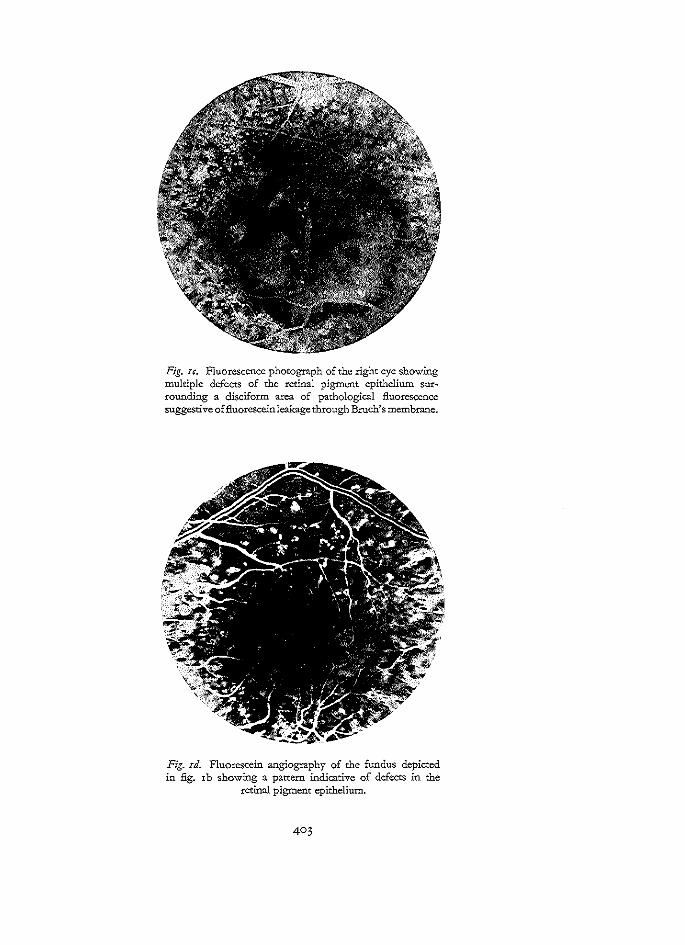
Fig. u. Fluorescence photograph of the right eye showing multiple defects of the retinal pigment epithelium surrounding a disciform area of pathological fluorescence suggestive of fluorescein leakage through Bruch's membrane.
Fig. rd. Fluorescein angiography of the fundus depicted in fig. I b showing a pattern indicative of defects in the
retinal pigment epithelium.

8. DARK ADAPTATION
Dark adaptation is generally unaffected. Franceschetti et a!. (1963), however, described a slightly delayed dark adaptation curve in z patients.
9· ELECTRORETINOGRAPHY
The ERG is normal in early stages, but becomes subnormal in advanced stages when a fair part of the retina is involved (Fran<;ois 195 z).
IO. ELECTRO-OCULOGRAPHY
So far as we know, no BOG's have been recorded in Sorsby's pseudo-inflammatory dystrophy. In advanced stages with extensive atrophy of the pigment epithelium and choroid, pathological BOG's are bound to be found.
II. PHOTOGRAPHY
Except in Bedell's article (r96r), none of the publications have so far presented photographs of Sorsby's dystrophy: nearly all publications present drawings (Sorsby eta!. 1959; Burn 1950; Franceschetti eta!. 1963).
I2. FLUORESCEIN ANGIOGRAPHY
Rosen and Leighton (1968) presented fluorescein angiograrns of Sorsby's dystrophy. They disclosed e:-:tensive defects of the pigment epithelium and pigmentations. Two individuals not immediately identifiable as patients showed slight but unmistakable changes of the pigment epithelium. The results of this method of examination, it should be borne in mind, are largely dependent on the stage which the dystrophy has reached. In exudative processes, as in disciform macular degeneration (] unius Kuhnt), diffuse fluorescein leakage will certainly be visible. The fluorescence photographs of our patient with bilateral lesions resembling Sorsby's pseudo-inflammatory dystrophy are seen in fig. red.
I3. CARRIERS
Regular dominant transiillSS!on occurred in the families so far described, and carriers have therefore not been identified.
I4. HISTOLOGICAL FINDINGS
Ashton and Sorsby (195 r) made a histological e.xamination of the eyes of 2 patients (aged 70 and 71) with Sorsby's pseudo-inflammatory dystrophy. A by no means negligible objection lies in the fact that precisely in these patients' families dominant transmission was not demonstrable. They found the following histological changes:

r. Sclerosis and atrophy of the choroid with fibrous mural degeneration of the remaining vessels.
z. Numerous ruptures of Bruch's membrane in the posterior fundus with degeneration of the elastic layer in the same area.
5· Subretinal newly formed vascular tissue, originating from the choroid and related to the dehiscences in Bruch's membrane.
4· Disturbance of the pigment epithelium. 5. Destruction of the outer layers of the retina with glial replacement. Babel (1958) examined the eyes of a sz-year-old man with a u-year history of diminishing vision, whose condition had been diagnosed as Sorsby's pseudoinflammatory dystrophy. The choriocapillaris of the right eye had largely disappeared. The corresponding pigment epithelium was atrophic but also showed areas of proliferation. The neuroepithelium and outer retinal layers likewise showed atrophy. The large choroidal vessels were either fibrotic or hyalinized.
Bruch's membrane was greatly changed: irregular rarefactions and deposits of fine granulations. Circumscribed e..-x:udates were found between the more or less marked pigment epithelium changes and the external limiting membrane, and small localized haemorrhages were visible in the inner retinal layers. The left eye showed a less completely affected choriocapillaris, and its neuroepithelium showed unmistakable degenerative changes.
Histopathological studies thus disclosed changes of the type usually found in disciform macular degeneration and angioid streaks. The principal common lesion in these 5 conditions is degeneration and rupture of Bruch's membrane, followed by formation of subretinal tissue.
15. PATHOGENESIS
The above indicates that in Sorsby's pseudo-inflammatory dystrophy the fundamental pathology seems to be a primary dystrophic change of Bruch's membr:\ne, followed by secondary organization of subretinal haemorrhages and exudates originating in the choriocapillaris and passing through the ruptures in Bruch's membrane.
Nevertheless, the physicochemical changes which lead to involvement of the elastic tissue of Bruch's membrane ate still obscure, and the possibility remains that this involvement results from pathological alteration of the choroid or the choriocapillaris proper. Histological examination in early stages of this dystrophy may lead to a solution of this problem.
For the time being it seems best to follow Ashton and Sorsby's suggestion (1951) and assume that pseudo-inflammatory dystrophy is primarily caused by genetically determined defects in Bruch's membrane, thus indicating a histopathological relationship of this condition to angioid streaks and disciform macular degeneration.
405

r6. MODE OF TRANSMISSION
The mode of transmission of this condition is probably always autosomal dominant (Sorsby 1940: Kr family; Sorsby et al. 1949:4 families, including the Randall family with r6 patients in 4 generations, the Ewbank family with ro patients in 2 generations, the Carver family with 2 5 patients in 4 generations, and the Kempster family with r6 patients in 3 generations; Burn 1950: II cases in 3 generations; Fran~ois 1958: 5 patients in 2 generations; Bedell 1961: father and daughter).
In the literature, an autosomal recessive mode of transmission is considered not impossible in view of some publications, but data so far available are too scanty to permit of a definite conclusion on the mode of transmission.
17. GENERAL PHYSICAL AND LABORATORY FINDINGS
No characteristic abnormalities have been found in these cases at general internal examination. Only Seedorff ( r 962) found renal glycosuria in a family suffering from a central macular degeneration, which resembled Sorsby's pseudo-inflammatory dystrophy.
18. ASSOCIATED CONDITIONS
No associated conditions have been described in patients.
19. DIFFERENTIAL DIAGNOSIS
Differential diagnosis of Sorsby's pseudo-inflammatory dystrophy encompasses the other dystrophies of the central retina and choroid discussed in this study, none of which shows really marked similarities to the entity Sorsby described.
Vitelliform dystrophy of the fovea can show eJ<:udative changes and must therefore be home in mind in differential diagnosis. Oedema, haemorrhages, e)..-udates, drusen, grOss pigment changes and subsequent generalized chorioretinal atrophy are more reminiscent of the complications of angioid streaks or of disciform macular degeneration and chorioiditis disseminata can also show similarities to this condition.
The entities thus defined are not accompanied by angioid streaks, and disciform macular degeneration usually is confined to the retinal centre, not affecting the retinal mid-periphery. Also, disciform macular degeneration rarely occurs before age 55 and has no dominant transmission.
In chorioretinitis disseminata an infectious cause can often be found, and hereditary factors are not at all involved.
In terminal stages an extensive choroidal atrophy may call for differentiation from one of the forms of choroidal atrophy, choroideremia, gyrate atrophy or chorioretinal atrophy associated with high myopia. For the more comprehensive differential diagnosis of these conditions, I refer to the differential diagnosis of central areolar choroidal dystrophy.

20. THERAPY
No therapy has been described. Photocoagulation of dehiscences in Bruch's membrane (demonstrated by fluorescein angiography) might prevent further progression of the process.
2!. FUTURE
Further reports on families with this affection are required for better analysis of this entity. Very few families with this affection have so far been described and the clearly detailed descriptions pertain only to families in England.
It should be stressed once again that only unequivocally familial cases (and probably only those with autosomal dominant transmission) warrant a diagnosis of pseudo-inflammatory dystrophy. In solitary cases not diagnosable with certainty, the diagnosis "Sorsby's pseudo-inflammatory dystrophy" should not be used as a "diagnosis of convenience".
22. CASE HISTORIES
x. Fam. Ot EOB-p.oi.Of At the age of 37 a rather sudden impairment of visual acuity in the right eye. VOD xj6o; VOS Sfxo; emmettopic. Media: Normal. Ftmdi: In the posterior pole of OD a large disciform e)..'Udative process, surrounded by small drusenlike configurations (fig. ra). In the posterior pole of the left eye a flecked retina, which resembles the initial stages described in Sorsby's pseudo-inflammatory dystrophy (fig. rb). F!ttOrescein angiography: The fluorescein angiograph of the posterior pole of the right eye shows a fluorescein pattern :resembling disciform macular degeneration (fig. xc). The left eye shows a pattern such as that seen in drusen and fundus fiavimaculatus (fig. xd). Famify history: The family is reported to have some more members with similar eye affections. A comprehensive family study has not so far been possible.
Summary: A 37-year-old woman with a foveal affection, resembling Sorsby's pseudo-inflammatory dystrOphy.
REFERENCES
ASHTON, N. and SORSBY, A.: Fundus dystrophy with unsual features. A histological study. Brit. J. Ophthal. 35:751-764 (195 I).
BABEL, J.: Le rOle de la choriocapillaire dans lcs affections d6g6nCratives du pOle posterieur. Bull. Soc. fran>. Ophtal. 71:389-399 (1958).
BEDELL, A. J·: Progressive bilateral chorioretinitis. Amer. ] . Ophthal. 5z: 545-347 (1961). BEHR, c.: Die Heredodegeneration der Makula. Klin. Mbl. Augenheilk. 65:465-505 (x9zo). BLUE, R.: Family degeneration of the macula lutea. J. Amer. Med. Ass. 73: 15z8-I35 I (1919). BURN, R. A.: Further cases of a fundus dystrophy with unusual features. Brit. ]. Ophthal. 34:393-403 (1950). CAVARA, v.: Sulla degenerazione faroigliare della maculae dei dintori. Boll. Ocul. 3:r.z.S-164 (1924). In
Z. Augenheilk. 6r:z73 (r9z4). cox, J.: Colour vision defects, acquired in diseases of the eye. Brit.]. Physiol. Opt. 17:195-zi6 (1960).
Brit.]. Physiol. Opt. x8:5-52(196I). Brit.]. Physiol. Opt. x8:67-84(I961).

DUKE-ELDER, s.: System of ophthalmology. Vol. IX. Diseases of the uveal tract. Kimpton, London, 1966. FRANCESCHETTI, A., FRAN901S, J. and BABEL, J.: Les h6r6dod6g6n6rescences chorior6tiniennes. Masson,
Paris, r96;. FRAN90IS, J.: L' 6lectro-r6tinographie dans les d6g6nCrescences tap6to-r6tiniennes p6riph6riques et centrales.
Ann. Ocul. 185:842-856 (195z). FRAN90IS, J.: L'h6r6dit6 en ophtalmologie. Masson, Paris, 1958. HUTCHINSON, J. and TAY, w.: Symmetrical central chorio-retinal disease occurring in senile persons. Roy.
Lond. Ophthal. Hosp. Rep. 8:z;1 (1875). LUTZ, A.: Dber eine Familie mit hereditirer familiater Chorio-retinitis. Klin. Mbl. Augenbeilk.49:699-7o;
(I9II). MA.Zzr, L.: Degenerazioni maculari retiniche famigliari associate ed emorragie della retina. Arch. Ottal. 41:
ZZ5-Z$9 (1934). PERDRIEL, G., BOUCHAT, J., CORNAND, G. and CHEVALERAUD, J.~ A propos d'un cas de dCg6n6rescences
maculai:re du type inflammatoire de Sorsby. Bull. Soc. Ophtal. France 68:455-458 (1968). ROSEN, E. s. and LEIGHTON, D.: Fluorescein photography of generalized dominant fundus dystrophy. Brit.
J. Ophtbal. )Z'8z8-83z (r968). SEEDORFF, H. H.: Hereditary renal glycosuria with central macular degeneration. Acta Ophthal. 40:91-95
(1962). soRSBY, A.: The dystrophies of the macula. Brit. ]. Ophthal. Z4:469-533 (1940). SORSBY, A.: Genetically determined lesions simulating inflammatory ocular reactions. Acta Neurol. Psychiat.
Belgica 49:6n-6rz (1949). SORSBY, A., JOLL MASON, M. E. and GARDENER, N.: A fundus dystrophy with unusual features. Brit. J. Ophthal.
33'67-97 (I949). soRSBY, A.: Generalized fundus dystrophy. In Modem trends in ophthalmology. ;rd series. Butterworth,
London, 1955. VERRIEST, c.: Les deficiences acquises de la discrimination chromacique. Memoires. Tome IV, fasc. 5,
p. 37-;z7. Acad6mie Royale de M6decine de Belgique. Imprimerie M6dicale et Scientiiique, Bruxelles, 1964.

XIV
Central areolar choroidal dystropry
I. INTRODUCTION
Central areolar choroidal dystrophy (atrophy) is one of several forms of choroidal dystrophy, often described as choroidal sclerosis. The recognized forms of choroidal dystrophy are:
r. diffuse, generalized choroidal dystrophy; z. peripapillary choroidal dystrophy; 3· central areolar choroidal dystrophy.
Solitary as well as familial cases of all three forms have been described. Another form is circinate choroidal dystrophy, in which an atypical chorio-retinal atrophy is limited to a circular area around the macula in both eyes (Knapp, 1907; Schocket and Ballin, r 970 ). Generalized choroidal dystrophy has been described by such authors as Morton (r885, r893), Frost (r896), Bednarski (r9oo), Harman (r9oz), Levinsohn (r9o3), Holloway (1914); familial cases have been reported by Fran~ois (1949), Sorsby and Davey (1955) and Stankovic (1958).
Haab (r895), Harman (r9oz), Haab (r9z8) and Di Marzio (r937) described peripapillary choroidal dystrophy, and familial cases were reported by Cuperus (r9o3), Guglianetti (r9o8, 1909), Gilbert (1938), Sorsby (1939) and Waardenburg (1952).
Central areolar choroidal dystrophy was observed in a Go-year-old woman by Jaeger (r855, r869). Other reports on this condition were published by Nettleship (r884) in a woman of 6o, Retze (r9oz) in a man of 66, and Thompson (r9o5) in a woman of 63.
Central areolar choroidal dystrophy is characterized in terminal stages by a bilateral, symmetrical, sharply defined areolar or oval-shaped area of choroidal atrophy, visible because pigment epithelium and choriocapillaris have disappeared at the site of the posterior pole.
Sorsby (193 5, 1939) was the first to call attention to the hereditary nature of central areolar choroidal dystrophy in two brothers. The family described by De Haas

Fig. I a. Irregularly shaped whitish flecks in the fovea of a 20-year-old girl (Fam. Fr). The pigment epithelium and the choriocapillaris appear to be absent in the patho-
logically changed areas.
Fig. rb. Fluorescein angiography reveals defectS in the pigment epithelium and presumably in the choriocapillaris. The fluorescence is less intensive than the one usually seen in cases with defects in the retinal pigment epithelium.
410

(I93 I), in which a man and three of his sons showed sharply defined white plaques with sclerotic choroidal vessels, probably also comes under this heading.
Sorsby and Crick (I95 3) published 4 families in which central areolar choroidal dystrophy occurred, and a follow-up on 2 brothers described in I935: a total of I4 cases in 5 families. In 4 of these families the condition was observed only in siblings but the fifth family included an affected mother and her affected daughter.
Friemann (I 9 53) observed this condition in 2 brothers out of 7 siblings, and in a second family 3 persons in 2 generations seemed to be affected.
A detailed study was published by Sandvig (I95 5, I959), who described a dominant form of central areolar choroidal dystrophy in I 3 patients in 4 generations, and 4 additional cases, including a brother and sister (I959).
Howard and Wolf (I 964) observed this condition in a sister and two brothers, aged 56, p and 4 7 years.
Carr (I965) described this condition in a 57-year-old woman and her 2 daughters (aged 22 and Ij).
The nomenclature of this condition is controversial. It was originally known as choroidal atrophy, and later the term choroidal sclerosis was introduced in view of the ophthalmoscopic features (Sorsby I935, I939; Sorsby and Crick I953)·
Histopathological studies by Ashton (I 9 53) demonstrated that the condition entails no sclerosis but atrophy of the choroidal vessels. These findings showed that the term atrophy is to be preferred to sclerosis. However, atrophy pertains to a state which occurs after some time; in initial sta.ges and during the course of the process it is best to speak of choroidal dystrophy.
Waardenburg eta!. (I96I) were right in introducing this designation, but the term central areolar choroidal atrophy is still general usage and correctly describes the unmistakable ophthalmoscopic picture of the terminal stages.
However, the question arises whether central areolar choroidal dystrophy is in fact always a separate entity. The reports by Sorsby and Crick (I 9 53), Sandvig (I 9 5 5) and Carr (I 96 5 ), which describe early stages of this condition, mention only slight pigment changes in the fovea and already notable disturbances of vision. The diagnosis was then made in view of older members of the family who showed the characteristic atrophic choroidal area. Without these older members, the diagnosis could have been Stargardes disease in the case of recessive transmission, or dominant progressive foveal dystrophy in the case of dominant transmission.
We observed Stargardt patients who developed central choroidal atrophy later in life, and found this atrophy fully developed in others.
On the other hand, there are reports on central areolar choroidal dystrophy (CACD) occurring at a later age and almost immediately assuming the atrophic aspect (De Haas I93 I; Sandvig I959) (fam. Kou, fam. Meu).
2. GENERAL CLINICAL PICTURE IN THE PATIENTS
Patients with incipient CACD are usually found in age group 20-50 (De Haas I93 I;

Fig. 2a-d. Development of central areolar choroidal atrophy in the right eye of a man in his forties (Fam, Kou).
Fig. 2a. Posterior pole on August .z, 1967.
Fig. 2b. Posterior pole on August 2.5, 1967.
412

Fig. 2c. Posterior pole on July 3, 1968.
Ft~f!,. 2d. Posterior pole on April4, 1970.

Fig. 2c. The posterior pole of the left eye of the same patient as seen in fig. za-d. There is a well defined areolar area of e:<.tensive atrophy of the retinal pigment epithelium and choriocapillaris. Only some of the larger choroidal
vessels have not yet disappeared.
Fig. 2j. Fluorescence photograph of the same fundus as depicted in :fig . .ze. E::..."tensive chodoretinal atrophy in the
centre surrounded by a normally appearing retina.
414

Sorsby and Crick I95 3). The initial symptoms can be mild but later, usually about age 6o, there is progressive loss of visual acuity resulting in a central scotoma. The condition is bilateral and symmetrical, and familial occurrence is frequently observed.
3· FUNDUS (OPHTHALMOSCOPIC FEATURES)
Initially, a slight e..-xudative oedematous reaction can be observed in previously normal fundi; but non-specific changes in the pigment epithelium in the form of pigmentations or white or variegated spots can also herald the beginning of the dystrophic process (fig. I, z). The specific character of this condition is not recognizable in the early stages. Only after many years does the pathognomonic circular or oval areolar area of atrophy occur in the posterior pole. Pigment epithelium and choriocapillaris have disappeared, and the large choroidal vessels are visible in a pale area which stands out clearly from the adjacent normal retina (fig. 2-4)·
4· REFRACTION
Myopia as well as hypermetropia with or without astigmatism have been described, but in many cases no mention is made of refraction.
5· VISUAL ACUITY
There is fairly rapid progressive loss of central visual acuity in CACD. Vision diminishes to I/Io or even lower.
6. VISUAL FIELDS
The peripheral visual fields remain quite intact, while a central scotoma develops which is initially relative but ultimately absolute.
7· COLOUR VISION
Colour vision changes as soon as visual acuity shows marked diminution. Both acquired blue-yellow dyschromatopsia and acquired red-green dyschromatopsia have been described in the patients (Franceschetti eta!. I963).
8. DARK ADAPTATION
As could be expected in an ophthalmoscopically purely central abnormality, dark adaptation is generally undisturbed (Carr I965). It was undisturbed in our patients. However, there have been reports on disturbed dark adaptation curves (Thompson I905; Guglianetti 1908; Frans;ois eta!. I956; Sandvig I959)·
4I5

Fig. ja. An acquired, well-defined round atea of chorioretinal atrophy in the left fovea of a 75-year-old man,
filmed on orthochromatic film (Farn. Meu).
Fig. jb. The same fundus as depicted in fig. ;a filmed on panchromatic film.

Fig. ;c. Fluorescein angiography is consistent with a sharply defined area of choriorctinal atrophy.
9· ELECTRORETINOGRAPHY
The ERG is generally normal in affections of the central retina, and it also is normal in CACD (Carr 1965). In cases in which a large part of the retina is involved, a subnormal ERG can be expected (Franceschetti eta!. 1963; Fran~ois eta!. 1969). We have recorded no F-ERG, but undoubtedly this must be subnormal in this condition.
IO. ELECTRO-OCULOGRAPHY
The EOG shows normal or slightly subnormal results (Carr 1965; Fran~ois et a!. 1969), as personal observations confirm (fam. Kou).
II. PHOTOGRAPHY
Not many photographs of CACD have been published (Sandvig 195 5, 1959; Carr 1965). Sandvig (1959) illustrated her article with very beautiful photographs. Our study disclosed that orthochromatic graphic film gives a much more exact picture of this condition than does panchromatic graphic film (fig. ;ab ).
!2. FLUORESCEIN ANGIOGRAPHY
After injection of fluorescein, some slight fluorescence occurs in the area where few choroidal vessels are left; no fluorescence occurs where choroidal vessels are lacking.

This is clearly visible because pigment epithelium and choriocapillaris have quite disappeared at the site of the focus (figs zf and 3c). At the site of persistent remnants of the choriocapil!aris, the small vessels leak fluorescein but the large vessels are impermeable to this dye (Maumenee 1968). If there were only a defect in the pigment epithelium, diffuse fluorescence of the choriocapillaris would be seen; an intact pigment epithelium would of course show no pathological fluorescence. If CACD is purely choroidal in origin, then it is probable that in its early stages normal fluorescein patterns still prevail.
Krill eta!. (1968) made a fluorescein-angiographic study of a carrier of choroideremia, and found widely scattered defects in the pigment epithelium. Partly on this basis they postulate that the original basic defect of this condition is to be found in the pigment epithelium. A similar situation might well be involved in CACD.
Follow-up fluorescein-angiographic studies of patients with incipient CACD are required to obtain more conclusive data on this question. Perhaps such an incipient picture is involved in our female patient Fr (fig. 1), but the problem is that the ultimate development of an ophthalmoscopic pattern is not predictable. Investigation and follow-up in a family in which this condition is dominant, however, could overcome this difficulty.
I). CARRIERS
Too little is so far known of the transmission of CACD to conclude with certainty that irregular dominance occurs. In the cases in which recessive transmission was described, the parents were (of course) quite normal. Since no specific changes in retinal function tests occur in CA CD, nothing can be expected of such tests in carrier investigation.
14. HISTOLOGICAL FINDINGS
Ashton (195 3) had occasion to make a histological examination of the eyes of a 56-year-old woman from a family described by Sorsby and Crick (1953). His findings were the following. a. A well demarcated avascular zone extending from the submacular region to the
disc was present in the posterior choroid. b. Histologically this avascular zone was found to be atrophic and fibrosed. No
arteriosclerotic changes were found either in the affected area or elsewhere in the choroid. Dissection of the posterior ciliary arteries failed to reveal constriction or occlusion.
c. The outer layers of the retina together with the pigment epithelium had disappeared without glial replacement, in an area exactly corresponding to the underlying choroidal atrophy.
d. Bruch's membrane was little affected by the failure of the choroidal blood supply. Babel (1958) e.';:amined the eyes of a 7o-year-old man with CACD and found numerous
418

Fig. 4· Areolar pattern of chorioretinal atrophy. There are some small areas of preserved pigment epithelium
centrally (Pam. Lod).
drusen surrounding the atrophic area from which choriocapillaris, pigment epithelium and neuroepithelium had disappeared. Bruch's membrane showed secondary irregularities, ruptures and a lamellar structure. Babel ascribed the clinical features of vascular sclerosis to the sharp contrast bet ween the remaining choroidal vessels and the atrophic area.
Klien (r964) described the histological features of CACD in the eye of a 71-yearold m..'"ln: "The macular area showed grossly an area z.o by z.o disc diameter in size, of pigmentary and choroidal atrophy, beginning near the temporal edge of the disc. Histologically this lesion represented a rather well-defined macular defect of the neuroepithelium, pigment epithelium and choriocapillaris. These three structures had normal appearance up to the edges of the defect where they ceased rather abruptly. In the peripheral portion of the atrophic area a few scattered lumens of capillaries were still visible, while in tl1e central portion all of these and the mediumsized vessels had disappeared, leaving only a few arteries surrounded by fibrosed stroma. No breaks were found in Bruch's membrane".
This man's family had no history of eye diseases. Klien also stated: "The histopathologic findings in this eye resemble closely those described by Ashton in central areolar choroidal atrophy and they serve well to contrast the findings in prin1ary heredo-degeneration of the first retinal neuron, described elsewhere (Klien 1950), in which no choroidal defects could be demonstrated". However, it is our impression

that in longstanding dystrophies of the first retinal neuron the choroid also becomes atrophic.
Howard and Wolf (I 964) carried out a histopathologic study in one case. They found atrophy and loss of the choriocapillaris, beginning near the equator and becoming more pronounced at the posterior pole. There were degenerative changes in Bruch's membrane and retinal pigment epithelium. A selective loss of the outer retinal elements was more marked posteriorly.
15. PATHOGENESIS
The pathogenesis of this condition is probably to be found in primary dystrophy of the choroidal vessels, although primary tapetochoroidal dystrophy or primary dystrophy of the pigment epithelium with secondary choroidal involvement cannot be ruled out. Sclerosis of the choroidal vessels as causative factor is unlikely in view of the histological absence of sclerotic vessels.
However, considering the early stages of this condition in which only slight pigment changes of the macula are seen with already markedly diminished vision (Sorsby and Crick I 9 53 ; Sandvig I 9 5 5 ; Carr I 96 5 ), the question arises whether this condition does not involve a primary tapetoretinal lesion, as does progress1ve foveal dystrophy.
16. MODE OF TRANSMISSION
There are reports on families with autosomal dominant transmission of CACD as well as on families in which an autosomal recessive pattern seems to prevail. In the patients we examined we were unable to demonstrate a hereditary factor. It should be pointed out that no complete family study could be made in these cases.
Dominant transmission is suggested by several case reports (De Haas I93I: father and 3 sons; Sorsby and Crick I95 3: mother and daughter; Friemann I95 3: 3 persons in z. generations; Sandvig 195 5 :r 3 patients in 4 generations; Carr 1965: mother and daughter). In other cases, recessive transmission seems probable (Sorsby I935, I939' z brothers; Sorsby and Crick I953: siblings of 4 family members; Friemann I953: 2 brothers; Sandvig I959' brother and sister).
17. GENERAL PHYSICAL AND LABORATORY FINDINGS
General physical and laboratory findings are generally normal, as they were in the patients we exarrrined.
18. ASSOCIATED CONDITIONS
Central areolar choroidal dystrophy is generally observed as an isolated condition. Kapuscinski (I9J4), however, described a progressive choroidal atrophy, not
entirely confined to the central retina, in 2 sisters who suffered from Friedreich's
420

ataxia. And Tiberi and Cuccagna (1959) reported on z brothers with CACD, one of whom (in a consanguineous marriage) sired 3 children with Stargardt's disease. The occurrence of these two different affections in the same family may have been a coincidence. The ophthalmoscopic features of an advanced stage of Stargardt's disease, however, can closely resemble those ofCACD, as demonstrated on page I 17.
Therefore, a recessive or dominant progressive foveal dystrophy cannot be ruled out in the cases of Tiberi and Cuccagna. Also, these cases are very similar to that described by Carr (1965) in a woman with CACD and her z daughters with Stargardtlike changes.
In terminal stages of vitelliform foveal dystrophy and Sorsby's pseudo-inflammatory dystrophy, too, a more or less marked form of choroidal atrophy can be observed.
19. DIFFERENTIAL DIAGNOSIS
The diflerenrial diagnosis of CACD encompasses the following conditions, the first two of which have been mentioned in the introduction.
a. Diffuse, generalized choroidal dystrophy.
b. Peripapillary choroidal t!Jstropry and circinate choroidal dystrophy. There are also forms which begin at the centre and spread to peripapillary or even peripheral areas, so that the diagnosis can be dependent on the developmental stage the process has reached.
c. Sorsby's pseudo-inflammato~y t!Jstrophy can show large areas of choroidal atrophy in the terminal stages. The extensive haemorrhages and exudates seen in early stages, however, are not observed in the more sharply defined CACD; and the overall picture is much more disorderly and less localized.
d. Choroideremia, also known as progressive tapetochoroidal dystrophy, shows a much more diffuse lesion than CACD. Its mode of transmission is intermediate X-chromosomal (Kurstjens 1965).
e. Chorioretinal atropry associated with high myopia is most extensive in the posterior pole of the eye, like CACD, and must therefore be differentiated.
f. Gyrate atropry of the choroid and retina in its initial stages shows well-defined, garland-shaped areas of choroidal and retinal atrophy in the retinal periphery; only in later stages do these areas extend in the direction of the posterior pole of the eye (Kurstjens 1965).
g. Terminal stages of Stargardt's disease may render differential diagnosis diflicult or even impossible (page 1 17). Patients with dominant drusen also have central choroidal atrophy as a late development (Neame 1954).
421

h. Macular colobomas show, even at birth, well-defined ovalshaped white areas in which no vessels are discernible in the posterior pole. This means that differential diagnosis is possible on the basis of the history.
20. THERAPY
There is no therapy. Many efforts made to treat the posterior pole process with vasodilators and anticoagulants have remained as futile as has placenta implantation.
ZI. FUTURE
It is important that further reports be published on families with central areolar choroidal dystrophy; this will facilitate differentiation from other similar affections and enlarge our knowledge of the mode of transmission.
Differentiation from Stargardt's disease or dominant progressive foveal dystrophy still offers difficulties, for in its early stages central areolar choroidal dystrophy may show features identical to those of these conditions (Sorsby and Crick 195 3; Sandvig 195 5, 1959; Carr 1965), and in the terminal stages of Stargardt's disease a picture of CACD is frequently encountered.
22. CASE HISTORIES
:r.Fam.Ko PK-99.04.I3 Poor visual acuity in the right eye since 1951. The family is reported to have normal eyes. r961: VOD r/3oo; VOS Sfro. I970: VOD r/3oo; VOS S+3·5o=C+o.5oX25° 8/ro. Media: Normal. Fundi: An eA"tcnsivc area of chorioretinal atrophy in each posterior pole. The left eye shows a remnant of retinal pigment epithelium, exactly in the centre of the fovea. This is the reason of the preserved visual acuity in this eye.
z. Faro.. Kou FK-26.II.02 Since some months a black spot in front of each eye. I967: VOD 4/ro; VOS o.5j6o Emmetropic. Fnndi: OD shows a yellowish, slightly pigmented area at the site of the fovea (fig. 2a). OS shows an areolar area of atrophic choroid and retina (fig. 2e). Vi .mal fteldr: Central scotoma. Colo"r vi.rion: OD normal. OS unrecordablc. Dr1rk adaptation: Normal. ERG: Scot. b-wavcs OD 2701-.V; OS 2651.1V.
Phot. b-waves OD IIo!~V; OS 10511V.
BOG, OD r.79; OS r.S2. Sy.rtemic examination: Normal. There is only a slightly positive Sabin-Feldmann reaction (1: 256). Has been treated with prednisone and pyrimethamine. Fanlify exwllination: Parents are not consanguineous. Three sisters, one brother and the two sons have completely normal eyes. I9JO: VOD 3/Go; VOS 1/300.
422

Fundi: Ovoid zone of chorioretinal atrophy in the right eye (fig. 2d). Large areolar area of chorioretinal atrophy in the left eye. Fluorescein angiography: OS: See fig. zf.
Sumtr~ary: A man, who developed a central areolar choroidal atrophy at the age of 4I. It is of interest to note the development of the ophthalmoscopic picture of the right eye (see fig. 2abcd). A yellowish-grey area, approximately I disc diameter in size, developed into an area of extensive cho:dorctinal atrophy.
3· Fam. Lod. j]LR-oS.o8.o4 In the last few years visual impairment. The family is reported to have normal eyes. VOD S+z.25=C+o.50X I00° 6/ro; VOS S+3=C+o.5oX Ioo0 6/Io. Media: Mild incipient senile cataract. Fzmdi: EA"tensive areolar chorioretinal atrophy in each eye (fig. 4). Visttal fields: Central and paracentral scotomata. Dark adaptation: The curve is approximately 2j3log. U. too high. ERG: Scot. b-waves OD 285.uV; OS zzo.uV.
Phot. b-waves OD 65.uV; OS So.uV. BOG: OD r.9z; OS r.3o.
4.Fam.Mcu G]M-94.r2.o4 r967: VOD S+r.5o=C+x X 30° 7/xo; VOS 6/xo, emmetropic. r968: VOD 8/xo; VOS 3/6o. Media: Incipient senile cataract ODS. Fundi: Mild pigmentary disturbances in the right fovea. A sharply defined area of chorioretinal atrophy in the posterior pole of the left eye (£g. 3). Fluorescein angiography: The pigment epithelium and the choriocapillaris have disappeared completely in a centrally located round area, which is slightly larger than I disc diameter. Some of the larger choroidal vessels arc left (fig. 3c).
5· Fam. Fr.
6 6 Mi , 2 ~ 4 5
104R
II-; ( M]F-4S.o;.o9) Has suffered from acute poliomyelitis and acute rheumatism. r96 J: Observes some tiny flecks with OS. r967: Same complaints with OD. r968: VOD S-4 ujro; VOS S-4·75=C-o.5oX50° ujro. There is some metamorphopsia (Amsler test). Fundi: Symmetrical foveal lesions. "Whitish, capricious flecks, suggestive of defects in the retinal pigment epithelium and the underlying choriocapillaris (fig. ra). VisHal fields: Normal. Colo~~r vision: Normal. Dark adaptation: Normal. ERG: Scot. b-waves ODS 4001tV; Phot. b-waves OD 95.uV; OS IIOJtV. BOG' OD 1.95; OS 1.97. Fluorescein angiography: Pathological fluorescence at the site of the lesions visible with normal ophthalmoscopy. The fluorescein pattern indicates defects in the retinal pigment epithelium and some atrophy of the choriocapillaris (fig. rb). Systemic examination: Slightly disturbed capillary resistence. No other abnormalities.

S!tmi!Jary: A fairly young female patient with irregularly shaped whitish flecks in both foveae. A definite diagnosis is impossible at this stage. The whitish flecks, located deeply in the retina are suggestive of choriorctinal atrophy. .A.n incipient central areolar choroidal atrophy seems to us one of the most likely of the few diagnostic possibilities.
REFERENCES
ASHTON, N.: Central areolar choroidal sclerosis. A histopathological study. Brit. J. Ophthal. ;57: 140-147 (1953)·
BABEL, J.: Le r6le de la choriocapillairc dans les affections degenCrativcs du p6le posterieur. Bull. MCm. Soc. fran<;. Opht. 7o:;89-398 (1958).
BEDNARSKI, A.: Ueber einen Fall von Atrophia gyrata chorioideae et retinae mit Sklerose dcr Adcrhaut. Arch. Augenhcilk. 40!4Z0-4z6 (1900).
CARR, R. £,: Central areolar choroidal dystrophy. Arch. Ophthal. 73: 32-35 (1965). CUPER us, N.J.: Ueber primfu'e Atrophic dcr Aderhaut mit Sklcrose der Gefasse. ''Atrophia alba chorioideac".
Arch. Augcnheilk. 48!235-242 (1903). DIMARZIO, Q.: Fundus oculi. L. Salomon, Roma, 1937. FRANCESCHE'I'TI, A., FRAN<;fOIS, ] and BABEL, ] • : Les h6r6dod6g6n6resccnccs choriorCtinicnncs. :Masson,
Paris, 196;. FRA:!'f<;fOIS, J.: Contribution i !'etude des h6r6do-d6g6n6resccnces tap6to-r6tiniennes. Ophthalmologica
u8:1-29 (1949). FRAN<;fOIS, J,. VERR!EST, G., DE ROUCK, A. and HUMBLE'!', M.; D6g6n6resccncc maculaire juvCnile avec atteinte
prCdominante de Ia vision photopique. Ophthalmologica r 3 I: 393-402 (1956). FRA:!'f<;fOIS, J., DE ROUCK, A. and FERNANDEZ-SASSO, D.: Electroretinography and electro-oculography in
diseases of the posterior pole of the eye. Adv. Ophthal. vol. 21, pp. 132.-163. Karger, Basel, 1969. FRIEM:ANN, w.: Beitrag zur Actiologie dcr zcntralen Netzhautcrkrankungcn. Sarnml. zwangl. Abhandl.
Geb. dcr Augcnhcilk. 6: 1-47 (195 3). FROST, w. A.: The fundus oculi. London, 1896. GILBERT, w.: Ueber sklcrotische Erkrankungcn dcr Adcrhautgef:isse. Ber. dtsch. Ophthal. Gcs. 52:447-
450 (1958). GUGLIANET'I'I, L.: Su di una forma di sclcrosi vascolare della coroide con carattere famigliare. Arch. Ottal.
15 !469-486 (1908). GUGLlA:-.lET'I'I, L.: Sclerosi vascularc diffusa con atrofia della coroidc di originc sifilitica. Arch. Ottal. 16:
JS7-J95 (1909). HAAII, o.: Atlas und Grundriss der Ophthalmoskopic. Pl. 62. J. F. Lehmann, Mi.inchen, 1895. HAAB, o.: Sklcrosc dcr Aderhautgefasse, disscminiertc chorioiditis und sckundiirc Pigmcntierung dcr
Nctzhaut. Atlas und Grundriss der Ophthalmoskopic und Ophthal. Diagnostik, p. 85, fig. 75· ]. F. Lehmann, MUnchen, 1928.
HAAS, H. L. DE: Een familic met erfclijkc ontaarding van de macula lutca. Ned. T. Genecsk. 75:4720 (1931). HARMA:!'f, N. B.: Choroidal angiosclerosis with pigmentary degeneration. Trans. Ophthal. Soc. U.K. 22:
156-159 (1902). HOLLOWAY, 'I'. w.: Atrophy of the choroid with e::-.."tensivc sclerosis of the choroidal vessels. Trans. Coli.
Phys. Philadelphia. 36:;r6-317 (1914). HOWARD, G. M. and WOLF, E.: Central choroidal sclerosis: Clinical and pathologic study. Trans. Amer. Acad.
Ophthal. Otolaryng. 68:647-660. July-Aug. 1964. JAEGER, E. VO:!'f: Bcitr. z. Path. des Auges, Wicn, 1855. JAEGER, E. vo:-r: Ophthalmoskopischer Hand Atlas. Veri. Hof- und Staatsarchiv, Wicn, r869. KAPUSCINSKI, w.: Ueber familifu:e Aderhautentartung mit ataktischcn StOrungcn. Ber. dtsch. Ophthal. Ges.
5o:r3-19 (1934). KLIEN, B. A.: Hercdodegeneration of the macula lutea: diagnostic and differential diagnostic considerations
and a histopathologic report, Amer.]. Ophthal. 33:371-379 (1950). KLIEX, B. A.: Some aspects of classification and differential diagnosis of senile macular degeneration. Amer.
]. Ophthal. 58:927-939 (1964).

KNAPP, P.: Ein seltener Augenspiegclbefund (sclerosis chorioideac circinata). Klin. Mbl. Augenheilk. 45: I7I-I77 (I907).
KRILL, A. E., NEWELL, F. w. and CHISHTI, M. I.: Fluorescein studies in diseases affecting the retinal pigment epithelium. Amer. J. Ophthal. 66 :470-484 (I968).
KURSTJENS, J· H.: Choroideremia and gyrate atrophy of the choroid and retina. Diss. Utrecht. Junk, 's-Gravenhage, 1965. Docum. Ophthal. 19:1-125 (r965).
LEVINSOHN, G.: Ueber sclerose der Aderhaut mit sekundarer Net:Zhautdegeneration. Arch. f. Augenheilk. ;8:268-275 (!899)-
MAUMENEE, A. E.: Fluorescein angiography in the diagnosis and treatment of lesions of the ocular fundus. Doyne memoriallectute. Trans. Ophthal. Soc. U.K. 88: 529-556 (1968).
MORTON STANFORD, A.: Atrophy of choroid. Trans. Ophthal. Soc. U.K. 5: 142-144 (1885); I 3: z8z (1893). NEAME, H.: Report on a case of macular degeneration of familial type, Tay's choroiditis, with central choroidal
atrophy as a late development, Proc. Roy. Soc. Med. 47: I63 (1954). :-<ETTLESHIP, E.: Central senile areolar choroidal atrophy. Trans. Ophthal. Soc. U.K. 4: I65-I66 (1884). RET7..E, w.: Einige interessante Spiegelfalle. Deutschmann's Beitr. zut Augenheilk. 5 :94-I07 (1902). SA!'I'DVIG, K.: Familial, central, areolar, choroidal atrophy of autosomal dominant inheritance. Acta Ophthal.
33: 7I-78 (195 5). SANDVIG, K.: Central areolar choroidal atrophy. Acta Ophthal. 37: 325-329 (I959). SCHOCKET, s. s. and BALLIN, N.: Circinate choroidal sclerosis. Trans. Amer. Acad. Ophthal. Otelaryng.
May-June I970, pp. 527-533· SORSBY, A.: Choroidal sclerosis. Proc. Royal Soc. Med. 28: 526-528 (I935). SORSBY, A.: Choroidal angio-sclerosis with special reference to its hereditary character. Brit. J. Ophthal.
23:433-444 (1939). SORSBY, A.: The choroidal abiotrophies. In Modem trends in ophthalmology. Vol. I, pp 474-480. Butter-
worth, London, I940. SORSBY, A.: Choroidal sclerosis. In Genetics in ophthalmology. pp. 148-I50. Butterworth, London, I95I. SORSBY, A. and CRICK, R. P.: Central areolar choroidal sclerosis. Brit. J. Ophthal. 37: 129-I39 (I95 3). SORSBY, A. and DAVEY, L. B.: Generalized choroidal sclerosis. Cause and mode of inheritance. Brit. J,
Ophtha!. 39'257-275 (I955)· STANKOVIC, I.: L'angiosclerose choroidienne familiale liCe au sexe. Bull. Soc. france Ophtal. I7!4ll-4I9
(I958). SUKUMLYN, s. w.: :Macular and choroidal degeneration. Amer. J. Ophthal. 30: II79-II8o (1947). TIBERI, G. F. and CUCCAGNA, F.: Degenerazione ercdo-familiare della macula e sclerosi coroidcalc centrale.
Gior. ital. oftal. 12:23-44 (I959). THOMPSON, A. J-I.: Central senile choroiditis. Trans. Ophthal. Soc. U.K. 25: u8-120 (1905). WAARDENBURG, P. J.: Angiosclerose familiale de la choroide. J. GCnet. hum. I: 83-93 (I95 2). WAARDENBURG, P. J., FRANCESCHETTI, A. and KLEIN, D.: GenetiCS and Ophthalmology. Van Gorcum,
Assen, 196I.
425

XV
Comment and conclusions
This study of 240 patients with hereditary dystrophies of the posterior pole and several times as many relatives shows that the hereditary dystrophies of the central retina and choroid include several readily distinguishable entities.
The classification presented on page 39 and repeated here (fig. 1) is very useful in general, and in clinical practice in particular, and easily lends itself to extension should new dystrophies be reported. On the other hand, if necessary, it can just as easily be restricted.
The long-accepted view that many diJferent manifestations of hereditary foveal dystrophies are all expressions of the same basic dystrophic process is quite untenable in my view, and I am confident that this study has demonstrated this. The characteristics which the various affections have in common are mainly determined by the localization of the pathological process, which is the same in all these dystrophies. Some of these common characteristics are: 1. familial, bilateral, symmetrical involvement of the posterior pole of the eye; 2. more or less marked diminution of visual acuity; 3· no general physical or laboratory abnormalities; 4· a generally early age of onset. The classification presented here might suggest that there are no more problems concerning the dystrophies of the posterior pole. However, there are still a few questions which remain moot points. A few of these may be briefly reviewed here.
It is difficult sharply to diJferentiate between Stargardt's disease with perifoveal yellow-white spots and fundus flavimaculatus. My own inipression is that in Stargardt's disease the dystrophic foveal process occurs first, to be followed by the development of perifoveal spots, whereas in fundus flaviniaculatus numerous spots are already in evidence in the posterior pole before the fovea is affected ("Stargardt inversa"). Moreover, the spots in fundus flavimaculatus are generally much more numerous.

OUTCFl: I>LCXIF"OFl:M LAYI:Fl:
OUTER NU~Lt:AR LAYt:R
t:XTCRNAL LIMIT IN!;; MEMBRANE:
ROOO AND CONI';B
I>II:OME:NT CI>ITHCLIUM
BRU~H·a MEMBRANE: --~~~~~~~~~tt~~~t.:St.~~:-::::~~~~t
SCLCRA
Fig. I.
Classification with regard to the tissue mainly and primarily affected.
Nerve fibre layer Neuroepithelium
Pigment epithelium
Bruch's membrane
Choroid
sex linked juvenile retinoschisis Stargardt's disease dominant progressive foveal dystrophy progressive cone dystrophy central retinopathia pigmentosa vitelliforrn dystrophy of the fovea fundus flavimaculatus reticular dystrophy of the retinal pigment epithelium buttcr£yshaped pigment dystrophy of the fovea grouped pigmentations of the foveal area dominant drusen of Bruch's membrane (hyaline dystrophies) pseudo inflammatory foveal dystrophy (Sorsby) central areolar choroidal dystrophy
Likewise, the difference between some forms of central areolar choroidal dystrophy and terminal stages of Stargardt's disease is not always marked, because in terminal stages of Stargardt's disease there is often marked choroidal atrophy_ Also, initial stages of what later becomes an extensive central areolar choroidal dystrophy are sometimes characterized by very inconsiderable changes in fundo. These facts once again emphasize the importance of follow-up studies on hereditary dystrophies of the posterior pole.
Nor is it qnite certain whether Stargardt's disease with dystrophy confined to the posterior pole and Stargardt's disease with involvement of the retinal periphery in the dystrophic process ( centroperipheral tapetoretinal dystrophy) are essentially different conditions or merely different manifestations of the same genotype. We
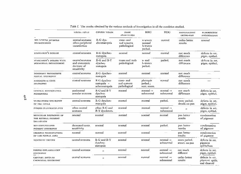
Table I. The results obtained by the various methods of investigation in all the condition studied. --YISUAI. FillLD COLOUlt VISION DARK ERG EOG PIIOTOGRAPIIY FLUORESCEIN
ADAPTATWN OR THO-PAN ANGIOGRAPIIY --
SHX LINKED )U\'E.."'ILE central scotoma R-G dys- cone- and a-waves normal ortho better normal RETL.-...OSCHISIS often peripheral chromatopsia rod-systems normal results
constriction pathological b-waves
STARGARDT'S DISEASE central scotoma R-G dyschro- normal normal normal not much defects in ret. matopsia difference pigm. cpithel.
----- --STARGARDT'S DISEASE WITH central scotoma R-G and B-Y cones and rods a- and pathol. not much defects in ret, PERIPHERAL INYOLYmtE:t-..7 and concentric dyschro- pathological b-waves difference pigm. epithel.
decrease of matopsia pat hoi. ivity
DO~IINANT PROGRESSI\'E central scotoma R-G dyschro- normal normal normal not much FO\'EAL DYSTROPHY matopsia difference
PROGRESSIYH CONE central scotoma R-G dyschro- cone- and photopic normal not much DYSTROPHY matopsia -) rod-systems pathol.; difference
achromatopsia pathological scot. nann.
Cfu'ITRAL RETL.-...OPATHIA pericentral R-Gand B-Y normal normal-) normal---... not much defects in 1ct. PIG~IENTOSA annular scotoma dyschro- subnormal subnormal difference pigm. epithel.
-!>-N 00
central scotoma R-G dyschro- normal normal pathol. more pathol. defects in ret. YITELLIFORM DYSTROPHY
OF TilE FOVEA matopsia details on pan pigm. epithel.
FUNDUS l'l.A\'l~fACULATUS often central often R-G and normal normal normal---... , defects in ret. scotoma B-Y dyschrom. subnormal pigm. epithel.
RETICULAR DYSTROPHY OF normal normal normal normal normal pan better condensation THE RETINAL PIG:\IENT results of pigment EPITHELIU~I
BU"lTERFLYSIIAPED decreased centr. normal normal normal pathol. pan better condensation PIG~ffu'IT DYSTROPHY sensitivit): results of pigment
GROUPllU PIG~!ENTATIONS normal normal normal normal pan better condensation OF THE FOVEAL AREA results of
---DO~nNA..."'~' DRUSEN central scotoma R-G and B-Y normal normal normal---... more pathol. defects in ret.
dyschro- subnormal struct. on pan pigment matopsia epithelium
PSEUDO-INPLA~BIATORY central scotoma " normal normal normal---... not much defects in ret.
DYSTROPHY subnormal different r_!gm. epithel. -CENTRAl. AREOLAR central scotoma , normal normal normal---... ortho better defects in ret. CHOROIDAL DYSTROPHY subnonnal results pigment epith.
and choroid

tend to prefer the latter possibility because follow-up studies have disclosed transitional forms and transitions.
The relations between the above discussed affections might be schematically presented as follows:
Stargardt's disease < fundus flavimaculatus ! I ___________ > centroperipheral TRD
central areolar choroidal dystrophy
Centroperipheral tapetoretinal dystrophy which diffusely affects the rods and cones throughout the retina, occupies an interesting position intermediate between progressive cone dystrophy, in which only the cones are involved, and retinopathia pigmentosa in which it is chiefly and primarily the rods that are affected (Berson et a!. 1968).
In this study, the value of various retinal function tests and modern photographic techniques has proved to be unmistakable. Especially the electrophysiological and photographic techniques used in this study have made it possible to differentiate the various conditions, and have given a more profound insight into the localization in depth of the various conditions and their extent through the retina.
Table I presents the results obtained by the various methods of investigation in all the conditions studied. The value of these methods will be discussed once again briefly, with special reference to their value in studying the dystrophies of the posterior pole. The various methods have on the one hand taught us more about the nature and localization of the various dystrophies, and on the other hand have given us a broader and more profound understanding of the basic retinal processes involved.
The methods used to examine the retina were methods to determine visual acuity, visual fields, colour vision and dark adaptation, and also included electroretinography, electro-oculography, photography with orthochromatic and panchromatic graphic film, and fluorescein angiography. The results and significance of these methods will be briefly recapitulated.
Vi:ttal actdty largely depends on the primary localization of the pathological process. Dystrophies which primarily involve the photoreceptors show rapid progressive diminution of vision, while dystrophies of the pigment epithelium cause slow or very slight diminution of vision. Conditions which involve Bruch's membrane and the choroid, can likewise cause rapid diminution of vision due to dehiscences in Bruch's membrane or deficient nutrition of the photoreceptors. In sex-linked juvenile retinoschisis, visual acuity is probably already affected at birth, and shows gradual further loss during life.
It is of importance that there is no parallel between the ophthalmoscopic features of the posterior pole and visual acuity. In fact, conditions with the least conspicuous

ophthalmoscopic changes often show the most marked diminution of vision, whereas extensive ophthalmoscopic changes can be found in association with rojro vision (intact vitelliform disc).
Refraction is inextricable from determination of visual acuity, and certain conditions prove to be associated with specific anomalies of refraction.
In se:<-linked juvenile rerinoschisis and in vitelliform dystrophy of the fovea, one nearly always finds hypermetropia, often with astigmatism.
The other dystrophies originating in the pigment epithelium are likewise often accompanied by hypermetropia, with or without astigmatism; in conditions primarily involving the photoreceptors, low myopia seems to be prevalent, not infrequently with astigmatism.
The t'i.rttal fields are usually characterized by an intact periphery and a central scotoma which increases in size and depth as the dystrophy progresses. In sex-linked juvenile retinoschisis there are often limitations in the peripheral visual field, especially in the upper nasal quadrant, but normal peripheral boundaries and very markedly reduced visual fields are also observed. In centroperipheral TRD, concentric limitation of sensitivity is always observed besides a central scotoma. Loss of peripheral visual fields is occasionally seen in these cases. In progressive cone dystrophy, the visual fields are much better under scotopic than under photopic conditions.
Colottr vision shows comparable results in all the various posterior pole dystrophies. No results specific for a given type of dystrophy are found, and differential diagnosis of these conditions is therefore not possible on the basis of disturbances in colour vision. Since acquired disturbances of colour vision are generally given little attention, they may be discussed in some detail here.
The generally accepted view is that the human retina contains three different pigments (red = protos; green = deuteros; blue = tritos), probably localized in three different types of cone. Congenital defects of colour vision must probably be ascribed to deficiency or absence of certain pigments or cones, and are designated according to the deficient pigments. Thus we have such terms as protanomaly (partial red pigment defect), protanopia (total red pigment defect) and protan defect (unspecified red pigment defect).
Acquired defects of colour vision are not present at birth but occur in the course of life in response to such influences as eye diseases, general diseases or drugs. Their classification of necessity differs from that of congenital defects of colour vision, because the acquired dyschromatopsias cannot be so sharply differentiated, and because various forms can occur in combination. The acquired dyschromatopsias are generally divided into red-green and blue-yellow dyschromatopsias, but a more detailed subdivision is also possible.
In his comprehensive study "Les deficiences acquises de Ia discrimination chromatique", V erriest (I 964) distinguished the following forms:
430

a. Type I acquired red-green dyschromatopsia. b. Type II acquired red-green dyschromatopsia. c. Acquired blue-yellow dyschromatopsia. d. Acquired dyschromatopsia with no specific axis. re a. This form is found in the hereditary dystrophies of the posterior pole. The
anomaloscope indicates reduced sensitivity to red, and the spectral sensitivity curve shows displacement to the short waves of the spectrum. Ultimately, when all the cones in the posterior pole have disappeared and acquired achromatopsia develops, the spectral sensitivity curve assumes a scotopic character.
re b. This form is found in affections of the optic nerve. The anomaloscope findings are the same as those found in congenital deutan defects, and the spectral sensitivity curve is normal. In the case of extensive optic ne:rve lesions, achromatopsia can then be found while the spectral sensitivity curve is normal.
re c. This form is often observed in affections of the retinal periphery in which dark adaptation and the scotopic ERG are affected also. It is also found in degenerative (not dystrophic!) posterior pole lesions, e.g. central serous choroidopathy and disciform degeneration of the posterior pole, and also in dominant optic nerve atrophy. A dyschromatopsia resembling a congenital tritan defect is found also in clouding of the refracting media.
re d. This is an ill-defined form which often shows trichomatic defects as a result of a wide variety of pathological changes.
As we mentioned, type I acquired red-green dyschromatopsia is observed in the hereditary posterior pole dystrophies, while an acquired tritan defect can also be observed in conditions in which the retinal periphery is involved as well. This is seen specifically in Stargardt's disease with peripheral involvement ( centroperipheral TRD). The marked decrease in red sensitivity in hereditary posterior pole dystrophies must probably be ascribed to the fact that the red-sensitive cones are for the most part localized in the posterior pole. However, selective affection of the red-sensitive cones in early stages cannot be ruled out (V erriest 1 964). In investigating acquired dyschromatopsias, it must always be borne in mind that congenital dyschromatopsias occur in about 8% of all males and some 0.5% of all females (Krill 1968).
Dark adaptation is generally normal in hereditary dystrophies of the posterior pole. Of the conditions discussed in this study, only sex-linked juvenile retinoschisis and the centroperipheral TRD are associated with an unmistakable delay in dark adaptation. The curve is rarely more than 1-2 log U too high, and in both conditions the photopic as well as the scotopic system is affected. Progressive cone dystrophy shows delayed adaptation of the photopic mechanism but an entirely normal scotopic mechanism. In our study the retina has always been tested in its totality. When even a small portion of the retina is still intact, a normal dark adaptation curve may be found. This is the case, for example, in chloroquine retinopathy in which some retinal areas remain intact while other areas are severely affected. In such cases a

400
350
300
250
200
150
100
50
.·
rNinoschisis
. . ..
vitellilorm without
I • I• I I
..
.. Stargnrdt
with
peripheral Involvement
.·
Fig. 2. ERG b-waves in se.-x linked juvenile rctinoschisis, vitellifonn dystrophy and Stargat:dt's disease. The latter designed separately with and without peripheral involvement as seen ophthalmoscopically. The :results of 10 unselected
patients (zo eyes) per entity are given (every black point means one eye).
decidedly pathological ERG and EOG can be obtained while the dark adaptation curves are virtually normal (Gouras and Gunkel 1965).
If the dark adaptation of localized retinal areas were tested, either normal or pathological results could be expected. Therefore, the fact that a pathological curve is obtained in sex-linked juvenile retinoschisis and centroperipheral TRD indicates that the whole retina is involved and that no islets of intact retinal function remain.
The e!ectroretinograJJJ (ERG) gives information on the overall function of the retina. The ERG is a rapid change in potential which follows a change in retinal illumination. The principal components of this response are the a-wave and the b-wave. The corneal negative a-wave seems to arise in the photo receptors, and the corneal positive b-wave in the bipolar cells.
The principal argument for the assumption that the source of the a-wave is in the
452

photoreceptors is the fact that it survives occlusion of the central retinal artery. In several patients with such occlusion we observed normal a-waves with distinctly subnormal b-waves. Moreover, the a-waves are best recorded by micro-electrodes placed close to the photoreceptors (Brown and Wiesel I96I).
The assumption that the b-wave arises in the bipolar cells is based on microelectrode probings (Brown and Wiesel I 96 I) and on the fact that destruction of most of the inner nuclear layer, be it by ligation of the central retinal artery (Noell I95 3, I954; Brown et aL I96I; Gouras and Carr I965) or by administration of glutamate (Potts et al. I 960 ), causes disappearance of the b-wave. In general, however, b-waves are more susceptible to noxae than a-waves (Fujino and Hamasaki I965; Gouras I968).
In hereditary dystrophies limited to the fovea the ERG is normal, photopically as well as scotopically. The fovea contains only rro,ooo of the total of 7 million cones, and the central retinal area with its diameter of 5 mm contains only 65o,ooo cones (Polyak I94I). If a subnormal photopic ERG is found in foveal dystrophies, this means only that the involvement of the photopic system is greater than would be expected in view of ophthalmoscopic findings, because changes confined to the fovea produce no changes in the diffuse ERG. Jacobson et al. (I 96o) demonstrated this in monkeys whose maculae had been photocoagulated; Ponte (I96z) demonstrated this in human individuals suffering from retinopathia heliocliptica (solar retinopathy).
Fig.J. Scotopic(left) and photopic(right) ERG in a 5o-year-old man with X-linkcd juvenile retinoschisis. The scotopic b-wavcs are unrecordable. The photopic a-waves are almost normal, whereas the photopic b-wavcs are definitely subnormal. (Stimulus onset zo msec after the
start of the sweep of the CAT).
Noell (discussion to Jacobson et aL I96o), however, maintained that "in every case in which there is a circumscribed lesion of disc-size or larger we find ERG changes which mainly concern to varying degrees the a-wave, the response to flicker, the response to red light and with regard to various other phenomena".
Fran~ois and De Rouck (I966) found changes in the photopic ERG only when the size of the posterior pole lesions exceeded 3 disc diameters. We obtained a pathological photopic and scotopic ERG only in sex-linked juvenile retinoschisis and in centroperipheral TRD (Stargardt's disease with involvement of the periphery) (fig. z). In sex-linked juvenile retinoschisis the a-waves proved to be normal or slightly subnormal, while the b-waves were decidedly subnormal (fig. 3). In centroperipheral TRD both the a-waves and the b-waves were subnormal.
433

1 2
ERP
3
--· ••
4
' I '
LIGHT STIMULUS
OPTIC NERVE
Fig. 4· Section through the retina (after Polyak) with the different components of the ERG. The localiization of the ERG-components is shown in relation with their presumed anatomical origin.
ERP = early receptor potential 1 = pigment epithelium a1 and x = photopic components 2. = photoreceptor layer ~ and b = scotopic components 3 = bipolar cell-layer OP = oscillatory potentials 4 = ganglion cell-layer
(after Henkes).
Progressive cone dystrophy proves to be characterized by a quite normal scotopic and a distinctly subnormal photopic ERG (Berson eta!. 1968).
The other conditions discussed in this study have a normal ERG, although subnormal results are occasionally observed in the case of marked peripheral spread of the lesions.
In view of the fact that a-waves arise in the photoreceptors and b-waves in the bipolar cells, the localization of pathological processes can be determined in some cases (fig. 4). For example, in sex-linked juvenile retinoschisis, in which the a-waves are normal while the b-waves are very subnormal, the principal pathological changes would seem to be in the superficial retinal layers, near the bipolar cells (fig. 3).
The local ERG of the fovea (F-ERG) and the visually evoked responses (VER) are subnormal in all cases showing unequivocal diminution of vision. These data, therefore, are not very informative and certainly are of no use in differential diagnosis of the various posterior pole dystrophies.
We never observed a subnormal F-ERG in the presence of normal visual acuity,
434

but in several cases of sex-linked juvenile retinoschisis there was a typical broadening of the curve (fig. 5). The great importance of the F-ERG combined with the VER lies in differential diagnosis of cases of loss of vision not accompanied by distinct ophthalmoscopic changes. There are incipient forms of foveal dystrophy, e.g. Stargardt's disease, in which there are no or hardly any ophthalmoscopic changes. Likewise there are affections of the optic nerve in which the disc shows a normal appearance. If in such cases we find a markedly subnormal or absent F-ERG, we can assume that foveal dystrophy is involved, even though we must bear in mind that poor fi.."<ation can be the cause of subnormal findings in young children.
The finding of a normal F-ERG with subnormal or absent VER warrants the assumption that conduction is disturbed, generally in the optic nerve. In two young patients in whom an une.,:plained low vision had been observed and who showed a
VER. right
VER. left
F~ ERG.
photocell
50 m. sec
normal
VOD 1.0
vltelllform
VOD 0.2
retlnoschlsls
VOD 0.7
Stargardt
VOD 0.5
Fig. f. The local ERG of the fovea and the visually evoked responses (VER) of the fovea in a normal individual, in a patient with sex-linked juvenile recinoschisis, in a patient with vitelliform
dystrophy and in a patient with Stargardt's disease.
435

normal F-ERG as well as normal VER, reduplication of skiascopic e.'amination disclosed a marked anomaly of refraction. Observations of tbis kind merely emphasize the value of tbis method of investigation.
The oscillatory potentials (OP) were examined in a few cases and showed a degree of parallelism with the b-waves of the ERG. The OP were normal in Stargardt's disease, vitelliform dystrophy of the fovea, butterfly-shaped pigment dystrophy and dominant drusen, but subnormal in sex-linked juvenile retinoschisis and Stargardt's disease with peripheral involvement. The OP generally proved to be more vulnerable than the b-waves. These findings are consistent with Algvere's conclusions (r968).
The electro-oculogram (EOG) proves to be made up of two different potentials: one sensitive and the other insensitive to light. The light-insensitive potential depends on the condition of the retinal pigment epithelium, but also represents some "-'trarerinal structures such as the cornea, lens and ciliary body (Noell 1952, 1963; Lasansky and De Fisch 1966). This potential can be measured in dark adaptation and supplies information on the function of the pigment epithelium without requiring stimulation of the photoreceptors. Unfortunately, the potential can be measured only indirectly, and consequently it depends on too many factors to be of much clinical value.
The light-sensitive responses of the EOG can be divided into two components (Gouras 1968). The only component clinically studied is the large, slow, corneal positive response which develops within a few minutes of photic srimulation of a previously dark-adapted eye; it reaches a peak after 8-10 minutes and then oscillates in a damped sinusoidal manner (Kris 1958). The other component of the lightsensitive EOG potential is a much smaller corneal negative response which is maintained throughout photic stimulation and becomes apparent once the large positive response is abolished (Gouras and Carr 1965). This response is probably part of the a-wave of the ERG, which persists after occlusion of the central retinal artery (Gouras 1968). The exact site in the retina where the large, slow light-sensitive corneal positive response of the EOG originates has not so far been established with certainty. Arden (1962), Arden, Barrada and Kelsey (r96z), Arden and Fojas (r96z) and Arden and Kolb (I 964) assumed on the basis of data obtained in e.,amination of patients with various aJfecrions, that this response reflects the activity of the pigment epithelium. Klien and Krill (r967) and Krill (1968) reached the same conclusion, in patt on the basis of their findings in a patient with fundus flavirnaculatus. The only pathological funcion test was a subnornral LpfDt-ratio of the EOG; histological examination disclosed changes only in the pigment epithelium.
Mter artificial interruption of the central retinal artery in monkeys, Gouras and Carr (1965) observed disappearance of the increase in the standing potential of the light-adapted eye, and in addition disappearance of the b-waves and intactness of the a-waves of the ERG. We made the same observation in three patients with occlusion of the central retinal artery. These findings indicate that the light-sensitive corneal

llllll111111111lllllllll11 EOG
L/0- rot!o ~llllllllll\1111111111111
I '""v {\
,b ~'""'":__) \___ pl>otoplc ERG
!"'----
SO m.sec
rotlnoschlala vltollllorm
Fig. 6. Representative EOG- and ERG recordings of an individual with X-linked juvenile retinoschisis, and of an individual with vitclliform dystrophy. Stimulus onset 2.0 msec after the statt of
the sweep of the CAT. (Note the differences in analysis time).
positive response of the EOG seems to depend on the inner nuclear Ia yers of the retina, because these layers are destroyed upon occlusion of the central retinal artery; and on the photoreceptors, because this response is elicited by light. Gouras and Carr (1965) maintained in view of their findings that this light-sensitive potential of the EOG, like the b-waves of the ERG, must arise in or very close to the bipolar cells. However, when Carr eta!. (1966) examined a patient with a recessive form of congenital night blindness and found a nortnal LpJDt-ratio of the EOG and normal ERG a-waves besides markedly reduced b-waves, they suggested that the BOG-lightrise must originate somewhere in a retinal structure interposed between the structures responsible for the a- and b-waves.
The results obtained in patients with vitelliform foveal dystrophy (normal ERG, absent BOG light rise; fig. 6, 7), however, do not support thls suggestion. But we may conclude that the EOG-lightrise and the ERG b-waves are independent of each other and therefore arise in different retinal structures. This was demonstrated not only in vitelliform foveal dystrophy, but also in sex-linked juvenile retinoschisis (fig. 6, 7), in which we obtained virtually the same results as Carr eta!. (1966) reported in a recessive form of congenital hemeralopia (normal EOG-lighrtise, normal ERG a-waves and subnormal scotopic ERG b-waves). A similar situation (normal EOG and subnormal photopic ERG b-waves) is encountered in achromatopsias. In sex-linked juvenile retinoschisis, like in sex-linked hemeralopia (hemeralopiamyopia syndrome) we found normal EOG, normal photopic a-waves and subnormal photopic and scotopic b-waves.
437

If we follow the reasoning of Gouras and Carr (1965) and Carr et al. (1966), the site of origin of the EOG-lightrise should be localized, in view of the findings in vitelliform foveal dystrophy, on the outside of the structures responsible for the ERG a- and b-waves, that is to say: in or very near the pigment epithelium. Since the choroid and Bruch's membrane show a normal appearance in vitelliform foveal dystrophy at fluorescein angiography, it is likely that only the pigment epithelium is dysfunctional. This would warrant the conclusion that the EOG-lightrise originates in the pigment epithelium. In that case, however, it remains difficult to explain the fact that the EOG-lightrise disappears upon occlusion of the central retinal artery. It is possible that a more e.'<tensive functional affection of the deeper retinal layers develops than histological findings would seem to suggest. · 'Arden ( 1962) postulated oedema due to retinal ischaemia, giving rise to what might
be called functional detachment. Carr and Siegel (1964) postulated a spasm of the proximal segment of the affected artery. "In this way there might be a transient effect on the posterior ciliary artery supply to the choroid, sufficient to eliminate the d-e potential but not sufficient to affect the photoreceptors, thus accounting for the presence of the ERG."
As long as no further data are available it seems best to assume that the lightinduced EOG response is produced in the pigment epithelium under the influence
L/D. ratio
3.00 ••
• • 2.50
.. • . . .
2.00 .. 1.85
1.65
1.50
1.00
0.50
retlnoschlsls
0
. . ·. ...
vltelllform
• •
•
• I ... I .. ·. I I • I .... I I I
Stargardt
' without I with
peripheral Involvement
Fig. 1· EOG light peak/dark trough ratio's in the three most common hereditary foveal dystrophies of childhood. The results of Io unselected patients (t.o eyes)
per entity are given (every black point means one eye).

of the photoreceptors (fig. 8). Thus, the finding of a subnormal LpjDt-ratio of the EOG warrants the assumption that a diffuse dysfunction of the retinal pigment epithelium is present.
This means that a normal EOG is to be expected in conditions ophthalmoscopically confined to the fovea or posterior pole. If a pathological EOG is nevertheless found, then we must assume that diffuse dysfunction of the pigment epithelium exists. A pathological EOG is observed in centroperipheral TRD, vitelliformfoveal dystrophy, butterfly-shaped pigment dystrophy of the fovea, and somewhat advanced cases of fundus flavimaculatus and drusen. In the remaining conditions we usually find a normal EOG, although a subnormal EOG can occasionally occur in advanced age and upon peripheral spread of the processes.
Centroperipheral TRD, fundus flavimaculatus and dominant drusen are conditions in which large retinal areas are ophthalmoscopically affected. In vitelliform dystrophy and butterfly-shaped pigment dystrophy, however, the changes are virtually confined to the central retina. The sporadic multiple vitelliform structures, however, support the theory of a diffuse affection of the pigment epithelium, as does the occurrence of unmistakable clumps of pigment in the retinal periphery in two patients with butterfly-shaped pigment dystrophy of the fovea.
CHOROID
BRUCH PIGM.EPITH.
PHOTORECEPTORS
HORIZONTAL+ BIPOL.ARS
AMACRINE$
GANGLION CELLS
PERIPHERY - MACULA - PERIPHERY
OPTIC NERVE -il~c~ii:=§=zJ;JJ:::::::t-----'
Fig. 8. A tentatively proposed model of the presumed sources (anatomical localization) of the different components of the ERG and EOG.
The fact that the EOG is decidedly pathological in ophthalmoscopically normal carriers of the pathological gene of vite!liform dystrophy of the fovea, likewise indicates that a pathological EOG does not result from localization of the vitelliform lesion in the foveal area, as Fran~ois eta!. (r967) assumed. A quite normal EOG is usually found also in other foveal affections such as central serous choroidopathy, central choroiditis and foveal lesions in angioid streaks (page 240). Stargardt's disease, too, demonstrates that the EOG is a test of the retina in its totality: the EOG is normal in the purely central, and pathological in the centroperipheral form (fig. 7) (Table II).
459

Table II. The results of the ERG b-waves and the EOG LJD-ratio's in the three most frequently occurring juvenile hereditary foveal dystrophies. Note that differentiation is possible on the basis of electro
physiological tests.
X- Linked juvenile retinoschisis
vitelliform dystrophy
without peripheral ·mvolvement
Storgordt 's disease - - - - -
with peripheral involvement
ERG-b-wove
+
+
+ +
It can be concluded that electro-oculography is a very important method of investigation in hereditary retinal affections, and that its importance is very evident especially in depth localization of pathological retinal processes.
This is clearly illustrated by sex-linked juvenile retinoschisis and vitelliform dystrophy of the fovea. In the former the EOG is normal, and this condition proves to be localized in the nerve fibre layer of the retina; in the latter the EOG is decidedly pathological, and all investigations point in the direction of localization in the pigment epithelium (fig. 6, 8).
Photography of fundi, using orthochromatic and panchromatic graphic film (Craandijk and Aan de Kerk 1969) has yielded interesting results. It was found that particularly pathological changes localized in the pigment epithelium are more clearly shown on panchromatic than on orthochromatic film (fig. 9). For example, the lesions of reticular pigment epithelium dystrophy (Sjogren) and butterfly-shaped pigment dystrophy are best seen on panchromatic film. In vitelliform dystrophy, fundus f!avimaculatus and dominant drusen, both types of film give good photographs, but panchromatic film discloses more extensive pathological changes than orthochromatic film. One obtains results comparable to those obtained with the aid of monochromatic light (Behrendt and Wilson 1965; Potts 1965; Behrendt and Duane 1966; Krill et al. 1966).
It is not quite clear why the pigment epithelium is so much more clearly shown on
440

Fig. 90-b. An individual with vitelliform dystrophy complicated by retinal detachment. The hole in the retinal pigment epithelium is beautifully visible on panchromatic film (see bottom), whereas the more superficially localized structures (folds etc.) arc better visible on orthochromatic
film (see top).
441

panchromatic film. Longer wavelengths penetrate deeper into the retina than shorter wavelengths (Behrendt and Duana I 966) (fig. 10 ), and panchromatic film has its ma:<imum sensitivity at a longer wavelength than orthochromatic film. The better penetration of the longer wavelengths into the retina might partially e>:plain the differences between orthochromatic and panchromatic film. It is probably not the only explanation, however, but contrast and absorption-reflection mechanisms are likely also to play a role. For e.-,:ample, the choroidal vascnlar pattern is sometimes better visible on orthochromatic than on panchromatic film. Also, orthochromatic and panchromatic films of a colour slide give the same results as those made directly of the patient. The difference in resnlt, therefore, cannot be ascribed solely to a difference in retinal penetration.
Whatever may be the exact mechanism underlying the marked difference in results between orthochromatic and panchromatic film, it is a fact that changes at the level of the pigment epithelium are especially clear on panchromatic film, while more superficially localized changes are shown in more detail on orthochromatic film.
Fluorescein angiography makes an important contribution in the assessment of the various hereditary affections of the posterior pole of the eye.
No pathological fluorescence is visible in processes which leave the pigment epithelium and deeper layers intact, such as sex-linked juvenile retinoschisis initially is. In affections of Bruch's membrane, leakage of fluorescein through the membrane forward can be observed, e.g. in central serous choroidopathy. But this is not seen in the hereditary dystrophies of the posterior pole discussed here. This might be found in Sorsby's pseudo-inflammatory dystrophy, because histological examination has revealed ruptures in Bruch's membrane in these cases.
Defects of the pigment epithelium are demonstrable in many of the conditions discussed in this study (Stargardt's disease, vitelliform dystrophy of the fovea, fundus flavimaculatus, and dominant drusen). In other conditions, however, the pigment epithelium shows densifications (reticular dystrophy of the pigment epithelium and butterfly shaped pigment dystrophy).
If there are defects of the pigment epithelium, it is possible to see the choriocapillaris and the large choroidal vessels, and sometimes even the sclera (central areolar choroidal dystrophy).
Fluorescein angiography is an important aid in the follow-up of hereditary dystrophies, and can enhance our knowledge of primarily affected retinal structures. As long as the retinal pigment epithelium is intact, however, pathological fluorescein angiograms can hardly be expected. Fluorescein angiography often reveals more pigment epithelium defects than can be disclosed by normal photography. A comparison of ophthalmoscopic findings with fluorescein angiograms can Improve our interpretation of ophthalmoscopic features, for fluorescein angiograms clearly show what is hardly discernible at ophthalmoscopy. Much of the value of fluorescein angiography also lies in the fact that the various dystrophies discussed here can be differentiated with greater confidence on the basis of the fluorescein pattern.

477mJJ
577mJJ 640mJJ
blue green
Fig. IO. Model of the depth-penetration of monochromatic light into the different retinal structures (..£ter Thiel and Behrendt).
We are hopeful that this study may have removed some of the confusion which prevails in the intricate field of the hereditary dystrophies of the posterior pole, and that it may have demonstrated that this group includes several essentially different conditions.
443

The classification presented in this study purports to be useful in actual practice, and readily lends itself to such expansion or restriction as may be necessary.
Histochemical and biochemical research, it is hoped, will in future yield more information on the pathological changes underlying this group of conditions. Histological examination, if possible, can teach us much in particular if the eyes examined are still free from senile changes.
It is a pleasure to observe that electrophysiological and photographic techniques can make a valuable contribution to a better understanding of the various conditions, thus facilitating correct differential diagnosis.
Unfortunately, future expectations are less hopeful in therapeutic terms. There is as yet very little that can be done beyond genetic counselling. Only in sex-linked juvenile retinoschisis is surgical intervention occasionally a sensible measure. Optical aids often make a grateful patient, and in many cases restore the patient's ability to read. It is an important assurance to the patient that virtually none of the conditions discussed here will lead to total blindness. Many patients prove capable of leading useful and happy lives in spite of their visual handicaps. Hospitalization and a comprehensive physical and neurological examination are quite unnecessary once the diagnosis is established. As long as no truly effective therapies are available, it will be much to the patients' benefit to observe the following rule, formulated by Berkley and Bussey (r949): "Much assistance to the patient may be offered in the form of correcting of refractive error, assurance that blindness will not ensue, and elimination of unnecessary empirical approaches to therapi'.
REFERENCES
ALGVERE, P.: Studies on the oscillatory potentials of the clinical electroretinogram. Thesis Stockholm. Acta Ophthal. suppl. 96. Munksgaa.rd, Copenhagen, 1968.
ARDEN, G. :s.: Alterations in the standing potential of the eye associ;~ted with retinal disease. Trans. Ophthal. Soc. U.K. Sz:63-72 (1962).
ARDEN, G. B., BARRADA, A. and KELSEY, J· :a.: New clinical test of retinal function based upon the swnding potential of the eye. Brit. J. Ophthal. 46:449-467 (r962).
ARDEN, G. B. and FOJAS, M. R.: Electrophysiological abnormalities in pigmentary degenerations of the retina. Arch. Ophthal. 6S:xo5-125 (196o).
ARDEN, G. B. and KOLB, H.: Electrophysiological investigations in retinal metabolic disease: their range and application. Exp. Eye Res. 3:334-347 (r964).
BEHRENDT, TH. and WILSO:N", L.A.: Spectral reflectance photography of the retina. Amer. J. Ophthal. 59: I079-Io88 (r965).
BEHRENDT, TH. and DUANE, T. D.: Investigation of fundus oculi with spectral reflectance photography. Arch. Ophthal. 75:375-379 (1966).
BERKLEY, w. L. and BUSSEY, F. R.: Heredodegeneration of the macula. Amer. J. Ophthal. 32:361-365 (1949). BERSON, E. L., GOURAs, P. and GUNKEL, R. o.: Rod responses in retinitis pigmentosa, dominantly inherited.
Arch. Ophthal. So: 58-67 (1968). BERSON, E. L., GOURAS. P. and GUNKEL, R. n.: Progressive cone-rod degeneration. Arch. Ophthal. So:68-76
(1968).
444

BERSON, E. L., GOURAS, P. and GUNKEL, R. D.: Progressive cone degeneration, dominantly inherited. Arch. Ophthal. 8o,n-8; (r968).
BROWN, K. T. and WIESEL, T. N,: Locali7..ation of origins of electro-retinogram components by intraretinal recording in the intact cat eye. J. Physiol. 158:257-280 (196I).
BROWN, K. T., WATANABE, K. and MURAKAM:I, M.: The early and late receptor potentials of monkey cones and rods. Cold Spring Harbor Symp. on quant. Bioi. 30:457-482 (1965).
CARR, R. E. and SIEGEL, I. M.: Electrophysiological aspects of several retinal diseases. Amer. J. Ophthal. 58'95·ro7 (x964).
CARR, R. E., RIPPS, H., SIEGEL, I, M. and WEALE, R. A.: Rhodopsin and the electrical activity of the retina in congenital nightblindness. Invest. Ophtal. 5:497-507 (I966).
CRAANDIJK, A, and AAN DE KERK, A. L.: Retinal photography using panchromatic and orthochromatic films. Edt. J. Ophthal. 53'568-573 (r969).
FRAN~OIS, ;. and DE ROUCK, A.: Behavior of ERG and EOG in localized retinal destruction by photocoagulation. In Clinical electroretinography; proceedings of the third ISCERG symp.1 october 1964; ed. by H. M. Burian and]. H. Jacobson, p. 191-202. Pergamon Press, Oxford, I966.
FRANYOIS, ;., DE ROUCK, A. and FER."<A.'JDEZ-SAsso, D.: Electro-oculography in vitelliform degeneration of the macula. Arch. Ophthal. 77:726-733 (1967).
FUJINO, T. and HA.\IASAKl, D. I.: The effect of occluding the retinal and choroidal circulations on the electroretinogram of monkeys. J. Physiol. 180: 837-845 (1965).
GOURAS, P. and GUNKEL, R. D.: The BOG in chloroquine and other retinopathies. Arch. Ophthal. 70:6:.!.9-6;9 (x96;).
GOUR.AS, P. and CARR, R. E.: Light-induced DC responses of monkey retina before and after central retinal artery interruption. Invest. Ophthal. 4: 3 1o-3 17 (I965).
GOURAS, P.: Relationship of the electro-oculogram to the electro-retinogram. In The clinical value of electroretinography; Proc. 5th ISCERG Syrnp. Ghent, 1966, p. 66-73. Karger, Basel, I968.
JACOBSON, J. H., NAJAC, H. T., STEPHENS, G., KARA, G. B. and GESTRING, G. F.: The role of the macula in the electroretinogram of monkey and man. Amer. J. Ophthal. 30:889-900 (I96o).
KLIE;:.;r, B. A. and KRILL, A. E.: Fundus fl.avimaculatus: clinical, functional and histopathological observations. Amex. J. Ophthal. 64"-z; (x967).
KRILL, A, E., MORSE, P. A., POTTS, A. M. and KLIEN, B. A.: Hereditary vitelliruptive macular degeneration. Amer. J. Ophthal. 61: 1405-1415 (1966).
KRILL, A. E.: a. Congenital color-vision defects; b. Round table discussion no. 2. In Congenital anomalies of the eye. Mosby, St. Louis, 1968.
KRrs, c.: Comeo-fundal potential variations during light and dark adaptation. Nature I82: 1027-1028 (1958). LASA.'JSKY, A. and DE FISCH, F. w.: Potential, current and ionic flu.">:es across the isolated retinal pigment
epithelium and choroid. J. Gen. Physiol. 49 :9I3-9=4 (1966). NOELL, w. K. : Azide-sensitive potential difference across the eye-bulb. Amer. J. Physiol. I 70: 217-238 (I 952). NOELL, w. K.: Studies on the electrophysiology and metabolism of vision. USAF School of Aviation Med.
Project 21:1:.!.02(1953). NOELL, w. K.: The origin of the electroretinogram. Amer. ]. Ophthal. 38:78-93 (1954). NOELL, w. K.: Discussion to Jacobson et al. Amer. J. Ophthal. 30:899-900 (196o). NOELL, w. K.: Cellular physiology of the retina. J. Opt. Soc. Amer. 58:36-48 (I963). POLYAK, s. L.: The retina. The University of Chicago Press, Chicago, 1941. PONTE, F.: Electroretinography in solar macular injury. First Symp. of ISCERG, Stockholm 196I. Acta
Ophthal. suppl. 70:238-244. Munksgaard, Copenhagen, I962. POTTS, A. M., MODRELL, K. W. and KINGSBURY, C.: Permanent fractionation of the electroretinogram by
sodium glutamate. Amer.]. Ophthal. 30:900-907 (I96o). Pons, A.M.: Monochromatic ophthalmoscopy. Trans. Amer. Ophthal. Soc. 6; :276-293 (1965). VERRIESl', G.: Les dt:ficiences acquises de Ia discrimination chromatique. Mt:moires IV. Imprimcrie mi:dicale
et scientifiquc, Bruxelles, 1964.
445


Samenvatting
Op het gebied van de erfelijke dystrofieen van de achterpool van het oog bestaat in de hedendaagse literatuur nog een aanzienlijke mate van verwarring. Aangezien na het werk ,The dystrophies of the macula" van Sorsby (1940) er geen studie meer speciaal aan dit onderwerp is gewijd werd het wenselijk geacht een zo uitgebreid mogelijk eigen onderzoek uit te voeren.
Franceschetti, Fran<;:ois en Babel (1963) gaven in hun uitgebreide studie ,Les heredo-degenerescences chorior€tiniennes" als hun mening te kennen, dat al de verschillende manifestaties van de erfelijke dystrofieen van de achterpool van het oog in wezen slechts uitingen van een en hetzelfde ziekte-proces waren. Duke-Elder (1967) handhaafde deze door Behr (192o) naar voren gebrachte monistische opvatting in ,The Retina", een van de delen van zijn ,System of Ophthalmology".
Waardenburg (1963) gaf echter in zijn standaardwerk ,Genetics and Ophthalmology" aan, dater verschillende van elkaar te differentieren aandoeningen zijn onder de achterpool-dystrofieen, een opvatting die eerder door Stargardt (r9r7) naar voren was gebracht. Waardenburg stelde, dater nog te weinig gegevens beschikbaar \Varen om een aanvaardbare classificatie op te stellen.
Om een eigen standpunt in te kunnen nemen bij deze divergerende opvattingen, werd een zo uitgebreid mogelijke literatuurstudie verricht, terwijl tevens een zo groot mogelijk aantal patienten werd opgespoord. Deze patienten werden aan een diepgaand onderzoek onderworpen, waarbij naast conventionele onderzoekingsmethoden van de retinafunctie als gezichtsscherpte, gezichtsveld, kleuren zien en donkeradaptatie, moderne en verfijnde electrofysiologische (electro-oculografie, electro-retinografie van de retina in zijn totaliteit en selectief van de fovea, visueel opgewekte corticale responsies en oscillatory potentials) en fotografische (fluorescentie angiografie, fotografie met ortho- en panchromatische films) technieken werden toegepast.
Meer dan 240 patienten met een erfelijk bepaalde achterpoolafwijking werden

onderzocht, terwijl vele malen dit aantal aan familieleden werd nagekeken. Bij dit onderzoek bleek, dat in vrij veel gevallen ten onrechte een infectieus proces als oorzaak van de oogaandoening was aangenomen. De grote waarde van familie-onderzoek bij moeilijk te diagnosticeren retina-afwijkingen werd aldus vastgesteld.
Aan de hand van onze studie hebben we in tegenstelling tot de wijd verbreide mening aan kunnen tonen, dat er een aantal, verschillende erfelijke dystrofieen van het centrale gedeelte van het netvlies en het vaatvlies bestaat. Deze dystrofieen zijn op velerlei wijze te onderscheiden, onder meer op grond van overervingsmodus, ophthalmoscopisch beeld en retinafunctie.
In bet eerste hoofdstuk wordt de afgrenzing van bet studiemateriaal besproken, waarbij nadrukkelijk wordt gesteld, dat een zeer stricte afscheiding van de te bestuderen afwijkingen moeilijk te maken valt. Slechts die aandoeningen werden in deze studie opgenomen, waarbij de achterpoolafwijking als enige aandoening voorkomt, of waarbij deze in het ziektebeeld een dominerende plaats inneemt. Vervolgens worden enkele begrippen als fovea, dystrofie en carrier aan een nadere beschouwing onderworpen.
De classificatie, die uit het onderzoek naar voren is gekomen wordt dan besproken. Het blijkt dat de veelal gebruikte classificatie op basis van de leeftijd waarop de aandoeningen zich gewoonlijk manifesteren niet voldoet. Onze classificatie is gebaseerd op grand van de uit ons onderzoek naar voren gekomen vermoedelijke primaire localisatie van de dystrofieen in de verschillende lagen van de retina en chorioidea: zenuwvezellaag geslachtsgebonden juveniele retinoschisis neuro-epitheel ziekte van Stargardt
dominante progressieve fovea dystrofie progressieve kegel dystrofie centrale retinopathia pigmentosa
pigment epitheel - vitelliforme fovea dystrofie fundus flavimaculatus reticulaire pigment epitheel dystrofie (Sjogren) v lindervormige pigment dystrofie van de fovea gegroepeerde pigmentaties in de achterpool
membraan van Bruch dominante drusen pseudo-inflammatoire fovea dystrofie
chorioidea centrale areolaire choroidea dystrofie. In de hierop volgende hoofdstukken worden de in deze classificarie genoemde
dystrofieen successievelijk behandeld. Bij deze dystrofieen worden enkele uiterst zeldzame aandoeningen zoals de reticulaire pigment epitheel dystrofie en de vlindervormige fovea dystrofie uitvoerig besproken.
In bet laatste hoofdstuk wordt diep ingegaan op de betekenis van de verschillende onderzoekingsmethoden van de retinafunctie. Het belang van electrodiagnostisch onderzoek wordt onder meer gelllustreerd door het uit deze studie naar voren gekomen feit, dat ophthalmoscopisch geheel normale dragers van het gen van de vitelliforme fovea dystrofie met behulp van het electro-oculogram als carrier kunnen

worden ontdekt. Het belang van fluorescentie angiografie wordt onder meet geillustreerd bij de vlindervormige pigment dystrofie van de fovea, waarbij met normaal oogspiegelonderzoek somtijds nauwelijks opvallende structuren duidelijk naar voren komen. De waarden en beperkingen van de moderne electrodiagnostische en fotografische technieken, vooral in het kader van de dieptelocalisatie van de verschillende dystrofieen in de retina en chorioidea, worden in dit laatste hoofdstuk uitvoerig besproken.


Colour-plates


I The pathognomonic foveal retinoschisis of X-linked juvenile retinoschisis, showing a radiate striation.
II Preretinal dendritiform structures and retinoschisis in the midperiphery of the retina in X-linked juvenile retinoschisis.


III Vitreous veil and a small haemorrhage in X-linked juvenile retinoschisis.
IV Classic case of Stargardt's disease: a central area of beaten bronze atrophy surrounded by a horizontal oval of yellowish flecks.
451


V Vitelliform dystrophy with almost normal visual acuity.
VI The intact "egg-yolk"-stage in vitelliform dystrophy.
45 3


VII "Scrarnbled-egg"-appearance in vitelliforrn dystrophy. Around the yellow centre there is atrophic pigment epithelium.
VIII Fundus flavimaculatus, showing the characteristic rather ill-defined pisciform lesions.
45 5


IX Reticular dystrophy of the retinal pigment epithelium (Sjogren).
X Butterfly-shaped pigment dystrophy of the fovea.
457


XI Numerous tiny drusen in dominantly inherited drusen of Bruch's membrane.
XII Dominant drusen of Bruch's membrane in a honeycomb pattern.
459


XIII Central areolar choro idal dystrophy, showing extensive atrophy of the retinal pigment epithelium and choriocap illaris.


Index of authors
Aan de Kerk,A. L.: 9, 39, 42, 241, 254, 295, 440,445 Accardi, V.: Ioz, 163 Adam, A: 97 Adams,]. E.: 198, 199, 203, 294 Adlakha, D.: 310, 322,385, 398 Agatston, H.: 102, 105, 134, r63, 321 Agopyan, M. : r 64 Aka bane, N.: 323 Albornoz, D. R.: 398 Albouy, J.: 24,41 Albrectsen, B.: 136, 163 Alfano,]. A.: 374, 387,395 Algvere, P.: 436, 444 Alkemade, P. P. H.: 239, 294 Alkio, V. V.: 22, 41, IOI, 102, 163 Alper, M.G.: 374, 387,395 Alvaro, M. E.: roz, 134, 163 Alvis, D. L.: 9, 45 Amalric, P.: 24, 27, 41, 76, 96, 105, 131, 163, 193,
195, 197, 302, 310, 311, 312, 321, 357, 359, 385,
395, 399 Ama:r, L.: 298 Ammann, F. : 43 Anastasi, G.: 102,117,134, r63 Anderson, ]. R.: 48, 96, 248 Androgue, E.: zo3, 256, 397 Archer, D.: 197 Arden, G. B.: 6, 7, 40, 41, 436, 438, 444 Argaiiaraz, R.: zoo, 203, 256, 294 Arnould, G.: zo, 41, 135, 163 Asheville, N. C.: 98 Ashton, N.: 404, 405, 407, 4xr, 418, 424 As peri, G.: 170
Babel, J.: 2, 3, 24, 26, 34, 41, 43, 46, 97, 103, 131, 163, 166, r8o, 197, 235, 237, 293, 295, 302, 310,
321, 322, 328, 339, 373, 734, 376, 378, 380, 385, 387, 395, 396, 405, 407, 408, 418, 424
Bacskulin, J.: 396 Bagolini, B.: 389,396 Baillart,]. P.: ro, I I, 41 Baillart, P.: 392, 395 Balen, A. Th. M. van: 266, 294 Balian,]. V.: 49, 50, 55, 6r, 75,96 Balik, J. : 9, 4 5 Ballin, N.: 409, 425 Balsacq, D.: 198,203,234,256, 257,299 Bankes,]. L. K.: 129, 163 Baquis, G.: 195, 197, 357,359 Barkman, Y.: 31, 37, 41, 199,203,210, 248, 256,
266, 294 Baronet, P.: 294 Barrada, A.: 41, 436, 444 Bartels, M. : 76, 96 Barut, C.: 48, 76, 96 Basar, A.: r67 Bassen, F. A.: 20, 41 Batten, F. E.: 18, 29, 41, 101,163 Batten, R. D.: 28, 29, 41, 1oo, 104, 113, 125, 163,
373, 397 Baumgarten, M.: 101, II3, 125, 163 Beach, S. J.: rox, 163 Bee, P.: 396 Becker, F.: 102, 104, 113, 163 Bedell, A. J.: 374, 396,400,404,406,407 Bednarski, A. : 409, 424 Bf:gaux, C.: 22, 41, 42 Behr, C.: x6, 24, 30, 31, 34, 35, 42, 125, 133, 134,
r63, 199, 203, 232, 256, 294, 400, 407 Behrendt, Th.: 440, 442, 443, 444 Bellavia, M.: ro2, 117, 134, 163 Belmonte, M.: 203, 234,235, 256, 257,294

Belz: 203, 294 Bengisu, D.: 103, 164 Bengtsson, B.: 49, 53, 62, 64, 65, 74, 75,96 BenkO, A.: 98 Berard, P. V.: 203,204, 257, 294 Bergstedt, M.: 20, 42, 135, 164 Berkley, W. L.: 199, 203, 216,232, 256, 294, 295,
444 Bernheim, M.: 102, 136, 164 Berson, E. L.: to3, 128,129,164,182,183,184,185,
187, 434, 444, 445 Bessiere, E.: 20, 22, 42,102, 103, u7, 124,125,126,
128, 129, 135, 164 Bessou, P.: 24, 41,321 Best, F.: 29, 34, 42, 198, 199, 203, 208, 216,232,
256, 294 Beuerman, V. A.: 49, 98 Biau, C.: 321 Bickerton, R. E.: 374, 396 Bielschowsky, M.: 18. 42 Biersdorf, W. R.: 238, 294 Biesheuvel, K.: 102, 117, 129, 159, r64 Bietti, G. B.: 32, 42, 396 BirO, I.: 102, 117, 164, 203, 294 Bischler, V.: 203, 204, zzo, 236, 294, 369, 388, 392,
396 BjOrk, A.: r85, 187 Blair, Ch.: 374, 396 Blank: 102, 164, 203, 294 Blervacque, A.: 296 Blodi, F. C.: 16, 34, 37, 42, 133, r64, 254,294 Blommestein, ]. D. A. van: 340, 3 59 Blue, R.: 172, r So, 400, 407 BOck, J.: 22, 42 Boet, D. J.: 239,294,357,359 Bogaert, L. van: 20, 22, 41, 42, 45, 46, 102, 135, 164,
r68, qo Bollack, J.: 102, 164 Bonamour, M.G.: 102,136, 164,195,197,203,294,
3)7, 359 Bonnet, J. L.: 203,204,234, 237,257,294 Bonnet, P.: 43, 102, 104, 136, r64, r66, 173, 177,
r8o, 199, 203, 204, 294, 374, 396 Bonnin, P.: 399 Borsellino, G.: 102, 136, 163 Barsotti, L: 117, 136, r64 Bosch, C. van den: 4, 44 Bouchard, ]. : 42, 164 Bouchat, J.: 408 Boudon, S.: 49, 62, 97 Boursin, P.: 361, 366 Boyer, R. L.: r67 Bozin, I.: 3n, 359, 361, 362, 363, 364, 265, 366 Bozzoni, F.: 102, r64 Braconnier, H. E.: 203, 294 Braley, A. E.: r6, 34, 42, 199, 203, 204, 208, 210,
216, 232, 234, 236, 237, 254, 256, 257, 266, 267, 278, 294> 295·
Braun, R.: 102, r64 Bregeat, P.: 117, r68 Brihaye-van Geertruyden, M.: 133, 164 Brini, A.: 302, 305, 309, 32 r Brockhurst, R. J.: 49, 78, 97, 99 Brognoli, C.: 102,104,113, 117, r64 Bran, A.].: 336,339 Bronner, A.: 203, 295 Brown, A. L.: 33, 34,42 Brown, D. J.: 182, 183, 184, r85, r88 Brown, I. A. R.: 3ro, 322,385, 398 Brown, K. T.: 433,445 Brown, N.: 302, 310, 311, 321 Brown, T. H.: 195, 197, 357, 359 Bruna, F.: 103, 164, 203,204, 216,256,257, 295 Budden, F. H.: 262, 295 BUcklers, M.: 357,359 Buiuc, C.: 302, 321 Buiuc, S.: 302, 32 r Bunge, E.: 136,164 Burian, H. M.: 22, 42 Burn, R. A.: 400, 404, 406, 407 Burnier, P.: 102, 134, 164 Burns, C. A.: 22, 42 Bussey, F. R.: 199, 203, 216, 232, 256, 294, 295,444 Butler, H.: 3 74, 396 Butler, I.: 239, 295, 357, 359 Buzzi, F.: 10, 42
Calmettes, L.: 102,136, r64, 203,220,294,369,388, 396
Camo, R. L.: r67 Caocci, G.: I02, 134, 164, 300, 312, 321 Capalbi, S.: 203,216, 232,235, 295 Cardello, G.: 191, 197 Carella, G. : r 70 Carpenter, S.: 20, 42, 135, r65 Carr, R. E.: 40, 43,105, r65, 302,307,321,411,415,
417, 420, 421, 422, 424, 433, 436, 437, 438, 445 Castellani, F.: 197 Cavara, V.: 102, r65, 400, 407 Chabot,].: 42, 102, 128, 164 Chalvaignac, A.: 165 Chan, E.: 355· 359, 36r, 366 Chance, B.: 102, 165 Charbonneau, R.: 177, r8o Chauvire, E.: 374, 396 Chavanne, H.: 366 Chen, C. W.: r65 Chevaleraud, ]. : 408 Chievitz, ]. H.: 14, 42 Chinaglia, V.: 203,234,237, 256, 295 Chishti, M. I.: 297, 425 Cibis, P. A.: 49, 77, 97, 98 Ciotola, G.: 361, 366 Clausen, W.: 26, 31, 42,125, r65, 172,174,176, r8o,
182, 187 Coats, G.: 387, 396

Cogan, D. G.: 20, 42, 203, 295 Cogan, J.: 136, 165 Cohen, ] . : 43 Collier,M.: 365,366 Collin, P.: 397 Colrat, A.: 43, 166 Col year, B. H.: 77, 97 Condon, R. A.: 49, 97 Cornand, G.: 408 Cow~a-Meyer, L.: 203, 256, 295 Cox, J.: 102, 125, 126, 165,401,407 Craandijk, A.: 9, 19, 20, 39, 42, 241, 254, 295, 440,
445 Crawford, ]. W.: 102, r65 Crema, C.: 297 Crick, R. P.: 411,415,418,420,422,425 Crishwick, V. G.: 77, 97 Cuccagna, F.: 102, 134, qo, 421,425 Cuendet, J. F.: 369, 388, 396 Cuperus, N. ]. : 409, 424 Curry, H. F: 203,234, 244, 251,254,295
Dailey, E. G : 99 Dalgleish, R.: 102, 165 Darnel, C. S : 200, 203, 295 Dank, M.: 102, I q, 165 Danielson, R. W.: 102, 117, r65 Danis, P.: 102, 191, 197 Darier, A.: 101, 165 Davenport, R. C.: 26, 42 Davey, J. B.: 172, 173, 174, q6, 177, q8, r8o, 298,
409, 425 Davis, C. T.: 173, 176, 177, r8o, 182, 187 Decock, G.: 22, 41,42 Dejean, Ch.: 136,165 Dekking, H. M.: 203,256, 295 Delfosse, ] : 203, 297 Delleman, J. W.: 339, 360 De Marco, P.: 20, 45, 135, r65 De Molfetta, V.: 203, 234, 236, 298, 322 Deaden, A.: 203, 208, 220, 237, 239, 257, 295 Denier van der Gon, ]. ].: 44 Deodati, F.: 164, 203, 220, 294, 396 DeRosa, G.: 136,165 Deutman, A. F.: 38, 42, 203, 208, 216, 237, 238,295,
324> 339.340,355, 359.376,384,396 Deweer, J.: 24, 43 Dhermy, P.: 298 Dieterle, P.: 43, 165, r8o Diller, D. A.: 238, 294 Di Marzio, Q.: 409, 424 Di Mauro, Z.: 322 DiPrima, J.: 102, r65 Doesschate, ]. ten, 324, 339 Doleans, ]. : 295 Dollfus, M. M.A.: 128, 165, 173, 174, 177, r8o Donaldson, D. D : 49, 97 Danek, D.: 43
Dome, P. A.: 203, 295 Douglas, A. A. : 2 7, 42 Doyne, R. W.: 29, 30, 33, 42, 43,369,374,376,387,
396 Duane, T. D.: 440, 442, 444 Dufour, D.: 296 Duguid, I. M.: 262, 295 Dujardin: 29, 43, 101, 134, 135, 165 Duke, ]. R.: 398 Duke-Elder, S.: 14, r6, 17, r8, 30, 34, 35, 43, 103,
165, 172, 173, 180, 189, 192, 194, 197, 208, 257, 262, 295. 337, 339. 369, 373, 376, 396, 400, 408
Dureux, J.: 41,163 Dutertre, F. : r 64 Dzodzo-Kukoc, R : 374, 396
Eberhardt, ]. : 203, 204, 297 Ego-Aguirre Benvenuto, J.: 203, 295 Eisler, P.: 10, rr, 14,43 Eisner, G.: 62, 97 Elenius, V.: 293, 299 Elwyn, H.: 32, 41, 43, r8o Erb: 374, 396 Eriksson, A. W.: 74, 97, 99,258,293,295,299 Ernest, J. F.: 310, 321, 374, 385, 396 Erslcr, G.: 103, 117, 165, 301, 321 Esteban Androgue: 200, 294 :Etienne, R.: 203,235,295 Etzine, S.: 102, 104, 117, 134, 165 Evans, P.] : 26, 43, 369, 374, 376, 387, 396 Ewing, C. C: 49, 53, 68, 97
Fabry: 18, 43 Falls, H. F.: r6, 17, 26, 34, 36, 43, 48, 49, 50, 52, 55,
6r, 64, 68, 75, 77, 96, 97, r89, 192, 197, 199, 203, 208, 210, 232, 235, 236, 254, 256, 295
Farber, S.: 20, 43 Farmer, Th. W.: 44, r66 Farnarier, G.: 294 Farpour, H.: 26, 41, 131, 163, 302, 310, 321, 374,
385, 395 Favre, M.: 23, 43, 76, 97 Feiler-Ofry, V.: 77, 97 Feingold, M.: 101, 134, r65 Feldman, A. W.: 102, rq, 165 Feldstein, M.: 44 Fellman, ]. : 97 Fernau, S. S.: 336, 339 Fernandez-Mendez, C. E.: 296 Fernandez-Sasso, D.: r66, 295, 396,424,445 Ferrer, J.: 41 FerriC, J.: 165, 208,.295 Fisch, F. W. de: 436, 445 Fischer, F. P.: 336, 339 Firjukowa, E.: 10.2, 113, 165 Fleischer, B.: 24, 43 Fojas, M. R.: 40, 41, 436, 444 Folk, R. M : ro2, r65, 202, 295

Follmann, P.: 396 Foos, R. Y.: 336, 339 Forgacs, ].: 355, 359, 361, 362, 363, 364, 365, 366 Forni, S.: 374, 376, 378, 38o, 385, 387, 396 Forsius, H.: 49, 56, 64, 65, 68, 97, 99,258,293,295,
'99 Foster,]. B.: 20, 43, 135, r65, 374,397 Franceschetti, A.: 2, 3, 34, 43, 47, 48, 52, 76, 97,
102, ro;, 105, rz6, 128, 135, 165, r66, 171, 172, 173, 176, 177, r8o, 189, 191, 194, 195, 197, 222, 235, 237, 262, 293, 295, 300, ;or, 302, 304, 305, 307, ;u, 312, 315, 316, 321, 322, 328, 336, 339, ;66, 373· 374, 376, ,so, 396, 400, 401, 404, 408, 415, 417· 424,425
Frans:ois, J.: z, 3, 24, 32, 33, 34, 43, 97, 102, 103, 105, 117, 126, 128, 129, 131, 134, 136, r66, 173, 180, r8z, 187, 191, 197, 198, 203, zo8, 216, 232, 235, 236, 237. 238, 240, 244, 256, z6z, 293, 295, 296, 300, 301, 302, 305, 307, ,xo, 315, 316, 321, 322, 328, 339, 369, 373, 374, 384, 388, 396, 400, 404, 406, 408, 409, 415, 417, 424, 433, 439, 445
Fran<_;ois, P.: 164,203, zo8, 256,296 Fraser, R. J. L.: 298 Freedman, A.: 296 Friedenwald,]. S.: 199, 203, 210, 220, 296 Friedmann, A. I.: 43 Friedmann, C. T.: 262 Friemann, W.: 102, 105, 136, r66, 199, 203, 256,
296, 300, 312, 322, 411, 420, 424 Froment, ].: 20, 43, 135, 166 Frost, W. A.: 409, 424 Fuchs, A.: 369, 374,387,397 Fuchs, E.: xo, 43 Fuhrmeister, H.: 322 Fujino, T.: 433,445 Fukunaga, K.: 389, 397
Gaba, V.: 102, 104, 113, r66 Galeazzi, C.: 202, 203, 220, 256, 296 Gallet, G.: 203, 237, 256, 296 Garcia Nocito, P. F.: 203, 296 Gardener, N.: 400, 408 Gartner, S.: 102, 134, 166 Gass,]. D. M.: 34, 43, 168, 201, 203, 244, 262, 296,
398 Gasteiger, H.: 173, 174, 180 Gaucher, P. Ch. E.: r8, 43 Gestring, G. F. : 44 5 Gieser, E. P.: 49, 64, 68, 75, 97 Gilbert, W.: 23, 43, 257, 266, 296, 409, 424 Girod, M.: 322 Goedbloed, J. : 40, 43 Goldmann, H.: 76, 97 Goodman, G.: 49, n. 97, 99, I28, 167, 181, 182,
184, 18), 187 Goodside, V.: 76, 97 Gorgone, G.: 203, 234,256, 296 Goslich, H.: 165, 173, 174, 176, 177, r8o
Gouras, P.: 38, 40, 43, 44, 138, 164, 166, 187, 239, 296, 432, 433, 436, 437, 438, 444, 445
Gourinat, J.: 295 Gowers, W. R.: 16, 44 Graefe, A. von: 336, 339 Graham, M. V.: 203,235,256,257,296 Green, W. R.: 99 Gregory, I. D. R.: 199, 203, 234, 256, 296 Griffiths, D. Lloyd, z6, 45 Grimm, R. ].: 203,208, zro, 236,254,256,296 Groenblad, E.: 392, 397 Grossman, H. P.: 298 Gruetzner, P.: 102, 103, 104, 117, 125, 126, 127,
144, x66, 167 Guglianetti, L.: 409, 415, 424 Gunkel, R. D.: 138, r64, 166, 187, 239, 296, 432,
444, 445 Gupta, A. N.: r68 Gupta, D. K.: r68 Guyot-Sionnest, Mme: 49, 62, 63, 97
Haab, 0.: 409, 424 Haas, J.: 29, 44, 48, 49, 97 Haas, E. B. H. de: 40, 44 Haas, H. L. de: 409, 411, 420, 424 Hache, ]. C.: 296 Hagedoorn, A.: 4, 22, 44 HaimbOck, K.: 203, 296 Halbertsma, K.: 36, 44, 125, 166 HaBermann, W.: 102, 105, 166, 322 Halsey, J. H.: 20, 44, 135, r66 Hamasaki, D. T.: 433, 445 Hambresin, L.: 203,256,257,296 Harman, N. B.: 409, 424 Harms, C.: 133, r66 Harris, G. S.: 49, 50, 63, 65, 66, 77, 79, 97, 98 Hartemann, P.: 41, r63 Hartmann, E.: 369,388,397 Hasegawa, K.: 397 Hauschild, E.: 49, 75, 97 Haustrate-Gosset, F.: 43 Havener, W. H.: 20, 44, 135, r66 Hazard, G. W.: 42 Heck,].: 128, r66 Hellner, K. A.: 302, 307, 322 Henkes, H. E.: 5, 6, 44, 45,128, 166, r67, 239,296,
359, 434 Rentsch, R.: 357, 359 Hermann,]. S.: 203, 234, 235, 296 Hermann, P.: 203,210,235,256,257,296 Hermans, R.: 199,203,210,216,220,222, z;6, 256,
Hervouet,F.: 133,136,169 Heydenreich, A.: 97 Hill, D. W.: ;oz., ;ro, 311,321 Hiller, H.: 97 Hine, M.: 191, 197 Hirschberg, J.: 374, 397

Hobbs, H. E.: 239, 296 H6eg, N.: 36r, 366 Hofe, vom: 102, 167 Hogan, M. J.: 71, 98 Hollenhorst, R. W.: 173, q6, 177, 180, 182, 187 Holloway, T. W.: 409, 424 Hollows, F. C.: 296 Hollwich, F.: 302, 305, 307, 3II, 322 Holmgren, S.: 336, 339 Bolthouse, E. H.: 373, 397 Hammer, K.: 191, 197,355, 36o, 366 Hong, S.: 102, 125, 126, r67 Hoppenbrouwers, R.: 365, 366 Horie, E. : 44 Horiguchi, T.: 102, 113, qo Hotz, G.: 298 Houtsmuller, A. J.: to, 20, 42, 44, 266, 294 Howard, G. M.: 4 r r, 420, 424 Hruby, K.: 203, 296 Huber, A.: 374, 397 Hugonnier, R.: q6, 164, 173, 177, 180 Humblet, M.: 187,424 Hunsec,]. le: 203, 204, 235, 256, 257, 296 Hurndall Gann, ]. : 99 Hutchinson, J.: 28, 30, 33, 34, 44, 373, 397,400, 408 Huysmans, J. H. B. M.: 199,202,203,210,216,232,
290, 296
Ibuki,R.: 102,134,167 Idil, M. K.: r64 Imaizumi, K.: 40, 44 Ingram, T. T. S.: 20, 43, 135, r65 Ioli-Spada, G.: 389, 396 Is bey jr., E. K.: 49, 98 Iser, G.: xz8, r67 Ishida, K.: 134, 167 Ives, E. ]. : 49, 53, 68, 97
Jackson, E.: tor, 134,167 Jacobson, ]. H.: 128, r67, 433, 445 Jaczynowska, Z.: 103, r q, 165, ;or, ;zr Jaeger, E. von: ;6r, 366, 409, 424 Jaeger, W.: 128, r67, 203,208,297 Jager, G. M.: 48, 49, 71, 74, 98 Jancke, G.: 76, 98 Jansen, L. M.A. A.: 376, 396 Jansky, ]. : 18, 44 Jayle, G. E.: 128, 167 Jendrassik, F.: r6, 30, 44 Jennings, J. E.: ror, 134, 167 J6quier, M.: 136,167 Jess, A.: 201,203,297 Johansson, S.: 42. 164 Johnson: 77, 98 Joll Mason, M. E.: 400, 408 Jones, C. K.: 296 Jones, D. B.: 336, 339 Jonkers, G. H.: 34, 44
Juler, F.: 49, 74, 98, 369, 374,397 Jung, E. E.: 198, 199, 203, 208, 256, 297 Junge,].: 22, 44 Jungschaffer: 79, 98 Justice jr., J.: 296 Ji.i.tte, A.: 131, r67, 369, 388, 397
Kahan, A.: 75, 98, 125, 126, 167 Kahan, I. L.: 98 Kamei, M.: 44 Kandori, F.: 389, 390, 391, 397 Kaplan, M. M.: 102, r67 Kapuscinski, W.: 191, 197, 420, 424 Kara, G. B.: 445 Karel, I.: 302, 312, 322 Karpe, G.: 128, 167 Keith, C. G.: 49, 7o, 74, 98 Kelsey, J. H.: 7, 41, 238,297,436,444 Keneyeres, P.: 131, r67 Kersting, G.: 367,387,398 Kingsbury, C.: 445 Klainguti, R.: 374, 397, 399 Klein, D.: 2, 3, 47, 102, 126, 135, 165, qr, 336,
339, 366 Klein, M.: 99 Kleinert, H.: 49, 61, 98 Klien, B. A.: 22, 24, 41, 44, 117,133, r67, 252,253,
255. 297, 298, 301, 302, 305, 307, 309, 310, 311, 322, 373, 374, 380, 384, 389, 397, 419, 424, 425, 436, 445
Klier, K. F.: 297 Klimkov3-Deutschovi, E.: 102, 136, 167 Kment, H.: 163, ;zr Knapp, A.: 102, 167 Knapp, P.: 409, 425 Knox, D. L.: 262, 297 Kodilinye, H. C.: 173, 174, q6, r8o Koh, K.: 168, 297 Kojima, K.: 374, 397 Kolb, H.: 40, 41, 436, 444 Konigsmark, B. W.: 47, 171 Korchmilros, I.: 48, 75, 76, 98 Kornzweig, A. L.: r, 20, 41, 44, 103, 136, r67 Koulischer: 2 57 Kozlowski, B.: 102, 117, 167,301, 322 Kranenburg, E. W.: 26, 44 Krause, U.: 97, 99 Krauthamer, S.: 165 Krill, A. E.: 39, 40, 41, 44, 74, 98, 125, rz8, 167,
195, 197, 199, 202, 208, 216, 232, 234, 235, 236, 237,238, 241, 244, 253, 254,255, 256, 262, 297, 301, 302, 305, 307, 309, 310, 311, 312, 321, 322, 355, 359, 373, 374, 376, 3so, 384, 385, 389, 396, 397,418,425,431,436,440,445
Kris, C.: 436, 445 Kri.ickmann, ]. : 374, 397 Kufs, H.: 18, 44 Kiilz, J.: 357, 359

Kurimoto, S.: 389, 397 Kurstjens, J. H.: 4o, 44,421,425 Kuwahara, F.: 42
Lachman, R.: ;or, 322 Laey,]. J. de: ro;, 131, r66, 203, 244, 296, 310, 322 Lafon, Ch.: 191, 197 Lafon, R.: r65 Lagrange, H.: 102, r67 Lammens, A. J. J.: 9, 45 Landau, J.· 133, r68 Landolt, E.: 102, 125, 126, r67 Lang, W.: 28, 29, 44, roo, 167 Larsson, T.: 23, 46 Lasansky, A.: 436, 445 Lauber, H.: 389, 397 Lawton Smith, J.: 43, r68, 296 Leber, Th.: 29, 44, roo, 104, 113, 125, r67 Ledic, P.: 20, 22, 45, 135, r68 Lefranc, D.: 203, 297 Leighton, D.: 400, 404, 408 Lemke, L.: r;r, r67 Lempereur, M.: 203, 236, 299 Lescure, F.: 41 Letchworth, T. W.: 102, r68 Levinsohn, G. : 409, 42 5 Levy,].: 49, 6r, 64, 98 Le\vina, L. S.: 102, r68 Liebrecht: 28, 45 Liebreich, R.: 336, 339 Liesenhoff: 78, 98 Ligorio, A.: 203, 297 Lij6 Pavia, J.: 301, 322, 369, 374, 388, 397 Lin, C. Y.: 102, r65 Lindblom, V.: 187 Linder, B.: 49, 53, 62, 64, 65, 74, 75,96 Lisch, K.: 49, 52, 62, 64, 98 Lith, G. H. M. van: 5, 9, 44, 45 Littann, K. E.: 203, 208, 220, 239, 297 Lloyd, R. L.: ro2, 171 Loewenstein, A.: 355, 360, 36r, 363, ;66 Lopez, F. S.: 365, 366 Lorentzen, S. E.: 367, 374, 388, 397 Louis-Bar, D.: 20, 45, r;6, r68 Lutz, A.: 101, 107, r q, 168, 400, 408 Lux, P.: 102, 103, 117, 167, r68, 190, 197
MacLean, A. L: 203, 240, 244, 254, 297 MacRae, A.: 98, 102, 134, r68 Mac Vicar, J. E.: 77, 98 Maeda, K.: 171 Maewsky: ror, 113, 168 Magers, J.: 29, 45, roo, 168 Maggi, C.: 203, 257, 297 Magistretti, A.: 45 Magladery, J. W.: 47, 171 Magnard, P. : I 69 Magnenat, P.: 396
Magnus, J. A.: 49, 6r, 98 Malbran, ]. : 374, 397 Malik, S. R. K.: 136, r68 Mann, I.: 49, 74, 98, 365, 366 Manschot, W. A.: 49, 66, 71, 98, 99 Marie, G. W. van: 44 Martenet, A. C.: 203, 204, 244, 256, 297 Martin, D.· 197 Marty, F.: 43 Massimeo, A.: 203, 297 Mastilovic, B.: 136, 166 Matsui, M.: 168, 297 Matsumoto, K.: r68, 297 Maumenee, A. E.: 199,203,210,220,262,296,297,
418, 425 Mauthner, L.: 361, 366 Mayou, M.S.: r8, 29, 45, ror, 168 Mazza, C.: 203, 297 Mazzi, L.: 201, 203, 232, 297, 400, 408 Mazzili, G.: 297 McFarland, C. B.: 203, 232,252,254,256, 297, 298 McGregor, I. S.: 365, 366 McQuarrie, M.D.· 172, 177, r8o Melodia, C.: 203, 234, 236, 256, 297 Medn, S.: 133, r68 Mesker, R. P.: 335,337,339, 353, 354, 360 Meunier, A': 361, 366 Michaelson, I.: 369, ;88, 397 Michiels, J.: 203, 297 Miglior, M.: 191, 192, 197, 322 Miki, H.: 298 Mirande, R. : r 64 Missirolli, G.: 102, r68 Mizuta, S.: qr
Modrell, K. W. : 44 5 Moffat, P. M.: 102, r68 Montaldi, M.: 203, 297 Montanelli, M.: 369, 374, 376, 388, 397 Mooren, A.: 389, 397 Moorman, LT.: 203, 234, 244, 251, 254, 295 Morax, V.: 28, 45 Morelli, E.: 102, 113, 125, 136, r68, ;or, 322, 374,
397 Morin, E.: 322 Morse, P. A.: 297, 397, 445 Morse, P. H.: 203, 240, 244, 254, 297 Mortelmans, L.: 102, r68, 322 Morton Stanford, A.: 409, 425 Moser, H.: 42, 203, 297 Mould, G. T.: 374, 397 Mournier, J.: r69 Moyano, R.: 203, 204, 244, 254 Mi.iller, H.: 14, 45, 164 MUller, R.: 42 Murakami, M.: 445 Muramoto, K.: 102, 117, r68 Mylius, K.: 102, 104, 105, r68, ;oo, 322

Nadrai, A.: 9S Nafarrate, J. A.: 296 Nagel, A.: 3 74, 397 Najac, H. T.: 167, 445 Nakajima, A.: 49, 99, 102 Nano, H.: 374, 3SS, 39S Nano, H. M.: 39S Naylor, E.: 102, 165 Neame, H.: 102, 134, 135, 168,374, 39S, 421,425 Nettleship, E.: 135, r6S, 369, 374, 3S9, 39S, 409,425 Newell, F. W.: 297, 425 Niccol, W.: 201, 203, 297 Nicodemi, V.: 203, 297 Niemann, A.: 20, 45 Niima, K.: 397 Noell, W. K.: 12S, 129, r6S, 169, 237, 298,433,436,
445 Nordmann, J.: 203, 204,297, 305, 322 Norman, R. M.: IS, 45 Norton, E. W. D.: 131, r6S, 296, 3S5, 39S Novotny, H. R.: 9, 45 Nuel, J. P.: 374, 39S Nyhan, W. L.: 43, 296
Oatman, E. L.: 1S, 29, 45, tor, r6S O'Brien, J. S: 35, 45, 102, 16S Oeller, J.: ;oo, 322 Offret, G.: 117, 16S Ogata, N.: 102, rq, 134, r6S Ogle, K. N.: 1So, 1SS Oguchi,Z.: 102,104, II3, 134, 16S Ohrr, V.: 102, 169, 191, 197 Oksala, A.: 76, 9S Okun, E.: 77, 9S Oosterhuis,]. A.: 9, 25, 45,263,334,337,339,356,
360 Orisaka, K.: 16S, 297 Orlando, E.: 99 O'Rourke, D. H.: 102, 169 bzerden, T.: 164
Pagenstecher, H. E.: 49, 9S PajtaS, ]. : 77, 98, ;So, 3S7, 39S Palmieri, L.: 203, 297 Pameyer, J. K.: 40, 45, 199, 203, 205, 2oS, 222, 232,
2 77, 2 97 Pampichler: 374, 39S Pansini, T.: 203, 297 Parsons, J. H.: 364, 366 Pastore, V.: 297 Paton, A.: 246, 29S, 3S5, 398 Paufique, L.: 133, 136, 169, 294, 365, ;66 Pearce, W. G.: 374,376, 37S, 3S7, 39S Peiker, F.: 102, 169 Peralta, S.: 4S, 76, 9S Perdriel, G.: 400, 408 Perera, C. A.: 361, 366 Perez, H.: 39S
Perini, S.: 203, 234,237,256,295 Peron, N.: 102, 167 Perron, R.: 102, ro;, 104, u;, 117, 129, 134, 169 Pesme, P.: 169 Peterson, B. H.: 102, 169 Phillips, C. I.: 26, 45 Pick, L.: 20, 45 Pierre, G.: 203, 204, 235, 256, 257, 296 Pillat, A.: 102, r69, 191, 197, 20S, 297 Pirot, G.: 20, 45, 136, 16S Pisano, E.: 3 74, 39S Pischel, D. K.: 49, 50, 77, 79, 9S Pitet, L.: 321 Polyak, S. L.: 12, 13, 14, 45,433,434,445 Pommier, M. L.: 203, 294, 295, 297 Ponte, F.: 64, 65, 98, 12S, 169,433,445 Potts, A.M.: 13S, 169, 1S7, 1SS, 240,297,357, ;6o,
397, 433. 440, 445 Psilas, K.: r69, 29S Pur, S.: 203, 297 Pusey, B.: 101, 169
Rahn, E. H.: 72, 99 Ragnetti, E.: 297 Ram pin, S.: 294 Rau, L.: 136, 169 Rausin, G.: 19S, 199, 203, 216, 232,235,236,299 Ravault, M.: 195, 197, 357, 359 Ravault, M.P.: 302, 305, 307, 311, 322 Rebeller, M. J. le: 164 Recagno, A.: 296 Redslob, E.: 10, 45, 102, 136, 169 Refsum, S.: 20, 45 Regenbogen, L.: 97 Remky, H.: r6;, 199, 203, 2oS, 216, 222, 234, 236,
237, 25 5, 256, 257, 297, 321 Renard, G.: 102, 169, 191, 197, 202, 203, 25 5, 256,
257, 29S, ;oo, 315, 322, 392, 39S Retze, W.: 409,425 Ricci, A.: 23, 45, 4S, 49, 52, 62, 64, 65, 9S, 169,203,
'9' Richards, ]. S. F.: 98 Rieger, G.: 102, 203, 29S Rieger, H.: 31, 45, 99, 104, II;, 125, 126, 134, 169,
201, 203, 29S Riehm, W.: 203, 204, zoS, 29S Rins, N.: 41 Rintelen, F.: 239, 29S Ripps, H.: 97, 1S7, 445 Riser, R. 0.: 202, 203, 29S Rix,J.: 297 Rochat, G. F.: 199, 202, 203, 29S Rochon-Duvigneaud, A.: 10, 14, 45 Roll, G. W.: 199, 203, 29S Roper, K. L.: 102, r6S Rosehr, K.: ;S, 45, 102, 104, u;, 169, ;or, 322 Rosen, E. S.: 203, 246, 29S, 400, 404, 40S Rosenthal, A.: 369, ;S8, 39S

Rosselet-Huguenin, G.: 77, 99 Rossi, V.: 102, 117, 169 Roth, A.: 295 ROtth, A. von: 33, 45, ro2, 104, 117, 169, 190, 197 Rouck, A. de: r66, 187, 197, 295, ;zz, 396,424,433,
445 Rouger-Houssin, ] . : r 64 Rougier,].; 169, ;66 Rouher, F.: 302, 305, 309, 322 Rubinstein, K.: 246,298, 385, 398 Rucker, C. W.: r8o, r88 Ruedemann jr., A. D.: 128, 129, r69, 237, 298 RU.mke, A.M. L.: 44,324,339, 355, 359 Ryerson, F. S.: 102, 117, r69 Rijnders, ] : 44
Sabates, F.: 49, 6r, 62, 68, 75, 77, 99 Sachs, B.: r8,45 Safar, K.: 374,398 Salvati: 369, ;88, 398 Salvi, G.: 203, 295 Samuels, L. S.: 102, 117, 169 Sandoz, Y. L.: 24, 4) Sandvig, K.: 411,415,417,420,422, 42) Sanna, M.: 3 74, 398 Santori, M.: 48, 76, 98 Saraux, H. : r 64 Sarin, L. K.: 49, 61, 99 Savory, M.: 298 Schenk, H.: 302, 305, 307, 322 Schepens, C. L.: 49, 62, 74, 77, 79, 97, 99 Schiff-Wertheimer, S : 102, n;, 169, 170 Schmidt, B.: 184, r88 Schneidemann, T. B.: 374, 398 Schneider, ] . : 44 Schocket, S. S.: 409, 425 SchOnfelder, T.: 102, r;6, 169 Schott, K.: 26, 45, r66 Schreinemachers, H. P. : 44 Schuler, D.: 98 Schum, U.: 19), 197, 357, 359 Schumacher, G. A.: 20, 42, 135, r65 Schwarz, G. T.: ;65, 366 Schweinitz, G. E. de: 374, 398 Schweitzer, N. M. ].: 336, 339 Scialfa, A.: ro), r69, ;o2, 307, 322 Scorciarini-Coppola, A.: 49, 74, 75, 99, ;69, ;88,
398 Scott, Th. R.: 44 Scuderi, G.: 102, 136, r69 Sebesty6n, ] . : 76, 99 Seedorff, H. H.: 400, 406, 408 Seitz, R.: 367,387,398 Serpin, G.: 322 Setogawa, T.: 397 Sheehan, B.: 49, 99 Shershevskaya, S. F.: 103, XI7, r69 Shimizu, K.: ;69, ;88, 398
Shoch, D.: 133, 170, 173, 174, q6, 177, r8o Sicault, R.: Il7, 129, 169 Siegel, I. M.: 97, 187, r88, 438, 445 Siliato, G.: 102, r;6, r69 Sip6s, M.: 125, 126, 167 SjOgren, H.: 135, 170, 324,326, ;z8, 336, 337, 339,
3)), 360 SjOgren, T.: 20, 23, 46 Slezak, H.: 131, 167, 355, ;6o, ;66 Sloan, L. L.: r82, r8;, 184, 185, r88 Smith, B. F.: 187, r88 Snyder, W. B.: 262, 299 Soemmerring, S. Th.: ro, 46 Sole, P.: 49, 62, 97, 322 Soler Sala,]. M.: ro;, 170 Solleveld-van Driest, E. : 3 59 Sollom, A. W.: 310,322, ;85, 398 Somerville-Large, L. B.: 49, 97 Sood, C. G.: r68 Sorel, R.: 164 Sorsby, A.: r, r6, 26, ;r, ;z, 33, 46, 49, 99, I02, 117,
''~'A'R•n.·~·~·n·~·~·'~ 197, 199, 202, 203, 232, 235, 236, 237, 246, 256, 296, 298, ;or, 323, 400, 401, 404, 405, 406, 407, 408, 409, 4II, 415,418,420,422,425.
Spallino, A.: 102, qo Spencer, W. H.: 49, 71,99 Spiclmeyer, W.: 18, 29, 46, ror, 170 Spinelli, D.: 197, 203, 234, 236, 298 Spivey, B. E.: 199, 203, 208, 216, 234, 236, 237,
2)4, 256, 257, 266, 294 Stadlin, W.: 20, 46, r 3 5, 170 Stankovic, I. : 409, 42 5 Stargardt, K.: 29, 30, 33, 46, roo, 101, ro;, 104,105,
II;, 125, 126, 153, 154, 170, 172, r8o, 190, 197, ;or, 315, 323
Steel,].: 355, ;6o, 361, ;6;, 366 Steindorff: 101, 170 Steinert, H.: 22, 46 Steinmetz, R. D.: 176, r8o, r8z, 183, 184, 188 Stepanik, J.: 49, 68, 99 Stephens, G.: r67, 445 Stephenson, S.: ror, 170 Steyn, ]. S.: 102, qo, 199, 203, 298 Stirling, A. W.: 101, qo Stock, W.: 18, 29, 46, 101, 170 Stoll,].: 47, 171 Straub, W.: 102, 128, 170 Streicher, T.: 203, 256, 298 Streiff, E. B.: 24, 46, 136, 167, 201, 203, 204, 298,
Jj6, 339 Strenger, ]. : 99 Sukumlyn, S. W.: 102, 170, ;65, 366, 42) Sun, S. F.: 102, 170 Suyama, T.: 397 Svendsen, I. B.: r;6, r63 SzabO, G.: 99 Szekler, R.: 102, 170
470

Tabacchi, G.: 203, 234, 236, 256, 297 Tajima, Y.: 323 Takahashi, F.: 44 Takeuchi, M.: 102, 113, qo Tarnai, A.: 397 Tanaka, K.: 374,398 Tay, W.: I 8, 28, 30, 33, 34, 44, 46, 373, 397, 400, 408 Tedford,].: 203, 208, 210, 236, 254, 256, 296 Teterina, T. P.: 173, r8o Thiel, R.: xo, 46, 443 Thompson, A. H.: 409,415,425 Thomson, E.: 48, 99 Tiberi, G. F.: ro2, 134,170,421,425 Tille, H.: 28, 46, 102, 105, rq, r69, 170 Tiscornia, A.: 199, 203, 256, 298, 398 Tiscornia, B.].: 374 Tognet, J.: 169 TOrnquist, R.: 77, 99 Torres-Lucena, M.: 355, 360 Tota, G.: 203, 266, 298 Toufic, D.: 128,170 Toyama, F.: 44 Treacher Collins, E.: 16, 46,385, 398 Tree, M.: 374, 387,398 Trevor-Roper, P. D.: 49, 6r, 99 Trimarchi, F.: qo Tsacopoulos, M.: r69, 298 Tsukahara, I.: 203, 235, 237, 244, 254, 298 Tudor, E.: 102, 136, 171 Turut, P. : 296
Uggur, A.: r64 Urbanek, J.: 102, 113, qo Urrets-Zavalia jr., A.: 203, 204, 244, 254, 298 Usher, C. H.: 337, 339 Utermann, D.: 77, 99 Uzman, L. L.: 43
Vail, D.: 133, qo, 173, 174, 176, 177, r8o Vainio-Matdla, B. A.: 48, 49, 64, 65, 66, 68, 74, 75,
97> 99 Valude, E.: ror, qo Vancea, P.: 102, 136, 171 Varga, M.: 203, 298 Velick:Y, J.: 102, q6, 167 Velzeboer, C. M. ]. : 34, 46, 203, 236, 254, 256, 298 Verduin, P. C.: 26, 46 Verger, P.: r69 Vergne, P.: 397 Verhoeff, F. H.: 262, 298, 369, 398 Verin, Ph. : r 64 Vernin, P.: 203,210,235, 256, 257,296 Verrey, F.: 22, 46, 239, 298 Verriest, G.: 125, 126, 127, 134, r66, 171, 187, 197,
236, 298, 322, 401, 408, 424, 430, 43 r, 445 Vogt, A.: 11, 18, 24, 29, 46, 336, 339, 374, 399 Vogt, H.: ror, qr Volmer, Ch.: 44
Vossius, A.: 198, 199,203,256,298 Vrabec, F.: 367, 399
Waardenburg, P. J.: 2, 3, ro, 11, 16, 24, 26, 33, 36, 45, 46, 47, 102, 103, 104, Il3, 134, 171, 293. 299, 34°, 359, 361, 366, 373, 374, 399. 409, 411, 425
Wadensten, L.: 128, qr, 187 Wagner, H.: 76, 99, 134, q6, qr, 374, 399 Wagner, P.: 298 Waheed, I.: 42 Wahlen, H. E.: 262, 299 Wakayama, Y.: 102, 104, 113, 171 Wald, G.: 10, 47 Walsche de: 203, 216, 232, 299 Walsh, F. B.: 47, 135, 136, 171 Watanabe, I.: 397 Watanabe, K.: 445 Watanabe, N.: 397 Watanabe, T.: 397 Watzke, R. C.: 262, 299 Weale, R. A.: 445 Weber, D.: 203, 299 Weiner, L. P.: 20, 47, 135, 171 Weisel, G.: 198, 203, 256, 299 Wertheimer,].: r64, 366 Wessing, A.: 310, 323, 385, 399 Weve, H.]. M.: 195, 197, 3)7, 360 Wiesel, T. N.: 433, 445 Wilbrandt, H. R.: 77, 98 Wilder, H. C.: 262, 299 Wilson, L. A.: 440, 444 Winkelman, ]. E.: 22, 47 Witmer, R.: 78, 79, 99 Wittmer, H.: 191, 197 Wolf, E.: 187,188,411,420,424 Wolter, J. R.: 367, 385, 387, 399 Wood, N.: r8, 45 Woods, A. C.: 262, 299 Wren, N.: 203, 235, 256, 298 Wright, R. E.: 171 Wyse, C. T : 42
Yamada, E.: 14, 47, 71, 99 Yano, F.: 102, 104, II3, 134, r68 Yanoff, M.: 49, 66, 69, 70, 71, 72, 77, 99 Yasukara, K.: 103, 134, 136, 171 Yazawa, K.: 49, 99 Yeates, F. M.: 389, 399 Yokotani, K.: 298 Yuri, H.: 302, 307, 323
Zanen, J.: 198, 199, 203, 210, 216, 220, 222, 232, 234, 236, 256, 257, 299
Zeeman, W. P. C.: 369, 399 Zimmerman, L. E.: 71, 72, 98, 99 Zuccoli, A.: 374, 385, 399 Zweifach, P. H.: 187, r88
471

Index of subjects
Abiotrophy, r6 - progressive bifocal chorioretinal, 27, z8 Ablatio retinae falciformis, 77 Acanthocytosis, 75 Achromatopsia, z6, 126, 127, 172, 174, r8r, 183,
185,431,437 - complete, r8r, r8z, 185 - incomplete, r8r, 183, 185 Albinism,
complete, z6 - incomplete, z6 - ocular, 17, z6 Amaurosis congenita - Leber's, z6, 257, 258 Amaurotic idiocy
See lipidoses Amblyopia, 124, r8r, 198, 232, 32.8 - test, r 24 Aland syndrome, 258, 293 Angioid streaks, zr, 22, 32, 234, 240, z63, z.6z., 310,
337, 392, 405, 406, 438 Angiokeratoma corporus diffusum
See Fabry's disease, r8 Angiomatosis retinae, r 5 Aniridia, z.6 Anomaloscope, 4, 126, 236, 380 Anterior dialysis of the young, 48 - mosaic dystrophy, 333, 336 Arborescent structures, 53, 58 Arteriosclerosis, - fundus changes in, 3 3 6, 3 3 7 Associated conditions - in butterfly-shaped pigment dystrophy of the
fovea, 354 - in central areolar choroidal dystrophy, 420 - in central retinopathia pigmentosa, 194
- in dominant drusen of Bruch's membrane, 388 - in dominant progressive foveal dystrophy, r 77 - in fundus flavimaculatus, 315 - in grouped pigmentations of the foveal area, 365
in progressive cone dystrophy, r85 - in pseudo-inflammatory dystrophy (Sorsby), 406 - in reticular dystrophy of the retinal pigment
epithelium (SjOgren), 3 3 7 - in sex-linked juvenile retinoschisis, 75 - in Stargardt's disease, I 3 5 - in vitelliform dystrophy of the fovea, 2 '57 Astigmatism, 50, 63, rq, 174, 183, 198, zo8, 232,
p8, 345, 415, 430 Ataxia - Friedrich's, 135, 136 - hereditary cerebellar, r85 - spinocerebellar, 133 Atrophy - beaten bronze, 113, 130, 131 - central areolar choroidal, r q, 163, 409 - chorioretinal, 113, 117, 123, 232,406,421 - choroidal, 105, 192, 406, 409 - essential iris, 388 - gyrate, 406, 42 r - Leber's optic nerve, 13 8
optic nerve, 138, 173, qS, r87, 431 - retinal, 229 Bassen-Kornzweig syndrome, 18, zo, 35 Beaten bronze atrophy, r r 3, r 30, r 3 r Behr's disease, 31, 32 Best's disease, 29, 30, 32, 34, 198, 199, 208 Bietti's tapetoretinal dystrophy, 389 Blindness, 63, 401, 444 Braley's polymorphic macular dystrophy, 35, 204,
208, 276, qS Buckling procedure, 79
47 2

Bull's eye, 138, 139, I86 Butterfly-shaped pigment dystrophy of the fovea,
340-360 associated conditions, 354 carriers, 3 52 case histories, 3 57 colour vision, 347 dark adaptation, 347 differential diagnosis, 354 electro-oculography, 350 electroretinography, 3 5o fluorescein angiography, 350 fundus, 343 future, 3 57 general clinical picture of the patient, 340 general physical and laboratory findings, 3 52 heredity, 3 52 histological findings, 3 52 introduction, 340 mode of transmission, 3 52 ophthalmoscopic features, 343 pathogenesis, 3 52 photography, 3 50 refraction, 345 therapy, 3 57 visual acuity, 347 visual fields, 347
Capillarosis, 389, 392 Carcinoma, squamous cell, I 3 3 Carrier, r6 Carriers
in butterfly-shaped pigment dystrophy of the fovea, 352 in central areolar choroidal dystrophy, 418 in central retinopathia pigmentosa, 194 in dominant drusen of Bruch's membrane, 385 in dominant progressive foveal dystrophy, I77 in fundus flavimaculatus, 3Io in grouped pigmentations of the foveal area, 364 in progressive cone dystrophy, I84 in pseudo-inflammatory dystrophy (Sorsby), 404 in reticular dystrophy of the retinal pigment epithelium (SjOgren), 3 3 3 in sex-linked juvenile retinoschisis, 67 in Stargardt' s disease, r 3 r in vitelliform dystrophy of the fovea, 246
Case histories butterfly, shaped pigment dystrophy of the fovea,
357 central areolar choroidal dystrophy, 422 central retinopathia pigmentosa, 195 dominant drusen of Bruch's membrane, 392 dominant progressive foveal dystrophy, I79 fundus flavimaculatus, 3 I 8 grouped pigmentations of the foveal area, 366 progressive cone dystrophy, r87 pseudo-inflammatory dystrophy (Sorsby), 407
reticular dystrophy of the retinal pigment epithelium (SjOgren), 3 3 8 sex-linked juvenile retinoschisis, 8o Stargardt's disease, 140 vitelliform dystrophy of the fovea, 267
Cataract, I, 5o Central areolar choroidal dystrophy, 409-425
associated conditions, 420 carriers, 4 r 8 case histories, 422 colour vision, 415 dark adaptation, 415 differential diagnosis, 421 electro-oculography, 417 electroretinography, 417 fluorescein angiography, 417 fundus, 415 future, 422 general clinical picture of the patient, 4I r general physical and laboratory findings, 420 heredity, 420 histological findings, 418 introduction, 409 mode of transmission, 420 ophthalmoscopic features, 415 pathogenesis, 420 photography, 417 refraction, 415 therapy, 422 visual acuity, 415 visual fields, 4I5
Central retinopathia pigmentosa, 189-197 associated conditions, 194 carriers, I 94 case histories, I 9 5 colour vision, I 9 I dark adaptation, 191 differential diagnosis, r 94 electro-oculography, 193 electro-retinography, r 92 fluorescein angiography, I94 fundus, I9I future, 195 general clinical picture of the patient, 19I general physical and laboratory findings, I94 heredity, I94 histological findings, I94 introduction, I 89 mode of transmission, 194 ophthalmoscopic features, 191 pathogenesis, I 94 photography, 194 refraction, I 9 I therapy, 195 visual acuity, 191 visual fields, r 91
Central retinal artery, occlusion, r8, 433, 436,437
473

Central serous choroidopathy, 26, 34, 20I, 204, 2I6, 234, 240, 244, 245, 254, 257, 2)8, 2)9, 431, 439
Central serous retinopathy See central serous choroidopathy
Cerebelloretinal degeneration, 20 Chloroquine, 138,139, r86, 187,239,257,431 Cholesterol, 35, 256, 257, 388 Chorioretinitis, 6r, 76, 195, 198, 200, 222, 243, 244,
262, 357 - disseminata, 300, 392, 4o6 Choroidal dystrophy - central areolar, 409
circinate, 409 - diffuse, generalized, 409, 421 - peripapillary, 409, 42I Choroideremia, q, 26,406, 418,421 Choroiditis, 28, 29, 212, 228, 229, 240, 260, 261 Choroidopathy central serous, 26, 34, 201, 204, 216,
234, 240, 244, 245, 254, 257, 258, 259, 431, 439 Chromosome, 75, 257 Classification, 30, 39, 426
of Behr, 30, 3 r - of Blodi, 34
of Braley, 34 of Deutman, 39, 427 of Duke-Elder, 3 5 of Falls, 34 of Franceschetti, 34 of Fran~ois, 3 2
of Leber, 30 of Stargardt, 3 o of Velzeboer, 34 of Waardenburg, 33
Clivus, 13 Coats' disease, 3 6) Colloid bodies, 3 87 Coloboma,
macular, 26, 27, 32, )O, 201, 262, 365, 422 Colour blindness, 12), 176, r8r Colour vision - in butterfly-shaped pigment dystrophy of the
fovea, 347 in central areolar choroidal dystrophy, 415 in central retinopathia pigmemosa, r 9 I in dominant drusen of Bruch's membrane, ;So in dominant progressive foveal dystrophy, I74 in fundus flavimaculatus, 3 05
in grouped pigmentation of the foveal area, 363 in hereditary dystrophies of the posterior pole of the eye, 430 methods, 4 in progressive cone dystrophy, I 8 3 in pseudo-inflammatory dystrophy (Sorsby), 401 in reticular dystrophy of the retinal pigmem epithelium (SjOgren), 329 in sex-linked juvenile retinoschisis, 64 in Stargardt's disease, I25 in vitelliform dystrophy of the fovea, 23 5
Cone, 18r, 184, 429 - dysfunction, rBr, 182, 185, I87 - monochromatism, r 8 I
progressive dystrophy, 137, I81, r88 - rod deficiency, rSr - rod dystrophy, 128 Congenital - cone dysfunction, I 82 - cystic retinal detachment, 49
deafness, 3 3 6 - grouped melanosis retinae, 361 - grouped pigmentation of the retina, 364 - hemeralopia, 389, 437 - ichthyosis, 22 - retinal detachment, 49 - toxoplasmosis, 26r, 262 - vascular veils, 49 Consanguinity, 68, 101, 107, 173, 256, 258, ;u,
312, 352 Cornea, 336 Corpora amylacea, ;67 Corti organ of, 3 3 6 Cortisone, 266 Counselling genetic, r Crater-like hole in the optic disc
with central serous chorioretinopathy, 25, 26 Crystalline retinal degeneration, ;69, 376 Cyst, 68, 78, 79 - macular, 199 - vitelliform, 206 Cystic disease of the retina, 49 - macular degeneration, 199, 202, 208 Cystoid structure, 66 Daraprim, 267 Dark adaptation - in butterfly-shaped pigment dystrophy of the
fovea, 347 in central areolar choroidal dystrophy, 415 in central retinopathia pigmentosa, 191
- in dominant drusen of Bruch's membrane, ;So - in dominant progressive foveal dystrophy, 176 - in fundus flavimaculatus, 305 - in grouped pigmentation of the foveal area,
363 - in hereditary dystrophies of the posterior pole
of the eye, 431 - methods, 4 - in progressive cone dystrophy, r84
in pseudo-inflammatory dystrophy (Sorsby), 404 - in reticular dystrophy of the retinal pigment
epithelium (SjOgren), 329 - in sex-linked juvenile retinoschisis, 64 - in Stargardt's disease, r 27 - in vitelliform dystrophy of the fovea, 23 7 Dayblindness, rB; Deafmutism, 24, 3 3 6 Deafness, 3 3 6, 3 3 7 Degeneration, r 6
474

Braley's polymorphic macular, 34, 204, 208, 276,
'78 cerebelloretinal, 20 cystic macular, 199, 202, 208 disciform macular, 254, 262, z63, 400, 405, 406,
43' Doyne's honeycomb, 30, p, 34, 369, 373, 374, J76, 3 87 haemorrhagic disciform macular, z6z heredo, 3, r6, p, 48, 199, 252 Junius-Kuhnt disciform posterior pole, 234 juvenile disciform macular, 259, 262 juvenile macular, p, 75 lattice-like, 77 pigmented paravenous retinal, 193, 195, 355 progressive cone-rod, I03 reticular cystoid, 3 3 6 tapeta-choroidal, See dystrophy tapeto-retinal, See dystrophy vitelline macular, 199 vitelliruptive macular, 203, 255, 266 vitreotapetoretinal, 23, 48, 52, 76 vitreous, 23, 73
Dendritiform structures, 53, 58 Detachment
of the anterior vitreous, 6I congenital retinal, 49 inherited retinal, 49 of the internal limiting membrane, 7I of the neuroepithelium, 259 of the posterior vitreous, 73 retinal, 62, 77, 79, 367 of the retinal pigment epithelium, 259
Deureranomalopia, 74, 236 Deuteranopia, I 8 I Dialysis of the young, inferior, 77 Diaphanous irides, 336, 337 Diathermy, 79 Differential diagnosis
in butterfly-shaped pigment dystrophy of the fovea, 354 in central areolar choroidal dystrophy, 42I in central retinopathia pigmentosa, 194 in dominant drusen of Bruch's membrane, 388 in dominant progressive foveal dystrophy, 177 in fundus flavimaculatus, 316 in grouped pigmentations of the foveal area, 366 in progressive cone dystrophy, 185 in pseudo-inflammatory dystrophy (Sorsby), 406 in reticular dystrophy of the retinal pigment epithelium (SjOgren), 337 in sex-linked juvenile retinoschisis, 75 in Stargardt's disease, I 3 7 in vitelliform dystrophy of the fovea, 25 8
Diplegia, spastic, 22 Disease
Behr's, 3 I, 32 Best's, 29, 30, 32, 34, 198, I99, 208
475
Coats', 365 Eales', 48, 76 Fabry's, r 8 Farber's, r8, 20 Gaucher's, r8 Goldmann-Favre, 23, 48, 52, 76 Graves', 78 Niemann-Pick, r8, 20 Paget's, 22 Spielmeyer-Vogt, r8, 20, 22, ror, 137 Stargardt's, I oo- I 7 I Steinert's, 22 Tay-Sachs, r8, 22, 35, 36, 65, ror Wilson's, 17
Disciform macular degeneration, 254, 26.2, .z63, 400, 405, 406, 43 I
Disseminated lipogranulomatous See Farber's disease, 20
Dominant drusen of Bruch's membrane, 367-399 associated conditions, 388 carriers, 3 8 5 case histories, 392 colour vision, 380 dark adaptation, 380 differential diagnosis, 3 8 8 electro-oculography, 384 electro-retinography, 3 So fluorescein angiography, 385 fundus, 376 future, 392 general clinical picture of the patient, 3 76 general physical and laboratory findings, 388 heredity, 387 histological findings, 385 introduction, 367 mode of transmission, 3 87 ophthalmoscopic features, 3 76 pathogenesis, 3 8 7 photography, 384 refraction, 3 78 therapy, 392 visual acuity, 3 78 visual fields, 380
Dominant progressive foveal dystrophy, 172-18o associated conditions, I77 carriers, I 77 case histories, I 79 colour vision, 174 dark adaptation, r 76 differential diagnosis, r 77 electro-oculography, q6 electro-retinography, 176 fluorescein angiography, q6 fundus, 174 future, 178 general clinical picture of the patient, I73 general physical and laboratory fin dints, I 77 heredity, I 77

histological 6ndings, 177 introduction, qz. mode of transmission, x 77 ophthalmoscopic features, 174 pathogenesis, 177 photography, q6 refraction, r 7 4 therapy, q8 visual acuity, 174 visual fields, 174
Doyne's honeycomb choroiditis degeneration, 30, p, 34, 369, 373, 374, n6, 387
Drusen, 367, 399 of Bruch's membrane, 2.20, 246, 300, 316, 327, 334, 336, 337, 345, 350, 356, 357> 406, 419 of the optic disc, 367, 368, 369
Duplicate disc, 365 Dyschondroplasia, 365 Dyschromatopsia, 31, 64, 125, 126, 127, 174, 183,
236, 305, 380, 401, 415, 430 Dyshorische herdchen, 316, 321, 392 Dysplasia foveal, 26, 27, 32, 5o, 201, 262, 365, 422 Dysproteinaemia, 369, 388 Dystrophia
macro-reticularis laminae pigmentosae retinae,
337 reticulads laminae pigmentosae retinae (SjOgren),
324, 34° tapetoretinalis stellata polycysroidia, 33, 34
Dystrophia myotonica, 22 See Steinert's disease
Dystrophy, r6 anterior mosaic of the cornea, 333, 336 Bietd's tapetoretinal, 389 Braley's polymorphic macular, 35, 204, 208, 276, 278 butterfly-shaped pigment of the fovea, 340-361, 337, 436, 439, 440, 442 central areolar choroidal, 409-425 circinate choroidal, 409 cone-rod, 128 corneal, r 3 6 diffuse generalized choroidal, 409 dominant progressive cone, r8r-x88 dominant progressive foveal, J72-r8o exudative foveal, 202 foveal, 24 hyaline, 28, 32, 34, 35, 369, 373 peripapillary choroidal mosaic of Descement's membrane, 333, 336 rod-cone, I 90 senile foveal, 2, 23, 24, 32, 240 Sorsby's pseudo-inflammatory, 400-408 spinocerebelloretinal, r 3 5 tapetochoroidal, 420, 42 r tapetoretinal, central, 103, 104, 105, 113, 134, 189, 199
centrally started, 103, ro5
centrifugal, 103 centroperipheral, 103, 105, 113, 114, 124, 125, 12.8 diffuse, 103, 300, 309 inverse, 103 mixed, 190 pericentral, 105
Eales' disease, 48, 76 Egg-yolk lesion, 200, 203, 220, 222, 246, 262 Ehlers-Danlos syndrome, 22
See fibrodysplasia hyperelastica Elastosis senile, 22 Electro-oculography
in butterfly-shaped pigment dystrophy of the fovea, 350 in central areolar choroidal dystrophy, 417 in central retinopathia pigmentosa, 193 in dominant drusen of Bruch's membrane, 384 in dominant progressive foveal dystrophy, r 76 in fundus Bavimaculatus, 309 in grouped pigmentations of che foveal area, 363 in hereditary dystrophies of the posterior pole of the eye, 436 methods, 6 in progressive cone dystrophy, 184 in pseudo-inflammatory dystrophy (Sorsby), 404 in reticular dystrophy of the retinal pigment epithelium (SjOgren), 330 in sex-linked juvenile retinoschisis, 66 in Stargardt's disease, 129 in vitelliform dystrophy of the fovea, 238
Electroretinography in butterfly-shaped pigment dystrophy of the fovea, 350 in central areolar choroidal dysuophy, 417 in central retinopathia pigmentosa, 192 in dominant drusen of Bruch's membrane, 380 in dominant progressive foveal dystrophy, q6 in fundus flavimaculatus, 307 in grouped pigmentations of the foveal area, 363 in hereditary dystrophies of the posterior pole of the eye, 43 2
methods, 5 in progressive cone dystrophy, r 84 in pseudo-inflammatory dystrophy (Sorsby), 404 in reticular dystrophy of the retinal pigment epithelium (SjOgren), 329 in sex-linked juvenile retinoschisis, 65 in Stargardt's disease, 128 in vitelliform dystrophy of the fovea, 237
Emmetropia, 63, 305, 345, 378 Essential iris atrophy, 388 Essential lipid histiocytosis
See Niemann-Pick disease, 20 Exudative central detachment of the retina, 202, 292
foveal dystrophy, 202 Fabry's disease, 18
See angiokeratoma corporis difficum

See hereditary dystopic lipidosis Farber's disease, r 8, 20
See disseminated lipogranulomatosis Farnsworth test, 4, I26, 235 Fibrodysplasia hyperelastica
See Ehlers-Danlos syndrome, 22 Film,
orthochromatic, 9, 39, 66, 131, 209, 241, 242, 243> 2)4, 310, p8, 331, ;42, 350, 384,416,417, 429, 440, 441, 442 panchromatic, 9, 39, 66, 68, 131, 209, 224, 241, 242, 243> 2)4, 310, 341, 344, 348, 350, 384, 416, 417, 429, 440, 441, 442
Fischer's Furchenbild, 336 Fleck retina Kandori, 389, 391 Flecked retina syndrome, 316, 384, 389 Fluorescein angiography
in butterfly-shaped pigment dystrophy of the fovea, 350 in central areolar choroidal dystrophy, 417 in central retinopathia pigmentosa, 194 in dominant drusen of Bruch's membrane, 385 in dominant progressive foveal dystrophy, q6 in fundus flavimacula-cus, 3 IO in grouped pigmentation of the foveal area, 364 in hereditary dystrophies of -che posterior pole of the eye, 442 methods, 9 in progressive cone dystrophy, r 84 in pseudo-inflammatory dystrophy (Sorsby), 404 in reticular dystrophy of the retinal pigment epithelium (SjOgren), 330 in sex-linked juvenile rerinoschisis, 66 in Stargardt's disease, r 3 I in vitelliform dystrophy of the fovea, 244
Folds, 71 chorioretinal, 77, 78 congenital retinal, 49 radial, 52, 55, I07, III, 2.15
Follow-up examination, 38 Fovea, ro, 14 Foveal ERG
See local ERG of the fovea Foveola, II, I3 Friedmann's visual field analyser, 234 Friedreich's ataxia, 135, 136 FrOhlich's syndrome, 365 Fuchs',
heterochromia, I 3 6 spot, 23
Fundus in butterfly-shaped pigment dystrophy of the fovea, 343 in central areolar choroidal dystrophy, 415 in central retinopathia pigmentosa, 191 in dominant drusen of Bruch's membrane, 3 76 in dominant progressive foveal dystrophy, 174 in fundus flavimaculatus, 304
in grouped pigmentations of the foveal area, 363 in progressive cone dystrophy, 183 in pseudo-inflammatory dystrophy (Sorsby), 401 in reticular dystrophy of the retinal pigment epithelium (SjOgren), p6 in sex-linked juvenile retinoschisis, 50 in Stargardt's disease, 107 in vitelliform dystrophy of the fovea, 2 I6
Fundus albipunctatus, 259, 3IO, 316, 384, 389, 390 Fundus flavimaculatus, 300-323
associated conditions, 3 r 5 carriers, 310 case histories, 3 r 8 colour vision, 305 dark adaptation, 305 differential diagnosis, 316 electro-oculography, 309 electro-retinography, 307 fluorescein angiography, 310 fundus, 304 future, 316 general clinical picture of the patient, 302 general physical and laboratory findings, 3 r 2
heredity, 3 I I
histological findings, 311 introduction, 300 mode of transmission, 3 I r ophthalmoscopic features, 304 pathogenesis, 3 I r photography, 310 refraction, 305 therapy, 316 visual acuity, 305 visual fields, 305
Fundus pulverulentus, 355, 366 Future, 444 Future
in butterfly-shaped pigment dystrophy of the fovea, 357 in central arolar choroidal dystrophy, 422 in central retinopathia pigmentosa, 195 in dominant drusen of Bruch's membrane, 392 in dominant progressive foveal dystrophy, I 78 in fundus flavimaculatus, 3I6 in grouped pigmentations of the foveal area, 366 in hereditary dystrophies of the posterior pole of the eye, 444 in progressive cone dystrophy, r87 in pseudo-inflammatory dystrophy (Sorsby) 407 in reticular dystrophy of the retinal pigment epithelium (SjOgren) 3 3 7 in sex-linked juvenile retinoschisis, 79 in Stargardt's disease, 138 in vitelliform dystrophy of the fovea, z67
Galacrosaemia, I 7 Gaucher's disease, IS Gene, 3, 336
pleiotropic, 337
477

General clinical picture of the patient butterfly-shaped pigment dystrophy of the fovea, J40 central areolar choroidal dystrophy, 41 r central retinoparhia pigmentosa, 191 dominant drusen of Bruch's membrane, 376 dominant progressive foveal dystrophy, 173 fundus flavimaculatus, 302. grouped pigmentations of the foveal area, 36I progressive cone dystrophy, r83 pseudo-inflammatory dystrophy (Sorsby), 401 reticular dystrophy of the retinal pigment epilium (SjOgren), p6 sex-linked juvenile retinoschisis, 50 Stargardt's disease, 105 vitelliform dystrophy of the fovea, zoS
General physical and laboratory findings in butterfly, shaped pigment dystrophy of the fovea, 3 52. in central areolar choroidal dystrophy, 420 in central retinopathia pigmentosa, 194 in dominant drusen of Bruch's membrane, 388 in dominant progressive foveal dystrophy, 177 fundus flavimaculatus, 312 in grouped pigmentation of the foveal area, ;65 in methods, ro in progressive cone dystrophy, 185 in pseudo-inflammatory dy-strophy (Sor-sby), 406 in reticular dystrophy of the retinal pigment epithelium (SjOgren), 336 in sex-linked juvenile retinoschisis, 75 in Stargardt's disease, I 3 5 in vitelliform dystrophy of the fovea, 256
Glaucoma, 75 Glioma (retinoblastoma), 77 Glycosuria, 406 Goldmann-Favre disease
See vitreotapetoretinal degeneration, 23, 48, p, 76
Goldmann's perimeter, 4 Goldmann-Weekers adaptometer, 4, 237 Graves' disease, 78 GrOnblad-Strandberg syndrome, 22, ;69, ;88 Grouped pigmentations of the foveal area, 36r-;66
associated conditions, ;65 carriers, 3 64 case histories, 3 66 colour vision, ;63 dark adaptation, ;63 differential diagnosis, 3 66 electro-oculography, 363 electro-retinography, ;6; fluorescein angiography, ;64 fundus, 363 future, 366 general clinical picture of the patient, ;6r general physical and laboratory findings, 365 heredity, 3 6 5
histological findings, 3 64 introduction, 3 6 I mode of transmission, ;65 ophthalmoscopic features, ;6; pathogenesis, ;65 photography, ;64 refraction, 363 therapy, ;66 visual acuity, 363 visual fields, ;6;
Grouped pigmentations of the retinae See melanosis retinae, ;6r, 364
Guttate choroiditis, ;69 Gyrate atrophy, 406-421 Haemorrhage, 21, 314, 3I5, ;r6, 401,406
intravitelline, 211, 215, 232 foveal, 234, 262, 266
Haemorrhagic disciform macular degeneration, 262 Hairgrowth, disturbance of, r36 Hardy-Rand-Rittler test, 4, 126 Hare-lip, q6, 257 Hemeralopia,
congenital, 389, 437 myopia syndrome, 258, 293, 294, 437 sex-linked, 26, 79, r8;, 257, 258, 274, 293, 437
Hereditary dystopic lipidosis, r 8 See Fabry's disease
Heredity in butterfly-shaped pigment dystrophy of the fovea, 3 52 in central areolar choroidal dystrophy, 420 in central retinopathia pigmentosa, 194 in dominant drusen of Bruch's membrane, 387 in dominant progressive foveal dystrophy, 177 in fundus fhvimaculatus, 3 I r in grouped pigmentations of the foveal area, 365 in progressive cone dystrophy, r 84 in pseudo-inflammatory dystrophy (Sorsby), 406 in reticular dystrophy of the retinal pigment epithelium (SjOgren), ;;6 in sex-linked juvenile rerinoschisis, 74 in Stargardt's disease, 134 in vitelliform dystrophy of the fovea, 2 55
Heredo-degeneration, 3, r6, ;z, 48, 199, 252 Heredopathia atactic-a polyneuritiformis
See Refsum's syndrome, r8, 20, 35 Heterochromia Fuchs', 136 Histological findings
in butterfly-shaped pigment dystrophy of the fovea, 3 52 in central areolar choroidal dystrophy, 418 in central retinopathia pigmentosa, 194 in dominant drusen of Bruch's membrane, 385 in dominant progressive foveal dystrophy, 177 in fundus flavimaculatus, 3 I I
in grouped pigmentations of the foveal area, 364 in progressive cone dystrophy, r84 in pseudo-inflammatory dystrophy (Sorsby), 404

~ in reticular dystrophy of the retinal pigment epithelium (SjOgren), 333
- in sex-linked juvenile retinoschisis, 69 - in Stargardt's disease, 133 ~ in vitelliform dystrophy of the fovea, 248, 249,
252
Histoplasmosis, 28, 259, 262, 264 Holthouse-Batten superficial chorioretinitis, 369,
373, 376 Honeycomb
See Doyne's honeycomb choroiditis Hutchinson-Tay choroiditis, 30, 369, 373, 376 Hyaline bodies, 3 8 5, 3 8 7 Hyaline dystrophies, 28, 32, 34, 35, 369, 373 Hypacusis, 136 Hypercholesteraemia, 369, 388 Hyperlipidaemia, 388 Hypermetropia, 50, 63, 1I7, I74, 198, zor, 208, 232,
234, 259, 305, 328, 345, 378, 4I5, 430 Hypoplasia foveal, 26, 36 Hypoprothrombinaemia, 75 Hypothyroidism, I36 Hysteria, I07 Ichthyosis, congenital, 22
Infection, I, 2, 24, 208, 262, 4oo, 406 Infections process
See infection Inflammatory process
See infection Inherited retinal detachment, 49 Introduction - butterfly-shaped pigment dystrophy of the fovea,
J40 central areolar choroidal dystrophy, 409
- central retinopathia pigmentosa, I 89 dominant drusen of Bruch's membrane, 367
- dominant progressive foveal dystrophy, 172 ~ fundus flavimaculatus, 300 - grouped pigmentations of the foveal area, 361 - hereditary dystrophies of the posterior pole of
the eye, I
- progressive cone dystrophy, r8r - pseudo-inflammatory dystrophy (Sorsby), 400 - reticular dystrophy of the retinal pigment epi-
thelium (SjOgren), 324 - sex-linked juvenile retinoschisis, 48
Stargardt's disease, roo - vitelliform dystrophy of the fovea, 198 Iris diaphanous, 336, 33 7 - essential atrophy, 388 Ishihara test plates, 176 Junius-Kuhnt disciform posterior pole degen-
eration, 234 Juvenile disciform macular degeneration, 259, 262 Juvenile macular degeneration, p, 75 Kandori's fleck retina, 389, 39I Karyotype studies, 2 57 Keratoconus, r 3 6
Kohlrausch kink, 237 Laurence-Moon-Biedl-Bardet syndrome, 26, 27, 136 Leber's congenital amaurosis, 26, 257, 258 Leber's optic nerve atrophy, I38 Legends to the pedigrees, So Lenticonus, 3 r 5 Linkage, 74 Lipidoses, q, 35, 101, 365 - systematic, r8, 22 - cerebroretinal, I 8, 22 Local ERG of the fovea
See foveal ERG, 65,129,131,138,176,178,238, 350,380,417,434,435,436
Lyon's hypothesis, 74 Macula lutea, ro, I r, 12, 13, 14 Malattia leventinese, 32, 34, 35, 369, 373, 374, 376 Marfan's syndrome, I 7 Material, 3 Measles, 3 57 Melanoma, I33, I36 Melanosis retinae, 3 6 r, 3 64 Mental retardation, 22 Metamorphopsia, 259, 324, 340, 380 Methods, 3-ro - colour vision, 4 - dark adaptation, 4 - electro-oculography, 6 - electro-retinography, 5 - fluorescein angiography, 9 - general physical and laboratory examination, ro - photography, 9 - visual acuity, 4 - visual fields, 4 Microcysts, 52, 55, 56, Go Micro-electrodes, 43 3 Microphthalmos, 26, 136 Micropsia, 259 Mode of transmission
in butterfly-shaped pigment dystrophy of the fovea, 3 52
- in central areolar choroidal dystrophy, 420 - in central retinopathia pigmentosa, 194 - in dominant drusen of Bruch's membrane, 387 - in dominant progressive foveal dystrophy, 177 - in fundus flavimaculatus, 31 I - in grouped pigmentations of the foveal area, 365 - in hereditary dystrophies of the posterior pole
of the eye, 36, 37 - in progressive cone dystrophy, r 84 - in pseudo-inflammatory dystrophy (Sorsby), 406 - in reticular dystrophy of the retinal pigment epi-
thelium (SjOgren), 336 - in sex-linked juvenile retinoschisis, 74 - in Stargardt' s disease, 134 - in vitelliform dystrophy of the fovea, 2445 5 Monochromatic light, 39, 241, 254, 440, 443 Monochromatism, 26-_ cone, I8r
479

~ rod, 181
Mosaic, 333 ~ anterior dystrophy, 333, 336
dystrophy of Bruch's membrane, 336
Mucopolysaccharide acid, 71, 255, 310, 311 Mliller's cells, 72, 73 1lultifocal inner choroiditis, z62, 266 Multiple sclerosis, 3 52. Multiple vitelliform lesions, 204, 205, zo6, 207, 208,
220, 239, 259, 392, 439 Mutism, 3 3 7 Myopia, 63, 117 174, 183, 234, 257, 328, 337, 345,
406, 415, 430 - chorioretinal atrophy, 421
congenital high with nyctalopia, 294 gravior, 23
- hemeralopia syndrome, 25 8, 293, 294, 43 7 Nagel's anomaloscope, 4, I 26 N"ephrocalcinosis, 136 Network, 324, p6, 333, 334 Neurasthenia, 107 Neutral density filter, 124 Niemann-Pick disease, r8, 20.
Nightblindness, 77, 301, 380, 437 Normal values,
EOG, 8, 9 ERG, 5, 6
Nystagmus, 50, 208, 324, 343 Oedema, 401, 406 Onchocerciasis, 262 OP- oscillatory potentials, 5, 6, 66, 129, 131, 238,
350, 384, 436 Ophthalmoscopic features - of butterfly-shaped pigment dystrophy of the
fovea, 343 of central areolar choroidal dystrophy, 415 of central retinopathia pigmentosa, 191 of dOminant drusen of Bruch's membrane, 3 76 of dominant progressive foveal dystrophy, r 74 of fundus flavimaculatus, 304 of grouped pigmentations of the foveal area, 363 of hereditary dystrophies of the posterior pole of the eye, 37,38 of progressive cone dystrophy, 183 of pseudo-inflammatory dystrophy (Sorsby), 401 of reticular dystrophy of the retinal pigment epithelium (SjOgren), 326 of sex-linked juvenile retinoschisis, 50 of Stargardt's disease, I07 of vitelliform dystrophy of the fovea, 216
Optic nerve, - atrophy, r38, 173, 178, 187 - lesions, 129, I38, 434 Orthochromatic film
See film Osteitis deformans
See Paget's disease, 22
Ovalocytosis, r 3 6
Paget's disease See osteitis deformans, 22
Panchromatic film See film
Paraplegia, I 36 Paravenous retinal degeneration, 193, 195, 355 Pathogenesis
of butterfly-shaped pigment dystrophy of the fovea, 3 52
- of central areolar choroidal dystrophy, 420 - central rednopathia pigmentosa, 194 - dominant drusen of Bruch's membrane, 387 - of dominant progressive foveal dystrophy, 177
of fundus flavimaculatus, 3 I I
- of grouped pigmentations of the foveal area, 365 - of progressive cone dystrophy, r 84 - of pseudo-inflammatory dystrophy (Sorsby), 405 - of reticular dystrophy of the retinal pigment
epithelium (SjOgren), 333 of sex-linked juvenile retinoschisis, 73
- of Stargardt' s disease, I 3 3 of vitelliform dystrophy of the fovea, 25 2
Pedigrees, legends to, So Periphlebitis, 48, 53, 76 Phacomatoses, 1 5
Phenothiazine, q8, r87, 239, 357 Photocoagulation, 77, 78, 79, 244, z67, 407,433 Photography - in butterfly-shaped pigment dystrophy of the
fovea, 350 - in central areolar choroidal dystrophy, 417 - in central retinopathia pigmentosa, 194 - in dominant drusen of Bruch's membrane, 384 - in dominant progressive foveal dystrophy, q6 - in fundus fiavimaculatus, 3 ro - in grouped pigmentation of the foveal area, 364 - in hereditary dystrophies of the posterior pole
of the eye, 440 - in methods, 9 - in progressive cone dystrophy, I84
in pseudo-inflammatory dystrophy (Sorsby), 404 - in reticular dystrophy of the retinal pigment epi-
thelium (SjOgren), 330 - in sex-linked juvenile retinoschsisis, 66 - in Stargardt' s disease, 13 I - in vitelliform dystrophy of the fovea, 241
Photophobia, 105, 182, 183, 340 Pigmented paravenous retinal degeneration, 193,
195, 3 55 Placenta implantation, 195 Polymorphic macular degeneration, 204, 208, 276,
'78 Polyserositis, 369, 388 Posterior crocodile shagreen
See mosaic dystrophy of Descemet's membrane,
336 Previtelliform stage, 218, 219, 232 Progressive bifocal chorioretinal abiotrophy, 27

Progressive cone dystrophy, r8r-r88 associated conditions, 185 carriers, r 84 case histories, r 8 7 colour vision, r 8 3 dark adaptation, 184 differential diagnosis, I 8 5 electro-oculography, 184 electro-retinography, I 84 fluorescein angiography, 184 fundus, r83 future, I 87 general clinical picture of the patient, 183 general physical and laboratory findings, I 8 5 heredity, 184 histological findings, r 84 introduction, r 8 I mode of transmission, 184 ophthalmoscopic features, 183 pathogenesis, 184 photography, I 84 refraction, r8 3 therapy, 187 visual acuity, 183 visual fields, I 8 3
Progressive cone-rod degeneration, 103 Protanomaly, 125, 430 Protanopia, 125, 181, 430 Pseudo-coloboma macular
See foveal dysplasia, 26, 50, 365 Pseudo-cysts macular, 199, 202, 292 Pseudo-Descemet mottling, 222 Pseudo-dominance, 134, 173 Pseudo-glioma, 77 Pseudo-hypopyon, 222,223,227,231,232,233,255 Pseudo-inflammatory dystrophy (Sorsby), 400-408
associated conditions, 406 carriers, 404 case histories, 422 colour vision, 415 dark adaptation, 415 differential diagnosis, 42 r electro-oculography, 417 electro-retinography, 417 fluorescein angiography, 417 fundus, 415 future, 422 general clinical picture of the patient, 4II general physical and laboratory findings, 420 heredity, 420 histological findings, 3418 introduction, 409 mode of transmission, 420 ophthalmoscopic features, 415 pathogenesis, 420 photography, 417 refraction, 41 5 therapy, 422
visual acuity, 415 visual fields, 415
Pseudo-papillitis, 49, 6r, 368 Pseudo-papilloedema, 49, 6I, 368 Pseudo-retinopathia pigmentosa viralis congenita,
355, 357 Pseudo-xanthoma elasticum
See Gri::inblad-Strandberg syndrome, 22, ;69, ;88 Pyrimethamine, 267 Red-free light, 324, 327, 345 Reflexes,
foveal, 26, 174, 345 foveolar, 174 metallic, roB
Refraction in butterfly-shaped pigment dystrophy of the fovea, 345 in central areolar choroidal dystrophy, 415 in central retinopathia pigmcntosa, 191 in dominant drusen of Bruch's membrane, 378 in dominant progressive foveal dystrophy, 174 in fundus flavimaculatus, 305 in grouped pigmentations of the foveal area, 363 in hereditary dystrophies of the posterior pole of the eye, 430 in progressive cone dystrophy, r8; in pseudo-inflammatory dystrophy (Sorsby), 401 in reticular dystrophy of the retinal pigment epithelium (SjOgren), 328 in sex-linked juvenile retinoschisis, 63 in Stargardt's disease, r I7 in vitelliform dystrophy of the fovea, 232
Refsum's syndrome, r8, 20, 35 Rendu-Osler familial haemorrhagic angiomatosis,
369, 388 Reticular cystoid degeneration, 336 Reticular dystrophy of the retinal pigment epi
thelium (SjOgren), 324, 3 39 associated conditions, 33 7 carriers, 3 3 3 case histories, 3 3 8 colour vision, 329 dark adaption, 329 differential diagnosis, 3 3 7 electro-oculography, 330 electro-retinography, 329 fluorescein angiography, ;;o fundus, 326 future, 33 7 general clinical picture of the patient, 324 general physical and laboratory findings, ;;6 heredity, 336 histological findings, 3 3 3 introduction, 324 mode of transmission, 3 3 6 ophthalmoscopic fe-atures, 326 pathogenesis, ; 33 photography, 3 ;o

refraction, 328 therapy, 337 visual acuity, 328 visual fields, 328
Retinitis, airolensis, 3 7 4 centralis serosa See central serous choroidopathy circinata, 3 74 pigmentosa See retinopathia pigmentosa proliferans, 74 punctata albescens, 300, 389
Retinitis punctata albescens, 3oo, 389 Retinopathia pigmentosa, 26, 40, IOI, 127, 172,I73,
I8I, I85, 192, 3I5, 336, 337,429 central, I85 diffuse, 3 57 inverse, 32, r89 pericentral, I 8 9, 3 57 sector, I89, I95 sex-linked, 17 unilateral, r 89
Retinopathy chloroquine, 138, 139, I86, r87, 239, 43I drug-induced, I 87 phenothiazine, 138, 187, 239 rubeolar, 24, 25 solar, 265, 266, 433
Retinoschisis, 23, 29, 49 autosomal recessive, 77 foveal, 50, 52, 57, 317 infantile, 72 senile, 68, 70, 77 sex-linked juvenile, 23, 34, 48-99, !28, I37, 317, 429,430,431,432,433,434,435,436,437,444
Rieger's syndrome, I I I
Rod-cone dystrophy, 190 Schwalbe's line, 75 Sclerectomy, 79 Scleroderma, 369, 388 Scrambled-egg stage in vitelliform dystrophy, 222,
2 32, 2 55 Senile foveal dystrophy, 2, 2;, 24, p, 240,
elastosis, 22 retinoschisis, 68, 70, 77
Sex-linked juvenile retinoschisis, 48-99 associated conditions, 75 carriers, 67 case histories, So colour vision, 64 dark adaptation, 64 differential diagnosis, 75 electro-oculography, 66 electro-retinography, 65 fluorescein angiography, 66 fundus, 50 future, 79
general clinical picture of the patient, 50 general physical and laboratory findings, 75 heredity, 74 histological findings, 69 introduction, 48 mode of transmission, 74 ophthalmoscopic features, 50 pathogenesis, 73 photography, 66 refraction, 63 therapy, 77 visual acuity, 63 visual fields, 64
Sex ratio, 74 Sickle-cell anaemia, 22
ocular disease, 77 SjOgren-Larsson syndrome, 22, 23, 257, 266 SjOgren's dystrophia reticularis laminae pigmentosae
retinae See reticular dystrophy of the retinal pigment epithelium
Solar retinopathy, 265, 266, 433 Sorsby's pseudo-inflammatory dystrophy
See pseudo-inflammatory dystrophy (Sorsby) Spherophakia, 336, 3 37 Spielmeyer-Vogt disease, r8, 20, 22, ror, 137 Spinocerebelloretinal dystrophies, 13 5 Staphyloma, scleral, 33 7 Stargardt's disease, roo-I7I
associated conditions, 13 5 carriers, I 3 r case histories, 140 colour vision, 125 dark adaptation, 127 differential diagnosis, 1 3 7 electro-oculography, I 29 electro-retinography, r 28 fluorescein angiography, r 3 I fundus, 107 future, 138 general clinical picture of the patient, I05 general physical and laboratory findings, r 3 5 heredity, 134 histological findings, I 3 3 introduction, 100 mode of transmission, I 34 ophthalmoscopic features, I07 pathogenesis, I 3 3 photography, 131 refraction, I I 7 therapy, I 3 8 visual acuity, r q visual fields, I 24
Stargardt flavimaculatus, ; r 5 Static perimetry, 125 Steinert's disease
See dystrophia myotonica, 22
Steroid, 266

Stereophotography, 66 Strabismus, 50, 198, 20I, 208,232, 324, p8, 337,343 Syndactylism, 365 Syndrome
Bassen-Kornzweig, I 8, 20, 3 5 Ehlers-Danlos, 22 FrOhlich's, 365 Groenblad-Strandberg, 22, 369, 388 Marfan's, I7 Laurence-Moon-Biedl-Bardet, 26, 27, 136 Refsum's, I8, 20, 35 Rieger's, rr r Waardenburg's I7, III
Syneresis, 62, 220, 25 5 Syphilis, 28, 74, 392 Tay's guttate choroiditis, 34 Tay-Sachs disease, r8, 22, 35, 36, 65, ror Thalassaemia minor, 75 Therapy
in butterfly-shaped pigment dystrophy of the fovea, 357 in central areolar choroidal dystrophy, 422 in central retinopathia pigmentosa, 195 in dominant drusen of Bruch's membrane, 392 in dominant progressive foveal dystrophy, 178 in fundus flavimaculatus, 3 r6 in grouped pigmentations of the foveal area, 366 in hereditary dystrophies of the posterior pole of the eye, 444 in progressive cone dystrophy, I 87 in pseudo-inflammatory dystrophy (Sorsby), 407 in reticular dystrophy of the retinal pigment epithelium (SjOgren), 337 in sex-linked juvenile retinoschisis, 77 in Stargardt's disease, 20 in vitelliform dystrophy of the fovea, 266
Thrombocytopathy, 75 Toxocara canis, 260, 262 Toxoplasmosis, 78, 212, 214, 260, 261, 262 Trichromasia anomalous, 236 Trichromatic defects, 43 I Tritanomaly, 183, 236 Tritanopia, r81 Tuberculosis, 28, 392 Tumours, 367 Twins, 34, 372, 375, 393, 394 Umbo, 13 Urbach-Wiethe's hyalinosis cutis et mucosae, 369,388 Uveitus, 367 Vaccinia, 357 Van Gogh photostimulator, 5 Vascular sheaths, 48, 53, 58, 6I
veils, 59, 6o Veils, 73
congenital vascular, 70 Vein, central retinal, occlusion, 136 VER =visually evoked cortical responses, 65, 129,
138, I76, q8, 238, 380, 434> 435, 436
Visual acuity in butterfly-shaped pigment dystrophy of the fovea, 347 in central areolar choroidal dystrophy, 415 in central retinopathia pigmentosa, I9I in dominant drusen of Bruch's membrane, 378 in dominant progressive foveal dystrophy, 174 fundus flavimaculatus, 305 in grouped pigmentation of the foveal area, 363 hereditary dystrophies of the posterior pole of the eye, 429 methods, 4 in progressive cone dystrophy, I 8 3 in pseudo-inflammatory dystrophy (Sorsby), 401 in reticular dystrophy of the retinal pigment epithelium (SjOgren), 328 in sex-linked juvenile retinoschisis, 63 in Stargardt's disease, II7 in vitelliform dystrophy of the fovea, 234
Visual fields in butterfly-shaped pigment dystrophy of the fovea, 347 in central areolar choroidal dystrophy, 415 in central retinopathia pigmentosa, 191 in dominant drusen of Bruch's membrane, 380 in dominant progressive foveal dystrophy, 174 in fundus flavimaculatus, 305 in grouped pigmentation of the foveal area, 3 6 3 in hereditary dystrophies of the posterior pole of the eye, 430 methods, 4 in progressive cone dystrophy, 183 in pseudo-inflammatory dystrophy (Sorsby), 401 in reticular dystrophy of the retinal pigment epithelium (SjOgren), 328 in sex-linked juvenile retinoschisis, 63, 64 in Stargardt's disease, 124 in vitelliform dystrophy of the fovea, 23 5
Vitelliform, cyst, 226, 231, 233 disc, 2or, 202, 207, 223, 225, 242, 243 stage, 232
Vitelliform dystrophy of the fovea, 198-299 associated conditions, 2 57 carriers, 246 case histories, 267 colour vision, 235 dark adaptation, 237 differential diagnosis, 258 electro-oculography, 23 8 electro-retinography, 237 fluorescein angiography, 244 fundus, 2I6 future, 267 general clinical picture of the patient, 208 general physical and laboratory findings, 256 heredity, 255 histological findings, 252

introduction, 198 mode of transmission, 2 5 5 ophthalmoscopic features, 2 r 6 pathogenesis, 252 photography, 241
refraction, 232
therapy, 266 visual acuity, 234 visual fields, 235
Vitelline macular degeneration, 199 Vitelliruptive macular degeneration, 203, 255, 266 Vitreoretinal dystrophies, 48 Vitreoretinopathy, 77
Vitreotapetoretinal degeneration See Goldmann-Favre disease, 23, 48, 52, 76
Vitreous, 50, 6z., 262 degeneration, 23 haemorrhage, 50, 62, 73 opacities, 48 veils, 49, 55
Von Hippel-Lindau disease, I5
Waardenburg's syndrome, 17, III
Wagner's vitreoretinal dystrophy, 48, 76 Wassermann reaction, ro, 337 White with pressure test, 62 Wilson's disease, 17 Yellow, macular, 10-14, 345

CURRICULUM VITAE
op verzoek van de Medische Faculteit
Deutman, August Frans geboren te Leiden 30 september I9J9 Middelbare Schoolopleiding aan het Lyceum, afdeling Gymnasium ~ te Meppel
(I9)I-I957) Inschrijving aan de Medische Faculteit der Rijksuniversiteit te Leiden (september
I 95 7) Doctoraal examen in de Geneeskunde te Leiden (november I962) Certificaat van het Educational Council for Foreign Medical Graduates (USA) (maart I965) Artsexamen te Leiden (I4 mei I965) Reserve Officier van Gezondheid ( mei I 96 5 -november I 966) Garnizoensarts te Bergen op Zoom Assistent bij de afdeling Oogheelkunde van het Militair Hospitaal te Utrecht (C. H. G. Botermans) (februari I966-november I966) Assistentschap bij Prof. Dr. H. E. Henkes, hoofd van de afdeling Oogheelkunde van de Medische Faculteit te Rotterdam (december I966-december I97o).