Case Report Hereditary Xerocytosis due to Mutations ... - CORE
9
Case Report Hereditary Xerocytosis due to Mutations in PIEZO1 Gene Associated with Heterozygous Pyruvate Kinase Deficiency and Beta-Thalassemia Trait in Two Unrelated Families Elisa Fermo, 1 Cristina Vercellati, 1 Anna Paola Marcello, 1 Anna Zaninoni, 1 Richard van Wijk, 2 Nadia Mirra, 3 Cristina Curcio, 4 Agostino Cortelezzi, 1,5 Alberto Zanella, 1 Wilma Barcellini, 1 and Paola Bianchi 1 1 U.O.C. Oncoematologia, U.O.S. Fisiopatologia delle Anemie, Fondazione IRCCS Ca’ Granda Ospedale Maggiore Policlinico, Milano, Italy 2 Department of Clinical Chemistry and Haematology, University Medical Center Utrecht, Utrecht, Netherlands 3 U.O.C. Pronto Soccorso, Pediatria Ambulatoriale e DH/MAC, Fondazione IRCCS Ca’ Granda Ospedale Maggiore Policlinico, Milano, Italy 4 U.O.S.D. Genetica Medica, Fondazione IRCCS Ca’ Granda Ospedale Maggiore Policlinico, Milano, Italy 5 Universita degli Studi di Milano, Milano, Italy Correspondence should be addressed to Elisa Fermo; [email protected] Received 14 October 2016; Accepted 15 February 2017; Published 6 March 2017 Academic Editor: Ramon Tiu Copyright © 2017 Elisa Fermo et al. is is an open access article distributed under the Creative Commons Attribution License, which permits unrestricted use, distribution, and reproduction in any medium, provided the original work is properly cited. Hereditary xerocytosis (HX) is a rare disorder caused by defects of RBC permeability, associated with haemolytic anaemia of variable degree and iron overload. It is sometimes misdiagnosed as hereditary spherocytosis or other congenital haemolytic anaemia. Splenectomy is contraindicated due to increased risk of thromboembolic complications. We report the clinical, haematological, and molecular characteristics of four patients from two unrelated Italian families affected by HX, associated with beta-thalassemia trait and heterozygous pyruvate kinase deficiency, respectively. Two patients had been splenectomised and displayed thrombotic episodes. All patients had iron overload in the absence of transfusion, two of them requiring iron chelation. e diagnosis of HX was confirmed by LoRRca Osmoscan analysis showing a leſt-shiſted curve. PIEZO1 gene sequencing revealed the presence of mutation p.E2496ELE, showing that this is one of the most frequent mutations in this disease. e concomitant defects did not aggravate the clinical phenotype; however, in one patient, the initial diagnosis of pyruvate kinase deficiency delayed the correct diagnosis of HX for many years and resulted in splenectomy followed by thrombotic complications. e study underlines the importance of a precise diagnosis in HX, particularly in view of splenectomy, and the need of a molecular confirmation of suspected RBC enzymopathy. 1. Introduction Dehydrated stomatocytosis, also known as hereditary xero- cytosis (HX, OMIM 194380), is the most frequent variant of hereditary stomatocytoses, a group of rare disorders charac- terized by a leak of monovalent cations (Na + and K + ) from the red blood cells (RBCs) [1, 2]. In particular, in HX red cells a decreased intraerythrocytic K + and total cations content is not accompanied by a proportional net gain of sodium and water, ultimately resulting in erythrocyte dehydration [3]. HX patients usually present with mild to moderate chronic haemolytic anaemia and splenomegaly, increased reticulo- cyte count, and slight macrocytosis; moreover, erythrocyte mean corpuscular haemoglobin concentration (MCHC) is increased and erythrocyte osmotic fragility is decreased. Blood smears show variable numbers of stomatocytes, but usually less than 10% [1]; HX is characterized by a propen- sity to iron overload [4]. e majority of HX cases has a Hindawi Case Reports in Hematology Volume 2017, Article ID 2769570, 8 pages https://doi.org/10.1155/2017/2769570
-
Upload
khangminh22 -
Category
Documents
-
view
1 -
download
0
Transcript of Case Report Hereditary Xerocytosis due to Mutations ... - CORE
Case ReportHereditary Xerocytosis due to Mutations in PIEZO1 GeneAssociated with Heterozygous Pyruvate Kinase Deficiency andBeta-Thalassemia Trait in Two Unrelated Families
Elisa Fermo,1 Cristina Vercellati,1 Anna Paola Marcello,1 Anna Zaninoni,1
Richard vanWijk,2 Nadia Mirra,3 Cristina Curcio,4 Agostino Cortelezzi,1,5
Alberto Zanella,1 Wilma Barcellini,1 and Paola Bianchi1
1U.O.C. Oncoematologia, U.O.S. Fisiopatologia delle Anemie, Fondazione IRCCS Ca’ Granda Ospedale Maggiore Policlinico,Milano, Italy2Department of Clinical Chemistry and Haematology, University Medical Center Utrecht, Utrecht, Netherlands3U.O.C. Pronto Soccorso, Pediatria Ambulatoriale e DH/MAC, Fondazione IRCCS Ca’ Granda Ospedale Maggiore Policlinico,Milano, Italy4U.O.S.D. Genetica Medica, Fondazione IRCCS Ca’ Granda Ospedale Maggiore Policlinico, Milano, Italy5Universita degli Studi di Milano, Milano, Italy
Correspondence should be addressed to Elisa Fermo; [email protected]
Received 14 October 2016; Accepted 15 February 2017; Published 6 March 2017
Academic Editor: Ramon Tiu
Copyright © 2017 Elisa Fermo et al. This is an open access article distributed under the Creative Commons Attribution License,which permits unrestricted use, distribution, and reproduction in any medium, provided the original work is properly cited.
Hereditary xerocytosis (HX) is a rare disorder caused by defects of RBC permeability, associated with haemolytic anaemia ofvariable degree and iron overload. It is sometimes misdiagnosed as hereditary spherocytosis or other congenital haemolyticanaemia. Splenectomy is contraindicated due to increased risk of thromboembolic complications. We report the clinical,haematological, and molecular characteristics of four patients from two unrelated Italian families affected by HX, associatedwith beta-thalassemia trait and heterozygous pyruvate kinase deficiency, respectively. Two patients had been splenectomised anddisplayed thrombotic episodes. All patients had iron overload in the absence of transfusion, two of them requiring iron chelation.The diagnosis of HX was confirmed by LoRRca Osmoscan analysis showing a left-shifted curve. PIEZO1 gene sequencing revealedthe presence of mutation p.E2496ELE, showing that this is one of the most frequent mutations in this disease. The concomitantdefects did not aggravate the clinical phenotype; however, in one patient, the initial diagnosis of pyruvate kinase deficiency delayedthe correct diagnosis ofHX formany years and resulted in splenectomy followed by thrombotic complications.The study underlinesthe importance of a precise diagnosis in HX, particularly in view of splenectomy, and the need of a molecular confirmation ofsuspected RBC enzymopathy.
1. Introduction
Dehydrated stomatocytosis, also known as hereditary xero-cytosis (HX, OMIM 194380), is the most frequent variant ofhereditary stomatocytoses, a group of rare disorders charac-terized by a leak of monovalent cations (Na+ and K+) fromthe red blood cells (RBCs) [1, 2]. In particular, in HX red cellsa decreased intraerythrocytic K+ and total cations content isnot accompanied by a proportional net gain of sodium and
water, ultimately resulting in erythrocyte dehydration [3].HX patients usually present with mild to moderate chronichaemolytic anaemia and splenomegaly, increased reticulo-cyte count, and slight macrocytosis; moreover, erythrocytemean corpuscular haemoglobin concentration (MCHC) isincreased and erythrocyte osmotic fragility is decreased.Blood smears show variable numbers of stomatocytes, butusually less than 10% [1]; HX is characterized by a propen-sity to iron overload [4]. The majority of HX cases has a
HindawiCase Reports in HematologyVolume 2017, Article ID 2769570, 8 pageshttps://doi.org/10.1155/2017/2769570
2 Case Reports in Hematology
mild presentation and may be overlooked for many yearsor sometimes misdiagnosed as hereditary spherocytosis orother congenital haemolytic anaemia [1]. However, unlikehereditary spherocytosis in which it is highly beneficial,splenectomy is contraindicated in HX due to increased riskof thromboembolic complications [5–7].
Osmotic gradient ektacytometry, whichmeasures red celldeformability, osmotic fragility, and cell hydration, is thegold standard test for the definitive diagnosis and typicallyshows a leftward shift of the bell-shaped curve [1, 8]. Sixteendifferent mutations in PIEZO1 gene have been identified in 27unrelated families with HX, primarily located in the highlyconserved COOH terminus of the protein [9–18]. Functionalstudies of HX-associated PIEZO1 mutations demonstratea partial gain-of-function phenotype with many mutantsdemonstrating delayed inactivation [10, 11, 19], suggestingincreased cation permeability that leads to HX erythrocytedehydration.
In this paper we report the clinical, haematological, andmolecular characterization of two unrelated Italian familieswith HX due to mutation p.E2496ELE in PIEZO1 gene asso-ciated with beta-thalassemia trait in one and heterozygouspyruvate kinase (PK) deficiency in the other. By reviewingthe other cases reported in literature carrying the samemutation, we exclude a synergetic effect between HX and theconcomitant disorders. In one patient the initial diagnosisof pyruvate kinase deficient haemolytic anaemia delayed thecorrect diagnosis of HX and resulted in splenectomy followedby thrombotic complications.
2. Case Report
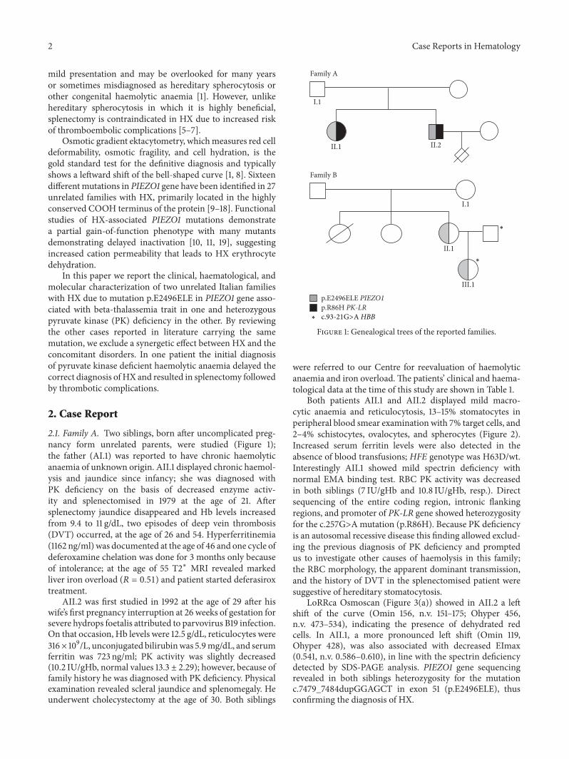
2.1. Family A. Two siblings, born after uncomplicated preg-nancy form unrelated parents, were studied (Figure 1);the father (AI.1) was reported to have chronic haemolyticanaemia of unknown origin. AII.1 displayed chronic haemol-ysis and jaundice since infancy; she was diagnosed withPK deficiency on the basis of decreased enzyme activ-ity and splenectomised in 1979 at the age of 21. Aftersplenectomy jaundice disappeared and Hb levels increasedfrom 9.4 to 11 g/dL, two episodes of deep vein thrombosis(DVT) occurred, at the age of 26 and 54. Hyperferritinemia(1162 ng/ml) was documented at the age of 46 and one cycle ofdeferoxamine chelation was done for 3 months only becauseof intolerance; at the age of 55 T2∗ MRI revealed markedliver iron overload (𝑅 = 0.51) and patient started deferasiroxtreatment.
AII.2 was first studied in 1992 at the age of 29 after hiswife’s first pregnancy interruption at 26 weeks of gestation forsevere hydrops foetalis attributed to parvovirus B19 infection.On that occasion, Hb levels were 12.5 g/dL, reticulocytes were316× 109/L, unconjugated bilirubinwas 5.9mg/dL, and serumferritin was 723 ng/ml; PK activity was slightly decreased(10.2 IU/gHb, normal values 13.3 ± 2.29); however, because offamily history he was diagnosed with PK deficiency. Physicalexamination revealed scleral jaundice and splenomegaly. Heunderwent cholecystectomy at the age of 30. Both siblings
Family A
I.1
II.1 II.2
p.E2496ELE PIEZO1p.R86H PK-LR
I.1
II.1
III.1
Family B
∗
∗
c.93-21G>A HBB∗
Figure 1: Genealogical trees of the reported families.
were referred to our Centre for reevaluation of haemolyticanaemia and iron overload.The patients’ clinical and haema-tological data at the time of this study are shown in Table 1.
Both patients AII.1 and AII.2 displayed mild macro-cytic anaemia and reticulocytosis, 13–15% stomatocytes inperipheral blood smear examination with 7% target cells, and2–4% schistocytes, ovalocytes, and spherocytes (Figure 2).Increased serum ferritin levels were also detected in theabsence of blood transfusions; HFE genotype was H63D/wt.Interestingly AII.1 showed mild spectrin deficiency withnormal EMA binding test. RBC PK activity was decreasedin both siblings (7 IU/gHb and 10.8 IU/gHb, resp.). Directsequencing of the entire coding region, intronic flankingregions, and promoter of PK-LR gene showed heterozygosityfor the c.257G>Amutation (p.R86H). Because PK deficiencyis an autosomal recessive disease this finding allowed exclud-ing the previous diagnosis of PK deficiency and promptedus to investigate other causes of haemolysis in this family;the RBC morphology, the apparent dominant transmission,and the history of DVT in the splenectomised patient weresuggestive of hereditary stomatocytosis.
LoRRca Osmoscan (Figure 3(a)) showed in AII.2 a leftshift of the curve (Omin 156, n.v. 151–175; Ohyper 456,n.v. 473–534), indicating the presence of dehydrated redcells. In AII.1, a more pronounced left shift (Omin 119,Ohyper 428), was also associated with decreased EImax(0.541, n.v. 0.586–0.610), in line with the spectrin deficiencydetected by SDS-PAGE analysis. PIEZO1 gene sequencingrevealed in both siblings heterozygosity for the mutationc.7479_7484dupGGAGCT in exon 51 (p.E2496ELE), thusconfirming the diagnosis of HX.

Case Reports in Hematology 3
Table 1: Clinical and haematological data of the patients at the time of the study.
AII.1# AII.2 BII.1# BIII.1 Normal values
Sex F M F F
Age 55 50 39 9
Detection of anaemia Infancy 29 yrs 15 yrs Birth
Splenectomy (age) Yes (21) No Yes (24) No
Cholecystectomy (age) No Yes (30) Yes (24) No
Transfusions No No No No
Hb (g/dL) 11 13 12 10.8 F 12–16; M 13.5–17.5
MCV (fl) 108 103 100 66.4∗ 78–99
MCH (pg) 39.7 39.2 38.1 24.1 25–35
MCHC (g/dL) 36.5 37.8 38 36.2 31–37
Reticulocytes (×109/L) 166 273 198 423 24–84
Stomatocytes (%) 13 15 33 22
PLTs (×109/L) 696 172 875 329 130–400
WBCs (×109/L) 8.4 7.68 9.31 8.15 4.8–10.8
Unconj bilirubin (mg/dL) 1.33 4.82 1.33 2.8 <0.8
Serum iron (𝜇g/dL) 101 150 167 n.a. 59–158
Serum ferritin (ng/mL) 745 1571 356 321 F 15–150; M 30–400
Transferrin (𝜇g/dL) 149 170 237 221 200–360
Transferrin saturation (%) 56 71 75 n.a. 16–54
GLT (sec) 180 68 480 195 23–45
AGLT (sec) >900 >900 >900 >900 >900
NaCl osmotic fragility Reduced Reduced Reduced Reduced
Pink test (%) 27 28 16 16 11–33
EMA binding test Normal Normal n.a. n.a.
Sp/Bd3 ratio 0.90 1.14 1.11 1.05 0.97–1.19
HFE genotype H63D/wt H63D/wt wt/wt n.a. wt/wt
Gilbert genotype 6/6 6/6 6/7 n.a. 6/6
PK activity (IU/gHb) 7 10.8 nd nd 11.9–16.1
2,3 DPG (nmol/gHb) n.a. 14183 nd nd 10540 ± 1720
ATP n.a. 5674 nd nd 4231 ± 630
PK-LR genotype c.257G>A/wt c.257G>A/wt nd nd∗𝛽-Thalassemia trait; n.a. = not available; nd = not determined.#Postsplenectomy.
2.2. Family B. Patient BII.1 was first recognized to be anaemicat the age of 15 years because of scleral jaundice; at the ageof 23 in 1995 she was diagnosed with hereditary stomato-cytosis based on the presence of stomatocytes at peripheralblood smear examination (12%); in that occasion, she hadHb 11.9 g/dL, MCV 95 fL, reticulocytes 550 × 109/L, serumferritin 421 ng/ml, transferrin saturation 70%, and uncon-jugated bilirubin 6.7mg/dL. Abdominal ultrasound showedsplenomegaly (18 × 12 cm), hepatomegaly, and gallstones.Family study revealed in the mother (BI.1) a compensated
haemolysis (Hb 12.6 g/dL, reticulocytes 311 × 109/L, andunconjugated bilirubin 2mg/dL) with 11% stomatocytes,suggesting dominant transmission of the disease.
One year later the patient underwent cholecystectomyfor gallstones, associated with splenectomy. Surgery had noeffect on Hb levels; postsplenectomy reticulocytes were 200–300 × 109/L and serum ferritin was 300–400 ng/ml. At theage of 30 years, the patient became pregnant and she gavebirth to a girl (BIII.1); pregnancy was complicated by gestosis,with delivery at 35 weeks. At birth the child was admitted

4 Case Reports in Hematology
AII.1 AII.2
BII.1 BIII.1
Figure 2: Light microscopy of peripheral blood, 63x.
0,7
0,6
0,5
0,4
0,3
0,2
0,1
050 150 250 350 450 550
EI
Osmolality (mOsm/Kg)
CtrAII.1AII.2
(a)
0,7
0,6
0,5
0,4
0,3
0,2
0,1
050 150 250 350 450 550
EI
Osmolality (mOsm/Kg)
CtrBII.1BIII.1
(b)
Figure 3: Results of LoRRca Osmoscan analysis in the affected patients compared to a normal control performed by Laser-Assisted OpticalRotation Cell Analyzer (LoRRca MaxSis, Mechatronics, Hoorn, The Netherlands). The osmotic gradient curves reflect RBC deformability asa continuous function of suspendingmedium osmolality.The following parameters were evaluated: EImax (maximal deformability, reflectingmean cellular surface area), Omin (osmolality at which deformability reaches its minimum, reflectingmean cellular surface-to-volume ratio),and Ohyper (the osmolality in the hypertonic region corresponding to 50% of the EImax, reflecting mean cellular hydration status).

Case Reports in Hematology 5
to the Intensive Care Unit for neonatal immaturity and res-piratory distress, and she was diagnosed with hereditarystomatocytosis associated with beta-thalassemia trait (Hb10.6 g/dL, MCV 71.9 fL, stomatocytes 22%, reticulocytes 260× 109/L, and unconjugated bilirubin 1.09mg/dL).
The mother and daughter were referred to our hospitalfor reevaluation of their anaemia and to confirm the diag-nosis at molecular level. At the last follow-up patient BII.1reported that a thromboembolic event occurred 20 years aftersplenectomy. Patient BII.1 displayed compensated haemolyticanaemia with 22% stomatocytes at peripheral blood smearexamination, 13% target cells, and 2% schistocytes (Table 1and Figure 2). Ferritin levels and transferrin saturation wereincreased in the presence of a normal HFE genotype. Bonemarrow evaluation showed marked dyserythropoiesis. CaseBIII.1 had mild microcytic anaemia, reticulocytosis, andincreased ferritin levels; 17% of stomatocytes were detected atblood smear examination (Table 1 and Figure 2); the molecu-lar testing for HBB gene revealed the presence of the splicingmutation c.93-21G>A at heterozygote level, confirming thebeta-thalassemia trait.
LoRRca Osmoscan (Figure 3(b)) showed the pronouncedleft shift of the curve typical of dehydrated stomatocytosis(Omin 71 and 76, resp., n.v. 136–151; Ohyper 342 and 352, n.v.447–474). PIEZO1 gene sequencing revealed in both cases thepresence of the mutation p.E2496ELE.
3. Discussion
There is an increasing interest in defects of RBC permeabilitydue to the recent advances in knowledge of their molecularbases. Considered to be a group of very rare diseases, thenumber of reported cases of HX in the past three years isincreased after the identification of the causative genes, suchas PIEZO1 [9–11] and, more recently, KCNN4 codifying forGardos channel [20–23].
The wide heterogeneity of clinical presentation and inparticular the occurrence of mild/compensated cases maycontribute to delay of the diagnosis [14, 16]. Moreover, somepatients displaying very few stomatocytes at the blood filmexamination may be misdiagnosed as hereditary spherocy-tosis or as suffering from an enzyme deficiency [1]. This wasthe case in patient AII.1, initially diagnosed as PK deficiencybased on decreased enzyme activity, who underwent splenec-tomy and was eventually identified as HX with 30-year delay:the apparently dominant transmission of the anaemia, theabsence of the expected increase of reticulocytes number aftersplenectomy, and PK-LR genotyping showing monoallelicmutation ruled out the diagnosis of PK deficiency as aprimary cause of haemolysis. This case therefore pinpointsthe need of confirming a suspected RBC enzymopathy at themolecular level.
More than in other haemolytic anaemia, a precise diagno-sis is of the utmost importance in HX in light of the increasedrisk of thrombotic complications following splenectomy [5–7] reported in all the splenectomised patients with PIEZO1mutations but one. Patient AII.1 displayed two DVT episodes5 and 33 years after splenectomy, and patient BII.1 had
pulmonary embolism 20 years after surgery. It is worthmentioning that the only patient reported in the literaturewithout thromboembolic complication after splenectomyhad a relatively short follow-up [17].
Although usually well compensated and requiring noblood transfusions, dehydrated stomatocytosis is a heavilyiron loading condition [4, 14], as also shown in our cases.The causes of this are not well established: haemolysis iscommonly mild and it is unlikely to be per se the main causeof iron overload. An important factor in this regard may bedyserythropoiesis, whichwas also present in patient BII.1.Theoccurrence of dyserythropoiesis has been documented in avariant of hereditary stomatocytosis due to Gly796Arg muta-tion of the erythroid anion exchanger [24] and in one casewithHXassociatedwith hereditary high phosphatidylcholinehaemolytic anaemia [17] but excluded in another PIEZO1mutated patient described by Archer et al. [14].
HX has been long known to be a pleiotropic syndromewhich combines in some families with haemolytic anaemiaand perinatal edema; the latter, usually transient and of rela-tively benign prognosis [25–28], has been recently correlatedwithPIEZO1mutation byAndolfo et al. [10].Thenonimmunefoetal hydrops recorded in family A is the most severe formso far described in patients with PIEZO1mutations.
LoRRca Osmoscan proved to be very useful in orientingthe diagnosis towardHX, later confirmed by the PIEZO1 geneanalysis. Interestingly, patient AII.1 also displayed a decreasein EImax, as found in the case reported by Grootenboer-Mignot et al. [27]. In our patient the decrease in EImax isin line with the SDS-PAGE finding of a moderate spectrindeficiency. Spectrin deficiency in the absence of anymutationin RBC cytoskeletal proteins documented by whole exomesequencing has been reported in hereditary stomatocytosisdue to KCNN4 mutation [23] and considered to be a sec-ondary effect of a membrane perturbation [29, 30] disclosedby splenectomy [31].
All the four patients here described were heterozygousformutation p.E2496ELE.Themutationwas already reportedin 8 unrelated cases described by Albuisson et al. [11] andvery recently in a Japanese family [17] (Table 2). Althoughbeing also detected in 2 of 600 healthy French controls, thismutation was considered pathogenic; in fact, patch-clampexperiments in transfected HEK293 cells demonstrated aconsiderable increase in the inactivation time in the mutantcompared to wild-type channel kinetics, indicating that itrepresents a gain-of-function mutation [11]. The finding ofp.E2496ELEmutation also in our unrelated families (the firsttwo families of Italian origin characterized atmolecular level)supports its pathogenicity and confirms that this is one ofthe most frequent mutations in HX, with 15 patients so farreported in literature.
Combined defects of red cell membrane and/or meta-bolism are very rare, and the fact that carriership for a meta-bolic defect may be a modifier for the clinical expressionof a membrane defect is still debated [32, 33]; in particular,the copresence of beta-thalassemia trait seems to reduce thedegree of haemolysis in patients with hereditary spherocy-tosis [34–37]. In the presented cases the clinical phenotypedoes not differ from the HX cases reported in literature

6 Case Reports in Hematology
Table2:Patie
ntsreportedin
literaturew
ithmutationp.E2
496E
LEin
PIEZ
O1g
ene.
Reference
Case
Age
atdiagno
sisFamily
histo
ryPerin
atal
edem
aSplenectom
y(age)
Thrombo
ticevents
Stom
atocytes
TypicalH
Xektacytometry
Hb
(g/dL)
MCV (fL)
MCH
C(%
)Re
tics
(109/L)
Ferritin
(ng/mL)
HFE
[11]
K116
No
No
n.a.
n.a.
n.a.
Yes
10.3
102.8
32.5
136
n.a.
n.a.
[11]
K215
n.a.
n.a.
n.a.
n.a.
n.a.
Yes
14.4
98.5
36202
n.a.
n.a.
[11]
K311
Yes
n.a.
n.a.
n.a.
n.a.
Yes
11.3
91.9
37.3
275
n.a.
n.a.
[11]
K521
Yes
No
n.a.
n.a.
n.a.
Yes
12.7
98.9
37.5
256
n.a.
n.a.
[11]
K618
Yes
No
n.a.
n.a.
n.a.
Yes
12.5
102.5
36378
n.a.
n.a.
[11]
K742
Yes
n.a.
n.a.
n.a.
n.a.
Yes
12.6
108.6
36.5
290
n.a.
n.a.
[11]
F230
Yes
Yes
No
—n.a.
Yes
9.499.1
n.a.
182
n.a.
n.a.
[11]
F326
Yes
No
Yes(27)
Yes
5%Yes
13.9
120
33.8
220
2910
n.a.
[17]
Daughter#
50Yes
No
Yes(38)
No
Yes
n.a.
7.9133.9
n.a.
3.9%
4315
wt
[17]
Son#
41Yes
No
No
—Yes
n.a.
6.6
101.1
n.a.
4.9%
4350
wt
Thisstu
dyAII.1
$55
Yes
No
Yes(21)
Yes
13%
Yes
11108
36.5
166
939
H63D/w
tTh
isstu
dyAII.2
$50
Yes
No
No
—15%
Yes
13103
37.8
273
1571
H63D/w
tTh
isstu
dyBII.1
23Yes
No
Yes(23)
No
33%
Yes
12100
38198
356
wt
Thisstu
dyBIII.1∗
Birth
Yes
No
No
—22%
Yes
10.8
66.4
36.2
423
321
n.a.
∗HXassociated
with𝛽-th
alassemiatrait;
$ HXassociated
with
heterozygotePK
deficiency.
# HXassociated
with
hereditary
high
phosph
atidylcholineh
aemolyticanaemia(H
HPC
HA).
n.a.=no
tavailable.

Case Reports in Hematology 7
(Table 2), thus excluding a synergetic effect between HX andthe concomitant disorders.
In conclusion, our data underline the importance of aprecise diagnosis in HX, particularly in view of splenectomybecause of the increased thrombotic risk, and the need of amolecular confirmation of a suspected RBC enzymopathy.
Competing Interests
The authors declare that there is no conflict of interestsregarding the publication of this paper.
Acknowledgments
This study was supported by the Italian Ministry of Health:Ricerca Finalizzata Conv no. RF-2010-2303934.
References
[1] J. Delaunay, “The hereditary stomatocytoses: genetic disordersof the red cell membrane permeability to monovalent cations,”Seminars in Hematology, vol. 41, no. 2, pp. 165–172, 2004.
[2] E. Glogowska and P. G. Gallagher, “Disorders of erythro-cyte volume homeostasis,” International Journal of LaboratoryHematology, vol. 37, no. 1, pp. 85–91, 2015.
[3] L. J. Bruce, “Hereditary stomatocytosis and cation leaky redcells—recent developments,” Blood Cells, Molecules, and Dis-eases, vol. 42, no. 3, pp. 216–222, 2009.
[4] P.-Y. Syfuss, A. Ciupea, S. Brahimi et al., “Mild dehydratedhereditary stomatocytosis revealed bymarked hepatosiderosis,”Clinical and Laboratory Haematology, vol. 28, no. 4, pp. 270–274, 2006.
[5] A. P.Marcello, C. Vercellati, E. Fermo et al., “A case of congenitalred cell pyruvate kinase deficiency associated with hereditarystomatocytosis,”Blood Cells, Molecules, andDiseases, vol. 41, no.3, pp. 261–262, 2008.
[6] G. W. Stewart, J. A. L. Amess, S. W. Eber et al., “Thrombo-embolic disease after splenectomy for hereditary stomatocyto-sis,” British Journal of Haematology, vol. 93, no. 2, pp. 303–310,1996.
[7] X. Jaıs, S. J. Till, T. Cynober et al., “An extreme consequenceof splenectomy in dehydrated hereditary stomatocytosis: grad-ual thrombo-embolic pulmonary hypertension and lung-hearttransplantation,” Hemoglobin, vol. 27, no. 3, pp. 139–147, 2003.
[8] L. Da Costa, J. Galimand, O. Fenneteau, and N. Mohandas,“Hereditary spherocytosis, elliptocytosis, and other red cellmembrane disorders,” Blood reviews, vol. 27, no. 4, pp. 167–178,2013.
[9] R. Zarychanski, V. P. Schulz, B. L. Houston et al., “Mutations inthe mechanotransduction protein PIEZO1 are associated withhereditary xerocytosis,” Blood, vol. 120, no. 9, pp. 1908–1915,2012.
[10] I. Andolfo, S. L. Alper, L. De Franceschi et al., “Multipleclinical forms of dehydrated hereditary stomatocytosis arisefrom mutations in PIEZO1,” Blood, vol. 121, no. 19, pp. 3925–3935, 2013.
[11] J. Albuisson, S. E. Murthy, M. Bandell et al., “Dehydratedhereditary stomatocytosis linked to gain-of-functionmutationsin mechanically activated PIEZO1 ion channels,” Nature Com-munications, vol. 4, article 1884, 2013.
[12] C. Beneteau, G.Thierry, S. Blesson et al., “Recurrentmutation inthe PIEZO1 gene in two families of hereditary xerocytosis withfetal hydrops,”Clinical Genetics, vol. 85, no. 3, pp. 293–295, 2014.
[13] B. E. Shmukler, D. H. Vandorpe, A. Rivera, M. Auerbach, C.Brugnara, and S. L. Alper, “Dehydrated stomatocytic anemiadue to the heterozygous mutation R2456H in the mechanosen-sitive cation channel PIEZO1: a case report,” Blood Cells,Molecules, and Diseases, vol. 52, no. 1, pp. 53–54, 2014.
[14] N. M. Archer, B. E. Shmukler, I. Andolfo et al., “Hereditaryxerocytosis revisited,” American Journal of Hematology, vol. 89,no. 12, pp. 1142–1146, 2014.
[15] M. B. Sandberg, M. Nybo, H. Birgens, and H. Frederik-sen, “Hereditary xerocytosis and familial haemolysis due tomutation in the PIEZO1 gene: a simple diagnostic approach,”International Journal of Laboratory Hematology, vol. 36, no. 4,pp. e62–e65, 2014.
[16] M. Paessler and H. Hartung, “Dehydrated hereditary stomato-cytosis masquerading as MDS,” Blood, vol. 125, no. 11, p. 1841,2015.
[17] S. Imashuku, H. Muramatsu, T. Sugihara et al., “PIEZO1gene mutation in a Japanese family with hereditary highphosphatidylcholine hemolytic anemia and hemochromatosis-induced diabetes mellitus,” International Journal of Hematology,vol. 104, no. 1, pp. 125–129, 2016.
[18] R. Del Orbe Barreto, B. Arrizabalaga, A. B. De la Hoz Rastrolloet al., “Hereditary xerocytosis, a misleading anemia,” Annals ofHematology, vol. 95, no. 9, pp. 1545–1546, 2016.
[19] C. Bae, R. Gnanasambandam, C. Nicolai, F. Sachs, and P. A.Gottlieb, “Xerocytosis is caused by mutations that alter thekinetics of the mechanosensitive channel PIEZO1,” Proceedingsof the National Academy of Sciences of the United States ofAmerica, vol. 110, no. 12, pp. E1162–E1168, 2013.
[20] R. Rapetti-Mauss, C. Lacoste, V. Picard et al., “A mutation inthe Gardos channel is associated with hereditary xerocytosis,”Blood, vol. 126, no. 11, pp. 1273–1280, 2015.
[21] I. Andolfo, R. Russo, F. Manna et al., “Novel Gardos chan-nel mutations linked to dehydrated hereditary stomatocytosis(xerocytosis),” American Journal of Hematology, vol. 90, no. 10,pp. 921–926, 2015.
[22] E. Glogowska, K. Lezon-Geyda, Y.Maksimova, V. P. Schulz, andP. G. Gallagher, “Mutations in theGardos channel (KCNN4) areassociated with hereditary xerocytosis,” Blood, vol. 126, no. 11,pp. 1281–1284, 2015.
[23] E. Fermo, A. Y. Bogdanova, P. Petkova-Kirova et al., “Gar-dos channel mutation is associated with hereditary dehydratestomatocytosis: a complex channelopathy,” Blood, vol. 126,supplement 1, article 3333, 2015.
[24] A. Iolascon, L. De Falco, F. Borgese et al., “A novel erythroidanion exchange variant (Gly796Arg) of hereditary stomatocy-tosis associated with dyserythropoiesis,”Haematologica, vol. 94,no. 8, pp. 1049–1059, 2009.
[25] S. Grootenboer, C. Barro, T. Cynober et al., “Dehydratedhereditary stomatocytosis: a cause of prenatal ascites,” PrenatalDiagnosis, vol. 21, no. 13, pp. 1114–1118, 2001.
[26] S. Grootenboer, P. O. Schischmanoff, T. Cynober et al., “Agenetic syndrome associating dehydrated hereditary stomato-cytosis, pseudohyperkalaemia and perinatal oedema,” BritishJournal of Haematology, vol. 103, no. 2, pp. 383–386, 1998.
[27] S. Grootenboer-Mignot, A. Cretien, I. Laurendeau et al., “Sub-lethal hydrops as a manifestation of dehydrated hereditarystomatocytosis in two consecutive pregnancies,” Prenatal Diag-nosis, vol. 23, no. 5, pp. 380–384, 2003.
8 Case Reports in Hematology
[28] M. Entezami, R. Becker, M.Marcinkowski, and H. T. Versmold,“Xerocytosis with concomitant intrauterine ascites: firstdescription and therapeutic approach,” Blood, vol. 87, no. 12,pp. 5392–5393, 1996.
[29] N. Fortier, L. M. Snyder, F. Garver, C. Kiefer, J. McKenney,and N. Mohandas, “The relationship between in vivo generatedhemoglobin skeletal protein complex and increased red cellmembrane rigidity,” Blood, vol. 71, no. 5, pp. 1427–1431, 1988.
[30] M. Salomao, K. Chen, J. Villalobos, N. Mohandas, X. An, and J.A. Chasis, “Hereditary spherocytosis and hereditary elliptocy-tosis: aberrant protein sorting during erythroblast enucleation,”Blood, vol. 116, no. 2, pp. 267–269, 2010.
[31] M. Mariani, W. Barcellini, C. Vercellati et al., “Clinical andhematologic features of 300 patients affected by hereditaryspherocytosis grouped according to the type of the membraneprotein defect,”Haematologica, vol. 93, no. 9, pp. 1310–1317, 2008.
[32] C. Vercellati, A. P. Marcello, E. Fermo, W. Barcellini, A.Zanella, and P. Bianchi, “A case of hereditary spherocytosismisdiagnosed as pyruvate kinase deficient hemolytic anemia,”Clinical Laboratory, vol. 59, no. 3-4, pp. 421–424, 2013.
[33] R. van Zwieten, B. A. van Oirschot, M. Veldthuis et al.,“Partial pyruvate kinase deficiency aggravates the phenotypicexpression of band 3 deficiency in a family with hereditaryspherocytosis,” American Journal of Hematology, vol. 90, no. 3,pp. E35–E39, 2015.
[34] M. K. Cihan, H. Gokce, M. Oruc, and L. Olcay, “Hemolyticcrisis as the initial presentation of hereditary spherocytosisinduced by parvovirus B19 and herpes virus infection in apatient with the thalassemia trait: a case report,”Turkish Journalof Hematology, vol. 29, no. 4, pp. 425–426, 2012.
[35] E. M. Del Giudice, S. Perrotta, B. Nobili, L. Pinto, L. Cutillo, andA. Iolascon, “Coexistence of hereditary spherocytosis (HS) dueto band 3 deficiency and 𝛽-thalassaemia trait: partial correctionof HS phenotype,” British Journal of Haematology, vol. 85, no. 3,pp. 553–557, 1993.
[36] M. Aksoy and S. Erdem, “TheCombination ofHereditary Sphe-rocytosis andHeterozygous Beta-Thalassaemia,”ActaHaemato-logica, vol. 39, no. 3, pp. 183–191, 1968.
[37] J. M. Andrien, A. Heusden, C. Lambotte, and J. Hugues,“[Hereditary spherocytosis and beta-thalassemia: coexistenceof 2 genes in a Sicilian family],” Acta paediatrica Belgica, vol.25, no. 1, pp. 35–43, 1971.

Submit your manuscripts athttps://www.hindawi.com
Stem CellsInternational
Hindawi Publishing Corporationhttp://www.hindawi.com Volume 2014
Hindawi Publishing Corporationhttp://www.hindawi.com Volume 2014
MEDIATORSINFLAMMATION
of
Hindawi Publishing Corporationhttp://www.hindawi.com Volume 2014
Behavioural Neurology
EndocrinologyInternational Journal of
Hindawi Publishing Corporationhttp://www.hindawi.com Volume 2014
Hindawi Publishing Corporationhttp://www.hindawi.com Volume 2014
Disease Markers
Hindawi Publishing Corporationhttp://www.hindawi.com Volume 2014
BioMed Research International
OncologyJournal of
Hindawi Publishing Corporationhttp://www.hindawi.com Volume 2014
Hindawi Publishing Corporationhttp://www.hindawi.com Volume 2014
Oxidative Medicine and Cellular Longevity
Hindawi Publishing Corporationhttp://www.hindawi.com Volume 2014
PPAR Research
The Scientific World JournalHindawi Publishing Corporation http://www.hindawi.com Volume 2014
Immunology ResearchHindawi Publishing Corporationhttp://www.hindawi.com Volume 2014
Journal of
ObesityJournal of
Hindawi Publishing Corporationhttp://www.hindawi.com Volume 2014
Hindawi Publishing Corporationhttp://www.hindawi.com Volume 2014
Computational and Mathematical Methods in Medicine
OphthalmologyJournal of
Hindawi Publishing Corporationhttp://www.hindawi.com Volume 2014
Diabetes ResearchJournal of
Hindawi Publishing Corporationhttp://www.hindawi.com Volume 2014
Hindawi Publishing Corporationhttp://www.hindawi.com Volume 2014
Research and TreatmentAIDS
Hindawi Publishing Corporationhttp://www.hindawi.com Volume 2014
Gastroenterology Research and Practice
Hindawi Publishing Corporationhttp://www.hindawi.com Volume 2014
Parkinson’s Disease
Evidence-Based Complementary and Alternative Medicine
Volume 2014Hindawi Publishing Corporationhttp://www.hindawi.com