
Gene-Expression Profiles in Hereditary Breast Cancer
10
The New England Journal of Medicine Copyright © 2001 by the Massachusetts Medical Society VOLUME 344 F EBRUARY 22, 2001 NUMBER 8 N Engl J Med, Vol. 344, No. 8 · February 22, 2001 · www.nejm.org · 539 GENE-EXPRESSION PROFILES IN HEREDITARY BREAST CANCER INGRID HEDENFALK, M.S., DAVID DUGGAN, PH.D., YIDONG CHEN, PH.D., MICHAEL RADMACHER, PH.D., MICHAEL BITTNER, PH.D., RICHARD SIMON, D.SC., PAUL MELTZER, M.D., PH.D., BARRY GUSTERSON, M.D., PH.D., MANEL ESTELLER, M.D., PH.D., OLLI-P. KALLIONIEMI, M.D., PH.D., BENJAMIN WILFOND, M.D., ÅKE BORG, PH.D., AND JEFFREY TRENT, PH.D. ABSTRACT Background Many cases of hereditary breast can- cer are due to mutations in either the BRCA1 or the BRCA2 gene. The histopathological changes in these cancers are often characteristic of the mutant gene. We hypothesized that the genes expressed by these two types of tumors are also distinctive, perhaps al- lowing us to identify cases of hereditary breast cancer on the basis of gene-expression profiles. Methods RNA from samples of primary tumors from seven carriers of the BRCA1 mutation, seven carriers of the BRCA2 mutation, and seven patients with spo- radic cases of breast cancer was compared with a mi- croarray of 6512 complementary DNA clones of 5361 genes. Statistical analyses were used to identify a set of genes that could distinguish the BRCA1 geno- type from the BRCA2 genotype. Results Permutation analysis of multivariate classi- fication functions established that the gene-expression profiles of tumors with BRCA1 mutations, tumors with BRCA2 mutations, and sporadic tumors differed sig- nificantly from each other. An analysis of variance between the levels of gene expression and the gen- otype of the samples identified 176 genes that were differentially expressed in tumors with BRCA1 muta- tions and tumors with BRCA2 mutations. Given the known properties of some of the genes in this panel, our findings indicate that there are functional differ- ences between breast tumors with BRCA1 mutations and those with BRCA2 mutations. Conclusions Significantly different groups of genes are expressed by breast cancers with BRCA1 muta- tions and breast cancers with BRCA2 mutations. Our results suggest that a heritable mutation influences the gene-expression profile of the cancer. (N Engl J Med 2001;344:539-48.) Copyright © 2001 Massachusetts Medical Society. From the Cancer Genetics Branch (I.H., D.D., Y.C., M.B., P.M., O.-P.K., J.T.) and the Medical Genetics Branch (B.W.), National Human Genome Research Institute, and the Division of Cancer Treatment and Diagnosis, National Cancer Institute (M.R., R.S.), National Institutes of Health, Be- thesda, Md.; the Department of Oncology, University of Lund, Lund, Sweden (I.H., Å.B.); the Department of Pathology, Western Infirmary, Uni- versity of Glasgow, Glasgow, Scotland (B.G.); and the Division of Tumor Biology, Johns Hopkins Oncology Center, Baltimore (M.E.). Address re- print requests to Dr. Trent at the National Human Genome Research In- stitute, National Institutes of Health, Bldg. 49, Rm. 4A22, Bethesda, MD 20892-4470, or at [email protected]. Other authors were Mark Raffeld, M.D. (Department of Pathology, Na- tional Cancer Institutes of Health, Bethesda, Md.); Zohar Yakhini, Ph.D., and Amir Ben-Dor, Ph.D. (Chemical and Biological Systems Department, Agilent Laboratories, Palo Alto, Calif.); Edward Dougherty, Ph.D. (De- partment of Electrical Engineering, Texas A&M University, College Sta- tion); Juha Kononen, M.D., Ph.D. (Cancer Genetics Branch, National Hu- man Genome Research Institute, National Institutes of Health, Bethesda, Md.); Lukas Bubendorf, M.D. (Cancer Genetics Branch, National Human Genome Research Institute, National Institutes of Health, Bethesda, Md., and the Institute of Pathology, University of Basel, Basel, Switzerland); Wilfrid Fehrle, M.D., and Stefania Pittaluga, M.D. (Department of Pathol- ogy, National Cancer Institute, National Institutes of Health, Bethesda, Md.); Sofia Gruvberger, M.S., Niklas Loman, M.D., Oskar Johannsson, M.D., Ph.D., and Håkan Olsson, M.D., Ph.D. (Department of Oncology, University of Lund, Lund, Sweden); and Guido Sauter, M.D. (Department of Pathology, University of Basel, Basel, Switzerland). NHERITANCE of a mutant BRCA1 or BRCA2 gene (numbers 113705 and 600185, respective- ly, in Online Mendelian Inheritance in Man, a catalogue of inherited diseases) confers a lifetime risk of breast cancer of 50 to 85 percent and a lifetime risk of ovarian cancer of 15 to 45 percent. 1-6 These germ-line mutations account for a substantial pro- portion of inherited breast and ovarian cancers, 7 but it is likely that additional susceptibility genes will be discovered. 8,9 Certain pathological features can help to distinguish breast tumors with BRCA1 mutations from those with BRCA2 mutations. Tumors with BRCA1 mutations are high-grade cancers with a high mitotic index, “pushing” tumor margins (i.e., noninfiltrating, smooth edges), and a lymphocytic infiltrate, whereas tumors with BRCA2 mutations are heterogeneous, are often relatively high grade, and display substantially less tu- I Copyright © 2001 Massachusetts Medical Society. All rights reserved. Downloaded from www.nejm.org at UNIVERSITY OF WISCONSIN on November 22, 2004 .
Transcript of Gene-Expression Profiles in Hereditary Breast Cancer

The New England
Journal
of
Medicine
Copyr ight © 2001 by the Massachusett s Medical Society
VOLUME 344
F
EBRUARY
22, 2001
NUMBER 8
N Engl J Med, Vol. 344, No. 8
·
February 22, 2001
·
www.nejm.org
·
539
GENE-EXPRESSION PROFILES IN HEREDITARY BREAST CANCER
I
NGRID
H
EDENFALK
, M.S., D
AVID
D
UGGAN
, P
H
.D., Y
IDONG
C
HEN
, P
H
.D., M
ICHAEL
R
ADMACHER
, P
H
.D., M
ICHAEL
B
ITTNER
, P
H
.D., R
ICHARD
S
IMON
, D.S
C
., P
AUL
M
ELTZER
, M.D., P
H
.D., B
ARRY
G
USTERSON
, M.D., P
H
.D.,M
ANEL
E
STELLER
, M.D., P
H
.D., O
LLI
-P. K
ALLIONIEMI
, M.D., P
H
.D., B
ENJAMIN
W
ILFOND
, M.D., Å
KE
B
ORG
, P
H
.D.,
AND
J
EFFREY
T
RENT
, P
H
.D.
A
BSTRACT
Background
Many cases of hereditary breast can-cer are due to mutations in either the
BRCA1
or the
BRCA2
gene. The histopathological changes in thesecancers are often characteristic of the mutant gene.We hypothesized that the genes expressed by thesetwo types of tumors are also distinctive, perhaps al-lowing us to identify cases of hereditary breast canceron the basis of gene-expression profiles.
Methods
RNA from samples of primary tumors fromseven carriers of the
BRCA1
mutation, seven carriersof the
BRCA2
mutation, and seven patients with spo-radic cases of breast cancer was compared with a mi-croarray of 6512 complementary DNA clones of 5361genes. Statistical analyses were used to identify aset of genes that could distinguish the
BRCA1
geno-type from the
BRCA2
genotype.
Results
Permutation analysis of multivariate classi-fication functions established that the gene-expressionprofiles of tumors with
BRCA1
mutations, tumors with
BRCA2
mutations, and sporadic tumors differed sig-nificantly from each other. An analysis of variancebetween the levels of gene expression and the gen-otype of the samples identified 176 genes that weredifferentially expressed in tumors with
BRCA1
muta-tions and tumors with
BRCA2
mutations. Given theknown properties of some of the genes in this panel,our findings indicate that there are functional differ-ences between breast tumors with
BRCA1
mutationsand those with
BRCA2
mutations.
Conclusions
Significantly different groups of genesare expressed by breast cancers with
BRCA1
muta-tions and breast cancers with
BRCA2
mutations. Ourresults suggest that a heritable mutation influencesthe gene-expression profile of the cancer. (N Engl JMed 2001;344:539-48.)
Copyright © 2001 Massachusetts Medical Society.
From the Cancer Genetics Branch (I.H., D.D., Y.C., M.B., P.M., O.-P.K.,J.T.) and the Medical Genetics Branch (B.W.), National Human GenomeResearch Institute, and the Division of Cancer Treatment and Diagnosis,National Cancer Institute (M.R., R.S.), National Institutes of Health, Be-thesda, Md.; the Department of Oncology, University of Lund, Lund,Sweden (I.H., Å.B.); the Department of Pathology, Western Infirmary, Uni-versity of Glasgow, Glasgow, Scotland (B.G.); and the Division of TumorBiology, Johns Hopkins Oncology Center, Baltimore (M.E.). Address re-print requests to Dr. Trent at the National Human Genome Research In-stitute, National Institutes of Health, Bldg. 49, Rm. 4A22, Bethesda, MD20892-4470, or at [email protected].
Other authors were Mark Raffeld, M.D. (Department of Pathology, Na-tional Cancer Institutes of Health, Bethesda, Md.); Zohar Yakhini, Ph.D.,and Amir Ben-Dor, Ph.D. (Chemical and Biological Systems Department,Agilent Laboratories, Palo Alto, Calif.); Edward Dougherty, Ph.D. (De-partment of Electrical Engineering, Texas A&M University, College Sta-tion); Juha Kononen, M.D., Ph.D. (Cancer Genetics Branch, National Hu-man Genome Research Institute, National Institutes of Health, Bethesda,Md.); Lukas Bubendorf, M.D. (Cancer Genetics Branch, National HumanGenome Research Institute, National Institutes of Health, Bethesda, Md.,and the Institute of Pathology, University of Basel, Basel, Switzerland);Wilfrid Fehrle, M.D., and Stefania Pittaluga, M.D. (Department of Pathol-ogy, National Cancer Institute, National Institutes of Health, Bethesda,Md.); Sofia Gruvberger, M.S., Niklas Loman, M.D., Oskar Johannsson,M.D., Ph.D., and Håkan Olsson, M.D., Ph.D. (Department of Oncology,University of Lund, Lund, Sweden); and Guido Sauter, M.D. (Departmentof Pathology, University of Basel, Basel, Switzerland).
NHERITANCE of a mutant
BRCA1
or
BRCA2
gene (numbers 113705 and 600185, respective-ly, in Online Mendelian Inheritance in Man, acatalogue of inherited diseases) confers a lifetime
risk of breast cancer of 50 to 85 percent and a lifetimerisk of ovarian cancer of 15 to 45 percent.
1-6
Thesegerm-line mutations account for a substantial pro-portion of inherited breast and ovarian cancers,
7
butit is likely that additional susceptibility genes will bediscovered.
8,9
Certain pathological features can help to distinguishbreast tumors with
BRCA1
mutations from those with
BRCA2
mutations. Tumors with
BRCA1
mutationsare high-grade cancers with a high mitotic index,“pushing” tumor margins (i.e., noninfiltrating, smoothedges), and a lymphocytic infiltrate, whereas tumorswith
BRCA2
mutations are heterogeneous, are oftenrelatively high grade, and display substantially less tu-
I
Copyright © 2001 Massachusetts Medical Society. All rights reserved. Downloaded from www.nejm.org at UNIVERSITY OF WISCONSIN on November 22, 2004 .

540
·
N Engl J Med, Vol. 344, No. 8
·
February 22, 2001
·
www.nejm.org
The New England Journal of Medicine
bule formation. The proportion of the perimeter withcontinuous pushing margins can distinguish bothtypes of tumors from sporadic cases of breast cancer.
10
Tumors with
BRCA1
mutations are generally negativefor both estrogen and progesterone receptors, where-as most tumors with
BRCA2
mutations are positive forthese hormone receptors.
11-14
These differences implythat the mutant
BRCA1
and
BRCA2
genes induce theformation of breast tumors through separate pathways.
The BRCA1 and BRCA2 proteins participate inDNA repair and homologous recombination andprobably other cellular processes.
15
A cell with a mu-tant
BRCA1
or
BRCA2
gene, which therefore lacksfunctional BRCA1 or BRCA2 protein, has a decreasedability to repair damaged DNA. In animal models, thisdefect causes genomic instability.
16
In humans, breasttumors in carriers of mutant
BRCA1
or
BRCA2
genesare characterized by a large number of chromosomalchanges, some of which differ depending on the gen-otype.
17
In this study, we examined breast-cancer tissuesfrom patients with
BRCA1
-related cancer, patientswith
BRCA2
-related cancer, and patients with spo-radic cases of breast cancer to determine whether thereare distinctive patterns of global gene expression inthese three kinds of tumors.
METHODS
Patients and Biopsy Specimens
Patients with primary breast cancer and who had a family historyof breast or ovarian cancer, or both, that was compatible with adominant mode of inheritance were referred for genetic counselingto the Oncogenetic Clinic of Lund University Hospital. These pa-tients were asked to provide a blood sample and to sign an informed-consent form authorizing an analysis for
BRCA1
and
BRCA2
mu-tations. Mutation analysis was performed as described previously.
18
Biopsy specimens of primary breast tumors from patients withgerm-line mutations of
BRCA1
(seven patients) or
BRCA2
(eighttumors from seven patients) were selected for analysis. In addition,seven patients with sporadic cases of primary breast cancer whosefamily history was unknown were also identified. These patientshad either estrogen-receptor–negative, aggressive tumors (charac-terized by aneuploidy and a high fraction of cells in S phase) orestrogen-receptor–positive, less aggressive tumors. Total RNA wasextracted from flash-frozen tumor specimens, which had been storedat ¡80°C, with the use of the RNeasy Maxi Kit (Qiagen) and Tri-zol reagent (GIBCO BRL) according to the manufacturers’ rec-ommendations.
19
The studies were approved by the institutional review boards ofboth Lund University and the National Human Genome ResearchInstitute of the National Institutes of Health.
Microarrays of Complementary DNA
We obtained samples of complementary DNA (cDNA) with ver-ified sequences
20
under a Cooperative Research and DevelopmentAgreement with Research Genetics. Gene names are listed accord-ing to build 110 of the UniGene human-sequence collection (avail-able at the UniGene Web site: http://www.ncbi.nlm.nih.gov/UniGene/build.html). The 6512 cDNAs we used represent 5361unique genes: 2905 are known and 2456 are unknown genes.
Microarrays were hybridized and scanned, and image analysis wasperformed as described previously (Fig. 1).
20-22
The reference cellline, MCF-10A (American Type Culture Collection, CRL-10317),a nontumorigenic breast-cell line, was an internal standard against
which each tumor was compared (not a biologic control). RNAfrom normal breast epithelial cells was included for comparison(Fig. 2B).
Tissue Microarrays
A microarray of breast-cancer tissue (Fig. 1), constructed as pre-viously described,
23
consisted of samples of 113 primary breast tu-mors, in duplicate, derived from a population-based series of patientsfrom southern Sweden in whom the disease had been diagnosed be-fore the age of 40 years. The patients consisted of 23 with
BRCA1
mutations, 17 with
BRCA2
mutations, 20 with familial breast can-cer (defined as a history of breast or ovarian cancer in at least onefirst-degree relative) but no
BRCA1
or
BRCA2
mutations, 19 withpossibly familial breast cancer (defined as a history of breast or ovar-ian cancer in at least one second-degree relative) but no
BRCA1
or
BRCA2
mutations, and 34 with sporadic breast cancer. The dupli-cate core-tissue–biopsy specimens (diameter, 0.6 mm) were ob-tained from the least differentiated regions of individual paraffin-embedded tumors.
Analysis of DNA Methylation
Patterns of DNA methylation in the CpG island of the
BRCA1
gene were determined by a methylation-specific polymerase chainreaction.
24
Statistical Analysis
Tests for associations between each type of mutation (
BRCA1
or
BRCA2
) and clinical variables were performed with Fisher’s exacttest for categorical variables and the Wilcoxon–Mann–Whitney testfor continuous and ordered variables. Reported P values are exactand have not been corrected for multiple comparisons (30 variableswere tested). All P values are two-sided.
In the analyses involving cDNA microarrays, a total of 3226 geneswith an average intensity (level of expression) of more than 2500pixels among all samples, an average spot area of more than 40 pix-els, and no more than one sample in which the size of the spot areawas 0 pixels were included.
22
A conservative estimate of experimen-tal variance (involving hybridization of pairs of cDNAs on differentdays) indicated that our observations fell within the 95 percent con-fidence interval of 0.61 to 1.65 for a mean value of 1.0.
We used a class-prediction method to determine whether the pat-terns of gene expression could be used to classify tumor samples intotwo classes according to the presence or absence of BRCA1 andBRCA2 mutations (positive or negative for BRCA1 mutations andpositive or negative for BRCA2 mutations), with use of a compoundcovariate predictor.25 We estimated the misclassification rate usingleave-one-out cross-validation and used random permutations of theclass-membership indicators to determine the significance of theresults.
We used three methods to generate lists of genes with differentlevels of expression among the groups of patients with breast can-cer: modified F tests and t-tests, a weighted gene analysis, and mu-tual-information scoring (InfoScore). InfoScore uses a ranking-based scoring system and combinatorial permutation of samplelabels to produce a rigorous statistical benchmarking of the over-abundance of genes whose differential expression pattern corre-lates with sample type (information available at http://www.labs.agilent.com/resources/techreports.html). An agglomerative hier-archical clustering algorithm was used to investigate any relationamong the statistically significant discriminator genes.19,20 We alsoused multidimensional scaling to show the correlation of expres-sion of given subgroups of genes among various tumor samples.20
In this three-dimensional rendering of the data, samples withsimilar expression profiles lie closer to each other than those withdissimilar profiles.
Supplemental Information
Additional information on the methods, clones, genes, samples,fluorescence-intensity ratios, and statistical methods is available at
Copyright © 2001 Massachusetts Medical Society. All rights reserved. Downloaded from www.nejm.org at UNIVERSITY OF WISCONSIN on November 22, 2004 .

GENE-EXPRESSION PROFILES IN HEREDITARY BREAST CANCER
N Engl J Med, Vol. 344, No. 8 · February 22, 2001 · www.nejm.org · 541
http://www.nejm.org and at http://www.nhgri.nih.gov/DIR/Microarray.
RESULTS
Characteristics of the Tumors
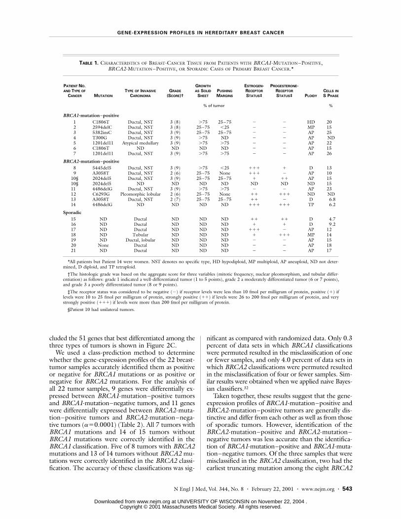
Mutations in seven carriers of BRCA1 mutationsand seven carriers of BRCA2 mutations were con-firmed by direct sequencing (Table 1). Specimens werealso obtained from seven patients with sporadic pri-mary breast cancer. Tumors were classified patholog-ically according to criteria of the Breast Cancer Link-
age Consortium10,26,27; all slides were read by a singlepathologist. Grading was performed according to apreviously described method.28 The pathological re-sults for our cohort were similar to those of earlierstudies.10,12,26,29-31 All tumors with BRCA1 mutationswere grade 3, most had lymphocytic infiltration andextensive pushing margins, most tended to grow insheets, and several had confluent necrosis; there wasone atypical medullary carcinoma. These features asa whole were not as common among patients withBRCA2 mutations.30,31 As expected, estrogen and pro-
Figure 1. Overview of Procedures for Preparing and Analyzing Microarrays of Complementary DNA (cDNA) and Breast-Tumor Tissue.As shown in Panel A, reference RNA and tumor RNA are labeled by reverse transcription with different fluorescent dyes (green forthe reference cells and red for the tumor cells) and hybridized to a cDNA microarray containing robotically printed cDNA clones.As shown in Panel B, the slides are scanned with a confocal laser scanning microscope, and color images are generated for eachhybridization with RNA from the tumor and reference cells. Genes up-regulated in the tumors appear red, whereas those with de-creased expression appear green. Genes with similar levels of expression in the two samples appear yellow. Genes of interest areselected on the basis of the differences in the level of expression by known tumor classes (e.g., BRCA1-mutation–positive andBRCA2-mutation–positive). Statistical analysis determines whether these differences in the gene-expression profiles are greaterthan would be expected by chance. As shown in Panel C, the differences in the patterns of gene expression between tumor classescan be portrayed in the form of a color-coded plot, and the relations between tumors can be portrayed in the form of a multidi-mensional-scaling plot. Tumors with similar gene-expression profiles cluster close to one another in the multidimensional-scalingplot. As shown in Panel D, particular genes of interest can be further studied through the use of a large number of arrayed, paraffin-embedded tumor specimens, referred to as tissue microarrays. As shown in Panel E, immunohistochemical analyses of hundredsor thousands of these arrayed biopsy specimens can be performed in order to extend the microarray findings.
A B
D
DonorFparaffinF
block
RecipientFparaffinF
block Tissue microarray
Multidimensional-scaling plot
E
StatisticalFanalysis
Tumors
Gen
es
C
cDNA
ReferenceFRNA
TumorFRNA
HybridizationFof probe toFmicroarray
Copyright © 2001 Massachusetts Medical Society. All rights reserved. Downloaded from www.nejm.org at UNIVERSITY OF WISCONSIN on November 22, 2004 .

542 · N Engl J Med, Vol. 344, No. 8 · February 22, 2001 · www.nejm.org
The New England Journal of Medicine
gesterone receptors were absent in tumors from all thepatients with BRCA1 mutations and also from one pa-tient with a BRCA2 mutation.11,12
Use of Gene-Expression Profiles to Identify Hereditary Breast Cancers
Fluorescence-intensity ratios were calculated andgene-expression profiles were generated for each sam-ple. The gene-expression profiles were used to deter-mine which of the genes expressed by the tumors cor-related with the BRCA1-mutation–positive tumors,
the BRCA2-mutation–positive tumors, and the spo-radic tumors. Figure 2A shows the results of a modifiedF test, which yielded 51 genes (a=0.001) whose vari-ation in expression among all experiments best differ-entiated among these types of cancers. The multidi-mensional-scaling plot of the 22 samples from patientswith primary breast cancer and 2 samples of normalmammary epithelial cells that included all 3226 genesthat met the criteria for inclusion is shown in Figure2B. The multidimensional-scaling plot of the 22 sam-ples from patients with primary breast cancer that in-
Figure 2. Identification of Genes That Can Be Used to Differentiate BRCA1-Mutation–Positive, BRCA2-Mutation–Positive, and Spo-radic Cases of Primary Breast Cancer.Panel A shows the 51 genes that best differentiated among the three types of tumors, as determined by a modified F test (a=0.001).Panel B shows the multidimensional-scaling plot of the seven samples from patients with BRCA1-mutation–positive breast tumors(blue circles), eight samples from patients with BRCA2-mutation–positive tumors (tan circles), seven samples from patients withsporadic tumors (gray circles), and two samples of normal mammary epithelial cells (pink circles) that included all 3226 genes thatmet the criteria for inclusion in the analysis. Panel C shows the multidimensional-scaling plot of the 22 primary-tumor samples thatincluded the 51 genes that best differentiated the three types of tumors, as evidenced by the clustering of the BRCA2-mutation–positive samples and the BRCA1-mutation–positive samples.
A
BRCA2-ŁMutation–F
PositiveFTumors
SporadicFTumors
BRCA1-FMutation–F
PositiveFTumors
BClone Gene897781F139354F809981F841617F823940F29054F810057F950682F26184F344109F36775F341130F417124F429135F44666F340644F246194F51209F949932F784830F26082F46019F247818F214731F236055F197176F566887F725680F823775F293104F46182F307843F366647F212198F42888F38763F366824F840702F564803F137638F73531F32231F274638F234150F701481F838568F31842F666377F50413F345645F810551F
KRT8�HSPC195�GPX4�ODC antizyme�TOB1�ACTR1A�CSDA�PFKP�PFKP�PCNA�HADHA�RBL2�APEX�ST13�G22P1�ITGB8�ESTs�PPP1CB�NSEP1�D123�VLDLR�MCM7�ESTs�KIAA0601�DKFZP564M2423�GDI2�HECH�TFAP2C�GNAI3�PHYH�CTPS�ESTs�BRF1�TP53BP2�ILF2�SPHAR�CDK4�SPS�FOXM1�ESTs�NIFU�KIAA0246�CAD�MTMR4�MX2�COX6C�UGTREL1�ZNF161�ARVCF�PDGFB�LRP1�
C
Copyright © 2001 Massachusetts Medical Society. All rights reserved. Downloaded from www.nejm.org at UNIVERSITY OF WISCONSIN on November 22, 2004 .
GENE-EXPRESSION PROFILES IN HEREDITARY BREAST CANCER
N Engl J Med, Vol. 344, No. 8 · February 22, 2001 · www.nejm.org · 543
cluded the 51 genes that best differentiated among thethree types of tumors is shown in Figure 2C.
We used a class-prediction method to determinewhether the gene-expression profiles of the 22 breast-tumor samples accurately identified them as positiveor negative for BRCA1 mutations or as positive ornegative for BRCA2 mutations. For the analysis ofall 22 tumor samples, 9 genes were differentially ex-pressed between BRCA1-mutation–positive tumorsand BRCA1-mutation–negative tumors, and 11 geneswere differentially expressed between BRCA2-muta-tion–positive tumors and BRCA2-mutation–nega-tive tumors (a=0.0001) (Table 2). All 7 tumors withBRCA1 mutations and 14 of 15 tumors withoutBRCA1 mutations were correctly identified in theBRCA1 classification. Five of 8 tumors with BRCA2mutations and 13 of 14 tumors without BRCA2 mu-tations were correctly identified in the BRCA2 classi-fication. The accuracy of these classifications was sig-
nificant as compared with randomized data. Only 0.3percent of data sets in which BRCA1 classificationswere permuted resulted in the misclassification of oneor fewer samples, and only 4.0 percent of data sets inwhich BRCA2 classifications were permuted resultedin the misclassification of four or fewer samples. Sim-ilar results were obtained when we applied naive Bayes-ian classifiers.32
Taken together, these results suggest that the gene-expression profiles of BRCA1-mutation–positive andBRCA2-mutation–positive tumors are generally dis-tinctive and differ from each other as well as from thoseof sporadic tumors. However, identification of theBRCA2-mutation–positive and BRCA2-mutation–negative tumors was less accurate than the identifica-tion of BRCA1-mutation–positive and BRCA1-muta-tion–negative tumors. Of the three samples that weremisclassified in the BRCA2 classification, two had theearliest truncating mutation among the eight BRCA2
*All patients but Patient 14 were women. NST denotes no specific type, HD hypodiploid, MP multiploid, AP aneuploid, ND not deter-mined, D diploid, and TP tetraploid.
†The histologic grade was based on the aggregate score for three variables (mitotic frequency, nuclear pleomorphism, and tubular differ-entiation) as follows: grade 1 indicated a well-differentiated tumor (1 to 5 points), grade 2 a moderately differentiated tumor (6 or 7 points),and grade 3 a poorly differentiated tumor (8 or 9 points).
‡The receptor status was considered to be negative (¡) if receptor levels were less than 10 fmol per milligram of protein, positive (+) iflevels were 10 to 25 fmol per milligram of protein, strongly positive (++) if levels were 26 to 200 fmol per milligram of protein, and verystrongly positive (+++) if levels were more than 200 fmol per milligram of protein.
§Patient 10 had unilateral tumors.
TABLE 1. CHARACTERISTICS OF BREAST-CANCER TISSUE FROM PATIENTS WITH BRCA1-MUTATION–POSITIVE, BRCA2-MUTATION–POSITIVE, OR SPORADIC CASES OF PRIMARY BREAST CANCER.*
PATIENT NO. AND TYPE OF
CANCER MUTATION
TYPE OF INVASIVE
CARCINOMA
GRADE
(SCORE)†
GROWTH
AS SOLID
SHEET
PUSHING
MARGINS
ESTROGEN-RECEPTOR
STATUS‡
PROGESTERONE-RECEPTOR
STATUS‡ PLOIDY
CELLS IN
S PHASE
% of tumor %
BRCA1-mutation–positive
1 C1806T Ductal, NST 3 (8) >75 25–75 ¡ ¡ HD 202 2594delC Ductal, NST 3 (8) 25–75 <25 ¡ ¡ MP 153 5382insC Ductal, NST 3 (9) 25–75 25–75 ¡ ¡ AP 254 T300G Ductal, NST 3 (9) >75 ND ¡ ¡ AP ND5 1201del11 Atypical medullary 3 (9) >75 >75 ¡ ¡ AP 226 C1806T ND ND ND ND ¡ ¡ AP 157 1201del11 Ductal, NST 3 (9) >75 >75 ¡ ¡ AP 26
BRCA2-mutation–positive
8 5445del5 Ductal, NST 3 (9) >75 <25 +++ + D 139 A3058T Ductal, NST 2 (6) 25–75 None +++ ¡ AP 10
10§ 2024del5 Ductal, NST 3 (9) 25–75 25–75 + ++ AP 1510§ 2024del5 ND ND ND ND ND ND ND 1511 4486delG Ductal, NST 3 (9) >75 >75 ¡ ¡ AP 2312 C6293G Pleomorphic lobular 2 (6) 25–75 None ++ +++ ND ND13 A3058T Ductal, NST 2 (7) 25–75 25–75 ++ ¡ D 6.814 4486delG ND ND ND ND +++ +++ TP 6.2
Sporadic
15 ND Ductal ND ND ND ++ ++ D 4.716 ND Ductal ND ND ND + + D 9.217 ND Ductal ND ND ND +++ ¡ AP 1218 ND Tubular ND ND ND + +++ MP 1419 ND Ductal, lobular ND ND ND ¡ ¡ AP 1520 None Ductal ND ND ND ¡ ¡ AP 1821 ND Ductal ND ND ND ¡ ¡ AP 17
Copyright © 2001 Massachusetts Medical Society. All rights reserved. Downloaded from www.nejm.org at UNIVERSITY OF WISCONSIN on November 22, 2004 .

544 · N Engl J Med, Vol. 344, No. 8 · February 22, 2001 · www.nejm.org
The New England Journal of Medicine
mutations identified in the study (Table 1), and theother came from a man with breast cancer. The gene-expression profile of his BRCA2-mutation–positivetumor was very similar to the profiles of the othersuch tumors, but the expression of a small subgroupof genes could have caused the misclassification.
Figure 3 shows the way in which we identified thegenes that are most important in distinguishinga BRCA1-mutation–positive breast cancer from aBRCA2-mutation–positive breast cancer. A total of176 such genes were identified by all three statisticalmethods (modified t-test, weighted gene analysis, andmutual-information scoring). This list shows thatBRCA1 and BRCA2 tumors differ significantly in theirgene-expression profiles. Within this list is a large blockof genes (shown in red in Fig. 3A) that are up-reg-ulated in BRCA1-mutation–positive samples but notin BRCA2-mutation–positive samples. Examinationof individual genes in this block suggests the coordi-nated transcriptional activation of two major cellularprocesses in BRCA1-mutation–positive samples: DNArepair and apoptosis. DNA-repair pathways are reflect-ed by genes (e.g., MSH2)33 that participate in the ac-tivation of cellular responses to stress. In addition,
BRCA1-mutation–positive tumors display increasedexpression of genes associated with inducing apoptosis(e.g., PDCD5)34 and decreased expression of genes in-volved in suppressing apoptosis (e.g., CTGF).35
This finding suggests that the mutation of BRCA1leads to a constitutive stress-type state. The cellularresponse to damaged DNA is complex and includesthe activation of “checkpoints” in the cell cycle, DNArepair, and changes in gene transcription — all thesefunctions involve the proteins encoded by BRCA1and BRCA2.15 The finding that BRCA1-mutation–positive tumors have increased expression of genes in-volved in a response to stress should provide furtherinsight into the different functions of the two genes.
High-Density Tissue Microarrays
A high-density microarray of breast-cancer tissue(Fig. 1D and 1E)23 was used to determine whetherlevels of proteins encoded by selected genes (as meas-ured by immunohistochemical analysis) correlate withthe cDNA microarray results. Figure 3C illustratesthe results for two genes (encoding cyclin D1 andmitogen-activated protein kinase kinase 1 [MEK-1])against a microarray containing 113 breast-cancer spec-
*The results were determined with use of the standard two-sample t-test (a=0.0001).
†There was a total of 1000 permutations for each classification.
‡Nine clones (212198, 897781, 840702, 566887, 307843, 247818, 26082, 46019, and 366647) were differentially expressed betweenBRCA1-mutation–positive tumors and BRCA1-mutation–negative tumors.
§Eleven clones (31842, 666377, 50413, 784830, 29054, 36775, 51209, 340644, 344109, 366824, and 345645) were differentially ex-pressed between BRCA2-mutation–positive tumors and BRCA2-mutation–negative tumors.
TABLE 2. CLASSIFICATION OF HEREDITARY BREAST CANCERS ACCORDING TO THE GENE-EXPRESSION PROFILE.
CLASSIFICATION
NO. OF
SAMPLES
ANALYZED
NO. OF DIFFERENTIALLY
EXPRESSED GENES* NO. OF MISCLASSIFIED SAMPLES
PERCENTAGE OF RANDOM
PERMUTATIONS WITH
MISCLASSIFICATIONS†
BRCA1-mutation–positive vs. BRCA1-mutation–negative
22 9‡ 1 (BRCA1-mutation–positive, 0; BRCA1-mutation–negative, 1)
With «1 misclassification, 0.3
BRCA2-mutation–positive vs. BRCA2-mutation–negative
22 11§ 4 (BRCA2-mutation–positive, 3; BRCA2-mutation–negative, 1)
With «4 misclassifications, 4.0
Figure 3 (facing page). Analysis of Genes Discriminating Breast Cancers with BRCA1 Mutations from Those with BRCA2 Mutations.Three statistical methods were used to generate lists of genes that discriminate between the BRCA1-mutation–positive and BRCA2-mutation–positive breast tumors; the three lists were then combined into a consensus list consisting of 176 genes. Panel A showsthe BRCA1-mutation–positive and BRCA2-mutation–positive samples of breast-cancer tissue with regard to the level of expressionof the 176 genes on the consensus list. Panel B shows the resulting multidimensional-scaling plot; it illustrates the ability of these176 genes to separate BRCA1-mutation–positive tumors (blue circles) from BRCA2-mutation–positive tumors (tan circles). Panel Cshows the results of staining of tissue microarrays with antibodies against cyclin D1 and MEK-1. The average nuclear intensity isconsidered to be 0 in the absence of staining, 1 in the presence of weak staining, 2 in the presence of moderate staining, and 3 inthe presence of strong staining. Each analysis included 23 BRCA1-mutation–positive samples and 17 BRCA2-mutation–positivesamples. Each tumor was represented on the array by two cores; the agreement in scores between each pair was high as measuredby a weighted kappa statistic. The Wilcoxon–Mann–Whitney test was used to test for differences between BRCA1-mutation–pos-itive and BRCA2-mutation–positive tissues (with use of the mean score for both cores). P values are two-sided and exact. The spec-imens used in the analysis of cDNA microarrays and the tumor-microarray analyses differed but were from the same institution(Lund University Hospital).
Copyright © 2001 Massachusetts Medical Society. All rights reserved. Downloaded from www.nejm.org at UNIVERSITY OF WISCONSIN on November 22, 2004 .

GENE-EXPRESSION PROFILES IN HEREDITARY BREAST CANCER
N Engl J Med, Vol. 344, No. 8 · February 22, 2001 · www.nejm.org · 545
0
70
CloneA
B C
BRCA2-FMutation–F
Positive Tumors
BRCA1-FMutation–F
Positive Tumors
BRCA2-FMutation–F
Positive Tumors
BRCA1-FMutation–F
Positive Tumors
26617F770080F21652F591281F290724F30502F897781F139354
366647F840702F127099F502369F37760F204299F43960
32790F376285F711450F47884F137638F235008F364716
841641F898138F38393F509682F838568F841617F365147
ALCAM�PXN�CTNNA1�LRP�MICA�DLL1�KRT8�ESTs
BRF1�SPS�ESTs�PDCD5�KIAA0095�RPA3�RSU1
MSH2�ADPRT�P84�MIF�ESTs�GNA12�MSH6
CCND1�UBE2B�CTGF�HDAC3�COX6C�ODC antizyme�ERBB2
Gene
0 1 2 3
102030405060
MEK-1 Staining Intensity
BRCA2-Mutation–FPositive Tumors
BRCA1-Mutation–FPositive Tumors
P=0.23
Per
cen
tag
e o
f S
amp
les
0 1 2 3
0
70
0 1 2 3
102030405060
Cyclin D1 Staining Intensity
P<0.001
0 1 2 3
Copyright © 2001 Massachusetts Medical Society. All rights reserved. Downloaded from www.nejm.org at UNIVERSITY OF WISCONSIN on November 22, 2004 .

546 · N Engl J Med, Vol. 344, No. 8 · February 22, 2001 · www.nejm.org
The New England Journal of Medicine
imens obtained from the same referring hospital thatprovided all the samples used in cDNA-microarrayanalyses.
The intensity of staining for cyclin D1 differed sig-nificantly (P<0.001): BRCA2-mutation–positive tu-mors displayed more intense staining than BRCA1-mutation–positive tumors, a finding that is consistentwith the expression of cyclin D1 in cDNA-microarrayexperiments (P<0.001 by the t-test) (Fig. 3C). Asexpected, the negative control MEK-1, the gene forwhich was not on the consensus gene list, had sim-ilar levels of expression in the two types of tumors(P=0.23) (Fig. 3C and http://www.nhgri.nih.gov/DIR/Microarray).
Effect of DNA Methylation on Gene Expression
In our analysis, only one tumor (from Patient 20,who had sporadic breast cancer) was misclassified aspositive for a BRCA1 mutation (Table 2 and Fig. 2C).As compared with the specimens from the other sixpatients with sporadic breast cancer, this specimen hada markedly reduced level of expression of BRCA1, per-haps because of an unrecognized mutation of BRCA1in this patient. On further investigation, the tumor wasfound to have phenotypic characteristics (e.g., nega-tivity for estrogen receptors, a high grade, and a duc-tal location) that were consistent with the commonclinical and pathological profiles of a BRCA1-muta-tion–positive breast cancer.
On approval by the institutional review board, thepatient was contacted and agreed to be tested for agerm-line mutation in BRCA1. Using sequence-basedmutation analysis and a chip-based system of muta-tion detection,36 we found no mutation in the BRCA1gene. We then analyzed the BRCA1 promoter regionfor aberrant methylation, which is known to silenceBRCA1 in sporadic cancers with no mutations in thegene.24,37 Testing (in a blinded fashion) of all speci-mens of sporadic tumors from our study indicated thatthe misclassified tumor (from Patient 20) was the onlyone with hypermethylation of the BRCA1 promoterregion, indicative of the inactivation of BRCA1 (Fig.4). This result was corroborated by the finding thatthis tumor exhibited by far the lowest level of BRCA1messenger RNA of all the samples in the study.
DISCUSSION
Studies of the pathological features of breast cancersuggest that cancers with underlying germ-line mu-tations in BRCA1 and BRCA2 differ from each oth-er and from cancers that do not carry these muta-tions.10,26 However, methods to classify these cancerson the basis of such features have been prone to errorand subjective interpretation. Our study, although lim-ited in terms of the number of specimens, indicatesthat gene-expression technology can increase the spec-ificity of the molecular classification of breast cancer.
Early reports suggested that there is a loss of es-
trogen and progesterone receptors in tumors withBRCA1 mutations,11-14 whereas tumors with BRCA2mutations are more variable in this respect but oftenhave such receptors.11 For this reason, some of thedifferences we found in the levels of expression ofvarious genes between BRCA1-mutation–positive andBRCA2-mutation–positive breast cancers are prob-ably due to differences in the genes whose expressionis associated with these receptors. Nevertheless, thesedifferences cannot explain all the findings. For exam-ple, one breast-cancer sample with a BRCA2 mutationlacked estrogen and progesterone receptors, yet itsgene-expression profile was very similar to those ofthe receptor-positive cancer specimens with BRCA2mutations. Also, many of the genes that were differ-entially expressed in receptor-positive and receptor-negative sporadic tumors did not distinguish betweenBRCA1-mutation–positive or BRCA2-mutation–pos-itive tumors. Conversely, many of the genes that iden-tified hereditary breast cancers were unable to sepa-rate receptor-positive from receptor-negative sporadicbreast cancers. These results, together with those of arecently published study by Perou et al.,38 indicate thatcDNA microarrays can readily distinguish estrogen-receptor–positive from estrogen-receptor–negativesporadic breast tumors.
We used several statistical approaches to evaluatethe patterns of gene expression in the breast-cancerspecimens. Of the 22 specimens that we studied, theclass-prediction method misclassified one sporadic tu-mor as positive for a BRCA1 mutation, three BRCA2-mutation–positive samples as negative for a BRCA2mutation, and one tumor sample as positive for aBRCA2 mutation. The different patterns of gene ex-pression among the three types of breast cancer onmicroarray analyses therefore represent useful ways ofdistinguishing these types, but the method is clearlyimprecise in determining the presence or absence ofBRCA2 mutations. The use of microarrays coveringa larger proportion of the genome and the analysis oflarger numbers of tumors may make possible a moreprecise molecular classification of breast cancer.
Our finding that a case of sporadic breast cancerappeared to arise from a BRCA1 mutation promptedus to investigate the mechanism of the inactivation ofthis gene in this specimen. We found that the down-regulation of the expression of BRCA1 in this tumorwas associated with hypermethylation of the promoterregion. This suggests that cDNA microarrays may beof use in identifying sporadic breast tumors with aphenotype resembling that of a BRCA1-mutation–positive breast cancer.37 This unexpected findingprompted consideration of whether to contact the pa-tient to request that she undergo testing for BRCA1mutations.
The institutional review boards of the participatingcenters initially waived the requirement to obtain thepatients’ consent to use these specimens, with the stip-
Copyright © 2001 Massachusetts Medical Society. All rights reserved. Downloaded from www.nejm.org at UNIVERSITY OF WISCONSIN on November 22, 2004 .

GENE-EXPRESSION PROFILES IN HEREDITARY BREAST CANCER
N Engl J Med, Vol. 344, No. 8 · February 22, 2001 · www.nejm.org · 547
ulation that the investigators would not contact sub-jects with the results. This approach was implementedto avoid providing results to subjects without theirprior consent to receive results. The investigatorsand the institutional review boards evaluated this un-anticipated finding, noting that patients with breastcancer who have a BRCA1 mutation are at greaterrisk for ovarian cancer and breast cancer in the con-tralateral breast than patients with breast cancer whodo not have a BRCA1 mutation39 and that preven-tive surgery (oophorectomy and mastectomy) mightincrease the life expectancy of such patients.40 In ad-dition, further research to determine why this spo-radic breast tumor had a gene-expression profile sim-ilar to that of the BRCA1-mutation–positive samplesmight improve our understanding of breast cancer.
The institutional review boards agreed that the pa-tient could be contacted to disclose the finding andrequest that she undergo further evaluation but askedthat her primary physician make the final decision andbe the initial conveyor of the information. The primaryphysician’s established relationship with the patientplaced him in the best position to weigh the clinicalbenefits and the harm of conveying this information.To avoid similar problems in future studies in whichpersonal identifiers are retained, obtaining subjects’consent to be contacted in the event of a relevant find-ing should be strongly considered. One approachwould be to incorporate such explicit consent in thesurgical consent process. Whenever there appears tobe a compelling need to contact a subject for clinicalreasons, this decision should involve both the insti-tutional review boards and the subject’s physician.
Perhaps the most striking finding of our study isthat tumor samples from patients with germ-line mu-tations in BRCA1 and those from patients with suchmutations in BRCA2 differ significantly in their glob-al patterns of gene expression, even though both mu-tant genes lead to breast and ovarian cancer. Study ofthe molecular differences between these cancers mayimprove our understanding of the way in which patho-logically different breast cancers arise in carriers ofBRCA1 or BRCA2 mutations. Our results indicatethat a heritable mutation influences the gene-expres-sion profile of a tumor.
Supported by grants from the Swedish Cancer Society, the Nordic Can-cer Union, Mrs. Berta Kamprad’s Foundation, the G.A.E. Nilsson Foun-dation, the Hospital of Lund Foundations, the F.M. Bergquist Foundation,the King Gustav V Jubilee Foundation, and Breakthrough Breast Cancer.
We are indebted to Dr. Joseph Hacia for technical assistance withthe chip-based mutation analysis.
REFERENCES
1. Miki Y, Swensen J, Shattuck-Eidens D, et al. A strong candidate for the breast and ovarian cancer susceptibility gene BRCA1. Science 1994;266:66-71.2. Wooster R, Bignell G, Lancaster J, et al. Identification of the breast can-cer susceptibility gene BRCA2. Nature 1995;378:789-92. [Erratum, Na-ture 1996;79:749.]3. Ford D, Easton DF, Bishop DT, Narod SA, Goldgar DE. Risk of cancer in BRCA1-mutation carriers. Lancet 1994;343:692-5.4. Easton DF, Ford D, Bishop DT. Breast and ovarian cancer incidence in BRCA1-mutation carriers. Am J Hum Genet 1995;56:265-71.5. Ford D, Easton DF, Stratton M, et al. Genetic heterogeneity and pen-etrance analysis of the BRCA1 and BRCA2 genes in breast cancer families. Am J Hum Genet 1998;62:676-89.6. Thorlacius S, Struewing JP, Hartage P, et al. Population-based study of risk of breast cancer in carriers of BRCA2 mutation. Lancet 1998;352:1337-9.
Figure 4. Methylation Analysis of the BRCA1 Promoter Region in Tumor Samples from Seven Patients with SporadicBreast Cancer.A methylation-specific polymerase-chain-reaction assay was used to distinguish unmethylated alleles (U) from methyl-ated alleles (M) of BRCA1 on the basis of sequence changes produced by treating DNA with bisulfite, which convertsunmethylated (but not methylated) cytosines to uracil, followed by a polymerase-chain-reaction assay involving primersdesigned for either methylated or unmethylated DNA.24 The methylated product is 75 bp long, and the unmethylatedproduct is 86 bp. DNA from normal lymphocytes was used as a negative control, and in vitro methylated DNA was usedas a positive control.
1
Patien
t 21
Patien
t 19
Patien
t 15
Patien
t 20
Patien
t 18
Patien
t 17
Patien
t 16
NormalF
lymphocy
tes
Met
hylate
dF
DNA
2 3 4 5 6 7 8 9 10 11 12 13 14 15 16 17
U M U M U M U M U M U M U M U M bp
86
75
Copyright © 2001 Massachusetts Medical Society. All rights reserved. Downloaded from www.nejm.org at UNIVERSITY OF WISCONSIN on November 22, 2004 .

548 · N Engl J Med, Vol. 344, No. 8 · February 22, 2001 · www.nejm.org
The New England Journal of Medicine
7. Szabo CI, King MC. Population genetics of BRCA1 and BRCA2. Am J Hum Genet 1997;60:1013-20.8. Kainu T, Juo SHH, Desper R, et al. Somatic deletions in hereditary breast cancers implicate 13q21 as a putative novel breast cancer suscepti-bility locus. Proc Natl Acad Sci U S A 2000;97:9603-8.9. Seitz S, Rohde K, Bender E, et al. Strong indication for a breast cancer susceptibility gene on chromosome 8p12-p22: linkage analysis in German breast cancer families. Oncogene 1997;14:741-3.10. Lakhani SR, Jacquemier J, Sloane JP, et al. Multifactorial analysis of differences between sporadic breast cancers and cancers involving BRCA1 and BRCA2 mutations. J Natl Cancer Inst 1998;90:1138-45.11. Loman N, Johannsson O, Bendahl PO, Borg A, Ferno M, Olsson H. Steroid receptors in hereditary breast carcinomas associated with BRCA1 or BRCA2 mutations or unknown susceptibility genes. Cancer 1998;83:310-9.12. Johannsson OT, Idvall I, Anderson C, et al. Tumour biological fea-tures of BRCA1-induced breast and ovarian cancer. Eur J Cancer 1997;33:362-71.13. Karp SE, Tonin PN, Begin LR, et al. Influence of BRCA1 mutations on nuclear grade and estrogen receptor status of breast carcinoma in Ash-kenazi Jewish women. Cancer 1997;80:435-41.14. Verhoog LC, Brekelmans CT, Seynaeve C, et al. Survival and tumour characteristics of breast-cancer patients with germline mutations of BRCA1. Lancet 1998;351:316-21.15. Welcsh PL, Owens KN, King MC. Insights into the functions of BRCA1 and BRCA2. Trends Genet 2000;16:69-74.16. Yu VPCC, Koehler M, Steinlein C, et al. Gross chromosomal rearrange-ments and genetic exchange between nonhomologous chromosomes fol-lowing BRCA2 inactivation. Genes Dev 2000;14:1400-6.17. Tirkkonen M, Johannsson O, Agnarsson BA, et al. Distinct somatic genetic changes associated with tumor progression in carriers of BRCA1 and BRCA2 germ-line mutations. Cancer Res 1997;57:1222-7.18. Håkansson S, Johannsson O, Johansson U, et al. Moderate frequency of BRCA1 and BRCA2 germ-line mutations in Scandinavian familial breast cancer. Am J Hum Genet 1997;60:1068-78.19. Bittner M, Meltzer P, Chen Y, et al. Molecular classification of cuta-neous malignant melanoma by gene expression profiling. Nature 2000;406:536-40.20. Kahn J, Simon R, Bittner M, et al. Gene expression profiling of alve-olar rhabdomyosarcoma with cDNA microarrays. Cancer Res 1998;58:5009-13.21. DeRisi J, Penland L, Brown PO, et al. Use of a cDNA microarray to analyse gene expression patterns in human cancer. Nat Genet 1996;14:457-60.22. Chen Y, Dougherty ER, Bittner ML. Ratio-based decisions and the quantitative analysis of cDNA microarray images. J Biomed Optics 1997;2:364-74.23. Kononen J, Bubendorf L, Kallioniemi A, et al. Tissue microarrays for high-throughput molecular profiling of tumor specimens. Nat Med 1998;4:844-7.
24. Esteller M, Silva JM, Dominguez G, et al. Promoter hypermethylation and BRCA1 inactivation in sporadic breast and ovarian tumors. J Natl Can-cer Inst 2000;92:564-9.25. Tukey JW. Tightening the clinical trial. Control Clin Trials 1993;14:266-85.26. Breast Cancer Linkage Consortium. Pathology of familial breast can-cer: differences between breast cancers in carriers of BRCA1 or BRCA2 mutations and sporadic cases. Lancet 1997;349:1505-10.27. Lakhani SR, Gusterson BA, Jacquemier J, et al. The pathology of fa-milial breast cancer: histological features of cancers in families not attrib-utable to mutations in BRCA1 or BRCA2. Clin Cancer Res 2000;6:782-9.28. Elston CW, Ellis IO. Pathological prognostic factors in breast cancer. I. The value of histological grade in breast cancer: experience from a large study with long-term follow-up. Histopathology 1991;19:403-10.29. Marcus JN, Watson P, Page DL, et al. Hereditary breast cancer: patho-biology, prognosis, and BRCA1 and BRCA2 gene linkage. Cancer 1996;77:697-709.30. Marcus JN, Watson P, Page DL, et al. BRCA2 hereditary breast cancer pathophenotype. Breast Cancer Res Treat 1997;44:275-7.31. Agnarsson BA, Jonasson JG, Bjornsdottir IB, Barkardottir RB, Egils-son V, Sigurdsson H. Inherited BRCA2 mutation associated with high grade breast cancer. Breast Cancer Res Treat 1998;47:121-7.32. Ben-Dor A, Bruhn L, Friedman N, Nachman I, Schummer M, Yakhini Z. Tissue classification with gene expression profiles. J Comput Biol 2000;7:559-83.33. Kolodner R. Biochemistry and genetics of eukaryotic mismatch repair. Genes Dev 1996;10:1433-42.34. Liu H, Wang Y, Zhang Y, et al. TFAR19, a novel apoptosis-related gene cloned from human leukemia cell line TF-1, could enhance apoptosis of some tumor cells induced by growth factor withdrawal. Biochem Bio-phys Res Commun 1999;254:203-10.35. Hishikawa K, Oemar BS, Tanner FC, Nakaki T, Luscher TF, Fujii T. Connective tissue growth factor induces apoptosis in human breast cancer cell line MCF-7. J Biol Chem 1999;274:37461-6.36. Hacia JG, Woski SA, Fidanza J, et al. Enhanced high density oligonu-cleotide array-based sequence analysis using modified nucleoside triphos-phates. Nucleic Acids Res 1998;26:4975-82.37. Catteau A, Harris WH, Xu CF, Solomon E. Methylation of the BRCA1 promoter region in sporadic breast and ovarian cancer: correlation with disease characteristics. Oncogene 1999;18:1957-65.38. Perou CM, Sørlie T, Eisen MB, et al. Molecular portraits of human breast tumours. Nature 2000;406:747-52.39. Burke W, Daly M, Garber J, et al. Recommendations for follow-up care of individuals with an inherited predisposition to cancer. II. BRCA1 and BRCA2. JAMA 1997;277:997-1003.40. Schrag D, Kuntz KM, Garber JE, Weeks JC. Life expectancy gains from cancer prevention strategies for women with breast cancer and BRCA1 or BRCA2 mutations. JAMA 2000;283:617-24.
Copyright © 2001 Massachusetts Medical Society.
Copyright © 2001 Massachusetts Medical Society. All rights reserved. Downloaded from www.nejm.org at UNIVERSITY OF WISCONSIN on November 22, 2004 .