Case Report Hereditary Xerocytosis due to Mutations ... - CORE
Upload
hs-regensburgCategory
view
0download
0
Am. J. Hum. Genet. 69:722–737, 2001
722
CNGA3 Mutations in Hereditary Cone Photoreceptor DisordersBernd Wissinger,1,3 Daphne Gamer,1,3 Herbert Jagle,2,3 Roberto Giorda,4 Tim Marx,1,3
Simone Mayer,1,3 Sabine Tippmann,1,3 Martina Broghammer,1,3 Bernhard Jurklies,5Thomas Rosenberg,6 Samuel G. Jacobson,7 E. Cumhur Sener,8 Sinan Tatlipinar,8Carel B. Hoyng,9 Claudio Castellan,11 Pierre Bitoun,12 Sten Andreasson,13 Gunter Rudolph,14
Ulrich Kellner,15 Birgit Lorenz,16 Gerhard Wolff,17 Christine Verellen-Dumoulin,18
Marianne Schwartz,6 Frans P. M. Cremers,10 Eckart Apfelstedt-Sylla,3 Eberhart Zrenner,3Roberto Salati,4 Lindsay T. Sharpe,2,3,19 and Susanne Kohl1,3
1Molekulargenetisches Labor, 2Psychophysisches Labor, and 3Universitats-Augenklinik Tubingen, Abt. Pathophysiologie des Sehens undNeuroophthalmologie, Tubingen, Germany; 4Laboratory of Molecular Biology and Department of Pediatric Ophthalmology, IRCCS EugenioMedea, Bosisio Parini, Italy; 5Universitats-Augenklinik Essen, Essen, Germany; 6National Eye Clinic, Copenhagen; 7Scheie Eye Institute,Philadelphia; 8Hacetteppe University, Ankara; Departments of 9Ophthalmology and 10Human Genetics, University Medical Center, Nijmegen,The Netherlands; 11Genetische Beratungsstelle, Bozen, Italy; 12University Hospital Jean Verdier Paris-Nord, Bondy, France; 13University EyeHospital Lund, Lund, Sweden; 14Universitats-Augenklinik Munchen, Munchen, Germany; 15Augenklinik, Klinikum Benjamin Franklin, FreeUniversity, Berlin; 16Abteilung fur Kinderophthalmologie, Strabismologie und Ophthalmogenetik, Universitats-Augenklinik Regensburg,Regensburg, Germany; 17Institut fur Humangenetik und Anthropologie, Universitat Freiburg, Freiburg, Germany; 18Centre de GenetiqueHumaine, Universite Catholique de Louvain, Louvain, Belgium; and 19Department of Psychology, University of Newcastle,Newcastle-upon-Tyne
We recently showed that mutations in the CNGA3 gene encoding the a-subunit of the cone photoreceptor cGMP-gated channel cause autosomal recessive complete achromatopsia linked to chromosome 2q11. We now report theresults of a first comprehensive screening for CNGA3 mutations in a cohort of 258 additional independent familieswith hereditary cone photoreceptor disorders. CNGA3 mutations were detected not only in patients with thecomplete form of achromatopsia but also in incomplete achromats with residual cone photoreceptor function and(rarely) in patients with evidence for severe progressive cone dystrophy. In total, mutations were identified in 53independent families comprising 38 new CNGA3 mutations, in addition to the 8 mutations reported elsewhere.Apparently, both mutant alleles were identified in 47 families, including 16 families with presumed homozygousmutations and 31 families with two heterozygous mutations. Single heterozygous mutations were identified in sixadditional families. The majority of all known CNGA3 mutations (39/46) are amino acid substitutions comparedwith only four stop-codon mutations, two 1-bp insertions and one 3-bp in-frame deletion. The missense mutationsmostly affect amino acids conserved among the members of the cyclic nucleotide gated (CNG) channel family andcluster at the cytoplasmic face of transmembrane domains (TM) S1 and S2, in TM S4, and in the cGMP-bindingdomain. Several mutations were identified recurrently (e.g., R277C, R283W, R436W, and F547L). These fourmutations account for 41.8% of all detected mutant CNGA3 alleles. Haplotype analysis suggests that the R436Wand F547L mutant alleles have multiple origins, whereas we found evidence that the R283W alleles, which areparticularly frequent among patients from Scandinavia and northern Italy, have a common origin.
Introduction
Human daylight and color vision relies on the presenceand functional integrity of three types of retinal pho-toreceptors—the cones sensitive to short (blue), middle(green), and long (red) wavelengths—which are char-
Received May 24, 2001; accepted for publication July 31, 2001;electronically published August 30, 2001.
Address for correspondence and reprints: Dr. Bernd Wissinger,Molekulargenetisches Labor, Universitats-Augenklinik, Auf der Mor-genstelle 15, D-72076 Tubingen, Germany. E-mail: [email protected]
� 2001 by The American Society of Human Genetics. All rights reserved.0002-9297/2001/6904-0007$02.00
acterized by the expression of specific visual pigments(cone opsins). Color discrimination depends on the dif-ferential excitation of the cone pigments by light stimuliof specific wavelengths and the appropriate processingof the postreceptor signals. Functional loss or alterationsin the spectral properties of one type of cone photore-ceptors—as caused by mutations, deletions, or structuralrearrangements of one of the opsin genes—may resultin selective color-vision deficiencies, such as the commonforms of X-linked red-green color blindness (protan anddeutan defects) or the less frequent autosomal dominantinherited blue-yellow (tritan) deficiency (Nathans et al.1986; Weitz et al. 1992; for a review, see Sharpe et al.1999). More-general forms of color blindness involve

Wissinger et al.: CNGA3 Mutations in Cone Photoreceptor Disorders 723
the degeneration, dysfunction, or absence of two or moretypes of cone photoreceptors, as in patients with achro-matopsia or cone dystrophies.
Whereas patients with cone dystrophy may secondarilydevelop total color blindness following the progressivedegeneration of cone photoreceptors, the term “achro-matopsia” denotes a group of congenital and stationaryretinal disorders with normal rod function but with ab-sent or limited cone photoreceptor function associatedwith photophobia and nystagmus. Complete achroma-topsia (also referred to as “rod monochromacy” or “totalcolor blindness”) is defined by the absence of measurablecone photoreceptor function. Patients are totally colorblind, visual acuity is !0.2, there is severe photophobiaunder daylight conditions, and nystagmus is evidentwithin the first month after birth. Complete achroma-topsia is inherited as an autosomal recessive trait. Itsprevalence has been estimated to be �1:30,000 (Francois1961; Sharpe and Nordby 1990; Sharpe et al. 1999).
Residual cone function—either measured by ERG re-cordings and/or assessed on the basis of performance oncolor tests—essentially distinguishes incomplete fromcomplete achromatopsia. The severity of symptoms (i.e.,visual-acuity loss, photophobia) is generally less pro-nounced than with complete achromats. However, thereis considerable variability in the clinical manifestationamong incomplete achromats. In particular, color testingperformance varies markedly—ranging from nearly nor-mal performance, with specific axes of confusion, to neg-ligible color-matching ability—and depends on the typeof test used (Jager 1953; Goodman et al. 1963; Neuhannet al. 1978; Jagle et al. 2001). Most cases of incompleteachromatopsia are sporadic or occur among siblings,consistent with an autosomal recessive mode of inheri-tance. The molecular basis of incomplete achromatop-sia is largely unknown. One notable exception is bluecone monochromacy (BCM [MIM 303700]), a particularform of incomplete achromatopsia that is caused by mu-tations in a solitary red or red-green hybrid opsin gene,simultaneous mutations in both red and green opsingenes, or deletions within the adjacent locus control re-gion (Nathans et al. 1993). Acceptance of blue filterglasses for improving contrast vision, color discrimina-tion along the blue-yellow axis, and X-linked inheritancedistinguish BCM from other forms of achromatopsia(Hansen 1979; Zrenner et al. 1988; Andreasson andTornqvist 1991; Sharpe et al. 1999).
By means of linkage analysis, two loci for completeachromatopsia, ACHM2 (MIM 216900) and ACHM3(MIM 262300), have been identified on chromosomes2q11 and 8q21, respectively (Arbour et al. 1997; Wis-singer et al. 1998; Milunsky et al. 1999; Winick et al.1999). Subsequently, we showed that mutations in theCNGA3 gene (MIM 600053) cause complete achro-
matopsia in families that show linkage to the ACHM2locus (Kohl et al. 1998), and, more recently, we andothers were able to identify the CNGB3 gene (MIM605080) that is mutated in families segregating diseasewith the ACHM3 locus (Kohl et al. 2000; Sundin et al.2000). CNGA3 and CNGB3 encode the a- and putativeb-subunits of the cone photoreceptor cGMP-gated chan-nel (cone CNG channel), which represents a crucial com-ponent of the cone phototransduction cascade. Whereashigh levels of cGMP in the photoreceptor outer segmentkeep the CNG channel open for cation influx in the dark,light stimulation results in a decrease of the cGMP leveland, subsequently, in the closure of the CNG channel.This closure shuts off the steady inward current and thusgenerates a membrane hyperpolarization signal that de-creases the glutamate release at the photoreceptor syn-apse (Muller and Kaupp 1998). When the importance ofthe CNG channel in phototransduction is considered, thephenotype of achromatopsia can be well explained as aresult of mutations in the CNGA3 or CNGB3 gene. Fur-thermore, it implies that the same CNG channel is com-mon to all three types of cone photoreceptors. Analysisof the homologous CNGA3 knockout–mouse modelshows complete absence of physiologically measurablecone function, a decrease in the number of cones in theretina, and morphological abnormalities of the remainingcones (Biel et al. 1999). These results accord with thefour histological examinations that have been made ofhuman purported achromat eyes, although the extent ofloss in cone number and their regional distribution variedconsiderably, and at least one of the cases was moreconsistent with cone dystrophy (Larsen 1921; Harrisonet al. 1960; Falls et al. 1965; Glickstein and Heath 1975;Sharpe and Norby 1990). Variability in histological pre-sentation may reflect heterogeneity in the genetic etiologyof achromatopsia and/or inclusion of cases with completeand incomplete achromatopsia.
In our initial article on CNGA3 mutations, we de-scribed eight different missense mutations segregatingin five families with complete achromatopsia (Kohl etal. 1998). Since then, we have completed a large-scalescreening of the CNGA3 gene, which now providesdeeper insight into this causal relationship, as well asinto a number of related issues: (i) the prevalence ofCNGA3 mutations in achromatopsia, (ii) the allelic het-erogeneity of CNGA3 mutations, (iii) the recurrencerate and origin of common CNGA3 mutations, and (iv)the spectrum of cone photoreceptor disorders caused byCNGA3 mutations. Although our original families withCNGA3 mutations have been categorized with com-plete achromatopsia, we hypothesized that CNGA3 al-leles might exist that do not completely abolish conefunction and thus may result in incomplete achroma-topsia. Given that mutations in the homologous gene

724 Am. J. Hum. Genet. 69:722–737, 2001
in rod photoreceptors cause retinitis pigmentosa, a pro-gressive form of retinal dystrophy (Dryja et al. 1995),and that degenerative processes have been noticed insome patients with BCM and incomplete achromatopsia(Goodman et al. 1963; Neuhann et al. 1978; Ayyagariet al. 1999), we also included patients diagnosed withprogressive cone dystrophy in this study.
Subjects, Material, and Methods
Patients and Clinical Examination
Patients were examined in several clinical centers inGermany, The Netherlands, Denmark, Italy, Sweden,France, Turkey, and the United States. The largest num-bers of patients were of apparent German (∼37%), Dutch(∼18%), Danish (∼12%), and Italian (∼12%) origins.Clinical diagnosis was based on disease history, routineophthalmological examination, and specialized visualfunction testing, such as color vision tests, scotopic andphotopic electroretinography (ERG), and dark-adaptedpsychophysics. In addition, a patients’ questionnaire wasused which addressed the family history, subjective viewsabout major visual complaints, onset and course of thedisease, a history or presence of additional nonretinaldiseases (namely heart or kidney diseases or male infer-tility), impairments of other sensory systems (namelyhearing, balance, taste, and smell) and the presence ofsleep disorders or impairment of day-night rhythms.
The index patient sample comprises 215 independentpatients (including those five described elsewhere byKohl et al. [1998]) with a diagnosis of congenital achro-matopsia, either complete or incomplete, and 48 inde-pendent patients with cone dystrophy. Criteria used inthis study for diagnosis of the three different types ofcone disorders were as follows:
Complete achromatopsia.—These patients have visualacuity !0.2, nystagmus or a history of nystagmus inchildhood, severe photophobia, no measurable responseto conventionally performed light-adapted (photopic) or30-Hz flicker cone ERGs but normal rod function, com-plete inability to perform color-discrimination tasks, anda normal fundus.
Incomplete achromatopsia.—These patients have re-duced visual acuity and may or may not have nystagmusor photophobia. Whereas the degree of cone dysfunctioncan vary considerably, rod function is not impaired. Pa-tients have residual cone ERG responses and/or color-matching capabilities which distinguish such patientsfrom those with complete achromatopsia. The fundus isnormal.
Cone dystrophy.—These patients have the visual func-tion findings of achromats, but there is evidence of pro-gressive loss of vision (sometimes including the rods)and fundus abnormalities, such as macular degenerative
changes or peripheral retinopathy. Venous blood was col-lected from the patients and available family membersafter informed consent was given, and total genomic DNAwas extracted according to standard procedures.
Mutation Screening
Exons and flanking intron sequences of the CNGA3gene were amplified by PCR from total genomic DNA(Kohl et al. 1998) and were subjected to direct DNAsequencing or SSCP analysis. Initially, mutation screen-ing was done by sequence analysis of both strands forall coding exons (exons 1–7) and flanking intron/un-translated sequences. In later stages of the project, weadopted a more practical screening protocol: (i) sequenceanalysis of exon 7, (ii) sequence or SSCP analysis ofexons 5 and 6, and (iii) sequence analysis of all remain-ing exons, including exons 2b and 0 in those patientsin whom only a single heterozygous mutation had beenidentified to that point.
For SSCP analysis, PCR products were separated on10% polyacrylamide gels (with or without 10% glycerol)in 1# TBE at 4�C or room temperature and were silverstained. For direct sequencing, PCR products were pu-rified by Centricon-100 ultrafiltration (Amicon) or Qia-quick columns (Qiagen) and were subjected to DNA se-quencing employing the AmpliTaq FS Dye Terminator or,in later stages, the BigDye Terminator Chemistry (PE Ap-plied Biosystems). Sequences were separated on an ABI373A or an ABI 377 DNA sequencer (PE Applied Bio-systems), were analyzed manually and independently bytwo experimenters and were assembled with the SeqManprogram (DNASTAR).
Cosegregation analysis and exclusion of the mutationsin control subjects ( ) was done either by PCR/n p 100RFLP or SSCP analysis. Primer sequences and RFLPassay conditions are available from the authors uponrequest.
Polymorphisms and Haplotype Analysis
Genotyping of microsatellite markers D2S2187,D2S2311, and CNGA3-STR was performed by PCRamplification from genomic DNA through use of fluo-rescence-labeled primers, separation of the products on5% polyacrylamide/8 M urea gels on an ABI 373 DNAsequencer (PE Applied Biosystems), and analysis withthe Genescan software program, as described elsewhere(Wissinger et al. 1998). Intragenic SNP genotyping wasdone by PCR/RFLP analysis: 72TrC results in the lossof a BglII site; 102-16ArG results in the loss of an NsiIsite after amplification with mismatch-containing (MM)primer CNGA3-PMNsiI (5′-TTG AAA TCA ATT CTGCTT GAT GC-3′ [mismatch underlined]); 215�46TrGresults in the gain of a BglI site after amplification withMM primer CNGA3-PMBglI (5′-TTA TTA TGG CCT

Wissinger et al.: CNGA3 Mutations in Cone Photoreceptor Disorders 725
GGG GGC CAC TGT-3′); and 215�151TrC results inthe gain of a Cac8I site after amplification with MMprimer CNGA3-PMCac8I (5′-ACA TCT GAC CCAAGA GGA AAT TGC TC-3′). Disease haplotypes werereconstructed on the basis of the allele segregation withinindividual families. In addition, nondisease haplotypeswere reconstructed to serve as controls for allele andhaplotype frequencies among the study population. Insome individuals with noninformative results for SNPs215�46TrG or 215�151TrC, the phase could be re-solved by double digests with BglI and Cac8I.
Haplotypes that could not be reconstructed unequiv-ocally—either because of noninformative allele segre-gation of markers or because samples from critical fam-ily members were not available—were considered asconforming with the common haplotype for this mu-tation if one of the observed alleles matched the commonallele.
RNA Isolation, RT-PCR, and RACE
Total RNA was isolated from human donor retinasand the human retinoblastoma cell line WERI-Rb1 byuse of Trizol reagent (Life Technologies). Three-micro-gram aliquots of total RNA were reverse transcribed intosingle-stranded cDNA with AMV reverse transcriptaseand oligo(dT) adaptor primers (RNA PCR Kit, Takara).cDNA aliquots were used to amplify overlapping seg-ments of the 5′ portion of CNGA3. For 5′ RACE, ∼20ng of Marathon-Ready Human Retinal cDNA (Clon-tech) was subjected to nested PCR amplifications withadaptor primers and CNGA3 cDNA primers. RT-PCRand RACE products were gel purified and were clonedwith the TA Cloning System (Invitrogen). The isolatedplasmid DNA was sequenced with standard pUC/M13forward and reverse primers.
Isolation of PAC Clones
PAC clones were isolated by hybridization against ahigh-density filter of a human chromosome 2 library(Gingrich et al. 1996) or by PCR screening of pools ofthe RPCI1 library (Ioannou and de Jong 1996) providedby the U.K. Human Genome Mapping Project (HGMP)resource center. PAC DNA was isolated by standard al-kaline lysis protocols, and the insert size was determinedby PFGE separation of NotI-digested PAC DNA. STScontent mapping was performed with 500 pg of PACDNA and PCRs limited to 23–25 thermal cycles. PACDNA was subcloned into the partially filled-in XhoI siteof pBluescript SKII� vectors by limited digestion of PACDNA with Bsp143I, partial fill-in of the 5′ protrudingends, and isolation of 8–15-kb fragments from prepar-ative agarose gels.
Database Sequences and Amino Acid–SequenceAlignment
The GenBank sequences used to construct an align-ment of CNG channels’ protein sequences and to analyzethe evolutionary conservation of corresponding aminoacid positions (table 1) were: the CNG channel a-sub-units of human cone photoreceptors (accession numberAF065314), of mouse cone photoreceptors (AJ243933),of chicken cone photoreceptors (X89598), of human rodphotoreceptors (S42457), and of bovine olfactory epi-thelium (X55010); the Drosophila melanogaster CNGchannel (X89601); and the Caenorhabditis elegans tax4CNG channel (D89601). Sequences were aligned us-ing the CLUSTAL algorithm implemented in the MEG-ALIGN program of the DNASTAR software package.Probability values for the equal distribution of muta-tions (i.e., evolutionarily conserved amino acid positionsvs. nonconserved positions) were calculated using the x2
test.
Results
Genomic Structure and Additional Exons of theCNGA3 Gene
We recently reported the entire coding sequence of thehuman CNGA3 gene and its genomic structure, com-posed of seven exons (Wissinger et al. 1997). UsingRACE and RT-PCR, we were able to extend the CNGA3cDNA by an additional 168 bp of 5′ untranslated se-quence. This sequence is encoded by an extra exon, des-ignated “exon 0” and localized ∼24 kb upstream of exon1 (fig. 1). Exon 0 and the downstream intron sequenceare enriched for -CG- dinucleotides, suggesting that thenewly identified sequence is in close proximity to theactual transcription initiation site. In addition, we founda 38-bp-long sequence segment overlapping with the ul-timate 5′ end of the human cDNA to be almost com-pletely identical to the 5′ end of the orthologous bovinecDNA (fig. 1b). The evolutionary conservation of thissequence implies that it may bear some specific func-tion—for example, in transcription regulation.
During our efforts to identify additional 5′ sequences,we recurrently observed a retinal splicing variant thatincludes an additional 165 bp of coding sequence be-tween exons 2 and 3. This additional exon, designated“exon 2b,” extends the low conserved aminoterminalpart of the CNGA3 polypeptide by 55 amino acids (fig.1c). No other known CNG channel contains sequenceshomologous to those encoded by exon 2b, and databasesearches provided no significant similarities with otherproteins. Thus, the functional relevance of this sequenceand the splice variant itself remain unclear.
On the basis of our sequence analysis of PAC clonesisolated for the CNGA3 gene and the database sequence

726 Am. J. Hum. Genet. 69:722–737, 2001
Figure 1 Genomic structure of the CNGA3 gene and sequences of newly identified exons. A, Schematic genomic map of the CNGA3gene covered by two finished BAC sequences (AC013751 and AC010134) and additional PAC clones isolated from the RPCI1 library and achromosome 2–specific LLNL library (AI-2-D12). The CNGA3 gene is composed of nine exons, including the newly identified exons 0 and 2b.The locations of STR and SNP markers used for haplotype analysis are indicated at the top. B, Nucleotide sequence of exons 0 and 1 andflanking sequences. cDNA sequences are boxed and are shown in uppercase letters, whereas flanking 5′ genomic and intron sequences are givenin lowercase letters. Note that the extent of exon 0 was deduced from the longest 5′ RACE clone. A 38-bp segment conserved between thehuman CNGA3 gene and the 5′ UTR of the orthologous bovine cDNA is highlighted by a gray box. C, Nucleotide sequence of the alternativelyspliced exon 2b and flanking intron sequences. The exon sequence is boxed and is shown in uppercase letters, with the deduced in-frame aminoacid sequence shown above it as single-letter codes.
of BAC clone RP11-629A22 (GenBank accession numberAC027241), the entire CNGA3 gene covers ∼53 kb ofgenomic sequence (fig. 1a). The markers D2S2311 andD2S2187 (located on the overlapping BAC clone RP11-127K18 [GenBank accession number AC010134]) wereidentified ∼1.6 kb and ∼90 kb downstream of the poly-adenylation site, respectively, and thus represent the in-dexed genetic markers closest to the CNGA3 gene (fig.1a). Another CA-dinucleotide repeat was found ∼27 kbupstream of the 5′-terminal exon 0 and was used to de-
velop an additional polymorphic dinucleotide marker,CNGA3-STR, for segregation analysis and haplotypeconstruction (see below).
Mutation Screening
Mutation screening was performed in a total sampleof 258 independent index patients. CNGA3 mutationswere identified in 53 of those patients. Notably, muta-tions were not confined to the group of patients with

Table 1
CNGA3 Mutations
ALTERATION OF
NUCLEOTIDE SEQUENCEa
ALTERATION
POLYPEPTIDEb,c
NO. OF
CHROMOSOMESd
EVOLUTIONARY CONSERVATIONe
HC MC CC HR BO DM CE
Exon 2:148insG G49fs 1
Exon 5:485ArT D162V 1 D D D D D D D488CrT P163L* 3 P P P P P P P542ArG Y181C 2 Y Y Y Y Y Y Y544ArT N182Y 1 N N N N N N N556CrT L186F 1 L L L V L V V
Exon 6:572GrA C191Y 3 C C C C C V V580GrA E194K 1 E E E E D E D667CrT R223W 3 R R R R R H R671CrG T224R 1 T T T T T E M
Exon 7:778GrA D260N 1 D D D D D D D800GrA G267D 1 G G G G G P I829CrT R277C 9 R R R R R R R830GrA R277H 2 R R R R R R R847CrT R283W* 19 R R R R R R R848GrA R283Q* 2 R R R R R R R872CrG T291R* 1 T T T T T T T934-936delATC I312del 3 I I I I I V/L/If I947GrA W316X 11021TrC S341P 1 S S S N T N N1106CrG T369S 1 T T T T T T V1114CrT P372S 3 P P P P P P P1139TrC F380S 1 F F F F F F F1217TrC M406T 1 M M M M M M M1228CrT R410W* 3 R R R R R R R1279CrT R427C 3 R R R R R R R1306CrT R436W 6 R R R R K R R1320GrA W440X 11350insG V451fs 11412ArG N471S 1 N N N N N Q Q1454ArT D485V 1 D D D D D D D1529GrC C510S 2 C C C C C C C1538GrA G513E 1 G G G G G G G1547GrA G516E 1 G G G G G G G1565TrC I522T 1 I I I I I V V1574GrA G525D 1 G G G G G G G1585GrA V529M* 1 V V V V V V V1609CrT Q537X 11641CrA F547L* 12 F F F F F F F1669GrA G557R* 2 G G G G G G G1688GrA R563H 3 R R R R R R R1694CrT T565M 2 T T T T T T T1706GrA R569H 1 R R R K R R R1718ArG Y573C 1 Y Y Y Y Y Y Y1777GrA E593K 1 E E E D D E D1963CrT Q655X 1
a Sequence position within the CNGA3 cDNA, with 1 denoting the first nucleotide of the ATG start codon.b fs p frameshift.c Mutations reported by Kohl et al. (1998) are marked by asterisks (*).d Number of observed mutant chromosomes.e Corresponding amino acid residues in the sequences of the following CNG gated channels: HC p human
cone; MC p murine cone; CC p chicken cone; HR p human rod; BO p bovine olfactory epithelium; DM pDrosophila melanogaster; and CE p Caenorhabditis elegans-tax4 (see Subjects, Material, and Methods).
f Alternatively aligned amino acids.

728 Am. J. Hum. Genet. 69:722–737, 2001
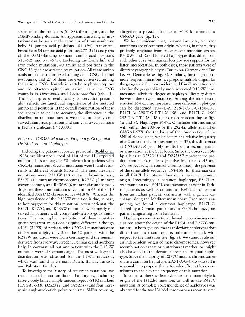
Figure 2 Location of the mutations with respect to the proposed topological model of the CNGA3 polypeptide including the six trans-membrane helices (S1–S6), the ion pore, and the cGMP-binding domain (modified according to Zagotta and Siegelbaum [1996]).
complete achromatopsia but were also found in patientswith incomplete forms of achromatopsia and even in afew patients diagnosed with cone dystrophy (see below).
Apparently homozygous mutations were found in 16patients, and another 31 patients carried two heterozy-gous mutations. However, homozygosity could be mim-icked by the presence of heterozygous deletions, and twoheterozygous mutations might be present in cis. To ex-clude these possibilities, segregation analysis of the mu-tations was performed within the respective families. Truehomozygosity could be established in all cases in whichboth parents could be genotyped (12/16). Similarly, forthe cases with two heterozygous mutations, we could ex-perimentally verify their independent inheritance (allel-ism) in all cases in which samples from unaffected familymembers were available (23/31). Moreover, the segrega-tion patterns of mutations within families were consistentwith respect to the affection status and confirmed thepresence of both mutant alleles in 16 other affected familymembers (not shown).
Only single heterozygous mutations were identified inan additional six index patients. In these patients, allexons of the CNGA3 gene (including exons 0 and 2b)were sequenced, and large structural alterations of theCNGA3 gene were ruled out by Southern blot hybrid-ization in selected patients.
We identified a total of 38 new CNGA3 mutations, in
addition to the 8 mutations reported elsewhere (table 1;Kohl et al. 1998). In the combined sample of knownCNGA3 mutations, the vast majority (39/46) representamino acid substitutions, compared with only 4 stop-co-don mutations, 2 single-codon insertions, and 1 single-codon in-frame deletion. Among the single-base–substi-tution mutations (missense plus stop-codon mutations),there were 35 transitions and 8 transversions, and a pre-ponderance of guanine and cytosine nucleotides (33/43)affected by mutations. We observed the typical increasedincidence of mutations in -CG- dinucleotides (10/43). Thisaccounts at least in part for the high frequency of sub-stitutions at arginine codons (10 of 39 missense muta-tions; table 1).
The distribution of mutations in CNGA3 is biasedtowards the central and terminal segments of the gene.Only a single mutation, 148insG (G49fs), was observedwithin the first six exons (exons 0, 1, 2, 2b, 3, and 4);all other mutations are located in the three terminal ex-ons (exons 5, 6, and 7). With the exception of the earlyframeshift mutation, G49fs, and the terminal stop-codonmutation, Q655X, all remaining mutations are concen-trated between amino acid positions 162 and 593, aregion that accounts for less than two-thirds of the cod-ing sequence (fig. 2). Thus the mutations are mainlyconfined to the functionally and structurally importantcentral parts of the CNGA3 polypeptide, including the
Wissinger et al.: CNGA3 Mutations in Cone Photoreceptor Disorders 729
six transmembrane helices (S1–S6), the ion pore, and thecGMP-binding domain. An apparent clustering of mu-tations can be seen at the terminus of transmembranehelix S1 (amino acid positions 181–194), transmem-brane helix S4 (amino acid positions 277–291) and partsof the cGMP-binding domain (amino acid positions510–529 and 557–573). Excluding the frameshift andstop codon mutations, 40 amino acid positions in theCNGA3 gene are affected by mutation. All these aminoacids are at least conserved among cone CNG channela-subunits, and 27 of them are even conserved amongthe various CNG channels in vertebrate photoreceptorsand the olfactory epithelium, as well as in the CNGchannels in Drosophila and Caenorhabditis (table 1).The high degree of evolutionary conservation presum-ably reflects the functional importance of the mutatedamino acid positions. If the overall conservation of thesesequences is taken into account, the difference in thedistribution of mutations between evolutionarily con-served amino acid positions and non-conserved positionsis highly significant ( ).P ! .0001
Recurrent CNGA3 Mutations: Frequency, GeographicDistribution, and Haplotypes
Including the patients reported previously (Kohl et al.1998), we identified a total of 110 of the 116 expectedmutant alleles among our 58 independent patients withCNGA3 mutations. Several mutations were found recur-rently in different patients (table 1). The most prevalentmutations were R283W (19 mutant chromosomes),F547L (12 mutant chromosomes), R277C (9 mutantchromosomes), and R436W (6 mutant chromosomes).Together, these four mutations account for 46 of the 110identified ACHM2 chromosomes (41.8%). Whereas thehigh prevalence of the R283W mutation is due, in part,to homozygosity for this mutation (seven patients), theF547L, R277C, and R436W mutations were mostly ob-served in patients with compound-heterozygous muta-tions. The geographic distribution of these most-fre-quent recurrent mutations is quite different: although140% (24/58) of patients with CNGA3 mutations wereof German origin, only 2 of the 12 patients with theR283W mutation were from Germany and the remain-der were from Norway, Sweden, Denmark, and northernItaly. In contrast, all but one patient with the R436Wmutation were of German origin. The most widespreaddistribution was observed for the F547L mutation,which was found in German, Dutch, Italian, Turkish,and Pakistani families.
To investigate the history of recurrent mutations, wereconstructed mutation-linked haplotypes, includingthree closely linked simple tandem repeat (STR) markers(CNGA3-STR, D2S2311, and D2S2187) and four intra-genic single-nucleotide polymorphisms (SNPs) covering,
altogether, a physical distance of ∼170 kb around theCNGA3 gene (fig. 1a).
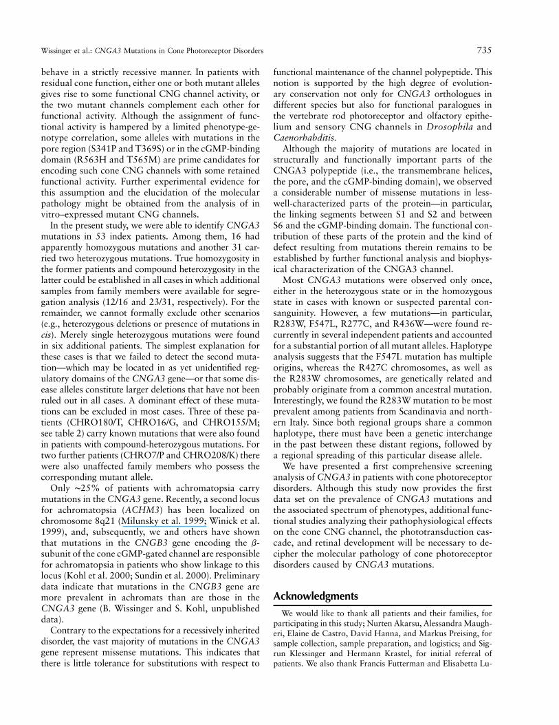
We found evidence that, in some instances, recurrentmutations are of common origin, whereas, in others, theyprobably originate from independent mutation events.R410W- and R563H-linked haplotypes that differ fromeach other at several marker loci provide support for thelatter interpretation. In both cases, those patients were ofdifferent geographic origin (Turkey vs. Germany and Tur-key vs. Denmark; see fig. 3). Similarly, for the group ofmore frequent mutations, we propose multiple origins forthe geographically most widespread F547L mutation andalso for the geographically more restricted R436W chro-mosomes, albeit the degree of haplotype diversity differsbetween these two mutations. Among the nine recon-structed F547L chromosomes, three different haplotypescan be discerned: F547L-A: 288-T-A-G-C-158-158;F547L-B: 290-T-G-T-T-158-158; and F547L-C: 290/292-T-A-T-T-158-158 (marker order according to figs.1a and 3). Haplotype F547L-C includes chromosomeswith either the 290-bp or the 292-bp allele at markerCNGA3-STR. On the basis of the conservation of theSNP allele sequence, which occurs at a relative frequencyof 1.2 on control chromosomes ( ), this differencen p 37at CNGA-STR probably results from a recombinationor a mutation at the STR locus. Since the observed 158-bp alleles at D2S2311 and D2S2187 represent the pre-dominant marker alleles (relative frequencies .42 and.57, respectively, in control chromosomes), the presenceof the same allele sequence (158-158) for these markersin all F547L haplotypes does not support a commonorigin. Interestingly, a common haplotype, F547L-A,was found on two F547L chromosomes present in Turk-ish patients as well as on another F547L chromosomefrom an Italian patient, consistent with a genetic ex-change along the Mediterranean coast. Even more sur-prising, we found a common haplotype, F547L-C,shared by a German patient and a F547L homozygouspatient originating from Pakistan.
Haplotype reconstruction allowed no convincing con-clusions about the origin of the P163L and R277C mu-tations. In both groups, there are deviant haplotypes thatdiffer from their counterparts only at one flank withrespect to the mutation site (fig. 3). We cannot rule outan independent origin of these chromosomes; however,recombination events or mutations at marker loci mightalso have led to the deviation from the original haplo-type. Since the majority of R277C mutant chromosomesshare a common haplotype, 292-T-A-G-C-158-158, it isreasonable to propose that a founder effect at least con-tributes to the elevated frequency of this mutation.
In contrast, there is clear evidence for a monophyleticorigin of the I312del mutation, as well as the R427Cmutation. A complete correspondence of haplotypes wasobserved for the two I312del chromosomes reconstructed

730 Am. J. Hum. Genet. 69:722–737, 2001
Figure 3 Haplotypes of CNGA3 mutant chromosomes. The origins of patients are given in the second column: D p Germany; DK pDenmark; GR p Greece; HT p Haiti; I p Italy; N p Norway; NL p The Netherlands; PK p Pakistan; S p Sweden; TK p Turkey; andUS p United States. Similar haplotypes for a given mutation (which probably reflect a common origin) are boxed, and deviations within agroup of similar haplotypes are represented by blank haplotype segments. The dark-shaded segment for CHRO180 represent a second-orderhaplotype deviation. The vertical line marked by an arrowhead represents the relative position of the mutations, between SNP 215�151TrCand D2S2311. Alleles of STR markers are given as fragment sizes.
in families CHRO13 and CHRO66 and can be also as-sumed for the R427C chromosomes, for which the phaseat some markers could not be resolved experimentally.
Finally, we suggest a common origin of the R283W
mutation, the most prevalent mutation in CNGA3. Hap-lotypes of 17 R283W chromosomes were reconstructedfrom 11 independent patients. Although parental con-sanguinity could not be established for the six patients

Wissinger et al.: CNGA3 Mutations in Cone Photoreceptor Disorders 731
with homozygous R283W mutations, all but one werealso homozygous for all tested markers and thus wereconsidered as bearing a single haplotype identical-by-descent (IBD). In contrast, the two R283W chromo-somes segregating in family CHRO214 differ at mark-er D2S2187. Therefore, we formally considered bothR283W chromosomes segregating in this family as in-dependent haplotypes. The final sample of R283Whaplotypes used for comparison thus comprises fivechromosomes representing the R283W homozygotesconsidered as IBD, the two R283W chromosomes seg-regating in family CHRO214, and another five R283Wchromosomes from compound-heterozygous patients (fig.3). Among this sample a common haplotype, 292-T-A-T-C-158-160, was seen on seven fully resolved R283Wchromosomes and can be traced in three further R283Wchromosomes with incompletely resolved allele assign-ment (CHRO74, CHRO124, and ZD95). Significantly,all these chromosomes share the 160-bp allele at markerD2S2187, which is uncommon in controls (relative fre-quency .16). The presence of this particular 160-bp alleleon the R283W chromosome in family ACH09-1 suggeststhat its deviation from the common haplotype at markerCNGA3-STR and SNP 102-16ArG results from an in-tragenic recombination proximal to the mutation. A sec-ond deviant R283W chromosome was reconstructed infamily CHRO214. Also, in this case, there are severalreasons that favor a differentiation from the common hap-lotype rather than an independent origin of the R283Wmutation. These include (a) the presence of a 162-bp alleleat the most distal marker D2S2187 being the only dif-ference from the common R283W haplotype, (b) the factthat the conserved haplotype segment (CNGA3-STRthrough D2S2311) is rare in controls (relative frequency.03), and (c) the presence of a second R283W chromo-some segregating in this family, which is unlikely to beencountered by chance.
In conclusion, these results strongly support the in-terpretation that all observed R283W chromosomesshare a common origin. Given the unusual geographicdistribution of the R283W mutation, we argue that,from a common ancestor, two separate founding pop-ulations have evolved that account for the elevated fre-quency of the R283W mutation in northern Europe andnorthern Italy.
Phenotypes of Patients with CNGA3 Mutations
Screening of a broad spectrum of cone photoreceptordisorders in the present study demonstrates that CNGA3mutations are not necessarily correlated with a phenotypeof complete achromatopsia, as described previously, butcan also be present in patients with other cone disorders.Clinical data were available for 67 patients with CNGA3mutations (table 2). Thirty-three patients from 26 families
had a diagnosis of complete achromatopsia. Their visualacuities ranged from 0.05 to 0.16, except that one patienthad a visual acuity of 0.2 in one eye. All patients lackedcone ERG responses under standard clinical setup andhad no color vision. Photophobia was present in all pa-tients and nystagmus in ∼90% of the patients. Some ofthose without nystagmus at the time of examination hada history of a transient period of nystagmus during child-hood. Refractive errors were quite variable, ranging from�6.0 to 13.0, but the majority of patients were hyperopic(mean spherical equivalent �1.72). There was no evi-dence for any disease progression after early childhood.
CNGA3 mutations were also found in 18 families com-prising 20 patients with diagnoses of incomplete achro-matopsia. The considerable variability in the clinical pre-sentation among these patients probably reflects theextent of disturbance of the cone photoreceptor system.Whereas, in some patients, residual cone function couldbe detected only by psychophysical color vision testing,others showed sizeable cone ERGs and only slightlyimpaired color vision (e.g., siblings CHRO21/N andCHRO21/A). Visual acuities ranged from 0.06 to 0.6 andwere, on average, better than those of complete achro-mats. Color-vision disturbances were rather nonuniform.Interestingly, some patients could perform color matchesonly for certain regions of the visual spectrum (e.g., re-sidual blue-yellow discrimination). Not all incompleteachromats complained about photophobia. Those withthe best preserved cone function had no or only moderateproblems with daylight conditions. As in the group ofcomplete achromats, hyperopia is the predominant re-fractive error in patients with incomplete achromatopsia(mean spherical equivalent �0.541).
The available clinical data for 11 patients did not suf-fice to categorize them reliably as complete or incompleteachromats. These cases are therefore presented as non-classified cases in table 2.
Finally, three patients had a diagnosis of cone dystro-phy. Current cone photoreceptor function parameterswere similar to those observed in patients with achro-matopsia, but there was evidence for a progression, eitherin follow-up examinations (ZD53/A) or subjectively re-ported by the patient (ZD82/H) and/or due to the pres-ence of funduscopic abnormalities (ZD95/K and ZD82/H). In addition, two of these patients (ZD53/A and ZD95/K) showed reduced rod function in ERGs.
In most instances, affected siblings or patients with thesame CNGA3 genotype showed a similar clinical phe-notype. A notable exception were the patients homozy-gous for the R283W mutation. Some of them were com-plete achromats with no detectable cone function, where-as others had residual cone ERG responses and/or colorvision, justifying the diagnosis of incomplete achroma-topsia (table 2; CHRO214/P and CHRO252/C).
Phenotype-genotype correlation in autosomal recessive

732
Table 2
Clinical Presentation of Patients with CNGA3 Mutations
Patienta,b Originc
Age atExamination/Sex Allele Ad Allele Bd Visual Acuity Refractione Cone ERG Color Vision Photophobia Nystagmus
Complete achromatopsia:CHRO135/H NL 33/M D162V D260N .16/.16 �.5/�1.0 No response None Yes NoCHRO14/B� D 54/F P163L* P163L* .1/.1 �.5/�.5 No response None Yes YesCHRO61/198 D 21/M P163L* R436W* .05/.05 �1.8/�2.75 No response None Yes NoCHRO178/M D 38/F N182Y F547L .12/.12 �1.5/�2.25 No response None Yes YesCHRO47/C S 43/F L186F* R277C* .15/.15 �1.75/�1.25 No response None Yes YesCHRO118/399 D 20/M C191Y* R277C* .2/.08 �1.5/�.8 No response None Yes YesCHRO71/S D 21/F G267D* Q655X* .1/.08 �5.0/�6.0 No response None Yes YesCHRO32/M D 9/M R277C* F547L* .1/.1 �6.0/�6.0 No response None Yes YesCHRO159/T NL 15/M R277C Y573C .1/.1 �1.5/�1.0 No response None Yes YesCHRO104/22 TK 6/M R277H* R277H* .1/.1 �7.0/�7.0 No response None Yes YesCHRO104/24 TK 15/F R277H* R277H* .05/.05 �13.0/�13.0 No response None Yes YesCHRO18/G� N 48/M R283W R283W .1/.1 Hyperopic No response None Yes YesCHRO18/B� N 50/F R283W R283W .1/.1 Hyperopic No response None Yes YesCHRO18/K� N 51/M R283W R283W .1/.1 �8.0/�7.7 No response None Yes YesCHRO127/L DK 62/F R283W R283W .05/.05 �1.25/�1.5 No response None Yes YesCHRO124/C D 10/F R283W* P372S* .1/.1 �1.7/�1.7 No response None Yes YesCHRO124/T D 11/M R283W* P372S* .1/.1 �2.2/�3.2 No response None Yes YesCHRO48/EL S 49/F R283W* F380S* .1/.1 �2.0/�2.0 No response None Yes YesCHRO66/G I 10/M I312del* I312del* .1/.1 �1.0/�1.5 No response None Yes YesCHRO13/A HT 9/F I312del* D485V* .1/.1 �2.0/�3.0 No response None Yes YesCHRO13/L HT 12/F I312del* D485V* .1/.05 �.5/�.5 No response None Yes YesCHRO7/P D 14/F M406T ? .1/.1 �3.75/�4.5 No response None Yes YesCHRO15/M� D 47/F R410W* V529M* .1/.1 �3.0/�5.25 No response None Yes YesCHRO202/J D 32/M R436W* F547L* .1/.1 �3.25/�3.25 No response None Yes YesCHRO103/Km TK 7/M W440X* G516E* .15/.15 �3.5/�3.0 No response None Yes YesCHRO103/Kz TK 7/F W440X* G516E* .1/.15 �7.0/�7.0 No response None Yes YesCHRO103/S TK 14/M W440X* G516E* .15/.15 �6.0/�5.0 No response None Yes TransientCHRO248/C D 28/M C510S C510S .1/.1 �4.5/�4.5 No response None Yes NoCHRO258/S I 9/F G513E* R569H* .05/.05 �.75/�1.75 No response None Yes YesCHRO235/1-1 I 12/M F547L* E194K* .16/.12 �.75/�.25 No response None Yes YesCHRO105/S TK 8/M F547L* F547L* .1/.1 �2.0/�1.75 No response None Yes YesCHRO113/J PK 27/M F547L F547L .16/.1 Myopic No response None Yes NoCHRO180/T D 13/M F547L ? .1/.1 �5.5/�6.0 No response None Yes Yes
Incomplete achromatopsia:CHRO179/U D 23/M Y181C Y181C .2/.125 �2.0/�3.25 Reduced Abnormal Yes YesCHRO241/7-1 I 13/M C191Y* C191Y* .1/.1 �.75/�.75 Residual Residual red Yes Yes

733
CHRO119/M I 3/M R223W* S341P* ∼.2/∼.2 �.375/�.375 Residual Abnormal Moderate YesCHRO21/A D 13/F T224R* T369S* .5/.3 �3.5/�3.75 Reduced Slightly abnormal Moderate YesCHRO21/N D 18/F T224R* T369S* .6/.4 �5.5/�5.0 Reduced Slightly abnormal Moderate YesCHRO65/A D 18/F R277C R427C .06/.05 �.75/�.25 Residual Severely defective Yes YesCHRO254/A US 7/M R277C* G557R* .2/.12 �.75/�.75 No response Residual blue Yes YesCHRO214/P I 3/F R283W* R283W* .1/.1 �3.5/�3.5 Strongly reduced ND Yes YesCHRO252/C I 4/M R283W* R283W* .1/.1 �1.75/�1.75 Residual None Yes YesCHRO74/B DK 12/M R283W R563H .15/.1 �3.75/�4.25 ND Residual red and yellow Yes YesCHRO243/9-1 I 17/M R283W* T565M* .1/.06 0/0 Response Residual blue Yes YesCHRO11/L� D 34/F T291R* F547L* .1/.1 �6.0/�5.5 Reduced Abnormal Yes YesCHRO11/W� D 36/M T291R* F547L* .2/.2 �3.0/�3.0 Reduced Abnormal Yes NoZD128/M D 34/M R427C R436W .125/.125 �12.0/�10.0 ∼80% reduced Abnormal Yes YesCHRO38/M US 40/F R427C* V451fs* .2/.2 �2.0/0 No response Abnormal Yes YesCHRO223/J D 63/M R436W R563H .1/.1 �.75/�.5 Strongly reduced Abnormal Yes MildCHRO16/G D 56/M R436W ? .1/.1 �5.6/�6.3 Residual Severely defective Yes NoCHRO171/M NL 16/M I522T F547L .16/.16 �7.25/�9.75 ND Abnormal Yes YesCHRO79/O DK 56/M G525D* T565M* .2/.16 �.25/0 Residual Slightly abnormal No YesCHRO208/K D 17/F E593K ? .1/.1 �4.0/�4.25 No response Abnormal Yes Yes
Cone dystrophy:ZD95/Kf D 15/F R277C* R283W* .05/.05 �6.5/�6.0 Absent Defective Yes YesZD82/Hg TK 23/F N471S ? .1/.1 �1.25/�1.0 Reduced Abnormal Yes NoZD53/Ah TK 32/F R563H* F547L* !.05/!.05 �3.5/�2.5 Absent Severely defective Yes Yes
Nonclassified:i
CHRO220/217 B ND/M G49fs* W316X* No data availableCHRO92/S D 2/M R223W* R223W* ND �5.0/�5.75 ND ND Yes YesCHRO185/M GR 2/F R277C* R436W* ND �1.5/�1.5 ND ND Yes YesCHRO34/831 D 59/F R277C* Q537X* .2/.2 �3.8/�2.8 No response None Yes NoCHRO6/S� US 2/F R283Q* G557R* No data availableCHRO155/M NL 25/M R283Q ? .16/.16 �3.0/�3.5 No response ND Yes NoCHRO50/A S 3/M R283W* R283W* ND ND No response ND Yes YesCHRO209/B I 14/M R283W* R283W* .2/.2 �3.5/�3.5 No response Abnormal Yes YesCHRO115/H I 25/M R283W* R283W* .1/.1 �1.25/�2.0 ND None Yes YesCHRO121/N TU 5/F P372S* P372S* .15/.15 �.75/�1.5 ND None Yes YesCHRO101/A TK 6/F R410W* R410W* .1/.1 �3.5/�3.5 No response ND Yes Yes
a Patients with identical identifiers to the left of the slash belong to the same family.b A plus sign (�) denotes patients from families reported by Kohl et al. (1998).c B p Belgium, D p Germany, DK p Denmark, GR p Greece, HT p Haiti, I p Italy, N p Norway, NL p The Netherlands, PK p Pakistan, S p Sweden, TK p Turkey, TU
p Tunisia, and US p United States.d An asterisk (*) denotes patients in whom homozygosity or allelism of heterozygous mutations could be established by segregation analysis. A question mark (?) denotes patients
with single heterozygous mutations.e Refractive errors are given in spherical equivalents.f The patient shows a bull’s eye maculopathy and a reduced scotopic ERG.g This patient’s age at onset was 10 years with rapid visual loss macular changes.h This patient showed a reduction in visual acuity in follow-up examination, a progressive visual field loss, and a reduced scotopic ERG.i Individuals not classified because of inconclusive clinical data.

734 Am. J. Hum. Genet. 69:722–737, 2001
conditions is principally complicated by the uncertainty,in compound heterozygotes, about which of the two mu-tant alleles retains the crucial functional activity, aboutwhether both mutant alleles act synergistically and aboutwhether they can complement each other. Despite theseinherent limitations, it is noteworthy that the R427C,R563H, and T565M mutations have been exclusivelyfound in patients with incomplete achromatopsia or conedystrophy and thus represent the best candidates for mu-tations that do not completely abolish cone photoreceptorfunction. Interestingly, we found that two of the patientswith the least severe visual complaints (CHRO21 andCHRO119) were both compound heterozygotes for mu-tations in the cytoplasmic linker between transmembranedomains S2 and S3 and in the channels’ pore regions(T224R/T369S and R223W/S341P, respectively).
It has been shown that the murine and bovine ortho-logues of the human CNGA3 gene are also expressedin nonretinal tissues, notably kidney, testis, and the pin-eal gland (Biel et al. 1994). However, none of the patientswith CNGA3 mutations had a positive medical historyfor systemic disease and evaluation of self-assessmentpatient questionnaires (13 respondents) revealed no ev-idence that mutations in the CNGA3 gene were asso-ciated with other, nonvisual illnesses or complaints.
Discussion
We have recently shown that mutations in the CNGA3gene cause complete achromatopsia (Kohl et al. 1998).One of the major findings of the present study is thatCNGA3 mutations do not only lead to complete achro-matopsia but can also cause incomplete achromatopsiawith residual cone function. The phenotypic presentationof such incomplete achromats is highly variable, rangingfrom cases with barely detectable remnants of color visionto those with well-preserved visual acuity and only slightlyabnormal color vision. Some of the incomplete achromatswith CNGA3 mutations showed residual color-sensitivityonly for parts of the visual spectrum rather than a uniformdefect. This peculiar variability in color sensitivity hasalready been noticed in early clinical descriptions of in-complete achromats (Jager 1953; Goodman et al. 1963).We also found that disease expression can vary amongsiblings or patients with the same CNGA3 genotype. Forexample, on detailed electrophysiological and psycho-physical investigations, apparent differences in the extentof cone dysfunction were found between the affected sib-lings in family CHRO21, who had the least severe visualcomplaints of all patients with CNGA3 mutations (H.Jagle and L. T. Sharpe, unpublished data). The phenotypicvariability in these patients with rather well-preservedcone function may result from differences in lens densityand macular pigment (Stockman and Sharpe 1999), aswell as from differences in the S- to M- to L-cone ratio
and distribution (Hagstrom et al. 1998; Roorda and Wil-liams 1999) or the presence of simultaneous alterationsin the pigment genes. Furthermore, some patients ho-mozygous for the R283W mutations present as completeachromats, whereas others had some residual cone func-tion, although at a low level. It is noteworthy that thelatter were young children (3 and 4 years of age at ex-amination), in contrast to the four R283W homozygotesdiagnosed as complete achromats, all of whom were �48years old. This may indicate that, in R283W homozy-gotes, there is a loss of the residual cone function duringthe patient’s lifetime, resulting in discrepant clinical pre-sentation depending on the patients’ age at examination.However, a progressive loss of cone function as a generalmechanism in achromatopsia is unlikely, since the meanages (at examination) of complete and incomplete achro-mats were nearly identical (24.39 years vs. 24.15 years).Thus, we have to consider that other factors—like theprecise sequence and expression of the channel b-subunitor the total number of defective cones that are actuallyformed in retinal development—determine the actual clin-ical phenotype in patients with the same genotype. Suchanticipated distortions in the development of cone pho-toreceptors and the cone mosaic are documented fromthe homologous knockout-mouse model (Biel et al. 1999)and the sparse histological studies of retinae from achro-mats (Larsen 1921; Harrison et al. 1960; Falls et al. 1965;Glickstein and Heath 1975).
The definite categorization of patients as complete orincomplete achromats can be difficult in those caseswith very low cone function and mainly relies on thesensitivity and reliability of noninvasive clinical andpsychophysical testing. Because of these diagnostic lim-itations and the fact that complete and incomplete a-chromatopsia are caused by mutations in the same gene,both disorders should rather be considered as part of acontinuous clinical spectrum of cone photoreceptor dys-function. Nevertheless, the availability of genotype datanow reinforces the demand for the development of im-proved noninvasive clinical tests and their standardi-zation, to resolve subtle phenotypic differences.
Some patients with CNGA3 mutations had a diag-nosis of cone dystrophy. These patients tended to be theones with the most-severe clinical symptoms among thegroup of cone dystrophy patients analyzed in this study(for a review about the clinical presentation, see thereport by Kellner [1996]). However, since the evidencefor the presence of a progressive retinal dystrophy isstill limited in these cases, further follow-up examina-tions and the identification of additional affected indi-viduals will be necessary to confirm these findings.
Heterozygotes for CNGA3 mutations do not com-plain of any visual problems and their cone photore-ceptor function is normal (H. Jagle and L. T. Sharpe,unpublished data), indicating that CNGA3 mutations
Wissinger et al.: CNGA3 Mutations in Cone Photoreceptor Disorders 735
behave in a strictly recessive manner. In patients withresidual cone function, either one or both mutant allelesgives rise to some functional CNG channel activity, orthe two mutant channels complement each other forfunctional activity. Although the assignment of func-tional activity is hampered by a limited phenotype-ge-notype correlation, some alleles with mutations in thepore region (S341P and T369S) or in the cGMP-bindingdomain (R563H and T565M) are prime candidates forencoding such cone CNG channels with some retainedfunctional activity. Further experimental evidence forthis assumption and the elucidation of the molecularpathology might be obtained from the analysis of invitro–expressed mutant CNG channels.
In the present study, we were able to identify CNGA3mutations in 53 index patients. Among them, 16 hadapparently homozygous mutations and another 31 car-ried two heterozygous mutations. True homozygosity inthe former patients and compound heterozygosity in thelatter could be established in all cases in which additionalsamples from family members were available for segre-gation analysis (12/16 and 23/31, respectively). For theremainder, we cannot formally exclude other scenarios(e.g., heterozygous deletions or presence of mutations incis). Merely single heterozygous mutations were foundin six additional patients. The simplest explanation forthese cases is that we failed to detect the second muta-tion—which may be located in as yet unidentified reg-ulatory domains of the CNGA3 gene—or that some dis-ease alleles constitute larger deletions that have not beenruled out in all cases. A dominant effect of these muta-tions can be excluded in most cases. Three of these pa-tients (CHRO180/T, CHRO16/G, and CHRO155/M;see table 2) carry known mutations that were also foundin patients with compound-heterozygous mutations. Fortwo further patients (CHRO7/P and CHRO208/K) therewere also unaffected family members who possess thecorresponding mutant allele.
Only ∼25% of patients with achromatopsia carrymutations in the CNGA3 gene. Recently, a second locusfor achromatopsia (ACHM3) has been localized onchromosome 8q21 (Milunsky et al. 1999; Winick et al.1999), and, subsequently, we and others have shownthat mutations in the CNGB3 gene encoding the b-subunit of the cone cGMP-gated channel are responsiblefor achromatopsia in patients who show linkage to thislocus (Kohl et al. 2000; Sundin et al. 2000). Preliminarydata indicate that mutations in the CNGB3 gene aremore prevalent in achromats than are those in theCNGA3 gene (B. Wissinger and S. Kohl, unpublisheddata).
Contrary to the expectations for a recessively inheriteddisorder, the vast majority of mutations in the CNGA3gene represent missense mutations. This indicates thatthere is little tolerance for substitutions with respect to
functional maintenance of the channel polypeptide. Thisnotion is supported by the high degree of evolution-ary conservation not only for CNGA3 orthologues indifferent species but also for functional paralogues inthe vertebrate rod photoreceptor and olfactory epithe-lium and sensory CNG channels in Drosophila andCaenorhabditis.
Although the majority of mutations are located instructurally and functionally important parts of theCNGA3 polypeptide (i.e., the transmembrane helices,the pore, and the cGMP-binding domain), we observeda considerable number of missense mutations in less-well-characterized parts of the protein—in particular,the linking segments between S1 and S2 and betweenS6 and the cGMP-binding domain. The functional con-tribution of these parts of the protein and the kind ofdefect resulting from mutations therein remains to beestablished by further functional analysis and biophys-ical characterization of the CNGA3 channel.
Most CNGA3 mutations were observed only once,either in the heterozygous state or in the homozygousstate in cases with known or suspected parental con-sanguinity. However, a few mutations—in particular,R283W, F547L, R277C, and R436W—were found re-currently in several independent patients and accountedfor a substantial portion of all mutant alleles. Haplotypeanalysis suggests that the F547L mutation has multipleorigins, whereas the R427C chromosomes, as well asthe R283W chromosomes, are genetically related andprobably originate from a common ancestral mutation.Interestingly, we found the R283W mutation to be mostprevalent among patients from Scandinavia and north-ern Italy. Since both regional groups share a commonhaplotype, there must have been a genetic interchangein the past between these distant regions, followed bya regional spreading of this particular disease allele.
We have presented a first comprehensive screeninganalysis of CNGA3 in patients with cone photoreceptordisorders. Although this study now provides the firstdata set on the prevalence of CNGA3 mutations andthe associated spectrum of phenotypes, additional func-tional studies analyzing their pathophysiological effectson the cone CNG channel, the phototransduction cas-cade, and retinal development will be necessary to de-cipher the molecular pathology of cone photoreceptordisorders caused by CNGA3 mutations.
Acknowledgments
We would like to thank all patients and their families, forparticipating in this study; Nurten Akarsu, Alessandra Maugh-eri, Elaine de Castro, David Hanna, and Markus Preising, forsample collection, sample preparation, and logistics; and Sig-run Klessinger and Hermann Krastel, for initial referral ofpatients. We also thank Francis Futterman and Elisabetta Lu-

736 Am. J. Hum. Genet. 69:722–737, 2001
chetta, for promoting information about and research on a-chromatopsia and for logistic help, and Dimitri Trankner,Reinhard Seifert, U. Benjamin Kaupp, and Klaus Benndorff,for helpful discussions on the physiology and biophysics ofCNG channels. We kindly acknowledge the provision of filtersof the Lawrence Livermore National Laboratory chromosome2 PAC library and pools of the Roswell Park Cancer InstitutePAC libraries through the Human Genome Mapping ProjectResource Center, Hinxton, United Kingdom. This work wassupported by a grant of the Federal Ministry of Education,Science, Research, and Technology (Fo. 01KS9602) and theInterdisciplinary Center for Clinical Research, Tubingen, (toL.T.S.) and grants of the Deutsche Forschungsmeinschaft(SFB430/A5) and the fortune research program (grant 583) ofthe Medical Faculty of the Tubingen University (to B.W.).
Electronic-Database Information
Accession numbers and URLs for data in this article are asfollows:
GenBank, http://www.ncbi.nlm.nih.gov/ (for the CNG channela-subunits of human cone photoreceptors [accession num-ber AF065314], of mouse cone photoreceptors [accessionnumber AJ243933], of chicken cone photoreceptors [acces-sion number X89598], of human rod photoreceptors [ac-cession number S42457], and of the bovine olfactory epi-thelium [accession number X55010], the Drosophila mela-nogaster CNG channel [accession number X89601], theCaenorhabditis elegans tax4 CNG channel [accession num-ber D89601], and the CGNA3-covering BAC clones RP11-127K18 [accession number AC010134] and RP11-127K18[accession number AC010134)
Online Mendelian Inheritance in Man (OMIM), http://www.ncbi.nlm.nih.gov/Omim/ (for ACHM2 [MIM 216900],ACHM3 [MIM 262300], BCM [MIM 303700], CNGA3[MIM 600053], and CNGB3 [MIM 605080])
References
Andreasson S, Tornqvist K (1991) Electroretinograms in pa-tients with achromatopsia. Acta Ophthalmol 69:711–716
Arbour NC, Zlotogora J, Knowlton RG, Merin S, RosenmannA, Kanis AB, Rokhlina T, Stone EM, Sheffield VC (1997)Homozygosity mapping of achromatopsia to chromosome2 using DNA pooling. Hum Mol Genet 6:689–694
Ayyagari R, Kakuk LE, Coats CL, Bingham EL, Toda Y, FeliusJ, Sieving PA (1999) Bilateral macular atrophy in blue conemonochromacy (BCM) with loss of the locus control region(LCR) and part of the red pigment gene. Mol Vis 5:13
Biel M, Seeliger M, Pfeifer A, Kohler K, Gerstner A, LudwigA, Jaissle G, Fauser S, Zrenner E, Hofmann F (1999) Se-lective loss of of cone function in mice lacking the cyclicnucleotide-gated channel CNG3. Proc Natl Acad Sci USA96:7553–7557
Biel M, Zong X, Distler M, Bosse E, Klugbauer N, MurakamiM, Flockerzi V, Hofmann F (1994) Another member of thecyclic nucleotide-gated channel family, expressed in testis,kidney, and heart. Proc Natl Acad Sci USA 91:3505–3509
Dryja TP, Finn JT, Peng YW, McGee TL, Berson EL, Yau KW
(1995) Mutations in the gene encoding the a-subunit of therod cGMP-gated channel in autosomal recessive retinitis pig-mentosa. Proc Natl Acad Sci USA 92:10177–10181
Falls HF, Wolter JR, Alpern M (1965) Typical total monochro-macy. Arch Ophthalmol 74:610–616
Francois J (1961) Heredity in ophthalmology. CV Mosby, StLouis
Gingrich JC, Boehrer D, Garnes JA, Johnson W, Wong B, Berg-mann A, Eveleth GG, Longlois RG, Carrano AV (1996)Construction and characterization of human chromosome2 specific cosmid, fosmid and PAC clone libraries. Genomics32:65–74
Glickstein M, Heath GG (1975) Receptors in the monochro-mat eye. Vision Res 15:633–636
Goodman G, Ripps H, Siegel IM (1963) Cone dysfunctionsyndromes. Arch Ophthalmol 70:214–231
Hagstrom SA, Neitz J, Neitz M (1998) Variations in the conepopulation for red-green color vision examined by analysisof mRNA. Neuroreport 9:1963–1967
Hansen E (1979) Typical and atypical monochromacy stud-ied by specific quantitative perimetry. Acta Ophthalmol 57:211–224
Harrison H, Hoefnagel D, Hayward JN (1960) Congenitaltotal colorblindness, a clinico-pathological report. ArchOphthalmol 64:685–692
Ioannou PA, de Jong PJ (1996) Construction of bacterial ar-tificial chromosome libraries using a modified P1 (PAC) sys-tem. In: Dracopoli NC, Haines JL, Korf BR, Moir DT, Mor-ton CC, Seidman CE, Seidman JG, Smith DR (eds) Currentprotocols in human genetics. John Wiley & Sons, New York
Jager W (1953) Typen der inkompletten Achromatopsie. BerlDtsch Ophthalmol Ges 58:44–47
Jagle H, Kohl S, Apfelstedt-Sylla E, Wissinger B, Sharpe LT(2001) Manifestation of rod monochromacy. Col Res Appl13:96–99
Kellner U (1996) Die progressiven Zapfendystrophien. Buch-erei des Augenarztes Bd. 135, Enke Verlag, Stuttgart
Kohl S, Baumann B, Broghammer M, Jagle H, Sieving P, Kell-ner U, Spegal R, Anastasi M, Zrenner E, Sharpe LT, Wis-singer B (2000) Mutations in the CNGB3 gene encoding theb-subunit of the cone photoreceptor cGMP-gated channelare responsible for achromatopsia (ACHM3) linked to chro-mosome 8q21. Hum Mol Genet 9:2107–2116
Kohl S, Marx T, Giddings I, Jagle H, Jacobson SG, Apfelstedt-Sylla E, Zrenner E, Sharpe LT, Wissinger B (1998) Totalcolourblindness is caused by mutations in the gene encodingthe a-subunit of the cone photoreceptor cGMP-gated cationchannel. Nat Genet 19:257–259
Larsen H (1921) Demonstration mikroskopischer Praparatevon einem monochromatischen Auge. Klin Mbl Augenheilk67:301–302
Milunsky A, Huang XL, Milunsky J, DeStefano A, BaldwinCT (1999) A locus for autosomal recessive achromatopsiaon human chromosome 8q. Clin Genet 56:82–85
Muller F, Kaupp, UB (1998) Signaltransduktion in Sehzellen.Naturwissenschaften 85:49–61
Nathans J, Maumenee IH, Zrenner E, Sadowski B, Sharpe LT,Lewis RA, Hansen E, Rosenberg T, Schwartz M, Hecken-lively JR, Traboulsi E, Klingaman R, Bech-Hansen NT,LaRoche GR, Pagon RA, Murphey WH, Weleber RG (1993)

Wissinger et al.: CNGA3 Mutations in Cone Photoreceptor Disorders 737
Genetic heterogeneity among blue-cone monochromats. AmJ Hum Genet 53:987–1000
Nathans J, Piantanida TP, Eddy RL, Shows TB, Hogness DS(1986) Molecular genetics of inherited variation in humancolor vision. Science 232:203–210
Neuhann T, Krastel H, Jaeger W (1978) Differential diagnosisof typical and atypical congenital achromatopsia: analysis ofa progressive foveal dystrophy and a nonprogressive oligo-cone trichromasy (general cone dysfunction without achro-matopsia), both of which at first had been diagnosed as achro-matopsia. Albert Von Graefes Arch Klin Exp Ophthalmol209:19–28
Roorda A, Williams DR (1999) The arrangement of the threecone classes in the living human eye. Nature 397:520–522
Sharpe LT, Nordby K (1990) Total colour blindness: an intro-duction. In: Hess RF, Sharpe LT, Nordby K (eds) Night vi-sion: basic, clinical and applied aspects. Cambridge Univer-sity Press, Cambridge, pp 253–289
Sharpe LT, Stockman A, Jagle H, Nathans J (1999) Opsingenes, cone photopigments and colorblindness. In: Gegen-furtner K, Sharpe LT (eds) Color vision: from genes to per-ception. Cambridge University Press, Cambridge, pp 3–52
Stockman A, Sharpe LT (1999) Cone spectral sensitivities andcolor matching. In: Gegenfurtner K, Sharpe LT (eds) Colorvision: from genes to perception. Cambridge UniversityPress, Cambridge, pp 53–87
Sundin OH, Yang JM, Li Y, Zhu D, Hurd JN, Mitchell TN,Silva ED, Hussels-Maumenee I (2000) Genetic basis of total
colourblindness among the Pingelapese islanders. Nat Genet25:289–293
Weitz CJ, Miyake Y, Shinzato K, Montag E, Zrenner, E, WentLN, Nathans N (1992) Human tritanopia associated withtwo amino acid substitutions in the blue sensitive opsin. AmJ Hum Genet 50:498–507
Winick JD, Blundell ML, Galke BL, Salam AA, Leal SM, Ka-rayiorgou M (1999) Homozygosity mapping of the achro-matopsia locus in the Pingelapese. Am J Hum Genet 64:1679–1685
Wissinger B, Jagle H, Kohl S, Broghammer M, Baumann B,Hanna DB, Hedels C, Apfelstedt-Sylla E, Randazzo G, Ja-cobson SG, Zrenner E, Sharpe LT (1998) Human rod mono-chromacy: linkage analysis and mapping of a cone photo-receptor expressed candidate gene on chromosome 2q11.Genomics 51:325–331
Wissinger B, Muller F, Weyand I, Schuffenhauer S, Thanos S,Kaupp UB, Zrenner E (1997) Cloning, chromosomal local-ization and functional expression of the gene encoding thealpha-subunit of the cGMP-gated channel in human conephotoreceptors. Eur J Neurosci 9:2512–2521
Zagotta WN, Siegelbaum SA (1996) Structure and function ofcyclic nucleotide-gated channels. Annu Rev Neurosci 19:235–263
Zrenner E, Magnussen S, Lorenz B (1988) Blauzapfenmono-chromasie: Diagnose, genetische Beratung, optische Hilfs-mittel. Klin Mbl Augenheilk 193:510–517
Copyright © 2022 FDOKUMEN