Clinical utility of dental cone-beam computed tomography
15
© 2014 Jaju and Jaju. This work is published by Dove Medical Press Limited, and licensed under Creative Commons Attribution – Non Commercial (unported, v3.0) License. The full terms of the License are available at http://creativecommons.org/licenses/by-nc/3.0/. Non-commercial uses of the work are permitted without any further permission from Dove Medical Press Limited, provided the work is properly attributed. Permissions beyond the scope of the License are administered by Dove Medical Press Limited. Information on how to request permission may be found at: http://www.dovepress.com/permissions.php Clinical, Cosmetic and Investigational Dentistry 2014:6 29–43 Clinical, Cosmetic and Investigational Dentistry Dovepress submit your manuscript | www.dovepress.com Dovepress 29 REVIEW open access to scientific and medical research Open Access Full Text Article http://dx.doi.org/10.2147/CCIDE.S41621 Clinical utility of dental cone-beam computed tomography: current perspectives Prashant P Jaju 1 Sushma P Jaju 2 1 Oral Medicine and Radiology, 2 Conservative Dentistry and Endodontics, Rishiraj College of Dental Sciences and Research Center, Bhopal, India Correspondence: Prashant P Jaju Department of Oral Medicine and Radiology, Rishiraj College of Dental Sciences and Research Center, Bhopal, 462003, India Tel +91 975 209 3011 Email [email protected] Abstract: Panoramic radiography and computed tomography were the pillars of maxillofacial diagnosis. With the advent of cone-beam computed tomography, dental practice has seen a paradigm shift. This review article highlights the potential applications of cone-beam computed tomography in the fields of dental implantology and forensic dentistry, and its limitations in maxillofacial diagnosis. Keywords: dental implants, cone-beam computed tomography, panoramic radiography, computed tomography Introduction Cone-beam computed tomography (CBCT) is a new diagnostic tool that has revolutionized diagnosis and treatment planning in the dental field. CBCT presents as a separate C-arm to computed tomography (CT) imaging. An early volumetric CT predecessor of CBCT, the dynamic spatial reconstructor, was developed in the late 1970s by the Biodynamics Research Unit at the Mayo Clinic (Rochester, MN, USA). 1 Initial interest has focused primarily on applications in angiography in which soft- tissue resolution could be sacrificed in favor of high temporal- and spatial-resolving capabilities. 1 CBCT provided an alternate method of cross-section image production to fan-beam CT using a comparatively less expensive radiation detector than conventional CT. The technology transfer of CBCT to dentistry first occurred in 1995. Italian coin- ventors, Tacconi and Mozzo, developed a CBCT system for the maxillofacial region that was designed and produced by QR Srl of Verona, Italy. 1 This unit, the NewTom QR-DVT 9000, became the first commercial CBCT unit marketed specifically to the dental market, and it was initially introduced in Europe in 1999. 1 Currently, numerous manufacturers have introduced CBCT machines in different fields of view (FOVs), as per clinical practice requirements (Table 1). CBCT was initially introduced for its indispensable role in the field of dental implantology. 1 Currently, the utility of CBCT encompasses field of dental implantology, oral surgery, orthodontics, endodontics, sleep apnea, temporomandibular joint (TMJ) disorders, and periodontics, and it is expanding its horizon in the field of ear, nose, and throat (ENT) medicine. 2,3 CBCT in dental implantology Dental implants are considered to be a boon to edentulous patients, thereby improving their overall health and longevity. 1 CBCT has revolutionized the way dental implant
-
Upload
khangminh22 -
Category
Documents
-
view
0 -
download
0
Transcript of Clinical utility of dental cone-beam computed tomography

© 2014 Jaju and Jaju. This work is published by Dove Medical Press Limited, and licensed under Creative Commons Attribution – Non Commercial (unported, v3.0) License. The full terms of the License are available at http://creativecommons.org/licenses/by-nc/3.0/. Non-commercial uses of the work are permitted without any further
permission from Dove Medical Press Limited, provided the work is properly attributed. Permissions beyond the scope of the License are administered by Dove Medical Press Limited. Information on how to request permission may be found at: http://www.dovepress.com/permissions.php
Clinical, Cosmetic and Investigational Dentistry 2014:6 29–43
Clinical, Cosmetic and Investigational Dentistry Dovepress
submit your manuscript | www.dovepress.com
Dovepress 29
R e v I e w
open access to scientific and medical research
Open Access Full Text Article
http://dx.doi.org/10.2147/CCIDE.S41621
Clinical utility of dental cone-beam computed tomography: current perspectives
Prashant P Jaju1
Sushma P Jaju2
1Oral Medicine and Radiology, 2Conservative Dentistry and endodontics, Rishiraj College of Dental Sciences and Research Center, Bhopal, India
Correspondence: Prashant P Jaju Department of Oral Medicine and Radiology, Rishiraj College of Dental Sciences and Research Center, Bhopal, 462003, India Tel +91 975 209 3011 email [email protected]
Abstract: Panoramic radiography and computed tomography were the pillars of maxillofacial
diagnosis. With the advent of cone-beam computed tomography, dental practice has seen a
paradigm shift. This review article highlights the potential applications of cone-beam computed
tomography in the fields of dental implantology and forensic dentistry, and its limitations in
maxillofacial diagnosis.
Keywords: dental implants, cone-beam computed tomography, panoramic radiography,
computed tomography
IntroductionCone-beam computed tomography (CBCT) is a new diagnostic tool that has
revolutionized diagnosis and treatment planning in the dental field. CBCT presents
as a separate C-arm to computed tomography (CT) imaging. An early volumetric CT
predecessor of CBCT, the dynamic spatial reconstructor, was developed in the late
1970s by the Biodynamics Research Unit at the Mayo Clinic (Rochester, MN, USA).1
Initial interest has focused primarily on applications in angiography in which soft-
tissue resolution could be sacrificed in favor of high temporal- and spatial-resolving
capabilities.1 CBCT provided an alternate method of cross-section image production to
fan-beam CT using a comparatively less expensive radiation detector than conventional
CT. The technology transfer of CBCT to dentistry first occurred in 1995. Italian coin-
ventors, Tacconi and Mozzo, developed a CBCT system for the maxillofacial region
that was designed and produced by QR Srl of Verona, Italy.1 This unit, the NewTom
QR-DVT 9000, became the first commercial CBCT unit marketed specifically to the
dental market, and it was initially introduced in Europe in 1999.1 Currently, numerous
manufacturers have introduced CBCT machines in different fields of view (FOVs),
as per clinical practice requirements (Table 1).
CBCT was initially introduced for its indispensable role in the field of dental
implantology.1 Currently, the utility of CBCT encompasses field of dental implantology,
oral surgery, orthodontics, endodontics, sleep apnea, temporomandibular joint (TMJ)
disorders, and periodontics, and it is expanding its horizon in the field of ear, nose,
and throat (ENT) medicine.2,3
CBCT in dental implantologyDental implants are considered to be a boon to edentulous patients, thereby improving
their overall health and longevity.1 CBCT has revolutionized the way dental implant

Clinical, Cosmetic and Investigational Dentistry 2014:6submit your manuscript | www.dovepress.com
Dovepress
Dovepress
30
Jaju and Jaju
practice is performed in dental clinics and hospitals. Past
decades have shown a paradigm shift from a surgically-driven
to a prosthetically-driven approach in dental implant therapy.3
To improve the overall success of implant therapy with pos-
sible reduction in surgical and postoperative implant com-
plications, implantologists should have three-dimensional
(3D) information of bone volume and topography prior to
implant placement.4 Presurgical assessment of implant site
by imaging technique thus allows for the accurate assessment
of the amount of bone volume available, bone density, and
proximity to anatomical structures (Table 2).
CBCT is the preferred option for implant dentistry as it
provides greater measurement accuracy when compared to
two-dimensional (2D) imaging, while utilizing lower doses
of radiation.5–8 Loubele et al9 reported that both CBCT and
CT yielded submillimeter accuracy for linear measurements
on an ex vivo specimen for implant measurements.
Ridge morphologyThe buccolingual ridge pattern cannot be viewed on 2D
radiographs, but CBCT provides the advantage of showing
the type of alveolar ridge pattern present.10 Cross-sectional
images provide the implantologist with the appearance of
ridge patterns, such as irregular ridge, narrow crestal ridge,
and knife shape ridge. Also, the loss of cortical plates and
undulating concavities can also be appreciated on cross-
sectional images, and they cannot be seen on panoramic
images. Mcginvney et al and Schwartz et al concluded that
3D images more accurately reflected true osseous topogra-
phy, and they considered it a valuable diagnostic aid.10 In the
Table 1 List of CBCT machines depending upon their FOv
Field of view (FOV)
CBCT machine Manufacturer Maximum FOV (cm) (height*width)
Large (FOvl) .15 cm
i-CAT® Imaging Sciences International Inc. (Hatfield, PA, USA)
17*23
CB MercuRay Hitachi Medical Systems America Inc. (Twinsburg, OH, USA)
20*20
NewTom 3G QR Srl (verona, Italy) 20*20KODAK 9500 Carestream Health (Rochester, NY, USA) 18*21ProMax® 3D Max Planmeca OY (Helsinki, Finland) 16*23
Medium (FOvM) 10–15 cm
GALILeOS Sirona Dental GmbH (Salzburg, Austria) 15*15KODAK CS 9300 Carestream Health 13.5*17SCANORA® 3D Soredex (Milwaukee, wI, USA) 7.5*14.5NewTom vGi QR Srl 15*15ProMax® 3D Mid Planmeca OY 9*16
Small (FOvS) ,10 cm
ORTHOPHOS XG 3D Sirona Dental GmbH 8*8KODAK 9000 3DC Carestream Health 4*5GXDP-7000TM Gendex Dental Systems (Hatfield, PA, USA) 6*8ProMax® 3D Planmeca OY 8*83D Accuitomo 80 J Morita USA, Inc. (Irvine, CA, USA) 8*8
Abbreviations: CBCT, cone-beam computed tomography; FOV, field of view.
Table 2 Comparison of imaging modalities in the field of dental implantology
Imaging goal Cephalometric Periapical OPG Tomography CT CBCT
Bone height 1 3 2 3 4 4Bone width 0 0 0 3 4 4Long axis or ridge 0 0 0 3 4 4Anatomy localization 1 1 1 3 4 4Bone quality 0 2 2 2 4 3Pathology identification 1 2 3 2 3 4Jaw boundary identification 1 0 2 3 4 4virtual planning 0 0 1 1 4 4Guide fabrication 0 0 0 0 4 4Communication aid 2 1 2 2 4 4Benefit/risk/cost ratio 1 1 2 2 3 4
Notes: Ranking score: 0= no score; 1= low value; 2= mild value; 3= moderate value; 4= high value. (Scoring type based on its performance divided into no; low; mild; moderate; high).Abbreviations: OPG, orthopantogram; CT, computed tomography; CBCT, cone-beam computed tomography.

Clinical, Cosmetic and Investigational Dentistry 2014:6 submit your manuscript | www.dovepress.com
Dovepress
Dovepress
31
Dental cone-beam CT
case of a compromised jaw bone (in terms of quality and/or
quantity of bone), the panoramic technique is an inefficient
imaging tool. In case of potential risks in treatment plan 3D
imaging may prove indispensable.10
Quality of bone at implant sitesThe term “bone quality” is commonly used in implant treat-
ment and in reports on implant success and failure.11 Lindh
et al11 emphasized that bone density (bone mineral density)
and bone quality are not synonymous. Bone quality encom-
passes factors other than bone density such as skeletal size,
bone architecture, the 3D orientation of the trabecula, and
matrix properties. Bone quality is not only a matter of min-
eral content, but also of structure. It has been shown that the
quality and quantity of bone available at the implant site are
very important local patient factors in determining the success
of dental implants.12 Bone quality is categorized into four
groups: groups 1–4 or types 1–4 (Bone Quality Index):13
• Type 1: homogeneous cortical bone;
• Type 2: thick cortical bone with marrow cavity;
• Type 3: thin cortical bone with dense trabecular bone of
good strength; and
• Type 4: very thin cortical bone with low density trabecular
bone of poor strength.
In the jaws, an implant placed in poor-quality bone with
thin cortex and low-density trabeculae (Type 4 bone) has a
higher chance of failure compared with the other types of
bones.11 This low-density bone is often found in the posterior
maxilla, and several studies report higher implant failure
rates in this region.10
Bone density can be obtained from CT units and expressed
in terms of Hounsfield units (HU). The HU is not part of the
system international (SI) system. In fact, it is a practical unit
that represents the relative deviation of the measured linear
attenuation of a material from that of water. X-ray beams used
for diagnostic radiology are not monochromatic and, hence,
are composed of photons with a broad spectrum of energies.14
This is the fundamental reason why the HU value for tissues
obtained from one CT system may not be the same as that
obtained using a different CT system, or even with the same
machine, if different technique-related factors are used.14
Unlike CT units, current CBCT units do not use a standard
scaling system. Methods have been proposed to convert CT
numbers measured on CBCT scans to HU. However, such
methods make the implicit assumption that the relation-
ship between CT numbers and X-ray attenuation is uniform
throughout the volume of the CBCT image.14 Several factors
like beam hardening, artifacts from metallic restorations, and
scattered radiation contribute to the heterogeneity of CT num-
bers on CBCT scans. Our experience shows us that even the
same anatomic region on CBCT scans may not demonstrate
the same CT numbers. In general, smaller irradiated and recon-
structed volumes (small FOVs) are less prone to inaccurate CT
numbers caused by scattered radiation and nonideal geometry;
therefore, whenever there is no need for large volume coverage,
a small FOV should be used instead of a large FOV.14
In order for HUs on CBCT to be used with confidence, the
accuracy of the HU should be known, so that the clinician can
assess whether or not the HUs can be used for specific clinical
tasks.14 With more advanced CBCT software and methods,
it should be possible to improve the accuracy of CBCT HU
values when determining bone densities at implant sites.
Thus, it can be concluded that CBCT provides a subjective
assessment of bone quality, and not objective assessment.
CBCT-guided implant surgeryType and size of the planned implant, its position within the
bone, its relationship to the planned restoration and adjacent
teeth and/or implants, and its proximity to vital structures can
be determined before performing surgery (Figure 1). This is
possible with the integration of CBCT scans with computer-
aided design/computer-aided manufacturing technology (for
example, CEREC; Sirona Dental GmbH, Salzburg, Austria).
Computer-generated surgical guides can be fabricated from
the virtual treatment plan. These surgical guides are used
by the implantologist to place the planned implants in the
patient’s mouth in the same position as in the virtual treatment
plan, allowing for more accurate and predictable implant
placement and reduced patient morbidity.15 Surgical guides
are not indicated for every case; indications are as follows:
• Three or more implants in a row
• Proximity to vital anatomic structures
• Problems related to the proximity of adjacent teeth
• Questionable bone volume
• Implant position that is critical to the planned
restoration
• Flapless implant placement
• Multiple unit or immediate full-arch restorations, with or
without extractions, and immediate placement.
Conventional surgical stents that aid in implant posi-
tioning have been used in implant dentistry for many years.
Guides are fabricated from vacuform shells with the buccal
or palatal/lingual facings of the planned restorations, or they
may be more complex with 2 mm drill holes or metal tubes
(Figure 2). There is a lack of a correlation in these appli-
ances between the planned restoration and the underlying

Clinical, Cosmetic and Investigational Dentistry 2014:6submit your manuscript | www.dovepress.com
Dovepress
Dovepress
32
Jaju and Jaju
bony anatomy. With the use of computer-guided implant
surgical guides, this anatomic relationship can be predictably
established and considered before surgery.15 Gutta-percha,
barium sulfate, and lead foils have been traditionally used
for the fabrication of surgical guides; bone and soft tissue
loss from periodontal disease, as well as atrophy, long-term
denture wear, and sinus pneumatization can make it difficult
to predictably use traditional surgical guides.15
Three types of computer-generated surgical guides are
currently available:15
1. Tooth supported;
2. mucosa supported; and
3. bone supported.
Tooth supported guides are used in partially edentulous
cases. This surgical guide is designed to rest on other teeth
in the arch for accuracy of guide fit. Mucosal supported
guides are used primarily in fully edentulous cases and are
designed to rest on the mucosa. Accurate interarch bite
registrations are of the utmost importance when using these
guides to assure accurate surgical guide positioning and
placement of securing screws/pins before the placement of
implants. Bone supported guides can be used in partially
or fully edentulous cases, but they are primarily used in
fully edentulous cases in which significant ridge atrophy
is present and good seating of a mucosa supported guides
is questionable.15 Currently, only SimPlant® (Materialise
Dental, Leuven, Belgium) manufactures bone supporting
surgical guides.15
Radiation doses delivered to the patient by CBCT need
to be evaluated properly. It has been reported that in implant
imaging, CT delivers the highest radiation dose to the sali-
vary glands, whereas the CBCT system studied delivered
the lowest dose.16
Recommendations by American Academy of Oral and Maxillofacial Radiology (AAOMR) for role of CBCT in dental implantologyThe following are recommendations for the role of CBCT in
dental implantology, as made by the AAOMR:4
Figure 1 virtual implant planning by selecting the desired implant from an implant library.
Figure 2 Gutta percha used as a radiographic template.

Clinical, Cosmetic and Investigational Dentistry 2014:6 submit your manuscript | www.dovepress.com
Dovepress
Dovepress
33
Dental cone-beam CT
• Recommendation 1: panoramic radiography should be
used as the imaging modality of choice in the initial
evaluation of the dental implant patient.
• Recommendation 2: use intraoral periapical radiography
to supplement the preliminary information from pano-
ramic radiography.
• Recommendation 3: do not use cross-sectional imag-
ing, including CBCT, as an initial diagnostic imaging
examination.
• Recommendation 4: the radiographic examination of
any potential implant site should include cross-sectional
imaging orthogonal to the site of interest.
• Recommendation 5: CBCT should be considered as
the imaging modality of choice for preoperative cross-
sectional imaging of potential implant sites.
• Recommendation 6: CBCT should be considered when
clinical conditions indicate a need for augmentation
procedures or site development before the placement
of dental implants: 1) sinus augmentation; 2) block or
particulate bone grafting; 3) ramus or symphysis grafting;
4) assessment of impacted teeth in the field of interest;
and 5) evaluation of prior traumatic injury.
• Recommendation 7: CBCT imaging should be consid-
ered if bone reconstruction and augmentation procedures
(for example, ridge preservation or bone grafting) have
been performed to treat bone volume deficiencies before
implant placement.
• Recommendation 8: in the absence of clinical signs or symp-
toms, use intraoral periapical radiography for the postopera-
tive assessment of implants. Panoramic radiographs may be
indicated for more extensive implant therapy cases.
• Recommendation 9: use cross-sectional imaging (par-
ticularly CBCT) immediately postoperatively, only if the
patient presents with implant mobility or altered sensation,
especially if the fixture is in the posterior mandible.
• Recommendation 10: do not use CBCT imaging for the
periodic review of clinically asymptomatic implants.
• Recommendation 11: cross-sectional imaging, optimally
CBCT, should be considered if implant retrieval is
anticipated.
CBCT in oral and maxillofacial surgeryA combination of low radiation dose, high-quality bony
definition, and compact design requiring minimum space has
made CBCT desirable as an in-office imaging system for the
examination of pathologies in the head and neck, extracranial,
paranasal, and temporal bone regions.17,18
Third molar evaluationDental clinics are routinely visited by young adults for the
removal of third molars.19 Damage to the inferior alveolar nerve
(IAN) is a serious complication following third molar removal.
The overall risk of temporary IAN injury associated with third
molar removal ranges from 0.4%–6%.20 It is important to pre-
operatively assess the position, and establish the relationship, of
the third molar with the mandibular canal to minimize the risk
of nerve injury.19 Clinicians use various radiographic markers
to indicate a close relationship between the third molar and the
mandibular canal.20 Panoramic and intraoral radiographs are
sufficient for preoperative imaging in most cases where there is
no overlap between the IAN and the lower third molar.19 Where
there are radiographic signs of an overlapping impacted tooth
with the mandibular canal, panoramic radiographs provide
limited information. The buccolingual relationship between
the IAN and the lower third molar cannot be evaluated from
panoramic radiographs. Furthermore, the presence or absence
of cortication around the IAN and the detailed anatomy of the
third molar may not be clearly evident with this method.20
In their study, Kamrun et al21 revealed that the visibility
of the superior border on panoramic images was very poor,
except for the most posterior area, which clearly confirmed the
limitations of panoramic radiography. The poor visualization
of the canal on panoramic images was considered to be
remarkably improved by the use of CBCT.21
Several studies have shown that panoramic radiography
has only limited accuracy in determining the number of roots
and in describing root morphology.19 CBCT proved to be
more reliable in determining the number of roots than did
panoramic radiography.19 Tantanapornkul et al22 concluded
that the 3D X-Ray CBCT (J Morita USA, Inc., Irvine, CA,
USA) was significantly more accurate when compared with
panoramic radiography in predicting IAN exposure during
third molar removal with a sensitivity of 93% and a specificity
of 77%.22 The patient can also be more adequately informed
about his or her risk profile. Almost all of the CBCT software
provided by manufacturers, as well as third-party software,
has a nerve tracing application. This allows for the identi-
fication and color coding of the mandibular nerve, thereby
assisting in easy recognition (Figure 3).
The maxillary permanent canine has a crucial esthetic
and functional value, but it is the second most frequently
impacted tooth following the third molar.23,24 The prevalence
of maxillary permanent canine impaction is about 1%–3%.25
The decision for interceptive treatment takes into account
several factors, including how to expose, recover, extract, or
not treat.19,20 Some factors include location of the impaction,

Clinical, Cosmetic and Investigational Dentistry 2014:6submit your manuscript | www.dovepress.com
Dovepress
Dovepress
34
Jaju and Jaju
prognosis of intervention on the impacted tooth and adjacent
teeth, surgical accessibility, impact of treatment on the final
functional occlusion, and possible surgical morbidity.19,20
Even though maxillary canine tooth buds develop labially to
adjacent tooth roots, the ratio of palatal impactions to labial
impactions is at least 3:1.26 As expected, most impacted teeth
can be accurately localized with traditional radiographs.
However, this is not true for every impacted canine; some
can be incorrectly localized, poorly accessed surgically, or
they can be recovered by using deleterious vectors (Figure 4).
Ericson and Kurol27 demonstrated that 8% of impacted
maxillary canines could not be accurately localized in the
labio-palatal dimension with periapical radiographs. CBCT
with its 3D orientation can precisely locate the position of
the impacted canine and thus assist the oral surgeon in plan-
ning the treatment.28 According to study done by Alqerban
et al,29 surgical treatment planning of impacted maxillary
canines was not significantly different between panoramic
and CBCT images.
Bony pathology assessmentsOral maxillofacial pathologies are routinely encountered
by clinicians; demographic data, clinical complaints, and
examination of the oral cavity are performed.3 After thorough
clinical examination, radiological and laboratory investiga-
tions are warranted to gather at a presumptive diagnosis. The
exposed images are examined to look for the lesion’s exact
location (maxilla, mandible, anterior, posterior, alveolar
process, and so forth). The exact size of the defect and its
relative density (radiolucent or radiopaque, or a combination
of the two) are determined.30 CBCT is recommended when
there is a need to diagnose a cyst, tumor, or infections in the
alveolar process and jaw bone (Figure 5).31
Rare calcifying lesions, such as a calcifying cystic
odontogenic tumor, can be examined in CBCT images for
their particular variations. CBCT has proven its worth in the
evaluation of intraosseous lesions that are in close proximity
to vital organs and vasculature in the head and neck region.31
Although the reliability of CBCT in detecting the invasion
Figure 3 Color coding of the mandibular nerve on Sirona software (Sirona Dental GmbH, Salzburg, Austria).
Figure 4 CBCT images (panoramic/3D reconstruction/cross-sectional) eases treat-ment planning for impacted mesiodens.Notes: (A) Panoramic radiograph showing mesiodens. (B) 3D reconstruction showing impacted mesiodens. (C) Cross sectional view showing relation of impacted mesiodens with central incisor.
Figure 5 Large radiolucent lesion in the anterior mandible with perforation of the labial cortical plates, as seen on CBCT.Notes: (A) 3D reconstruction showing extensive radiolucent lesion in anterior mandible. (B) Loss of buccal cortical plate clearly demonstrated on axial view.Abbreviation: CBCT, cone-beam computed tomography.

Clinical, Cosmetic and Investigational Dentistry 2014:6 submit your manuscript | www.dovepress.com
Dovepress
Dovepress
35
Dental cone-beam CT
or erosion of oral malignancy such as oral squamous cell
carcinoma is still under investigation, a study has suggested
that the combination of dynamic contrast-enhanced magnetic
resonance imaging (MRI) and CBCT may be a useful tool in
delineating tumor boundaries and in developing appropriate
surgical interventions.32
In their study, Dreiseidler et al33 showed a slightly supe-
rior overall diagnostic reliability for the assessment of a
malignancy’s bone infiltration to CBCT compared with CT
and single-photon emission CT. They concluded that the
FOV-to-radiation dose ratio, easy panoramic view recon-
struction, and clinical accessibility were responsible for the
better performance of CBCT in diagnosis over multislice
CT (MSCT) for the preoperative maxillofacial assessment
of tumor invasion into the bone.33 CBCT can also be teamed
with stereolithographic model construction, which can be
used in conjunction with dental implant placement or in the
reconstruction of jaws resected due to pathology.30
Maxillofacial traumaDentoalveolar fracture, maxillary bone fracture, zygomatic
complex fracture, mandibular fracture, or gunshot injuries
require radiographs for precise location of the fracture.
Panoramic radiographs and numerous extra oral radiographs
are routinely used in maxillofacial trauma cases.34 Authors
recommend the use of CBCT when compared with panoramic
radiograph in identifying the location of a cortical plate
fracture that is not complete.34 Additionally, CBCT is more
sensitive and accurate in imaging the maxilla and mandible.
It is reported that mandibular fractures that are not evident
in conventional CT can be identified using CBCT. Also,
when using CBCT, as compared to CT and conventional
radiograph, information about dentoalveolar fractures is more
detailed. This makes CBCT uniquely useful in the diagnosis
of alveolar fractures.34–36
CBCT also eases the surgeon’s decision making regarding
whether a fracture exists or not. The diagnostic certainty is
higher for the surgeon that uses CBCT imaging compared to
conventional radiography.37 Contrary to this statement, Sirin
et al38 found no statistically significant difference between
CBCT and MSCT in artificially created condylar fractures
of 63 sheep. However, the authors recommend the use of
CBCT as a confirmatory imaging modality in maxillofacial
trauma. Furthermore, 3D imaging captures skeletal and soft
tissue details. Both can be displayed together to examine the
relationship between a fracture and soft tissue, or they can
be displayed individually to examine the details of either.
A single CBCT following a traumatic event quickly captures
a significant amount of patient information, which is useful
for diagnosis.
Bone graft analysisVolumetric analysis offers better prediction of defect
morphology, like in the case of a cleft palate. Understanding
the morphology of a traumatic defect is critical in developing
the implant site before planned implant placement. Defect
size and shape affect the factors that guide treatment-planning
decisions. For example, defect size and shape form the
basis for calculating how much graft material is needed,
for predicting the likely stability of the postgraft arch, for
estimating the quality of the bone graft over time and, in
growing patients, for predicting how treatment will affect
overall facial growth.39
Temporomandibular joint assessmentThe diagnosis and treatment planning of TMJ disorders are
often quite challenging. Although MRI remains the gold stan-
dard for imaging the intra-articular components of the TMJ,
evaluation of its bony components is often left to conventional
panoramic radiographs.40 Panoramic radiographs can provide
a general impression of the joint in two dimensions, but they
have low sensitivity in evaluating changes in the condyle, and
they also have poor reliability and low accuracy in evaluat-
ing the temporal components of the joint.41 Current CBCT
machines have been shown to provide a complete radiographic
evaluation of the bony components of the TMJ.41 The resulting
images are of high diagnostic quality.
A recent study suggests that CBCT should be considered
as a complementary method of imaging when limitations in
mandibular movement and function, stiffness of the jaw, and
pain in the TMJ upon palpation are present, and when it is not
possible to visualize the articular eminence upon panoramic
radiography.42 Given the significantly reduced radiation dose
and cost compared with conventional CT, CBCT may soon
become the investigational tool of choice for evaluating bony
changes of the TMJ.40
Craniofacial surgeryCleft lip and palate pose unique challenges to dentists.43 Timely
treatment of a cleft lip and palate is of paramount importance.
Due to the young age of the patients and concerns surround-
ing radiation exposure, conventional CT is not always used.43
Panoramic and occlusal radiographs are used to evaluate the
extent of cleft, but sometimes minor defects are concealed by
conventional imaging. Other considerations include palatal
Clinical, Cosmetic and Investigational Dentistry 2014:6submit your manuscript | www.dovepress.com
Dovepress
Dovepress
36
Jaju and Jaju
expansion, as well as segmental alignment. CBCT allows for
the better evaluation of dental age, arch segment position-
ing, and cleft size compared with traditional radiography.39
Albuquerque et al43 demonstrated that MSCT and CBCT
are reliable techniques in the volumetric assessment of bone
defects in the alveolar and palatal regions.
Orthognathic surgeryLateral cephalography has been long considered the stan-
dard imaging modality when diagnosing skeletal and dental
deformities.43 Superimposition of the right and left side, along
with machine magnification, accurate surgical prediction, and
treatment planning is difficult. With the advent of 3D imaging
like CBCT, a 3D model can be virtually created and reliably
adopted for orthodontic and orthognathic analysis, thereby
accurately predicting the surgical protocol and the final
prognosis. Complicated cases like hemifacial microsomia –
severe facial asymmetries that were previously considered to
be difficult cases – are now planned and treated with geometric
accuracy.39 Three-dimensional imaging of the hard and soft
tissue makes all of the images available; the only question is
how best to apply and manipulate that data for more accurate
surgery and treatment planning.39 With the FaceScanner inte-
grated in certain CBCT machines like GALILEOS (Sirona
Dental GmbH, Bensheim, Germany), a virtual “mirror image”
of the patient is plotted. The X-ray scan and 3D face scan
are superimposed, fully automatically and accurately, thus
proving to be great patient education tools, and they can also
assist surgeons in treatment planning.
Cone beam CT in endodonticsRadiography is essential to the successful diagnosis of
odontogenic and nonodontogenic pathoses, biomechanical
instrumentation, final canal obturation, and in the assessment
of healing.44 Imaging serves at all stages in endodontics.44 In
endodontics, a machine with a limited FOV should suffice.
In general, the smaller the scan volume, the higher the spatial
resolution of the image. Given that the earliest sign of periapi-
cal pathology is discontinuity in the lamina dura and widen-
ing of the periodontal ligament space, it is desirable that the
optimal resolution of any CBCT imaging system that is used
in endodontics not exceed 200 µm – the average width of the
periodontal ligament space. It has been reported that when
comparing intraoral radiography, panoramic radiography,
computerized radiography, and digital volume tomography
with histologic specimens, CBCT has been shown to display
the periodontal ligament space more accurately.45 The 3D
Accuitomo (J Morita USA, Inc.) – the first of the small FOV
systems – provided a resolution of 0.125 mm.44
Assessment of root canal morphologyThe success of endodontic treatment depends on the iden-
tification of all root canals so that they can be accessed,
cleaned, shaped, and obturated.46 The prevalence of a second
mesiobuccal canal (MB2) in the maxillary first molars has
been reported to vary from 69%–93%, depending on the
study method employed (Figure 6). This variability occurs
in the buccolingual plane, where the superimposition of
anatomic structures impedes the detection of small structural
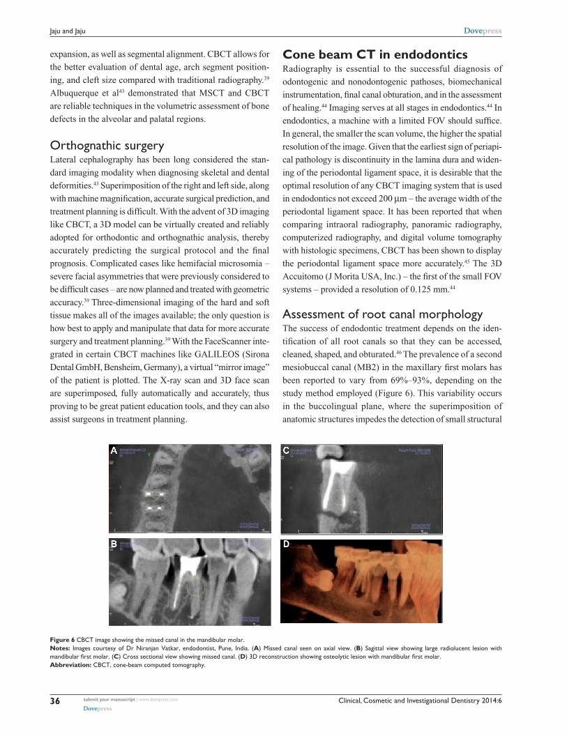
Figure 6 CBCT image showing the missed canal in the mandibular molar.Notes: Images courtesy of Dr Niranjan vatkar, endodontist, Pune, India. (A) Missed canal seen on axial view. (B) Sagittal view showing large radiolucent lesion with mandibular first molar. (C) Cross sectional view showing missed canal. (D) 3D reconstruction showing osteolytic lesion with mandibular first molar.Abbreviation: CBCT, cone-beam computed tomography.

Clinical, Cosmetic and Investigational Dentistry 2014:6 submit your manuscript | www.dovepress.com
Dovepress
Dovepress
37
Dental cone-beam CT
density changes.47,48 Conventional radiographic techniques, at
best, can only detect up to 55% of these configurations.49
Ramamurthy et al49 and Matherne et al50 have described
the limitations of 2D imaging in the detection of the MB2
canal. A study conducted by Neelakantan et al51 in an Indian
population revealed that the number of MB2 canals was more
apparent in the maxillary first molar, as compared to the sec-
ond molar. Also, type 4 canal anatomy was more prevalent
when compared to the Mongoloid population.51
Baratto Filho et al52 investigated the internal morphol-
ogy of extracted maxillary first molars by comparing their
detection rates, as obtained using an operating microscope
and CBCT to ex vivo sections. The authors reported an
ex vivo prevalence of a fourth canal in 67.14% of teeth
and additional root canals in 92.85% of mesiobuccal roots.
Clinical assessment provided slightly lower overall (53.26%),
but higher (95.63%), MB2 detection rates, whereas CBCT
results showed the lowest overall (37.05%) detection rate.
The authors further indicated that CBCT provided a good
method for the initial evaluation of maxillary first molar
internal morphology, but that the use of operating micro-
scopes was optimal. CBCT imaging has also been reported
to characterize the high prevalence of the distolingual canal
in Taiwanese individuals, it highlights anomalies in the root
canal system of mandibular premolars, and it assists in the
determination of root curvature.53,54 Some authors also wish
to report on the variations in mandibular incisor anatomy, as
detected in an Indian population by CBCT.55
With the advent of newer software provided by CBCT
companies like ORTHOPHOS XG3D/GALILEOS (Sirona
Dental GmbH), working length determination of the root
canal can be performed. However, its clinical success and
accuracy in clinical setup needs to be evaluated.
Dental periapical pathosesThe most common pathologic conditions that involve teeth
are inflammatory lesions of the pulp and periapical areas.4
CBCT technology now provides the clinician with the ability
to observe an area in three different planes, and thus he or she
can acquire 3D information. Lesions confined to cancellous
bone with little or no cortical plate erosion can be difficult
to diagnose with intraoral film.4 Lofthag-Hansen et al56,57
compared the accuracy of limited FOV CBCT to an intraoral
radiographic paralleling technique. The authors reported that
CBCT provides greater diagnostic accuracy (61%) compared
with digital (39%) and conventional (44%) radiographs. They
also indicated that while detection rates for CBCT were
higher, they did not advocate the replacement of intraoral
radiography for detecting periapical lesions in routine clinical
practice due to financial and dose considerations.
CBCT, when compared with periapical and panoramic
radiography, detected apical periodontitis at a higher rate.58
Estrela et al58 proposed a periapical index based on CBCT
for the identification of apical periodontitis (AP). The CBCT
periapical index is a six-point (0–5) scoring system calculated
from determining the largest lesional measurement in either
the buccopalatal, mesiodistal, or diagonal dimension, and it
also takes into account expansion and destruction of cortical
bone. The index is outlined in Table 3.
Using this periapical index, Low et al59 concluded that
CBCT performed better in the detection of periapical lesions
compared with conventional imaging. CBCT technology and
the assignment of gray values might aid in the diagnosis of
cysts versus granulomas.44 The generally higher detection
rates afforded by CBCT are similar to those reported for
conventional CT.44 This may be of clinical importance in
patients who present with pain or who have poorly localized
symptoms associated with an untreated or previously root-
treated tooth with no evidence of a pathology, as identified
by conventional imaging.44
Root fracturesThe usefulness and importance of CBCT in the diagnosis
and management of specific aspects of dentoalveolar trauma,
especially root fractures, luxation, displacement, and alveolar
fracture, has been studied extensively.60,61
The superiority of CBCT in the detection of vertical
and horizontal root fractures has been described in the
literature.62,63 Elimination of superimposition of anatomic
structures allows the clinician to analyze the fracture clearly.
Authors recommend the use of limited CBCT for the detec-
tion of horizontal root fractures when compared with con-
ventional imaging (Figure 7).
Table 3 CBCT periapical index
Score Quantitative bone alterations in mineral structures
0 Intact periapical bone structures1 Diameter of periapical radiolucency .0.5–1 mm2 Diameter of periapical radiolucency .1–2 mm3 Diameter of periapical radiolucency .2–4 mm4 Diameter of periapical radiolucency .4–8 mm5 Diameter of periapical radiolucency .8 mm+e expansion of periapical cortical bone
+D Destruction of periapical cortical bone
Abbreviation: CBCT, cone-beam computed tomography.

Clinical, Cosmetic and Investigational Dentistry 2014:6submit your manuscript | www.dovepress.com
Dovepress
Dovepress
38
Jaju and Jaju
Root resorptionRoot resorption is the loss of dental hard tissues as a result of
clastic activities. It might occur as a physiologic or pathologic
phenomenon.64 Root resorption might be broadly classified
into external or internal resorption by the location of the
resorption in relation to the root surface. The accuracy of
CBCT in the detection of surface defects, while higher than
conventional imaging modalities, is not perfect, and appears
to increase with increasing voxel resolution of the volumetric
dataset (greater resolution is achieved with increased voxel
size).44 CBCT has also been shown to have particular appli-
cations in the assessment of the postorthodontic apical root
resorption and, in particular, of the roots of lateral maxillary
incisors impacted by maxillary canines.44
CBCT has been used successfully to confirm the presence
of internal root resorption and it can differentiate this type of
resorption from external root resorption.65 Conventional radio-
graphy is often unable to identify the true extent, location, or the
portal of entry of a resorptive lesion.44 CBCT has been shown to
help determine treatment complexity, as well as aid the clinician
in offering an accurate prognosis on the basis of the extent of
the resorptive lesion.44 As a result, both treatment and treatment
outcomes are likely to become more predictable.44
Postoperative assessmentMonitoring the healing of apical lesions is an important aspect
of postoperative assessment in endodontics. Similarly, the
adequacy of root canal obturation is an important determinant
of endodontic success; it might be considered that CBCT is
used in the initial and subsequent monitoring of the integrity
of root canal fillings. Sogur et al66 reported that the images
acquired from phosphor plates and conventional films were
better when compared to CBCT images, as the presence of
streaking artifacts from the gutta percha and the sealer com-
promise the quality of those images with regards to root filling
evaluations. The utility of CBCT in determining the precise
nature of a perforation and the role of this on subsequent
treatment has been illustrated by Young.67
Preoperative assessment of the periapical surgical site is
of utmost importance to avoid postoperative complications.
Proximity to the mandibular canal, mental foramen, and
maxillary sinus during periapical surgery can be assessed
accurately on CBCT images. Jaju44 first described the value
of CBCT in the planning of endodontic surgery. The impor-
tance of CBCT for apical surgery of the teeth adjacent to the
maxillary sinus has subsequently been illustrated by Jaju,44
who presented a case report localizing the presence of a
periradicular lesion to a specific root.
Recommendation of the AAe and AAOMRThe following are recommendations from the American
Association of Endodontists (AAE) and AAOMR:68
• Identification of potential accessory canals in teeth with
suspected complex morphology based on conventional
imaging.
• Identif ication of root canal system anomalies and
determination of root curvature.
• Diagnosis of dental periapical pathosis in patients who
present with contradictory or nonspecific clinical signs
and symptoms, who have poorly localized symptoms
associated with an untreated or previously endodonti-
cally treated tooth with no evidence of pathosis, as
identified by conventional imaging, and in cases where
anatomic superimposition of roots or areas of the max-
illofacial skeleton is required to perform task-specific
procedures.
• Diagnosis of nonendodontic origin pathosis in order to
determine the extent of the lesion and its effect on sur-
rounding structures.
• Intra- or postoperative assessment of endodontic
treatment complications, such as overextended root
canal obturation material, separated endodontic instru-
ments, calcified canal identification, and localization of
perforations.
Figure 7 Axial view of CBCT showing the fracture line on the upper-right second premolar.Abbreviation: CBCT, cone-beam computed tomography.

Clinical, Cosmetic and Investigational Dentistry 2014:6 submit your manuscript | www.dovepress.com
Dovepress
Dovepress
39
Dental cone-beam CT
• Diagnosis and management of dentoalveolar trauma,
especially root fractures, luxation and/or displacement
of teeth, and alveolar fractures.
• Localization and differentiation of external and internal
root resorption, or invasive cervical resorption from
other conditions, and the determination of appropriate
treatment and prognosis.
• Presurgical case planning to determine the exact location
of the root apex/apices and to evaluate the proximity of
adjacent anatomical structures.
• Dental implant case planning when cross-sectional imag-
ing is deemed essential, as based on the clinical evaluation
of the edentulous ridge.
Applications in orthodonticsThe introduction of new software in orthodontic assess-
ment, such as Dolphin (Dolphin Imaging and Management
Solutions, Chatsworth, CA, USA) and Invivo Dental
(Anatomage, San Jose, CA, USA), has allowed dentists to use
CBCT images for cephalometric analysis, making it the tool
of choice for assessing facial growth, age, airway function,
and disturbances in tooth eruption.69 Katkar et al,70 in their
study, confirmed that CBCT was reliable in demonstrating
cephalometric landmarks accurately.
Moreira et al71 verified the accuracy of linear measure-
ments from CBCT cephalograms and compared them
to traditional cephalograms. CBCT proved to be more
accurate than lateral cephalogram when compared to skull
measurement.71
CBCT is a reliable tool for assessing the proximity of
impacted teeth to vital structures that could interfere with
orthodontic movement.72 When mini-implants are required
as temporary anchors, CBCT offers visual guides for safe
insertion, thus avoiding accidental and irreparable damage
to the existing roots.73 Assessing bone density before, dur-
ing, and after treatment can show whether it is decreasing
or remaining the same.73 CBCT images are self-corrected
for magnification, producing orthogonal images with a
practical 1:1 measuring ratio; as a result, CBCT is consid-
ered a more accurate option than panoramic and traditional
2D images.73
CBCT in periodonticsAccording to Vandenberghe et al,74 intraoral radiography
is the most common imaging modality used for diagnos-
ing bone morphology, such as periodontal bone defects.
However, the limitations of 2D radiography could under-
estimate the amount of bone loss or available bone due to
projection errors.74 These findings confirm the observation
by Alamri et al,73 that 2D radiographs are inadequate for
detecting changes at the bone level or for determin-
ing the architecture of osseous defects. CBCT provides
accurate measurements of intrabony defects and allows
clinicians to assess dehiscence, fenestration defects, and
periodontal cysts.73 CBCT has been used to obtain detailed
morphologic descriptions of bone that are as accurate as
those obtained via direct measurement with a periodon-
tal probe.73 CBCT can also be used to assess furcation
involvement of periodontal defects and allow clinicians
to evaluate the postsurgical results of regenerative peri-
odontal therapy.73
Moreira et al71 investigated the accuracy of CBCT in
linear measurements of bone defects and concluded that
CBCT can be an accurate diagnostic tool for the evaluation
of small osseous defects. The authors are of the view that
more scientific literature is required to conclusively prove
that CBCT is a more superior imaging technology that can
be utilized in periodontal applications.
CBCT in operative dentistryHigher radiation dose and lower resolution of CBCT com-
pared with intraoral radiography hampers its role in the
detection of occlusal caries.3
Applications in forensic dentistryAge estimation is an important aspect of forensic dentistry.
The pulpo-dentinal complex (dentin, cementum, and the
dental pulp) shows physiologic and pathological changes
with advancing age.72 Typically, extraction and sectioning
are required to quantify these morphological changes, which
is not always a viable option.75 CBCT, however, provides a
noninvasive alternative.75
The visualization of cervical vertebral morphology holds
potential in skeletal age assessment. Shi et al,76 in their study,
concluded that segmentation of individual vertebrae was
possible using CBCT volumetric datasets. This provides a
3D approach to the biologic aging of orthodontic patients
by using images of the cervical spine. It also holds poten-
tial in studying disease processes such as spinal fractures
consequent to osteoporosis.76
CBCT in obstructive sleep apnea (OSA)OSA is a common respiratory sleep disorder character-
ized by snoring and episodes of breathing cessation or the
absence of respiratory airflow (10 seconds) during sleep,

Clinical, Cosmetic and Investigational Dentistry 2014:6submit your manuscript | www.dovepress.com
Dovepress
Dovepress
40
Jaju and Jaju
despite respiratory effort.77 Any technology that would
enhance clinicians’ ability to visualize where in the air-
way obstruction occurs would help identify those subsets
of patients who may or may not benefit from a choice
of treatment modalities. CBCT, with its 3D presentation of
the airway and the airway’s surrounding structures, offers
this increased visualization of both untreated obstruction
tendencies and potentially of changes in the airway by treat-
ment modality. Ogawa et al78 demonstrated the utility of
the diagnosis of anatomy with the 3D airway imaging
with CBCT (CBCT allows to visualize the anatomy of
airspace in three dimension and helps in the diagnosis of
any anatomic variation or diseases). They noted the ability
to describe significant group differences in total airway
volume and the anteroposterior dimension of the oropha-
ryngeal airway between OSA and sex-matched controls.78
Farman et al79 examined the possibilities of using different
software packages to analyze changes in the upper airway
with and without placement of a mandibular advancement
device. Excellent segmentation was achieved, and it was
possible to make airway minimum cross-sectional area and
volumetric assessment.
CBCT in ear imaging (ENT)Improvement in CBCT technology has revolutionized not
only dentomaxillofacial imaging, but it has also improved
imaging in the field of ENT.
Sinuses and nasal fossaeHigher resolution images and operator-friendly CBCT
software with multiplanar reformation (MPR) visualization
of each examination make them particularly practical in
understanding sinonasal anatomy.80
Inflammatory pathologyCBCT shows excellent air–mucosa–bone contrast, allow-
ing for the very interesting study of air cavity anatomy
and ventilation.81 Effusion, mucosal thickening, and ostial
obstruction are perfectly visible, with precision equal to or
greater than that of CT.81 Any inflammatory or infectious
sinus pathology is accessible to CBCT examination, with
complete topographic exploration.81 However, as with CT
without contrast injection, it is not possible to distinguish
between simple mucosal thickening, mucosal cysts, polyps,
and retention cysts (Figure 8).
Fungal sinusitisWith the introduction of metal artifact reduction software
and good spatial resolution enable the detection of fine
calcifications associated with Aspergillus grafts around
intrasinus metallic foreign bodies, usually of dental origin.81
Bone remodeling and mucosal calcificationThe study of fine bone remodeling requires good spatial
resolution. This is the case for the assessment of bone exten-
sion of infectious processes of dental or sinus origin, fine
perforation, intraosseous fistular trajectories, and thinning or
blurring of walls.81 Mucosal calcification along the wall by the
osseous metaplasia of Schneider’s membrane during subacute
or chronic inflammatory processes can also be detected.81
Postoperative assessmentCBCT, with its low radiation intensity, is well adapted for
postoperative follow-up. It is, of course, restricted to benign
sinus lesion surgeries, with CT and MRI still used for the
postoperative follow-up of malignancies.81
Tumoral pathologyIf a tumor invading the soft tissue is discovered, CT and/or
MRI are mandatory. CBCT without contrast enables excellent
topographic study of bone extension, any intratumoral
calcification, or of the perilesional thin osseous wall.81
Temporal bone/cranial base aspect on cone-beam imageA lower signal-to-noise ratio gives a slightly different aspect
than on reference (CT) imaging.81 The esthetic and diagnostic
Figure 8 CBCT coronal image showing pansinusitis.Note: Image courtesy of Dr Shikha Rathi Diplomat (AAOMR, USA).Abbreviations: CBCT, cone-beam computed tomography; AAOMR, American Academy of Oral and Maxillofacial Radiology.

Clinical, Cosmetic and Investigational Dentistry 2014:6 submit your manuscript | www.dovepress.com
Dovepress
Dovepress
41
Dental cone-beam CT
qualities of an image, however, are not the same thing. CBCT
gives access to plentiful and sufficient diagnostic information
if the indications for CBCT are correct. It should always be
borne in mind that minimal radiation is an important factor
in indications for imaging assessment by X-ray, and that
radiation-free or radiation–light techniques of equal diag-
nostic quality should always be preferred.82
ConclusionWhile clinical applications of CBCT have expanded, current
CBCT technology has limitations related to the projection
geometry, detector sensitivity, and contrast resolution that
produce images that lack the clarity and usefulness of con-
ventional CT images. The clarity of CBCT images is affected
by artifacts, noise, and poor soft tissue contrast. With soft-
ware improvements, these limitations would be gradually
overcome in the future. It is also necessary to respect the
“as low as reasonably achievable” radiation dose concept.
However, this should not deter dental surgeons from utilizing
CBCT for providing necessary information. Some authors
are of the opinion that the judicious use of CBCT technology
can outweigh inherent risks over its extraordinary benefits.
Proper training and education in CBCT for oral maxillofacial
radiologists and dentists is required to ensure astute use of
CBCT technology.
AcknowledgmentsThe authors wish to thank Dr Niranjan Vatkar, Dr Shikha
Rathi, and JSD Technodent, Bangalore, India for providing
CBCT images.
DisclosureThe authors report no conflicts of interest in this work.
References1. Tyndall DA, Rathore S. Cone-beam CT diagnostic applications: caries,
periodontal bone assessment, and endodontic applications. Dent Clin North Am. 2008;52(4):825–841, vii.
2. Zoller JE, Neugebauer J. Cone-Beam Volumetric Imaging in Dental, Oral and Maxillofacial Medicine: Fundamentals, Diagnostics and Treatment Planning. Chicago, IL: Quintessence Publishing; 2008.
3. Tetradis S, Anstey P, Graff-Radford S. Cone beam computed tomography in the diagnosis of dental disease. J Calif Dent Assoc. 2010;38(1):27–32.
4. Tyndall DA, Price JB, Tetradis S, Ganz SD, Hildebolt C, Scarfe WC; American Academy of Oral and Maxillofacial Radiology. Position state-ment of the American Academy of Oral and Maxillofacial Radiology on selection criteria for the use of radiology in dental implantology with emphasis on cone beam computed tomography. Oral Surg Oral Med Oral Pathol Oral Radiol. 2012;113(6):817–826.
5. Macleod I, Heath N. Cone-beam computed tomography (CBCT) in dental practice. Dent Update. 2008;35(9):590–592, 594.
6. Howerton WB, Mora MA. Advancements in digital imaging: what is new and on the horizon? J Am Dent Assoc. 2008;139 Suppl:20S–24S.
7. Dreiseidler T, Mischkowski RA, Neugebauer J, Ritter L, Zöller JE. Comparison of cone-beam imaging with orthopantomography and computerized tomography for assessment in presurgical implant dentistry. Int J Oral Maxillofac Implants. 2009;24(2):216–225.
8. Tischler M. In-off ice cone beam computerized tomography: technology review and clinical examples. Dent Today. 2008;27(6):102, 104, 106.
9. Loubele M, Van Assche N, Carpentier K, et al. Comparative localized linear accuracy of small-field cone-beam CT and multislice CT for alveolar bone measurements. Oral Surg Oral Med Oral Pathol Oral Radiol Endod. 2008;105(4):512–518.
10. Jaju PP, Jaju SP, Suvarna PV, Dedhia P, editors. Dental CT: Third Eye in Dental Implants. New Delhi, India: Jaypee Brothers Medical Publishers Ltd; 2013.
11. Lindh C, Obrant K, Petersson A. Maxillary bone mineral density and its relationship to the bone mineral density of the lumbar spine and hip. Oral Surg Oral Med Oral Pathol Oral Radiol Endod. 2004;98(1):102–109.
12. Drage NA, Palmer RM, Blake G, Wilson R, Crane F, Fogelman I. A comparison of bone mineral density in the spine, hip and jaws of edentulous subjects. Clin Oral Implants Res. 2007;18(4):496–500.
13. Lekholm U, Zarb GA. Patient selection and preparation. In: Brånemark PI, Zarb GA, Albrektsson T, editors. Tissue-Integrated Prostheses: Osseointegration in Clinical Dentistry. Chicago, IL: Quintessence Publishing; 1985:199–209.
14. Molteni R. Prospects and challenges of rendering tissue density in Hounsfield units for cone beam computed tomography. Oral Surg Oral Med Oral Pathol Oral Radiol. 2013;116(1):105–119.
15. Orentlicher G, Abboud M. Guided surgery for implant therapy. Dent Clin North Am. 2011;55(4):715–744.
16. Chau AC, Fung K. Comparison of radiation dose for implant imaging using conventional spiral tomography, computed tomography, and cone-beam computed tomography. Oral Surg Oral Med Oral Pathol Oral Radiol Endod. 2009;107(4):559–565.
17. Miracle AC, Mukherji SK. Conebeam CT of the head and neck, part 2: clinical applications. AJNR Am J Neuroradiol. 2009;30(7): 1285–1292.
18. Adibi S, Zhang W, Servos T, O’Neill P. Cone beam computed tomogra-phy for general dentists. Open Access Scientific Reports. 2012;1:519.
19. Suomalainen A, Ventä I, Mattila M, Turtola L, Vehmas T, Peltola JS. Reliability of CBCT and other radiographic methods in preoperative evaluation of lower third molars. Oral Surg Oral Med Oral Pathol Oral Radiol Endod. 2010;109(2):276–284.
20. Ghaeminia H, Meijer GJ, Soehardi A, Borstlap WA, Mulder J, Bergé SJ. Position of the impacted third molar in relation to the mandibular canal. Diagnostic accuracy of cone beam computed tomography compared with panoramic radiography. Int J Oral Maxillofac Surg. 2009;38(9):964–971.
21. Kamrun N, Tetsumura A, Nomura Y, et al. Visualization of the supe-rior and inferior borders of the mandibular canal: a comparative study using digital panoramic radiographs and cross-sectional computed tomography images. Oral Surg Oral Med Oral Pathol Oral Radiol. 2013;115(4):550–557.
22. Tantanapornkul W, Okouchi K, Fujiwara Y, et al. A comparative study of cone-beam computed tomography and conventional panoramic radiography in assessing the topographic relationship between the mandibular canal and impacted third molars. Oral Surg Oral Med Oral Pathol Oral Radiol Endod. 2007;103(2):253–259.
23. Leifert S, Jonas IE. Dental anomalies as a microsymptom of palatal canine displacement. J Orofac Orthop. 2003;64(2):108–120.
24. Liu DG, Zhang WL, Zhang ZY, Wu YT, Ma XC. Localization of impacted maxillary canines and observation of adjacent incisor resorption with cone-beam computed tomography. Oral Surg Oral Med Oral Pathol Oral Radiol Endod. 2008;105(1):91–98.
25. Stewart JA, Heo G, Glover KE, Williamson PC, Lam EW, Major PW. Factors that relate to treatment duration for patients with palatally impacted maxillary canines. Am J Orthod Dentofacial Orthop. 2001;119(3):216–225.

Clinical, Cosmetic and Investigational Dentistry 2014:6submit your manuscript | www.dovepress.com
Dovepress
Dovepress
42
Jaju and Jaju
26. Fournier A, Turcotte JY, Bernard C. Orthodontic considerations in the treatment of maxillary impacted canines. Am J Orthod. 1982;81(3): 236–239.
27. Ericson S, Kurol J. Radiographic assessment of maxillary canine eruption in children with clinical signs of eruption disturbance. Eur J Orthod. 1986;8(3):133–140.
28. Walker L, Enciso R, Mah J. Three-dimensional localization of maxillary canines with cone-beam computed tomography. Am J Orthod Dentofacial Orthop. 2005;128(4):418–423.
29. Alqerban A, Hedesiu M, Baciut M, et al; SedentexCT Consortium. Pre-surgical treatment planning of maxillary canine impactions using panoramic vs cone beam CT imaging. Dentomaxillofac Radiol. 2013;42(9):20130157.
30. Guttenberg SA. Oral and maxillofacial pathology in three dimensions. Dent Clin North Am. 2008;52(4):843–873, viii.
31. Marques YM, Botelho TD, Xavier FC, Rangel AL, Rege IC, Mantesso A. Importance of cone beam computed tomography for diagnosis of calcifying cystic odontogenic tumour associated to odontoma. Report of a case. Med Oral Patol Oral Cir Bucal. 2010;15(3): e490–e493.
32. Hendrikx AW, Maal T, Dieleman F, Van Cann EM, Merkx MA. Cone-beam CT in the assessment of mandibular invasion by oral squamous cell carcinoma: results of the preliminary study. Int J Oral Maxillofac Surg. 2010;39(5):436–439.
33. Dreiseidler T, Alarabi N, Ritter L, et al. A comparison of multislice computerized tomography, cone-beam computerized tomography, and single photon emission computerized tomography for the assessment of bone invasion by oral malignancies. Oral Surg Oral Med Oral Pathol Oral Radiol Endod. 2011;112(3):367–374.
34. Palomo L, Palomo JM. Cone beam CT for diagnosis and treat-ment planning in trauma cases. Dent Clin North Am. 2009;53(4): 717–727, vi.
35. Heiland M, Schulze D, Rother U, Schmelzle R. Postoperative imaging of zygomaticomaxillary complex fractures using digi-tal volume tomography. J Oral Maxillofac Surg. 2004;62(11): 1387–1391.
36. Mischkowski RA, Zinser MJ, Ritter L, Neugebauer J, Keeve E, Zöller JE. Intraoperative navigation in the maxillofacial area based on 3D imag-ing obtained by a cone-beam device. Int J Oral Maxillofac Surg. 2007;36(8):687–694.
37. Kaeppler G, Cornelius CP, Ehrenfeld M, Mast G. Diagnostic efficacy of cone-beam computed tomography for mandibular fractures. Oral Surg Oral Med Oral Pathol Oral Radiol. 2013;116(1):98–104.
38. Sirin Y, Guven K, Horasan S, Sencan S. Diagnostic accuracy of cone beam computed tomography and conventional multislice spiral tomog-raphy in sheep mandibular condyle fractures. Dentomaxillofac Radiol. 2010;39(6):336–342.
39. Quereshy FA, Savell TA, Palomo JM. Applications of cone beam computed tomography in the practice of oral and maxillofacial surgery. J Oral Maxillofac Surg. 2008;66(4):791–796.
40. Tsiklakis K, Syriopoulos K, Stamatakis HC. Radiographic examination of the temporomandibular joint using cone beam computed tomography. Dentomaxillofac Radiol. 2004;33(3):196–201.
41. Dahlström L, Lindvall AM. Assessment of temporomandibular joint disease by panoramic radiography: reliability and validity in relation to tomography. Dentomaxillofac Radiol. 1996;25(4):197–201.
42. de Boer EW, Dijkstra PU, Stegenga B, de Bont LG, Spijkervet FK. Value of cone-beam computed tomography in the process of diagnosis and management of disorders of the temporomandibular joint. Br J Oral Maxillofac Surg. 2014;52(3):241–246.
43. Albuquerque MA, Gaia BF, Cavalcanti MG. Comparison between multislice and cone-beam computerized tomography in the volumetric assessment of cleft palate. Oral Surg Oral Med Oral Pathol Oral Radiol Endod. 2011;112(2):249–257.
44. Jaju SP. Cone beam CT in endodontics: a paradigm shift in clinical practice. Smile Dental Journal. 2013;8(2):22–28.
45. Jervøe-Storm PM, Hagner M, Neugebauer J, et al. Comparison of cone-beam computerized tomography and intraoral radiographs for determination of the periodontal ligament in a variable phantom. Oral Surg Oral Med Oral Pathol Oral Radiol Endod. 2010;109(2): e95–e101.
46. Vertucci FJ. Root canal anatomy of the human permanent teeth. Oral Surg Oral Med Oral Pathol. 1984;58(5):589–599.
47. Pineda F. Roentgenographic investigation of the mesiobuccal root of the maxillary first molar. Oral Surg Oral Med Oral Pathol. 1973;36(2):253–260.
48. Nance R, Tyndall D, Levin LG, Trope M. Identification of root canals in molars by tuned-aperture computed tomography. Int Endod J. 2000;33(4):392–396.
49. Ramamurthy R, Scheetz JP, Clark SJ, Farman AG. Effects of imaging system and exposure on accurate detection of the second mesio-buccal canal in maxillary molar teeth. Oral Surg Oral Med Oral Pathol Oral Radiol Endod. 2006;102(6):796–802.
50. Matherne RP, Angelopoulos C, Kulild JC, Tira D. Use of cone-beam computed tomography to identify root canal systems in vitro. J Endod. 2008;34(1):87–89.
51. Neelakantan P, Subbarao C, Ahuja R, Subbarao CV, Gutmann JL. Cone-beam computed tomography study of root and canal morphology of maxillary first and second molars in an Indian population. J Endod. 2010;36(10):1622–1627.
52. Baratto Filho F, Zaitter S, Haragushiku GA, de Campos EA, Abuabara A, Correr GM. Analysis of the internal anatomy of maxillary first molars by using different methods. J Endod. 2009;35(3):337–342.
53. Tu MG, Huang HL, Hsue SS, et al. Detection of permanent three-rooted mandibular first molars by cone-beam computed tomog-raphy imaging in Taiwanese individuals. J Endod. 2009;35(4): 503–507.
54. Cleghorn BM, Christie WH, Dong CC. Anomalous mandibular pre-molars: a mandibular first premolar with three roots and a mandibular second premolar with a C-shaped canal system. Int Endod J. 2008;41(11):1005–1014.
55. Jaju SP, Jaju PP, Garcha V. Root canal assessment of mandibular incisors in an Indian population using cone beam CT. Endod Prac. 2013;7(2):105–111.
56. Estrela C, Bueno MR, Sousa-Neto MD, Pécora JD. Method for determination of root curvature radius using cone-beam computed tomography images. Braz Dent J. 2008;19(2):114–118.
57. Lofthag-Hansen S, Huumonen S, Gröndahl K, Gröndahl HG. Limited cone-beam CT and intraoral radiography for the diagnosis of periapical pathology. Oral Surg Oral Med Oral Pathol Oral Radiol Endod. 2007;103(1):114–119.
58. Estrela C, Bueno MR, Azevedo BC, Azevedo JR, Pécora JD. A new periapical index based on cone beam computed tomography. J Endod. 2008;34(11):1325–1331.
59. Low KM, Dula K, Bürgin W, von Arx T. Comparison of periapical radiography and limited cone-beam tomography in posterior maxillary teeth referred for apical surgery. J Endod. 2008;34(5):557–562.
60. Cohenca N, Simon JH, Roges R, Morag Y, Malfaz JM. Clinical indications for digital imaging in dento-alveolar trauma. Part 1: traumatic injuries. Dent Traumatol. 2007;23(2):95–104.
61. Ilgüy D, Ilgüy M, Fisekcioglu E, Bayirli G. Detection of jaw and root fractures using cone beam computed tomography: a case report. Dentomaxillofac Radiol. 2009;38(3):169–173.
62. Hassan B, Metska ME, Ozok AR, van der Stelt P, Wesselink PR. Detection of vertical root fractures in endodontically treated teeth by a cone beam computed tomography scan. J Endod. 2009;35(5): 719–722.
63. Kamburoglu K, Ilker Cebeci AR, Gröndahl HG. Effectiveness of limited cone-beam computed tomography in the detection of horizontal root fracture. Dent Traumatol. 2009;25(3):256–261.
64. Patel S, Pitt Ford TR. Is the resorption external or internal? Dent Update. 2007;34:218–229.

Clinical, Cosmetic and Investigational Dentistry
Publish your work in this journal
Submit your manuscript here: http://www.dovepress.com/clinical-cosmetic-and-investigational-dentistry-journal
Clinical, Cosmetic and Investigational Dentistry is an international, peer-reviewed, open access, online journal focusing on the latest clini-cal and experimental research in dentistry with specific emphasis on cosmetic interventions. Innovative developments in dental materials, techniques and devices that improve outcomes and patient satisfaction
and preference will be highlighted. The manuscript management system is completely online and includes a very quick and fair peer-review system, which is all easy to use. Visit http://www.dovepress.com/testimonials.php to read real quotes from published authors.
Clinical, Cosmetic and Investigational Dentistry 2014:6 submit your manuscript | www.dovepress.com
Dovepress
Dovepress
DovepressDovepress
43
Dental cone-beam CT
65. Cohenca N, Simon JH, Mathur A, Malfaz JM. Clinical indications for digital imaging in dento-alveolar trauma. Part 2: root resorption. Dent Traumatol. 2007;23(2):105–113.
66. Sogur E, Baksi BG, Gröndahl HG. Imaging of root canal fillings: a comparison of subjective image quality between limited cone-beam CT, storage phosphor and film radiography. Int Endod J. 2007;40(3): 179–185.
67. Young GR. Contemporary management of lateral root perforation diagnosed with the aid of dental computed tomography. Aust Endod J. 2007;33(3):112–118.
68. Joint Position Statement of the American Association of Endodontists and the American Academy of Oral and Maxillofacial Radiology. Use of Cone-Beam Computed Tomography in Endodontics. 2010. Available from: http://c.ymcdn.com/sites/www.aaomr.org/resource/resmgr/Docs/AAOMR-AAE_postition_paper_CB.pdf.
69. Bjerklin K, Ericson S. How a computerized tomography examination changed the treatment plans of 80 children with retained and ectopically positioned maxillary canines. Angle Orthod. 2006;76:43–51.
70. Katkar RA, Kummet C, Dawson D, et al. Comparison of observer reliability of three-dimensional cephalometric landmark identifica-tion on subject images from Galileos and i-CAT cone beam CT. Dentomaxillofac Radiol. 2013;42(9):20130059.
71. Moreira CR, Sales MA, Lopes PM, Cavalcanti MG. Assessment of linear and angular measurements on three-dimensional cone-beam computed tomographic images. Oral Surg Oral Med Oral Pathol Oral Radiol Endod. 2009;108(3):430–436.
72. Erickson M, Caruso JM, Leggitt L. Newtom QR-DVT 9000 imaging used to confirm a clinical diagnosis of iatrogenic mandibular nerve paresthesia. J Calif Dent Assoc. 2003;31(11):843–845.
73. Alamri HM, Sadrameli M, Alshalhoob MA, Sadrameli M, Alshehri MA. Applications of CBCT in dental practice: a review of the literature. Gen Dent. 2012;60(5):390–400; quiz 401.
74. Vandenberghe B, Jacobs R, Yang J. Diagnostic validity (or acuity) of 2D CCD versus 3D CBCT-images for assessing periodontal breakdown. Oral Surg Oral Med Oral Pathol Oral Radiol Endod. 2007;104(3): 395–401.
75. Yang F, Jacobs R, Willems G. Dental age estimation through volume matching of teeth imaged by cone-beam CT. Forensic Sci Int. 2006; 159 Suppl 1:S78–S83.
76. Shi H, Scarfe WC, Farman AG. Three-dimensional reconstruc-tion of individual cervical vertebrae from cone-beam computed-tomography images. Am J Orthod Dentofacial Orthop. 2007;131(3): 426–432.
77. McCrillis JM, Haskell J, Haskell BS, et al. Obstructive sleep apnea and the use of cone beam computed tomography in airway imaging: a review. Semin Orthod. 2009;15(1):63–69.
78. Ogawa T, Enciso R, Memon A, Mah JK, Clark GT. Evaluation of 3D airway imaging of obstructive sleep apnea with cone-beam computed tomography. Stud Health Technol Inform. 2005;111:365–368.
79. McCrillis J, Farman A, Scarfe W, et al. Analysis of Airway Changes using CBCT with and without Placement of a Mandibular Advancement Device. Louisville, KY: University of Louisville School of Dentistry; 2008.
80. Mathew R, Omami G, Hand A, Fellows D, Lurie A. Cone beam CT analysis of Haller cells: prevalence and clinical significance. Dentomaxillofac Radiol. 2013;42(9):20130055.
81. Hodez C, Griffaton-Taillandier C, Bensimon I. Cone-beam imaging: applications in ENT. Eur Ann Otorhinolaryngol Head Neck Dis. 2011;128(2):65–78.
82. Faccioli N, Barillari M, Guariglia S, et al. Radiation dose saving through the use of cone-beam CT in hearing-impaired patients. Radiol Med. 2009;114(8):1308–1318.