Transactivation of Abl by the Crk II adapter protein requires a PNAY sequence in the Crk C-terminal...
13
Transactivation of Abl by the Crk II adapter protein requires a PNAY sequence in the Crk C-terminal SH3 domain Charles Reichman 1 , Kamalendra Singh 1 , Yan Liu 1 , Sukhwinder Singh 1 , Hong Li 1 , J Eduardo Fajardo 2 , Andras Fiser 2 and Raymond B Birge* ,1 1 Department of Biochemistry and Molecular Biology, UMDNJ-New Jersey Medical School, 185 South Orange Avenue, Newark, NJ 07103, USA; 2 Department of Biochemistry, Albert Einstein College of Medicine, Bronx, NY 10461, USA To gain a better understanding of how Crk II regulates the function of the Abl tyrosine kinase, we explored the function of the C-terminal linker and SH3 domain, a region of Crk II that is still poorly understood. Molecular modeling, tryptophan fluorescence, and covariation se- quence alignment indicate that the Crk-SH3-C has a unique binding groove and RT loop not observed in typical SH3 domains. Based on these models, we made a series of mutations in the linker and in residues predicted to destabilize the putative binding pocket and RT loop. In Abl transactivation assays, Y222F and P225A mutations in the linker resulted in strong transactivation of Abl by Crk II. However, mutations predicted to be at the surface of the Crk SH3-C were not activators of Abl. Interest- ingly, combinations of activating mutations of Crk II with mutations in the highly conserved PNAY sequence in the SH3-C inactivated the activating mutations, suggesting that the SH3-C is necessary for activation. Our data provide insight into the role of highly conserved residues in the Crk-SH3-C, suggesting a mechanism for how the linker and the Crk-SH3-C function in the transactivation of the Abl tyrosine kinase. Oncogene (2005) 24, 8187–8199. doi:10.1038/sj.onc.1208988; published online 12 September 2005 Keywords: Crk; Abl; SH3 domain; atypical; covaria- tion; protein modeling; kinase activation Introduction The Crk II protein is an SH2–SH3 domain containing adapter protein involved in cytoskeletal reorganization and events associated with cellular adhesion, migration, phagocytosis, cellular proliferation, and oncogenic transformation (Feller, 2001). Crk II, the cellular homolog of the v-Crk oncoprotein, is expressed from two alternatively spliced mRNAs to produce Crk II (p38) and Crk I (p28). Crk II and Crk I differ in their C- terminal regions (Matsuda et al., 1992; Reichman et al., 1992; Feller, 2001). Crk II contains an N-terminal SH2 domain and two SH3 domains (SH3-N and SH3-C). Crk II also contains an approximately 45 amino-acid linker between the two SH3 domains. In contrast, v-Crk and Crk I encode proteins with C-terminal truncations that do not possess a regulatory tyrosine residue 222 (Y222) between the SH3 domains, or the C-terminal SH3 domain (Mayer et al., 1988; Reichman et al., 1992). Expression of v-Crk or Crk I in cells robustly increases cellular phosphotyrosine levels, and transforms cells, despite the fact that neither protein has intrinsic tyrosine kinase activity. While several cellular proteins have been identified that bind to the SH2 and SH3-N domain, Abl and its paralog Arg are the only tyrosine kinases that are known to directly associate with v-Crk or Crk II (Feller et al., 1994; Ren et al., 1994; Wang et al., 1996). Binding of v-Crk or Crk I to Abl results in Abl transactivation, and possibly contributes to the transforming potential of Abl (Ren et al., 1994; Sattler and Salgia, 1998; Hemmeryckx et al., 2002). In contrast, the Crk II–Abl interaction is more transient, and by a signal that is not well understood, Abl-bound Crk becomes phosphorylated on Y222 by the Abl kinase, resulting in the intramolecular association of Y222 with the Crk SH2 domain, and dissociation of Crk from Abl (Feller et al., 1994; Rosen et al., 1995). Expression of Y222F Crk II enhances Abl transactivation, increases cellular phosphotyrosine, and promotes cellular transformation (Shishido et al., 2001; Zvara et al., 2001). Presently, the molecular mechanism by which Crk II activates Abl is not well understood, although previous studies suggest that the SH2 and SH3-C domains are necessary (Shishido et al., 2001). The present study was carried out to investigate the structural and functional characteristics of the Crk-SH3-C. We present data that the linker and the Crk-SH3-C, through conserved residues in the modeled binding pocket and RT loop of the SH3-C, play important roles in the transactivation of the Abl tyrosine kinase. Results and discussion Threading analysis, computational molecular modeling, and biophysical studies of the C-terminal Crk SH3 domain (Crk SH3-C) Previous efforts to identify binding partners of the Crk- SH3-C domain failed to detect an interaction with Received 2 March 2005; revised 23 June 2005; accepted 24 June 2005; published online 12 September 2005 *Correspondence: RB Birge; E-mail: [email protected] Oncogene (2005) 24, 8187–8199 & 2005 Nature Publishing Group All rights reserved 0950-9232/05 $30.00 www.nature.com/onc
-
Upload
independent -
Category
Documents
-
view
4 -
download
0
Transcript of Transactivation of Abl by the Crk II adapter protein requires a PNAY sequence in the Crk C-terminal...
Transactivation of Abl by the Crk II adapter protein requires a PNAY
sequence in the Crk C-terminal SH3 domain
Charles Reichman1, Kamalendra Singh1, Yan Liu1, Sukhwinder Singh1, Hong Li1,J Eduardo Fajardo2, Andras Fiser2 and Raymond B Birge*,1
1Department of Biochemistry and Molecular Biology, UMDNJ-New Jersey Medical School, 185 South Orange Avenue, Newark,NJ 07103, USA; 2Department of Biochemistry, Albert Einstein College of Medicine, Bronx, NY 10461, USA
To gain a better understanding of how Crk II regulates thefunction of the Abl tyrosine kinase, we explored thefunction of the C-terminal linker and SH3 domain, aregion of Crk II that is still poorly understood. Molecularmodeling, tryptophan fluorescence, and covariation se-quence alignment indicate that the Crk-SH3-C has aunique binding groove and RT loop not observed in typicalSH3 domains. Based on these models, we made a series ofmutations in the linker and in residues predicted todestabilize the putative binding pocket and RT loop. InAbl transactivation assays, Y222F and P225A mutationsin the linker resulted in strong transactivation of Abl byCrk II. However, mutations predicted to be at the surfaceof the Crk SH3-C were not activators of Abl. Interest-ingly, combinations of activating mutations of Crk II withmutations in the highly conserved PNAY sequence in theSH3-C inactivated the activating mutations, suggestingthat the SH3-C is necessary for activation. Our dataprovide insight into the role of highly conserved residues inthe Crk-SH3-C, suggesting a mechanism for how thelinker and the Crk-SH3-C function in the transactivationof the Abl tyrosine kinase.Oncogene (2005) 24, 8187–8199. doi:10.1038/sj.onc.1208988;published online 12 September 2005
Keywords: Crk; Abl; SH3 domain; atypical; covaria-tion; protein modeling; kinase activation
Introduction
The Crk II protein is an SH2–SH3 domain containingadapter protein involved in cytoskeletal reorganizationand events associated with cellular adhesion, migration,phagocytosis, cellular proliferation, and oncogenictransformation (Feller, 2001). Crk II, the cellularhomolog of the v-Crk oncoprotein, is expressed fromtwo alternatively spliced mRNAs to produce Crk II(p38) and Crk I (p28). Crk II and Crk I differ in their C-terminal regions (Matsuda et al., 1992; Reichman et al.,1992; Feller, 2001). Crk II contains an N-terminal SH2domain and two SH3 domains (SH3-N and SH3-C).
Crk II also contains an approximately 45 amino-acidlinker between the two SH3 domains. In contrast, v-Crkand Crk I encode proteins with C-terminal truncationsthat do not possess a regulatory tyrosine residue 222(Y222) between the SH3 domains, or the C-terminalSH3 domain (Mayer et al., 1988; Reichman et al., 1992).Expression of v-Crk or Crk I in cells robustly increasescellular phosphotyrosine levels, and transforms cells,despite the fact that neither protein has intrinsic tyrosinekinase activity. While several cellular proteins have beenidentified that bind to the SH2 and SH3-N domain, Abland its paralog Arg are the only tyrosine kinases that areknown to directly associate with v-Crk or Crk II (Felleret al., 1994; Ren et al., 1994; Wang et al., 1996). Bindingof v-Crk or Crk I to Abl results in Abl transactivation,and possibly contributes to the transforming potential ofAbl (Ren et al., 1994; Sattler and Salgia, 1998;Hemmeryckx et al., 2002). In contrast, the Crk II–Ablinteraction is more transient, and by a signal that is notwell understood, Abl-bound Crk becomes phosphorylatedon Y222 by the Abl kinase, resulting in the intramolecularassociation of Y222 with the Crk SH2 domain, anddissociation of Crk from Abl (Feller et al., 1994; Rosenet al., 1995). Expression of Y222F Crk II enhances Abltransactivation, increases cellular phosphotyrosine, andpromotes cellular transformation (Shishido et al., 2001;Zvara et al., 2001). Presently, the molecular mechanism bywhich Crk II activates Abl is not well understood,although previous studies suggest that the SH2 andSH3-C domains are necessary (Shishido et al., 2001). Thepresent study was carried out to investigate the structuraland functional characteristics of the Crk-SH3-C. Wepresent data that the linker and the Crk-SH3-C, throughconserved residues in the modeled binding pocket and RTloop of the SH3-C, play important roles in thetransactivation of the Abl tyrosine kinase.
Results and discussion
Threading analysis, computational molecular modeling,and biophysical studies of the C-terminal Crk SH3domain (Crk SH3-C)
Previous efforts to identify binding partners of the Crk-SH3-C domain failed to detect an interaction with
Received 2 March 2005; revised 23 June 2005; accepted 24 June 2005;published online 12 September 2005
*Correspondence: RB Birge; E-mail: [email protected]
Oncogene (2005) 24, 8187–8199& 2005 Nature Publishing Group All rights reserved 0950-9232/05 $30.00
www.nature.com/onc

canonical PxxPxK,R containing motifs. Feller et al.(1994) previously reported that the Crk-SH3-N, but notthe Crk-SH3-C, bound Abl. It has also been demon-strated that the Crk-SH3-N can interact with PXXPXKmotifs from C3G, whereas Crk-SH3-C cannot (Knudsenet al., 1994). Using a 35S peptide overlay assay (Felleret al., 1995), Crk-SH3-N was shown to bind to a numberof cellular proteins, although no binding partners ofCrk-SH3-C were detected. We also failed to detect Crk-SH3-C binding to a PPPALPPKK peptide by trypto-phan fluorescence (data not shown). It has beenreported, however, that the Crk-SH3-C can bind tothe nuclear export protein Crm1 (Smith et al., 2002).
To define the structural elements of the Crk-SH3-Cand ascertain whether its structure conforms to that ofcanonical SH3 domains, we utilized unbiased threadinganalysis, sequence alignment, template structure-basedmodeling, and tryptophan fluorescence spectroscopy onpurified recombinant proteins in the present paper. Weemployed an unbiased threading analysis to determinestructures in the databases that most closely match thestructure of the Crk-SH3-C (aa 239–293). Threadinganalysis (using Fugue and 3D-PSSM databases) identi-fied several structures, all SH3 domains (Figure 1a), and10 models were built on each selected structure. The bestmodel produced, based on the evaluations performedwith Prosa, was one based on the crystal structure of theSH3-C domain of human Grb2 (Lowenstein et al.,1992), an SH2/SH3 domain containing adapter protein.High-quality models were also produced from the SH3
domains of Caenorhabditis elegans Sem-5 (Rozakis-Adcock et al., 1992), and of the mouse Grb2-like proteinMona/Gads (Lewitzky et al., 2004). All are atypical SH3domains that bind PXXP-independent motifs. Crk-SH3-C also had structural similarities to p47phox, the Vavproto-oncogene SH3-N, and Amphiphysin II(Figure 1a). Based on the threading, the best templatewe found was Grb2-SH3-C (1gri). Therefore, we builtthe homology model using Grb2-SH3-C as a template.However, the homology-based model using the N-terminal SH3 domain of Crk superimposed on the onebuilt with Grb-2 SH3-C had an RMS deviation of 1.73A. This suggests that models built on either templatecould be used for comparison purposes to Crk SH3-C,and for further analysis.
We wanted to explore how the SH3-C might differ inits structure and peptide-binding activity from those ofcanonical SH3 domains. Since the binding pocket of theCrk-SH3-N is well established to bind conventionalPXXP peptide-containing proteins, the minimal energymolecular modeling program Look (version 3.5) wasemployed to model the structure of the SH3-C using thecrystal structure of mouse Crk SH3-N bound to aninteracting peptide as the template. The SH3-N inter-acting peptide is PPPALPPKK, from the Crk interact-ing protein C3G (Wu et al., 1995). The predictedstructure of Crk SH3-C is consistent with conventionalSH3 domains, comprising five antiparallel b sheets, andtwo flexible variable loops called the RT loop and a 310turn between the fourth and fifth b sheets that flank a
Crk(N)SH3 VEYVRALFDFNGNDDED--LPFKKGDILKIRDKPEEQWWNAEDMD-GKRGMIPVPYVEKCrk(C)SH3 FYARVIQKRVPNAYDKTALALEVGELVKVTKINMSGQWEGECN--GKRGHFPFTHVRLLDCed-2(C)SH3 AKAKVTFDRVPNAYDPTQLRVKKGQTVLVTQKMSNGMYKAELD--GQIGSVPHTYLRFTgrb2(C)SH3 QPTYVQALFDFDPQEDGELGFRRGDFIHVMDNSDPNWWKGACH--GQTGMFPRNYVTPVNRNV
Consensus GKYVRALYDYEAREDDE--LSFKKGDIITVLEKSDDGWWKGRLNDTGREGLFPSNYVEEIDSPPKAVVIF FDPQNPGD--ITLRE EVLEIINREEGD YEAENLRD QR WI A FLRPVEE K GEDEE R KV LVSDDNEE LRVK S KS YV KLL
Strand a Strand eStrand dStrand cStrand bRT-Src-loop
PDB Chain Gene OrganismCodes1h3h A GRB2-RELATED ADAPTOR PROTEIN 2 Mus
(GADS, GRBLG, GRB2L); C-TERMINAL SH3 1sem A SEM-5; (GRB2, DRK); C-TERMINAL SH3 C. Elegans1udl A INTERSECTIN 2; (KIAA1256); SH3 DOMAIN; Human1gri A GROWTH FACTOR BOUND PROTEIN 2; Human
(GRB2); SH3 DOMAIN1ng2 A NEUTROPHIL CYTOSOLIC FACTOR 1;(P47-PHOX); Human
SH3 DOMAIN;1gcq VAV PROTO-ONCOGENE; Human
N-TERMINAL SH3 DOMAIN1mv3 A MYC BOX DEPENDENT INTERACTING PROTEIN 1; Human
(BIN1; AMPHIPHYSIN II)
a
b
A
Figure 1 Sequence alignment and threading analysis. (a) Structures identified by threading analysis as suitable templates for modelingthe Crk-SH3-C domain. The PDB code, chain, a brief description of the chain, and the source organism are provided. (b) The SH3consensus sequence as determined by the covariation analysis of Larson and Davidson (2000) (op cit) was aligned with the Crk SH3-N,Crk SH3-C and human Grb2 SH3-C using Clustal W software; blue¼ hydrophobic core residues; red¼binding to PXXPXK/R. Notethat in Crk-SH3C and Ced-SH3C, the canonical peptide-binding amino acids are poorly conserved. Underlined residues¼ short,identical region in Ced-2 and Crk SH3-C
Transactivation of Abl by Crk IIC Reichman et al
8188
Oncogene

possible binding pocket (Figure 2a). Superimposition ofthe modeled N- and C-terminal Crk SH3 domains, whileindicating subtle differences between the RT loop and bcand bd loops, nevertheless suggests an overall conserva-tion of structure between the two SH3 domains(Figure 2a). Moreover, while only 17% identical to themammalian proteins, the model of the Ced-2 SH3-Calso had the basic b-barrel structure characteristic of allSH3 domains, when modeled using the SH3-N of Crk IIas the template. It may fold in order to generate amodular domain (data not shown).
Covariation analysis of protein sequences involves thestudy of statistical variations in amino-acid sequences inhomologous proteins or protein domains, and analyzeswhether a change in an amino acid at one positioncorrelates with a change at another site in the protein. Ifsuch covarying sites are found, then it is hypothesizedthat these two sites likely interact in some manner, forexample by contributing to the overall tertiary structure,or to a complex binding region. Using covariationanalysis of 266 SH3 domains, consensus sequences for
SH3 domains have been derived. Two types of aminoacids have been identified in the consensus sequence,those conserved for folding into the SH3 hydrophobiccore, and those typically present at the surface andconserved for interaction with PXXP containing pep-tides (Larson and Davidson, 2000; Larson et al., 2000).
We compared the SH3 domains of Crk II, Ced-2, andthe Grb2 SH3-C to the SH3 consensus sequence derivedby Larson et al. (2000). As illustrated in Figure 1b, CrkSH3-C and Ced-2-SH3-C are remarkably conserved inthe hydrophobic core residues that maintain the overalltertiary structure of SH3s (10/10 hydrophobic coreamino acids are either identical or conserved in Crk IIand 9/10 in Ced-2). In contrast, Crk SH3-C and Ced-2SH3-C had a general lack of conservation in the peptide-binding surface, whereby only three of 16 amino acidswere conserved in Crk II and five of 16 in Ced-2. EvenGrb2-SH3-C, which had the most similar structure toCrk II as evidenced by the threading analysis, had moreconservation in the peptide-binding amino acids, com-pared to Crk-SH3-C (Figure 1b).
We performed tryptophan measurements on recom-binant Crk SH3-C domain proteins. While most SH3domains contain a contiguous tryptophan duet (WW) inthe c strand, the Crk SH3-C domain contains a singletryptophan (W276). This fact led us to test whether thisTrp is unconstrained (unfolded, Emax B353–360 nM) orconstrained (Emax B333–342 nM). As shown inFigure 2b, Trp fluorescence measurements of the full-length SH3 domain (aa 239–293) or the SH3 domaincontaining parts of the linker-C-terminal SH3 domainhad Emax values of B339–342 nM, consistent with afolded polypeptide structure. Another N-terminal trun-cated linker-SH3C protein (aa 230–293), and an SH3-Cprotein comprising aa 239–297, also had a similar Emax
(data not shown). Finally, we generated an N-terminaltruncated SH3-C domain (DFYARVIQ) that deletes thefirst seven amino acids in the putative b1 SH3 strand(Crk DSH3-C). This generates a protein with Emax ofB358 nM, consistent with an unfolded, denaturedstructure (Figure 2b). These data do not contradict themodel of the SH3-C.
The vast majority of SH3 domains recognize specificproline-rich sequences that adopt the conformation ofpolyproline type I or type II helices (PPII), containingthe core sequence PXXP (Kay et al., 2000). In typicalSH3 domains, the PXXP core motif is stabilized bystacking of the aromatic residues, Trp, Phe, and Tyr,which interact directly with the pyrrolidine rings of thePXXP. Additional binding specificity occurs outside ofthis core interaction and confers specificity amongdistinct SH3 domains. Recently, a number of SH3domains have been reported to bind unconventionalnon-PXXP-dependent motifs in target proteins. Suchexamples include an RKXXYXXY motif in SKAP55that binds the Fyn and Lck SH3 domains (Kang et al.,2000), a PXXDY motif in several proteins that binds theEps8 SH3 (Mongiovi et al., 1999), a WXXXFXXLEmotif in p67phox and Pex5p that binds the Pex13p SH3domain (Barnett et al., 2000; Kami et al., 2002), a PX(V/I)(D/N)RXXKP motif in Gab1 and SLP-76 proteins
SH3[N]
SH3[C]RT-loop
30
14.36.2
3.5
L-SH3
∆SH3
a b c d e f
SH2 SH3 [N] SH3 [C]Y222
K190CRP F239YAR THVR293
a
b
c
d
e
f
aa Emax
190-293
200-293
210-293
220-293
239-293
246-293
332 nm
340 nm
340 nm
340 nm
340 nm
358 nm
305
a
b
Figure 2 Basic SH3-C model and tryptophan fluorescence onrecombinant proteins. (a) Ribbon structure of the Crk SH3-N(gray), superimposed on the modeled structure of the Crk SH3-C(dark gray). The RT loop is seen on the upper left. (b) Tryptophanfluorescence analysis of the SH3-C domain containing proteins. Alinear map of Crk II depicting the linker region (aa 190–238) andSH3-C (aa 239–293), and the boundaries of linkerSH3-C peptides,(a) aa 190–293; (b) aa 200–293; (c) aa 210–293; (d) aa 220–293; (e)aa 239–293 (SH3-C domain), and (f) aa 246–293 (DSH3-C) areshown. At right in this panel are the normalized fluorescences ofpeptides a–f; at lower left is a Coomassie blue stained gel of thepeptides, with size markers at left and SH3-C and Linker-SH3-C(L-SH3) at right. All proteins were analysed by mass spectrometryto verify mass
Transactivation of Abl by Crk IIC Reichman et al
8189
Oncogene

that bind the Mona/Gads SH3 domain (Lewitzky et al.,2001; Harkiolaki et al., 2003; Lewitzky et al., 2004), anda PX(P/A)XXR motif that binds to the CIN85/SETA/Rut protein SH3 domains (Kurakin and Bredesen, 2002;Kurakin et al., 2003). For some atypical SH3 domains,for example Mona/Gad, SKAP55, and Eps8, theunconventional peptides have been suggested to bindto regions including the classical PXXP binding site(Harkiolaki et al., 2003). However, studies with Pex13pshow that it can bind both conventional PXXP-contain-ing peptides in the usual way, as well as a second, non-PXXP peptide, that binds at a novel, noncanonicalpocket on the same SH3. Both peptides can bind to thePex13p SH3 simultaneously (Pires et al., 2003). There-fore, it would not be unprecedented to find anSH3 whose overall structure is similar to that of mostother SH3s, but whose binding surface is very different,as we have presented evidence here concerning theSH3-C of Crk II.
The covariation analysis suggests that the bindingresidues of the SH3-C may be very different from thoseof canonical SH3s. As we show below, when themodeled SH3-C is superimposed on the template SH3-N and compared in detail, the potential peptide-bindingresidues of the SH3-C also look very different. For thesereasons, we believe it is probable that the SH3-C ofCrk II will be shown to be atypical in the peptides itcan bind, and will likely not bind conventionalPXXP peptides.
In order to predict which residues might occupy thebinding surface of the SH3-C, and for later mutagenesisstudies, we compared in detail the residues in the SH3-Npeptide that made direct contact with P3, P6, and K8positions of the PPPALPPKK peptide to the corre-sponding substituted amino acids in the SH3-C. Thepeptide residues at the P3, P6, and K8 positions of theinteracting peptide are the most important because theyconform to the PXXPXK consensus sequence. There area total of 12 residues in the Crk SH3-N domain withinpotential interacting distance (3.8 A) of the C3G peptide(Figure 3). These residues are L140, F141, Y186, P185,P183, Q168, W169, E167, E166, D147, E149, and D150.The topologically equivalent residues in the modeledC-terminal Crk SH3 domain are K246, R247, H291,T290, P288, G274, Q275, S273, M272, D253, T255, andA256. Some of these are illustrated in Figure 3. If theregion in the SH3-C model analogous to the bindinggroove in the SH3-N also binds a peptide, thecomparison of the properties of the binding site residuesin the two domains suggests that the nature of thepeptides that bind the two proteins are probablydissimilar. Interestingly, several of the hydrophobicresidues in the N-terminal SH3 domain that makedirect contact with the pyrrolidine rings of the PXXPmotif are replaced with basic or polar amino acids in themodel of the SH3-C. For example, F141 is replaced byK246 (Figure 3a), Y186 is replaced by H291 (Figure 3a),and W169 is replaced by Q275 (Figure 3b). Anothermajor difference is the characteristics of the binding sitefor interacting residues with K8 and K9 of the peptide.The two lysine residues of the C3G peptide form
extensive salt bridges with negatively charged residuesof the Crk SH3-N (Figure 3c; Wu et al., 1995). K8 formsthree ion pairs with D147, E149, and D150, and onlyone of the residues (equivalent to D147) is conserved inthe SH3-C. The other two residues are replaced by a Thr(T255) and Ala (A256). Similarly, the possibility of thesalt bridge between K9 and E167 of Crk SH3-N alsodoes not exist in Crk SH3-C, because the equivalentposition to E167 of the SH3-N is occupied by a Serresidue (not shown). These dissimilarities clearly estab-lish the differences in the nature of recognized andbound peptides by the SH3-N versus the modeledstructure of the SH3-C, and suggest that the Crk SH3-C does not bind conventional PXXP-containing ligands.
In Figure 3d, we show a space-filling model of theSH3-C, together with a peptide in the place analogous tothe peptide binding groove of the SH3-N. Each of thesimilar regions that interact with P3, P6, and K8 in theSH3-N are highlighted in blue, green, and yellow,respectively. Note that a potential binding groove stillexists where the peptide is positioned, although asdetailed above, important specific interactions forPXXPXK peptides are not present in the SH3-C model.Finally, we note that a portion of the RT loop of SH3-Chas residues that point away from the SH3-C domain(red, Figure 3d), and because of this might be moreavailable to interact with other molecules. These includeR247 to T255. In a study of the structures of 17 SH3domains, investigators noted the surprising presence ofstabilizing interactions among residues of the RT-Srcloop that are not directly involved in peptide binding.They speculated that these conserved interactions mightstabilize the RT loop as a whole for interactions withother peptides (Larson and Davidson, 2000). The Capositions of the SH3-C residues R247, V248, P249,A251, Y252, D253, and T255, and the correspondingresidues in Ced-2 (oriented similarly) are highlyconserved in a wide range of species of Crk and CrkL,including chicken, mammals, Xenopus laevis, Drosophilamelanogaster, and C. elegans (Table 1). Later, we detailhow we used a mutational analysis of residues in each ofthe four blocks highlighted in Figure 3d to understandthe functions of the SH3-C. We did this by investigatinghow they affect the activation of the Abl tyrosine kinaseby Crk II.
Contributions of the linker (aa 190–238) and the Crk-SH3-C to regulating the interaction between Abl and Crk
Previous studies have shown that Crk binding to Ablhas a transactivating function, and therefore we canscore the extent of Abl activation when coexpressed withdifferent Crk mutants (Shishido et al., 2001; Zvara et al.,2001). To explore the functions of the SH3 linker andthe proposed surface of the Crk SH3-C in further detail,we generated three types of mutants in the C-terminalregion of Crk, and coexpressed these with Abl to seehow they affected Abl binding and Abl transactivation.These mutants include substitutions of (i) individualproline residues of the linker, (ii) residues predicted tobe on the binding surface of the Crk SH3-C, and
Transactivation of Abl by Crk IIC Reichman et al
8190
Oncogene

(iii) residues that comprise the putative RT loop of theCrk-SH3-C.
Prolines have many roles in interacting with adaptermolecules, and can thereby strongly affect the regulationof these and other signaling molecules, includingkinases. The best known examples of proline-bindingactivity are the canonical PXXP peptides that interactwith most SH3 domains, and short sequences like theP221YAQP225 in the Crk linker which, when phosphory-lated on tyrosine, bind to SH2 domains. Proline residuesin linker/connector regions have also been shown to beinhibitors in cis of some kinases (Macias et al., 2002).For example, in Hck, the SH3 folds over and interactsintramolecularly with a small proline-containing region,blocking the kinase domain (Sicheri et al., 1997).
Finally, in p47phox, an SH3 domain is inhibited frombinding to interacting proteins by an intramolecularinteraction with a proline-containing peptide, providinga precedent for the regulation of SH3 domain bindingby such intramolecular interactions (Ago et al., 1999).
Shown in Figure 4A is the Clustal W alignment of thelinker regions of chicken, rat, and Xenopus Crk IIproteins, as well as murine CrkL. The alignment ofprolines is highlighted; many of these are present as PXPsequences. As the linker region of Crk II contains 10prolines out of 49 amino acids, we decided to mutateeach one individually to alanine, and test the ability ofthese mutants to activate Abl kinase activity. Themutants generated were P193A, P211A, P213A,P217A, P219A, P221A, P225A, P230A, and P238A
H291
Y186P3
PPPALPPKKK peptide
P3 interactions:crkSH3(N) Y186 P185 F141crkSH3(C) H291 T290 K246
F141
W169
Q275
P6
PPPALPPKKK Peptide
P6 interactions:crkSH3(N) W169 P183 P185crkSH3(C) Q275 P288 T290
PPPALPPKKK Peptide K8
2.81 A
2.49 A
E149
D150
D147
A256D253
K8 interactions:crkSH3(N) D147 E149 D150crkSH3(C) D253 T255 A256
P3
P6
RT-loop
K8
a b
dc
Figure 3 Peptide/SH3 interaction details. (a) Details of the interactions of the P3 residue of the PPPALPPKK peptide with the SH3-N(gray), superimposed on the corresponding residues of the SH3-C (purple). Side chains of SH3-N (gray, red for oxygen) and side chainsof SH3-C (purple) are shown. (b) Details of the interactions of the P6 residue of the PPPALPPKK peptide with the SH3-N (gray),superimposed on the corresponding residues of the SH3-C (purple). Side chains of SH3-N (gray, red for oxygen) and side chains ofSH3-C (purple) are shown. (c) Details of the interactions of the K8 residue of the PPPALPPKK peptide with the SH3-N (gray),superimposed on the corresponding residues of the SH3-C (purple). Side chains of SH3-N (gray, red for oxygen), side chains of SH3-C(purple), and intermolecular distances between SH3-N residues and K8 are shown. (d) Space filling model of the SH3-C with thePPPALPPKK peptide shown in the similar location to its position when binding to the SH3-N. The region that interacts with P3 of thepeptide (blue), P6 (green), and K8 (yellow) are highlighted. The part of the RT loop that does not interact with PPPALPPKK andprojects away from the SH3 is in red. This is the most conserved region in the alignment of Crk II and Ced-2
Transactivation of Abl by Crk IIC Reichman et al
8191
Oncogene

Table 1 Taxonomic alignment of the Crk-SH3-C and CrkL SH3-C domains
CLUSTAL W (1.74) multiple sequence alignment
mus_CrkII PIYARVIQKRVPNAYDKTALALEVGELVKVTKINVSGQWEGECNGKRGHFPFTHVRLLDQhum PIYARVIQKRVPNAYDKTALALEVGELVKVTKINVSGQWEGGCNGKRGHFPFTHVRLLDQrat PIYARVIQKRVPNAYDKTALALEVGELVKVTKINVSGQWEGECNGKRGHFPFTHVRLLDQgallus PFYARVIQKRVPNAYDKTALALEVGELVKVTKINMSGQWEGECNGKRGHFPFTHVRLLDQXenopus PIFARVIQKRVPNAYDKTALALEVGDLVKVTKINVSGQWEGECNGKYGHFPFTHVRLLEQDanio PVYARAIQKRVPNAYDKTALALEVGDMVKVTKINVNGQWEGECKGKHGHFPFTHVRLLDQDrosophila PAYARVKQSRVPNAYDKTALKLEIGDIIKVTKTNINGQWEGELNGKNGHFPFTHVEFVDDCed-2 PAKAKVTFDRVPNAYDPTQLRVKKGQTVLVTQKMSNGMYKAELDGQIGSVPHTYLRFTAV
hum_CrkL PVFAKAIQKRVPCAYDKTALALEVGDIVKVTRMNINGQWEGEVNGRKGLFPFTHVKIFDPDanioRerio_CrkL PVLAKAIQKRVPCAYDKTALALEVGDIVKVTRMNISGQWEGEVNNRRGLFPFTHVKILDP
* *:. .*** *** * * :: *: : **: .* ::. ..: * .*.*::.:
Using the Clustal W 1.74 program at the EMBnet Swiss site, we aligned the SH3-C of Crk II and CrkL from several species, and found that themost conserved string of residues in Crk II was in the RT loop. This was also true when the entire Crk II alignments were examined (not shown).Shown are the SH3-C domains from the Crk II of murine (Ogawa et al. 1994), human (Matsuda et al. 1992), rat (Kizaka-Kondoh et al. 1996),Gallus gallus (Reichman et al. 1992), X. laevis (Evans et al. 1997), D. rerio (IMAGE clone AAH77088; (Lennon et al. 1996), and D. melanogaster(Galletta et al. 1999); the SH3-C from ced-2 of C. elegans (Reddien and Horvitz, 2000), and CrkL from human (ten Hoeve et al. 1993) and crkL D.rerio (IMAGE clone AAH56763; (Lennon et al. 1996). The PNAY sequence studied in this paper (and the aligned PCAY of crkL) is shown in gray.‘*’ indicates identical or equivalent residues in all sequences in the alignment, ‘:’ indicates conserved substitutions, ‘.’ indicates semi-conservedsubstitutions
66
46
30
Crk
W17
0K
no D
NA
Crk
-
wtC
rk
Crk
Y22
2F
Crk
W27
6K
Crk
P19
3A
Crk
P21
1A
Crk
P21
7A
Crk
P21
9A
Crk
P22
1A
Crk
P22
5A
Crk
P23
0A
Crk
P23
8A
Crk
P21
3A
+ Abl
GST-Crk120-225
(1.0
)
(1.7
)
(0.2
)
(1.8
)
(1.1
)
(0.7
)
(08)
(0.6
)
(1.2
)
(1.8
)
(5.3
)
(1.9
)
(1.1
)
Chicken KCRPSSASVSTLTGGNQDSSHPQPLGGPEPG-PYAQPSINTPLPNLQNGPFYARVIQRat KYRPASASVSALIGGNQEGSHPQPLGGPEPG-PYAQPSVNTPLPNLQNGPIYARVIQXenopus KYRPPSSPGSALIGGNQENSHPQPLGGPEPG-PYAQPSVNTPLPNLQNGPIFARVIQMousecrkL GNRNSNSYG.. IPEPAHAYAQPQTTTPLPTVASTPGAAINPLPSTQNGPVFAKAIQ
193
211
213
217
219
221
225
230
232
239
Crk
W17
0K
no D
NA
Crk
-
wtC
rk
Crk
Y22
2F
Crk
W27
6K
Crk
P19
3A
Crk
P21
1A
Crk
P21
7A
Crk
P21
9A
Crk
P22
1A
Crk
P22
5A
Crk
P23
0A
Crk
P23
8A
Crk
P21
3A
a Kinase Assay
c Abl
Crkd
b Densitometry (0.1
)
(0.1
)
--Abl
--Crk II
A
B
Figure 4 Proline to alanine mutations in the Crk linker. (A) Alignment of the Crk II linker from three species, together with the CrkLprotein from murine. This shows the strong conservation in the linker region, including the Tyr phosphorylated by Abl, and the YXXPsite which binds SH2. The Pro mutated to Ala is labeled above . (B) (a) and (b), in vitro kinase assay from 293T cells coexpressing Abland Crk II constructs mutated at each of the sites shown. Kinase assays were separated by 10% SDS–PAGE and [32P] labeled bandswere detected by autoradiography. Shown at the left are sizes of protein markers, and at the right the substrate used in the kinase assay.Quantitation was on a phosphoimager (normalized to the Crk II wild-type sample). (c) and (d) Western blots of Abl and Crkexpression. Equal amounts of protein from cell lysates were detected by Western blotting to confirm expression. Abl DNA wastransfected in all samples except ‘no DNA’
Transactivation of Abl by Crk IIC Reichman et al
8192
Oncogene

(Figure 4B). Wild-type Crk or individual Pro to Alamutants were coexpressed with Abl in HEK 293T cells.Subsequently, detergent lysates were prepared andimmunoprecipitated with anti-Crk RF51 Ab (a CrkSH2-domain specific antibody), and assayed for asso-ciated Abl kinase activity (Figure 4B a, b). For positivecontrols, we used Y222F, which mutates the Ablphosphorylation site, and W276K Crk, an SH3-Cdestabilizing mutant that we have previously shown toincrease the association between Abl and Crk (Zvaraet al., 2001). As shown in Figure 4B a, b, most of the Pto A mutants did not significantly increase Crk-associated Abl kinase activity, although consistently,P217A and P219A slightly reduced the association ofAbl and Crk as indicated by the kinase pull-down assay.However, mutations in the P225 of the P221YAQP225
sequence strongly increased the transactivation of Abl,as well as increasing cellular tyrosine phosphorylation
(Figure 5a; Figure 7a). Since P225 and Y222 are both apart of the pYAQP motif that interacts with the Crk-SH2 domain, they likely have a similar function inpreventing SH2 docking to the Y222 when tyrosinephosphorylated. These data are consistent with thefinding that the minimal SH2 recognition sequence ofphospho-Y222 includes the P225, as has been reportedfor the Crk SH2 domain (Songyang et al., 1993).Coexpression of Abl in the absence of Crk (lane 2,Figure 4A), or with W170K Crk (lane 5), an N-terminalSH3 mutant that abrogates Abl binding, produced nodetectable Crk-associated Abl activity, or Abl protein(Figure 4B a, b; Figure 5d; Figure 6c). This indicatesthat the assay conditions specifically measure Ablactivity associated with the specified Crk mutants.
Next, we studied mutants expected to disrupt thepredicted binding groove of Crk SH3-C. Since thenatural ligand of the Crk SH3-C is not known, we
220 --
90 --66 --
46 --
30 --
--Crk II
--Abl
--Abl
--Crk II
noC
r k
Crk
II
Crk
I
Crk
R38
K
Cr k
W1 7
0K
Cr k
Y2 2
2F
Cr k
P2 2
5A
Crk
K2 4
6A
Cr k
R24
7A
Cr k
K,R
246,
7 A
,A
Crk
A25
6D
Crk
Q27
5W
Crk
W27
6K
+ Abl
--GST-Crk
(0.2
)
(1)
(1.9
)
(0.4
)
(2.2
)
(4.4
)
(1.2
)
(1.6
)
(1.2
)
(4.7
)
(3.5
)
(3.1
)
(1.1
)
--Abl
a p-Tyr
d
b Abl
c Crk
e Densitometry
f Crk ip/Abl blot
g Crk ip/Crk blot --Crk II
Kinase Assay
Figure 5 In vitro kinase assay and Western blots of AblþCrk mutants. (a) 293T cells were cotransfected with DNAs encoding Ablplus Crk mutants as indicated. Detergent lysates were prepared 48 h later, and centrifuged to remove insoluble materials. Equalamounts of lysates were loaded on an SDS–PAGE gel and Western blotted with anti-p-Tyr antibody. (b) and (c) Western blots oflysates using the indicated antibodies. (d) Lysates were immunoprecipitated by anti-Crk antibodies, washed, and used for in vitrokinase assays in the presence of [g-32P]ATP and GST-Crk (110–225) as exogenous substrate. Kinase assays were run on SDS–PAGEgels and quantitated on a phosphoimager. (e) Densitometry of kinase assay above (normalized to the Crk II wild-type sample). (f) and(g) Kinases assays were Western blotted with the indicated antibodies
Transactivation of Abl by Crk IIC Reichman et al
8193
Oncogene
mutated the corresponding amino acids that most likelyreplaced residues that interacted directly with thePPPALPPKK peptide in the SH3-N. These mutationsinclude K246A, R247A, K246A/R247A, and H291A(P3-equivalent position amino acids), Q275W (P6-equivalent positions), and A256D (K8-equivalent posi-tion) (Figure 3). Subsequently, we made the additionalmutations A256K and Q275A in order to mutate theseresidues away from the canonical SH3 residues typicallyfound at these positions (in the case of A256, manySH3s have a D at the equivalent position, whichinteracts with a K or R in the K/RXXPXXP orPXXPXK/R interacting peptide; in the case of Q275,the equivalent position is usually occupied by W, as inSH3-N). As above, CrkSH3-C mutants were co-expressed with Abl for coimmunoprecipitation andtransactivation analysis (Figure 5). There was a moder-ate increase in Abl kinase activity when Crk II wascoexpressed with Abl, compared to overexpression ofAbl alone. However, the strongest activators of Ablwere Crk I, Y222F, and P225A (Figure 5a). Comparedto expression of Abl with wild-type Crk II, there wasessentially no change in cellular phosphotyrosine levels
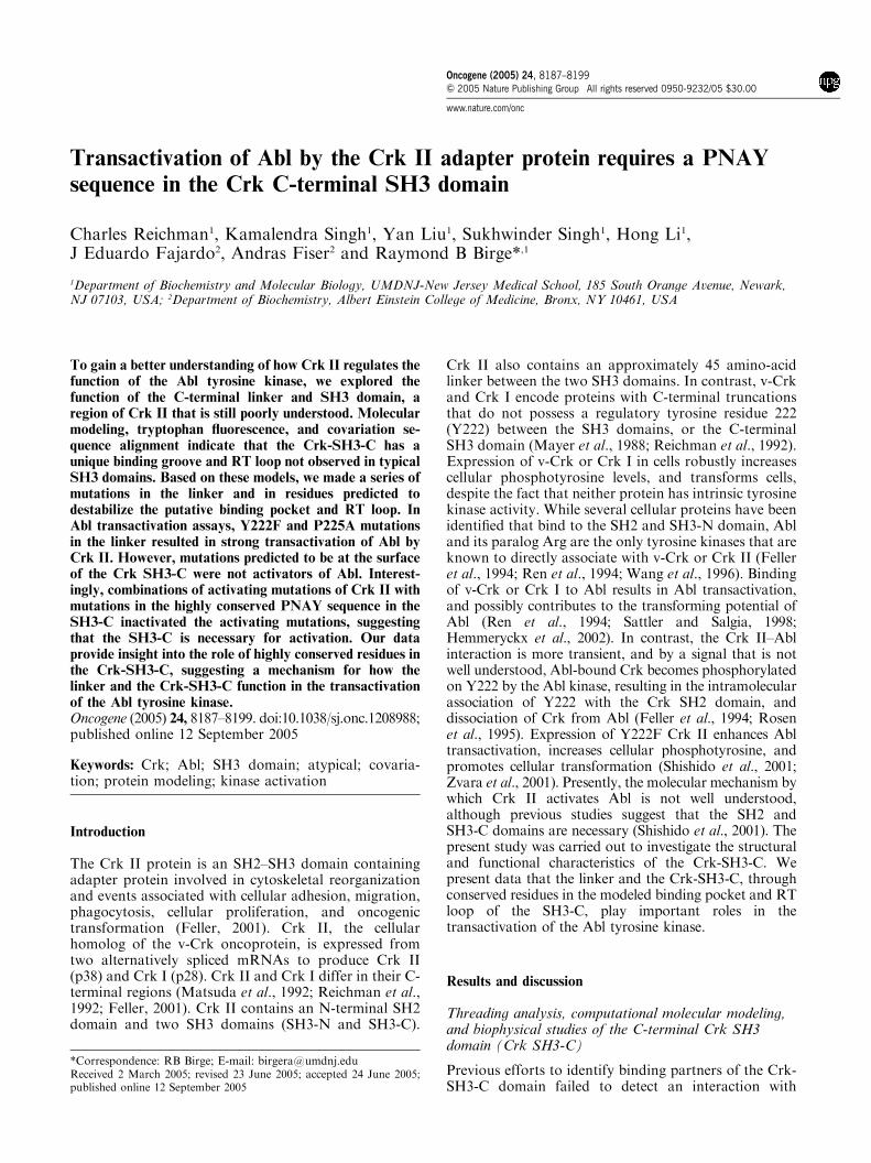
with the Crk II SH3-C mutants K246A, R247A,K246A/R247A, A256D, Q275W, and W276K.
Thus, mutations at residues modeled to be at the SH3-C surface did not lead to upregulation of Abl kinaseactivity in vivo compared to wild-type Crk II. However,when we examined the in vitro kinase activity of Crkimmunoprecipitates, we found increased Abl kinaseactivity not only in Crk I, Y222F and P225A, but also insome of the SH3-C surface mutations, A256D, Q275W,and W276K (Figure 5d, e); the Crk II sample in lane 2 isnormalized to 1). Our results with A256K and Q275Awere similar to those with A256D and Q275W; neitherincreased in vivo cellular phosphotyrosine (Figure 7B a),but both showed increased in vitro tyrosine kinaseactivity (Figure 6a, b). Interestingly, when lysates ofthese transfected cells were co-immunoprecipitated withanti-Crk Abs, there was a correlation between the kinaseactivity and the extent of Abl co-immunoprecipitation(Figure 6c; Figure 5f). One possible explanation is thatthese mutants bind more strongly to Abl than the wild-type Crk II protein, although they do not activate itskinase activity. Thus, the increased in vitro kinaseactivity may be due to increased Abl protein complexed
Crk
-
Crk
II
Crk
W17
0K
Crk
P22
5A
Crk
P24
9A
Crk
Y25
2A
Crk
∆P
NA
Y
Crk
Y22
2F/∆
PN
AY
Crk
P22
5A/∆
PN
AY
Crk
A25
6K
Crk
Q27
5A
Crk
W27
6K
Crk
Y22
2F/W
276K
Crk
P22
5A/W
276K
Crk
Y22
2F
Kinase -GST-Crk
Densitometry (3.2
)
(5.3
)
(2.9
)
(2.3
)
(3.9
)
(1.3
)
(1.7
)
(0.6
)
(1.5
)
(1.6
)
(5.5
)
(5.4
)
(0.3
)
(1)
(0.2
)
a
b
Crk ip/Abl blot c
Crk ip/Crk blotd
-96
+ Abl
Abl pY245-96
Abl pY412 -148
-96
Abl -96
e
f
g
-148
Figure 6 In vitro kinase assay and Western blots of AblþCrk mutants using phospho-specific antibodies. (a) 293T cells werecotransfected with DNAs encoding Abl plus Crk mutants, as indicated. Detergent lysates were prepared 48 h later and centrifuged toremove insoluble materials. Lysates were immunoprecipitated by anti-Crk antibodies, washed, and used for in vitro kinase assays in thepresence of [g-32P]ATP and GST-Crk (110–225) as substrate. Kinase assays were run on SDS–PAGE gels and quantitated on aphosphoimager. (b) Densitometry of kinase assay above (normalized to the Crk II wild-type sample). (c) and (d) Replicate sampleswere immunoprecipitated and Western blotted with the indicated antibodies (e), (f) and (g) are Western blots of lysates using theindicated antibodies
Transactivation of Abl by Crk IIC Reichman et al
8194
Oncogene
with Crk in the immunoprecipitation, rather than anincrease in Abl kinase specific activity.
Importantly, however, while Q275A, Q275W, A256K,A256D, and W276K Crk II may have increased theassociation between Crk and Abl, their mode of Ablactivation was distinct from that of the Y222F andP225A mutations in the linker. In this respect, the C-terminal SH3 domain mutants did not transactivate Ablto result in increased cellular tyrosine phosphorylation(Figure 5, compare a, d; also compare Figure 6a withFigure 7B a). As a result, some linker mutants, but notSH3-C mutants tested, increase Abl kinase activity invivo, it suggests that the linker region and the SH3-Cperform different functions. For example, the wild-typelinker may be directly inhibiting transactivation, per-
haps by binding to the kinase domain in a mannersimilar to the Abl kinase domain activation loop, whichis also a substrate of the Abl kinase. The SH3-C maybind elsewhere on Abl (see below), or may serve tomodulate the affinity of Crk II for Abl.
Abl transactivation by CrkY222F and P225A is mediatedby a PNAY sequence in the RT loop of the Crk-SH3-C
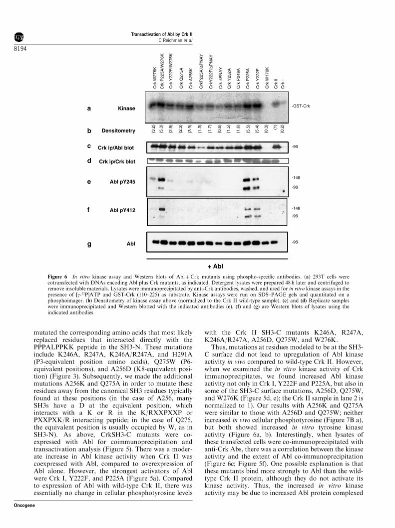
In addition to the residues of the RT loop of SH3-N thatinteract with K8 of the PPPALPPKK peptide, otherresidues point away from the binding groove and do notinteract with PPPALPPKK, notably D148. Similarly,there are residues in our model of the SH3-C that alsopoint away from the main body of the SH3-C.Interestingly, these comprise the most conserved stretchof residues between the evolutionarily distant Crk II ofvertebrates and the ortholog Ced-2 protein of C. elegans(Table 1 and data not shown). Shown in the ribbondiagrams in Figure 7A are a number of those residues ofthe SH3-C (P249, N250, A251 and Y252), together withsimilar superimposed residues in the structure of theSH3-N (F144, N146).
We substituted a number of these residues in order totest their effect on Abl activation. When co-expressed incells, the individual mutants P249A, Y252A andDPNAY had little effect on cellular phosphotyrosinecompared to the wild-type Crk II protein (Figure 7B a).In addition, their effect on the in vitro Abl kinase assaywas not dramatic (Figure 6a). This suggests that the RTloop residues are not sufficient for the direct activationof Abl by Crk II. However, we also made a number ofmutants in which we combined activating mutants ofCrk II with either DPNAY or the SH3-C structure-disrupting mutant W276K (W276 is predicted to beburied in the hydrophobic core). Interestingly, thesep-Tyr Western blots show that the W276K and DPNAYmutations were dominant over the activating mutations;the enhanced co-immunoprecipitation and activationwas either abrogated (Y222F/DPNAY, P225A/DPNAY,and Y222F/W276K) or reduced (P225A/W276K)(Figure 7B a). In addition, the enhanced co-immuno-precipitation and activation was also abrogated in thecase of the Y222F/DPNAY and P225A/DPNAY muta-tions, and reduced for the Y222F/W276K mutant(Figure 6a–c). This suggests that the RT loop isnecessary for activation of Crk II, although it is notsufficient.
Detection of Crk mutants and Abl proteins withanti-phospho-Abl antibodies
The kinase activity of Abl can be auto-inhibited throughinternal interactions in which its myristoylatedN-terminus, and SH2 and SH3 domains bind to thetwo lobes of the kinase domain, termed the N- andC-terminal lobes (Nagar et al., 2003). The crystalstructure of the N-terminal cap region of Abl boundto its own kinase domain has shown that the auto-inhibited structure requires a P242TIY245 sequence, whichis just N-terminal to the kinase domain. The side chain
Y252
A251
N250
F144P249
N145
N146
Crk
P22
5A
Crk
Y25
2A
Crk
-
Crk
II
Crk
W17
0K
Crk
P24
9A
Crk
∆P
NA
Y
Crk
Y22
2F/∆
PN
AY
Crk
P22
5A/∆
PN
AY
Crk
A25
6K
Crk
Q27
5A
Crk
W27
6K
Crk
Y22
2F/W
276K
Crk
P22
5A/W
276K
Crk
Y22
2F
-50
-64p-Tyr
pY245Abl ab
a
b
Crkc+ Abl
-148
-96
-36
-22
-36
-148
-96
-50
-64
-36
-22
A
B
Figure 7 Predicted RT loop structure and phosphotyrosineWestern blots of AblþCrk mutants. (A) Superimposed ribbondiagrams of SH3-N (light gray) and SH3-C (dark gray). Residuesof the SH3-N that do not interact with the PPPALPPKK peptide(green), and corresponding residues of the SH3-C (purple) are alsoshown. These residues are predicted to point away from the SH3-C.(B) (a) Equal amounts of lysates from 293T cells transfected withAbl plus Crk mutants were loaded on 8/11% step-gradient SDS–PAGE gels and Western blotted with anti-p-Tyr antibody. (b) and(c) Western blots of lysates with the indicated antibodies
Transactivation of Abl by Crk IIC Reichman et al
8195
Oncogene
of the unphosphorylated Y245 projects into the kinasedomain, stabilizing the structure and inhibiting thekinase domain. This structure is further stabilized by theSH3 of Abl, which interacts with the unphosphorylatedY245 as it rests in the kinase domain (Figure 8). Autophosphorylation of Y245, or mutations that disrupt thisregion, activate Abl (Brasher and Van Etten, 2000). Inaddition, the kinase domain has an activation loopincluding Y412, which sticks into the kinase domain andinhibits it. Upon activation, this tyrosine is alsoautophosphorylated, and the activation loop becomesplaced outside the kinase domain (Nagar et al., 2003;reviewed in Hantschel and Superti-Furga, 2004; Wang,2004).
We utilized antibodies made against Abl phosphopep-tides containing either pY245 or pY412 to explorewhether Abl was activated when coexpressed with CrkII. We found that both antibodies detected Abl intransfections in which cellular phosphotyrosine waselevated: Y222F, P225A and P225A/W276K (Figure 6e,f; Figure 7B a). For pY245, the amount of detectioncorrelated with the levels of increased phosphotyrosinein cells, with P225A/W276K less than the other two. Inaddition, there was some detection on the othertwo W276K lysates, W276K alone and Y222F/W276K. The pY412 antibody detected Abl coexpressedwith the three Crk II mutants most active in increasingcellular phosphotyrosine. As Abl activation in thepresence of Crk mutants correlates with the tyrosinephosphorylation on Abl seen in other modes of Ablactivation, it suggests that the transactivation of Abl byCrk occurs by a mechanism similar to other kinds ofactivation of Abl.
Most interestingly, we also found that the Abl pY245antibody crossreacted with Crk in several lysates,notably P225A, Q275A, W276K/P225A, and W276K
(Figure 7B b). While the meaning of this is not clear atpresent, there are instances where an antibody preparedagainst one activation-specific epitope can cross-reactwith another epitope, which selects the same bindingpartner as the first epitope. For example, the STAT5SH2 domain binds both pY694 of STAT5 to form aSTAT5 homodimer, and pY343 and/or pY401 of theerythropoietin receptor (Epo R). Two separate anti-bodies raised against the pY694 of STAT5 crossreactedwith the tyrosine phosphorylated Epo R; this cross-reaction was localized to epitopes at pY343 and/orpY401 (Barber et al., 2001).
By analogy, there may exist one or several PXXYmotifs in Crk that are structurally similar to the negativeregulatory P242TVY245 motif in human Abl (PTIY inmouse Abl). There are three PXXY motifs in the C-terminus of Crk (PVPY186 in the C-terminus of the SH3-N; PGPY222 at the conventional Abl phosphorylationsite, and P249NAY252 in the RT loop of the SH3-C). TheP249NAY252 sequence is particularly interesting not onlybecause it may point away from the predicted SH3domain core, but because it is identical in Crk II from awide range of species (human, rat, mouse, chicken,zebrafish, Drosophila, and X. laevis). It is also the mostconserved contiguous sequence between the entiremammalian Crk II and Ced-2 proteins (Table 1 anddata not shown). It is also interesting that the Y222Fand P2225A Crk proteins were tyrosine phosphorylatedwhen expressed with Abl, suggesting that sites distinctfrom the negative regulatory Y222 are phosphorylatedby Abl. Although we have yet to map these phosphor-ylation sites, it is noteworthy that Crk strongly cross-reacted with anti-Abl pY245, but not anti-Abl pY412,again suggesting that one or more of the PXXYsequences of Crk are phosphorylated. Consistent withthis idea, expression of P225A/DPNAY or Y222F/DPNAY abrogated Crk II phosphorylation, suggestingthat the putative RT loop of the SH3-C is necessary forAbl activation by Crk II. Taken together, we posit thatthe Crk SH3-C, in particular the RT loop residues, isrequired for the transient activation of Abl by Crk II,possibly by mimicking and destabilizing the negativeregulatory PTIY motif in Abl.
As shown in Figures 6 and 7, phosphorylation onY245 of Abl was not sufficient to activate Abl in thecase of coexpression of Crk mutants containingW276K, suggesting that this mutation can block Ablactivation, perhaps by remaining bound to Abl underconditions in which wild-type Crk II would normallydissociate from Abl. This suggests that W276K trapsthe Crk II/Abl interaction in a bound, partiallyactivated state. If W276K binds more strongly to Ablthan wild-type Crk II, it would bring down more Abl inthe Crk immunoprecipitates. This may explain whythe W276K mutants show increased Abl kinase acti-vity in the in vitro kinase assay and increasedco-immunoprecipitation with Abl, although they donot increase the levels of cellular phosphotyrosine(‘in vivo kinase assay’).
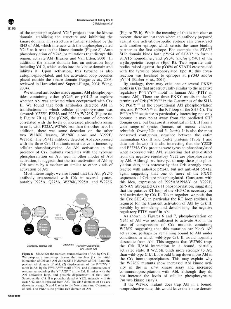
If the W276K mutant does trap Abl in a bound,nonproductive state, this would leave the kinase domain
Figure 8 Model for the transient transactivation of Abl by Crk II.We propose a multi-step process that involves (1) the initialinteraction of Crk and Abl via the SH3-N domain of Crk II and theproline-rich domain of Abl, (2) displacement of the P242TVY245
motif in Abl by the P249NAY252 motif of Crk, and (3) interaction ofresidues surrounding the Y222AQP225 in the Crk II linker with theAbl activation loop, and possible displacement of that loop.Subsequently, Crk II is phosphorylated at Y222, interacts with itsown SH2, and is released from Abl. The SH3 domains of Crk areshown in orange. N and C refer to the N-terminus and C-terminusof Abl. The PRD is the proline-rich domain of Abl
Transactivation of Abl by Crk IIC Reichman et al
8196
Oncogene

inactive, with Crk II rather than the Abl N-terminaldomains controlling/inhibiting Abl kinase activity. Thisalso suggests that the wild-type Crk II/Abl heterodimerawaits some signal to Crk or Abl itself that results inactivation of Abl. It would be interesting to test whetherDPNAY or W276K Crk-SH3-C mutants act as domi-nant negative mutants for the physiological activation ofthe Abl or Bcr-Abl kinases.
Since it has been shown that phosphorylation of AblY245 relieves the intramolecular binding of the AblP242TVY245 region to the Abl kinase domain, it isconceivable that phosphorylation of one or more ofthe PXXY sequences in Crk II could result in Ablactivation. If W276K mutants of Crk II remain boundto Abl in a state in which the heterodimer complex ispartially activated, they may become targets for addi-tional phosphorylation on PXXY regions of Crk and/orAbl, without the subsequent activation of Abl. Thismight explain why W276K mutants had increasedphosphorylation on Abl of Y412 compared to Crk IIalone, on Abl and Crk of Y245 epitopes, and increasedAbl in vitro kinase activity (i.e., possibly increasedbinding of Crk to Abl).
The binding of Crk to Abl could change the ability ofAbl to interact with other proteins. The Abl P242TVY245-kinase intramolecular interaction is stabilized by the AblSH3 domain; should the P242TVY245 of Abl be displacedby Crk II, then the SH3 of Abl might become availableto bind to other partners. Subsequently, this could alsopotentially make the SH2 of Abl available as well, sinceit is also part of the Abl intramolecular N-terminal-to-kinase domain interaction (Nagar et al., 2003).
It is already known that Abl phosphorylates CrkY222, and autophosphorylates on Abl Y245 and AblY412. It would be interesting to explore whether otherkinases can phosphorylate these sites, and also whetherCrk can be phosphorylated at PXXY sites by Abl oranother kinase. Phosphorylation of Abl at Y245 andY412 may occur by autophosphorylation, or perhapstransphosphorylation by another Abl molecule; it hasbeen shown that Abl can be activated by homodimer-ization (Smith and Van Etten, 2001). Since Src and Hckhave been shown to phosphorylate and activate Abl, it isalso possible that a Src family member, or anothertyrosine kinase, could be involved in phosphory-lating Abl or Crk when the two are in a complex (Taniset al., 2003).
Overall, these and other observations suggest thatwhile Crk I can constitutively activate Abl kinaseactivity in vivo, Crk II activation of Abl (towards thephosphorylation of exogenous substrates) is moretightly regulated and requires a multistep process.Furthermore, it appears that any activation of Abl byCrk II that does occur requires an initiating signal inwhich, at the minimum, an inhibitory effect of the Crk IIlinker (centering on Y222) is relieved, and anotheractivating step involving the SH3-C occurs as well. Asshown in Figure 8, we propose a multistep process in theinteraction and transient activation of Abl by Crk II.These include (1) the initial interaction of Crk and Ablvia the SH3-N domain of Crk and the proline-rich
domain of Abl, (2) displacement of the P242TVY245 motifin Abl by the P249NAY252 motif of Crk, and (3)interaction of residues surrounding the Y222AQP225 inthe Crk II linker with the Abl kinase domain, replacingthe Abl activation loop (the Abl kinase activation loopmaintains Abl in the inactive configuration). Subse-quently, phosphorylation by Abl of Crk at Y222, andintramolecular binding of Crk SH2 to this phosphotyr-osine, leads to dissociation of Crk from Abl and signaldownmodulation.
In summary, we examined the structural and func-tional properties of the Crk II linker and SH3-C domainto understand how they regulate the biological proper-ties of Crk II. Using several independent criteriaincluding tryptophan fluorescence, homology-basedmolecular modeling, and informatic analysis, we pro-vide a model where the minimal functional unit of theCrk SH3-C (aa 239–293) is a modular bundle containingantiparallel b-sheets packed against each other withW276 at the hydrophobic core. However, in contrast tocanonical SH3 domains, Crk SH3-C and Ced-2 SH3-Care unlikely to bind conventional PXXP-containingpeptides, because the residues that are required for theseinteractions are mostly unconserved. Our data suggestthat the SH3-C domains of Crk and Ced-2 are stable-folded entities characteristic of all known SH3 domains,but contain binding residues unique among SH3domains. We also present evidence that the Crk-SH3-C, through a conserved PNAY sequence in the putativeRT loop, is necessary for the transient activation of Ablfollowing Crk II binding. Consistent with this study,studies by Kizaka-Kondoh et al. (1996) have alsoshown that this region of Crk is important for EGF-induced signaling to ras in rodent fibroblasts, althoughthese investigators did not explore the role of Abl in thispathway. SH3 and SH2 domains not only bind to pYpeptides and PXXP peptides intermolecularly, but theyact intramolecularly in some kinases as inhibitors (Abl,Src, Hck). We hypothesize that Crk, as a protein thathas been demonstrated to regulate Abl kinase activity, isusing the same themes in its regulation of Abl. Thus, themethod of regulation of Abl already seen intramolecu-larly may be conserved when Crk binds Abl, except thatin the heterodimer the domains of Crk take over theregulatory roles from those of Abl. This study suggestsanother degree of modularity in the function of the Crkadapter protein, and a possible explanation for why theC-terminal SH3 domain is so conserved.
Materials and methods
Threading analysis, homology-derived molecular modeling, andcovariation sequence analysis
A homology model of the Crk SH3-C was generated using theprogram Look (version 3.5; Molecular Application Group,Palo Alto, CA, USA). The template for homology modeling(Grb-2 SH3-C) was identified by threading analysis usingFUGUE (Shi et al., 2001) and 3D-PSSM (Kelley et al., 2000).The quality of models produced by FUGUE and 3D-PSSMwas evaluated with PROSA (Sippl, 1993).
Transactivation of Abl by Crk IIC Reichman et al
8197
Oncogene
Larson and Davidson (2000) used 266 SH3 domainsequences for covariation analysis, and derived a set ofconsensus sequences for SH3 domains. We used the first,most highly conserved consensus sequence in our alignments.A multiple sequence alignment of four sequences comprisingthe SH3 consensus sequence, Crk SH3-N, Crk-SH3-C, andCed-2 SH3-C, was generated with Clustal W version 1.74. Thealignment was scored for conservation of structural (hydro-phobic core) and protein-binding residues.
Tryptophan fluorescence measurements
Fluorescence of constrained versus unconstrained tryptophanwas measured as an indicator of protein folding. Fluorescencewas measured using a Photon Technology Inc. (Lawrenceville,NJ, USA) fluorometer with a model 840 photomultiplier.Excitation was at 295 nm. Emission data were collectedbetween 320 and 400 nm, with a step size of 0.5 nM andintegration time of 0.25 s. Slit width for both excitation andemission was set at 5 nm. Measurements were made at roomtemperature in buffer containing 50mM Tris pH 7.4, 150mM
NaCl, 10% glycerol, and 1mM DTT.
Plasmids and mutagenesis
pEBB plasmids containing Crk mutant DNAs were generatedusing the PCR-based Quikchanget mutagenesis system(Stratagene, La Jolla, CA, USA). The following mutants weregenerated: K246A, R247A, K246A/R247A, A256D, A256K,Q275W, Q275A, H291A, P193A, P211A, P213A, P217A,P219A, P221A, P225A, P230A, P238A, Y252A, and P249A.The DP249NAY252 deletion mutant was generated using amodified Quikchange PCR procedure, using primers contain-ing the desired deletion (Wang and Malcolm, 1999). The CrkII mutants, R38K, W170K, Y222F, and W276K, have beendescribed earlier (Zvara et al., 2001). For all studies, themurine Abl IV gene was used (Van Etten et al., 1989).
Construction of GST-fusion proteins
cDNA amplicons encoding the linker, SH3-C and deletionmutants were generated by PCR, with primers allowing fordirectional cloning into the pGEX 6P-1 vector (AmershamBiosciences Corp, Piscataway, NJ, USA), for expressing GST-fusion proteins. Primer pairs were as follows:
GST-linker-SH3-C; (aa 190–293); 50 AAGAATTCAAGTGTAGACCTTCCTCTGCTTCA 30 and 50 ATAGATATCTCAGCGGACATGTGTGAATGGAAAGTG 30
GST D1Linker-SH3-C (aa 200–293); 50 ATGAATTCACTCTGACTGGAGGTAACCAG 30 and 50 ATAGATATCTCAGCGGACATGTGTGAATGGAAAGTG 30;GST D2-Linker-SH3-C (aa 210–293); 50 ATGAATTCCACCCACAACCACTGGGTGG 30 and 50 ATAGATATCTCAGCGGACATGTGTGAATGGAAAGTG 30;GST D3-Linker-SH3-C (aa 220–293); 50 ATGAATTCGGGCCCTATGCCCAGCCC 30 and 50 ATAGATATCTCAGCGGACATGTGTGAATGGAAAGTG 30;GST D4-Linker-SH3-C (aa 230–293); 50 ATGAATTCCCGCTCCCTAACCTTCAGAA 30 and 50 ATAGATATCTCAGCGGACATGTGTGAATGGAAAGTG 30.GST-SH3-C (aa 239–293); 50 ATGAATTCTTTTATGCCCGGGTTATCCAG 30 and 50 ATAGATATCTCAGCGGACATGTGTGAATGGAAAGTG 30;D50 GST-SH3-C (aa 246–293); 50 AAGAATTCAAGCGAGTCCCTAATGCCTACG 30 and 50 ATAGATATCTCAGCGGACATGTGTGAATGGAAAGTG 30;
GST-SH3-C (aa 239–297); 50 ATGAATTCTTTTATGCCCGGGTTATCCAG 30 and 50 ATAGATATCTCATTGATCCAGCAGGCGGACATG 30
All inserts were cloned into the EcoRI and SmaI sites ofpGEX6P-1 and sequenced to verify the integrity of the plasmidDNA.
Expression and purification of GST-fusion proteins
GST-fusion proteins were produced by standard methods frombacteria expressing inserts cloned into the pGEX 6P-1 vector(Amersham Biosciences Corp, Piscataway, NJ, USA). Briefly,plasmid constructs were transformed into competent Escher-ichia coli. Approximately 500ml of fresh Luria-Bertani (LB)media containing ampicillin (100 mg/ml) were inoculated with asmall aliquot of an overnight bacterial culture grown at 371C.The culture was subsequently induced with 0.15mM isopropyl-b-D-thiogalactopyranoside (IPTG) for 2 h at 371C. The purifi-cation and elution of GST-fusion proteins was by standardprotocols. The fusion proteins were liberated from GST with2.5U of PreScission Protease (Amersham Biosciences Corp,Piscataway, NJ, USA) at 41C overnight. The identity andintegrity of cleaved proteins was further verified using aMALDI-TOF mass spectrometer and Voyager software(Applied Biosystems, Foster City, CA, USA).
Mammalian cell transfection, electrophoresis, and kinase assays
Human embryonic kidney (HEK) 293T cells were maintainedin DMEM with 10% FBS (heat inactivated) and penicillin/streptomycin. Cells were transfected with 0.5 mg of each DNAplasmid using the Lipofectaminet transfection reagent (In-vitrogen Corp., Carlsbad, CA, USA), and collected 2 dayspost-transfection. Cells were lysed in HNTG 1% buffer (Hepes20mM, pH 7.5; NaCl 150mM; glycerol 10% and Triton X-1001%; plus protease and phosphatase inhibitors). Lysates wereexamined by Western blotting and/or immunoprecipitation.Western blots were analyzed on electrophoretically resolvedproteins on PVDF membranes (Millipore Corp., Billerica,MA, USA), using antiphosphotyrosine antibody PY99 (SantaCruz Biotechnology Inc., Santa Cruz, CA, USA), anti-Abl Ab-3 (Calbiochem brand, EMD Biosciences, Inc., San Diego, CA,USA), anti-pY245, and anti-pY412 Abl IB (Cell SignalingTechnology Inc., Beverly, MA, USA). The pY245 and pY412Abl antibodies crossreact with mouse Abl. For Crk immuno-blots, we used anti-Crk RF-51, a polyclonal antibody raisedagainst the SH2 domain of Crk. Crk immunoprecipitates wereperformed with anti-Crk RF-51 Ab, followed by Protein Asepharose beads (Amersham Biosciences Corp., Piscataway,NJ, USA). Kinase assays were performed on Crk immuno-precipitates in kinase buffer (HNTG buffer containing 0.1%Triton X-100, 10mM MnCl2, 2mg/reaction of GST-Crkamino-acid 120–225, 100mM ATP, and 5mCi [g32P] ATP(3000Ci/mmol). After 30min mixing at room temperature,reactions were terminated by the addition of SDS–PAGEsample buffer. Reactions were examined by separatingproteins by SDS–PAGE and exposing the gels directly to filmor to a phosphoimager plate, and by quantification using aTyphoon Storm Phosphoimager (Amersham BiosciencesCorp., Piscataway, NJ, USA).
AcknowledgementsWe thank Drs Cecile Bougeret, Brain Kay, Tom Muir, StephanFeller, Sally Kornbluth, and Allan Davidson for insightfuldiscussions and sharing unpublished data. This work wassupported by a Public Health Service Award (NIH-GMS55760)to RBB and an NJCCC fellowship to Sukhwinder Singh.
Transactivation of Abl by Crk IIC Reichman et al
8198
Oncogene
References
Ago T, Nunoi H, Ito T and Sumimoto H. (1999). J. Biol.Chem., 274, 33644–33653.
Barber DL, Beattie BK, Mason JM, Nguyen MH, Yoakim M,Neel BG, D’Andrea AD and Frank DA. (2001). Blood, 97,2230–2237.
Barnett P, Bottger G, Klein AT, Tabak HF and Distel B.(2000). EMBO J., 19, 6382–6391.
Brasher BB and Van Etten RA. (2000). J. Biol. Chem., 275,35631–35637.
Evans EK, Lu W, Strum SL, Mayer BJ and Kornbluth S.(1997). EMBO J., 16, 230–241.
Feller SM. (2001). Oncogene, 20, 6348–6371.Feller SM, Knudsen B and Hanafusa H. (1994). EMBO J., 13,2341–2351.
Feller SM, Knudsen B, Wong TW and Hanafusa H. (1995).Methods Enzymol., 255, 369–378.
Galletta BJ, Niu XP, Erickson MR and Abmayr SM. (1999).Gene, 228, 243–252.
Hantschel O and Superti-Furga G. (2004). Nat. Rev. Mol. CellBiol., 5, 33–44.
Harkiolaki M, Lewitzky M, Gilbert RJ, Jones EY, BouretteRP, Mouchiroud G, Sondermann H, Moarefi I and FellerSM. (2003). EMBO J., 22, 2571–2582.
Hemmeryckx B, Reichert A, Watanabe M, Kaartinen V, deJong R, Pattengale PK, Groffen J and Heisterkamp N.(2002). Oncogene, 21, 3225–3231.
Kami K, Takeya R, Sumimoto H and Kohda D. (2002).EMBO J., 21, 4268–4276.
Kang H, Freund C, Duke-Cohan JS, Musfelleracchio A,Wagner G and Rudd CE. (2000). EMBO J., 19, 2889–2899.
Kay BK, Williamson MP and Sudol M. (2000). FASEB J., 14,231–241.
Kelley LA, MacCallum RM and Sternberg MJ. (2000). J. Mol.Biol., 299, 499–520.
Kizaka-Kondoh S, Matsuda M and Okayama H. (1996). Proc.Natl. Acad. Sci. USA, 93, 12177–12182.
Knudsen BS, Feller SM and Hanafusa H. (1994). J. Biol.Chem., 269, 32781–32787.
Kurakin A and Bredesen D. (2002). J. Biomol. Struct. Dyn.,19, 1015–1029.
Kurakin AV, Wu S and Bredesen DE. (2003). J. Biol. Chem.,278, 34102–34109. Epub 2003 June 26.
Larson SM and Davidson AR. (2000). Protein Sci., 9, 2170–2180.Larson SM, Di Nardo AA and Davidson AR. (2000). J. Mol.
Biol., 303, 433–446.Lennon G, Auffray C, Polymeropoulos M and Soares MB.(1996). Genomics, 33, 151–152.
Lewitzky M, Harkiolaki M, Domart MC, Jones EY and FellerSM. (2004). J. Biol. Chem., 279, 28724–28732 Epub 2004April 20..
Lewitzky M, Kardinal C, Gehring NH, Schmidt EK, KonkolB, Eulitz M, Birchmeier W, Schaeper U and Feller SM.(2001). Oncogene, 20, 1052–1062.
Lowenstein EJ, Daly RJ, Batzer AG, Li W, Margolis B,Lammers R, Ullrich A, Skolnik EY, Bar-Sagi D andSchlessinger J. (1992). Cell, 70, 431–442.
Macias MJ, Wiesner S and Sudol M. (2002). FEBS Lett., 513,30–37.
Matsuda M, Tanaka S, Nagata S, Kojima A, Kurata T andShibuya M. (1992). Mol. Cell. Biol., 12, 3482–3489.
Mayer BJ, Hamaguchi M and Hanafusa H. (1988). Nature,332, 272–275.
Mongiovi AM, Romano PR, Panni S, Mendoza M, WongWT, Musacchio A, Cesareni G and Di Fiore PP. (1999).EMBO J., 18, 5300–5309.
Nagar B, Hantschel O, Young MA, Scheffzek K, Veach D,Bornmann W, Clarkson B, Superti-Furga G and Kuriyan J.(2003). Cell, 112, 859–871.
Ogawa S, Toyoshima H, Kozutsumi H, Hagiwara K, Sakai R,Tanaka T, Hirano N, Mano H, Yazaki Y and Hirai H.(1994). Oncogene, 9, 1669–1678.
Pires JR, Hong X, Brockmann C, Volkmer-Engert R,Schneider-Mergener J, Oschkinat H and Erdmann R.(2003). J. Mol. Biol., 326, 1427–1435.
Reddien PW and Horvitz HR. (2000). Nat. Cell. Biol., 2,
131–136.Reichman CT, Mayer BJ, Keshav S and Hanafusa H. (1992).
Cell Growth Differ., 3, 451–460.Ren R, Ye ZS and Baltimore D. (1994). Genes Dev., 8,
783–795.Rosen MK, Yamazaki T, Gish GD, Kay CM, Pawson T andKay LE. (1995). Nature, 374, 477–479.
Rozakis-Adcock M, McGlade J, Mbamalu G, Pelicci G,Daly R, Li W, Batzer A, Thomas S, Brugge J,Pelicci PC, Schlessinger J and Pawson T. (1992). Nature,360, 689–692.
Sattler M and Salgia R. (1998). Leukemia, 12, 637–644.Shi J, Blundell TL and Mizuguchi K. (2001). J. Mol. Biol., 310,243–257.
Shishido T, Akagi T, Chalmers A, Maeda M, Terada T,Georgescu MM and Hanafusa H. (2001). Genes Cells, 6,
431–440.Sicheri F, Moarefi I and Kuriyan J. (1997). Nature, 385,
602–609.Sippl MJ. (1993). Proteins, 17, 355–362.Smith JJ, Richardson DA, Kopf J, Yoshida M, HollingsworthRE and Kornbluth S. (2002). Mol. Cell. Biol., 22, 1412–1423.
Smith KM and Van Etten RA. (2001). J. Biol. Chem., 276,24372–24379.
Songyang Z, Shoelson SE, Chaudhuri M, Gish G, Pawson T,Haser WG, King F, Roberts T, Ratnofsky S, Lechleider RJ,Neel B, Birge RB, Fajardo JE, Chou MM, Hanafusa H,Schaffhausen B and Cantley LC. (1993). Cell, 72,767–778.
Tanis KQ, Veach D, Duewel HS, Bornmann WG and KoleskeAJ. (2003). Mol. Cell. Biol., 23, 3884–3896.
ten Hoeve J, Morris C, Heisterkamp N and Groffen J. (1993).Oncogene, 8, 2469–2474.
Van Etten RA, Jackson P and Baltimore D. (1989). Cell, 58,669–678.
Wang JY. (2004). Nat. Cell Biol., 6, 3–7.Wang B, Mysliwiec T, Feller SM, Knudsen B, Hanafusa Hand Kruh GD. (1996). Oncogene, 13, 1379–1385.
Wang W and Malcolm BA. (1999). BioTechniques, 26,680–682.
Wu X, Knudsen B, Feller SM, Zheng J, Sali A, Cowburn D,Hanafusa H and Kuriyan J. (1995). Structure, 3,215–226.
Zvara A, Fajardo JE, Escalante M, Cotton G, Muir T, KirschKH and Birge RB. (2001). Oncogene, 20, 951–961.
Transactivation of Abl by Crk IIC Reichman et al
8199
Oncogene