Detection and Characterization of Partially Unfolded Oligomers of the SH3 Domain of alpha-Spectrin
Upload
independentCategory
view
3download
0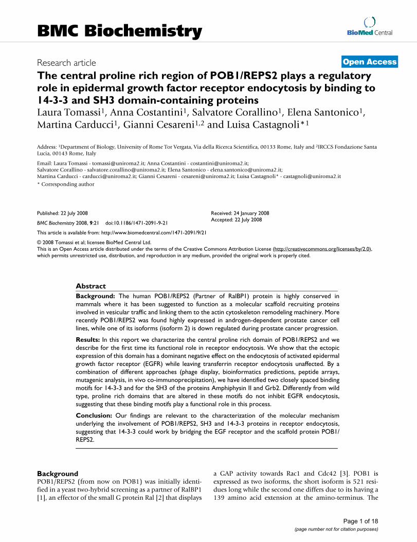
BioMed CentralBMC Biochemistry
ss
Open AcceResearch articleThe central proline rich region of POB1/REPS2 plays a regulatory role in epidermal growth factor receptor endocytosis by binding to 14-3-3 and SH3 domain-containing proteinsLaura Tomassi1, Anna Costantini1, Salvatore Corallino1, Elena Santonico1, Martina Carducci1, Gianni Cesareni1,2 and Luisa Castagnoli*1Address: 1Department of Biology, University of Rome Tor Vergata, Via della Ricerca Scientifica, 00133 Rome, Italy and 2IRCCS Fondazione Santa Lucia, 00143 Rome, Italy
Email: Laura Tomassi - [email protected]; Anna Costantini - [email protected]; Salvatore Corallino - [email protected]; Elena Santonico - [email protected]; Martina Carducci - [email protected]; Gianni Cesareni - [email protected]; Luisa Castagnoli* - [email protected]
* Corresponding author
AbstractBackground: The human POB1/REPS2 (Partner of RalBP1) protein is highly conserved inmammals where it has been suggested to function as a molecular scaffold recruiting proteinsinvolved in vesicular traffic and linking them to the actin cytoskeleton remodeling machinery. Morerecently POB1/REPS2 was found highly expressed in androgen-dependent prostate cancer celllines, while one of its isoforms (isoform 2) is down regulated during prostate cancer progression.
Results: In this report we characterize the central proline rich domain of POB1/REPS2 and wedescribe for the first time its functional role in receptor endocytosis. We show that the ectopicexpression of this domain has a dominant negative effect on the endocytosis of activated epidermalgrowth factor receptor (EGFR) while leaving transferrin receptor endocytosis unaffected. By acombination of different approaches (phage display, bioinformatics predictions, peptide arrays,mutagenic analysis, in vivo co-immunoprecipitation), we have identified two closely spaced bindingmotifs for 14-3-3 and for the SH3 of the proteins Amphiphysin II and Grb2. Differently from wildtype, proline rich domains that are altered in these motifs do not inhibit EGFR endocytosis,suggesting that these binding motifs play a functional role in this process.
Conclusion: Our findings are relevant to the characterization of the molecular mechanismunderlying the involvement of POB1/REPS2, SH3 and 14-3-3 proteins in receptor endocytosis,suggesting that 14-3-3 could work by bridging the EGF receptor and the scaffold protein POB1/REPS2.
BackgroundPOB1/REPS2 (from now on POB1) was initially identi-fied in a yeast two-hybrid screening as a partner of RalBP1[1], an effector of the small G protein Ral [2] that displays
a GAP activity towards Rac1 and Cdc42 [3]. POB1 isexpressed as two isoforms, the short isoform is 521 resi-dues long while the second one differs due to its having a139 amino acid extension at the amino-terminus. The
Published: 22 July 2008
BMC Biochemistry 2008, 9:21 doi:10.1186/1471-2091-9-21
Received: 24 January 2008Accepted: 22 July 2008
This article is available from: http://www.biomedcentral.com/1471-2091/9/21
© 2008 Tomassi et al; licensee BioMed Central Ltd. This is an Open Access article distributed under the terms of the Creative Commons Attribution License (http://creativecommons.org/licenses/by/2.0), which permits unrestricted use, distribution, and reproduction in any medium, provided the original work is properly cited.
Page 1 of 18(page number not for citation purposes)
BMC Biochemistry 2008, 9:21 http://www.biomedcentral.com/1471-2091/9/21
most prominent structural/functional features, which arecommon to both isoforms, include an amino-terminalEH (Eps15 Homology) domain, a central region contain-ing two adjacent proline-rich regions and a carboxy-termi-nal portion mediating the binding to RalBP1. Via its EHdomain, POB1 associates with Eps15 and Epsin [4,5].
The observed interactions with proteins involved in theendocytic pathway and the report that over-expression ofthe EH domain or the RalBP1 binding region inhibits EGFand insulin internalization, implicates POB1 in receptorendocytosis [4]. Accordingly, POB1 is tyrosine-phosphor-ylated in response to EGF in COS cells and co-purifieswith activated EGF receptor [1]. Furthermore POB1 co-immunoprecipitates with over expressed Grb2 (growthfactor receptor-bound protein 2) and with PAG2(DDEF1), a paxillin-associated protein, at endogenouslevels. The sequence responsible for this latter interactionwas mapped to a proline-rich motif (423PSKPIR428) thatbinds to the SH3 domain of PAG2 [6].
Because of its links to receptor endocytosis and to actincytoskeleton remodeling, POB1 has been proposed as amolecular scaffold recruiting proteins involved in vesicu-lar trafficking and linking them to actin cytoskeleton.
In addition, more recently, it has been reported that lossof POB1 expression in prostate cancer cells results inderegulation of growth factor signaling, while an increaseof POB1 expression has been correlated with silencing ofEGF signaling. Moreover, POB1 isoform 2 down-regula-tion has been observed during progression of prostatecancer from androgen to EGF dependency [7-9].
Although this scattered information suggest a central roleof POB1 in the complex regulatory mechanisms modulat-ing growth factor response, no simple model has been putforward to explain all the observations.
In this work, we show that, in addition to the amino-ter-minal EH domain and the carboxy-terminal RALBP1binding region, a third central area of POB1, containingmotifs recognized by the SH3 domains of Grb2 andAmphiphysin II and by the 14-3-3 family proteins, plays arole in modulating receptor endocytosis. Over-expressionof this POB1 fragment negatively affects epidermalgrowth factor receptor endocytosis by titration and delo-calization of essential components. Conversely, pointmutations in the conserved binding motifs eliminate thedominant negative effect on receptor endocytosis. Thesedata provide support for a role of POB1 as a regulator ofgrowth factor receptor endocytosis and corroborate itssuggested role as a tumor suppressor.
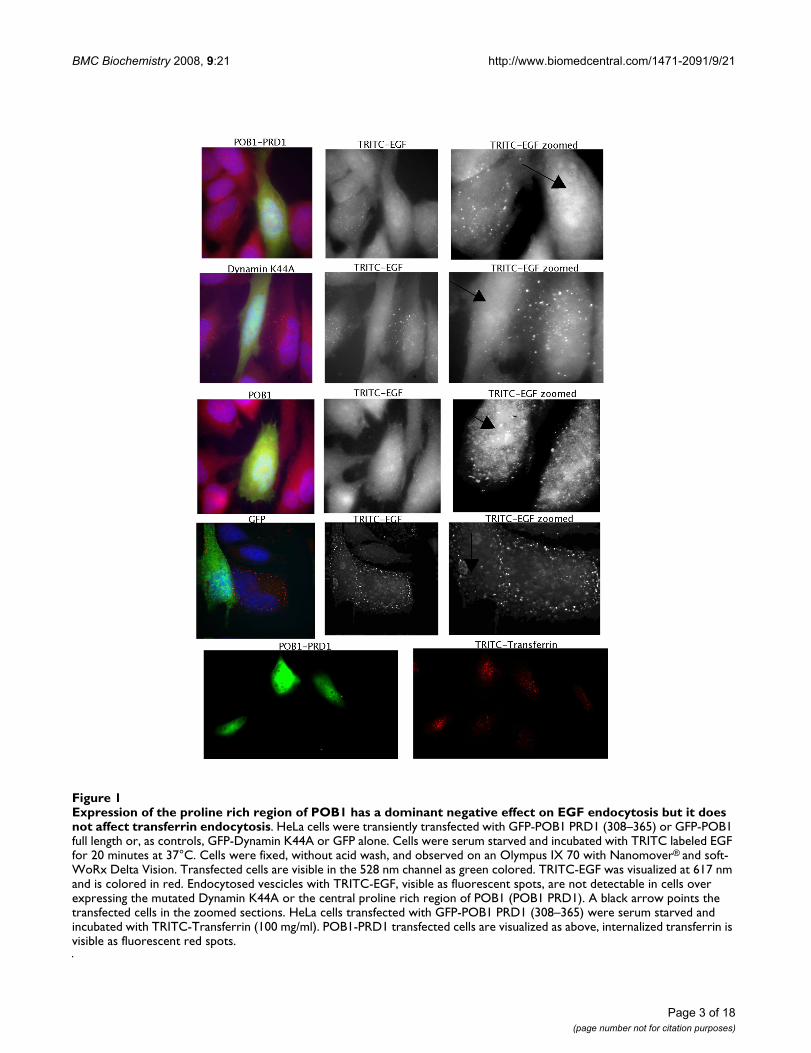
ResultsExpression of the POB1 proline rich region from Pro308 to Vel365 inhibits EGFR endocytosisWhile expression of full-length POB1 in A431 cells doesnot affect either binding or internalization of EGF, over-expression of either the EH domain or the C-terminalregion of POB1 affects the ligand dependent internaliza-tion pathway of EGF and insulin without interfering withthe constitutive transferrin pathway [4]. We were inter-ested in assessing whether the POB1 proline-rich regionplays an additional role in the ligand-dependent endocy-tosis of EGFR. To this end, we transiently transfected HeLacells with green fluorescent protein (GFP) fusion con-structs expressing either the POB1 isoform 2 full length orPRD1, the POB1 proline rich region from Pro308 toVal365 (Fig. 1). As controls we performed parallel trans-fections with two plasmids expressing GFP or GFP-Dynamin K44A, a dynamin mutant that is an establishedinhibitor of both EGF and transferrin endocytosis [10].Transfected cells were serum starved and subsequentlyincubated with TRITC labeled EGF or transferrin to mon-itor the two internalization pathways. As already reported,the ectopic expression of full length POB1 had no influ-ence on transferrin or EGF endocytosis [4]. Similarly,POB1 PRD1, the construct expressing a POB1 fragmentfrom amino acid 308 to 365, did not affect the constitu-tive transferrin endocytic pathway. On the other hand,HeLa cells over-expressing the POB1 PRD1, when treatedwith TRITC-EGF, have a much reduced number of inter-nalized EGF-stained vesicles and show a diffuse fluores-cence, resembling Dynamin K44A expression, indicatingthat POB1-PRD1 over-expression interferes with EGFendocytosis, (Fig. 1, first row). This phenotype has beenobserved in five independent experiments with a pene-trance of 65% of transfected cells. In similar control exper-iments, over-expression of Dynamin K44A mutantinhibited EGF internalization with a penetrance of 75%.
The C-terminal SH3 domain of Grb2 and the SH3 domain of Amphiphysin II bind to full-length POB1 and to POB1 PRD1 (308–365)To further characterize the interaction network centeredon POB1, we sought to isolate proteins binding the pro-line-rich core region. The POB1 central region fromPro308 to Val365 was expressed in bacteria as a GSTfusion protein (GST-POB1 PRD1), immobilized on glu-tathione-sepharose and used as a bait for affinity selectionof a human brain cDNA library displayed on lambdaphage [11]. The selected phage clones were sequenced andshown to encode either the Src homology 3 domain(SH3) of Amphiphysin II or the carboxy-terminal SH3domain of the molecular adaptor Grb2, which was for-merly shown to bind tagged POB1 in a GST pull-downassay [1].
Page 2 of 18(page number not for citation purposes)
BMC Biochemistry 2008, 9:21 http://www.biomedcentral.com/1471-2091/9/21
Page 3 of 18(page number not for citation purposes)
Expression of the proline rich region of POB1 has a dominant negative effect on EGF endocytosis but it does not affect trans-ferrin endocytosisFigure 1Expression of the proline rich region of POB1 has a dominant negative effect on EGF endocytosis but it does not affect transferrin endocytosis. HeLa cells were transiently transfected with GFP-POB1 PRD1 (308–365) or GFP-POB1 full length or, as controls, GFP-Dynamin K44A or GFP alone. Cells were serum starved and incubated with TRITC labeled EGF for 20 minutes at 37°C. Cells were fixed, without acid wash, and observed on an Olympus IX 70 with Nanomover® and soft-WoRx Delta Vision. Transfected cells are visible in the 528 nm channel as green colored. TRITC-EGF was visualized at 617 nm and is colored in red. Endocytosed vescicles with TRITC-EGF, visible as fluorescent spots, are not detectable in cells over expressing the mutated Dynamin K44A or the central proline rich region of POB1 (POB1 PRD1). A black arrow points the transfected cells in the zoomed sections. HeLa cells transfected with GFP-POB1 PRD1 (308–365) were serum starved and incubated with TRITC-Transferrin (100 mg/ml). POB1-PRD1 transfected cells are visualized as above, internalized transferrin is visible as fluorescent red spots.
BMC Biochemistry 2008, 9:21 http://www.biomedcentral.com/1471-2091/9/21
Page 4 of 18(page number not for citation purposes)
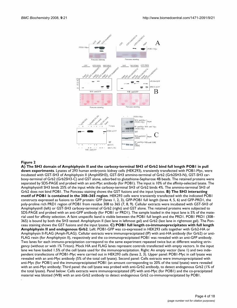
A) The SH3 domain of Amphiphysin II and the carboxy-terminal SH3 of Grb2 bind full length POB1 in pull down experimentsFigure 2A) The SH3 domain of Amphiphysin II and the carboxy-terminal SH3 of Grb2 bind full length POB1 in pull down experiments. Lysates of 293 human embryonic kidney cells (HEK293), transiently transfected with POB1-Myc, were incubated with GST-SH3 of Amphiphysin II (AmphIISH3), GST-SH3 ammino-terminal of Grb2 (Grb2SH3-N), GST-SH3 car-boxy-terminal of Grb2 (Grb2SH3-C) and GST alone, adsorbed to glutathione-Sepharose 4B beads. The retained proteins were separated by SDS-PAGE and probed with an anti-Myc antibody (for POB1). The input is 10% of the affinity-selected lysate. The AmphiphysinII SH3 binds 25% of the input while the carboxy-terminal SH3 of Grb2 binds 4%. The ammino-terminal SH3 of Grb2 does not bind POB1. The Ponceau staining shows the GST fusions and the input lysates. B) The SH3 interacting motif of POB1 is contained in the 308–365 region. HEK293 cells were transiently transfected with the indicated POB1 constructs expressed as fusions to GFP protein: GFP (lanes 1, 2, 3), GFP-POB1 full length (lanes 4, 5, 6) and GFP-PRD1, the poly-proline rich PRD1 region of POB1 from residue 308 to 365 (7, 8, 9). Cellular extracts were incubated with GST-SH3 of AmphiphysinII (left) or GST-SH3 carboxy-terminal of Grb2 (right) and GST alone. The retained proteins were subjected to SDS-PAGE and probed with an anti-GFP antibody (for POB1 or PRD1). The sample loaded in the input lane is 5% of the mate-rial used for affinity selection. A faint unspecific band is visible between the POB1 full length and the PRD1. POB1 PRD1 (308–365) is bound by both the SH3 tested: Amphiphysin II (last lane in leftmost gel) and Grb2 (last lane in rightmost gel). The Pon-ceau staining shows the GST fusions and the input lysates. C) POB1 full length co-immunoprecipitates with full length Amphiphysin II and endogenous Grb2. Left: POB1-GFP was co-expressed in HEK293 cells together with Grb2-HA or Amphiphysin II-FLAG (Amph-FLAG). Cellular extracts were immunoprecipitated (IP) with anti-HA antibody (for Grb2) or anti-FLAG resin (for Amphiphysin II), respectively and the co-immunoprecipitated POB1 was revealed with an anti-GFP antibody. Two lanes for each immuno-precipitation correspond to the same experiment repeated twice but at different washing strin-gency (without or with 1% Triton). Mock HA and FLAG lanes represent controls transfected with empty vectors. In the input lane we have loaded 1.5% of the cell lysate used for the immunoprecipitation. Right: An empty vector (lane 1) and two inde-pendent transfections of POB1-Myc were carried out in HEK293 cells (lanes 2, 3). Upper panel: POB1-Myc in cell lysate was revealed with an anti-Myc antibody (5% of the total cell lysate). Second panel: Cells extracts were immunoprecipitated with anti-Myc (for POB1) and the immunoprecipitated POB1 (an amount corresponding to 20% of the total lysate) were revealed with an anti-Myc antibody; Third panel: the cell lysate was probed with anti-Grb2 antibody, to detect endogenous Grb2 (1% of the total lysate). Panel below: Cells extracts were immunoprecipitated (IP) with anti-Myc (for POB1) and the co-precipitated material was blotted (WB) with an anti-Grb2 antibody to detect endogenous Grb2 co-immunoprecipitated by POB1.
BMC Biochemistry 2008, 9:21 http://www.biomedcentral.com/1471-2091/9/21
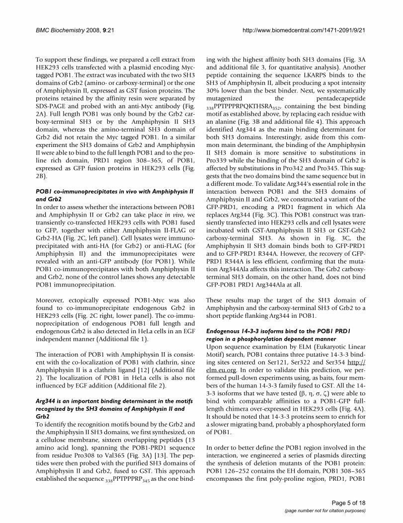
To support these findings, we prepared a cell extract fromHEK293 cells transfected with a plasmid encoding Myc-tagged POB1. The extract was incubated with the two SH3domains of Grb2 (amino- or carboxy-terminal) or the oneof Amphiphysin II, expressed as GST fusion proteins. Theproteins retained by the affinity resin were separated bySDS-PAGE and probed with an anti-Myc antibody (Fig.2A). Full length POB1 was only bound by the Grb2 car-boxy-terminal SH3 or by the Amphiphysin II SH3domain, whereas the amino-terminal SH3 domain ofGrb2 did not retain the Myc tagged POB1. In a similarexperiment the SH3 domains of Grb2 and AmphiphysinII were able to bind to the full length POB1 and to the pro-line rich domain, PRD1 region 308–365, of POB1,expressed as GFP fusion proteins in HEK293 cells (Fig.2B).
POB1 co-immunoprecipitates in vivo with Amphiphysin II and Grb2In order to assess whether the interactions between POB1and Amphiphysin II or Grb2 can take place in vivo, wetransiently co-transfected HEK293 cells with POB1 fusedto GFP, together with either Amphiphysin II-FLAG orGrb2-HA (Fig. 2C, left panel). Cell lysates were immuno-precipitated with anti-HA (for Grb2) or anti-FLAG (forAmphiphysin II) and the immunoprecipitates wererevealed with an anti-GFP antibody (for POB1). WhilePOB1 co-immunoprecipitates with both Amphiphysin IIand Grb2, none of the control lanes shows any detectablePOB1 immunoprecipitation.
Moreover, ectopically expressed POB1-Myc was alsofound to co-immunoprecipitate endogenous Grb2 inHEK293 cells (Fig. 2C right, lower panel). The co-immu-noprecipitation of endogenous POB1 full length andendogenous Grb2 is also detected in HeLa cells in an EGFindependent manner (Additional file 1).
The interaction of POB1 with Amphiphysin II is consist-ent with the co-localization of POB1 with clathrin, sinceAmphiphysin II is a clathrin ligand [12] (Additional file2). The localization of POB1 in HeLa cells is also notinfluenced by EGF addition (Additional file 2).
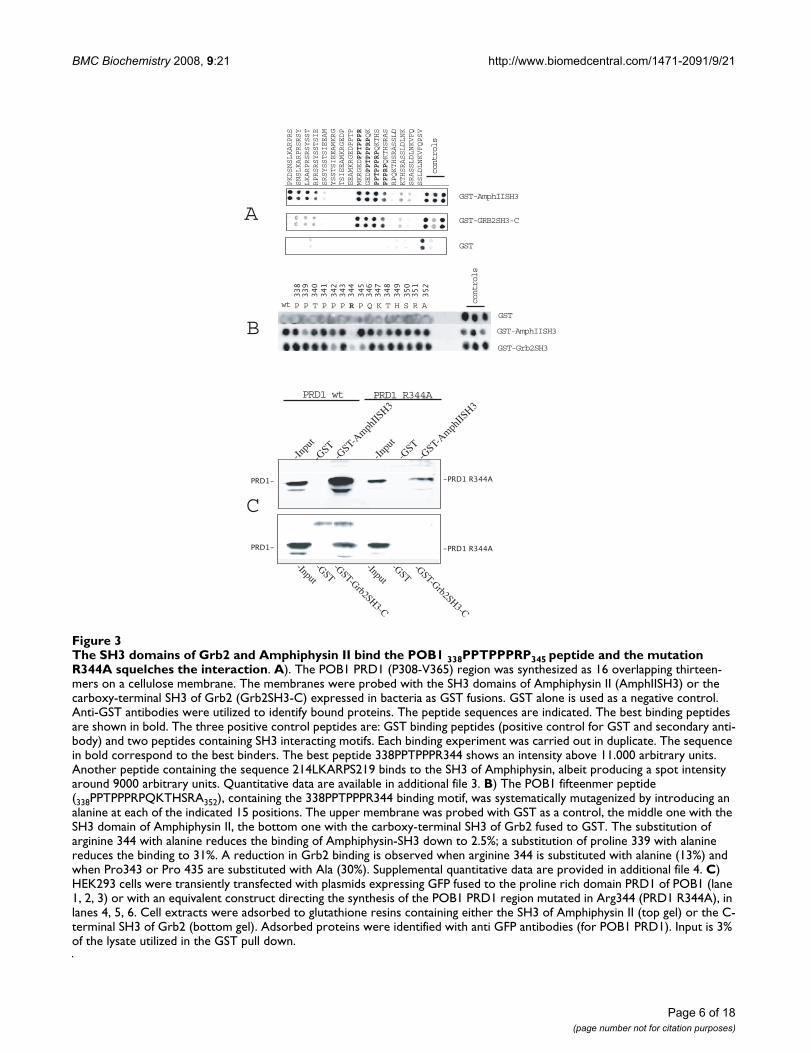
Arg344 is an important binding determinant in the motifs recognized by the SH3 domains of Amphiphysin II and Grb2To identify the recognition motifs bound by the Grb2 andthe Amphiphysin II SH3 domains, we first synthesized, ona cellulose membrane, sixteen overlapping peptides (13amino acid long), spanning the POB1-PRD1 sequencefrom residue Pro308 to Val365 (Fig. 3A) [13]. The pep-tides were then probed with the purified SH3 domains ofAmphiphysin II and Grb2, fused to GST. This approachestablished the sequence 338PPTPPPRP345 as the one bind-
ing with the highest affinity both SH3 domains (Fig. 3Aand additional file 3, for quantitative analysis). Anotherpeptide containing the sequence LKARPS binds to theSH3 of Amphiphysin II, albeit producing a spot intensity30% lower than the best binder. Next, we systematicallymutagenized the pentadecapeptide
338PPTPPPRPQKTHSRA352, containing the best bindingmotif as established above, by replacing each residue withan alanine (Fig. 3B and additional file 4). This approachidentified Arg344 as the main binding determinant forboth SH3 domains. Interestingly, aside from this com-mon main determinant, the binding of the AmphiphysinII SH3 domain is more sensitive to substitutions inPro339 while the binding of the SH3 domain of Grb2 isaffected by substitutions in Pro342 and Pro345. This sug-gests that the two domains bind the same sequence but ina different mode. To validate Arg344's essential role in theinteraction between POB1 and the SH3 domains ofAmphiphysin II and Grb2, we constructed a variant of theGFP-PRD1, encoding a PRD1 fragment in which Alareplaces Arg344 (Fig. 3C). This POB1 construct was tran-siently transfected into HEK293 cells and cell lysates wereincubated with GST-Amphiphysin II SH3 or GST-Grb2carboxy-terminal SH3. As shown in Fig. 3C, theAmphiphysin II SH3 domain binds both to GFP-PRD1and to GFP-PRD1 R344A. However, the recovery of GFP-PRD1 R344A is less efficient, confirming that the muta-tion Arg344Ala affects this interaction. The Grb2 carboxy-terminal SH3 domain, on the other hand, does not bindGFP-POB1 PRD1 Arg344Ala at all.
These results map the target of the SH3 domain ofAmphiphysin and the carboxy-terminal SH3 of Grb2 to ashort peptide flanking Arg344 in POB1.
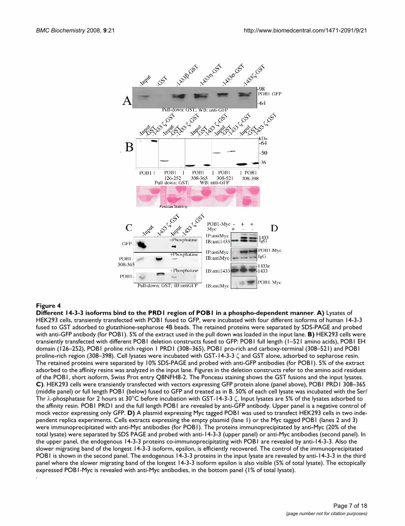
Endogenous 14-3-3 isoforms bind to the POB1 PRD1 region in a phosphorylation dependent mannerUpon sequence examination by ELM (Eukaryotic LinearMotif) search, POB1 contains three putative 14-3-3 bind-ing sites centered on Ser121, Ser322 and Ser354 http://elm.eu.org. In order to validate this prediction, we per-formed pull-down experiments using, as baits, four mem-bers of the human 14-3-3 family fused to GST. All the 14-3-3 isoforms that we have tested (β, η, σ, ζ) were able tobind with comparable affinities to a POB1-GFP full-length chimera over-expressed in HEK293 cells (Fig. 4A).It should be noted that 14-3-3 proteins seem to enrich fora slower migrating band, probably a phosphorylated formof POB1.
In order to better define the POB1 region involved in theinteraction, we engineered a series of plasmids directingthe synthesis of deletion mutants of the POB1 protein:POB1 126–252 contains the EH domain, POB1 308–365encompasses the first poly-proline region, PRD1, POB1
Page 5 of 18(page number not for citation purposes)
BMC Biochemistry 2008, 9:21 http://www.biomedcentral.com/1471-2091/9/21
Page 6 of 18(page number not for citation purposes)
The SH3 domains of Grb2 and Amphiphysin II bind the POB1 338PPTPPPRP345 peptide and the mutation R344A squelches the interactionFigure 3The SH3 domains of Grb2 and Amphiphysin II bind the POB1 338PPTPPPRP345 peptide and the mutation R344A squelches the interaction. A). The POB1 PRD1 (P308-V365) region was synthesized as 16 overlapping thirteen-mers on a cellulose membrane. The membranes were probed with the SH3 domains of Amphiphysin II (AmphIISH3) or the carboxy-terminal SH3 of Grb2 (Grb2SH3-C) expressed in bacteria as GST fusions. GST alone is used as a negative control. Anti-GST antibodies were utilized to identify bound proteins. The peptide sequences are indicated. The best binding peptides are shown in bold. The three positive control peptides are: GST binding peptides (positive control for GST and secondary anti-body) and two peptides containing SH3 interacting motifs. Each binding experiment was carried out in duplicate. The sequence in bold correspond to the best binders. The best peptide 338PPTPPPR344 shows an intensity above 11.000 arbitrary units. Another peptide containing the sequence 214LKARPS219 binds to the SH3 of Amphiphysin, albeit producing a spot intensity around 9000 arbitrary units. Quantitative data are available in additional file 3. B) The POB1 fifteenmer peptide (338PPTPPPRPQKTHSRA352), containing the 338PPTPPPR344 binding motif, was systematically mutagenized by introducing an alanine at each of the indicated 15 positions. The upper membrane was probed with GST as a control, the middle one with the SH3 domain of Amphiphysin II, the bottom one with the carboxy-terminal SH3 of Grb2 fused to GST. The substitution of arginine 344 with alanine reduces the binding of Amphiphysin-SH3 down to 2.5%; a substitution of proline 339 with alanine reduces the binding to 31%. A reduction in Grb2 binding is observed when arginine 344 is substituted with alanine (13%) and when Pro343 or Pro 435 are substituted with Ala (30%). Supplemental quantitative data are provided in additional file 4. C) HEK293 cells were transiently transfected with plasmids expressing GFP fused to the proline rich domain PRD1 of POB1 (lane 1, 2, 3) or with an equivalent construct directing the synthesis of the POB1 PRD1 region mutated in Arg344 (PRD1 R344A), in lanes 4, 5, 6. Cell extracts were adsorbed to glutathione resins containing either the SH3 of Amphiphysin II (top gel) or the C-terminal SH3 of Grb2 (bottom gel). Adsorbed proteins were identified with anti GFP antibodies (for POB1 PRD1). Input is 3% of the lysate utilized in the GST pull down.
BMC Biochemistry 2008, 9:21 http://www.biomedcentral.com/1471-2091/9/21
Page 7 of 18(page number not for citation purposes)
Different 14-3-3 isoforms bind to the PRD1 region of POB1 in a phospho-dependent mannerFigure 4Different 14-3-3 isoforms bind to the PRD1 region of POB1 in a phospho-dependent manner. A) Lysates of HEK293 cells, transiently transfected with POB1 fused to GFP, were incubated with four different isoforms of human 14-3-3 fused to GST adsorbed to glutathione-sepharose 4B beads. The retained proteins were separated by SDS-PAGE and probed with anti-GFP antibody (for POB1). 5% of the extract used in the pull down was loaded in the input lane. B) HEK293 cells were transiently transfected with different POB1 deletion constructs fused to GFP: POB1 full length (1–521 amino acids), POB1 EH domain (126–252), POB1 proline rich region 1 PRD1 (308–365), POB1 pro-rich and carboxy-terminal (308–521) and POB1 proline-rich region (308–398). Cell lysates were incubated with GST-14-3-3 ζ and GST alone, adsorbed to sepharose resin. The retained proteins were separated by 10% SDS-PAGE and probed with anti-GFP antibodies (for POB1). 5% of the extract adsorbed to the affinity resins was analyzed in the input lane. Figures in the deletion constructs refer to the amino acid residues of the POB1, short isoform, Swiss Prot entry Q8NFH8-2. The Ponceau staining shows the GST fusions and the input lysates. C). HEK293 cells were transiently transfected with vectors expressing GFP protein alone (panel above), POB1 PRD1 308–365 (middle panel) or full length POB1 (below) fused to GFP and treated as in B. 50% of each cell lysate was incubated with the Ser/Thr λ-phosphatase for 2 hours at 30°C before incubation with GST-14-3-3 ζ. Input lysates are 5% of the lysates adsorbed to the affinity resin. POB1 PRD1 and the full length POB1 are revealed by anti-GFP antibody. Upper panel is a negative control of mock vector expressing only GFP. D) A plasmid expressing Myc tagged POB1 was used to transfect HEK293 cells in two inde-pendent replica experiments. Cells extracts expressing the empty plasmid (lane 1) or the Myc tagged POB1 (lanes 2 and 3) were immunoprecipitated with anti-Myc antibodies (for POB1). The proteins immunoprecipitated by anti-Myc (20% of the total lysate) were separated by SDS PAGE and probed with anti-14-3-3 (upper panel) or anti-Myc antibodies (second panel). In the upper panel, the endogenous 14-3-3 proteins co-immunoprecipitating with POB1 are revealed by anti-14-3-3. Also the slower migrating band of the longest 14-3-3 isoform, epsilon, is efficiently recovered. The control of the immunoprecipitated POB1 is shown in the second panel. The endogenous 14-3-3 proteins in the input lysate are revealed by anti-14-3-3 in the third panel where the slower migrating band of the longest 14-3-3 isoform epsilon is also visible (5% of total lysate). The ectopically expressed POB1-Myc is revealed with anti-Myc antibodies, in the bottom panel (1% of total lysate).
BMC Biochemistry 2008, 9:21 http://www.biomedcentral.com/1471-2091/9/21
308–398 comprises two adjacent proline rich regionswhile POB1 308–521 further extends to the carboxy-ter-minal region including the RalBP1 binding domain (Fig-ure 7). The pull down experiment in Figure 4B confirmsthe interaction of 14-3-3 with the carboxy-terminal por-tion of POB1 (from 308 to 521) and restricts the minimalessential binding site to the PRD1 region of POB1 (resi-dues 308 to 365) including both Ser322 and Ser354.
In order to assess whether POB1 binds to endogenous 14-3-3, we transiently transfected HEK293 cells with Myc-POB1 in two independent experimental repeats (Fig. 4D).Cell lysates were immunoprecipitated with anti-Myc anti-bodies (for POB1) while the co-immunoprecipitatedendogenous 14-3-3 proteins were detected with specificantibodies. The results in Figure 4D, upper panel, showthat endogenous 14-3-3 proteins, including the largestisoform (epsilon), co-immunoprecipitate with POB1.
Since most 14-3-3 interactions are phosphorylationdependent [14-17]we tested whether the interactionbetween 14-3-3 and POB1 PRD1 is mediated by a phos-pho-peptide. We performed a pull down experiment asdescribed in Fig. 4B after incubating half of the cell extractwith λ-phosphatase, active towards phosphorylated serineand threonine (Fig. 4C). When treated with phosphatase,neither the full length POB1 (bottom panel, lane 4) northe POB1 PRD1 region (middle panel, lane 4) is effi-ciently recovered by 14-3-3. The same absence of co-immunoprecipitation occurred after wortmannin treat-ment suggesting an involvement of Akt Ser/Thr kinase(data not shown).
Taken together, these results suggest that the interactionbetween POB1 and the 14-3-3 proteins can occur in vivo,the recognized motif is encompassed in the region 308–365 of POB1 and the binding is Ser/Thr phosphorylationdependent.
Coimmunoprecipitation of POB1 with the isoform ζ isobserved also at endogenous levels in HeLa cells (Addi-tional file 1).
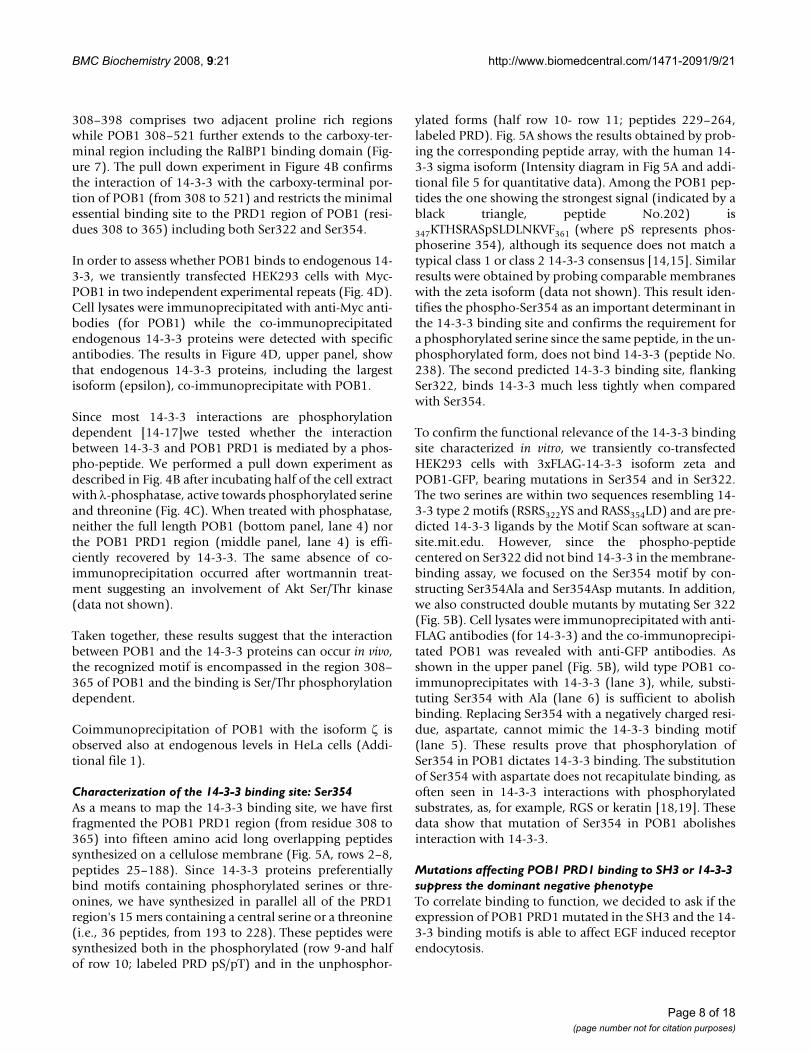
Characterization of the 14-3-3 binding site: Ser354As a means to map the 14-3-3 binding site, we have firstfragmented the POB1 PRD1 region (from residue 308 to365) into fifteen amino acid long overlapping peptidessynthesized on a cellulose membrane (Fig. 5A, rows 2–8,peptides 25–188). Since 14-3-3 proteins preferentiallybind motifs containing phosphorylated serines or thre-onines, we have synthesized in parallel all of the PRD1region's 15 mers containing a central serine or a threonine(i.e., 36 peptides, from 193 to 228). These peptides weresynthesized both in the phosphorylated (row 9-and halfof row 10; labeled PRD pS/pT) and in the unphosphor-
ylated forms (half row 10- row 11; peptides 229–264,labeled PRD). Fig. 5A shows the results obtained by prob-ing the corresponding peptide array, with the human 14-3-3 sigma isoform (Intensity diagram in Fig 5A and addi-tional file 5 for quantitative data). Among the POB1 pep-tides the one showing the strongest signal (indicated by ablack triangle, peptide No.202) is
347KTHSRASpSLDLNKVF361 (where pS represents phos-phoserine 354), although its sequence does not match atypical class 1 or class 2 14-3-3 consensus [14,15]. Similarresults were obtained by probing comparable membraneswith the zeta isoform (data not shown). This result iden-tifies the phospho-Ser354 as an important determinant inthe 14-3-3 binding site and confirms the requirement fora phosphorylated serine since the same peptide, in the un-phosphorylated form, does not bind 14-3-3 (peptide No.238). The second predicted 14-3-3 binding site, flankingSer322, binds 14-3-3 much less tightly when comparedwith Ser354.
To confirm the functional relevance of the 14-3-3 bindingsite characterized in vitro, we transiently co-transfectedHEK293 cells with 3xFLAG-14-3-3 isoform zeta andPOB1-GFP, bearing mutations in Ser354 and in Ser322.The two serines are within two sequences resembling 14-3-3 type 2 motifs (RSRS322YS and RASS354LD) and are pre-dicted 14-3-3 ligands by the Motif Scan software at scan-site.mit.edu. However, since the phospho-peptidecentered on Ser322 did not bind 14-3-3 in the membrane-binding assay, we focused on the Ser354 motif by con-structing Ser354Ala and Ser354Asp mutants. In addition,we also constructed double mutants by mutating Ser 322(Fig. 5B). Cell lysates were immunoprecipitated with anti-FLAG antibodies (for 14-3-3) and the co-immunoprecipi-tated POB1 was revealed with anti-GFP antibodies. Asshown in the upper panel (Fig. 5B), wild type POB1 co-immunoprecipitates with 14-3-3 (lane 3), while, substi-tuting Ser354 with Ala (lane 6) is sufficient to abolishbinding. Replacing Ser354 with a negatively charged resi-due, aspartate, cannot mimic the 14-3-3 binding motif(lane 5). These results prove that phosphorylation ofSer354 in POB1 dictates 14-3-3 binding. The substitutionof Ser354 with aspartate does not recapitulate binding, asoften seen in 14-3-3 interactions with phosphorylatedsubstrates, as, for example, RGS or keratin [18,19]. Thesedata show that mutation of Ser354 in POB1 abolishesinteraction with 14-3-3.
Mutations affecting POB1 PRD1 binding to SH3 or 14-3-3 suppress the dominant negative phenotypeTo correlate binding to function, we decided to ask if theexpression of POB1 PRD1 mutated in the SH3 and the 14-3-3 binding motifs is able to affect EGF induced receptorendocytosis.
Page 8 of 18(page number not for citation purposes)
BMC Biochemistry 2008, 9:21 http://www.biomedcentral.com/1471-2091/9/21
Page 9 of 18(page number not for citation purposes)
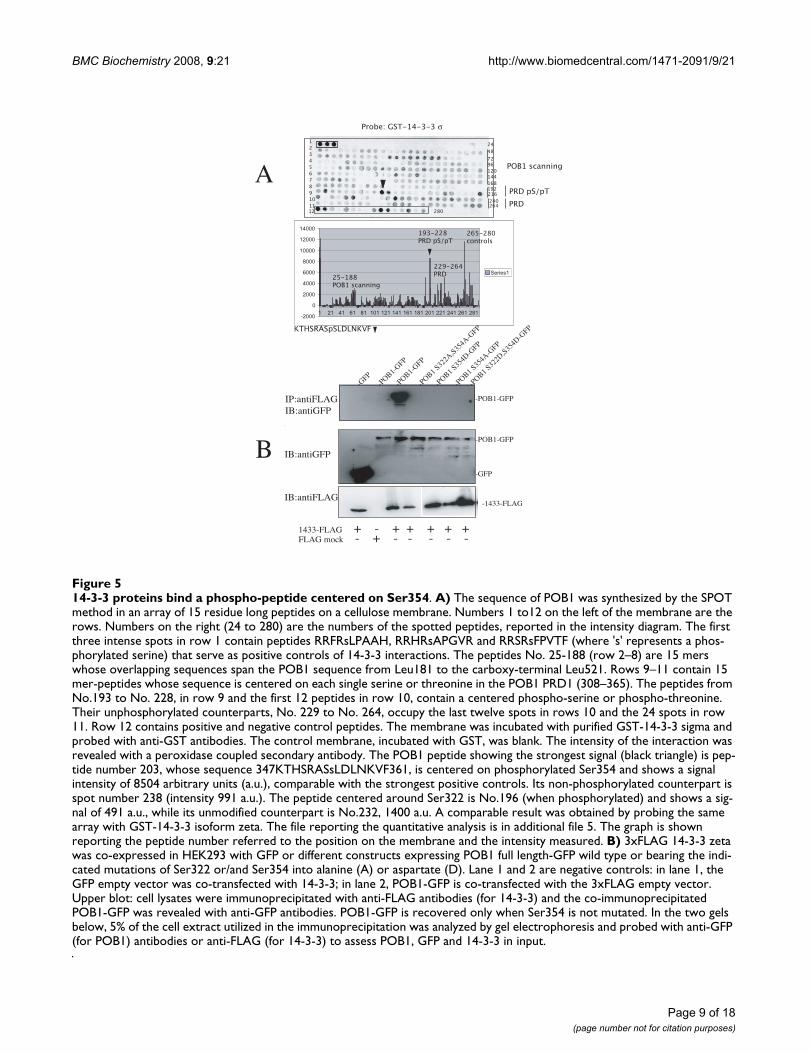
14-3-3 proteins bind a phospho-peptide centered on Ser354Figure 514-3-3 proteins bind a phospho-peptide centered on Ser354. A) The sequence of POB1 was synthesized by the SPOT method in an array of 15 residue long peptides on a cellulose membrane. Numbers 1 to12 on the left of the membrane are the rows. Numbers on the right (24 to 280) are the numbers of the spotted peptides, reported in the intensity diagram. The first three intense spots in row 1 contain peptides RRFRsLPAAH, RRHRsAPGVR and RRSRsFPVTF (where 's' represents a phos-phorylated serine) that serve as positive controls of 14-3-3 interactions. The peptides No. 25-188 (row 2–8) are 15 mers whose overlapping sequences span the POB1 sequence from Leu181 to the carboxy-terminal Leu521. Rows 9–11 contain 15 mer-peptides whose sequence is centered on each single serine or threonine in the POB1 PRD1 (308–365). The peptides from No.193 to No. 228, in row 9 and the first 12 peptides in row 10, contain a centered phospho-serine or phospho-threonine. Their unphosphorylated counterparts, No. 229 to No. 264, occupy the last twelve spots in rows 10 and the 24 spots in row 11. Row 12 contains positive and negative control peptides. The membrane was incubated with purified GST-14-3-3 sigma and probed with anti-GST antibodies. The control membrane, incubated with GST, was blank. The intensity of the interaction was revealed with a peroxidase coupled secondary antibody. The POB1 peptide showing the strongest signal (black triangle) is pep-tide number 203, whose sequence 347KTHSRASsLDLNKVF361, is centered on phosphorylated Ser354 and shows a signal intensity of 8504 arbitrary units (a.u.), comparable with the strongest positive controls. Its non-phosphorylated counterpart is spot number 238 (intensity 991 a.u.). The peptide centered around Ser322 is No.196 (when phosphorylated) and shows a sig-nal of 491 a.u., while its unmodified counterpart is No.232, 1400 a.u. A comparable result was obtained by probing the same array with GST-14-3-3 isoform zeta. The file reporting the quantitative analysis is in additional file 5. The graph is shown reporting the peptide number referred to the position on the membrane and the intensity measured. B) 3xFLAG 14-3-3 zeta was co-expressed in HEK293 with GFP or different constructs expressing POB1 full length-GFP wild type or bearing the indi-cated mutations of Ser322 or/and Ser354 into alanine (A) or aspartate (D). Lane 1 and 2 are negative controls: in lane 1, the GFP empty vector was co-transfected with 14-3-3; in lane 2, POB1-GFP is co-transfected with the 3xFLAG empty vector. Upper blot: cell lysates were immunoprecipitated with anti-FLAG antibodies (for 14-3-3) and the co-immunoprecipitated POB1-GFP was revealed with anti-GFP antibodies. POB1-GFP is recovered only when Ser354 is not mutated. In the two gels below, 5% of the cell extract utilized in the immunoprecipitation was analyzed by gel electrophoresis and probed with anti-GFP (for POB1) antibodies or anti-FLAG (for 14-3-3) to assess POB1, GFP and 14-3-3 in input.
BMC Biochemistry 2008, 9:21 http://www.biomedcentral.com/1471-2091/9/21
Page 10 of 18(page number not for citation purposes)
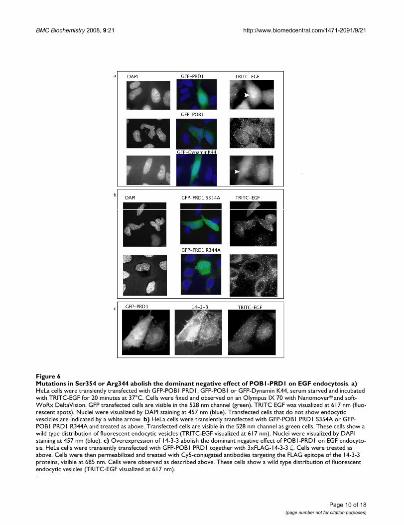
Mutations in Ser354 or Arg344 abolish the dominant negative effect of POB1-PRD1 on EGF endocytosisFigure 6Mutations in Ser354 or Arg344 abolish the dominant negative effect of POB1-PRD1 on EGF endocytosis. a) HeLa cells were transiently transfected with GFP-POB1 PRD1, GFP-POB1 or GFP-Dynamin K44, serum starved and incubated with TRITC-EGF for 20 minutes at 37°C. Cells were fixed and observed on an Olympus IX 70 with Nanomover® and soft-WoRx DeltaVision. GFP transfected cells are visible in the 528 nm channel (green). TRITC EGF was visualized at 617 nm (fluo-rescent spots). Nuclei were visualized by DAPI staining at 457 nm (blue). Transfected cells that do not show endocytic vescicles are indicated by a white arrow. b) HeLa cells were transiently transfected with GFP-POB1 PRD1 S354A or GFP-POB1 PRD1 R344A and treated as above. Transfected cells are visible in the 528 nm channel as green cells. These cells show a wild type distribution of fluorescent endocytic vesicles (TRITC-EGF visualized at 617 nm). Nuclei were visualized by DAPI staining at 457 nm (blue). c) Overexpression of 14-3-3 abolish the dominant negative effect of POB1-PRD1 on EGF endocyto-sis. HeLa cells were transiently transfected with GFP-POB1 PRD1 together with 3xFLAG-14-3-3 ζ. Cells were treated as above. Cells were then permeabilized and treated with Cy5-conjugated antibodies targeting the FLAG epitope of the 14-3-3 proteins, visible at 685 nm. Cells were observed as described above. These cells show a wild type distribution of fluorescent endocytic vesicles (TRITC-EGF visualized at 617 nm).
BMC Biochemistry 2008, 9:21 http://www.biomedcentral.com/1471-2091/9/21
In contrast with its wild type counterpart (Fig. 6A), theover-expression of GFP-POB1 PRD1 S354A, mutated inthe serine residue that dictates 14-3-3 binding, does notaffect endocytosis. (Fig. 6B). Also, cells over expressingPOB1 PRD1 R344A, which is defective in binding toAmphiphysin II and Grb2 SH3, do not show any interfer-ence in the wild type EGF internalization (Fig. 6B). Theseresults demonstrate that the POB1 proline-rich region actsas a dominant negative for EGF internalization and thatthis negative effect can be rescued by mutations in the res-idues that affect binding to 14-3-3 or to specific SH3.
Interestingly, the normal phenotype of endocytosis isrestored upon co-transfection of the wild type POB1PRD1 region with 3xFLAG-14-3-3 ζ in over-expression(Fig. 6C). We conclude that over-expression of 14-3-3 ζremoves the endocytosis block caused by the dominantnegative effect of the PRD1 region of POB1.
DiscussionThe observation that over-expression of the EH domain orof the carboxy-terminal RalBP1 binding region of POB1/REPS2 results in a 30–40% reduction of EGF or insulininternalization has linked POB1 to the molecular machin-ery modulating regulated endocytosis [4]. More recently,it was reported that loss of POB1 expression duringhuman prostate cancer progression, from androgen-dependent to growth factors dependent, results in loss ofcontrol of cell growth signaling while induced expressionof POB1 causes a reduction of several EGF-responsivegenes (e.g., Fos and Jun). In accordance, we find that anincrease of POB1 isoform 2 expression correlates with adecrease of EGF-induced phosphorylation of Erk1–2 andShc (Additional file 6). Moreover, POB1 isoform 2 down-regulation was observed during the progression of pros-tate cancer [7-9].
In our study we confirm the involvement of POB1 in EGFreceptor endocytosis and we provide evidence for a func-tional role of the central proline-rich region. In fact, weobserved that over-expression of the proline-rich region ofPOB1 (308–365) interferes with EGFR endocytosis whileleaving transferrin uptake unaffected (Fig. 1). This sug-gests that, in addition to the N and C-terminal regions, thecentral proline-rich domain (PRD1) of POB1, fromPro308 to Val365, also plays an important function in reg-ulated endocytosis and that over-expression of this por-tion of POB1 disturbs the dynamics of proteininteractions by titration of elements required for regulatedendocytosis.
By panning phage displayed cDNA libraries, with a POB1construct containing the proline-rich domain PRD1, weenriched for phages displaying the carboxy-terminal SH3domain of Grb2 and the SH3 domain of Amphiphysin II.
These in vitro identified physical interactions were furtherconfirmed in vivo, with full length POB1 (Fig. 2). A com-plex between POB1 and Grb2 has already been reported[6], We add that POB1 can bind Grb2 at endogenous lev-els (Fig. 2 and Additional file 1) and we report that theinteraction occurs by means of the carboxy-terminal SH3domain of Grb2. Interestingly the Grb2 binding motif inPOB1 (341PPPRP345) is exactly conserved in the Gab motif(337PPPRPP342) that is also recognized by Grb2. Thismotif is also conserved in the highly homologous humanprotein REPS1. POB1 shows a punctate cytoplasmic local-ization that is not much affected by EGF induction (Addi-tional file 2). A substantial fraction of POB1 co-stains withthe late endosome markers CD63 or LAMP-1, as alsofound for tyrosine-phosphorylated EGFR, Shc, Cbl andGrb2 [20].
We describe Amphiphysin II as a new POB1 interactor.POB1 cellular colocalization with clathrin is consistentwith this interaction, since Amphiphysin II is also a clath-rin ligand (Additional file 2) (20). By alanine scanningmutagenesis we have shown that both Grb2 andAmphiphysin II SH3 bind to the same POB1 peptide,however, different residues seem to be the main determi-nants of recognition specificity. Amphiphysin II bindingis affected by changes in Arg344 and Pro339 while Grb2binding is sensitive to changes in Pro342 and Pro 345(Fig. 3).
We have characterized a second linear motif of POB1 con-taining a phosphorylated serine and mediating binding tothe 14-3-3 isoforms (Fig. 4). This binding site wasmapped approximately ten amino acids away from theSH3 binding motif (Fig. 5). 14-3-3 binding was shown tobe sensitive to phosphatase treatment (Fig. 4) and bindingwas abolished when Ser354 was mutated to Ala or to Asp(Fig. 5). This suggests that POB1 binds 14-3-3 in a phos-phorylated form. Significantly, the characterized POB1peptide sequence HSRASsLDLNK, only displays poor sim-ilarity to the classical 14-3-3 binding motifs [15]. In fact,while presenting the conserved Arg at position -3 from thephosphorylated serine, it displays a few uncommon fea-tures, such as an Asp at position +2, in place of the pre-ferred Pro. This divergence is not anomalous, given thatphysiological substrates often bind 14-3-3 through pep-tide ligands which do not exactly match the most com-mon motifs [21]. Interestingly binding is abolished ifSer353, immediately preceding the phosphorylatedSer354, is also phosphorylated (data not shown). 14-3-3have been described as forming complexes with morethan 200 partners in high throughput studies and poten-tially engaging 0.6% of the human proteome [22]. Someof these complexes have been implicated in regulatingprotein membrane trafficking. For instance, 14-3-3 wereshown to reduce endoplasmic reticulum (ER) localiza-
Page 11 of 18(page number not for citation purposes)
BMC Biochemistry 2008, 9:21 http://www.biomedcentral.com/1471-2091/9/21
tion, thereby promoting surface expression of membraneproteins [17,23].
It has to be emphasized that 14-3-3 zeta has already beenreported as associating with the epidermal growth factorreceptor cytoplasmic tail and co-localizing along theplasma membrane with EGFR upon EGF stimulation [24].Thus a 14-3-3 dimer could bridge POB1 to the EGFR uponEGF induction. Similarly, 14-3-3 proteins appear to be theessential molecular bridge between the alfa1-subunit ofthe Na+, K+, -ATPase in the plasma membrane and the PI3-kinase, thus providing the signal to initiate endocytosisof the protein in response to natriuretic hormones [25].Finally 14-3-3 sigma is phosphorylated in prostate cells inresponse to EGF [26]. It is interesting to note that the 14-3-3 binding motif HSRxSSLD flanking Ser354 of POB1,that we find essential for interaction with 14-3-3, is con-served in the mouse orthologs and flanks the Ser510 ofhuman REPS1, that is found phosphorylated in vivo inA431 cells (Stover DR et al. Phosphosite: http://www.phosphosite.org). This serine site is predicted to bephosphorylated by Akt or calcium/calmodulin-dependentprotein kinase II gamma, CaMK2G. In agreement, weobserved absence of 14-3-3-coimmunoprecipitated POB1when cells where treated with phosphatase or wortman-nin (not shown), an inhibitor of PI3K and consequentlyof Akt.
To further test the model we assayed the POB1 proline-rich construct harboring the point mutation R344A,which prevents binding of Grb2 and Amphiphysin II and,as expected, we did not observe the dominant negativeeffect on EGFR internalization (Fig. 6). Thus, theArg344Ala mutation both affects the formation of thePOB1/Grb2 and POB1/Amphiphysin II complexes andalso abolishes the dominant negative phenotype affectingendocytosis. This suggests that titration of either SH3 con-taining partner affects EGFR internalization. Similarly, aPOB1 PRD1 construct whose ability to bind 14-3-3 wasimpaired by a Ser354Ala mutation, was unable to inhibitendocytosis in over-expression conditions (Fig. 6). Inaddition, we have found that co-expression of 14-3-3relieves the dominant negative effect of POB1-PRD1 onEGFR internalization (Fig. 6). Mechanistically, 14-3-3 ζcould offset the negative POB1-PRD1 over-expressioneffect, by binding POB1-PRD1 and by preventing therecruitment and trapping of a second essential physiolog-ical ligand.
ConclusionWe confirm that POB1 plays an active role in controllingthe dynamics of ligand-dependent receptor internaliza-tion and signaling. This is mediated by several interac-tions with diverse protein partners [4,5]. In this work, weprovide evidence of the central role played by the proline
rich region of POB1 through binding to 14-3-3 and to theSH3 of Grb2 and Amphyphisin II (Fig. 7). We observe thatthese interactions are not EGF dependent and are proba-bly exclusive, since the binding motifs are only nineamino acid apart.
The artificial over-expression of this portion of POB1affects endocytosis by titration of essential components,leading to formation of partial complexes that are notfunctional [7]. Thus POB1 acts as a sort of scaffold to enu-cleate a complex that is required for receptor internaliza-tion. The assembly and composition of this complex isfinely regulated by protein concentration. Besides contrib-uting to expanding and further characterizing the func-tional endocytosis "interactome", these results add to themolecular interpretation of POB1 as a tumor suppressor,due to its acting as a scaffold to bridge proteins which areestablished promoters of endocytosis and, subsequently,of receptor down regulation.
MethodsCell culture and transient transfectionHeLa and human embryonic kidney cell line HEK293Phoenix were grown at 37°C in a 5% CO2 incubator, inDMEM (Gibco/Invitrogen) supplemented with 10% fetalbovine serum (SIGMA-Aldrich), penicillin and streptomy-cin (Gibco). HeLa cells were transfected with Lipo-fectamine 2000 reagent (Invitrogen) followingmanufacturer's instructions while HEK293 cells weretransfected with the calcium-phosphate method.
PlasmidsThe plasmid vectors encoding for GFP-POB1, GFP-Dynamin K44A and FLAG-Amphiphysin II were a gener-ous gift from A. Kikuchi, G. Cestra and H. Daub, respec-tively. The remaining plasmids were constructed bystandard recombinant DNA techniques. Briefly, the cDNAencoding for the Amphiphysin II Src homology 3 domain(SH3) was cloned using SalI and BglII pYEX (modifiedpGEX-2TK, Amersham) unique sites, and GST-Grb2 car-boxy-terminal SH3 was generated by cloning the corre-sponding cDNA in pYEX BamHI and NotI sites. Theconstruct expressing GST-POB PRD1 was generated byamplifying the corresponding POB1 cDNA region andcloning it into BamHI and EcoRI pYEX unique sites. Thesame POB1 cDNA region (POB1/REPS2 Swiss-prot:Q8NFH8, isoform 2, 521 amino acids) was cloned intothe BglII and EcoRI sites of pEGFP-C1 vector (BD Bio-science Clontech) for expression in eukaryotic cells. PRD1region was amplified by PCR with the primers 5'-ATC-CAGGATCCCCCAAGGATTCCAACAGT-3' and 5'-TGGT-GAATTCCACACTGGGCTGGAAGAC-3' and sub clonedinto BglII and EcoRI sites; the PRD1–PRD2 (308–398)fragment was amplified by PCR with the oligos 5'ATC-CAGGATCCCCCAAGGATTCCAACAGT-3' and
Page 12 of 18(page number not for citation purposes)
BMC Biochemistry 2008, 9:21 http://www.biomedcentral.com/1471-2091/9/21
5'GGCAAGCTTTTACACTTGTTCAGACTGTGA-3' andcloned into the BglII-HindIII sites; the NH2 terminal-EHregion was amplified by PCR with the oligos 5'TGCCT-GGATCCATGATGTCAAAGAAT-3' and 5'GATGAAT-TCGCTGCAGAGTTGGAGGGAGGC and subcloned intothe BglII-EcoRI sites; the PRO-RICH and carboxyterminalregion (308–521) was amplified by PCR with oligos:5'ATCCAGGATCCCCCAAGGATTCCAACAGT-3' and5'TGTCTAGACAACACAGTGACCGGACG-3' and clonedinto the BglII-SalI sites. Site-directed mutagenesis of GFP-
POB PRD1 to obtain GFP-POB1 PRD1 R344A was carriedout by assembly of two overlapping DNA fragmentsobtained by PCR amplification with pairs of complemen-tary primers (R1299 5'ACCCCGCCACCTGCACCACA-GAAAACC-3', R13005'GGTTTTCTGTGGTGCAGGTGGCGGGGT-3'), each car-rying the mutated sequence, and two oligos priming fromthe 5'- and 3'-ends of the wild type POB1 proline-richsequence. This latter pair of primers contained the BamHIand EcoRI sites for cloning into the pEGFP-C1 vector. The
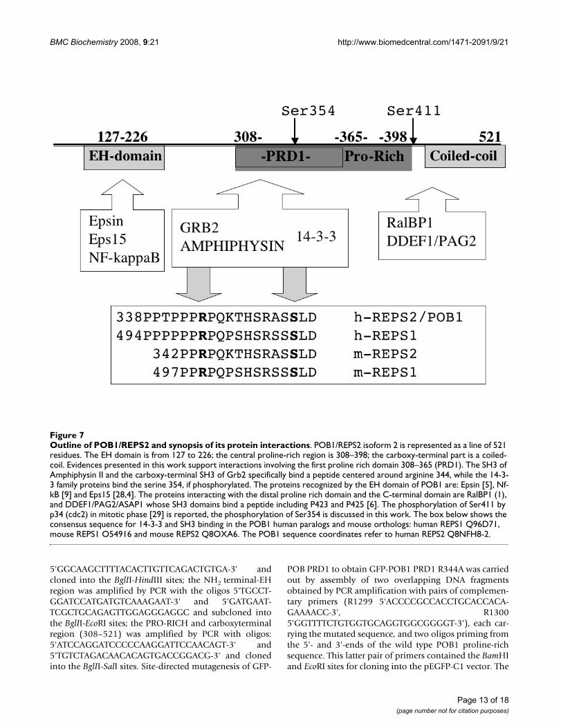
Outline of POB1/REPS2 and synopsis of its protein interactionsFigure 7Outline of POB1/REPS2 and synopsis of its protein interactions. POB1/REPS2 isoform 2 is represented as a line of 521 residues. The EH domain is from 127 to 226; the central proline-rich region is 308–398; the carboxy-terminal part is a coiled-coil. Evidences presented in this work support interactions involving the first proline rich domain 308–365 (PRD1). The SH3 of Amphiphysin II and the carboxy-terminal SH3 of Grb2 specifically bind a peptide centered around arginine 344, while the 14-3-3 family proteins bind the serine 354, if phosphorylated. The proteins recognized by the EH domain of POB1 are: Epsin [5], Nf-kB [9] and Eps15 [28,4]. The proteins interacting with the distal proline rich domain and the C-terminal domain are RalBP1 (1), and DDEF1/PAG2/ASAP1 whose SH3 domains bind a peptide including P423 and P425 [6]. The phosphorylation of Ser411 by p34 (cdc2) in mitotic phase [29] is reported, the phosphorylation of Ser354 is discussed in this work. The box below shows the consensus sequence for 14-3-3 and SH3 binding in the POB1 human paralogs and mouse orthologs: human REPS1 Q96D71, mouse REPS1 O54916 and mouse REPS2 Q8OXA6. The POB1 sequence coordinates refer to human REPS2 Q8NFH8-2.
Page 13 of 18(page number not for citation purposes)
BMC Biochemistry 2008, 9:21 http://www.biomedcentral.com/1471-2091/9/21
cDNA encoding for POB1 was cloned into pcDNA3.1/Myc-His EcoRI and NotI unique sites. Finally, HA-Grb2was constructed by amplifying the cDNA coding for Grb2and cloning it into BglII and EcoRI sites of pcDNA-HA(Natoli et al., 1997).
The plasmid vectors pFLAG CMV2 encoding the 14-3-3isoforms β,ε,η,θ,σ,ζ were a generous gift from TakashiTsuruo. The same isoforms were subcloned in a pCDNA/FRT/TO Invitrogen modified by insertion of a box encod-ing a 3xFLAG tag and a cloning site suitable for the LICcloning method (52), flanked by HindIII-BamHI sites. The3xFLAG box was inserted utilizing the oligos:5'CCCAAGCTTACCATGGACTACAAAGACCAT-3' and5'CGGGATCCCCGTTATCCACTTTCCCCGGGGGATT-GGAAGTACAGCTTGTCATCGTCATC-3'. The 14-3-3 iso-forms were amplified by PCR and cloned in this box usingthe oligos:
5'TACTTCCAATCCCCCGGCATGACAATGGATAAAAGTand
5'TTATCCACTTTCCCCGTTAGTTCTCTCCCTCCCC for β;
5'TACTTCCAATCCCCCGGCATGGATAAAAATGAGCT-GGTT and
5'TTATCCACTTTCCCCGTTAATTTTCCCCTCCTTCTC forζ;
5'TACTTCCAATCCCCCGGCATGGATGATCGAGAG-GATCT and
5'TTATCCACTTTCCCCGTCACTGATTTTCGTCTTCCA forε;
5'TACTTCCAATCCCCCGGCATGGGGGACCGGGAG-CAGC and
5'TTATCCACTTTCCCCGTCAGTTGCCTTCTCCTGCTT forη;
5'TACTTCCAATCCCCCGGCATGGAGAGAGCCAGTCT-GATC and
5'TTATCCACTTTCCCCGTCAGCTCTGGGGCTCCTGGGfor σ. The cDNA encoding for GST 14-3-3 β,ε,η,θ,σ,ζ wascloned into the LIC site of a pGEX-2T-K modified by inser-tion of a box suitable for LIC cloning method in BamHIand EcoRI sites, by the annealed oligos
5'GATCCCTGTACTTCCAATCCCCCGGGGAAAGTGGA-TAACGGG-3' and
5'AATTCCCGTTATCCACTTTCCCCGGGGGATTGGAAG-TACAGG.
Site directed mutagenesis of GFP-POB1 and GFP-POB1-PRD1 to obtain GFP-POB1 S354A, GFP-POB1 S322A,GFP-POB1 S322/354A and GFP-POB1 PRD1 S354A wascarried out by using the QuikChange Site-directed Muta-genesis Kit (Stratagene) with primers R1528 for S354D:5'ACCCATTCCAGAGCCTCCGACTTGGATCTGAATAAA;R1529 for S322A:
5'GCAAGACCAAGATCCAGAGCTTACTCTAGCACC;R1530 for S322D:
5'GCAAGACCAAGATCCAGAGATTACTCTAGCACC;R1532 for S354A:
5'ACCCATTCCAGAGCCTCCGCCTTGGATCTGAATAAA.It should be noted that all the residue numbering ofPOB1/REPS2 refers to the isoform 2, Q8NFH8-2.
AntibodiesThe antibodies used in these experiments were a mousemonoclonal anti-T7 (Novagen), a rabbit polyclonal anti-lambda serum, a mouse monoclonal anti-HA (SIGMA-Aldrich), a mouse monoclonal anti-FLAG (SIGMA-Aldrich), a rabbit polyclonal anti-GFP (Santa Cruz), amouse monoclonal anti-Grb2, a mouse monoclonal anti-CHC, a mouse monoclonal anti-EEA1, a mouse mono-clonal anti-CD63, a mouse monoclonal anti-Lamp2, amouse monoclonal anti-GM130 (all from Becton Dickin-son Transduction Laboratories); rabbit anti 14-3-3 zeta(Santa Cruz); a mouse monoclonal anti 14-3-3 sigma(Santa Cruz); mouse monoclonal anti 14-3-3 (Neomar-ker); polyclonal anti-REPS2 raised in rabbit (Atlas Anti-body). A rabbit polyclonal anti-POB1 serum wasgenerated against a N-terminal part of POB1, tested onwestern blot in parallel with commercial anti-REPS2. Sec-ondary antibodies used in this work were an alkalinephosphatase-conjugated anti-rabbit IgG, an alkalinephosphatase-conjugated anti-mouse IgG (both fromSIGMA-Aldrich), a peroxidase-conjugated anti-mouse andanti-rabbit IgG, a Rhodamine Red-X-coupled anti-rabbit(all from Jackson ImmunoResearch). Cy5-coupled antimouse (from Jackson ImmunoResearch) and an AlexaFluor 488-coupled anti-mouse (Molecular Probes).
Pull-down experimentsGST and GST-fusion proteins (100 μg) expressed in bacte-ria and adsorbed to glutathione-sepharose 4B beads(Amersham) were incubated for two hours at 4°C with 1mg of HEK293 cellular extract in JS lysis buffer (50 mMHepes, 150 mM NaCl, 1% Glycerol, 1% Triton-X100, 1.5mM MgCl2, 5 mM EGTA, 1 mM PMSF, 10 μg/ml Leupep-tin, 10 μg/ml Aprotinin, 1 μM NaVO4). Beads were
Page 14 of 18(page number not for citation purposes)
BMC Biochemistry 2008, 9:21 http://www.biomedcentral.com/1471-2091/9/21
washed three times in ice-cold PBS. Bound proteins wereelectrophoresed on a 10% polyacrylamide gel, transferredonto nitrocellulose membranes and detected with specificantibodies.
Protein dephosphorylationTo release phosphate groups from serine or threonines,cell lysis was performed by adding JS buffer without phos-phatase inhibitor (50 mM Hepes, 150 mM NaCl, 1%Glycerol, 1% Triton X-100, 1.5 mM MgCl2, 5 mM EGTA,protease inhibitor cocktail Sigma-Aldrich 200×). The cellextracts were incubated with 300 units of phosphoSer/Thrλ protein phosphatase (for 500 micrograms of total pro-tein extract) for 2 h at 30°C. Otherwise, to inhibit Akt Ser/Thr kinase, cells were serum-starved for 4 hours and incu-bated with 200 nM wortmannin in Dimethyl-Sulfoxide(DMSO) for 30 minutes at 37°C. Controls were carriedout in similar conditions in the absence of λ phosphataseor in DMSO without wortmannin.
Co-immunoprecipitationTransfected HEK293 cells were washed in ice-cold PBSand lysed in ice-cold JS buffer (50 mM Hepes, 150 mMNaCl, 1% Glycerol, 1% Triton-X100, 1.5 mM MgCl2, 5mM EGTA, 1 mM PMSF, 10 μg/ml Leupeptin, 10 μg/mlAprotinin, 1 μM NaVO4). Cellular extracts were incubatedwith anti-FLAG M2 affinity gel (SIGMA-Aldrich) or withanti-HA antibody for 2 hours at 4°C. After incubation theaffinity resins were washed three times in NET buffer (50mM Tris-HCl pH 7.4, 150 mM NaCl, 0.1% Triton X-100,1 mM EDTA, 1 mM PMSF, 10 μg/ml Leupeptin, 10 μg/mlAprotinin, 1 mM NaVO4) and the adsorbed proteins ana-lyzed by electrophoresis on a 10% polyacrylamide gel,transferred onto nitrocellulose membranes and revealedwith specific antibodies.
ImmunofluorescenceHeLa cells were grown on glass cover slips, serum-starvedfor 4 hours and incubated with 100 ng/ml of EGF for 20minutes at 37°C and 5% CO2. Cells were cooled on ice,washed three times in ice-cold PBS, then fixed in 4% para-formaldehyde (SIGMA-Aldrich) for 30 minutes at roomtemperature and washed in 0.1 M glycine in PBS. Cellswere made permeable with 0.1% Triton X-100 for 5 min-utes and incubated in blocking solution (10% FBS in PBS)for 40 minutes. HeLa cells were incubated over-night at4°C with primary antibodies diluted in blocking solution,washed three times in PBS and incubated with secondaryantibodies coupled to different fluorochromes (AlexaFluor 488-coupled anti-mouse from Molecular Probesand Rhodamine Red-X-coupled anti-rabbit from JacksonImmuno-Research) for 1 hour at 37°C.
After washing in PBS, coverslips were mounted in ProlongAntifade (Molecular Probes) and images were acquired on
a confocal microscope (Leica TCS SP2) or on Olympus IX70 with Nanomover® and softWoRx DeltaVision (AppliedPrecision) with a U-PLAN-APO 100× objective.
For assaying endocytosis, HeLa cells were grown on cover-slips and transiently transfected with various cDNAs. After24 hours from transfection cells were serum-starved for 4hours, then incubated with TRITC-EGF (2 μg/ml) orTRITC-Transferrin (100 μg/ml) (Molecular Probes) for 20minutes at 37°C and 5% CO2. Internalization of the fluo-rescence signal was monitored as described above. Nucleiwere stained with Hoechst 33258 or 4,6-diamidino-2-phenylindole dihydrochloride (DAPI).
Spot synthesisCellulose membrane-bound peptides were automaticallysynthesized according to standard SPOT synthesis proto-cols [27] using a Spot synthesizer MultiPep Spotter(Intavis AG, Germany). To limit background signals, allcysteines were replaced by serines. The generated arrays ofpeptides were synthesized on Amino-PEG500-UC540Sheet (acid hardened) (Intavis AG, Germany). The mem-brane, was activated in Ethanol (3 times for 10 minutes),washed 3 times in PBS, blocked in PBS/BSA 5% and thenincubated with Glutathione S-Transferase (GST) fusionproteins (10 micrograms/ml) in PBS/BSA 5% Incubationwith GST alone at the same concentration in the samebuffer is used as negative control. The membrane was thentreated with anti GST antibody and finally with peroxi-dase labeled anti goat antibody. Chemiluminescent sig-nals were acquired with a LAS3000 instrument (FujiFilm)and analyzed with the software AIDA.
Authors' contributionsLT carried out the characterization of the 14-3-3 interac-tions, AC performed the initial experiments and the char-acterization of the SH3 dependent interactions, ES andMC performed the localization and co-immunoprecipita-tions, SC performed the signaling experiments, GC andLC participated in the design of the study and LC draftedthe manuscript.
Page 15 of 18(page number not for citation purposes)
BMC Biochemistry 2008, 9:21 http://www.biomedcentral.com/1471-2091/9/21
Additional material
Additional file 1Co-immunoprecipitation of POB1 with Grb2 and 14-3-3 at endog-enous level is not EGF dependent. HeLa cells were starved for 4 hours in serum deprived medium and induced by addition of EGF at 100 ng/ml, when indicated. Cells were lysed and treated as described. Protein lysates were immunoprecipitated with anti-POB1 (lane 2,3) and mock antibody in lane 1. The co-immunoprecipitated proteins were separated on SDS-PAGE, transfered onto nitrocellulose membranes and probed with anti-Grb2 or anti-14-3-3 zeta. Input is 5% of the amount loaded in the lanes of co-immunoprecipitations.Click here for file[http://www.biomedcentral.com/content/supplementary/1471-2091-9-21-S1.tiff]
Additional file 2POB1 localization in different sub cellular compartments: Localiza-tion is not EGF dependent. HeLa cells were stained with anti-POB1 antibodies together with antibodies for markers of specific cellular struc-tures: anti-clathrin CHC (staining coated pits), anti-EEA1 (early endo-somes), anti-CD63 (late endosomes), anti-GM130 (Golgi), and anti-LAMP2 (lysosomes) Secondary antibodies were: Alexa-Fluor 488-coupled anti mouse, to stain the cell structure markers (green) and Rhodamine-conjugated anti rabbit, to stain POB1 (red). Images were acquired on Olympus IX 70 with Nanomover® and softWoRx DeltaVision. In "merge", a yellow color indicates co localization. In panel A, HeLa were not treated with EGF. In panel B HeLa were treated with EGF 100 ng/ml.Click here for file[http://www.biomedcentral.com/content/supplementary/1471-2091-9-21-S2.tiff]
Additional file 3POB1 scanning: SH3 binding. This file provides additional data to Fig. 3A. Scanning of the POB1 proline-rich from Pro308 to Val365, with the SH3 domain of Amphihphysin II (No 2-38), Grb2 carboxy-terminal SH3 (No. 39-76) and GST (No.77-114) as negative control. The sequence of the peptides is reported. Signal intensity of binding is reported in column labeled QL, as arbitrary units. Best binders are shown in red.Click here for file[http://www.biomedcentral.com/content/supplementary/1471-2091-9-21-S3.xls]
Additional file 4POB1 338–352 Alanine scanning. Additional data to Fig. 3B. Alanine Scanning Mutagenesis of the region POB1 338-PPTPPPRPQKTHSRA-352. The mutagenized peptides are indicated in the first column. The sig-nal intensity obtained by probing with each SH3 domain or the GST alone is reported in second column. The intensity belonging to mutagenized pep-tides that have lost binding are in red.Click here for file[http://www.biomedcentral.com/content/supplementary/1471-2091-9-21-S4.xls]
Additional file 5POB1 scanning: 14-3-3 binding. Additional data to Fig. 5A. The first three spot positions are positive control peptides. From spot No. 25 to No. 188 (row 2–8) are the POB1 peptides obtained by scanning the POB1 sequence from L181 to L521. From spot No. 193 to No. 228 are the pep-tides containing a phosphorylated serine or threonine in the eighth posi-tion, as indicated by a s/t. From No. 229 to No. 264 are the same peptides as above but bearing an unphosphorylated Ser or Threo, indicated by S/T. From No. 265 to No. 280 are controls. The peptides were challenged with 14-3-3 sigma fused to GST. Intensity is reported in arbitrary units.Click here for file[http://www.biomedcentral.com/content/supplementary/1471-2091-9-21-S5.xls]
Page 16 of 18(page number not for citation purposes)
BMC Biochemistry 2008, 9:21 http://www.biomedcentral.com/1471-2091/9/21
AcknowledgementsThis work was supported by the Interaction Proteome Integrated Project, by PRIN 2005 and by AIRC. We are endebted to Palma Mattioli for her assistance in microscopy and image analysis.
References1. Ikeda M, Ishida O, Hinoi T, Kishida S, Kikuchi A: Identification and
characterization of a novel protein interacting with Ral-bind-ing protein 1, a putative effector protein of Ral. J Biol Chem1998, 273(2):814-821.
2. Cantor SB, Urano T, Feig LA: Identification and characterizationof Ral-binding protein 1, a potential downstream target ofRal GTPases. Mol Cell Biol 1995, 15(8):4578-4584.
3. Jullien-Flores V, Dorseuil O, Romero F, Letourneur F, Saragosti S,Berger R, Tavitian A, Gacon G, Camonis JH: Bridging Ral GTPaseto Rho pathways. RLIP76, a Ral effector with CDC42/RacGTPase-activating protein activity. J Biol Chem 1995,270(38):22473-22477.
4. Nakashima S, Morinaka K, Koyama S, Ikeda M, Kishida M, Okawa K,Iwamatsu A, Kishida S, Kikuchi A: Small G protein Ral and itsdownstream molecules regulate endocytosis of EGF andinsulin receptors. Embo J 1999, 18(13):3629-3642.
5. Morinaka K, Koyama S, Nakashima S, Hinoi T, Okawa K, Iwamatsu A,Kikuchi A: Epsin binds to the EH domain of POB1 and regu-lates receptor-mediated endocytosis. Oncogene 1999,18(43):5915-5922.
6. Oshiro T, Koyama S, Sugiyama S, Kondo A, Onodera Y, Asahara T,Sabe H, Kikuchi A: Interaction of POB1, a downstream mole-cule of small G protein Ral, with PAG2, a paxillin-bindingprotein, is involved in cell migration. J Biol Chem 2002,277(41):38618-38626.
7. Oosterhoff JK, Kuhne LC, Grootegoed JA, Blok LJ: EGF signallingin prostate cancer cell lines is inhibited by a high expressionlevel of the endocytosis protein REPS2. Int J Cancer 2005,113(4):561-567.
8. Oosterhoff JK, Penninkhof F, Brinkmann AO, Anton Grootegoed J,Blok LJ: REPS2/POB1 is downregulated during human pros-tate cancer progression and inhibits growth factor signallingin prostate cancer cells. Oncogene 2003, 22(19):2920-2925.
9. Penninkhof F, Grootegoed JA, Blok LJ: Identification of REPS2 asa putative modulator of NF-kappaB activity in prostate can-cer cells. Oncogene 2004, 23(33):5607-5615.
10. Damke H, Baba T, Warnock DE, Schmid SL: Induction of mutantdynamin specifically blocks endocytic coated vesicle forma-tion. J Cell Biol 1994, 127(4):915-934.
11. Zucconi A, Dente L, Santonico E, Castagnoli L, Cesareni G: Selec-tion of ligands by panning of domain libraries displayed onphage lambda reveals new potential partners of synaptojanin1. J Mol Biol 2001, 307(5):1329-1339.
12. Brett TJ, Traub LM, Fremont DH: Accessory protein recruitmentmotifs in clathrin-mediated endocytosis. Structure 2002,10(6):797-809.
13. Frank R: The SPOT-synthesis technique. Synthetic peptidearrays on membrane supports--principles and applications. JImmunol Methods 2002, 267(1):13-26.
14. Muslin AJ, Tanner JW, Allen PM, Shaw AS: Interaction of 14-3-3with signaling proteins is mediated by the recognition ofphosphoserine. Cell 1996, 84(6):889-897.
15. Yaffe MB, Rittinger K, Volinia S, Caron PR, Aitken A, Leffers H, Gam-blin SJ, Smerdon SJ, Cantley LC: The structural basis for 14-3-3:phosphopeptide binding specificity. Cell 1997, 91(7):961-971.
16. Ganguly S, Weller JL, Ho A, Chemineau P, Malpaux B, Klein DC:Melatonin synthesis: 14-3-3-dependent activation and inhibi-tion of arylalkylamine N-acetyltransferase mediated byphosphoserine-205. Proc Natl Acad Sci U S A 2005,102(4):1222-1227.
17. Shikano S, Coblitz B, Sun H, Li M: Genetic isolation of transportsignals directing cell surface expression. Nat Cell Biol 2005,7(10):985-992.
18. Ward RJ, Milligan G: A key serine for the GTPase-activatingprotein function of regulator of G protein signaling proteinsis not a general target for 14-3-3 interactions. Mol Pharmacol2005, 68(6):1821-1830.
19. Ku NO, Liao J, Omary MB: Phosphorylation of human keratin 18serine 33 regulates binding to 14-3-3 proteins. Embo J 1998,17(7):1892-1906.
20. Oksvold MP, Skarpen E, Wierod L, Paulsen RE, Huitfeldt HS: Re-localization of activated EGF receptor and its signal trans-ducers to multivesicular compartments downstream ofearly endosomes in response to EGF. Eur J Cell Biol 2001,80(4):285-294.
21. Gardino AK, Smerdon SJ, Yaffe MB: Structural determinants of14-3-3 binding specificities and regulation of subcellularlocalization of 14-3-3-ligand complexes: a comparison of theX-ray crystal structures of all human 14-3-3 isoforms. SeminCancer Biol 2006, 16(3):173-182.
Additional file 6POB1 increase affects EGF-dependent phosphorylation of Erk1–2 and Shc. Fig 1. HEK293 were transfected with a vector encoding the full lenght protein POB1 isoform2, fused to the Myc epitope or the control of the empty vector. As a control, HEK293 were transfected with a vector encoding the Dynamin K44A fused to the Green Fluorescent protein (GFP). After 24 hours, cells were starved for 16 hours in serum deprived medium and induced by addition of Epidermal Growth factor (EGF) at 100 ng/ml final concentration. After 1 or 5 minutes (as indicated), 1 mg. of the cell lysate was immunoprecipitated with anti-phosphotyrosine anti-body 4G10, while 50 micrograms of the lysate was loaded to visualize the input. Protein lysate were separated on SDS-PAGE, transfered onto nitro-cellulose membranes and probed with anti-Shc antibody (panel a) or anti-Erk1–2 antibody (panel b). Cell extracts were normalized probing with anti-tubulin antibody. The immunoprecipitations were normalized by probing with anti-IgG light chain (25 kDa). Transfection efficiency is shown (panel c), by probing with anti-Myc antibody (for POB1) or anti-GFP antibody (for Dynamin K44A). Fig. 2. Band intensity from the experiment shown in the previous figure is acquired and measured by means of the AIDA program (Raytest). The phosphorylated proteins immunoprecipitated were normalized by comparison with the IgG (left) or to the unmodified counterpart: The figures on the right are the ratio between the tyrosin-phosphorylated and the total protein without EGF induction and after 1 or 5 minutes of EGF induction. The input protein was normalized by comparison with the tubulin. Fig. 3. Cellule HEK293 were transfected with a vector encoding the central portion (308–365) of the protein POB1 (PRD1) fused to the Green Fluorescent protein (GFP) and the empty vector, encoding GFP, as a negative control. After 24 hours, cells were starved for 16 hours in serum deprived medium and induced by addition of Epidermal Growth factor (EGF) at 100 ng/ml final concen-tration. After 1 or 5 minutes (as indicated), 1 mg. of the lysate was immu-noprecipitated with anti-phosphotyrosine antibody 4G10, while 50 micrograms of the lysate was loaded to visualize the input. Protein lysate were separated on SDS-PAGE, transfered onto nitrocellulose membranes and probed with anti-SHC antibody (panel a) or anti-Erk1–2 antibody (panel b). Transfection efficiency is shown (panel c), by probing with anti-GFP antibody. Cell extracts were normalized probing with anti-tubu-lin antibody. The immunoprecipitations were normalized by probing with anti-IgG light chain (25 kDa). Fig. 4. Band intensity from the experiment in previous figure was acquired and measured by means of the AIDA pro-gram (Raytest). The figures on the right are the ratio between the tyrosin-phosphorylated and the total protein without EGF induction and after 1 or 5 minutes. Phosphorylated and input proteins were normalized. Fig. 5. EGF induction control. Cellule HEK293 Phoenix, Hela and A431 were serum starved for 16 hours (lane 1,3,5) and stimulated by EGF (100 ng/ml) addition (lane 2,4,6). Proteins were separated on SDS-PAGE and probed with anti-phosphotyrosine antibody 4G10.Click here for file[http://www.biomedcentral.com/content/supplementary/1471-2091-9-21-S6.doc]
Page 17 of 18(page number not for citation purposes)
BMC Biochemistry 2008, 9:21 http://www.biomedcentral.com/1471-2091/9/21
Publish with BioMed Central and every scientist can read your work free of charge
"BioMed Central will be the most significant development for disseminating the results of biomedical research in our lifetime."
Sir Paul Nurse, Cancer Research UK
Your research papers will be:
available free of charge to the entire biomedical community
peer reviewed and published immediately upon acceptance
cited in PubMed and archived on PubMed Central
yours — you keep the copyright
Submit your manuscript here:http://www.biomedcentral.com/info/publishing_adv.asp
BioMedcentral
22. Jin J, Smith FD, Stark C, Wells CD, Fawcett JP, Kulkarni S, MetalnikovP, O'Donnell P, Taylor P, Taylor L, Zougman A, Woodgett JR, Lange-berg LK, Scott JD, Pawson T: Proteomic, functional, anddomain-based analysis of in vivo 14-3-3 binding proteinsinvolved in cytoskeletal regulation and cellular organization.Curr Biol 2004, 14(16):1436-1450.
23. Coblitz B, Wu M, Shikano S, Li M: C-terminal binding: anexpanded repertoire and function of 14-3-3 proteins. FEBSLett 2006, 580(6):1531-1535.
24. Oksvold MP, Huitfeldt HS, Langdon WY: Identification of 14-3-3zeta as an EGF receptor interacting protein. FEBS Lett 2004,569(1-3):207-210.
25. Efendiev R, Chen Z, Krmar RT, Uhles S, Katz AI, Pedemonte CH, Ber-torello AM: The 14-3-3 protein translates the NA+,K+-ATPase {alpha}1-subunit phosphorylation signal into bindingand activation of phosphoinositide 3-kinase during endocyto-sis. J Biol Chem 2005, 280(16):16272-16277.
26. Huang D, Liu X, Plymate SR, Idowu M, Grimes M, Best AM, McKinneyJL, Ware JL: Proteomic identification of 14-3-3 sigma as a com-mon component of the androgen receptor and the epider-mal growth factor receptor signaling pathways of the humanprostate epithelial cell line M12. Oncogene 2004,23(41):6881-6889.
27. Kramer A, Schneider-Mergener J: Synthesis and screening of pep-tide libraries on continuous cellulose membrane supports.Methods Mol Biol 1998, 87:25-39.
28. Santonico E, Panni S, Falconi M, Castagnoli L, Cesareni G: Binding toDPF-motif by the POB1 EH domain is responsible for POB1-Eps15 interaction. BMC Biochem 2007, 8(1):29.
29. Kariya K, Koyama S, Nakashima S, Oshiro T, Morinaka K, Kikuchi A:Regulation of complex formation of POB1/epsin/adaptorprotein complex 2 by mitotic phosphorylation. J Biol Chem2000, 275(24):18399-18406.
Page 18 of 18(page number not for citation purposes)
Copyright © 2022 FDOKUMEN