The Src homology domain 3 (SH3) of a yeast type I myosin, Myo5p, binds to verprolin and is required...
14
The Rockefeller University Press, 0021-9525/98/06/1357/14 $2.00 The Journal of Cell Biology, Volume 141, Number 6, June 15, 1998 1357–1370 http://www.jcb.org 1357 The Src Homology Domain 3 (SH3) of a Yeast Type I Myosin, Myo5p, Binds to Verprolin and Is Required for Targeting to Sites of Actin Polarization Blake L. Anderson,* Istvan Boldogh, ‡ Marie Evangelista, § Charles Boone, § Lloyd A. Greene,* and Liza A. Pon ‡ *Department of Pathology and ‡ Department of Anatomy and Cell Biology, Columbia University College of Physicians and Surgeons, New York 10032; and § Department of Biology, Queen’s University, Kingston, Ontario, Canada, K7L 3N6 Abstract. The budding yeast contains two type I myo- sins, Myo3p and Myo5p, with redundant functions. De- letion of both myosins results in growth defects, loss of actin polarity and polarized cell surface growth, and ac- cumulation of intracellular membranes. Expression of myc-tagged Myo5p in myo3D myo5D cells fully restores wild-type characteristics. Myo5p is localized as punc- tate, cortical structures enriched at sites of polarized cell growth. We find that latrunculin-A–induced depo- lymerization of F-actin results in loss of Myo5p patches. Moreover, incubation of yeast cells at 378C results in transient depolarization of both Myo5p patches and the actin cytoskeleton. Mutant Myo5 proteins with dele- tions in nonmotor domains were expressed in myo3D myo5D cells and the resulting strains were analyzed for Myo5p function. Deletion of the tail homology 2 (TH2) domain, previously implicated in ATP-insensitive actin binding, has no detectable effect on Myo5p function. In contrast, myo3D myo5D cells expressing mutant Myo5 proteins with deletions of the src homology domain 3 (SH3) or both TH2 and SH3 domains display defects including Myo5p patch depolarization, actin disorgani- zation, and phenotypes associated with actin dysfunc- tion. These findings support a role for the SH3 domain in Myo5p localization and function in budding yeast. The proline-rich protein verprolin (Vrp1p) binds to the SH3 domain of Myo3p or Myo5p in two-hybrid tests, coimmunoprecipitates with Myo5p, and colocalizes with Myo5p. Immunolocalization of the myc-tagged SH3 domain of Myo5p reveals diffuse cytoplasmic staining. Thus, the SH3 domain of Myo5p contributes to but is not sufficient for localization of Myo5p either to patches or to sites of polarized cell growth. Consis- tent with this, Myo5p patches assemble but do not lo- calize to sites of polarized cell surface growth in a VRP1 deletion mutant. Our studies support a multistep model for Myo5p targeting in yeast. The first step, as- sembly of Myo5p patches, is dependent upon F-actin, and the second step, polarization of actin patches, requiresVrp1p and the SH3 domain of Myo5p. T he myosin superfamily of molecular motors has a crucial role in actin-dependent processes in all eu- karyotic cells. This superfamily consists of conven- tional (type II or muscle-like) myosins and at least ten classes of unconventional myosins (for review see Cheney and Mooseker, 1995; Sellers and Goodson, 1995). All un- conventional myosin proteins have conserved “head” and “neck” regions. The head or motor domain contains bind- ing sites for ATP and actin. The necks of myosin proteins have a variable number of isoleucine- and glutamine-rich IQ motifs, possible binding sites for calmodulin or calmod- ulin-like light chains. The carboxy-terminal “tail” regions, which are highly divergent among classes of unconventional myosins, contain sequences implicated in protein–protein interactions, membrane binding, and intracellular signaling. The type I myosin proteins were the first unconven- tional myosins to be discovered and remain the best stud- ied class (Pollard and Korn, 1973). Representatives of this class are present in phylogenetically diverse organisms in- cluding filamentous fungi, ameba, yeast, and vertebrates. This points to a central role for these proteins in eukary- otic cell function (for review see Cheney and Mooseker, 1995). Phylogenetic sequence comparisons of the head re- gions of a host of novel myosins-I reveals at least four sub- classes. Our study focuses on the subclass of myosins-I known as “classic” or “ameboid” myosins-I. In Saccharomyces cerevisiae, there are two highly re- lated classic type I myosin isoforms, MYO3 and MYO5, which appear to have overlapping activities (Goodson and Address all correspondence to Liza A. Pon, Department of Anatomy and Cell Biology, Columbia University P&S 12-425, 630 West 168th Street, New York, NY 10032. Tel.: (212) 305-1947. Fax: (212) 305-3970. E-mail: [email protected]
-
Upload
independent -
Category
Documents
-
view
0 -
download
0
Transcript of The Src homology domain 3 (SH3) of a yeast type I myosin, Myo5p, binds to verprolin and is required...
The Rockefeller University Press, 0021-9525/98/06/1357/14 $2.00The Journal of Cell Biology, Volume 141, Number 6, June 15, 1998 1357–1370http://www.jcb.org 1357
The Src Homology Domain 3 (SH3) of a Yeast Type IMyosin, Myo5p, Binds to Verprolin and Is Requiredfor Targeting to Sites of Actin Polarization
Blake L. Anderson,* Istvan Boldogh,
‡
Marie Evangelista,
§
Charles Boone,
§
Lloyd A. Greene,*and Liza A. Pon
‡
*Department of Pathology and
‡
Department of Anatomy and Cell Biology, Columbia University College of Physicians and Surgeons, New York 10032; and
§
Department of Biology, Queen’s University, Kingston, Ontario, Canada, K7L 3N6
Abstract.
The budding yeast contains two type I myo-sins, Myo3p and Myo5p, with redundant functions. De-letion of both myosins results in growth defects, loss of actin polarity and polarized cell surface growth, and ac-cumulation of intracellular membranes. Expression of myc-tagged Myo5p in
myo3
D
myo5
D
cells fully restores wild-type characteristics. Myo5p is localized as punc-tate, cortical structures enriched at sites of polarized cell growth. We find that latrunculin-A–induced depo-
lymerization of F-actin results in loss of Myo5p patches.
Moreover, incubation of yeast cells at 37
8
C results in transient depolarization of both Myo5p patches and the actin cytoskeleton. Mutant Myo5 proteins with dele-tions in nonmotor domains were expressed in
myo3
D
myo5
D
cells and the resulting strains were analyzed for Myo5p function. Deletion of the tail homology 2 (TH2) domain, previously implicated in ATP-insensitive actin binding, has no detectable effect on Myo5p function. In contrast,
myo3
D
myo5
D
cells expressing mutant Myo5 proteins with deletions of the src homology domain 3
(SH3) or both TH2 and SH3 domains display defects including Myo5p patch depolarization, actin disorgani-zation, and phenotypes associated with actin dysfunc-tion. These findings support a role for the SH3 domain in Myo5p localization and function in budding yeast. The proline-rich protein verprolin (Vrp1p) binds to the SH3 domain of Myo3p or Myo5p in two-hybrid tests, coimmunoprecipitates with Myo5p, and colocalizes with Myo5p. Immunolocalization of the myc-tagged SH3 domain of Myo5p reveals diffuse cytoplasmic staining. Thus, the SH3 domain of Myo5p contributes to but is not sufficient for localization of Myo5p either to patches or to sites of polarized cell growth. Consis-tent with this, Myo5p patches assemble but do not lo-calize to sites of polarized cell surface growth in a
VRP1
deletion mutant. Our studies support a multistep model for Myo5p targeting in yeast. The first step, as-sembly of Myo5p patches, is dependent upon F-actin, and the second step, polarization of actin patches, requiresVrp1p and the SH3 domain of Myo5p.
T
he
myosin superfamily of molecular motors has acrucial role in actin-dependent processes in all eu-karyotic cells. This superfamily consists of conven-
tional (type II or muscle-like) myosins and at least tenclasses of unconventional myosins (for review see Cheneyand Mooseker, 1995; Sellers and Goodson, 1995). All un-conventional myosin proteins have conserved “head” and“neck” regions. The head or motor domain contains bind-ing sites for ATP and actin. The necks of myosin proteinshave a variable number of isoleucine- and glutamine-richIQ motifs, possible binding sites for calmodulin or calmod-ulin-like light chains. The carboxy-terminal “tail” regions,
which are highly divergent among classes of unconventionalmyosins, contain sequences implicated in protein–proteininteractions, membrane binding, and intracellular signaling.
The type I myosin proteins were the first unconven-tional myosins to be discovered and remain the best stud-ied class (Pollard and Korn, 1973). Representatives of thisclass are present in phylogenetically diverse organisms in-cluding filamentous fungi, ameba, yeast, and vertebrates.This points to a central role for these proteins in eukary-otic cell function (for review see Cheney and Mooseker,1995). Phylogenetic sequence comparisons of the head re-gions of a host of novel myosins-I reveals at least four sub-classes. Our study focuses on the subclass of myosins-Iknown as “classic” or “ameboid” myosins-I.
In
Saccharomyces cerevisiae
, there are two highly re-lated classic type I myosin isoforms,
MYO3
and
MYO5
,which appear to have overlapping activities (Goodson and
Address all correspondence to Liza A. Pon, Department of Anatomy andCell Biology, Columbia University P&S 12-425, 630 West 168th Street,New York, NY 10032. Tel.: (212) 305-1947. Fax: (212) 305-3970. E-mail:[email protected]
The Journal of Cell Biology, Volume 141, 1998 1358
Spudich, 1995; Goodson et al., 1996). Deletion of
MYO3
or
MYO5
alone leads to only subtle defects. In contrast,deletion of both
MYO3
and
MYO5
results in severe de-fects in actin organization and phenotypes associated withactin cytoskeletal dysfunction including slow growth, accu-mulation of intracellular membranes and vesicles, cellrounding, random bud site selection, and impaired secre-tion and endocytosis (Geli and Riezman, 1996; Goodsonet al., 1996). Myo5p is localized as punctate, cortical struc-tures that are enriched at sites of polarized growth and ac-tin cytoskeletal polarization. These observations support ageneral role for Myo5p in organization of the yeast actincytoskeleton.
Recent studies also support a role for the classic myosinI of
Dictyostelium discoideum
, myoB, in control of actinorganization (Temesvari et al., 1996). Moreover, otherclassic myosin I proteins have been implicated in a varietyof processes that are dependent upon actin cytoskeletalfunction (Jung and Hammer, 1990; Jung et al., 1993, 1996;Titus et al., 1993; Peterson et al., 1995; Novak et al., 1995;McGoldrick et al., 1995; Goodson et al., 1996). myoB,myoC, and myoD of
Dictyostelium
contribute to endocy-tosis, motility, streaming, and phagocytosis (Jung andHammer, 1990; Titus et al., 1993; Jung et al., 1996). myoA,a classic type I myosin that is essential for viability in
As-pergillus nidulans,
is required for polarized growth and se-cretion (McGoldrick et al., 1995). Finally, myosin IC of
Acanthamoeba castellani
is essential for contractile vacu-ole function (Doberstein et al., 1993).
Consistent with this, classic myosin I proteins in yeastand other organisms (Fukui et al., 1989; Jung et al., 1993;Baines et al., 1995; McGoldrick et al., 1995; Goodson et al.,1996) are localized to actin-rich regions or sites of actin re-organization. First, many other myosin I proteins includ-ing myosin IC of
Acanthamoeba
, myoA and B of
Dictyo-stelium,
and myoA of
Aspergillus
display cortical localization.Second, classic myosin I proteins are localized at sites ofactin organization in budding yeast,
Aspergillus
, and
Dic-tyostelium
. Third, the classic myosin I protein of
Aspergil-lus
occurs in punctate structures similar to cortical Myo5ppatches of budding yeast.
Here, we report studies on the role of the tail homologydomain 2 (TH2)
1
and src homology domain 3 (SH3) do-mains of the Myo5p tail in Myo5p-control of actin organi-zation and function in vivo. These regions are present inall classic myosin I proteins; however, their contribution tomyosin I function within the context of the cell is not wellunderstood (for review see Sellers and Goodson, 1995).The TH2 domain is a glycine-proline-alanine–rich regionimplicated in ATP-insensitive actin binding and actincross-linking in vitro (Lynch et al., 1986; Pollard et al.,1991; Jung and Hammer, 1994; Rosenfeld and Rener,1994). The SH3 domain, a region that binds proline-richsequences, has been implicated in a variety of cell func-tions. Early studies indicate that the SH3 domain mediatesprotein–protein interactions that regulate enzymatic activ-ity. For example, binding of p85 to the SH3 domain of
phosphatidylinositol-3
9
kinase (PI-3 kinase) increases PI-3kinase activity five- to sevenfold (Pleiman et al., 1994).Since classic myosin I proteins contain proline-rich regionswithin their TH2 domain, it is possible that their SH3 do-main participates in intramolecular links which regulatefunction (Goodson and Spudich, 1995). Other studies indi-cate that the SH3 domain contributes to protein targeting.For example, SH3 domains of the NADPH oxidase sub-units, p47phox and p67phox, mediate protein–protein in-teractions required to recruit p47phox and p67phox to themembrane to form an activated oxidase (de Mendez et al.,1994, 1996). Moreover, microinjection studies with theSH3 domain of phospholipase C-
g
indicate that the SH3domain mediates localization of phospholipase C-
g
to themicrofilament network (Bar-Sagi et al., 1993). Indeed,many cytoskeleton-associated proteins including myosin I(Bement et al., 1994; Stoffler et al., 1995; Goodson andSpudich, 1995; Goodson et al., 1996), fodrin (Merilainenet al., 1993), nebulin (Wang et al., 1996), cortactin (Wuand Parsons, 1993), and the actin-associated proteins ofyeast Abp1p (Drubin et al., 1990; Lila and Drubin, 1997),Rvs167p (Bauer et al., 1993), Bem1p (Chenevert et al.,1993), Boi1p and Boi2p (Bender et al., 1996; Matsui et al.,1996) contain SH3 domains, which underscores the impor-tance of the SH3 domain in protein–protein interactionswith the cytoskeleton.
We find that the SH3 domain is critical for Myo5p patchpolarization and Myo5p-dependent actin organization,and identify Vrp1p as a likely binding partner for the SH3domain of yeast myosin I proteins. Moreover, we find thattargeting of Myo5p in yeast is a multistep process.
Materials and Methods
Yeast and Bacterial Manipulations
S. cerevisiae
strains used in this study are the following: (
a
) HA10-1b(wild-type), MAT
a
can1-100 ade2-1 his3-11 leu2-3,112 ura3-1 trp1-1
(Goodson and Spudich, 1995); (
b
) HA31-9c (
myo3
D
myo5
D
), MAT
a
can1-100 ade2-1 his3-11 leu2-3,112 ura3-1 trp1-1 myo3::HIS3 myo5::TRP1
;(
d
) HA31-9a (
myo3
D
)
, and (
e
) MAT
a
can1-100 ade2-1 his3-11 leu2-3,112ura3-1 trp1-1 myo3::HIS3
(Goodson et al., 1996). VHA-1 is an HA31-9c–derived strain in which the DNA encoding three copies of the hemaggluti-nin (HA) tag was inserted into the 3
9
end of the chromosomal copy of the
VRP1
gene (see below). Two-hybrid assays were carried out using thestrain Y704 (MAT
a
lexAop-LEU2 lex-Aop-lacZ sst1
D
his3 trp1 ura3-52leu2
). The effect of verprolin deletion on Myo5p localization was evalu-ated in the
vrp1
D
strain (GVY1 - MAT
a
/MAT
a
leu2-3,112/leu2-3,112ade1/ade1 ura3-52/ura3-52 Ile-/Ile- MEL1/MEL1 vrp1::LEU2/rrp1::LEU2
) and the corresponding wild-type strain (Sc467-MAT
a
/MAT
a
leu2-3,112/leu2-3,112 ade1/ade1 ura3-52/ura3-52 Ile-/Ile- MEL1/MEL1).
Yeast manipulations including cell culture, transformation, and tetradanalysis were carried out according to Guthrie and Fink (1991). Bacterialmanipulations were carried out according to Sambrook et al. (1989).
Construction of Plasmids Containing Mutantmyo5 Constructs
Expression vectors for these studies will be referred to using the followingconvention: (
a
) p
MYO5,
plasmid-borne wild-type
MYO5
gene bearingthree copies of the myc epitope at its carboxy terminus; (
b
) pTH2
D
, plas-mid-borne myc-tagged
MYO5
gene bearing a deletion of the TH2 region;(
c
) pSH3
D
, plasmid-borne myc-tagged
MYO5
gene bearing a deletion ofthe SH3 region; (
d
) pTH2
D
–SH3
D
, plasmid-borne myc-tagged
MYO5
gene bearing deletions of the TH2 and SH3 regions; and (
e
) pSH3, plas-mid-borne myc-tagged SH3 domain of the
MYO5
gene.All
myo5
containing plasmids used in this study were generated using
1.
Abbreviations used in this paper
: aa, amino acids; HA, hemagglutinin;LAT-A, latrunculin-A; RT, room temperature; SH3, src homology do-main 3; TH1, tail homology domain 1; TH2, tail homology domain 2;YPD, rich, glucose-based media.
Anderson et al.
Myosin I Function and Localization in Yeast
1359
the pRS-Y2
MYO5
-myc construct described in our previous work (Good-son et al., 1996). pTH2
D
was created using the Transformer Site-DirectedMutagenesis Kit from Clontech Laboratories, Inc. (Palo Alto, CA) ac-cording to the manufacturer’s protocol. The selection oligonucleotideprimer used for this construct (5
9
-GCGGTGGCGGTCGCTCTAGAAC-3
9
) destroys a unique NotI site in the noncoding region of the vector. Themutagenic primer used was 5
9
GCAGCCCAGCATGTTAAAGAACC-CATGTTGAAGC-3
9
. The pSH3
D
and pTH2
D
–SH3
D
constructs werecreated using overlapping PCR products with site-directed mutations. ForpSH3
D
and pTH2
D
–SH3
D
constructs, the outer primers used were 5
9
-CTCGAGGTCGACGGTATCGA-3
9
and 5
9
-CAATATCAGTTCGTC-GTGG-3
9
. The inner primers were 5
9
-CCAAAAGAACCCATGTT-TGAAAAACCACATTCCGGAAATAATAATATC-3
9
and 5
9
-GATA-TTATTATTTCCGGAATGTGGTTTTTCAAACATGGGTTCTTT-TGG-3
9
for pSH3
D
and 5
9
-GCTGCAGCCCAGCATGTTAAACCA-CATTCCGGAAATAATAATATC-3
9
and 5
9
-GATATTATTATTTCCGG-AATGTGGTTTAACATGCTGGGCTGCAGC-3
9
for pTH2
D
–SH3
D
. Thefull-length PCR products from the pSH3
D
and pTH2
D
–SH3
D
mutationswere cloned in place of the endogenous BstEII–SalI fragment of pRS-Y2
MYO5
-myc.The plasmid containing the SH3 domain of
MYO5
(pSH3) was con-structed by PCR using the primers 5
9
-TTATGTGGCCAATACG-AATTTAACCGCTTTATAGAAATGGCTATCCCAAAAGAACCCA-TGTTTGAAGCGGCTTACG-3
9
and 5
9
-GACCATGATTACGCCAAG-CTC-3
9
. The PCR product was digested with MscI and XhoI and cloned inplace of the endogenous MscI–XhoI fragment of pRS-Y2
MYO5
-myc. Allconstructs were confirmed by DNA sequencing.
Construction of Plasmids for Two-Hybrid Analysis
To create p1360, carrying a DNA fragment coding for Vrp1p (1–200), weamplified a 0.6-kb fragment of
VRP1
by PCR with primers (5
9
-GGATC-CAAATGGCAGGTGCTCCAGCTCCT-39) and (59-GCGGCCGCTCA-CCTTACTGAAGGCATATGCGG-39) that incorporated BamHI andNotI sites. The product was ligated into pCR-Script/SK1 (Stratagene, LaJolla, CA). To create p1361, carrying a DNA fragment coding for Vrp1p(195–817), we amplified a 1.9-kb fragment of VRP1 by PCR with primers(59-GGATCCATATGCCTTCAGTAAGGCCAGCAC-39) and (59-GCG-GCCGCTCACGTAAATAATGTTAAGTCCAATGGCACACTACT-AC-39) that incorporated BamHI and NotI sites. The product was ligatedinto pCR-Script/SK1 (Stratagene, La Jolla, CA).
To create p1645, carrying a DNA fragment coding for Myo3p (1,118–1,271),we amplified a 0.5-kb fragment of MYO3 by PCR with primers (59-CAA-CCAAAGGATCCGAAATTCGAAGCT-39) and (59-TCGCGACCAGT-CATCATCATC ATCGCCATCGT-39) that used an internal BamHI sitein the MYO3 sequence and incorporated an NruI site before the stopcodon of the MYO3 sequence, and then the product was ligated into pCR2.1 TOPO (Invitrogen, Carlsbad, CA). p1686 is similar to p1645 exceptthat the NruI site is replaced by a stop codon followed by a NotI site.p1705 contains a BamHI to NotI fragment encoding for Myo3p (1,118–1,271) cloned into KS1 (Stratagene). To create p1408, carrying a DNAfragment coding for Myo5p (1,082–1,219), we amplified a 0.4-kb fragmentof MYO5 by PCR with primers (59-TGGATCCGCGAATCAAAGC-CAAAAGAACCCATGTTTGAAG-39) and (59-TGCGGCCGCTGAT-TACCAATCATCTTCCTCTTCATCT-39) that incorporated BamHIand NotI sites and then the product was ligated into pCR-Script/SK1.
Two-hybrid constructs were based on pEG202 (Gyuris et al., 1991),which encodes the LexA DNA-binding domain, pJG4-5 (Gyuris et al.,1991), which encodes the B42 transcription activation domain, and pACT(Durfee et al., 1993), which encodes the Gal4p transcription activation do-main. pEG202-derived plasmids used were p1386, which contains aBamHI to NotI fragment encoding Vrp1p (1–200); p1388, which containsa BamHI to NotI fragment encoding Vrp1p (195–817); p1707, which con-tains a BamHI to NotI fragment encoding Myo3p (1,118–1,271); andp1456, which contains a BamHI to NotI fragment encoding Myo5p(1,082–1,219). pJG4-5–derived plasmids used were p1387, which containsa BamHI to NotI fragment encoding Vrp1p (1–200); p1389, which con-tains a BamHI to NotI fragment encoding Vrp1p (195–817); p1706, whichcontains a BamHI to NotI fragment encoding Myo3p (1,118–1,271);p1457, which contains a BamHI to NotI fragment encoding Myo5p(1,082–1,219). p1962 is a pACT-derived plasmid that encodes full-lengthVrp1p (Vaduva et al., 1997).
Epitope Tagging of Vrp1pVrp1p was tagged at its carboxy terminus with three copies of the peptide
epitope from the HA protein of human influenza virus using a derivativeof a gene disruption cassette (Wach, 1996; Longtine et al., 1998). This cas-sette codes for three copies of the HA epitope and the heterologous dom-inant resistance marker, kanMX, which confers geneticin (G418) resis-tance in yeast. The cassette with long-flanking VRP1 homology regionswas produced using PCR amplification and the forward and reverse prim-ers: 59-AAAGGGTAGTAGTGTGCCATTGGACTTAACATTATTTA-CGCGGATCCCCGGGTTAATTAA-39 and 59-TGTTCTTCAGTGAT-TTATTGTAACCATGGAGAAATGCTCAGAATTCGAGCTCGTTT-AAAC-3. myo3D myo5D cells bearing pMYO5 were transformed with thePCR product. Cells bearing the integrated cassette were selected bygrowth on rich, glucose-based media (YPD) plates containing 200 mg/literof G418 (Sigma Chemical Co., St. Louis, MO). HA tagging of Vrp1p wasconfirmed by Western blot analysis using an anti-HA antibody (clone3F10; Boehringer Mannheim, Indianapolis, IN). HA-tagged Vrp1p is a 93-kD protein based on its amino acid content. However, HA-Vrp1p mi-grates on SDS gels as a 120-kD protein, presumably due to its high prolinecontent.
Light MicroscopyActin cytoskeletal structure and chitin deposition were visualized usingrhodamine–phalloidin, a rabbit polyclonal antibody raised against yeastactin (Drubin et al., 1988), and calcofluor (Sigma Chemical Co.) accordingto published procedures (Adams et al., 1991; Pringle et al., 1991). Immuno-fluorescence detection of myc-tagged Myo5p was carried out as describedpreviously (Goodson et al., 1996). HA-tagged Vrp1p was visualized usinga rat monoclonal antibody (see above), and FITC-coupled goat anti–ratantibody (Kirkegaard and Perry Laboratories, Inc., Gaithersburg, MD).Other methods (fixation, mounting, and DAPI staining) were as describedby Pringle et al. (1991). Photomicroscopy was performed on a Leitz Di-alux microscope (Rockleigh, NJ) and a Zeiss Axioplan II microscope(Oberkochen, Germany). Images were collected using a cooled charge-coupled device camera (model Star-1; Photometrics, Tucson, AZ). Lightoutput from the 100-W mercury arc lamp was controlled using a shutterdriver (model Uniblitz D122; Vincent Associates, Rochester, NY) and at-tenuated using neutral density filters (Omega Optical Corporation, Brat-tleboro, VT). Image enhancement and analysis were performed on a Mac-intosh Quadra 800 computer (Cupertino, CA) using the public domainprogram NIH Image 1.60 (National Institutes of Health, Bethesda, MD).Images were stored on a magnetic optical disk drive (Peripheral LandInc., Fremont, CA).
Electron MicroscopyPreparation of samples for transmission electron microscopy was carriedout according to Stevens (1977). Yeast were fixed by addition of glutaral-dehyde (Sigma Chemical Co.) to growth medium to a final concentrationof 5%. After incubation for 3 h at room temperature (RT), cells were con-centrated by centrifugation at 10,000 g for 10 min at RT and then washedtwo times with 0.9% NaCl. Samples were resuspended in 4% KMnO4 in0.1 M Na-cacodylate, pH 7.4 (Electron Microscopy Sciences, Fort Wash-ington, PA), and incubated at 48C for 1 h with gentle rotation. After twowashes with 0.9% NaCl, samples were resuspended in 2% uranyl acetate(Electron Microscopy Sciences) and then incubated for 1 h at RT. Sam-ples were washed three times, dehydrated in a graded series of ethanol so-lutions, infiltrated with propylene oxide for 10 min, and then embedded inEpon-812 (Tousimis Research Co., Rockville, MD). Ultrathin sectionswere stained for 5 min with 1% lead citrate before viewing with a JEOL1200 transmission electron microscope (Peabody, MA).
Immunoprecipitation50-ml cultures of various yeast strains were grown to mid-log phase. Cellswere collected and lysed by vortexing for 6 min at 48C with 0.5-mm glassbeads in a solution consisting of 10% glycerol, 10 mM EGTA, 1% TritonX-100, 50 mM Tris-HCl, pH 7.5, 150 mM NaCl, 1 mM MgCl2, 1 mM ATP,2 mM PMSF, and protease inhibitor cocktail as described in Lazzarino et al.(1994). The protein extracts were clarified by centrifugation for 5 min at10,000 g and the supernatant was used for immunoprecipitation experi-ments. 200 ml of 2 mg/ml protein extract were incubated with 6 mg of anti-myc antibody for 2 h at 48C. Thereafter, 15 ml of protein–G Sepharose 4Fast Flow (Pharmacia Biotech. Inc., Piscataway, NJ) pretreated with BSAwas added. This mixture was further incubated for 1 h at 48C. TheSepharose beads were then pelleted and washed two times with a bufferconsisting of 20 mM Tris-HCl, pH 7.5, 10% glycerol, 1% Triton X-100,
The Journal of Cell Biology, Volume 141, 1998 1360
150 mM NaCl, 1 mM PMSF, and protease inhibitor cocktail. Proteins re-covered on Sepharose beads were separated by SDS-PAGE (Laemmli,1970) and analyzed by immunoblots (Towbin et al., 1979).
Other MethodsTo detect mutant and wild-type Myo5p protein expression levels, wholecell extracts were prepared by vortexing yeast cells with 0.5-mm glassbeads in a solution consisting of 10% glycerol, 10 mM EGTA, 1% TritonX-100, 50 mM Tris-HCl, pH 7.5, 150 mM NaCl, 2 mM PMSF, and proteaseinhibitor cocktail. Protein concentrations were determined using thebicinchoninic acid assay following the vendor's protocol (Pierce ChemicalCo., Rockford, IL). Gel electrophoresis and immunoblots were performedas described above. Latrunculin-A (Molecular Probes, Eugene OR) treat-ment was carried out as described in Ayscough et al. (1997).
Results
Construction and Expression of myo5 Mutants
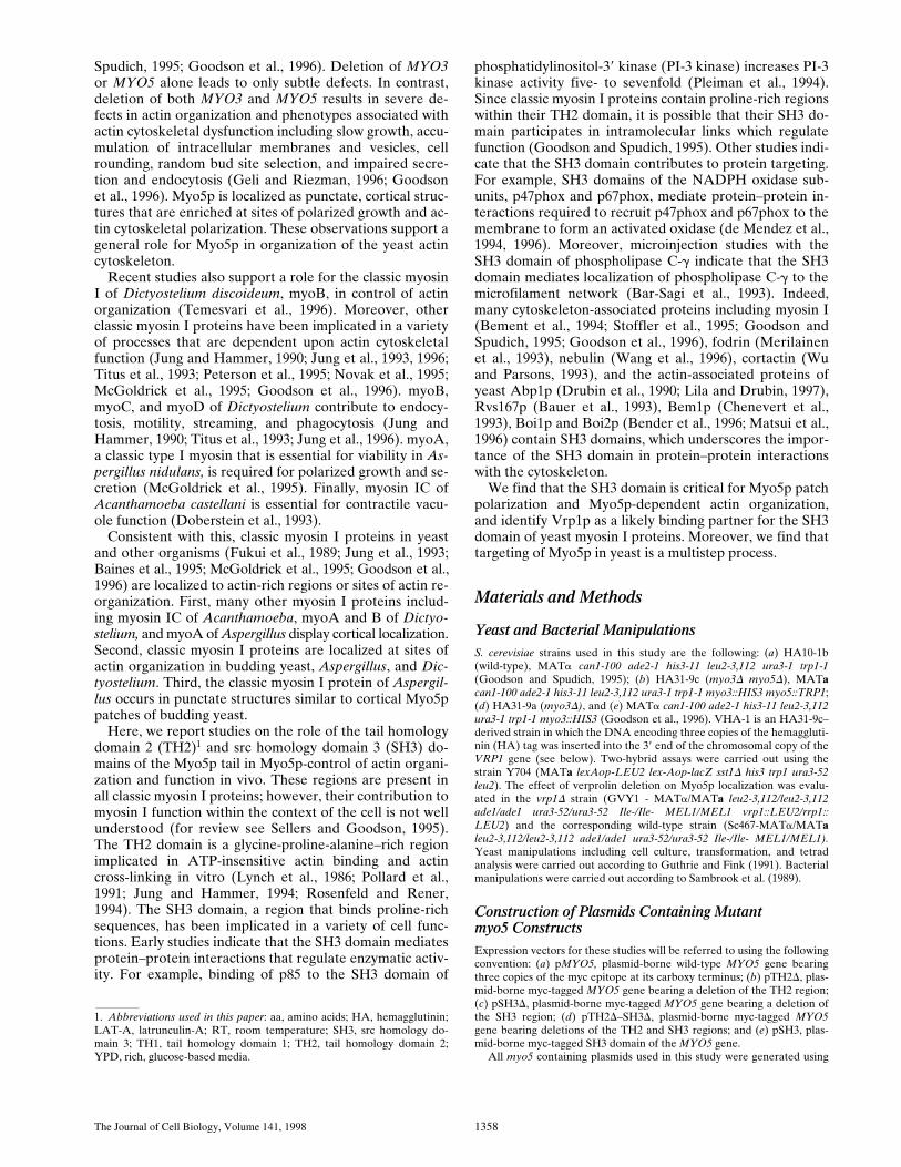
Expression of myc-tagged Myo5p on a low-copy numberplasmid under control of the MYO5 promoter (pMYO5)complements slow growth rate and defects in actin organi-zation and function produced by deletion of endogenousmyosin I genes (Goodson et al., 1996). To investigate thefunctional roles of various domains within the tail ofMyo5p, we created MYO5–myc constructs containing de-letions of either the TH2, SH3, or TH2 and SH3 domains(Fig. 1). We defined the TH2 domain as amino acids (aa)1,000–1,091, a region enriched in glycine, proline, and ala-nine that is amino-terminal to the SH3 domain. However,in some Acanthamoeba and Dictyostelium myosin I pro-teins, the TH2 region is believed to be bipartite: part of theTH2 region is amino-terminal to the SH3 domain, and partis carboxy-terminal to the SH3 domain. Since a proline-and alanine-rich region is present carboxy-terminal to theSH3 domain in Myo5p (aa 1,141–1,200), it is formally pos-sible that the TH2 region of yeast myosin I proteins ex-tends beyond the region deleted in this study. The SH3 do-main of MYO5 encompasses aa 1,092–1,140. We expressedthese mutant myosin I constructs using low-copy, cen-tromere-based plasmids and the endogenous MYO5 pro-moter in cells bearing deletions of endogenous myosin Igenes, and then examined the effect of mutant myosin Iproteins on actin organization and function in vivo.
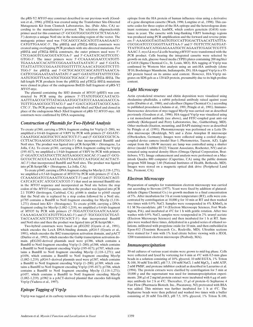
Expression of various epitope-tagged and untaggedMyo5p in myo3D myo5D mutants was examined by West-ern blot analysis using an anti-myc antibody (Evan et al.,1985) and an antibody raised against a myosin I–specificpeptide (Ruppert et al., 1993) (Fig. 2). Western blot analy-sis using a myosin I–specific antipeptide antibody indicatedthat the level of expression of plasmid-borne wild-typeand mutant Myo5p in the myo3D myo5D cells is similar tothat of endogenously expressed myosin I proteins in wild-type cells. Moreover, myc-tagged myosin I proteins of theexpected electrophoretic mobility were detected in cellsexpressing wild-type and mutant MYO5 genes. The levelof expression of Myo5p bearing deletions in both SH3 andTH2 domains is lower than that of wild-type Myo5p. How-ever, the level of the other mutant proteins was compara-ble to that of protein expressed from pMYO5.
Effect of Mutant myo5 Constructs on Cell Growth
Deletion of MYO3 and MYO5 results in severe growth de-
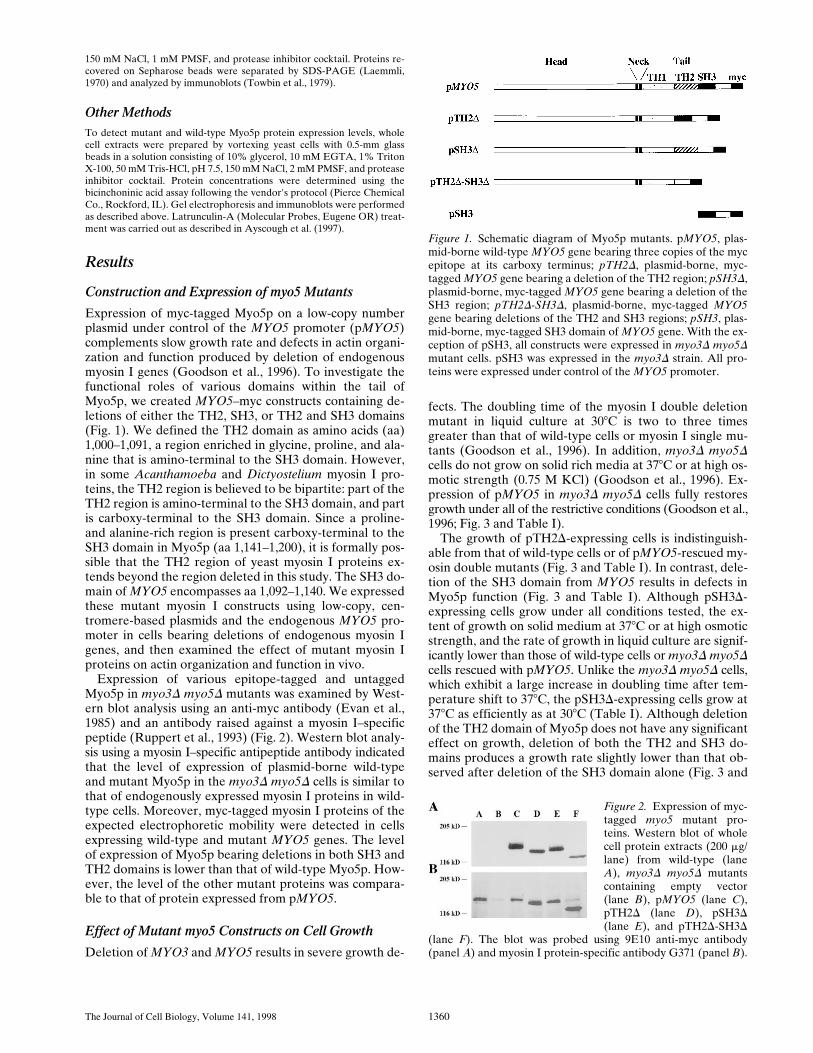
fects. The doubling time of the myosin I double deletionmutant in liquid culture at 308C is two to three timesgreater than that of wild-type cells or myosin I single mu-tants (Goodson et al., 1996). In addition, myo3D myo5Dcells do not grow on solid rich media at 378C or at high os-motic strength (0.75 M KCl) (Goodson et al., 1996). Ex-pression of pMYO5 in myo3D myo5D cells fully restoresgrowth under all of the restrictive conditions (Goodson et al.,1996; Fig. 3 and Table I).
The growth of pTH2D-expressing cells is indistinguish-able from that of wild-type cells or of pMYO5-rescued my-osin double mutants (Fig. 3 and Table I). In contrast, dele-tion of the SH3 domain from MYO5 results in defects inMyo5p function (Fig. 3 and Table I). Although pSH3D-expressing cells grow under all conditions tested, the ex-tent of growth on solid medium at 378C or at high osmoticstrength, and the rate of growth in liquid culture are signif-icantly lower than those of wild-type cells or myo3D myo5Dcells rescued with pMYO5. Unlike the myo3D myo5D cells,which exhibit a large increase in doubling time after tem-perature shift to 378C, the pSH3D-expressing cells grow at378C as efficiently as at 308C (Table I). Although deletionof the TH2 domain of Myo5p does not have any significanteffect on growth, deletion of both the TH2 and SH3 do-mains produces a growth rate slightly lower than that ob-served after deletion of the SH3 domain alone (Fig. 3 and
Figure 1. Schematic diagram of Myo5p mutants. pMYO5, plas-mid-borne wild-type MYO5 gene bearing three copies of the mycepitope at its carboxy terminus; pTH2D, plasmid-borne, myc-tagged MYO5 gene bearing a deletion of the TH2 region; pSH3D,plasmid-borne, myc-tagged MYO5 gene bearing a deletion of theSH3 region; pTH2D-SH3D, plasmid-borne, myc-tagged MYO5gene bearing deletions of the TH2 and SH3 regions; pSH3, plas-mid-borne, myc-tagged SH3 domain of MYO5 gene. With the ex-ception of pSH3, all constructs were expressed in myo3D myo5Dmutant cells. pSH3 was expressed in the myo3D strain. All pro-teins were expressed under control of the MYO5 promoter.
Figure 2. Expression of myc-tagged myo5 mutant pro-teins. Western blot of wholecell protein extracts (200 mg/lane) from wild-type (laneA), myo3D myo5D mutantscontaining empty vector(lane B), pMYO5 (lane C),pTH2D (lane D), pSH3D(lane E), and pTH2D-SH3D
(lane F). The blot was probed using 9E10 anti-myc antibody(panel A) and myosin I protein-specific antibody G371 (panel B).
Anderson et al. Myosin I Function and Localization in Yeast 1361
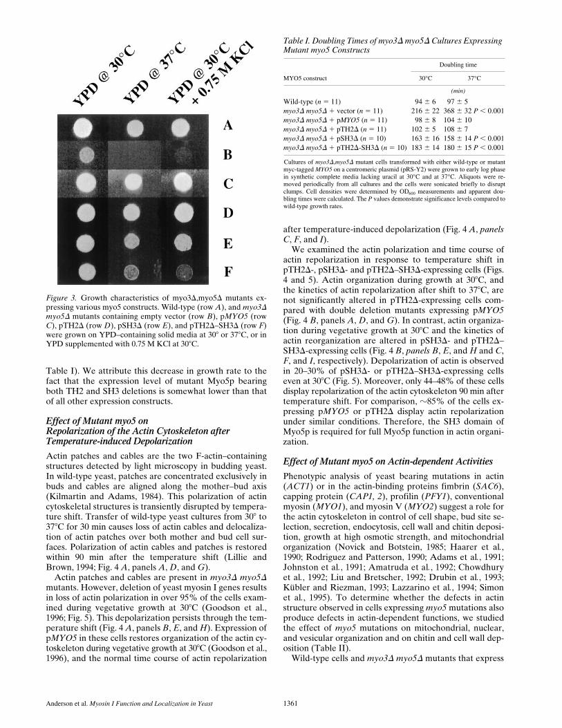
Table I). We attribute this decrease in growth rate to thefact that the expression level of mutant Myo5p bearingboth TH2 and SH3 deletions is somewhat lower than thatof all other expression constructs.
Effect of Mutant myo5 onRepolarization of the Actin Cytoskeleton after Temperature-induced Depolarization
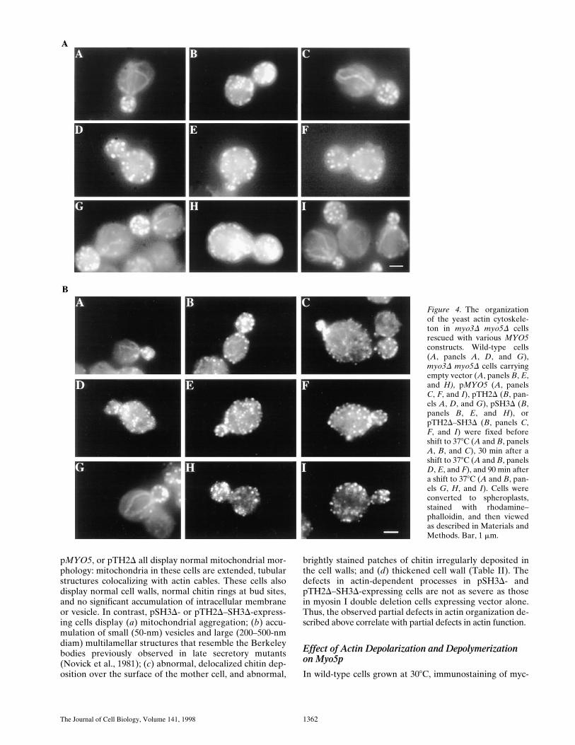
Actin patches and cables are the two F-actin–containingstructures detected by light microscopy in budding yeast.In wild-type yeast, patches are concentrated exclusively inbuds and cables are aligned along the mother–bud axis(Kilmartin and Adams, 1984). This polarization of actincytoskeletal structures is transiently disrupted by tempera-ture shift. Transfer of wild-type yeast cultures from 308 to378C for 30 min causes loss of actin cables and delocaliza-tion of actin patches over both mother and bud cell sur-faces. Polarization of actin cables and patches is restoredwithin 90 min after the temperature shift (Lillie andBrown, 1994; Fig. 4 A, panels A, D, and G).
Actin patches and cables are present in myo3D myo5Dmutants. However, deletion of yeast myosin I genes resultsin loss of actin polarization in over 95% of the cells exam-ined during vegetative growth at 308C (Goodson et al.,1996; Fig. 5). This depolarization persists through the tem-perature shift (Fig. 4 A, panels B, E, and H). Expression ofpMYO5 in these cells restores organization of the actin cy-toskeleton during vegetative growth at 308C (Goodson et al.,1996), and the normal time course of actin repolarization
after temperature-induced depolarization (Fig. 4 A, panelsC, F, and I).
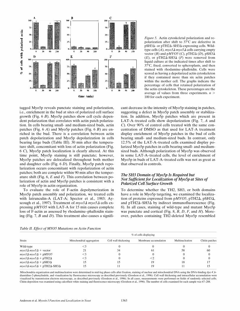
We examined the actin polarization and time course ofactin repolarization in response to temperature shift inpTH2D-, pSH3D- and pTH2D–SH3D-expressing cells (Figs.4 and 5). Actin organization during growth at 308C, andthe kinetics of actin repolarization after shift to 378C, arenot significantly altered in pTH2D-expressing cells com-pared with double deletion mutants expressing pMYO5(Fig. 4 B, panels A, D, and G). In contrast, actin organiza-tion during vegetative growth at 308C and the kinetics ofactin reorganization are altered in pSH3D- and pTH2D–SH3D-expressing cells (Fig. 4 B, panels B, E, and H and C,F, and I, respectively). Depolarization of actin is observedin 20–30% of pSH3D- or pTH2D–SH3D-expressing cellseven at 308C (Fig. 5). Moreover, only 44–48% of these cellsdisplay repolarization of the actin cytoskeleton 90 min aftertemperature shift. For comparison, z85% of the cells ex-pressing pMYO5 or pTH2D display actin repolarizationunder similar conditions. Therefore, the SH3 domain ofMyo5p is required for full Myo5p function in actin organi-zation.
Effect of Mutant myo5 on Actin-dependent Activities
Phenotypic analysis of yeast bearing mutations in actin(ACT1) or in the actin-binding proteins fimbrin (SAC6),capping protein (CAP1, 2), profilin (PFY1), conventionalmyosin (MYO1), and myosin V (MYO2) suggest a role forthe actin cytoskeleton in control of cell shape, bud site se-lection, secretion, endocytosis, cell wall and chitin deposi-tion, growth at high osmotic strength, and mitochondrialorganization (Novick and Botstein, 1985; Haarer et al.,1990; Rodriguez and Patterson, 1990; Adams et al., 1991;Johnston et al., 1991; Amatruda et al., 1992; Chowdhuryet al., 1992; Liu and Bretscher, 1992; Drubin et al., 1993;Kübler and Riezman, 1993; Lazzarino et al., 1994; Simonet al., 1995). To determine whether the defects in actinstructure observed in cells expressing myo5 mutations alsoproduce defects in actin-dependent functions, we studiedthe effect of myo5 mutations on mitochondrial, nuclear,and vesicular organization and on chitin and cell wall dep-osition (Table II).
Wild-type cells and myo3D myo5D mutants that express
Figure 3. Growth characteristics of myo3D,myo5D mutants ex-pressing various myo5 constructs. Wild-type (row A), and myo3Dmyo5D mutants containing empty vector (row B), pMYO5 (rowC), pTH2D (row D), pSH3D (row E), and pTH2D–SH3D (row F)were grown on YPD–containing solid media at 308 or 378C, or inYPD supplemented with 0.75 M KCl at 308C.
Table I. Doubling Times of myo3D myo5D Cultures Expressing Mutant myo5 Constructs
MYO5 construct
Doubling time
30°C 37°C
(min)
Wild-type (n 5 11) 94 6 6 97 6 5myo3D myo5D 1 vector (n 5 11) 216 6 22 368 6 32 P , 0.001myo3D myo5D 1 pMYO5 (n 5 11) 98 6 8 104 6 10myo3D myo5D 1 pTH2D (n 5 11) 102 6 5 108 6 7myo3D myo5D 1 pSH3D (n 5 10) 163 6 16 158 6 14 P , 0.001myo3D myo5D 1 pTH2D-SH3D (n 5 10) 183 6 14 180 6 15 P , 0.001
Cultures of myo3D,myo5D mutant cells transformed with either wild-type or mutantmyc-tagged MYO5 on a centromeric plasmid (pRS-Y2) were grown to early log phasein synthetic complete media lacking uracil at 30°C and at 37°C. Aliquots were re-moved periodically from all cultures and the cells were sonicated briefly to disruptclumps. Cell densities were determined by OD600 measurements and apparent dou-bling times were calculated. The P values demonstrate significance levels compared towild-type growth rates.
The Journal of Cell Biology, Volume 141, 1998 1362
pMYO5, or pTH2D all display normal mitochondrial mor-phology: mitochondria in these cells are extended, tubularstructures colocalizing with actin cables. These cells alsodisplay normal cell walls, normal chitin rings at bud sites,and no significant accumulation of intracellular membraneor vesicle. In contrast, pSH3D- or pTH2D–SH3D-express-ing cells display (a) mitochondrial aggregation; (b) accu-mulation of small (50-nm) vesicles and large (200–500-nmdiam) multilamellar structures that resemble the Berkeleybodies previously observed in late secretory mutants(Novick et al., 1981); (c) abnormal, delocalized chitin dep-osition over the surface of the mother cell, and abnormal,
brightly stained patches of chitin irregularly deposited inthe cell walls; and (d) thickened cell wall (Table II). Thedefects in actin-dependent processes in pSH3D- andpTH2D–SH3D-expressing cells are not as severe as thosein myosin I double deletion cells expressing vector alone.Thus, the observed partial defects in actin organization de-scribed above correlate with partial defects in actin function.
Effect of Actin Depolarization and Depolymerization on Myo5p
In wild-type cells grown at 308C, immunostaining of myc-
Figure 4. The organizationof the yeast actin cytoskele-ton in myo3D myo5D cellsrescued with various MYO5constructs. Wild-type cells(A, panels A, D, and G),myo3D myo5D cells carryingempty vector (A, panels B, E,and H), pMYO5 (A, panelsC, F, and I), pTH2D (B, pan-els A, D, and G), pSH3D (B,panels B, E, and H), orpTH2D–SH3D (B, panels C,F, and I) were fixed beforeshift to 378C (A and B, panelsA, B, and C), 30 min after ashift to 378C (A and B, panelsD, E, and F), and 90 min aftera shift to 378C (A and B, pan-els G, H, and I). Cells wereconverted to spheroplasts,stained with rhodamine–phalloidin, and then viewedas described in Materials andMethods. Bar, 1 mm.
Anderson et al. Myosin I Function and Localization in Yeast 1363
tagged Myo5p reveals punctate staining and polarization,i.e., enrichment in the bud at sites of polarized cell surfacegrowth (Fig. 6 B). Myo5p patches show cell cycle depen-dent polarization that correlates with actin patch polariza-tion. In cells bearing small- and medium-sized buds, actinpatches (Fig. 6 A) and Myo5p patches (Fig. 6 B) are en-riched in the bud. There is a correlation between actinpatch depolarization and Myo5p depolarization in cellsbearing large buds (Table III). 30 min after the tempera-ture shift, concomitant with loss of actin polarization (Fig.6 C), Myo5p patch localization is clearly altered. At thistime point, Myo5p staining is still punctate; however,Myo5p patches are delocalized throughout both motherand daughter cells (Fig. 6 D). Finally, Myo5p patch repo-larization occurs concomitant with repolarization of actinpatches; both are complete within 90 min after the temper-ature shift (Fig. 6, E and F). This correlation between po-larization of actin and Myo5p patches is consistent with arole of Myo5p in actin organization.
To evaluate the role of F-actin depolymerization inMyo5p patch assembly and polarization, we treated cellswith latrunculin-A (LAT-A; Spector et al., 1983; Ay-scough et al., 1997). Treatment of myo3D myo5D cells ex-pressing pMYO5 with LAT-A for 15 min causes completeloss of F-actin as assessed by rhodamine–phalloidin stain-ing (Fig. 7, B and D). This treatment also causes a signifi-
cant decrease in the intensity of Myo5p staining in patches,suggesting a defect in Myo5p patch assembly or stabiliza-tion. In addition, Myo5p patches which are present inLAT-A–treated cells show depolarization (Fig. 7, A andC). Over 90% of control cells treated with the same con-centration of DMSO as that used for LAT-A treatmentdisplay enrichment of Myo5p patches in the bud of cellsbearing small- and medium-sized buds. In contrast, only12.5% of the LAT-A–treated cells examined display po-larized Myo5p patches in cells bearing small- and medium-sized buds. Although polarization of Myo5p was observedin some LAT-A–treated cells, the level of enrichment ofMyo5p in buds of LAT-A–treated cells was not as great asthat observed in controls.
The SH3 Domain of Myo5p Is Required butNot Sufficient for Localization of Myo5p at Sites of Polarized Cell Surface Growth
To determine whether the TH2, SH3, or both domainshave a role in Myo5p targeting, we examined the localiza-tion of proteins expressed from pMYO5, pTH2D, pSH3D,and pTH2D–SH3D by indirect immunofluorescence (Fig.8). In all cases, staining of wild-type and mutant Myo5pwas punctate and cortical (Fig. 8, B, D, F, and H). More-over, patches containing TH2-deleted Myo5p resembled
Figure 5. Actin cytoskeletal polarization and re-polarization after shift to 378C are defective inpSH3D- or pTH2D–SH3D-expressing cells. Wild-type cells (A), myo3D myo5D cells carrying emptyvector (B) and pMYO5 (C), pTH2D (D), pSH3D(E), or pTH2D-SH3D (F) were removed fromliquid culture at the indicated times after shift to378C, fixed, converted to spheroplasts, and thenstained with rhodamine–phalloidin. Cells werescored as having a depolarized actin cytoskeletonif they contained more than six actin patcheswithin the mother cell. The graphs indicate thepercentage of cells that retained polarization ofthe actin cytoskeleton. These percentages are theaverage of values from three experiments. n .100 for each experiment.
Table II. Effect of MYO5 Mutations on Actin Function
Strain
% of cells displaying
Mitochondrial aggregation Cell wall thickening Membrane accumulation Multinucleation Chitin patches
Wild-type ,3 0 0 0 0myo3D myo5D 1 vector 21 34 61 58 60myo3D myo5D 1 pMYO5 ,3 0 0 0 0myo3D myo5D 1 pTH2D ,3 0 ,2 0 0myo3D myo5D 1 pSH3D 15 15 19 18 17myo3D myo5D 1 pTH2D-SH3D 15 11 19 11 15
Mitochondria organization and multinucleation were determined in mid-log phase cells after fixation, staining of nuclear and mitochondrial DNA using the DNA-binding dye 49,6-diamidino-2-phenylindole, and visualization by fluorescence microscopy as described previously (Goodson et al., 1996). Cell wall thickening and intracellular accumulation werevisualized by transmission electron microscopy, as described previously (Goodson et al., 1996). In all cases, measurements were performed on fields of randomly selected cells.Chitin deposition was examined using calcofluor white staining and fluorescence microscopy (Goodson et al., 1996). The number of cells examined for each sample was 67–288.
The Journal of Cell Biology, Volume 141, 1998 1364
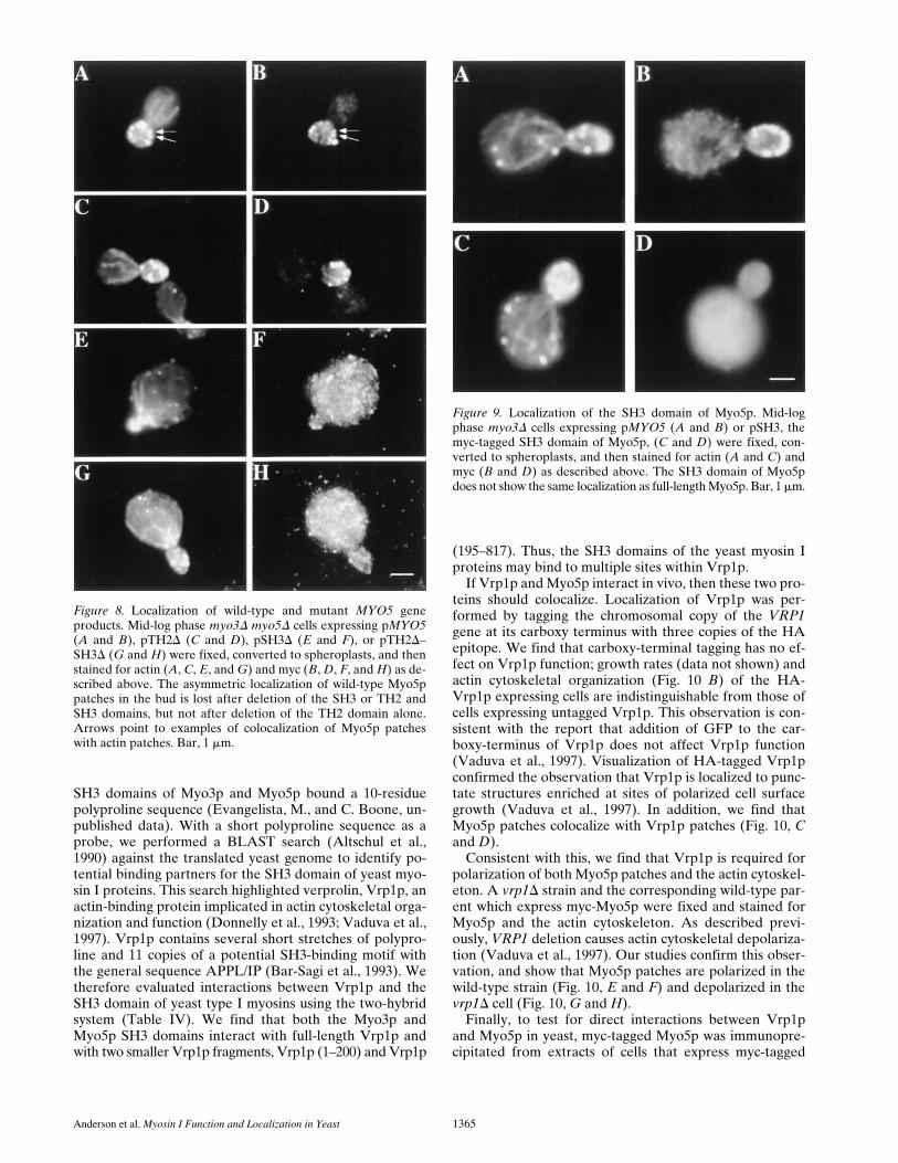
wild-type Myo5p patches and were enriched in the bud(n 5 74). In contrast, patches containing Myo5p bearingdeletions in the SH3 domain alone, or in the TH2 and SH3domains, were evenly distributed in mother cells and bud:only 3% of cells examined showed enrichment of theseproteins in the bud (n 5 101 and n 5 111, respectively; Fig.8, F and H). Although partial depolarization of the actincytoskeleton is observed in pSH3D- and pTH2D–SH3D-expressing cells, depolarization of Myo5p patches in thesecells is not due to effects on actin organization. Patches con-taining mutant Myo5p protein are delocalized even inpSH3D- and pTH2D–SH3D-expressing cells that containpolarized actin patches and cables.
According to the results described above, the SH3 do-main is required for targeting of Myo5p to growing buds.To examine the role of this domain in (a) assembly ofMyo5p into patches, and (b) enrichment of Myo5p patchesat sites of polarized cell surface growth, we constructed aplasmid, pSH3, carrying the SH3 domain and the subse-quent 79-aa carboxy terminus of Myo5p (aa 1,086–1,219)fused to three copies of the myc epitope. This constructwas expressed and localized in a MYO3 deletion mutant.myo3D cells were used for expression because they con-tain a polarized actin cytoskeleton, and only one of twoendogenous myosin I genes. Western blot analysis using
the anti-myc antibody indicates that expression of pSH3using a low-copy plasmid and the endogenous MYO5 pro-moter produces a protein of the expected apparent molec-ular weight at levels comparable to that of protein expressedfrom pMYO5 (data not shown). Immunolocalization ofthis fusion protein using the anti-myc antibody reveals dif-fuse staining which is distributed throughout the cyto-plasm (Fig. 9). Thus, the SH3 domain of Myo5p is not suf-ficient for either association of Myo5p with patches or forenrichment of Myo5p patches at sites of polarized cell sur-face growth.
The Proline-rich, SH3-binding Protein, Verprolin, Is a Myo5p Binding Partner
In two-hybrid tests for protein–protein interaction, the
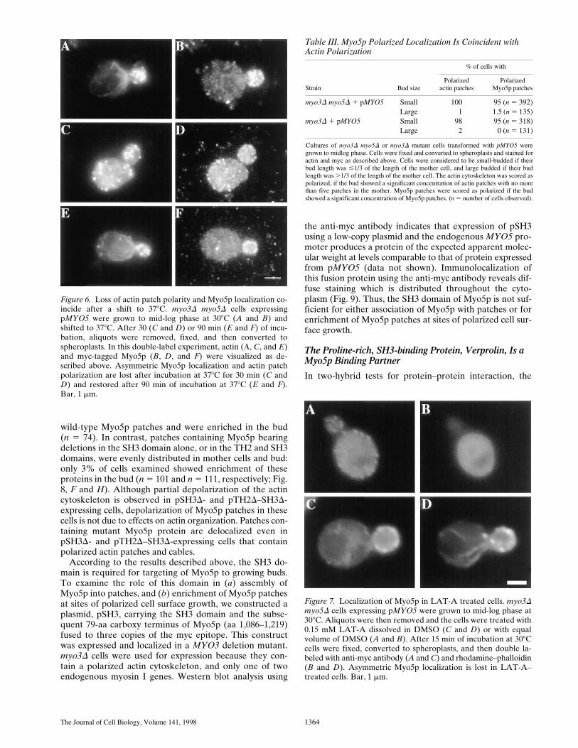
Figure 6. Loss of actin patch polarity and Myo5p localization co-incide after a shift to 378C. myo3D myo5D cells expressingpMYO5 were grown to mid-log phase at 308C (A and B) andshifted to 378C. After 30 (C and D) or 90 min (E and F) of incu-bation, aliquots were removed, fixed, and then converted tospheroplasts. In this double-label experiment, actin (A, C, and E)and myc-tagged Myo5p (B, D, and F) were visualized as de-scribed above. Asymmetric Myo5p localization and actin patchpolarization are lost after incubation at 378C for 30 min (C andD) and restored after 90 min of incubation at 378C (E and F).Bar, 1 mm.
Table III. Myo5p Polarized Localization Is Coincident with Actin Polarization
Strain Bud size
% of cells with
Polarizedactin patches
PolarizedMyo5p patches
myo3D myo5D 1 pMYO5 Small 100 95 (n 5 392)Large 1 1.5 (n 5 135)
myo3D 1 pMYO5 Small 98 95 (n 5 318)Large 2 0 (n 5 131)
Cultures of myo3D myo5D or myo3D mutant cells transformed with pMYO5 weregrown to midlog phase. Cells were fixed and converted to spheroplasts and stained foractin and myc as described above. Cells were considered to be small-budded if theirbud length was #1/3 of the length of the mother cell, and large budded if their budlength was .1/3 of the length of the mother cell. The actin cytoskeleton was scored aspolarized, if the bud showed a significant concentration of actin patches with no morethan five patches in the mother. Myo5p patches were scored as polarized if the budshowed a significant concentration of Myo5p patches. (n 5 number of cells observed).
Figure 7. Localization of Myo5p in LAT-A treated cells. myo3Dmyo5D cells expressing pMYO5 were grown to mid-log phase at308C. Aliquots were then removed and the cells were treated with0.15 mM LAT-A dissolved in DMSO (C and D) or with equalvolume of DMSO (A and B). After 15 min of incubation at 308Ccells were fixed, converted to spheroplasts, and then double la-beled with anti-myc antibody (A and C) and rhodamine–phalloidin(B and D). Asymmetric Myo5p localization is lost in LAT-A–treated cells. Bar, 1 mm.
Anderson et al. Myosin I Function and Localization in Yeast 1365
SH3 domains of Myo3p and Myo5p bound a 10-residuepolyproline sequence (Evangelista, M., and C. Boone, un-published data). With a short polyproline sequence as aprobe, we performed a BLAST search (Altschul et al.,1990) against the translated yeast genome to identify po-tential binding partners for the SH3 domain of yeast myo-sin I proteins. This search highlighted verprolin, Vrp1p, anactin-binding protein implicated in actin cytoskeletal orga-nization and function (Donnelly et al., 1993; Vaduva et al.,1997). Vrp1p contains several short stretches of polypro-line and 11 copies of a potential SH3-binding motif withthe general sequence APPL/IP (Bar-Sagi et al., 1993). Wetherefore evaluated interactions between Vrp1p and theSH3 domain of yeast type I myosins using the two-hybridsystem (Table IV). We find that both the Myo3p andMyo5p SH3 domains interact with full-length Vrp1p andwith two smaller Vrp1p fragments, Vrp1p (1–200) and Vrp1p
(195–817). Thus, the SH3 domains of the yeast myosin Iproteins may bind to multiple sites within Vrp1p.
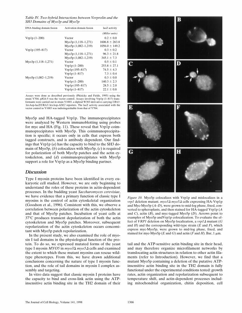
If Vrp1p and Myo5p interact in vivo, then these two pro-teins should colocalize. Localization of Vrp1p was per-formed by tagging the chromosomal copy of the VRP1gene at its carboxy terminus with three copies of the HAepitope. We find that carboxy-terminal tagging has no ef-fect on Vrp1p function; growth rates (data not shown) andactin cytoskeletal organization (Fig. 10 B) of the HA-Vrp1p expressing cells are indistinguishable from those ofcells expressing untagged Vrp1p. This observation is con-sistent with the report that addition of GFP to the car-boxy-terminus of Vrp1p does not affect Vrp1p function(Vaduva et al., 1997). Visualization of HA-tagged Vrp1pconfirmed the observation that Vrp1p is localized to punc-tate structures enriched at sites of polarized cell surfacegrowth (Vaduva et al., 1997). In addition, we find thatMyo5p patches colocalize with Vrp1p patches (Fig. 10, Cand D).
Consistent with this, we find that Vrp1p is required forpolarization of both Myo5p patches and the actin cytoskel-eton. A vrp1D strain and the corresponding wild-type par-ent which express myc-Myo5p were fixed and stained forMyo5p and the actin cytoskeleton. As described previ-ously, VRP1 deletion causes actin cytoskeletal depolariza-tion (Vaduva et al., 1997). Our studies confirm this obser-vation, and show that Myo5p patches are polarized in thewild-type strain (Fig. 10, E and F) and depolarized in thevrp1D cell (Fig. 10, G and H).
Finally, to test for direct interactions between Vrp1pand Myo5p in yeast, myc-tagged Myo5p was immunopre-cipitated from extracts of cells that express myc-tagged
Figure 8. Localization of wild-type and mutant MYO5 geneproducts. Mid-log phase myo3D myo5D cells expressing pMYO5(A and B), pTH2D (C and D), pSH3D (E and F), or pTH2D–SH3D (G and H) were fixed, converted to spheroplasts, and thenstained for actin (A, C, E, and G) and myc (B, D, F, and H) as de-scribed above. The asymmetric localization of wild-type Myo5ppatches in the bud is lost after deletion of the SH3 or TH2 andSH3 domains, but not after deletion of the TH2 domain alone.Arrows point to examples of colocalization of Myo5p patcheswith actin patches. Bar, 1 mm.
Figure 9. Localization of the SH3 domain of Myo5p. Mid-logphase myo3D cells expressing pMYO5 (A and B) or pSH3, themyc-tagged SH3 domain of Myo5p, (C and D) were fixed, con-verted to spheroplasts, and then stained for actin (A and C) andmyc (B and D) as described above. The SH3 domain of Myo5pdoes not show the same localization as full-length Myo5p. Bar, 1 mm.
The Journal of Cell Biology, Volume 141, 1998 1366
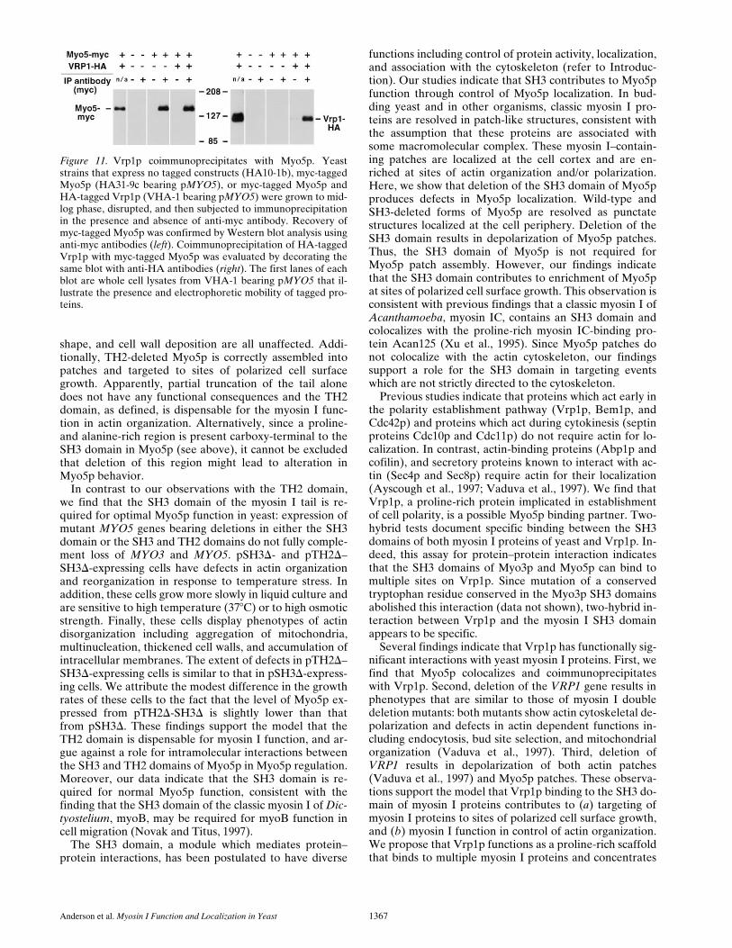
Myo5p and HA-tagged Vrp1p. The immunoprecipitateswere analyzed by Western immunoblotting using probesfor myc and HA (Fig. 11). These reveal that Vrp1p coim-munoprecipitates with Myo5p. This coimmunoprecipita-tion is specific; it occurs only in cells that express bothtagged constructs, and is antibody dependent. Our find-ings that Vrp1p (a) has the capacity to bind to the SH3 do-main of Myo5p, (b) colocalizes with Myo5p, (c) is requiredfor polarization of both Myo5p patches and the actin cy-toskeleton, and (d) coimmunoprecipitates with Myo5psupport a role for Vrp1p as a Myo5p binding partner.
DiscussionType I myosin proteins have been identified in every eu-karyotic cell studied. However, we are only beginning tounderstand the roles of these proteins in actin-dependentprocesses. In the budding yeast Saccharomyces cerevisiae,we have evidence that a primary function of classic type Imyosins is the control of actin cytoskeletal organization(Goodson et al., 1996). Consistent with this, we observe acorrelation between polarization of the actin cytoskeletonand that of Myo5p patches. Incubation of yeast cells at378C produces transient depolarization of both the actincytoskeleton and Myo5p patches. Moreover, subsequentrepolarization of the actin cytoskeleton occurs concomi-tant with Myo5p patch repolarization.
In the present study, we also examined the role of myo-sin I tail domains in the physiological function of the pro-tein. To do so, we expressed mutated forms of the yeasttype I myosin MYO5 in myo3D myo5D cells and examinedthe extent to which these mutant myosins can rescue wild-type phenotypes. From this, we have drawn additionalconclusions concerning the nature of type I myosin func-tion, and the role of tail domains in myosin I complex as-sembly and targeting.
In vitro data suggest that classic myosin I proteins havethe capacity to bind and cross-link actin using the ATP-insensitive actin binding site in the TH2 domain of their
tail and the ATP-sensitive actin binding site in their head,and may therefore organize microfilament networks bytranslocating actin structures in relation to other actin fila-ments (refer to Introduction). However, we find that amutant Myo5p containing a deletion of the putative ATP-insensitive actin binding site in the TH2 domain is fullyfunctional under the experimental conditions tested: growthrates, actin organization and repolarization subsequent totemperature shift, and actin-dependent processes includ-ing mitochondrial organization, chitin deposition, cell
Table IV. Two-hybrid Interactions between Verprolin and the SH3 Domains of Myo3p and Myo5p
DNA-binding domain fusion Activation domain fusion lacZ activity
(Miller units)
Vrp1p (1–200) Vector 0.2 6 0.0Myo3p (1,118–1,271) 1606.8 6 263.8Myo5p (1,082–1,219) 1056.0 6 149.2
Vrp1p (195–817) Vector 0.3 6 0.2Myo3p (1,118–1,271) 96.3 6 21.8Myo5p (1,082–1,219) 345.1 6 7.3
Myo3p (1,118–1,271) Vector 0.5 6 0.1Vrp1p (1–200) 253.8 6 27.1Vrp1p (195–817) 74.5 6 4.3Vrp1p (1–817) 7.3 6 0.4
Myo5p (1,082–1,219) Vector 0.3 6 0.0Vrp1p (1–200) 140.3 6 2.3Vrp1p (195–817) 28.5 6 2.0Vrp1p (1–817) 22.1 6 0.8
Assays were done as described previously (Phizicky and Fields, 1995) using thestrain Y704; pJG4-5 was the vector control. Assays involving Vrp1p (1–817) trans-formants were carried out in strain Y1003, a diploid W303 derivative carrying URA3::lexAop-lacZ/URA3::lexAop-ADE2 reporters. The lacZ activity associated with thevector control in Y1003 was indistinguishable from that of Y704.
Figure 10. Myo5p colocalizes with Vrp1p and mislocalizes in avrp1 deletion mutant. myo3D myo5D cells expressing HA-Vrp1pand Myc-Myo5p (A–D), were grown to mid-log phase, fixed, con-verted to spheroplasts, and then stained for HA-tagged Vrp1p (Aand C), actin (B), and myc-tagged Myo5p (D). Arrows point toexamples of Myo5p andVrp1p colocalization. To evaluate the ef-fect of VRP1 deletion on Myo5p localization, a vrp1D mutant (Gand H) and the corresponding wild-type strain (E and F), whichexpress myc-Myo5p, were grown to mid-log phase, fixed, andstained for myc-Myo5p (E and G) and actin (F and H). Bar, 1 mm.
Anderson et al. Myosin I Function and Localization in Yeast 1367
shape, and cell wall deposition are all unaffected. Addi-tionally, TH2-deleted Myo5p is correctly assembled intopatches and targeted to sites of polarized cell surfacegrowth. Apparently, partial truncation of the tail alonedoes not have any functional consequences and the TH2domain, as defined, is dispensable for the myosin I func-tion in actin organization. Alternatively, since a proline-and alanine-rich region is present carboxy-terminal to theSH3 domain in Myo5p (see above), it cannot be excludedthat deletion of this region might lead to alteration inMyo5p behavior.
In contrast to our observations with the TH2 domain,we find that the SH3 domain of the myosin I tail is re-quired for optimal Myo5p function in yeast: expression ofmutant MYO5 genes bearing deletions in either the SH3domain or the SH3 and TH2 domains do not fully comple-ment loss of MYO3 and MYO5. pSH3D- and pTH2D–SH3D-expressing cells have defects in actin organizationand reorganization in response to temperature stress. Inaddition, these cells grow more slowly in liquid culture andare sensitive to high temperature (378C) or to high osmoticstrength. Finally, these cells display phenotypes of actindisorganization including aggregation of mitochondria,multinucleation, thickened cell walls, and accumulation ofintracellular membranes. The extent of defects in pTH2D–SH3D-expressing cells is similar to that in pSH3D-express-ing cells. We attribute the modest difference in the growthrates of these cells to the fact that the level of Myo5p ex-pressed from pTH2D-SH3D is slightly lower than thatfrom pSH3D. These findings support the model that theTH2 domain is dispensable for myosin I function, and ar-gue against a role for intramolecular interactions betweenthe SH3 and TH2 domains of Myo5p in Myo5p regulation.Moreover, our data indicate that the SH3 domain is re-quired for normal Myo5p function, consistent with thefinding that the SH3 domain of the classic myosin I of Dic-tyostelium, myoB, may be required for myoB function incell migration (Novak and Titus, 1997).
The SH3 domain, a module which mediates protein–protein interactions, has been postulated to have diverse
functions including control of protein activity, localization,and association with the cytoskeleton (refer to Introduc-tion). Our studies indicate that SH3 contributes to Myo5pfunction through control of Myo5p localization. In bud-ding yeast and in other organisms, classic myosin I pro-teins are resolved in patch-like structures, consistent withthe assumption that these proteins are associated withsome macromolecular complex. These myosin I–contain-ing patches are localized at the cell cortex and are en-riched at sites of actin organization and/or polarization.Here, we show that deletion of the SH3 domain of Myo5pproduces defects in Myo5p localization. Wild-type andSH3-deleted forms of Myo5p are resolved as punctatestructures localized at the cell periphery. Deletion of theSH3 domain results in depolarization of Myo5p patches.Thus, the SH3 domain of Myo5p is not required forMyo5p patch assembly. However, our findings indicatethat the SH3 domain contributes to enrichment of Myo5pat sites of polarized cell surface growth. This observation isconsistent with previous findings that a classic myosin I ofAcanthamoeba, myosin IC, contains an SH3 domain andcolocalizes with the proline-rich myosin IC-binding pro-tein Acan125 (Xu et al., 1995). Since Myo5p patches donot colocalize with the actin cytoskeleton, our findingssupport a role for the SH3 domain in targeting eventswhich are not strictly directed to the cytoskeleton.
Previous studies indicate that proteins which act early inthe polarity establishment pathway (Vrp1p, Bem1p, andCdc42p) and proteins which act during cytokinesis (septinproteins Cdc10p and Cdc11p) do not require actin for lo-calization. In contrast, actin-binding proteins (Abp1p andcofilin), and secretory proteins known to interact with ac-tin (Sec4p and Sec8p) require actin for their localization(Ayscough et al., 1997; Vaduva et al., 1997). We find thatVrp1p, a proline-rich protein implicated in establishmentof cell polarity, is a possible Myo5p binding partner. Two-hybrid tests document specific binding between the SH3domains of both myosin I proteins of yeast and Vrp1p. In-deed, this assay for protein–protein interaction indicatesthat the SH3 domains of Myo3p and Myo5p can bind tomultiple sites on Vrp1p. Since mutation of a conservedtryptophan residue conserved in the Myo3p SH3 domainsabolished this interaction (data not shown), two-hybrid in-teraction between Vrp1p and the myosin I SH3 domainappears to be specific.
Several findings indicate that Vrp1p has functionally sig-nificant interactions with yeast myosin I proteins. First, wefind that Myo5p colocalizes and coimmunoprecipitateswith Vrp1p. Second, deletion of the VRP1 gene results inphenotypes that are similar to those of myosin I doubledeletion mutants: both mutants show actin cytoskeletal de-polarization and defects in actin dependent functions in-cluding endocytosis, bud site selection, and mitochondrialorganization (Vaduva et al., 1997). Third, deletion ofVRP1 results in depolarization of both actin patches(Vaduva et al., 1997) and Myo5p patches. These observa-tions support the model that Vrp1p binding to the SH3 do-main of myosin I proteins contributes to (a) targeting ofmyosin I proteins to sites of polarized cell surface growth,and (b) myosin I function in control of actin organization.We propose that Vrp1p functions as a proline-rich scaffoldthat binds to multiple myosin I proteins and concentrates
Figure 11. Vrp1p coimmunoprecipitates with Myo5p. Yeaststrains that express no tagged constructs (HA10-1b), myc-taggedMyo5p (HA31-9c bearing pMYO5), or myc-tagged Myo5p andHA-tagged Vrp1p (VHA-1 bearing pMYO5) were grown to mid-log phase, disrupted, and then subjected to immunoprecipitationin the presence and absence of anti-myc antibody. Recovery ofmyc-tagged Myo5p was confirmed by Western blot analysis usinganti-myc antibodies (left). Coimmunoprecipitation of HA-taggedVrp1p with myc-tagged Myo5p was evaluated by decorating thesame blot with anti-HA antibodies (right). The first lanes of eachblot are whole cell lysates from VHA-1 bearing pMYO5 that il-lustrate the presence and electrophoretic mobility of tagged pro-teins.
The Journal of Cell Biology, Volume 141, 1998 1368
these proteins at sites of polarized cell surface growth.Since multiple proline-rich proteins including Srv2p andBni1p are localized at sites of polarized cell surface growth(Freeman et al., 1996; Evangelista et al., 1997) it is possiblethat multiple SH3-mediated protein–protein interactionscontribute to myosin I targeting in yeast.
Several findings indicate that the SH3 domain contrib-utes to, but is not sufficient for, Myo5p targeting. First,pSH3D-expressing cells show only partial loss of actin or-ganization and only partial defects in actin dependent pro-cesses. That is, deletion of the SH3 domain results in adecrease in the efficiency of Myo5p function in actin orga-nization. Thus, some fraction of the SH3-deleted Myo5pmust reach the correct site of action. Indeed, we find thatpatches containing SH3-deleted Myo5p are present butare not enriched at sites of polarized cell surface growth.Second, we find that a fusion protein containing of theSH3 domain of Myo5p shows diffuse cytosolic stainingwhen expressed in yeast at levels comparable to wild-type,endogenous Myo5p. Thus, the SH3 domain is sufficientneither for the assembly of Myo5p patches, nor for Myo5ppatch polarization. Together, these observations indicatethat multiple factors in addition to the SH3 domain are re-quired for Myo5p assembly and targeting. Indeed, sincethe SH3 domain of myoB, a myosin I protein of Dictyoste-lium, is not required for myoB targeting (Novak and Titus,1997), it is possible that the relative contribution of theSH3 domain to myosin I targeting may vary among celltypes.
One region that may participate in Myo5p targeting isits TH1 sequence, a highly basic region found in all myosinI subclasses. Previous studies indicate that the TH1 do-main of myosin proteins can bind to acidic phospholipids(Adams and Pollard, 1989; Doberstein and Pollard, 1992).Moreover, the basic TH1 region of brush border myosin Iprotein is sufficient to target myosin molecules to actin-rich apical structures (Footer and Bretscher, 1994). Wefind that deletion of the hyperbasic segment of the Myo5pTH1 region, or deletion of the motor domain of Myo5p,impairs protein expression and/or stability (Swayne, T.,and L. Pon, unpublished results). Therefore, we cannotdraw conclusions regarding the role of the TH1 region inMyo5p targeting from this line of experiments.
Another region that may participate in Myo5p targetingis the actin-binding motor domain. We find that Myo5ppatch assembly requires F-actin. LAT-A treatment resultsin rapid, quantitative actin depolymerization in yeast andalso produces a decrease in total staining intensity of myc-tagged Myo5p in punctate structures and an increase indiffuse, cytosolic staining. Myo5p-containing patches thatare detected are cortical, but delocalized and equally dis-tributed in mother and daughter cells. Thus, a drug thatcauses F-actin depolymerization produces partial defectsin Myo5p patch assembly and defects in enrichment ofMyo5p patches at sites of polarized cell surface growth.Since myosins are actin-binding proteins, the simplest in-terpretation of this finding is that Myo5p patch assemblyrequires association of Myo5p with F-actin, presumablythrough its high-affinity F-actin binding site in the motordomain. Current studies focus on analysis of the actin-binding activity of the Myo5p motor domain, and the roleof this activity in Myo5p targeting.
The data we present supports a model for myosin I lo-calization and function in yeast. In this model, Myo5p isassembled into a protein complex at the cell cortex. As-sembly of cortical Myo5p patches does not require eitherthe TH2 domain or the SH3 domain of the Myo5p tail.However, Myo5p patch assembly is at least partially de-pendent upon F-actin. Cortical Myo5p patches are ulti-mately transported to and concentrated at sites of polar-ized cell surface growth where they function in polarizationof the actin cytoskeleton. This final targeting event re-quires the SH3 domain of the Myo5p tail, a region thatbinds to Vrp1p. We propose that Vrp1p serves as a scaf-fold to bind and concentrate Myo5p patches within thebud. This is the first example of protein-mediated localiza-tion of a myosin I protein. Moreover, since Vrp1p hasbeen implicated in actin polarization, our finding provides(a) additional support for a role of myosin I in actin polar-ization, and (b) an important link between the cell polaritymachinery and a mediator of actin organization.
We thank F. Chang and P. Brandt (both from Columbia University, NewYork), and members of the Pon laboratory for critical evaluation of themanuscript; G. Vaduva and A. Hopper for yeast strains and helpful dis-cussion regarding verprolin; S. Swamy for assistance in immunofluores-cence microscopy; K. Brown for expert assistance with the electron mi-croscopy; T. Leeuw for helpful discussion regarding Myo3p localization,and I. Pot for two-hybrid analysis of Myo3p protein–protein interactions.
This work was supported by research grants from the American CancerSociety (RPG-97-163-01-C) and National Institutes of Health (GM45735)to L.A. Pon, a National Institutes of Health Grant (NS16036) to L.A.Greene, a Medical Scientists Training Program Award (5T32 GM07367)to B.L. Anderson, and grants from the Natural Sciences and EngineeringResearch Council of Canada, and the National Cancer Institute of Canadato C. Boone.
Received for publication 28 October 1997 and in revised form 23 April1998.
References
Adams, R.J., and T.D. Pollard. 1989. Binding of myosin I to membrane lipids.Nature. 340:565–568.
Adams, A.E., D. Botstein, and D.G. Drubin. 1991. Requirement of yeast fim-brin for actin organization and morphogenesis in vivo. Nature. 354:404–408.
Altschul, S.F., W. Gish, W. Miller, E.W. Myers, and D.J. Lipman. 1990. Basiclocal alignment search tool. J. Mol. Biol. 215:403–410.
Amatruda, J.F., D.J. Gattermeir, T.S. Karpova, and J.A. Cooper. 1992. Effectsof null mutations and overexpression of capping protein on morphogenesis,actin distribution, and polarized secretion in yeast. J. Cell Biol. 119:1151–1162.
Ayscough, K.R., J. Stryker, N. Pokala, M. Sanders, P. Crews, and D.G. Drubin.1997. High rates of actin filament turnover in budding yeast and roles for ac-tin in establishment and maintenance of cell polarity revealed using the actininhibitor latrunculin-a. J. Cell Biol. 137:399–416.
Baines, I.C., A. Corigliano-Murphy, and E.D. Korn. 1995. Quantification andlocalization of phosphorylated myosin I isoforms in Acanthamoeba castel-lani. J. Biol. Chem. 130:591–603.
Bar-Sagi, D., D. Rotin, A. Batzer, V. Mandiyan, and J. Schlessinger. 1993. SH3domains direct cellular localization of signaling molecules. Cell. 74:83–91.
Bauer, F., M. Urdaci, M. Aigle, and M. Crouzet. 1993. Alteration of a yeast SH3protein leads to conditional viability with defects in cytoskeletal and buddingpatterns. Mol. Cell Biol. 13:5070–5084.
Bement, W.M., J.A. Wirth, and M.S. Mooseker. 1994. Cloning and mRNA ex-pression of human unconventional myosin 1C. A homologue of amoeboidmyosins-I with a single IQ motif and an SH3 domain. J. Mol. Biol. 243:356–363.
Bender, L., H. Lo, H. Lee, V. Kokojan, J. Peterson, and A. Bender. 1996. Asso-ciations among PH and SH3-containing proteins and Rho-type GTPases inyeast. J. Cell Biol. 133:879–894.
Chenevert, J., K. Corrado, A. Bender, J. Pringle, and I. Herskowitz. 1992. Ayeast gene (BEM1) necessary for cell polarization contains two SH3 do-mains. Nature. 356:77–79.
Cheney, R.E., and M.S. Mooseker, M.S. 1995. Unconventional myosins. Annu.Rev. Cell Dev. Biol. 11:633–675.
Chowdhury, S., K.W. Smith, and M.C. Gustin. 1992. Osmotic stress and the ac-
Anderson et al. Myosin I Function and Localization in Yeast 1369
tin cytoskeleton: phenotype-specific suppression of an actin mutation. J. CellBiol. 118:561–571.
de Mendez, I., M.C. Garrett, A.G. Adams, and T.L. Leto. 1994. Role of thep67-phox SH3 domains in assembly of the NADPH oxidase system. J. Biol.Chem. 269:16326–16332.
de Mendez., I., A.G. Adams, R.A. Sokolic, H.L. Malech, and T.L. Leto. 1996.Multiple SH3 domain interactions regulate NADPH oxidase assembly inwhole cells. EMBO (Eur. Mol. Biol. Organ.) J. 15:1211–1220.
Doberstein, S.K., and T.D. Pollard. 1992. Localization and specificity of thephospholipid and actin binding sites on the tail of Acanthamoeba myosin IC.J. Cell Biol. 117:1241–1249.
Doberstein, S.K., I.C. Baines, G. Wiegand, E.D. Korn, and T.D. Pollard. 1993.Inhibition of contractile vacuole function in vivo by antibodies against myo-sin-1. Nature. 365:841–843.
Donnelly, S.F.H., M.J. Pocklington, D. Pallotta, and E. Orr. 1993. A proline-rich protein, verprolin, involved in cytoskeletal organization and cellulargrowth in the yeast Saccharomyces cerevisiae. Mol. Microbiol. 10:585–596.
Drubin, D.G., K.G. Miller, and D. Botstein. 1988. Yeast actin-binding proteins:evidence for a role in morphogenesis. J. Cell Biol. 107:2551–2561.
Drubin, D.G., J. Mulholland, Z. Zhu, and D. Botstein. 1990. Homology of ayeast actin-binding protein to signal transduction proteins and myosin-1. Na-ture. 343:288–290.
Drubin, D.G., H.D. Jones, and K.F. Wertman. 1993. Actin structure and function:roles in mitochondrial organization and morphogenesis in budding yeast andidentification of the phalloidin-binding site. Mol. Biol. Cell. 4:1277–1294.
Durfee, T., K. Becherer, P.L. Chen, S.H. Yeh, Y. Yang, A.E. Kilburn, W.H.Lee, and S.J. Elledge. 1993. The retinoblastoma protein associates with theprotein phosphatase type 1 catalytic subunit. Genes Dev. 7:555–569.
Evan, G.I., G.K. Lewis, G. Ramsay, and J.M. Bishop. 1985. Isolation of mono-clonal antibodies specific for human c-myc proto-oncogene product. Mol.Cell. Biol. 5:3610–3616.
Evangelista, M., K. Blundell, M.S., Longtine, C.J. Chow, N. Adames, J.R. Prin-gle, M. Pete, and C. Boone. 1997. Bni1p, a yeast formin linking Cdc42p andthe actin cytoskeleton during polarized morphogenesis. Science. 276:118–122.
Footer, M., and A. Bretscher. 1994. Brush border myosin I microinjected intocultured cells is targeted to actin-containing surface structures. J. Cell Sci.107:1623–1631.
Freeman, N.L., T. Lila, K.A. Mintzer, Z. Chen, A.J. Pahk, R. Ren, D.G.Drubin, and J. Field. 1996. A conserved proline-rich region of the Saccharo-myces cerevisiae cyclase-associated protein binds SH3 domains and modu-lates cytoskeletal localization. Mol. Cell Biol. 16:548–556.
Fukui, Y., T.J. Lynch, H. Brzeska, and E.D. Korn. 1989. Myosin I is located atthe leading edge of locomoting Dictyostelium amoebae. Nature. 341:328–331.
Geli, M.I., and H. Riezman. 1996. Role of type I myosins in receptor-mediatedendocytosis in yeast. Science. 272:533–535.
Goodson, H.V., and J.A. Spudich. 1995. Identification and molecular character-ization of a yeast myosin I. Cell Motil. Cytoskeleton. 30:73–84.
Goodson, H.V., B.L. Anderson, H.M. Warrick, L.A. Pon, and J.A. Spudich.1996. Synthetic lethality screen identifies a novel yeast myosin I gene(MYO5): myosin I proteins are required for organization of the actin cytoskel-eton. J. Cell Biol. 133:1277–1291.
Guthrie, C., and G.R. Fink. 1991. Guide to Yeast Genetics and Molecular Biol-ogy. In Methods in Enzymology Vol. 194. J.N. Abelson and M.I. Simon, edi-tors. Academic Press, Inc., San Diego, CA. 931 pp.
Gyuris, J., E. Golemis, H. Chertkov, and R. Brent. 1993. Cdi1, a human G1 andS phase protein phosphatase that associates with Cdk2. Cell. 75:791–803.
Haarer, B.K., S.H. Lillie, A.E. Adams, V. Magdolen, W. Bandlow, and S.S.Brown. 1990. Purification of profilin from Saccharomyces cerevisiae andanalysis of profilin-deficient cells. J. Cell Biol. 110:105–114.
Johnston, G.C., J.A. Prendergast, and R.A. Singer. 1991. The Saccharomycescerevisiae MYO2 gene encodes an essential myosin for vectorial transport ofvesicles. J. Cell Biol. 113:539–551.
Jung, G., and J.A. Hammer. 1990. Generation and characterization of Dictyo-stelium cells deficient in a myosin I heavy chain isoform. J. Cell Biol. 110:1955–1964.
Jung, G., and J.A. Hammer. 1994. The actin binding site in the tail domain ofDictyostelium myosin IC (myoC) resides within the glycine- and proline-richsequence (tail homology region 2). FEBS (Fed. Eur. Biochem. Soc.) Lett.342:197–202.
Jung, G., X. Wu, and J.A. Hammer. 1996. Dictyostelium mutants lacking multi-ple classic myosin I isoforms reveal combinations of shared and distinct func-tions. J. Cell Biol. 133:305–323.
Jung, G., Y. Fukui, B. Martin, and J.A. Hammer. 1993. Sequence, expressionpattern, intracellular localization, and targeted disruption of the Dictyostel-ium myosin ID heavy chain isoform. J. Biol. Chem. 268:14981–14990.
Kilmartin, J.V., and A.E.M. Adams. 1984. Structural rearrangements of tubulinand actin during cell cycle of the yeast Saccharomyces. J. Cell Biol. 98:922–933.
Kübler, E., and H. Riezman. 1993. Actin and fimbrin are required for the inter-nalization step of endocytosis in yeast. EMBO (Eur. Mol. Biol. Organ.) J. 12:2855–2862.
Laemmli, U.K. 1970. Cleavage of structural proteins during the assembly of thehead of bacteriophage T4. Nature. 227:680–685
Lazzarino, D., I. Boldogh, M.G. Smith, J. Rosand, and L.A. Pon. 1994. Yeastmitochondria contain ATP-sensitive, reversible actin-binding activity. Mol.Biol. Cell. 5:807–818.
Lila, T., and D.G. Drubin. 1997. Evidence for physical and functional interac-tions among two Saccharomyces cerevisiae SH3 domain protein, adenylyl cy-clase-associated protein and the actin cytoskeleton. Mol. Biol. Cell. 8:367–385.
Lillie, S.H., and S.S. Brown. 1994. Immunofluorescence localization of the un-conventional myosin, Myo2p, and the putative kinesin-related protein,Smy1p, to the same regions of polarized growth in Saccharomyces cerevisiae.J. Cell Biol. 125:825–842.
Liu, H., and A. Bretscher. 1992. Characterization of TPM1 disrupted yeast cellsindicates an involvement of tropomyosin in directed vesicular transport. J.Cell Biol. 118:285–299.
Longtine, M.S., A. McKenzie III, D.J. DeMarini, N.G. Shah, A. Wach, A.Brachat, P. Philippsen, and J. Pringle. 1998. Additional molecules for versatileand economical PCR-based gene deletion and modification in Saccharomy-ces cerevisiae. Yeast. In press.
Lynch, T., J.P. Albanesi, E.D. Korn, E.A. Robinson, B. Bowers, and H.Fujisaki. 1986. ATPase activities and actin-binding properties of subfrag-ments of Acanthamoeba myosin IA. J. Biol. Chem. 261:17156–17162.
Matsui, Y., R. Matsui, R. Akada, and A. Toh-e. 1996. Yeast src homology 3 do-main binding proteins involved in bud formation. J. Cell Biol. 133:865–878.
McGoldrick, C.A., C. Gruver, and G.S. May. 1995. MyoA of Aspergillus nidu-lans encodes an essential myosin I required for secretion and polarizedgrowth. J. Cell Biol. 128:577–587.
Merilainen, J., R. Palvuori, R. Sormunen, V.M. Wasenius, and V.P. Lehto.1993. Binding of the alpha-fodrin SH3 domain to the leading lamellae of lo-comoting chicken fibroblasts. J. Cell Sci. 105:647–654.
Novak, K.D., and M.A. Titus. 1997. Myosin I overexpression impairs cell migra-tion. J. Cell Biol. 136:633–647.
Novick, P., and D. Botstein. 1985. Phenotypic analysis of temperature sensitiveyeast actin mutants. Cell. 40:405–416.
Novak, K.D., M.D. Peterson, M.C. Reedy, and M.A. Titus. 1995. Dictyosteliummyosin I double mutants exhibit conditional defects in pinocytosis. J. CellBiol. 131:1205–1221.
Novick, P., S. Ferro, and R. Schekman. 1981. Order of events in the yeast secre-tory pathway. Cell. 25:461–469.
Peterson, M.D., K.D. Novak, M.C. Reedy, J.L. Ruman, and M.A. Titus. 1995.Molecular genetic analysis of myoC, a Dictyostelium myosin I. J. Cell Sci.108:1093–1103.
Phizicky, E.M., and S. Fields. 1995. Protein–protein interactions: methods fordetection and analysis. Microbiol. Rev. 59:94–123.
Pleiman, C.M., W.M. Hertz, and J.C. Cambier. 1994. Activation of phosphati-dylinositol-39 kinase by Src-family kinase SH3 binding to the p85 subunit.Science. 263:1609–1612.
Pollard, T.D., and E.D. Korn. 1973. Acanthamoeba myosin. I. Isolation fromAcanthamoeba castellanii of an enzyme similar to muscle myosin. J. Biol.Chem. 248:4682–4690.
Pollard, T.P., S.K. Doberstein, and H.G. Zot. 1991. Myosin-I. Annu. Rev. Phys-iol. 53:653–681.
Pringle, J.R., A.E. Adams, D.G. Drubin, and B.K. Haarer. 1991. Immunofluo-rescence methods for yeast. Methods Enzymol. 194:565–601.
Rodriguez, J.R., and B.M. Paterson. 1990. Yeast myosin heavy chain mutant:maintenance of the cell type specific budding pattern and the normal deposi-tion of chitin and cell wall components requires an intact myosin heavy chaingene. Cell Motil. Cytoskeleton. 17:301–308.
Rosenfeld, S.S., and B. Rener. 1994. The GPQ-rich segment of Dictyosteliummyosin IB contains an actin binding site. Biochemistry. 33:2322–2328.
Ruppert, C., R. Kroschewski, and M. Bähler. 1993. Identification, characteriza-tion and cloning of myr 1, a mammalian myosin-I. J. Cell Biol. 120:1393–1403.
Sambrook, J., E.F. Fritsch, and T. Maniatis. 1989. Molecular Cloning: A Labo-ratory Manual. Second edition. Cold Spring Harbor Laboratory Press, ColdSpring Harbor, NY. 545 pp.
Sellers, J.R., and H.V. Goodson. 1995. Motor proteins 2: myosins. Protein Pro-files. 2:1323–1423.
Simon, V.R., T.C. Swayne, and L.A. Pon. 1995. Actin-dependent mitochondrialmotility in mitotic yeast and cell-free systems: identification of a motor activ-ity on the mitochondrial surface. J Cell Biol. 130:345–354.
Spector, I., N.R. Shochet, Y. Kashman, and A. Groweiss. 1983. Latrunculins:novel marine toxins that disrupt microfilament organization in cultured cells.Science. 219:493–495.
Stevens, B. 1977. Variation in number and volume of the mitochondria in yeastaccording to growth conditions. A study based on serial sectioning and com-puter graphics reconstruction. Biologie Cellulaire. 28:37–56.
Stoffler, H.E., C. Ruppert, J. Reinhard, and M. Bähler. 1995. A novel mamma-lian myosin I from rat with an SH3 domain localizes to Con A-inducible,F-actin-rich structures at cell-cell contacts. J. Cell Biol. 129:819–830.
Temesvari, L.A., J.M. Bush, M.D. Peterson, K.D. Novak, M.A. Titus, and J.A.Cardelli. 996. Examination of the endosomal and lysosomal pathways in Dic-tyostelium discoideum myosin I mutants. J. Cell Sci. 109:663–673.
Titus, M.A., D. Wessels, J.A. Spudich, and D. Soll. 1993. The unconventionalmyosin encoded by the myoA gene plays a role in Dictyostelium motility.Mol. Biol. Cell. 4:233–246.
Towbin, H., T. Staehelin, and J. Gordon. 1979. Electrophoretic transfer of pro-teins from polyacrylamide to nitrocellulose sheets: procedure and some ap-plications. Proc. Natl. Acad. Sci. USA. 76:4350–4354.
Vaduva, G., N.C. Martin, and A.K. Hopper. 1997. Actin-binding verprolin is a
The Journal of Cell Biology, Volume 141, 1998 1370
polarity development protein required for the morphogenesis and functionof the yeast actin cytoskeleton. J. Cell Biol. 139:1821–1833.
Wach, A. 1996. PCR-synthesis of marker cassettes with long flanking homologyregions for gene disruption in S. cerevisiae. Yeast. 12:259–265.
Wang, K., M. Knipfer, Q.Q. Huang, A. van Heerden, L.C. Hsu, G. Gutierrex,X.L. Quian, and H. Stedman. 1996. Human skeletal muscle nebulin se-quence encodes a blueprint for thin filament architecture. Sequence motifsand affinity profiles of tandem repeats and terminal SH3. J. Biol. Chem. 271:
4304–4314.Wu., H., and J.T. Parsons. 1993. Cortactin, an 80/85-kilodalton Pp60src sub-
strate, is a filamentous actin-binding protein enriched in the cell cortex. J.Cell Biol. 120:1417–1426.
Xu, P., A.S. Zot, and H. Zot. 1995. Identification of Acan125 as a myosin-I-binding protein present with myosin I on cellular organelles in Acan-thamoeba. Proc. Natl. Acad. Sci. USA. 270:25316–25319.