Active multistage coarsening of actin networks driven by myosin motors
Upload
independentCategory
view
1download
0
© 2012 Landes Bioscience.
Do not distrib
ute.
BioArchitecture 1:5, 230-235; September/October 2011; © 2011 Landes Bioscience
AutOphAgic punctum
230 BioArchitecture Volume 1 issue 5
tubular and vesicular carrier precursors. The current model regarding membrane remodeling is largely based on studies of plasma membrane deformations that lead to membrane invagination during endo-cytosis. This model involves two major steps for membrane deformation and a third step that implies change in mem-brane continuity and triggers the scission of vesicles from the plasma membrane.
In the first step, membrane bend-ing can be achieved by different mecha-nisms. Change in lipid composition or integral membrane proteins can influence membrane bending.1 Proteins that insert amphipatic helices into the proximal lipid monolayer, such as proteins with ENTH (Epsin N-Terminal Homology) or ANTH (AP180N-Terminal Homology) domains, also induce membrane curva-ture.2 These proteins have been involved at the plasma membrane for endocytosis and at the trans-Golgi network (TGN) for the exit of CI-mannose-6-phosphate receptor (MPR). Scaffolding proteins, including proteins with BAR domains, make membrane curvature by imposing their crescent shape on the membrane with which they interact.3,4 F-BAR pro-teins with a positive curvature have been implicated at the plasma membrane for endocytosis.5,6 Clathrin, or other coats that are recruited to membrane domains by adaptor complexes like AP1, AP2, AP3, AP4, are believed to also contribute to membrane curvature.7 Interestingly, proteins with ENTH, ANTH or BAR domains interact with the adaptor com-plexes, suggesting that the adaptor/coat machinery coordinates the formation of membrane curvature with the sorting of cargo addressed to different cellular destinations.2,8
Keywords: Myosins, actin, membrane remodeling, membrane traffic, trans-Golgi network
Abbreviations: F-Actin, actin fila-ments; G-actin, globular actin; KD, knockdown; KO, knockout; MPR, CI-mannose-6-phophate receptor; NPF, nucleation-promoting factors; TGN, trans-Golgi network
Submitted: 10/06/11
Revised: 10/10/11
Accepted: 10/12/11
http://dx.doi.org/10.4161/bioa.1.5.18406
Correspondence to: Evelyne Coudrier; Email: [email protected]
Cellular functions are intimately associated with rapid changes in
membrane shape. Different mechanisms interfering with the lipid bilayer, such as the insertion of proteins with amphipatic helices or the association of a protein scaffold, trigger membrane bending. By exerting force on membranes, molecular motors can also contribute to membrane remodeling. Previous studies have shown that actin and myosin 1 participate in the invagination of the plasma membrane during endocytosis while kinesins and dynein with microtubules provide the force to elongate membrane buds at recy-cling endosomes and at the trans-Golgi network (TGN). Using live cell imaging we have recently shown that a myosin 1 (myosin 1b) regulates the actin depen-dent post-Golgi traffic of cargo and gen-erates force that controls the assembly of F-actin foci and promotes with the actin cytoskeleton the formation of tubules at the TGN. Our data provide evidence that actin and myosin 1 can regulate membrane remodeling of organelles as well as having an unexpected role in the spatial organization of the actin cyto-skeleton. Here, we discuss our results together with the role of actin and other myosins that have been implicated in the traffic of cargo.
Introduction
During embryonic development numer-ous biological processes, including cell migration, membrane traffic and tissue reorganization, require rapid changes in membrane shape. For example dur-ing intracellular transport of cargo, membranes of organelles acquire curved shapes leading to the formation of
Myosin 1 controls membrane shape by coupling F-Actin to membrane
Evelyne Coudrier1,2,* and Claudia G. Almeida1,2
1Institut Curie; Centre de Recherche, Paris, France; 2Morphogenesis and Cell Signalization CNRS; UMR144; Paris, France
© 2012 Landes Bioscience.
Do not distrib
ute.
www.landesbioscience.com BioArchitecture 231
AutOphAgic punctum pERSpEctiVE
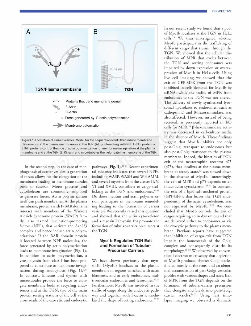
In the second step, in the case of mor-phogenesis of carrier vesicles, a generation of forces allows for the elongation of the membrane leading to membrane tubules prior to scission. Motor proteins and cytoskeleton are commonly employed to generate forces. Actin polymerization itself can push membranes. At the plasma membrane, proteins with F-BAR domains interact with members of the Wiskott Aldrich Syndrome protein (WASP) fam-ily, also named nucleation-promoting factors (NPF), that activate the Arp2/3 complex and hence induce actin polym-erization.5 If the BAR domain protein is located between NPF molecules, the force generated by actin polymerization leads to membrane invagination (Fig. 1). In addition to actin polymerization, a yeast myosin from class I has been pro-posed to contribute to membrane invagi-nation during endocytosis (Fig. 1).9,10 In contrast, kinesins and dynein with microtubules provide the force to elon-gate membrane buds at recycling endo-somes and at the TGN, two of the main protein sorting stations of the cell at the cross roads of the exocytic and endocytic
pathways (Fig. 1).11-13 Recent experimen-tal evidence indicates that several NPFs, including WASP, WASH and WHAMM, and several myosins from the classes II, V, VI and XVIII, contribute to cargo traf-ficking at the TGN and endosomes.14-19 Do these myosins and actin polymeriza-tion participate in membrane remodel-ing leading to the formation of carrier vesicles? We recently raised this question and showed that the actin cytoskeleton and a myosin I (myosin 1b) promote the formation of tubular-carrier precursors at the TGN.
Myo1b Regulates TGN Exit and Formation of Tubular-
Precursor Carriers
We have shown previously that myo-sin1b (Myo1b) localizes at the plasma membrane in regions enriched with actin filaments, and at early endosomes, mul-tivesicular endosomes and lysosomes.20,21 Furthermore, Myo1b was involved in the traffic of cargo along the endocytic path-way and together with F-actin it modu-lated the shape of sorting endosomes.20,22
In our recent study we found that a pool of Myo1b localizes at the TGN in HeLa cells.23 We thus investigated whether Myo1b participates in the trafficking of different cargo that transit through the TGN. We showed that the cellular dis-tribution of MPR that cycles between the TGN and sorting endosomes was impaired by down expression or overex-pression of Myo1b in HeLa cells. Using live cell imaging we showed that the exit of GFP-MPR from the TGN was inhibited in cells depleted for Myo1b by siRNA, while the traffic of MPR from endosomes to the TGN was not altered. The delivery of newly synthesized lyso-somal hydrolases to endosomes, such as cathepsin D and β-hexosaminidase, was also affected. However, instead of being secreted, as previously reported in KO cells for MPR,24 β-hexosaminidase activ-ity was decreased in cell-culture media in the absence of Myo1b. These findings suggest that Myo1b inhibits not only post-Golgi transport to endosomes but also post-Golgi transport to the plasma membrane. Indeed, the kinetics of TGN exit of the neurotrophin receptor p75 (p75), that localizes at the plasma mem-brane at steady-state,12 was slowed down in the absence of Myo1b. Interestingly, the exit of MPR and p75 depends on an intact actin cytoskeleton.25-27 In contrast, the exit of a lipid-raft anchored protein (GPI-GFP), that leaves the TGN inde-pendently of the actin cytoskeleton, was not regulated by Myo1b.26,27 We con-cluded that Myo1b controls the exit of cargos requiring actin dynamics and that are delivered either to endosomes or via the exocytic pathway to the plasma mem-brane. Previous reports have suggested that inhibition of cargo exit from TGN impairs the homeostasis of the Golgi complex and consequently disturbs its morphology.28-30 We observed by conven-tional electron microscopy that depletion of Myo1b produced shorter Golgi stacks, dilated mostly at the rims, and an abnor-mal accumulation of peri-Golgi vesicular profiles with various shapes and sizes. Exit of MPR from the TGN depends on the formation of tubular-carrier precursors that elongate and break into post-Golgi carrier vesicles.14,31 Using fast time-lapse imaging we observed a dramatic
Figure 1. Formation of carrier vesicles. model for the sequential events that induce membrane deformation at the plasma membrane or at the tgn. (A) By interacting with npF, F-BAR protein or Etnh proteins control the side of actin polymerization for membrane invagination at the plasma membrane and at the tgn. (B) Kinesin and microtubules then elongate the membrane of the tgn.

© 2012 Landes Bioscience.
Do not distrib
ute.
232 BioArchitecture Volume 1 issue 5
reduction of tubular-carrier precursors and cytoplasmic carrier vesicles in absence of Myo1b. Conversely, overexpression of Cherry-Myo1b induced the formation of long GFP-MPR tubular-precursors. To determine whether the force generated by Myo1b triggers cargo exit from the TGN and could be at the origin of membrane deformation, we analyzed whether Myo1b rigor mutant (N160A) with a single muta-tion in the motor domain and a Myo1b mutant (E476K) with a single mutation in the “switch 2” of the motor domain, both being unable to generate force to move F-actin in vitro, could rescue the normal distribution of MPR upon endog-enous Myo1b KD. We found that expres-sion of recombinant wild-type Myo1b, but not Myo1b-rigor or Myo1b-E476K, rescued the normal steady-state distribu-tion of MPR. We concluded that Myo1b regulates the exit of cargo from the TGN by generating a force that could contrib-ute to the formation of tubular-precursor carriers.
Myo1b Controls the Assembly of F-Actin Foci at the TGN
Growing evidence indicates that the actin cytoskeleton plays a role in membrane trafficking at the TGN.14,25,28,29 and that actin foci localizes in the TGN region.28,32
However, the role of actin dynamics at an early stage of post-Golgi carrier biogen-esis, such as formation of tubular-carrier precursors, was unknown. By monitoring the behavior of F-actin foci at the TGN in cells expressing Cherry-LifeAct and GFP-MPR, we observed that F-actin foci remained relatively non motile and stable at the opposite of the behavior of F-actin at the plasma membrane where a burst of actin occurs during the for-mation of coated pits to enable vesicle formation and ligand uptake.33 Thirty percent of F-actin foci codistributed with MPR associated to the TGN and F-actin foci were in close proximity to the TGN membrane when MPR tubules formed, suggesting that F-actin foci interact with the TGN membrane. We showed that F-actin foci are required for the formation of tubular-carrier precursors emanating from the TGN and give rise to the forma-tion of post-Golgi carriers. The Arp2/3
complex has been proposed to nucleate the actin filaments that compose F-actin foci at the TGN membrane.34 The reduc-tion of F-actin foci in the TGN region, upon Arp2/3 complex KD by siRNA, phenocopied the accumulation of MPR in the TGN, as seen in the Myo1b KD. Furthermore, KD of the Arp2/3 complex reduced the formation of tubular-carrier precursors and consequently the num-ber of cytoplasmic MPR carrier vesicles. Myosins generate force by interacting in an ATP dependant manner with F-actin filaments. We found that Myo1b was in association with F-actin foci in the TGN region and that its depletion reduced the number of F-actin foci at the TGN by 60%. Myo1b depletion also decreased the number of Arp2/3 structures in the vol-ume occupied by MPR in the TGN area,
but not at the ventral plasma membrane. While Myo1b overexpression increased the number of Arp2/3 structures in the Golgi region with no significant increase at the ventral plasma membrane.
Together, these findings allowed us to propose that Myo1b tethers F-actin foci to the TGN membrane. Recently, Kobama and colleagues reported the ability of Myo1b, to bind to phosphoinositides.35 As Myo1b contains, in its tail domain, a cluster of basic amino acids as well as a PH-like motif, Myo1b could bind the TGN membrane via its PH motif. Similar to the other members of the subgroup of short tail myosins 1, Myo1b does not con-tain a second actin binding domain or a protein-protein interacting motif, such as a single Src homology domain 3 (SH3), that can bind proteins involved in actin
Figure 2. model for the role of myo1b in membrane remodeling. myo1b moves toward the plus end of F-actin. if myo1b is immobilized because of its interaction with the membrane then the myo1b motor activity will move F-actin backward thereby facilitating addition of new g actin next to the plasma membrane. (A) Shows the orientation of myo1b movement (see blue arrows) and (B) the consequence of this movement on actin dynamics (see gray arrows and new addition of g-actin).

© 2012 Landes Bioscience.
Do not distrib
ute.
www.landesbioscience.com BioArchitecture 233
polymerization.36 However, another mem-ber of the short tail myosin 1 subgroup, myosin 1c has been proposed to spatially control actin assembly to the plasma mem-brane in Xenopus Oocytes via its motor domain.37 Myo1b, similarly to Myo1c, could spatially control actin assembly at the TGN membrane by interacting with F-actin via its motor domain and at the membrane via its PH motif. In agreement with this hypothesis, we found that the force generated by Myo1b was required to tether F-actin foci to the TGN mem-brane. Using an in vitro motility assay, we observed that the rigor mutant remained bound to F-actin while the E476K mutant displayed a weak affinity for F-actin. Myo1b, but not Myo1b-rigor or Myo1b-E476K, rescued the normal steady-state distribution of F-actin foci at the TGN.
Our findings can be summarized in a model where the force generated by Myo1b controls F-actin foci orientation and binding to the TGN membrane. The force generated by Myo1b could thereby facilitate actin polymerization and con-tribute to membrane curvature of the TGN (Fig. 2). According to this model, Myo1b-Tail interactions with the TGN membrane should be strong enough to avoid Myo1b-Tail slipping and Myo1b dis-sociation in the TGN membrane during the force produced by Myo1b-motor on F-actin. Further work needs to be done to test this hypothesis.
Is Myo1b Function Coordinated with Actin Nucleation?
The Arp2/3 complex can be activated and recruited to the TGN through the inter-action of clathrin coat with Cyfip1/2, triggering WAVE/N-WASP actin nucle-ation.14 However, this mechanism can-not account for the actin dependent exit of p75 since this receptor exits the TGN independently of clathrin.38 ARF1 (ADP-ribosylation factor 1) recruits COPI and clathrin coat complexes on Golgi sub-domains and also activates the Arp2/3 complex via cdc42 and N-WASP.39-41 Furthermore, expression of an active cdc42 mutant activates the TGN exit of p75 (ref. 42). In addition, several NPFs, including WASP and WHAMM, con-tribute to the trafficking of cargo at the
Golgi and TGN.14,17 These data, together, highlights the existence of multiple TGN exit pathways with distinct mechanisms to recruit and activate the Arp2/3 com-plex at the TGN membrane. It is likely that Myo1b functions in these different pathways by tethering the newly polym-erized F-Actin to the TGN membrane. Furthermore, we have previously reported that Myo1b participates in membrane traf-ficking along the endocytic pathway and in particular to the transfer of cargo from the membrane of the sorting endosomes to the internal vesicles.22 We observed that over-expression of Myo1b increases membrane extensions at the surface of these endo-somes. Conversely, we recently observed that Myo1b KD decreases the formation of such extensions (Almeida CG, unpub-lished data). Thus, Myo1b may function to tether newly polymerized F-actin to the membranes of different types of organelles and thereby trigger membrane remodeling of organelles.
Do Other Myosins Implicated in Membrane Traffic Contribute
to Membrane Remodeling of Organelles?
Myosins from classes I and II could con-tribute to organelle and plasma mem-brane remodeling. Yeast myosins 1, with a long tail that interacts with NPF via an acidic motif at its C-terminus, were the first myosins reported to be involved in membrane remodeling.9,10 More recently, another long tail myosin 1, myosin 1e, that could interact with NSF via its SH3 domain, has been suggested to play a similar role in mammalian cells.43,44 It has been proposed that these myosins elon-gate the plasma membrane and/or cleave the newly-formed membrane invagina-tion leading to the endosomes.10 A recent report showed that depletion of myosin II inhibits scission of tubular-carrier pre-cursors positive for Rab6.16 We observed that inhibition of the ATPase activity of this myosin inhibits the scission of MPR tubules. This suggests that myosin II con-tributes to membrane remodeling leading to scission of tubular carrier precursors downstream of Myo1b function at the TGN (Almeida CG, unpublished data). Whether this myosin contributes directly
to the scission or whether it acts in coor-dination with dynamin remains to be clarified.
Recent experimental evidence indicates that members of other classes of myosins contribute to membrane trafficking along both the endocytic and exocytic pathways and suggests that these myosins function by using distinct molecular mechanisms in distinct pathways. For example, myo-sin VI has been found to be involved in the first step of endocytosis as well as in the endocytic recycling pathway and more recently in the fusion of secretory vesicles to the plasma membrane.19,45-47 Although depletion of myosin VI reduces tubule formation in the endocytic recycling path-way, its direct role in membrane remodel-ing for these different steps of membrane trafficking needs to be further examined.19 Myosins V contribute to endocytic recy-cling of a variety of receptors and cargos. Yet, although the isoform Vb is associ-ated with tubular networks, there is no evidence indicating that myosins V can remodel membrane of the recycling endo-somes.48-50 It is likely that myosin 18a par-ticipates in retrograde trafficking since its membrane receptor, GOLPH3, has been shown to contribute to retrograde Golgi trafficking in yeast and to be associated with the retromer complex in mammalian cells.51 Myosin 18a depletion inhibits the frequency of formation of Golgi carriers. However, due to the question raised by the sequence of its motor domain, it is not clear whether this myosin can generate a force to remodel membrane. Myosin 18a presents a non-conserved residue in the “switch 2” region of the motor domain that abolishes cellular function of myosin II in dictyostellium and instead, myosin 18a presents an actin-binding site in an unusual PDZ domain at its N-terminus in order to have an ATP dependent actin-binding site.52 Further work needs to be done to determine whether these myosins contribute to membrane remodeling of organelles by generating, with F-actin, a force on organelle membrane.
In conclusion, our recent work with that of Miserey-Lenkei and colleagues, pro-vides two examples of myosins involved in membrane trafficking that control mem-brane remodeling of organelles.16,23 These new results provide an emerging concept

© 2012 Landes Bioscience.
Do not distrib
ute.
234 BioArchitecture Volume 1 issue 5
that the force generated by myosins is part of the machinery implicated in membrane elongation and/or membrane scission, two steps of membrane remodeling that lead to the formation of carrier vesicles. Furthermore, they suggest that the func-tion of some of these myosins needs to be coordinated to achieve the formation of carrier vesicles.
Acknowledgments
This work has been supported by the Institut Curie, the CNRS and the Agence Nationale pour la Recherche (grant ANR 09-BLAN-0027). C.G.A. has been the recipient of an EMBO long-term fellow-ship (ALTF 607-2006) and a Marie Curie action intra-European fellowship for career development (FP7-PEOPLE-2007–2-1-IEF N. 2200088).
References1. McMahon HT, Gallop JL. Membrane curvature and
mechanisms of dynamic cell membrane remodelling. Nature 2005; 438:590-6; PMID:16319878; http://dx.doi.org/10.1038/nature04396
2. Legendre-Guillemin V, Wasiak S, Hussain NK, Angers A, McPherson PS. ENTH/ANTH proteins and clathrin-mediated membrane budding. J Cell Sci 2004; 117:9-18; PMID:14657269; http://dx.doi.org/10.1242/jcs.00928
3. Peter BJ, Kent HM, Mills IG, Vallis Y, Butler PJ, Evans PR, et al. BAR domains as sensors of mem-brane curvature: the amphiphysin BAR structure. Science 2004; 303:495-9; PMID:14645856; http://dx.doi.org/10.1126/science.1092586
4. Suetsugu S, Toyooka K, Senju Y. Subcellular mem-brane curvature mediated by the BAR domain super-family proteins. Semin Cell Dev Biol 2010; 21:340-9; PMID:19963073; http://dx.doi.org/10.1016/j.semcdb.2009.12.002
5. Fricke R, Gohl C, Bogdan S. The F-BAR protein family Actin’ on the membrane. Commun Integr Biol 2010; 3:89-94; PMID:20585497; http://dx.doi.org/10.4161/cib.3.2.10521
6. Zhao H, Pykalainen A, Lappalainen P. I-BAR domain proteins: linking actin and plasma mem-brane dynamics. Curr Opin Cell Biol 2011; 23:14-21; PMID:21093245; http://dx.doi.org/10.1016/j.ceb.2010.10.005
7. Anitei M, Hoflack B. Exit from the trans-Golgi network: from molecules to mechanisms. Curr Opin Cell Biol 2011; 23:443-51; PMID:21550789; http://dx.doi.org/10.1016/j.ceb.2011.03.013
8. Henne WM, Boucrot E, Meinecke M, Evergren E, Vallis Y, Mittal R, et al. FCHo proteins are nucleators of clathrin-mediated endocytosis. Science 2010; 328:1281-4; PMID:20448150; http://dx.doi.org/10.1126/science.1188462
9. Geli MI, Lombardi R, Schmelzl B, Riezman H. An intact SH3 domain is required for myosin I-induced actin polymerization. EMBO J 2000; 19:4281-91; PMID:10944111; http://dx.doi.org/10.1093/emboj/19.16.4281
10. Idrissi FZ, Grotsch H, Fernandez-Golbano IM, Presciatto-Baschong C, Riezman H, Geli MI. Distinct acto/myosin-I structures associate with endocytic profiles at the plasma membrane. J Cell Biol 2008; 180:1219-32; PMID:18347067
11. Delevoye C, Hurbain I, Tenza D, Sibarita JB, Uzan-Gafsou S, Ohno H, et al. AP-1 and KIF13A coor-dinate endosomal sorting and positioning during melanosome biogenesis. J Cell Biol 2009; 187:247-64; PMID:19841138; http://dx.doi.org/10.1083/jcb.200907122
12. Kreitzer G, Marmorstein A, Okamoto P, Vallee R, Rodriguez-Boulan E. Kinesin and dynamin are required for post-Golgi transport of a plas-ma-membrane protein. Nat Cell Biol 2000; 2 :125-7; PMID:10655593; http://dx.doi.org/10.1038/35000081
13. Roux A, Cappello G, Cartaud J, Prost J, Goud B, Bassereau P. A minimal system allowing tubula-tion with molecular motors pulling on giant lipo-somes. Proc Natl Acad Sci USA 2002; 99:5394-9; PMID:11959994; http://dx.doi.org/10.1073/pnas.082107299
14. Anitei M, Stange C, Parshina I, Baust T, Schenck A, Raposo G, et al. Protein complexes contain-ing CYFIP/Sra/PIR121 coordinate Arf1 and Rac1 signalling during clathrin-AP-1-coated carrier bio-genesis at the TGN. Nat Cell Biol 2010; 12:330-40; PMID:20228810
15. Derivery E, Sousa C, Gautier JJ, Lombard B, Loew D, Gautreau A. The Arp2/3 activator WASH controls the fission of endosomes through a large multiprotein complex. Dev Cell 2009; 17:712-23; PMID:19922875; http://dx.doi.org/10.1016/j.dev-cel.2009.09.010
16. Miserey-Lenkei S, Chalancon G, Bardin S, Formstecher E, Goud B, Echard A. Rab and acto-myosin-dependent fission of transport vesicles at the Golgi complex. Nat Cell Biol 2010; 12:645-54; PMID:20562865; http://dx.doi.org/10.1038/ncb2067
17. Dippold HC, Ng MM, Farber-Katz SE, Lee SK, Kerr ML, Peterman MC, et al. GOLPH3 bridges phosphatidylinositol-4- phosphate and actomyosin to stretch and shape the Golgi to promote budding. Cell 2009; 139:337-51; PMID:19837035; http://dx.doi.org/10.1016/j.cell.2009.07.052
18. Au JS, Puri C, Ihrke G, Kendrick-Jones J, Buss F. Myosin VI is required for sorting of AP-1B-dependent cargo to the basolateral domain in polarized MDCK cells. J Cell Biol 2007; 177:103-14; PMID:17403927; http://dx.doi.org/10.1083/jcb.200608126
19. Chibalina MV, Seaman MN, Miller CC, Kendrick-Jones J, Buss F. Myosin VI and its interacting protein LMTK2 regulate tubule formation and transport to the endocytic recycling compartment. J Cell Sci 2007; 120:4278-88; PMID:18029400
20. Raposo G, Cordonnier MN, Tenza D, Menichi B, Durrbach A, Louvard D, et al. Association of myosin I alpha with endosomes and lysosomes in mammalian cells. Mol Biol Cell 1999; 10:1477-94; PMID:10233157
21. Cordonnier MN, Dauzonne D, Louvard D, Coudrier E. Actin filaments and myosin I alpha cooperate with microtubules for the movement of lysosomes. Mol Biol Cell 2001; 12:4013-29; PMID:11739797
22. Salas-Cortes L, Ye F, Tenza D, Wilhelm C, Theos A, Louvard D, et al. Myosin Ib modulates the morphol-ogy and the protein transport within multi-vesicular sorting endosomes. J Cell Sci 2005; 118:4823-32; PMID:16219689; http://dx.doi.org/10.1242/jcs.02607
23. Almeida CG, Yamada A, Tenza D, Louvard D, Raposo G, Coudrier E. Myosin 1b promotes the formation of post-Golgi carriers by regulating actin assembly and membrane remodelling at the trans-Golgi network. Nat Cell Biol 2011; 13:779-89; PMID:21666684; http://dx.doi.org/10.1038/ncb2262
24. Ludwig T, Munier-Lehmann H, Bauer U, Hollinshead M, Ovitt C, Lobel P, et al. Differential sorting of lysosomal enzymes in mannose 6-phos-phate receptor-deficient fibroblasts. EMBO J 1994; 13:3430-7; PMID:8062819
25. Cao H, Weller S, Orth JD, Chen J, Huang B, Chen JL, et al. Actin and Arf1-dependent recruitment of a cortactin-dynamin complex to the Golgi regulates post-Golgi transport. Nat Cell Biol 2005; 7:483-92; PMID:15821732; http://dx.doi.org/10.1038/ncb1246
26. Lázaro-Diéguez F, Colonna C, Cortegano M, Calvo M, Martinez SE, Egea G. Variable actin dynamics requirement for the exit of different cargo from the trans-Golgi network. FEBS Lett 2007; 581:3875-81; PMID:17651738; http://dx.doi.org/10.1016/j.febs-let.2007.07.015
27. Salvarezza SB, Deborde S, Schreiner R, Campagne F, Kessels MM, Qualmann B, et al. LIM kinase 1 and cofilin regulate actin filament population required for dynamin-dependent apical carrier fission from the trans-Golgi network. Mol Biol Cell 2009; 20:438-51; PMID:18987335; http://dx.doi.org/10.1091/mbc.E08-08-0891
28. Carreno S, Engqvist-Goldstein AE, Zhang CX, McDonald KL, Drubin DG. Actin dynamics cou-pled to clathrin-coated vesicle formation at the trans-Golgi network. J Cell Biol 2004; 165:781-8; PMID:15210728; http://dx.doi.org/10.1083/jcb.200403120
29. Poupon V, Girard M, Legendre-Guillemin V, Thomas S, Bourbonniere L, Philie J, et al. Clathrin light chains function in mannose phosphate receptor trafficking via regulation of actin assembly. Proc Natl Acad Sci USA 2008; 105:168-73; PMID:18165318; http://dx.doi.org/10.1073/pnas.0707269105
30. von Blume J, Duran JM, Forlanelli E, Alleaume AM, Egorov M, Polishchuk R, et al. Actin remodeling by ADF/cofilin is required for cargo sorting at the trans-Golgi network. J Cell Biol 2009; 187:1055-69; PMID:20026655; http://dx.doi.org/10.1083/jcb.200908040
31. Waguri S, Dewitte F, Le Borgne R, Rouille Y, Uchiyama Y, Dubremetz JF, et al. Visualization of TGN to endosome trafficking through fluorescently labeled MPR and AP-1 in living cells. Mol Biol Cell 2003; 14:142-55; PMID:12529433; http://dx.doi.org/10.1091/mbc.E02-06-0338
32. Percival JM, Hughes JA, Brown DL, Schevzov G, Heimann K, Vrhovski B, et al. Targeting of a tropo-myosin isoform to short microfilaments associated with the Golgi complex. Mol Biol Cell 2004; 15:268-80; PMID:14528022; http://dx.doi.org/10.1091/mbc.E03-03-0176
33. Kaksonen M, Toret CP, Drubin DG. Harnessing actin dynamics for clathrin-mediated endocytosis. Nat Rev Mol Cell Biol 2006; 7:404-14; PMID:16723976; http://dx.doi.org/10.1038/nrm1940
34. Campellone KG, Webb NJ, Znameroski EA, Welch MD. WHAMM is an Arp2/3 complex activator that binds microtubules and functions in ER to Golgi transport. Cell 2008; 134:148-61; PMID:18614018; http://dx.doi.org/10.1016/j.cell.2008.05.032
35. Komaba S, Coluccio LM. Localization of myo-sin 1b to actin protrusions requires phosphoinosit-ide binding. J Biol Chem 2010; 285:27686-93; PMID:20610386; http://dx.doi.org/10.1074/jbc.M109.087270
36. Loubéry S, Coudrier E. Myosins in the secretory pathway: tethers or transporters? Cell Mol Life Sci 2008; 65:2790-800; PMID:18726179
37. Sokac AM, Schietroma C, Gundersen CB, Bement WM. Myosin-1c couples assembling actin to mem-branes to drive compensatory endocytosis. Dev Cell 2006; 11:629-40; PMID:17084356; http://dx.doi.org/10.1016/j.devcel.2006.09.002
38. Deborde S, Perret E, Gravotta D, Deora A, Salvarezza S, Schreiner R, et al. Clathrin is a key regula-tor of basolateral polarity. Nature 2008; 452:719-23; PMID:18401403; http://dx.doi.org/10.1038/nature06828

© 2012 Landes Bioscience.
Do not distrib
ute.
www.landesbioscience.com BioArchitecture 235
39. D’Souza-Schorey C, Chavrier P. ARF proteins: roles in membrane traffic and beyond. Nat Rev Mol Cell Biol 2006; 7:347-58; PMID:16633337; http://dx.doi.org/10.1038/nrm1910
40. Heuvingh J, Franco M, Chavrier P, Sykes C. ARF1-mediated actin polymerization produces movement of artificial vesicles. Proc Natl Acad Sci USA 2007; 104:16928-33; PMID:17942688; http://dx.doi.org/10.1073/pnas.0704749104
41. Matas OB, Martinez-Menarguez JA, Egea G. Association of Cdc42/N-WASP/Arp2/3 signal-ing pathway with Golgi membranes. Traffic 2004; 5:838-46; PMID:15479449; http://dx.doi.org/10.1111/j.1600-0854.2004.00225.x
42. Müsch A, Cohen D, Kreitzer G, Rodriguez-Boulan E. cdc42 regulates the exit of apical and basolat-eral proteins from the trans-Golgi network. EMBO J 2001; 20:2171-9; PMID:11331583; http://dx.doi.org/10.1093/emboj/20.9.2171
43. Krendel M, Osterweil EK, Mooseker MS. Myosin 1E interacts with synaptojanin-1 and dynamin and is involved in endocytosis. FEBS Lett 2007; 581:644-50; PMID:17257598; http://dx.doi.org/10.1016/j.febslet.2007.01.021
44. Jung G, Remmert K, Wu X, Volosky JM, Hammer JA 3rd. The Dictyostelium CARMIL protein links capping protein and the Arp2/3 complex to type I myosins through their SH3 domains. J Cell Biol 2001; 153:1479-97; PMID:11425877; http://dx.doi.org/10.1083/jcb.153.7.1479
45. Morris SM, Arden SD, Roberts RC, Kendrick-Jones J, Cooper JA, Luzio JP, et al. Myosin VI binds to and localises with Dab2, potentially linking receptor-mediated endocytosis and the actin cytoskeleton. Traffic 2002; 3:331-41; PMID:11967127; http://dx.doi.org/10.1034/j.1600-0854.2002.30503.x
46. Spudich G, Chibalina MV, Au JS, Arden SD, Buss F, Kendrick-Jones J. Myosin VI targeting to clathrin-coated structures and dimerization is mediated by binding to Disabled-2 and PtdIns(4,5)P(2). Nat Cell Biol 2007; 9:176-83; PMID:17187061; http://dx.doi.org/10.1038/ncb1531
47. Bond LM, Peden AA, Kendrick-Jones J, Sellers JR, Buss F. Myosin VI and its binding partner opti-neurin are involved in secretory vesicle fusion at the plasma membrane. Mol Biol Cell 2011; 22:54-65; PMID:21148290; http://dx.doi.org/10.1091/mbc.E10-06-0553
48. Röder IV, Choi KR, Reischl M, Petersen Y, Diefenbacher ME, Zaccolo M, et al. Myosin Va cooperates with PKA RIalpha to mediate mainte-nance of the endplate in vivo. Proc Natl Acad Sci USA 2010; 107:2031-6; PMID:20133847; http://dx.doi.org/10.1073/pnas.0914087107
49. Provance DW Jr., Addison EJ, Wood PR, Chen DZ, Silan CM, Mercer JA. Myosin-Vb functions as a dynamic tether for peripheral endocytic compart-ments during transferrin trafficking. BMC Cell Biol 2008; 9:44; PMID:18687135; http://dx.doi.org/10.1186/1471-2121-9-44
50. Roland JT, Kenworthy AK, Peranen J, Caplan S, Goldenring JR. Myosin Vb interacts with Rab8a on a tubular network containing EHD1 and EHD3. Mol Biol Cell 2007; 18:2828-37; PMID:17507647; http://dx.doi.org/10.1091/mbc.E07-02-0169
51. Bugarcic A, Zhe Y, Kerr MC, Griffin J, Collins BM, Teasdale RD. VPS26A and VPS26B subunits define distinct Retromer complexes. Traffic 2011; 12:1759-73; PMID:21920005
52. Isogawa Y, Kon T, Inoue T, Ohkura R, Yamakawa H, Ohara O, et al. The N-terminal domain of MYO18A has an ATP-insensitive actin-binding site. Biochemistry 2005; 44:6190-6; PMID:15835906; http://dx.doi.org/10.1021/bi0475931
Copyright © 2022 FDOKUMEN