SKAP2, a novel target of HSF4b, associates with NCK2/F-actin at membrane ruffles and regulates actin...
13
SKAP2, a novel target of HSF4b, associates with NCK2/F-actin at membrane ruffles and regulates actin reorganization in lens cell Li Zhou a , Zhenguo Zhang a , Yufang Zheng b , Yufei Zhu a , Zejun Wei a , Heng Xu a , Quan Tang a , Xiangyin Kong a, c , Landian Hu a, * a The Key Laboratory of Stem Cell Biology, Institute of Health Sciences, Shanghai Institutes for Biological Sciences (SIBS), Chinese Academy of Sciences (CAS) & Shanghai Jiao Tong University School of Medicine (SJTUSM), Shanghai, People’s Republic of China b Department of Physiology and Biophysics, School of Life Sciences, Fudan University, Shanghai, People’s Republic of China c State Key Laboratory of Medical Genomics, Ruijin Hospital, Shanghai Jiaotong University, Shanghai, People’s Republic of China Received: November 18, 2009; Accepted: February 23, 2010 Abstract In addition to roles in stress response, heat shock factors (HSFs) play crucial roles in differentiation and development. Heat shock tran- scription factor 4 (HSF4) deficiency leads to defect in lens epithelial cell (LEC) differentiation and cataract formation. However, the mech- anism remains obscure. Here, we identified Src kinase-associated phosphoprotein 2 (SKAP2) as a downstream target of HSF4b and it was highly expressed at the anterior tip of lens elongating fibre cells in vivo. The HSF4-deficient lenses showed reduced SKAP2 expression and defects in actin reorganization. The disassembly of stress fibres and formation of cortical actin fibres are critical for the initiation of LEC differentiation. SKAP2 localized at actin-rich ruffles in human LECs (SRA01/04 cells) and knockdown SKAP2 using RNA interference impaired the disassembly of cellular stress fibres in response to fibroblast growth factor (FGF)-b. Overexpression of SKAP2, but not the N-terminal deletion mutant of SKAP2, induced the actin remodelling. We further found that SKAP2 interacted with the SH2 domain of non- catalytic region of tyrosine kinase adaptor protein 2 (NCK2) via its N-terminus. The complex of SKAP2-NCK2-F-actin accumulated at the leading edge of the lamellipodium, where FGF receptors and focal adhesion were also recruited. These results revealed an essential role for HSF4-mediated SKAP2 expression in the regulation of actin reorganization during lens differentiation, likely through a mechanism that SKAP2 anchors the complex of NCK2/focal adhesion to FGF receptors at the lamellipodium in lens epithelial cells. Keywords: lens cell differentiation • actin reorganization • SKAP2 • NCK2 • HSF4b J. Cell. Mol. Med. Vol 15, No 4, 2011 pp. 783-795 © 2011 The Authors Journal of Cellular and Molecular Medicine © 2011 Foundation for Cellular and Molecular Medicine/Blackwell Publishing Ltd doi: 10.1111/j.1582-4934.2010.01048.x Introduction The lens starts development during the embryonic stage and con- tinues to grow after birth, with the new secondary fibres being added from the outer mono-layer epithelium. Lens epithelial cells (LECs) start to differentiate, elongate, express lens fibre cell spe- cific genes, lose contact with the capsule and epithelium, and finally lose their organelles as they become mature lens fibres [1]. Various signalling proteins are involved in this process, including fibroblast growth factor (FGF)-b, insulin-like growth factor (IGF)-1, transforming growth factor-, N-cadherin and integrin [2–4]. Actin reorganization, which is profoundly affected by those sig- nals, is essential for the differentiation induction and survival of LECs [5], and precise LEC differentiation is important for the transparency of the lens. Previously we have reported that a DNA-binding domain mutant allele of heat shock factor (HSF)4 is associated with auto- somal dominant lamellar and Marner cataracts [6]. Then, it has been reported that HSF4 knockout mice have defects in LECs dif- ferentiation and form cataracts [7, 8]. HSF4 belongs to the tran- scriptional regulator family of heat shock proteins (HSPs), and it binds to the heat shock element (HSE) that is composed of consensus inverted repeats of nGAAn [9]. Apart from the HSP *Correspondence to: Landian HU, IHS Bldg 5, 225 Chongqing Rd, Shanghai 200025, People’s Republic of China. Tel.: 86-21-63852639 Fax: 86-21-63844476 E-mail: [email protected]
Transcript of SKAP2, a novel target of HSF4b, associates with NCK2/F-actin at membrane ruffles and regulates actin...
SKAP2, a novel target of HSF4b, associates with
NCK2/F-actin at membrane ruffles and regulates
actin reorganization in lens cell
Li Zhou a, Zhenguo Zhang a, Yufang Zheng b, Yufei Zhu a, Zejun Wei a, Heng Xu a, Quan Tang a, Xiangyin Kong a, c, Landian Hu a, *
a The Key Laboratory of Stem Cell Biology, Institute of Health Sciences, Shanghai Institutes for Biological Sciences (SIBS),Chinese Academy of Sciences (CAS) & Shanghai Jiao Tong University School of Medicine (SJTUSM),
Shanghai, People’s Republic of Chinab Department of Physiology and Biophysics, School of Life Sciences, Fudan University, Shanghai, People’s Republic of China
c State Key Laboratory of Medical Genomics, Ruijin Hospital, Shanghai Jiaotong University, Shanghai, People’s Republic of China
Received: November 18, 2009; Accepted: February 23, 2010
Abstract
In addition to roles in stress response, heat shock factors (HSFs) play crucial roles in differentiation and development. Heat shock tran-scription factor 4 (HSF4) deficiency leads to defect in lens epithelial cell (LEC) differentiation and cataract formation. However, the mech-anism remains obscure. Here, we identified Src kinase-associated phosphoprotein 2 (SKAP2) as a downstream target of HSF4b and it washighly expressed at the anterior tip of lens elongating fibre cells in vivo. The HSF4-deficient lenses showed reduced SKAP2 expressionand defects in actin reorganization. The disassembly of stress fibres and formation of cortical actin fibres are critical for the initiation ofLEC differentiation. SKAP2 localized at actin-rich ruffles in human LECs (SRA01/04 cells) and knockdown SKAP2 using RNA interferenceimpaired the disassembly of cellular stress fibres in response to fibroblast growth factor (FGF)-b. Overexpression of SKAP2, but not theN-terminal deletion mutant of SKAP2, induced the actin remodelling. We further found that SKAP2 interacted with the SH2 domain of non-catalytic region of tyrosine kinase adaptor protein 2 (NCK2) via its N-terminus. The complex of SKAP2-NCK2-F-actin accumulated at theleading edge of the lamellipodium, where FGF receptors and focal adhesion were also recruited. These results revealed an essential rolefor HSF4-mediated SKAP2 expression in the regulation of actin reorganization during lens differentiation, likely through a mechanism thatSKAP2 anchors the complex of NCK2/focal adhesion to FGF receptors at the lamellipodium in lens epithelial cells.
Keywords: lens cell differentiation • actin reorganization • SKAP2 • NCK2 • HSF4b
J. Cell. Mol. Med. Vol 15, No 4, 2011 pp. 783-795
© 2011 The AuthorsJournal of Cellular and Molecular Medicine © 2011 Foundation for Cellular and Molecular Medicine/Blackwell Publishing Ltd
doi:10.1111/j.1582-4934.2010.01048.x
Introduction
The lens starts development during the embryonic stage and con-tinues to grow after birth, with the new secondary fibres beingadded from the outer mono-layer epithelium. Lens epithelial cells(LECs) start to differentiate, elongate, express lens fibre cell spe-cific genes, lose contact with the capsule and epithelium, andfinally lose their organelles as they become mature lens fibres [1].Various signalling proteins are involved in this process, including
fibroblast growth factor (FGF)-b, insulin-like growth factor (IGF)-1,transforming growth factor-�, N-cadherin and integrin [2–4].Actin reorganization, which is profoundly affected by those sig-nals, is essential for the differentiation induction and survival ofLECs [5], and precise LEC differentiation is important for thetransparency of the lens.
Previously we have reported that a DNA-binding domainmutant allele of heat shock factor (HSF)4 is associated with auto-somal dominant lamellar and Marner cataracts [6]. Then, it hasbeen reported that HSF4 knockout mice have defects in LECs dif-ferentiation and form cataracts [7, 8]. HSF4 belongs to the tran-scriptional regulator family of heat shock proteins (HSPs), and it binds to the heat shock element (HSE) that is composed ofconsensus inverted repeats of nGAAn [9]. Apart from the HSP
*Correspondence to: Landian HU,IHS Bldg 5, 225 Chongqing Rd, Shanghai 200025, People’s Republic of China.Tel.: �86-21-63852639Fax: �86-21-63844476E-mail: [email protected]
784
genes, HSFs may also regulate distinct, non-HSP genes.Candidate targets of HSF4b include Crygf, Fgf7, Hspb2 and Bfsp2[7, 8, 10]. There are two splicing isoforms of HSF4: HSF4a andHSF4b. However, only HSF4b is highly expressed in mouse lens.Although HSF4 knockout mice have obvious defects in lens devel-opment, the function of HSF4 in lens development remains elu-sive. To understand the role of HSF4 in lens development, it isimperative to identify its downstream targets.
In searching for the downstream targets of HSF4, we com-pared two independent sets of microarray expression data fromHSF4 knockout mice with different backgrounds (C57BL/6-129/S3and C57BL/6-129/SvJ) and found that the Src kinase-associatedphosphoprotein 2 (SKAP2) gene is down-regulated in bothHSF4�/– mouse lenses [8, 10].
SKAP2 (also called SCAP2, RA70, SKAP-HOM and SKAP55R)is a homolog of SKAP55 (encoded by Skap1). Unlike SKAP55,which is expressed exclusively in the thymus and in T lympho-cytes, SKAP2 is expressed ubiquitously [11, 12]. The proteinstructure of SKAP2 is similar to that of SKAP55, both containinga pleckstrin homology (PH) domain, which can bind to mem-brane lipids [13], and both having a carboxyl-terminal src homol-ogy3 (SH3) domain. However, SKAP2 has a unique N-terminalcoiled-coil (CC) domain and tyrosine phosphorylation sites. Theamino acid sequence of mouse SKAP2 is 90% identical to humanSKAP2, suggesting functional conservation of this protein.Previous studies on haematopoietic and immune systems haveshown that SKAP2 binds to FYB (the Fyn binding protein) via itsSH3 domain and is a substrate of Fyn kinase, which suggests arole of SKAP2 in T-cell receptor signalling similar to that ofSKAP55 [12, 14]. It has also been reported that adhesion of acti-vated B cells to fibronectin and to ICAM-1 is strongly reduced inthe SKAP2�/– mouse, implying that SKAP2 might also beinvolved in the B-cell adhesion processes by coupling the B-cellreceptor with the activation of integrin [15]. However, the mech-anism of how SKAP2 is involved in integrin adhesion remainsunclear, and much less is known for the function of SKAP2beyond the immune system. Here, our results illustrate an essen-tial role for SKAP2, a downstream target of HSF4b, in actin reor-ganization, providing a potential explanation for the cataracts for-mation in HSF4 knockout mice.
Material and methods
Cell culture and LEC differentiation induction
Cells from the human lens epithelial cell line SRA01/04 (a gift fromZhejiang University, China) were cultured at 37�C in low-glucose DMEM(Invitrogen, Carlsbad, CA, USA) supplemented with 15% FBS (GIBCO,Invitrogen, Grand Island, NY, USA) and 1� penicillin/streptomycin antibi-otics (PAA Labs, Pasching, Austria). For the in vitro differentiation assays,the cells were starved for 24 hrs in DMEM with 0.15% FBS before treat-ment with 20 ng/ml of human recombinant basic FGF-b (ProSpec-TanyTechnoGene, Rehovot, Israel) to induce differentiation [16].
Plasmid transfection and antibodies used
Full-length SKAP2 was cloned into pcDNA3.1-triHA-5� [the trihemagglutinin(HA) sequence was inserted into pcDNA3.1 (Invitrogen)], while full-length NCK2 or NCK1 from mouse lens were cloned into pcDNA3.1-myc-3� [the myc sequence was inserted into pcDNA3.1 (Invitrogen)]. The Y to Fmutation at position 75 of SKAP2 was made using a site-directed mutationkit (Sai Bai Sheng, Shanghai, China). The N-terminal 106 amino acids dele-tion mutant of SKAP2 (SKAP2�106aa) plasmid was also inserted intopcDNA3.1-triHA5�. The myc-tagged SH2 domain of NCK2 comprisesresidues 284-380. For knockdown assays using SRA01/04 cells, twoduplexes that target different regions of hSKAP2 (5’-GATCCGCAAAGGAA-GATGAGTCA GGTTCAAGAGACCTGACTCATCTTCCTTTGTTTTTTGGAAA-3�
and 5’-GATCCGCT GATGACCAACAGTTCCATTCAAGAGATGGAACTGTTGGT-CATCAGTTTTTTGGAAA-3�, referred to as shRNA #1 and shRNA #2, respec-tively) were cloned into Psilencer 3.0 (Applied Biosystems/Ambion, Austin,TX, USA). SRA01/04 cells were transfected using Lipofectamine 2000(Invitrogen) according to the manufacturer’s instructions.
Mouse anti-HA, rabbit anti-myc, rabbit anti-FAK and the monoclonalmouse anti-Phospho-fibroblast growth factor receptor (FGFR) antibodieswere from Cell Signaling Technology, Inc. (Beverly, MA, USA). The primarygoat anti-SKAP2 antibody was from Abcam (Cambridge, UK), and the rabbitanti-SKAP2 antibody was from Proteintech Group, Inc. (Chicago, IL, USA).The rabbit anti-HA antibody and the monoclonal mouse NCK antibody werefrom BD Biosciences (Franklin Lakes, NJ, USA). Phalloidin Alexa Fluor555,Alexa Fluor568-conjugated rabbit anti-goat IgG and goat anti-rabbit AlexaFluor488 IgG were from Invitrogen/Molecular Probes. The goat antimouseCY3, donkey antimouse CY5, donkey anti-rabbit CY2 and donkey anti-goatCY3 secondary antibodies were from Jackson ImmunoResearch Labs (WestGrove, PA, USA). The anti-actin antibody and Phalloidin-fluorescein isothio-cyanate (FITC) were from Sigma-Aldrich (St. Louis, MO, USA).
Preparation and treatment of primary lens culture
The primary lens cell cultures from neonatal HSF4�/� or HSF4�/� mice wereprepared as described previously [5, 17]. Briefly, lenses were isolated fromthree HSF4�/� or HSF4�/– mice at postnatal day 3, respectively. Lenes werethen trypsinized in 2� trypsin- ethylenediaminetetraacetic acid (EDTA)/PBSbuffer (GIBCO, Invitrogen) at 37�C for 5 min. and agitated. The collected lenscells were plated on 48-well dish (Greiner Bio-one, Stuttgart, Germany) andcultivated in M199 media supplemented with 20% FBS and 1� penicillin/streptomycin. The primary lens had well-spread epithelial morphology after 1 week culture. For the in vitro differentiation assays, the lens cells weretreated with 40 ng/ml of FGF-b for 36 hrs after serum starvation in M199supplemented with 0.15% FBS for 24 hrs.
Quantitative PCR
RNA from mouse lens or the SRA01/04 cells was extracted using Trizol(Invitrogen) and reverse transcribed using the MLV Transcription Kit(Invitrogen). Quantitative PCR was performed with the SYBR Green PCRkit (Applied Biosystems, Streetsville, ON, Canada) and the sequence detec-tion system (ABI 7900HT). The following primers were used: 5�-ACCAGTTTCCTC CCATTGCA-3� and 5�-CCATTCAAACCCCAGAAAGC-3�.
Chromatin immunoprecipitation
Lenses were isolated from postnatal, day-9 mice and treated as describedpreviously [7]. Briefly, after cross-linking, the lens cells were lysed in cell
© 2011 The AuthorsJournal of Cellular and Molecular Medicine © 2011 Foundation for Cellular and Molecular Medicine/Blackwell Publishing Ltd
J. Cell. Mol. Med. Vol 15, No 4, 2011
785
lysis buffer [5 mM N-2-hydroxyethylpiperazine-N-ethane-sulphonicacid(HEPES) including 85 mM KCl, 0.5% NP40, PMSF (phenylmethanesul-fonylfluoride) and protease inhibitor cocktail] and centrifuged. The precip-itated fractions were then lysed in nucleus lysis buffer (50 mM Tris-HClincluding 10 mM EDTA, 1% SDS, PMSF and protease inhibitor cocktail)and sonicated. The cross-linked chromatin fragments were immunoprecip-itated using the anti-HSF4b antibody (Santa Cruz Biotechnology, Inc.,Santa Cruz, CA, USA) or Normal IgG, following the Upstate manufacturer’sinstructions. We determined the enrichment of specific DNA sequences inthe immunoprecipitated material using PCR with the following primers: 5�-TGGGACTCCGTTA CCTTC-3�and 5�-TTGCGTAGACCTTGC TTT-3�.
Immunostaining
The cultured cells were fixed with 4% paraformaldehyde in PBS for 10 min.,permeabilized with 0.2% Triton X-100 in PBS for 10 min. and then blockedin 1% bovine serum albumin in PBS for 1 hr, all at room temperature. Thecells were then incubated overnight at 4�C with the primary antibody. Afterwashing, the cells were incubated with the secondary antibody in the darkor counterstained for polymerized actin (F-actin) using phalloidin for 1 hr atroom temperature. The nuclei were stained with 4,6-diamidino-2-phenylin-dole (DAPI), and cover slips were mounted on glass slides using theantifade mounting medium from Sigma. Images were taken using a fluores-cence microscope (ECLIPSE 80i, Nikon, Tokyo, Japan) or a confocal micro-scope (TCS sp5, Leica, Wetzlar, Germany). Quantification of the cells withactin remodelling (as visualized using a microscope) was described in [18].
Mouse eyes were isolated and fixed in 4% paraformaldehyde in PBS andembedded in paraffin (Sigma) using paraffin-embedding apparatus fromLeica (EG1140H), and 7 m serial sections were cut using a paraffin section-ing apparatus (Leica RM2126, Leica, Shanghai, China). After removal of theparaffin and retrieval of the antigen, the slices were blocked and incubatedovernight at 4�C with the primary anti-SKAP2 antibody. The slices were thenincubated with a fluorescent secondary antibody or counterstained with phal-loidin. The animal protocol has been approved by the Ethics Committee of HIS.
Co-immunoprecipitation (co-IP) and immunoblotting
The cells were lysed in ice-cold lysis buffer (KangCheng, Shanghai, China)containing a protease inhibitor cocktail (Sigma). Normalized cell lysates weremixed with the primary antibodies at 4�C overnight and 30 l of M-280 sheepanti-rabbit IgG paramagnetic Dynabeads (Dynal Biotech, Hamburg, Germany)was added following the manufacturer’s instructions. After vigorous washing,the immunoprecipitated samples were boiled for 10 min. in Laemmli buffer,separated on 4–12% NUPAGE Bis-tris gels (Invitrogen) and transferred tonitrocellulose membranes. After incubation with the HRP-conjugated sec-ondary antibody, electrochemiluminescence (ECL) detection reagents (GEHealthcare, Little Chalfont, UK) were added to the membranes. The membraneswere then exposed to photographic film. In some experiments, the membraneswere labelled with IRDye 800 (LI-COR Bioscience, Lincoln, NE, USA) and thenvisualized using the ODYSSEY infrared imaging system (LI-COR Bioscience).
Results
SKAP2 is a downstream target of HSF4b in lens cells
HSF4b is expressed mainly in the lens, and it plays an importantrole in lens development [8]. To identify its downstream target, we
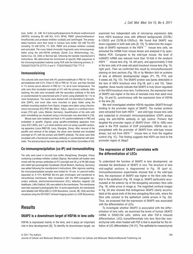
examined two independent sets of microarray expression datafrom HSF4 knockout mice with different backgrounds (C57BL/6-129/S3 and C57BL/6-129/SvJ). We found that SKAP2 wasdown-regulated in both date sets [8, 10]. To determine the magni-tude of SKAP2 repression in the HSF4�/� mouse lens cells, weextracted the mRNA from mouse lenses and analysed it by quan-titative PCR. Compared to the wild-type mouse, the level ofmSKAP2 mRNA was reduced more than 10-fold in the newbornHSF4�/� mouse lens (Fig. 1A, left part), and approximately 2-foldin the lens cells of 8-week-old adult knockout mouse lens (Fig. 1A,right part). Then we confirmed this result by immunofluorescentstaining on lens sections (Fig. 1B) or by Western blot on proteinsof lens at different developmental stages (P1, P5, P10, and 4 weeks old, Fig. 1D). The SKAP2 protein was barely detectable inthe lens of HSF4 knockout mice (Fig.1B, part c and 1D). Takentogether, these results indicate that SKAP2 is truly down-regulatedin the HSF4 knockout mice lens. Furthermore, the expression levelof SKAP2 was higher in the lens of neonatal mice than adult mice(Fig. 1A and D), synchronous with the expression time of HSF4bin the lens [8].
We next investigated whether HSF4b regulates SKAP2 throughbinding to the promoter region of SKAP2. The nuclear proteinswere extracted from both HSF4�/�and wild-type mouse lens cellsand subjected to chromatin immunoprecipitation (ChIP) assaysusing the anti-HSF4b antibody or IgG control. Primers thattargeted the promoter region of SKAP2 (from -100 to -500) wereused in the PCR reaction of ChIP precipitates. HSF4b was co-precipitated with the promoter of SKAP2 from wild-type mouselenses, but not from HSF4�/� mouse lens or from the negativecontrol (Fig. 1C). This finding suggests that HSF4 can bind to thepromoter region of Skap2.
The expression of SKAP2 correlates with the differentiation of LECs
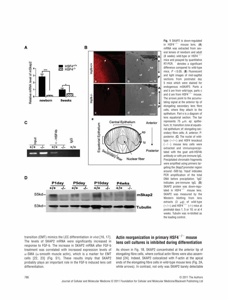
To understand the function of SKAP2 in lens development, wechecked the distribution of SKAP2 in vivo. The structure of lensmid-sagittal sections is diagrammed in Fig. 1B (part e).Immunofluorescence experiments showed that in the wild-typelens, the expression of SKAP2 was higher in the fibre cells thanthat in the epithelium (Fig. 1B, image a). SKAP2 particularly accu-mulated at the anterior tip of the elongating secondary fibre cells(Fig. 1B, white arrow in image a). The magnified confocal imagesin Fig. 2A also showed that endogenous SKAP2 clearly accumu-lated at the apical ends of the lens elongating fibre cells, where thefibre cells connect to the epithelium (white arrows in Fig. 2A).Thus, we proposed that the expression of SKAP2 was associatedwith the differentiation of LECs.
To investigate whether SKAP2 is associated with the differ-entiation of lens cells, we examined the levels of human SKAP2mRNA in SRA01/04 cells, before and after FGF-b induced differentiation. LECs transdifferentiate into lens fibre-like mes-enchymal cells when treated with FGF-b that is essential for the ini-tiation of LEC differentiation [19–21]. This epithelial-to-mesenchymal
© 2011 The AuthorsJournal of Cellular and Molecular Medicine © 2011 Foundation for Cellular and Molecular Medicine/Blackwell Publishing Ltd
786
transition (EMT) mimics the LEC differentiation in vivo [16, 17].The levels of SKAP2 mRNA were significantly increased inresponse to FGF-b. The increase in SKAP2 mRNA after FGF-btreatment was correlated with increased expression levels of -SMA (-smooth muscle actin), which is a marker for EMTcells [22, 23] (Fig. S1). These results imply that SKAP2 probably plays an important role in the FGF-b induced lens celldifferentiation.
Actin reorganization in primary HSF4�/� mouselens cell cultures is inhibited during differentiation
As shown in Fig. 1B, SKAP2 concentrated at the anterior tip ofelongating fibre cells, where cortical actin fibres were also assem-bled [24]. Indeed, SKAP2 colocalized with F-actin at the apicalends of the elongating fibre cells in wild-type mouse lens (Fig. 2A,white arrows). In contrast, not only was SKAP2 barely detectable
© 2011 The AuthorsJournal of Cellular and Molecular Medicine © 2011 Foundation for Cellular and Molecular Medicine/Blackwell Publishing Ltd
Fig. 1 SKAP2 is down-regulatedin HSF4�/� mouse lens. (A)mRNA was extracted from sev-eral lenses of newborn and adult(8 weeks) wild-type or HSF4�/�
mice and assayed by quantitativeRT-PCR. * denotes a significantdifference compared to wild-typemice, P �0.05. (B) Fluorescentand light images of mid-sagittalsections from postnatal day 5 mice which were stained forendogenous mSKAP2. Parts aand b are from wild-type, parts cand d are from HSF4�/� mouse.The arrows point to the accumu-lating signal at the anterior tip ofelongating secondary lens fibrecells, where they attach to theepithelium. Part e is a diagram oflens equatorial section. The barrepresents 75 m. ep: epithe-lium; tz: transition zone at equato-rial epithelium; ef: elongating sec-ondary fibre cells; A: anterior; P:posterior. (C) The nuclei of wild-type (�/�) and HSF4 knockout(�/�) mouse lens cells wereextracted and immunoprecipi-tated with the goat anti-HSF4bantibody or with pre-immune IgG.Precipitated chromatin fragmentswere amplified using primers tar-geting the Skap2 promoter regionaround –500 bp. ‘Input’ indicatesPCR amplification of the totalDNA before precipitation. ‘IgG’indicates pre-immune IgG. (D)SKAP2 protein was down-regu-lated in HSF4�/� mouse lens.SKAP2 was measured by theWestern blotting from lensextracts (3 g) of wild-type(�/�) and HSF4�/� (-/-) mice atpostnatal days 1, 5 or 10; or at 4weeks. Tubulin was re-blotted asthe loading control.
J. Cell. Mol. Med. Vol 15, No 4, 2011
787
in the HSF4�/� mouse lens, but also there were fewer corticalactin fibres in the anterior tip of the elongating fibre cells indicatedby phalloidin staining (Fig. 2A, yellow arrows).
The primary lens culture system has been proved to be an idealmodel to examine the mechanisms of differentiation [5, 17, 25].Primary LECs cultured in medium with high serum have well-spread epithelial morphology [5, 17]. When the cells are starvedwith serum-free medium or treated with FGF-b, they begin totransdifferentiate and elongate; withdraw from the cell cycle,express differentiation-specific markers and assemble stable N-cadherin cell–cell junctions, similar to what happens in vivo [4,17, 26]. At the beginning of lens differentiation, the disassemblyof centre stress fibres and reorganization to cortical actin fila-
ments provide an initiation signal for lens differentiation [5]. Weused primary LECs culture to examine the effect of SKAP2 on actin reorganization in HSF4�/– lens cells during differentiation. The primary actin filament structures in the undifferentiated lens cellsare actin stress fibres (Fig. 2B, parts a, c), as previously reportedin studies of the LEC cultures [5]. After FGF-b treatment, LECsfrom wild-type mice started transdifferentiation, which wasaccompanied by the centre actin stress fibres disassembly and cellelongation (Fig. 2B, part b). In contrast, HSF4�/� LECs still con-tained strong actin stress fibres and had no morphological changeafter starvation and FGF-b treatment (Fig. 2B, part d). Theseresults suggest that HSF4�/– LECs fail to reorganize their actincytoskeleton during differentiation.
© 2011 The AuthorsJournal of Cellular and Molecular Medicine © 2011 Foundation for Cellular and Molecular Medicine/Blackwell Publishing Ltd
Fig. 2 Defective reorganizationof the actin cytoskeleton inHSF4�/� mouse lens cells. (A)SKAP2 was colocalized with F-actin at the anterior tip of elon-gating fibre cells in HSF4�/�
lens (white arrows), but not inHSF4�/� lens (yellow arrows).Mid-sagittal lens sections thatwere prepared from wild-typeor HSF4�/– mice at postnatalday 4 were stained with phal-loidin-Alexa Fluor 555. Theimages were taken under aconfocal microscope. The barrepresents 50 m. ep: epithe-lium; tz: transition zone; ef:elongating fibre cell; A: ante-rior; P: posterior. (B) TheHSF4�/� cells failed to reor-ganize their actin cytoskeletonin vitro. The primary undiffer-entiated lens cells (parts a, c)were cultured in medium withserum. To induce transdifferen-tiation in vitro, the primary lenscells were starved for 24 hrsand subsequently treated with40 ng/ml of FGF-b for 36 hrs(parts b, d). The cells werefixed and stained with phal-loidin-FITC. Bar 100 m.
788 © 2011 The AuthorsJournal of Cellular and Molecular Medicine © 2011 Foundation for Cellular and Molecular Medicine/Blackwell Publishing Ltd
Next we tried to overexpress SKAP2 in the primary HSF4�/–
cells for rescue experiment. Due to the low expression level in pri-mary cells, we observed partial disassembly of centre stress fibrein the cells overexpressing SKAP2 after starvation and FGF-btreatment (Fig. S2, white arrowheads). In contrast, there are stillplenty of centre stress fibres in the control HSF4�/�cells express-ing GFP alone (Fig. S2, top parts) or in the untransfected cellsnearby (Fig. S2, yellow arrows in lower parts). These results sug-gest that SKAP2 plays an important role in the remodelling of theactin cytoskeleton during the differentiation of lens cells.
SKAP2 associates with cortical actin fibres at thelamellipodium and is essential for the disassemblyof centre stress fibre in SRA01/04 cells
Bourette et al. found that phosphorylated SKAP2 can be precipi-tated with -actin in myeloid cells treated with M-CSF [27]. To
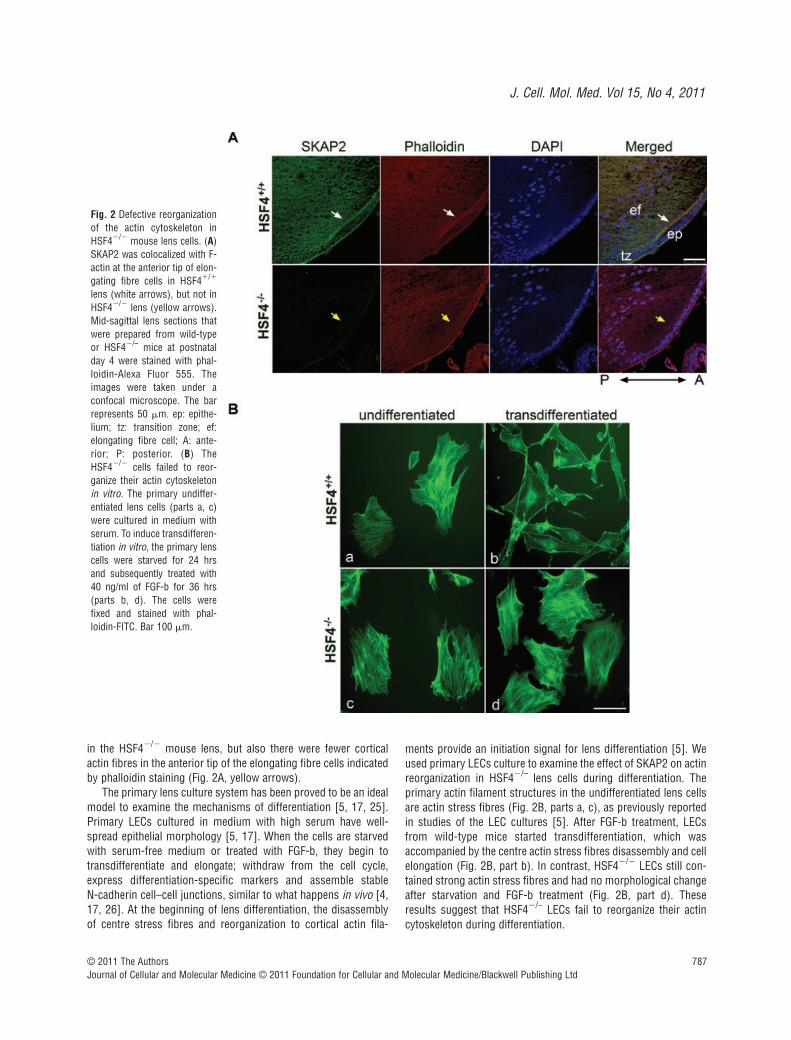
confirm that SKAP2 localized in actin-rich membrane ruffles, wecarried co-IP and immunostaining experiments in SRA01/04 LECstreated by FGF-b. Indeed, -actin could be co-IPed with HA-SKAP2 in SRA01/04 cells (Fig. 3A). We also observed the sub-cellular colocalization of HA-SKAP2 and cortical actin fibres at thelamellipodia in the EMT cells (Fig. 3B), similar to what happens in vivo. As shown in Fig. 3B, lamellipodium are broad, flat, sheet-like structures at the leading edges and contain actin meshwork,which produce the major driving force of migration [28].Meanwhile, the colocalization of endogenous SKAP2 and corticalactin fibres was showed in row 1 of Fig. 3D.
Rapid loss of actin stress fibres, the reorganization of actinfrom cellular stress fibres to cortical actin fibres, and the assem-bly of N-cadherin-based cell–cell junctions are characteristicmarkers for the initiation of LEC differentiation [1]. The FGF-binduced human LECs differentiation in vitro is also accompaniedby the disassembly of actin stress fibres and the formation of cor-tical actin fibres (Fig. 3D, row 1). The function of the actin-binding
Fig. 3 SKAP2 is essential foractin reorganization in lensepithelial cells. (A) SKAP2 wasco-precipitated with -actin inLECs. The SRA01/04 cells thathad been transfected with HA-SKAP2 were lysed andimmunoprecipitated with rab-bit anti-HA antibody or withnormal IgG. The immunopre-cipitates were probed with rab-bit anti--actin or mouse anti-HA antibodies. The arrowheadpoints to the band correspon-ding to -actin, above which isthe band corresponding to theIgG heavy chain. (B) HA-SKAP2 is colocalized with cor-tical actin fibres at the lamel-lipodia. The SRA01/04 cellsthat had been transfected withHA-SKAP2 were treated withFGF-b. Then cells were fixedand stained with anti-HA anti-body (red) or counter-stainedwith phalloidin-FITC (green)and visualized using confocalmicroscopy. These results arerepresentative of three inde-pendent experiments. Bar 10 m. (C) The efficiency ofSKAP2 depletion. SRA 01/04cells were transiently trans-fected with either control or
SKAP2 shRNA (#1 or #2) and then lysed and blotted with SKAP2 and GAPDH. (D) SKAP2 was necessary for actin remodelling during differentiation.After starvation and FGF-b treatment, the control (parts a–c) or shRNA (parts d–i) transfected SRA 01/04 cells were fixed and stained with rabbit anti-SKAP2 antibody and counterstained with phalloidin Alexa Fluor 555. Cells with high levels of SKAP2 have less F-actin (yellow arrows), while cells withless SKAP2 have much higher F-actin (white arrows). Bar 25 m.
J. Cell. Mol. Med. Vol 15, No 4, 2011
789
protein SKAP2 in actin reorganization was then examined. In fact,overexpressing SKAP2 alone in the SRA01/04 cells could cause aclear disassembly of the centre stress fibres (Fig. 4A, top part),similar to that seen in differentiated LECs that had been inducedby FGF-b (Fig. 3D, row 1).
To confirm the effect of SKAP2 on actin reorganization, weknocked down SKAP2 expression in human LECs with shRNA andthen examined the remodelling of actin during the initiation of LECdifferentiation induced by FGF-b. The knockdown efficiency isshown in Fig. 3C. The SRA01/04 LECs were transfected with con-trol shRNA, SKAP2 shRNA#1, SKAP2 shRNA#2, respectively. They
were next treated by FGF-b to induce differentiation. In the controlcells, the cellular actin stress fibres were disassembled as expected(Fig. 3D, parts a, b, c). The cells that had been transfected withSKAP2 shRNA had different knockdown efficiency. This turned outto be a very useful tool to analyse the effect of SKAP2 on thedynamics of actin. The cells with down-regulated SKAP2 (Fig. 3D,white arrows in parts f, i) exhibited clear cellular actin stress fibres.However, nearby cells, that had a higher level of SKAP2, disassem-bled their centre actin stress fibres in response to FGF-b (Fig. 3D,yellow arrows in parts f, i). These results suggest that SKAP2 isessential for actin reorganization during lens differentiation.
© 2011 The AuthorsJournal of Cellular and Molecular Medicine © 2011 Foundation for Cellular and Molecular Medicine/Blackwell Publishing Ltd
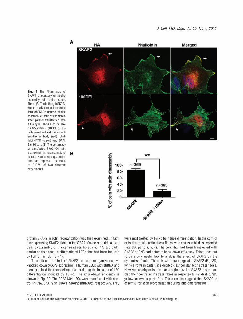
Fig. 4 The N-terminus ofSKAP2 is necessary for the dis-assembly of centre stressfibres. (A) The full length SKAP2but not the N-terminal truncatedform of SKAP2 induced the dis-assembly of actin stress fibres.After parallel transfection withfull-length HA-SKAP2 or HA-SKAP2�106aa (106DEL), thecells were fixed and stained withanti-HA antibody (red), phal-loidin-FITC (green) and DAPI.Bar 10 m. (B) The percentageof transfected SRA01/04 cellsthat exhibit the disassembly ofcellular F-actin was quantified.The bars represent the mean� S.E.M. of two differentexperiments.
790
The N-terminal 106 amino acids of SKAP2 are necessary for actin remodelling
To understand the mechanism of SKAP2 in actin reorganization,we first compared the two SKAP homologues. SKAP2 has uniquetyrosine phosphorylation sites in the N-terminal region. To inves-tigate the function of the N-terminal 106 amino acids, especially inthe SKAP2-dependent reorganization of actin, we then transfectedSRA01/04 cells with plasmids encoding either HA-tagged full-length SKAP2 or HA-tagged SKAP2�106aa (the N-terminal106 amino acids deletion mutant of SKAP2). The cells were sub-sequently fixed and stained with anti-HA antibody and phalloidin-FITC. Both the full-length SKAP2 and SKAP2�106aa localized atmembrane ruffles (Fig. 4A, arrows). Centre actin stress fibreswere absent in the cells overexpressing full-length SKAP2 (Fig. 4A,top part), whereas the cells overexpressing SKAP2�106aa
exhibited obvious cellular stress fibres without actin reorganiza-tion (Fig. 4A, bottom part). The efficiency of cellular F-actin dis-assembly in SKAP2-expressing cells was much higher than that inthe SKAP2�106aa cells (Fig. 4B). These results suggest that the N-terminal fragment of SKAP2 has an important role in actinremodelling.
Identification of the interaction between SKAP2and NCK2 at membrane ruffles
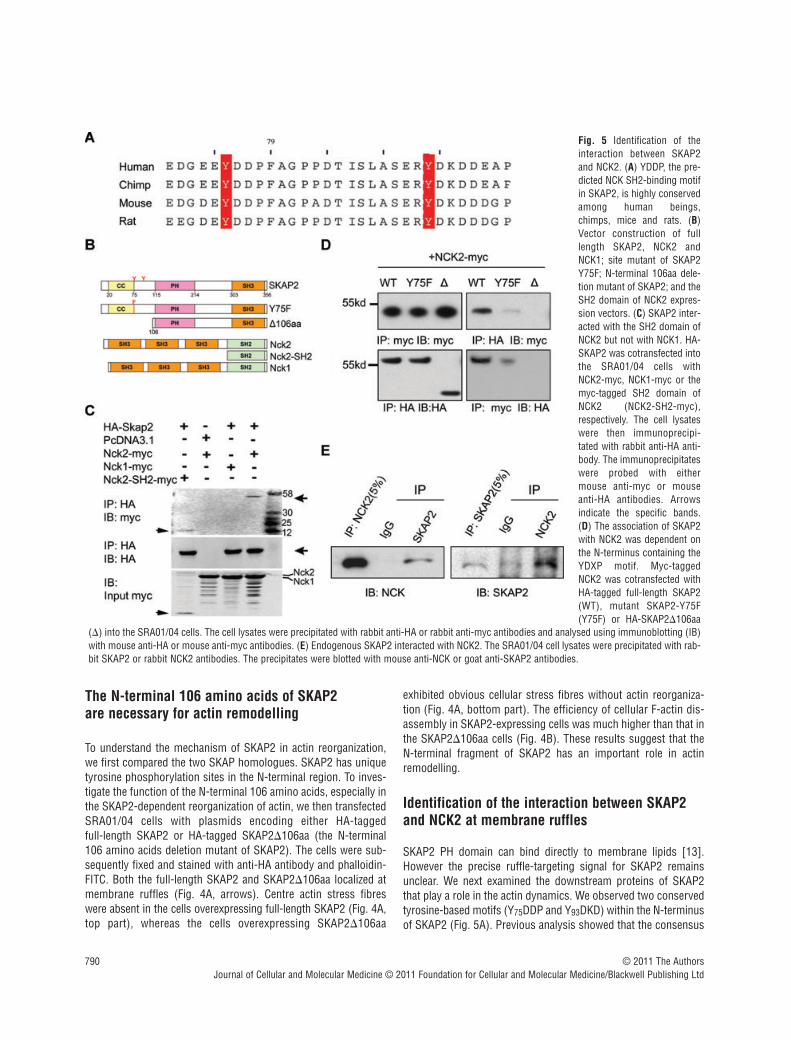
SKAP2 PH domain can bind directly to membrane lipids [13].However the precise ruffle-targeting signal for SKAP2 remainsunclear. We next examined the downstream proteins of SKAP2that play a role in the actin dynamics. We observed two conservedtyrosine-based motifs (Y75DDP and Y93DKD) within the N-terminusof SKAP2 (Fig. 5A). Previous analysis showed that the consensus
© 2011 The AuthorsJournal of Cellular and Molecular Medicine © 2011 Foundation for Cellular and Molecular Medicine/Blackwell Publishing Ltd
Fig. 5 Identification of theinteraction between SKAP2and NCK2. (A) YDDP, the pre-dicted NCK SH2-binding motifin SKAP2, is highly conservedamong human beings,chimps, mice and rats. (B)Vector construction of fulllength SKAP2, NCK2 andNCK1; site mutant of SKAP2Y75F; N-terminal 106aa dele-tion mutant of SKAP2; and theSH2 domain of NCK2 expres-sion vectors. (C) SKAP2 inter-acted with the SH2 domain ofNCK2 but not with NCK1. HA-SKAP2 was cotransfected intothe SRA01/04 cells withNCK2-myc, NCK1-myc or themyc-tagged SH2 domain ofNCK2 (NCK2-SH2-myc),respectively. The cell lysateswere then immunoprecipi-tated with rabbit anti-HA anti-body. The immunoprecipitateswere probed with eithermouse anti-myc or mouseanti-HA antibodies. Arrowsindicate the specific bands.(D) The association of SKAP2with NCK2 was dependent onthe N-terminus containing theYDXP motif. Myc-taggedNCK2 was cotransfected withHA-tagged full-length SKAP2(WT), mutant SKAP2-Y75F(Y75F) or HA-SKAP2�106aa
(�) into the SRA01/04 cells. The cell lysates were precipitated with rabbit anti-HA or rabbit anti-myc antibodies and analysed using immunoblotting (IB)with mouse anti-HA or mouse anti-myc antibodies. (E) Endogenous SKAP2 interacted with NCK2. The SRA01/04 cell lysates were precipitated with rab-bit SKAP2 or rabbit NCK2 antibodies. The precipitates were blotted with mouse anti-NCK or goat anti-SKAP2 antibodies.
J. Cell. Mol. Med. Vol 15, No 4, 2011
791
motif phospho(p)-Y-D-E/D/K-V/P is a binding site for the NCK SH2domain [29]. Thus, the Y75DDP motif is a potential binding site forthe NCK adaptor proteins. There are two NCK proteins in mam-mals, NCK1 and NCK2, each of which contains three consecutiveSH3 domains and a C-terminal SH2 domain. NCK2 is highlyexpressed in the mouse lens as we cloned full length NCK2 fromlens sample. It has been shown that NCK2 functions in couplingphosphotyrosine (pTyr) signals to actin cytoskeletal reorganiza-tion, through a mechanism that the SH2 domian binds to
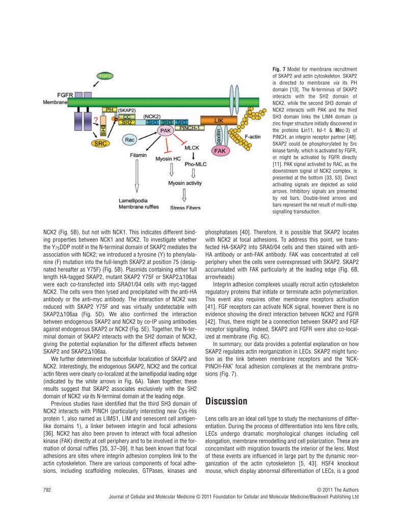
phosphotyrosine (pTyr) signals, the second SH3 domain interactswith PAK (p21-activated kinase) that leads to the disassembly ofstress fibre and the formation of lamellipodia upon Rac activation,and the third SH3 domain recruits focal adhesions [30–35] (Fig. 7). Therefore, it is probable that SKAP2 signals to actin viaNCK2/PAK pathway.
We then examined the interaction between SKAP2 and NCK1 orNCK2 by co-IP experiments. As shown in Fig. 5C, HA-SKAP2 wasco-precipitated with full-length NCK2 and the SH2 domain of
© 2011 The AuthorsJournal of Cellular and Molecular Medicine © 2011 Foundation for Cellular and Molecular Medicine/Blackwell Publishing Ltd
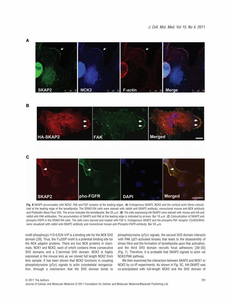
Fig. 6 SKAP2 accumulates with NCK2, FAK and FGF receptor at the leading edges. (A) Endogenous SKAP2, NCK2 and the cortical actin fibres colocal-ized at the leading edge of the lamellipodia. The SRA01/04 cells were stained with rabbit anti-SKAP2 antibody, monoclonal mouse anti-NCK antibodyand Phalloidin Alexa Flour 555. The arrow indicates the lamellipodia. Bar 25 m. (B) The cells expressing HA-SKAP2 were stained with mouse anti-HA andrabbit anti-FAK antibodies. The accumulation of SKAP2 and FAK at the leading edge is indicated by arrows. Bar 10 m. (C) Colocalization of SKAP2 andphospho-FGFR in the SRA01/04 cells. The cells were starved and treated with FGF-b. Endogenous SKAP2 and the phospho-FGF receptor (Tyr653/654)were visualized with rabbit anti-SKAP2 antibody and monoclonal mouse anti-Phospho-FGFR antibody. Bar 50 m.
792
NCK2 (Fig. 5B), but not with NCK1. This indicates different bind-ing properties between NCK1 and NCK2. To investigate whetherthe Y75DDP motif in the N-terminal domain of SKAP2 mediates theassociation with NCK2; we introduced a tyrosine (Y) to phenylala-nine (F) mutation into the full-length SKAP2 at position 75 (desig-nated hereafter as Y75F) (Fig. 5B). Plasmids containing either fulllength HA-tagged SKAP2, mutant SKAP2 Y75F or SKAP2�106aawere each co-transfected into SRA01/04 cells with myc-taggedNCK2. The cells were then lysed and precipitated with the anti-HAantibody or the anti-myc antibody. The interaction of NCK2 wasreduced with SKAP2 Y75F and was virtually undetectable withSKAP2�106aa (Fig. 5D). We also confirmed the interactionbetween endogenous SKAP2 and NCK2 by co-IP using antibodiesagainst endogenous SKAP2 or NCK2 (Fig. 5E). Together, the N-ter-minal domain of SKAP2 interacts with the SH2 domain of NCK2,giving the potential explanation for the different effects betweenSKAP2 and SKAP2�106aa.
We further determined the subcellular localization of SKAP2 andNCK2. Interestingly, the endogenous SKAP2, NCK2 and the corticalactin fibres were clearly co-localized at the lamellipodial leading edge(indicated by the white arrows in Fig. 6A). Taken together, theseresults suggest that SKAP2 associates exclusively with the SH2domain of NCK2 via its N-terminal domain at the leading edge.
Previous studies have identified that the third SH3 domain ofNCK2 interacts with PINCH (particularly interesting new Cys-Hisprotein 1, also named as LIMS1, LIM and senescent cell antigen-like domains 1), a linker between integrin and focal adhesions[36]. NCK2 has also been proven to interact with focal adhesionkinase (FAK) directly at cell periphery and to be involved in the for-mation of dorsal ruffles [35, 37–39]. It has been known that focaladhesions are sites where integrin adhesion complexs link to theactin cytoskeleton. There are various components of focal adhe-sions, including scaffolding molecules, GTPases, kinases and
phosphatases [40]. Therefore, it is possible that SKAP2 locateswith NCK2 at focal adhesions. To address this point, we trans-fected HA-SKAP2 into SRA0/04 cells and then stained with anti-HA antibody or anti-FAK antibody. FAK was concentrated at cellperiphery when the cells were overexpressed with SKAP2. SKAP2accumulated with FAK particularly at the leading edge (Fig. 6B,arrowheads)
Integrin adhesion complexes usually recruit actin cytoskeletonregulatory proteins that initiate or terminate actin polymerization.This event also requires other membrane receptors activation[41]. FGF receptors can activate NCK signal, however there is noevidence showing the direct interaction between NCK2 and FGFR[42]. Thus, there might be a connection between SKAP2 and FGFreceptor signalling. Indeed, SKAP2 and FGFR were also co-local-ized at membrane (Fig. 6C).
In summary, our data provides a potential explanation on howSKAP2 regulates actin reorganization in LECs. SKAP2 might func-tion as the link between membrane receptors and the ‘NCK-PINCH-FAK’ focal adhesion complexes at the membrane protru-sions (Fig. 7).
Discussion
Lens cells are an ideal cell type to study the mechanisms of differ-entiation. During the process of differentiation into lens fibre cells,LECs undergo dramatic morphological changes including cellelongation, membrane remodelling and cell polarization. These areconcomitant with migration towards the interior of the lens. Mostof these events are influenced in large part by the dynamic reor-ganization of the actin cytoskeleton [5, 43]. HSF4 knockoutmouse, which display abnormal differentiation of LECs, is a good
© 2011 The AuthorsJournal of Cellular and Molecular Medicine © 2011 Foundation for Cellular and Molecular Medicine/Blackwell Publishing Ltd
Fig. 7 Model for membrane recruitmentof SKAP2 and actin cytoskeleton. SKAP2is directed to membrane via its PHdomain [13]. The N-terminus of SKAP2interacts with the SH2 domain of NCK2, while the second SH3 domain ofNCK2 interacts with PAK and the thirdSH3 domain links the LIM4 domain (azinc finger structure initially discovered inthe proteins Lin11, Isl-1 & Mec-3) ofPINCH, an integrin receptor partner [48].SKAP2 could be phosphorylated by Srckinase family, which is activated by FGFR,or might be activated by FGFR directly[11]. PAK signal activated by RAC, as thedownstream signal of NCK2 complex, ispresented at the bottom [33, 53]. Directactivating signals are depicted as solidarrows. Inhibitory signals are presentedby red bars. Double-lined arrows andbars represent the net result of multi-stepsignalling transduction.
J. Cell. Mol. Med. Vol 15, No 4, 2011
793
model for identifying novel target genes that are essential for thedifferentiation of LECs.
In our study, we identified a downstream target of HSF4b,SKAP2. ChIP assay revealed that HSF4 could bind to the promoterregion of Skap2 (Fig. 1C). The perfect HSF binding site is composedof at least three inverted repeats of nGAAn (nTTCnnGAAnnTTCn) [9]and the atypical HSEs contain gap-type HSE (nTTCnnGAAn(5 bp)nGAAn), step-type HSE (nTTCn(5 bp)nTTCn(5 bp)nTTCn)and DR-type HSE (nnGAAnnnnnnnGAAn) [44]. Besides, Fujimoto et al. have reported that the motif with at least three inverted repeatsof nGnnn is also HSF4 binding site [45]. Sequence analysis onmSkap2 promoter region (from –650 to 0) suggests that there arenine putative HSEs in this region, seven of which are motifs with atleast three inverted repeats of nGnnn, one is DR-type HSE and theother is gap-type HSE (Fig. S3). Further experiments will be neededto identify which motif is essential for HSF4 binding.
Previous studies have shown that SKAP2 is involved in the differentiation of myeloid cells and in cell cycle arrest [27, 46].Consistent with that finding, expression of SKAP2 in the lens alsocorrelated with the differentiation of LECs, indicating that SKAP2may be involved in some process of lens cell differentiation. Thefacts that SKAP2 was phosphorylated after stimulation by MCS-Fand that it was co-immunoprecipitated with -actin in myeloidcells in vitro [27], inspired us to investigate the role of SKAP2 inregulating actin cytoskeleton in LECs. Our study demonstratesthat SKAP2 is apt to accumulate with cortical actin fibres at mem-brane ruffles and is essential for the disassembly of actin stressfibres in response to FGF-b. The disruption of actin stress fibresregulates LEC differentiation [5, 43]. The failure of actin disassem-bly unmasks the mechanism of the abnormal differentiation inHSF4�/� len cells. In agreement with our findings, Reinhold andhis colleagues recently revealed that actin polymerization wasenhanced when triggered by integrin in SKAP2-deficient dendriticcells [47]. Furthermore, we found that the N-terminal domain ofSKAP2 is important for its function in actin reorganization asSKAP2 interacts with NCK2 via its N-terminal domain .
NCK2 is one of the proteins that control the rearrangement ofthe actin cytoskeleton by linking membrane receptors to actin reg-ulatory proteins. The SH3 domain of NCK2 has been reported tobind to the proline-rich motifs of N-WASP, WASP-interacting pro-tein (WIP), PAK serine/threonine kinase, which are all involved incontrol of cellular actin dynamics [36, 39, 48–50]. When cells arestimulated, the complex of NCK-PAK-PINCH-PKL (the paxillinkinase linker) is recruited to nascent focal complexes at the lead-ing edge upon intergrin engagement and Rac activation, wherelamellipodia forms and the regulatory signal of actin cytoskeletionare transduced [31]. As the downstream effector of NCK signal,the membrane-attached and activated PAK induces the disassem-bly of stress fibres through inhibiting myosin light chain kinaseand myosin HC activity [33, 51–53], and promotes the formationof lamellipodia through filamin [32, 54] (Fig. 7). In the upstreamsignal, the SH2 domain of NCK binds to specific pTyr-containingsites on activated receptors directly, such as the ß-platelet-derivedgrowth factor-receptor (ß-PDGFR) and EGFR [34, 55, 56]. NCK
has also been reported to link to FGF receptors indirectly [42]. Ourobservations of the association of SKAP2 with the SH2 domain ofNCK2 at the lamellipodia, and the essential role for SKAP2 in actinremodelling induced by FGF-b, suggest that SKAP2 may be themissing link between NCK2 and FGF receptor. Here, we alsoobserved that SKAP2 and the activated FGF receptor colocalized inLECs (Fig. 6B). However, whether they interact with each otherdirectly or through other proteins requires further examination.Importantly, there is a positive feedback that the expression ofSKAP2 is significantly increased when LECs were induced by FGF-b. Our data raises the possibility that SKAP2 might act coopera-tively with NCK2 at the leading edge to regulate the PAK activity.However, further investigations are required to fully understandthe signal transduction in lens downstream of FGFR/SKAP2/NCK2.
SKAP2 can also be phosphorylated by SRC kinases, includingSrc, Fyn, Lyn and Hck [11]. Src and related kinases can phospho-rylate tyrosine sites of many actin-associated proteins, such asFAK, paxillin, vinculin and so on, which will regulate the formationof various phosphotyrosine-based macromolecular signaling com-plexes like focal adhesion complexes, or directly affect the activityof the downstream substrates, and thereby influence the actincytoskeleton and cell shape [57, 58]. In addition, kinases in the Srcfamily are known to play a central role in the migration of epithelialcells. They accomplish this through their ability to recruit proteinsto the sites of integrin attachment for regulating the focal adhesionturn-over and actin reorganization and to regulate the activity of thesmall GTPases Rac and Rho [57–59]. Therefore, it is probable thatSKAP2, as the actin-binding protein phosphorylated by SRCkinases or directly by FGFR, recruits NCK2/F-actin to transmem-brane integrin /focal adhesion in the lamella.
Taken together, these results reveal an essential role for SKAP2in actin remodelling and uncover a novel ruffle-targeting complex.Our new findings are crucial to understand the role of HSF4 in LECdifferentiation and the molecular mechanism of cataract.
Acknowledgements
We thank Dr. Dangsheng Li for helpful discussions. We also thank threeanonymous referees for their constructive comments. This work was sup-ported by the National Natural Science Foundation of China, Key Program(No. 30530450), the National High Technology Research and DevelopmentProgram of China (2006AA02Z330, 2006AA02A301), the National BasicResearch Program of China (Nos. 2007CB512202, 2004CB518603) andthe Knowledge Innovation Program of the Chinese Academy of Sciences(Grant No. KSCX1-YW-R-74).
Supporting Information
Additional Supporting Information may be found in the online ver-sion of this article.
© 2011 The AuthorsJournal of Cellular and Molecular Medicine © 2011 Foundation for Cellular and Molecular Medicine/Blackwell Publishing Ltd
794
Fig. S1 SKAP2 is associated with lens epithelial cells differentiation.Semi-quantitative RT-PCR of 1 g of polyA� mRNA from theSRA01/04 cells was performed with primers for the genes that arespecified. The cells were incubated in the presence of FBS (�),serum-free DMEM– or 20 ng/ml FGF-b for different amounts of time.
Fig. S2 Overexpression of SKAP2 in HSF4�/� lens cells partiallycauses the disassembly of centre stress fibres. The primaryHSF4�/– lens culture cells were transfected with SKAP2-GFP orpEGFP vectors. After 24 hrs, the cells were starved for 24 hrs andthen treated with 40 ng/ml FGF-b for 12 hrs. Finally, the cells werefixed and stained with phalloidin Alexa Fluor 555 to visualize F-actin. The magnified images were from the region pointed by thewhite arrows. Bar 50 m.
Fig. S3 The nucleotide sequences of putative HSEs in the pro-moter region of mouse Skap2. The sequences consisting of atleast three inverted repeats of nGnnn include #2, #3, #4, #6, #7,#8, #9 sequences. Among these, #2 sequence has two perfectinverted repeats of nGAAn; #3 sequence has one perfect repeatof nGAAn. #1 sequence is DR-type HSE (nnGAAnnnnn nnGAAn)and #5 sequence is the gap-type HSE (nTTCnnGAAn(5bp)nGAAn) . The inverted repeats of nGnnn or nGAAn sequencesare shown in red.
Please note: Wiley-Blackwell are not responsible for the content orfunctionality of any supporting materials supplied by the authors.Any queries (other than missing material) should be directed tothe corresponding author for the article.
References
1. Sue Menko A. Lens epithelial cell differen-tiation. Exp Eye Res. 2002; 75: 485–90.
2. Chamberlain CG, McAvoy JW. Inductionof lens fibre differentiation by acidic andbasic fibroblast growth factor (FGF).Growth Factors. 1989; 1: 125–34.
3. Walker JL, Zhang L, Zhou J, et al. Rolefor alpha 6 integrin during lens develop-ment: evidence for signaling through IGF-1R and ERK. Dev Dyn. 2002; 223: 273–84.
4. Ferreira-Cornwell MC, Veneziale RW,Grunwald GB, et al. N-cadherin functionis required for differentiation-dependentcytoskeletal reorganization in lens cells in vitro. Exp Cell Res. 2000; 256: 237–47.
5. Weber GF, Menko AS. Actin filamentorganization regulates the induction oflens cell differentiation and survival. DevBiol. 2006; 295: 714–29.
6. Bu L, Jin Y, Shi Y, et al. Mutant DNA-bind-ing domain of HSF4 is associated withautosomal dominant lamellar and Marnercataract. Nat Genet. 2002; 31: 276–8.
7. Fujimoto M, Izu H, Seki K, et al. HSF4 isrequired for normal cell growth and differ-entiation during mouse lens development.EMBO J. 2004; 23: 4297–306.
8. Min JN, Zhang Y, Moskophidis D, et al.Unique contribution of heat shock tran-scription factor 4 in ocular lens develop-ment and fiber cell differentiation. Genesis.2004; 40: 205–17.
9. Xiao H, Lis JT. Germline transformationused to define key features of heat-shockresponse elements. Science. 1988; 239:1139–42.
10. Shi X, Cui B, Wang Z, et al. Removal ofHsf4 leads to cataract development in mice
through down-regulation of gamma S-crystallin and Bfsp expression. BMC MolBiol. 2009; 10: 10.
11. Kouroku Y, Soyama A, Fujita E, et al.RA70 is a src kinase-associated proteinexpressed ubiquitously. Biochem BiophysRes Commun. 1998; 252: 738–42.
12. Marie-Cardine A, Verhagen AM,Eckerskorn C, et al. SKAP-HOM, a noveladaptor protein homologous to the FYN-associated protein SKAP55. FEBS Lett.1998; 435: 55–60.
13. Swanson KD, Tang Y, Ceccarelli DF, et al.The Skap-hom dimerization and PHdomains comprise a 3’-phosphoinositide-gated molecular switch. Mol Cell. 2008;32: 564–75.
14. Liu J, Kang H, Raab M, et al. FYB (FYNbinding protein) serves as a binding part-ner for lymphoid protein and FYN kinasesubstrate SKAP55 and a SKAP55-relatedprotein in T cells. Proc Natl Acad Sci USA.1998; 95: 8779–84.
15. Togni M, Swanson KD, Reimann S, et al.Regulation of in vitro and in vivo immunefunctions by the cytosolic adaptor proteinSKAP-HOM. Mol Cell Biol. 2005; 25:8052–63.
16. Lovicu FJ, McAvoy JW. FGF-induced lenscell proliferation and differentiation isdependent on MAPK (ERK1/2) signalling.Development. 2001; 128: 5075–84.
17. Menko AS, Klukas KA, Johnson RG.Chicken embryo lens cultures mimic dif-ferentiation in the lens. Dev Biol. 1984;103: 129–41.
18. Lim MA, Yang L, Zheng Y, et al. Roles ofPDK-1 and PKN in regulating cell migra-
tion and cortical actin formation of PTEN-knockout cells. Oncogene. 2004; 23:9348–58.
19. Symonds JG, Lovicu FJ, ChamberlainCG. Posterior capsule opacification-likechanges in rat lens explants cultured withTGFbeta and FGF: effects of cell coverageand regional differences. Exp Eye Res.2006; 82: 693–9.
20. Huang JX, Feldmeier M, Shui YB, et al.Evaluation of fibroblast growth factor sig-naling during lens fiber cell differentiation.Invest Ophthalmol Vis Sci. 2003; 44:680–90.
21. Robinson ML. An essential role for FGFreceptor signaling in lens development.Semin Cell Dev Biol. 2006; 17: 726–40.
22. Garcia CM, Kwon GP, Beebe DC. alpha-Smooth muscle actin is constitutivelyexpressed in the lens epithelial cells of sev-eral species. Exp Eye Res. 2006; 83:999–1001.
23. Nagamoto T, Eguchi G, Beebe DC. Alpha-smooth muscle actin expression in cul-tured lens epithelial cells. InvestOphthalmol Vis Sci. 2000; 41: 1122–9.
24. Lee A, Fischer RS, Fowler VM.Stabilization and remodeling of the mem-brane skeleton during lens fiber cell differ-entiation and maturation. Dev Dyn. 2000;217: 257–70.
25. Chandrasekher G, Sailaja D. Differentialactivation of phosphatidylinositol 3-kinasesignaling during proliferation and differen-tiation of lens epithelial cells. InvestOphthalmol Vis Sci. 2003; 44: 4400–11.
26. Le AC, Musil LS. FGF signaling in chick lensdevelopment. Dev Biol. 2001; 233: 394–411.
© 2011 The AuthorsJournal of Cellular and Molecular Medicine © 2011 Foundation for Cellular and Molecular Medicine/Blackwell Publishing Ltd
J. Cell. Mol. Med. Vol 15, No 4, 2011
795
27. Bourette RP, Therier J, Mouchiroud G.Macrophage colony-stimulating factorreceptor induces tyrosine phosphorylationof SKAP55R adaptor and its associationwith actin. Cell Signal. 2005; 17: 941–9.
28. Condeelis J. Life at the leading edge: theformation of cell protrusions. Annu RevCell Biol. 1993; 9: 411–44.
29. Frese S, Schubert WD, Findeis AC, et al.The phosphotyrosine peptide bindingspecificity of Nck1 and Nck2 Src homology2 domains. J Biol Chem. 2006; 281:18236–45.
30. Bladt F, Aippersbach E, Gelkop S, et al.The murine Nck SH2/SH3 adaptors areimportant for the development of meso-derm-derived embryonic structures andfor regulating the cellular actin network.Mol Cell Biol. 2003; 23: 4586–97.
31. Brown MC, West KA, Turner CE.Paxillin-dependent paxillin kinase linker andp21-activated kinase localization to focaladhesions involves a multistep activationpathway. Mol Biol Cell. 2002; 13: 1550–65.
32. Sells MA, Chernoff J. Emerging from thePak: the p21-activated protein kinase fam-ily. Trends Cell Biol. 1997; 7: 162–7.
33. Burridge K, Wennerberg K. Rho and Ractake center stage. Cell. 2004; 116: 167–79.
34. Li W, Fan J, Woodley DT. Nck/Dock: anadapter between cell surface receptors andthe actin cytoskeleton. Oncogene. 2001;20: 6403–17.
35. Schlaepfer DD, Broome MA, Hunter T.Fibronectin-stimulated signaling from afocal adhesion kinase-c-Src complex:involvement of the Grb2, p130cas, andNck adaptor proteins. Mol Cell Biol. 1997;17: 1702–13.
36. Tu Y, Li F, Wu C. Nck-2, a novel Srchomology2/3-containing adaptor proteinthat interacts with the LIM-only proteinPINCH and components of growth factorreceptor kinase-signaling pathways. MolBiol Cell. 1998; 9: 3367–82.
37. Goicoechea SM, Tu Y, Hua Y, et al. Nck-2interacts with focal adhesion kinase andmodulates cell motility. Int J Biochem CellBiol. 2002; 34: 791–805.
38. Ruusala A, Pawson T, Heldin CH, et al.Nck adapters are involved in the formationof dorsal ruffles, cell migration, and Rhosignaling downstream of the platelet-derived growth factor beta receptor. J BiolChem. 2008; 283: 30034–44.
39. Velyvis A, Vaynberg J, Yang Y, et al.Structural and functional insights intoPINCH LIM4 domain-mediated integrinsignaling. Nat Struct Biol. 2003; 10:558–64.
40. Geiger B, Bershadsky A, Pankov R, et al.Transmembrane crosstalk between theextracellular matrix–cytoskeleton crosstalk.Nat Rev Mol Cell Biol. 2001; 2: 793–805.
41. Westhoff MA, Serrels B, Fincham VJ, et al. SRC-mediated phosphorylation offocal adhesion kinase couples actin andadhesion dynamics to survival signaling.Mol Cell Biol. 2004; 24: 8113–33.
42. Liu JF, Chevet E, Kebache S, et al.Functional Rac-1 and Nck signaling net-works are required for FGF-2-induced DNAsynthesis in MCF-7 cells. Oncogene. 1999;18: 6425–33.
43. Rao PV, Maddala R. The role of the lensactin cytoskeleton in fiber cell elongationand differentiation. Semin Cell Dev Biol.2006; 17: 698–711.
44. Sakurai H, Takemori Y. Interactionbetween heat shock transcription factors(HSFs) and divergent binding sequences:binding specificities of yeast HSFs andhuman HSF1. J Biol Chem. 2007; 282:13334–41.
45. Fujimoto M, Oshima K, Shinkawa T, et al. Analysis of HSF4 binding regionsreveals its necessity for gene regulationduring development and heat shockresponse in mouse lenses. J Biol Chem.2008; 283: 29961–70.
46. Curtis DJ, Jane SM, Hilton DJ, et al.Adaptor protein SKAP55R is associatedwith myeloid differentiation and growtharrest. Exp Hematol. 2000; 28: 1250–9.
47. Reinhold A, Reimann S, Reinhold D, et al. Expression of SKAP-HOM in DCs isrequired for an optimal immune responsein vivo. J Leukoc Biol. 2009; 86: 61–71.
48. Wu C. PINCH, N(i)ck and the ILK: networkwiring at cell-matrix adhesions. TrendsCell Biol. 2005; 15: 460–6.
49. Anton IM, Lu W, Mayer BJ, et al. TheWiskott-Aldrich syndrome protein-inter-acting protein (WIP) binds to the adaptorprotein Nck. J Biol Chem. 1998; 273:20992–5.
50. Bagrodia S, Taylor SJ, Creasy CL, et al.Identification of a mouse p21Cdc42/Racactivated kinase. J Biol Chem. 1995; 270:22731–7.
51. Sanders LC, Matsumura F, Bokoch GM,et al. Inhibition of myosin light chainkinase by p21-activated kinase. Science.1999; 283: 2083–5.
52. van Leeuwen FN, van Delft S, Kain HE, et al. Rac regulates phosphorylation of themyosin-II heavy chain, actinomyosin dis-assembly and cell spreading. Nat Cell Biol.1999; 1: 242–8.
53. Manser E, Huang HY, Loo TH, et al.Expression of constitutively active alpha-PAK reveals effects of the kinase on actinand focal complexes. Mol Cell Biol. 1997;17: 1129–43.
54. Vadlamudi RK, Li F, Adam L, et al.Filamin is essential in actin cytoskeletalassembly mediated by p21-activatedkinase 1. Nat Cell Biol. 2002; 4: 681–90.
55. Cowan CA, Henkemeyer M. The SH2/SH3adaptor Grb4 transduces B-ephrin reversesignals. Nature. 2001; 413: 174–9.
56. Chen M, She H, Kim A, et al. Nckbetaadapter regulates actin polymerization inNIH 3T3 fibroblasts in response to platelet-derived growth factor bb. Mol Cell Biol.2000; 20: 7867–80.
57. Brown MT, Cooper JA. Regulation, sub-strates and functions of src. BiochimBiophys Acta. 1996; 1287: 121–49.
58. Parsons JT, Parsons SJ. Src family protein tyrosine kinases: cooperating withgrowth factor and adhesion signaling path-ways. Curr Opin Cell Biol. 1997; 9: 187–92.
59. Frame MC, Fincham VJ, Carragher NO,et al. v-Src’s hold over actin and cell adhe-sions. Nat Rev Mol Cell Biol. 2002; 3:233–45.
© 2011 The AuthorsJournal of Cellular and Molecular Medicine © 2011 Foundation for Cellular and Molecular Medicine/Blackwell Publishing Ltd