Expression of unconventional myosin genes during neuronal development in zebrafish
Upload
independentCategory
view
0download
0
Proc. Natl. Acad. Sci. USAVol. 94, pp. 4205–4210, April 1997Physiology
Adult fast myosin pattern and Ca21-induced slow myosin patternin primary skeletal muscle culture
HANS-PETER KUBIS, ERNST-AUGUST HALLER, PETRA WETZEL, AND GEROLF GROS*Zentrum Physiologie, Medizinische Hochschule Hannover 30623 Hannover, Germany
Communicated by Robert E. Forster, University of Pennsylvania School of Medicine, Philadelphia, PA, January 30, 1997 (received for reviewJanuary 11, 1995)
ABSTRACT A primary muscle cell culture derived fromnewborn rabbit muscle and growing on microcarriers insuspension was established. When cultured for several weeks,the myotubes in this model develop the completely adultpattern of fast myosin light and heavy chains. When Ca21
ionophore is added to the culture medium on day 11, raisingintracellular [Ca21] about 10-fold, the myotubes develop toexhibit properties of an adult slow muscle by day 30, express-ing slow myosin light as well as heavy chains, elevated citratesynthase, and reduced lactate dehydrogenase. The remarkableplasticity of these myotubes becomes apparent, when 8 daysafter withdrawal of the ionophore a marked slow-to-fasttransition, as judged from the expression of isomyosins andmetabolic enzymes, occurs.
While the alterations that occur in mammalian muscle duringfast-to-slow transition in vivo are known in great detail, mostlyfrom chronic electrostimulation experiments with fast hindlimbmuscles (1), the information on the mechanism initiating thisprocess is sparse. A reduced intracellular phosphorylation poten-tial and an elevated intracellular Ca21 concentration ([Ca21]i), asthey occur during sustained contractile activity, have been dis-cussed as possible trigger events (2–5). It was our aim to testwhether an increase in [Ca21]i imposedon themuscle cell is indeedable to induce a fast-to-slow transition. Because such an experi-ment cannot be performed in vivo, wewere searching for a suitablemyogenic cell culture system in which this would become feasible.Thus, this paper has two goals: (i) to report how a primary
muscle culture can be grown that develops into an adult-like state,expressing adult myosin of the fast type only, and (ii) to studywhethermanipulationof [Ca21]i induces a shift between ‘‘fast’’ and‘‘slow’’ fiber properties in the cultured myotubes. We show, first,thatmyotubes derived fromnewborn rabbitmuscles and grown for4 weeks on microcarriers in suspension possess a purely adult fastmyosin pattern. Second, we show that these myotubes exhibit asimilarly remarkable plasticity as it is known for adult rabbitmuscles (1). This plasticity becomes apparent during exposure ofthe myotubes to high [Ca21]i, which causes development of theslow isoforms of myosin light chains (MLCs) and myosin heavychains (MHCs) instead of their fast counterparts, of an increasedaerobicmetabolic capacity accompanied by a decreased anaerobiccapacity as evidenced from elevated citrate synthase (CS) andreduced lactate dehydrogenase (LDH) levels, and of an increasedactivity ratio of the carbonic anhydrases CA III/CA II. Lowering[Ca21]i back to normal levels is followed by a reversal of all thesechanges within a few days.
EXPERIMENTAL PROCEDURESCulture and Harvesting of Muscle Cells. Newborn White New
Zealand rabbits were killed by decapitation. Hindlimb muscleswere cut in small pieces and incubated in BSS, pH 7.0 (4.56 mMKCly0.44 mM KH2PO4y0.42 mM Na2HPO4y25 mM NaHCO3y119.8 mM NaCly50 mg/liter penicilliny100 mg/liter streptomycin)with 0.125% trypsin under stirring at 378C for 1 h. The suspensionwas centrifuged at 800 3 g for 5 min, the pellet resuspended inDMEMwith 10% neonatal calf serum (NCS), and the entire pro-cedure repeated once. The final pellet was suspended in DMEMy10%NCSand then filtered througha sievewith 0.4-mmpores.Thefiltrate was transferred into culture bottles where the fibroblastswere allowed to settle and attach themselves to the bottom for 30min. The supernatant suspension was decanted and diluted to afinal cell density of 83 105yml in DMEMwith 10%NCS. A totalof 15ml of this suspensionwere filled into one 260-ml culture flaskand 0.04 g cross-linked gelatin beads with a diameter of 100–300mm (CultiSpher-GL; Percell Biolytica, Astorp, Sweden) wereadded per flask. The flasks were kept at 378C in 8%CO2 in air and95% humidity while being shaken gently to ensure adequate O2supply to the cells and to prevent cells and beads from settlingdown. Twenty-four hours later the cell suspension was diluted toa cell concentration of 43 105 cellsyml.Myoblasts attached them-selves to the gelatin beads and began to fuse after 3 days in culture.After 2 weeks fusion appeared to be complete and only myotubeswere detectable. To collect the myotubes after 3–5 weeks ofculture, cell-covered beads were allowed to sediment, washedtwice in BSS (pH 7.0) with 0.02%EDTA, and resuspended in BSS(pH 7.9) containing 0.35% trypsin, 1.8 mM CaCl2, and 0.8 mMMgSO4. After incubation for 30 min at 378C under shaking, theisolated cells were spun down at 800 3 g for 5 min, washed twicein BSS (pH 7.0) with 0.02% EDTA, and then suspended in BSS(pH 7.0). This suspension was sonicated 63 5 s with 60W at 08C.Scanning Electron Microscopy. The carrier suspension is
pipetted onto a collagen-coated glass slide and incubated for3 hr in a chamber saturated with water vapor. Thereafter thecarriers are firmly attached to the slide and are washed twotimes with 0.1 M cacodylate (pH 7.3). Glutardialdehyde(2.5%) is pipetted onto the carriers, and the slides are incu-bated again for 2 hr in a humid chamber for fixation, then againrinsed in cacodylate buffer. This is followed by standardtreatment for scanning electron microscopy (6).MLC Electrophoresis. Cell culture homogenates were centri-
fuged at 100,000 3 g for 1 hr and the pellets were incubated inextraction buffer with 0.6 M KCl, 10 mM EGTA, 0.5 mMdithiotreitol, 1 mM phenylmethylsulfonyl fluoride, 10 mM phos-phate (pH 6.8) (1:7 volyvol) for 1 hr at 48C. After centrifugationat 10,000 3 g for 10 min, the supernatant was diluted 1:10 withice-cold water to precipitate actomyosin over night at 08C. Aftercentrifugation at 20,000 3 g the pellet was solubilized withextraction buffer, mixed 1:1 with glycerol, and stored at 2208CThe publication costs of this article were defrayed in part by page charge
payment. This article must therefore be hereby marked ‘‘advertisement’’ inaccordance with 18 U.S.C. §1734 solely to indicate this fact.
Copyright q 1997 by THE NATIONAL ACADEMY OF SCIENCES OF THE USA0027-8424y97y944205-●$2.00y0PNAS is available online at http:yywww.pnas.org.
Abbreviations: [Ca21]i, intracellular Ca21 concentration; MLC, myo-sin light chain; MHC, myosin heavy chain; CA, carbonic anhydrase;CS, citrate synthase; LDH, lactate dehydrogenase; CK, creatine kinase.*To whom reprint requests should be addressed.
4205
until used. MLC isoforms were analyzed by two-dimensionalelectrophoresis as described by O’Farrell (7). The first dimensionwas carried out by isoelectric focusing of 10 mg protein aliquots ofmyosin extract solubilized in O’Farrell’s lysis buffer. The 5% slabgel contained 9 M urea and 2% ampholyte (pH 3.5–9.5). Afterfocusing, stripes were cut out and equilibrated in 50 mM TriszHCl(pH 6.8), 9 M urea, 30% glycerol, 3% SDS, and 0.25% dithiot-reitol. The seconddimensionwas then carried out in a 5%stackingand 15% separating gel. Thereafter gels were silver-stained.MHCElectrophoresis.MHCelectrophoresis was carried out on
myosin extracts using amodification of themethod of Carraro andCatani (8) as described (17) using a slab gel with a 3.5% stacking(pH 6.8) and a 6.6% separating gel (pH 8.8) followed by a second6.6%stacking (pH6.8)with a second8.8%separating gel (pH8.8).The latter three gels contained 4.8%, 14.5%, and 24.5% glycerol,respectively. After runs, gels were silver stained according toHeukeshoven and Dernick (9).[Ca21]i Measurements. [Ca21]i concentrations were deter-
mined by loading myotubes with Fura-2 AM. The optical mea-surement was performed in a microscope photometer over singlemyotubes as described (10). By successive excitation of the dyewith light at wavelengths of 340 and 380 nm and measuring theemitted light at 5106 10 nm, the ratio, R340/380, of light intensitiesemitted at the two excitation wavelengths was obtained. This ratiois related to the [Ca21]i in the myotubes.CA Activities. CA activities were measured using the barbital
system in Maren’s micromethod as described by Bruns and Gros(11). CA III activity was determined in the presence of 5 3 1026
M of the CA inhibitor acetazolamide, and CA II activity wasobtained by subtracting CA III activity from the total activitymeasured in the absence of inhibitor (12).CS, LDH, and Creatine Kinase (CK). These enzymes were
determined using standard optically coupled tests (13, 14).
RESULTS AND DISCUSSIONShahar and Reuveny (15) demonstrated that skeletal muscle cul-tures can be grown on microcarriers. The present newborn rabbit-derivedmyoblasts show a strong tendency to fuse to givemyotubesunder normal culture conditions in the gelatin bead culture usedhere. Fig. 1a shows a scanning electron micrograph of the type ofgelatin beads employed. Their surface is quite irregular andcharacterized by deep valleys and holes. Fig. 1b shows a myotube-covered gelatin bead after 28 days in culture. Practically all cells onthe beads are myotubes at this age with very few myoblasts. Many
myotubes extend across the ‘‘mountains’’ of the bead surface andtheir ends are fixed to the valleys and holes. These myotube cul-tures could be held for up to 5 weeks in a vital and increasinglymature state as judged from the expression of muscle-specificproteins and morphological properties such as the appearance ofT-tubule openings on the sarcolemmal surface (the latter are notvisible at the magnification of Fig. 1b). Preliminary experimentswith beads possessing smoother surfaces had shown an earlydetachment of the myotubes from the beads. It may be notedthat the myotubes contracted vigorously and spontaneouslyduring the first two weeks, whereupon spontaneous contrac-tion began to disappear. This is perhaps, in conjunction withthe appearance of a pure adult myosin pattern and the develop-ment of T-tubules, another sign of increased maturity of themyotubes in the later stages of this culture.We have also grown primary muscle cultures from the same
source andusing the samemedium in collagen-coatedPetri dishes.Here, long cylindrical myotubes develop in a manner similar, forexample, to figure 17 of Nag and Foster (16). When the myotubesbegin to contract, their cell body, except the very ends of themultinucleated cell, detaches from the collagen layer of the dish.Five days after they have begun to contract they are lost, probablybecause the increasing contractile force disrupts the attachment ofthe myotube endings to the collagen. They are then replaced bynewly formed myotubes. Thus, in contrast to the bead culture, inwhich a virtually complete fusion and a myotube age of severalweeks are achieved, the conventional Petri dish culture is charac-terized by a constant disappearance and reformation of myotubeswith the lasting presence of a major number of myoblasts and amyotube age that does not exceed a few days.Time Course of Isomyosin Expression in Culture. We have
previously shown that with the present electrophoretic techniqueMHC isoforms can be separated with good resolution and appearon the SDS gel in the sequence MHCemb , MHC IIa , MHCIId , MHCneo , MHC IIb , MHC I , MHC Ia (17). Here,MHCemb is the embryonic isoform;MHCneo the neonatal form;MHC IIa, IIb, and IId are fast adult isoforms; MHC I is the slowisoform of adult skeletal muscle; and MHC Ia is one of the slowcardiac forms. Fig. 2 shows MHC expression versus time underdifferent culture conditions. In Fig. 2a conventional Petri disheswere used to grow primary rabbit muscle cultures. Fig. 2a showsSDSelectrophoreses ofMHCfrom these cultures at different ages.They demonstrate that after 9 days in culture essentially only theneonatalMHC,MHCneo, is expressed, and this situation remains
FIG. 1. Scanning electron micrograph of (a) the gelatin bead employed in the culture system (CultiSpher-GL), and (b) a bead covered withmyotubes on day 28 of the culture. (Bars 5 50 mm.)
4206 Physiology: Kubis et al. Proc. Natl. Acad. Sci. USA 94 (1997)
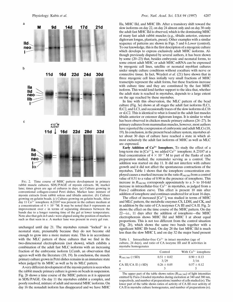
unchanged until day 21. The myotubes remain ‘‘locked’’ in aneonatal state, presumably because they do not become oldenough to grow into a more mature state. This is in accordancewith the MLC pattern of these cultures that we find in thetwo-dimensional electrophoresis (not shown), which exhibits acombination of the adult fast MLC isoforms with an increasingfraction of the embryonic isoform LCemb, an observation thatagrees well with the literature (18, 19). In conclusion, the muscleprimary culture grown inPetri dishes remains in an immature statewhen judged by its MHC as well as by its MLC pattern.Averydifferent developmentof themyosinpattern is seenwhen
the rabbit muscle primary culture is grown on beads in suspension.Fig. 2b shows a time course of the MHC pattern as it is apparentin SDSyPAGE. On day 11 the myotubes exhibit a complex, andpoorly resolved, mixture of adult and neonatalMHC isoforms. Onday 16 the nonadult isoform has disappeared and we have MHC
IIa, MHC IId, and MHC IIb. After a transitory shift toward theslow isoforms on day 22, on day 24 almost only and on day 30 onlythe adult fastMHC IId is observed, which is the dominatingMHCof many fast adult rabbit muscles (e.g., tibialis anterior, extensordigitorum longus, plantaris, psoas). Other examples with a similarsequence of patterns are shown in Figs. 5 and 6 Lower (control).Toour knowledge, this is the first descriptionof amyogenic culturewhich develops to express exclusively adult MHC isoforms. Al-though previously disputed by several authors, it has been shownby some (20–23) that, besides embryonic and neonatal forms, tosome extent adult MHC or adult MHC mRNA can be expressedby myogenic cell lines, satellite or neonatal myoblast culturesunder simple culture conditions without coculture with nerve orconnective tissue. In fact, Weydert et al. (21) have shown that inthree myogenic cell lines initially very small fractions of MHCtranscripts represent the adult forms, but these fractions increasewith culture time and they are constituted by the fast MHCisoform. This would lend further support to the idea that, whetherthe adult state is reached in myotubes, depends to a large extenton the age reached by these myotubes.In line with this observation, the MLC pattern of the bead
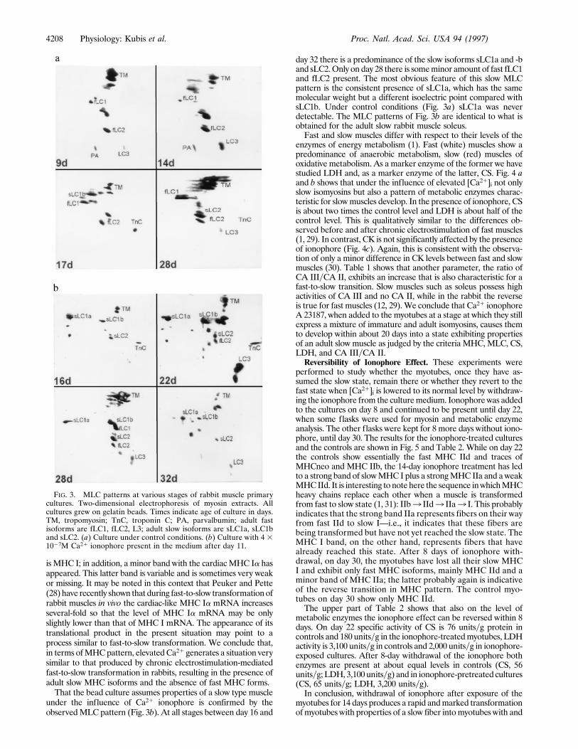
culture (Fig. 3a) shows at all stages the adult fast isoforms fLC1,fLC2, andLC3, and occasionally traces of the slow isoforms sLC1bor sLC2. This is identical to what is found in the adult fast musclestibialis anterior or extensor digitorum longus. It is similar to whathas been observed in chicken muscle primary cultures (24–27). Inprimary cultures frommammalianmuscles, however,most authorshave reported the coexpression of embryonic and adultMLCs (18,19). In conclusion, in the present bead culture system,myotubes af-ter about 30 days of culture have reached a state in which al-most exclusively the adult fast isoforms of MHC as well as MLCare expressed.Early Addition of Ca21 Ionophore. To study the effect of a
long-term rise in [Ca21]i, we added Ca21 ionophore A 23187 at afinal concentration of 4 3 1027 M to part of the flasks of eachpreparation studied, the remainder serving as a control. Theaddition was started on day 11. It did not interfere with culturegrowth and it did not affect the spontaneous contractions of themyotubes. Table 1 shows that the ionophore concentration em-ployed causes amarked increase in the ratioR340/380 froma controlvalue of 0.51 to a value of 0.90 in the presence of ionophore. Thisincrease in R340/380 corresponds appoximately to a 3- to 10-foldincrease in intracellular-free Ca21 in myotubes, as judged from aFura-2 calibration curve. This effect is present 10 min afteraddition of ionophore and continues unaltered for at least 16 days.The effect of increased [Ca21]i was studied in terms of MHC
andMLC pattern, the metabolic enzymes CS, LDH, and CK, andin addition by the ratio of CA isozymes CA III and CA II. Fig. 2cshows the effect on the time course of the MHC pattern. On day22—i.e., 11 days after the addition of ionophore—the MHCelectrophoresis shows MHC IId and MHC I at about equalproportions. This is not too different from the control situation(Fig. 2b), which shows the same two bands together with asignificant MHC IIb band. On day 28 the fast MHC IId is muchless than the slow MHC I, and on day 32 the major band present
FIG. 2. Time course of MHC pattern development in primaryrabbit muscle cultures. SDSyPAGE of myosin extracts. M, markerlane; times given are age of cultures in days. (a) Culture growing inconventional collagen-coated Petri dishes. Marker lane: mixture ofmyosin extracts from rabbit soleus and tibialis anterior. (b) Culturegrowing on gelatin beads. (c) Culture growing on gelatin beads. Afterday 11 Ca21 ionophore A23187 was present in the culture medium ata concentration of 4 3 1027M. It may be noted that b represents animprovement over c in terms of separating distances between thebands due to a longer running time of the gel at lower temperature.Note also that gels in b and cwere aligned using the position of markerssuch as that seen in a. A marker lane was present in every gel run.
Table 1. Intracellular-free Ca21 in intact myotubes (age ofculture, 24 days), and ratio of CA isozyme III and II activities inmyotube homogenates
Control With Ca21 ionophore
R340y380 (6SD) 0.51 6 0.02 0.90 6 0.13n 3y29 3y16CA IIIyCA II (6SD) 0.26 6 0.05 0.57 6 0.12n 3 5
The upper part of the table shows ratios (R340y380) of light intensitiesemitted by Fura-2-loaded myotubes during excitation at 340 and 380 nm,respectively, andnumber of preparationsynumber of cells studied (n). Thelower part of the table shows ratios of activity of CA III over activity ofCA II in myotube culture homogenates, and number of preparations (n).
Physiology: Kubis et al. Proc. Natl. Acad. Sci. USA 94 (1997) 4207
is MHC I; in addition, a minor band with the cardiacMHC Ia hasappeared. This latter band is variable and is sometimes very weakor missing. It may be noted in this context that Peuker and Pette(28) have recently shown that during fast-to-slow transformationofrabbit muscles in vivo the cardiac-like MHC Ia mRNA increasesseveral-fold so that the level of MHC Ia mRNA may be onlyslightly lower than that of MHC I mRNA. The appearance of itstranslational product in the present situation may point to aprocess similar to fast-to-slow transformation. We conclude that,in terms ofMHCpattern, elevatedCa21 generates a situation verysimilar to that produced by chronic electrostimulation-mediatedfast-to-slow transformation in rabbits, resulting in the presence ofadult slow MHC isoforms and the absence of fast MHC forms.That the bead culture assumes properties of a slow type muscle
under the influence of Ca21 ionophore is confirmed by theobservedMLC pattern (Fig. 3b). At all stages between day 16 and
day 32 there is a predominance of the slow isoforms sLC1a and -band sLC2.Only on day 28 there is someminor amount of fast fLC1and fLC2 present. The most obvious feature of this slow MLCpattern is the consistent presence of sLC1a, which has the samemolecular weight but a different isoelectric point compared withsLC1b. Under control conditions (Fig. 3a) sLC1a was neverdetectable. The MLC patterns of Fig. 3b are identical to what isobtained for the adult slow rabbit muscle soleus.Fast and slow muscles differ with respect to their levels of the
enzymes of energy metabolism (1). Fast (white) muscles show apredominance of anaerobic metabolism, slow (red) muscles ofoxidative metabolism. As a marker enzyme of the former we havestudied LDH and, as a marker enzyme of the latter, CS. Fig. 4 aand b shows that under the influence of elevated [Ca21]i not onlyslow isomyosins but also a pattern of metabolic enzymes charac-teristic for slowmuscles develop. In the presence of ionophore, CSis about two times the control level and LDH is about half of thecontrol level. This is qualitatively similar to the differences ob-served before and after chronic electrostimulation of fast muscles(1, 29). In contrast, CK is not significantly affected by the presenceof ionophore (Fig. 4c). Again, this is consistent with the observa-tion of only a minor difference in CK levels between fast and slowmuscles (30). Table 1 shows that another parameter, the ratio ofCA IIIyCA II, exhibits an increase that is also characteristic for afast-to-slow transition. Slow muscles such as soleus possess highactivities of CA III and no CA II, while in the rabbit the reverseis true for fast muscles (12, 29).We conclude that Ca21 ionophoreA 23187, when added to the myotubes at a stage at which they stillexpress a mixture of immature and adult isomyosins, causes themto develop within about 20 days into a state exhibiting propertiesof an adult slow muscle as judged by the criteria MHC, MLC, CS,LDH, and CA IIIyCA II.Reversibility of Ionophore Effect. These experiments were
performed to study whether the myotubes, once they have as-sumed the slow state, remain there or whether they revert to thefast state when [Ca21]i is lowered to its normal level by withdraw-ing the ionophore from the culturemedium. Ionophorewas addedto the cultures on day 8 and continued to be present until day 22,when some flasks were used for myosin and metabolic enzymeanalysis. The other flasks were kept for 8 more days without iono-phore, until day 30. The results for the ionophore-treated culturesand the controls are shown in Fig. 5 and Table 2. While on day 22the controls show essentially the fast MHC IId and traces ofMHCneo and MHC IIb, the 14-day ionophore treatment has ledto a strong band of slowMHC I plus a strongMHC IIa and a weakMHCIId. It is interesting to note here the sequence inwhichMHCheavy chains replace each other when a muscle is transformedfrom fast to slow state (1, 31): IIb3 IId3 IIa3 I. This probablyindicates that the strong band IIa represents fibers on their wayfrom fast IId to slow I—i.e., it indicates that these fibers arebeing transformed but have not yet reached the slow state. TheMHC I band, on the other hand, represents fibers that havealready reached this state. After 8 days of ionophore with-drawal, on day 30, the myotubes have lost all their slow MHCI and exhibit only fast MHC isoforms, mainly MHC IId and aminor band of MHC IIa; the latter probably again is indicativeof the reverse transition in MHC pattern. The control myo-tubes on day 30 show only MHC IId.The upper part of Table 2 shows that also on the level of
metabolic enzymes the ionophore effect can be reversed within 8days. On day 22 specific activity of CS is 76 unitsyg protein incontrols and 180 unitsyg in the ionophore-treatedmyotubes, LDHactivity is 3,100 unitsyg in controls and 2,000 unitsyg in ionophore-exposed cultures. After 8-day withdrawal of the ionophore bothenzymes are present at about equal levels in controls (CS, 56unitsyg; LDH, 3,100 unitsyg) and in ionophore-pretreated cultures(CS, 65 unitsyg; LDH, 3,200 unitsyg).In conclusion, withdrawal of ionophore after exposure of the
myotubes for 14 days produces a rapid andmarked transformationofmyotubes with properties of a slow fiber intomyotubes with and
FIG. 3. MLC patterns at various stages of rabbit muscle primarycultures. Two-dimensional electrophoresis of myosin extracts. Allcultures grew on gelatin beads. Times indicate age of culture in days.TM, tropomyosin; TnC, troponin C; PA, parvalbumin; adult fastisoforms are fLC1, fLC2, L3; adult slow isoforms are sLC1a, sLC1band sLC2. (a) Culture under control conditions. (b) Culture with 4 31027M Ca21 ionophore present in the medium after day 11.
4208 Physiology: Kubis et al. Proc. Natl. Acad. Sci. USA 94 (1997)

properties of a fast fiber.By the criteriaMHCpatternCSandLDHlevels this represents an example of a slow-to-fast transition.Late Addition of Ca21 Ionophore. This experiment was per-
formed to study whether a myotube that has lost embryonic andneonatal MHCs and has reached an ‘‘adult’’ state can be shiftedfrom fast to slow state by the addition of ionophore. A23187 wasadded to the cultures on day 22, and the myotubes were exposedfor 8 days until day 30. Fig. 6 shows the effects on MHC pattern.On day 22 the major MHC band is IIb, and there are also
significant amounts ofMHCIIa andMHCI. There is a clear effectof the ionophore, although it is not as impressive as in thereversibility experiment shown in Fig. 5: on day 30 the majorisoform isMHCIIa, the ‘‘transition form;’’ there is alsoMHCIandsome MHC IId. In view of the transition sequence IIb3 IId3IIa3 I, it is noteworthy that the large MHC IIb band of day 22has disappeared under the influence of ionophore. Both thedisappearance of IIb and the relative increase in IIa indicate aMHC rearrangement compatible with some shift from fast towardslow properties. Fig. 6 Lower shows that between day 22 and day30 controls, on the other hand, reach stable fast properties withonly MHC IId being expressed. Signs of a fast-to-slow transitionare also apparent at the level of metabolic enzymes (lower half ofTable 2): from a control level on day 22 of 110 unitsyg CS activityrises to 140 unitsyg after ionophore exposure for 8 days, while thecontrol activity falls to 63unitsyg. Similarly, LDHfalls fromavalueof 2,800 unitsyg on day 22 to 2,200 unitsyg after 8 days’ treatmentwith ionophore, while the control remains almost unchanged with2,900 unitsyg.
FIG. 4. Marker enzymes of oxidative (CS) and anaerobic (LDH)metabolism and of CK in myotubes cultured for different times. Opensymbols, controls; filled symbols, with ionophore A23187 present afterday 11. p, Significance of P , 0.02 of differences between control andionophore-treatedmyotubes; n.s., lack of significant differences (P. 0.1).
FIG. 5. Reversibility of the effect of ionophore treatment on MHCpattern of myotubes by SDSyPAGE. (Upper) Day 22, MHCs afterionophore exposure for 14 days; day 30, MHCs after ionophore with-drawal for 8 days. (Lower) Controls without ionophore.Marker lane (M):mixture of myosin extracts from soleus and tibialis anterior of the rabbit.
Table 2. Ionophore effects on marker enzymes of oxidative andanaerobic metabolism: reversibility after early ionophore additionand effect of late ionophore addition
Age,day Treatment
CS,unitsygprotein
LDH,unitsygprotein
22 Control (22 days) 76 3,10022 Control (8 days) 1 ionophore (14 days) 180 2,00030 Control (30 days) 56 3,20030 Control (8 days) 1 ionophore (14 days)
1 control (8 days)65 3,200
22 Control (22 days) 110 2,80030 Control (30 days) 63 2,90030 Control (22 days) 1 ionophore (8 days) 140 2,200
The upper part of table demonstrates reversibility of effects afterearly addition of ionophore A23187. All measurements were per-formed on the same culture preparation derived from the muscles ofseveral newborn rabbits. The lower part of table demonstrates theeffect of late addition of ionophore. Again, all measurements wereperformed on the same culture preparation.
Physiology: Kubis et al. Proc. Natl. Acad. Sci. USA 94 (1997) 4209

Weconclude that late addition ofCa21 ionophore does producesome indications of a fast-to-slow transition in terms of MHCpattern and metabolic enzymes. The effects, however, are subtleand by far not as marked as those generated by early addition orby withdrawal of ionophore.Conclusions. The primary rabbit muscle culture system using
gelatin beads described here allows one to keep myotubes forabout 30 days in standard culture medium. During such a periodthe myotubes appear to reach a degree of maturity in which theyexpress only fast adult myosin as it is observed, for example, inadult fast rabbit tibialis anterior. By early addition of the Ca21ionophore A23187 from day 11 onwards the myotubes developwithin about 20 days to assume properties of a slow muscle, suchas soleus. This becomes apparent from the complete absence of allfast isoforms of MHC and MLC and their replacement by slowadult isoforms. This is accompanied by an elevated level of CS, adecreased activity of LDH, and an increased ratio of CA IIIyCAII. All these observations correspond to well-known differencesbetween slow red and fast white muscles. Thus, in the presence ofnormal [Ca21]i levels a fast myotube develops in culture, whereasin the presence of elevated [Ca21]i a slow myotube grows.This ionophore effect is rapidly reversible. When, after 14-day
exposure, the ionophore is withdrawn on day 22, the myotubeshave on day 30 lost the slow MHC I and possess only fast MHC.They also show nearly complete reversals of the previously ele-vated level of CS and the diminished level of LDH.This representsa fairly extensive slow-to-fast transformation, of a degree that evenby cross-innervation experiments is difficult to achieve in vivo.The demonstration of a transformation of a fast myotube into
a slowmyotube by late addition of ionophore to the culture ismoredifficult. Addition of ionophore on day 22 does not produce amarked increase in the slow MHC I but rather a rearrangementamong the different fast MHC isoforms which points to a begin-ning transformation from a fast toward a slow myotube. Concom-itantly, there is a moderate increase in CS and a decrease in LDHlevels. Whether the smaller changes induced by late (on day 22)compared with early (on day 11) addition of ionophore are due tothemoremature state of themyotubeswith adiminishedplasticity,or to the shorter time of exposure to ionophore (8 vs. 20 days),cannot be decided from the present data. The experiment neces-sary to solve this problem, keeping the cultures under controlconditions for 22 days and then for another 20 days with iono-phore, has not been possible so far.Also, it is not clear why withdrawal of ionophore between day
22and30 causes amarked slow-to-fast transformationbut addition
of ionophore during the same time interval is not sufficient for adrastic fast-to-slow transformation. A possible explanation mightbe a slower degradation of fast MHC isoforms in the presence ofionophore A23187 than of slowMHC I in its absence. Since Silverand Etlinger (32) found no significant effect of A23187, at aconcentration identical to ours, on MHC degradation in chickenmuscle cultures, a more rapid degradation of slow than of fastMHCs would have to be an intrinsic property of these cells. Analternative explanation is a decreased rate of synthesis ofMHCs inthe presence of ionophore, and indeed, a decreased rate ofsynthesis of MHC (of an unspecified type) in the presence of A23187 has been described by Silver and Etlinger (32). Furtherexperiments are required to resolve this question.We have shown by a variety of criteria that the level of [Ca21]i
in rabbitmyotubes, whenmodified by addition or omission ofCa21ionophoreA23187, determines whether thesemuscle cells assumeproperties of fast or slow muscle fibers. This suggests that theincreased levels of [Ca21]i that have been observed by Sreter et al.(4) during fast-to-slow muscle transformation by chronic electro-stimulation in vivo may be a causal event, initiating the chain ofprocesses leading to such a transformation, rather than being asecondary phenomenon. Our results do not exclude possible sideeffects and thus other mechanisms of action of the Ca21 iono-phore, a possibility which has to be investigated, and they do notanswer the exciting question what the physiological stimulus is thatmediates the long-lasting increase in [Ca21]i described by Sreter etal. (4). However, the data reported here suggest that [Ca21]i playsan important role in the development of a fast vs. a slow muscleand they demonstrate that the present primary muscle culture is apowerful model for further studies of the mechanism of musclefiber transformation.
We thank Ms. Ulrike Fuhrmann for expert technical assistance. Weare indebted to Dr. B. Decker (Abt. Zellbiologie und Elektroneuni-kopie der Medizinischen Hochschule, Hannover) for scanning elec-tron micrographs. This study was supported by the Gesellschaft derFreunde der Medizinischen Hochschule Hannover.
1. Pette, D. & Vrbova, G. (1992) Rev. Physiol. Biochem. Pharmacol. 120, 115–202.2. Shoubridge, E. A., Challiss, R. A. J., Hayes, D. J. &Radda, G. K. (1985)Biochem.
J 232, 125–131.3. Henriksson, J., Salmons, S., Chi, M. M.-Y., Hintz, C. S. & Lowry, O. H. (1988)
Am. J. Physiol. 255, C543–C551.4. Sreter, F. A., Lopez, J. R., Alamo, L., Mabuchi, K. & Gergely, J. (1987) Am. J.
Physiol 253, C296–C300.5. Green, H. J., Dusterhoft, S., Dux, L. & Pette, D. (1990) in The Dynamic State of
Muscle Fibers, ed. Pette, D. (de Gruyter, Berlin), pp. 617–627.6. Robinson, D. G., Ehlers, U., Herken, R., Herrmann, B., Mayer, F. & Schurmann,
F.-W (1985) Praparationsmethodik in der Elektronenmikros-kopie (Springer, Ber-lin), pp. 155–187.
7. O’Farrell, P. H. (1975) J. Biol. Chem. 250, 4007–4021.8. Carraro, U. & Catani, C. (1983) Biochem. Biophys. Res. Commun. 116, 793–802.9. Heukeshoven, J. & Dernick, R. (1985) Electrophoresis 6, 103–112.10. Wetzel, P., Liebner, T. & Gros, G. (1990) FEBS Lett. 267, 66–70.11. Bruns, W. & Gros G. (1991) in The Carbonic Anhydrases-Cellular Physiology and
Molecular Genetics, eds. Dodgson, S. J., Tashian, R. E., Gros, G. & Carter, N. D.(Plenum, New York), pp. 127–131.
12. Geers, C., Kruger, D., Siffert, W., Schmid, A., Bruns, W. & Gros, G. (1992)Biochem. J 282, 165–171.
13. Bergmeyer, H. U. (1984) Methods of Enzymatic Analysis (Verlag Chemie, Wein-heim, Germany), Vol. 4, 3rd Ed., pp. 353–358.
14. Bass, A., Brdiczka, D., Eyer, P., Hofer, S. & Pette, D. (1969) Eur. J. Biochem. 10,198–206.
15. Shahar, A. & Reuveny, S. (1987) Adv. Biochem. Eng. Biotechol. 34, 33–35.16. Nag, A. C. & Foster, J. D. (1981) J. Anat. 132, 1–18.17. Kubis, H.-P. & Gros, G. (1997) Electrophoresis 18, 64–66.18. Strohmann, R. C., Micou-Eastwood, J., Glass, C. A. & Matsuda, R. (1983)
Science 221, 955–957.19. Barton, P. J. & Buckingham, M. E. (1985) Biochem. J. 231, 249–261.20. Silberstein, L., Webster, S. G., Travis, M. & Blau, H. M. (1986) Cell 46,
1075–1081.21. Weydert, A., Barton, P., Harris, A. J., Pinset, Ch. & Buckingham, M. (1987) Cell
49, 121–129.22. Dusterhoft, S. & Pette, D. (1993) Differentiation (Berlin) 53, 25–33.23. Naumann, K. & Pette D. (1994) Differentiation (Berlin) 55, 203–211.24. Rubinstein, N. & Holtzer, H. (1979) Nature (London) 280, 323–325.25. Srihari, T. & Pette, D. (1981) FEBS Lett. 123, 312–314.26. Stockdale, F. E., Baden, H. & Raman, N. (1981) Dev. Biol. 82, 168–171.27. Crow, M. T. & Stockdale, F. E. (1985) J. Cell Biol. 100, 1415–1422.28. Peuker, H. & Pette, D. (1995) FEBS Lett. 367, 132–136.29. Gros, G. & Dodgson, S. J. (1988) Annu. Rev. Physiol. 50, 669–694.30. Lowry, C. V., Kimmey, J. S., Felder, S., Chi, M. Y., Kaiser, K. K., Passonneau
P. N., Kirk, K. A. & Lowry, O. H. (1978) J. Biol. Chem. 253, 8269–8277.31. Staron, R. S. & Pette, D. (1993) Histochemistry 100, 149–153.32. Silver, G. & Etlinger, J. D. (1985) J. Cell Biol. 101, 2383–2391.
FIG. 6. Effects of late addition of Ca21 ionophore to the cultureson MHC patterns by SDSyPAGE. (Upper) MHC patterns afteraddition of the ionophore from day 22 on. (Lower) Controls withoutionophore. Marker lane (M): mixture of myosin extracts from tonguemuscle, soleus, and young masseter of the rabbit.
4210 Physiology: Kubis et al. Proc. Natl. Acad. Sci. USA 94 (1997)
Copyright © 2022 FDOKUMEN