Myosin Class XIV And Other Myosins In Protists
20
CHAPTER 15 MYOSIN CLASS XIV AND OTHER MYOSINS IN PROTISTS KARINE FRÉNAL 1 , BERNARDO J. FOTH 2 , AND DOMINIQUE SOLDATI 1 * 1 Department of Microbiology and Molecular Medicine, University of Geneva, CMU, 1 Rue Michel-Servet, CH-1211 Geneva 4, SWITZERLAND 2 School of Biological Sciences, Nanyang Technological University, 60 Nanyang Drive, SINGAPORE 637551 Abstract: Myosins are actin-based molecular motors that convert chemical energy released by ATP hydrolysis into directed movement along tracks of actin filaments. They are found in eukaryotes and are implicated in a number of important cell functions, such as nuclear and cell division, transport of molecules, vesicles, and organelles, signal transduction, and motility. The myosin superfamily was previously described as containing at least 18 different classes with class XIV reported to be specific to apicomplexan parasites and subdivided into two subclasses. But a recent reassessment of myosin phylogeny and classification incorporating a number of novel sequences uncovered by several genome sequencing initiatives has expanded the known repertoire of myosin heavy chains from apicomplexans and other protists. It established six new myosin classes, three of which are restricted to alveolates (XXII, XXIII, XXIV), and showed that class XIV encompasses myosins of both apicomplexans and ciliates and is subdivided into four subclasses. Moreover, several sequences include protein domains (ATS1-like, WD40) previously unknown to be associated with myosin motors. In this chapter, we discuss the current classification of myosin heavy chains with particular emphasis on the apicomplexan myosins. Most of them have not yet been studied experimentally and we discuss their possible function based on their classification and their protein domains. Keywords: Myosins, parasitic protists, Apicomplexa ∗ Corresponding author: Department of Microbiology and Molecular Medicine, University of Geneva, CMU, 1 Rue Michel-Servet, CH-1211 Geneva 4, SWITZERLAND 421 L.M. Coluccio (ed.), Myosins: A Superfamily of Molecular Motors, 421–440. © 2008 Springer.
Transcript of Myosin Class XIV And Other Myosins In Protists

CHAPTER 15
MYOSIN CLASS XIV AND OTHER MYOSINSIN PROTISTS
KARINE FRÉNAL1, BERNARDO J. FOTH2, AND DOMINIQUE SOLDATI1�*1 Department of Microbiology and Molecular Medicine, University of Geneva, CMU,1 Rue Michel-Servet, CH-1211 Geneva 4, SWITZERLAND2 School of Biological Sciences, Nanyang Technological University, 60 Nanyang Drive,SINGAPORE 637551
Abstract: Myosins are actin-based molecular motors that convert chemical energy released byATP hydrolysis into directed movement along tracks of actin filaments. They are foundin eukaryotes and are implicated in a number of important cell functions, such as nuclearand cell division, transport of molecules, vesicles, and organelles, signal transduction,and motility. The myosin superfamily was previously described as containing at least18 different classes with class XIV reported to be specific to apicomplexan parasitesand subdivided into two subclasses. But a recent reassessment of myosin phylogenyand classification incorporating a number of novel sequences uncovered by severalgenome sequencing initiatives has expanded the known repertoire of myosin heavychains from apicomplexans and other protists. It established six new myosin classes,three of which are restricted to alveolates (XXII, XXIII, XXIV), and showed thatclass XIV encompasses myosins of both apicomplexans and ciliates and is subdividedinto four subclasses. Moreover, several sequences include protein domains (ATS1-like,WD40) previously unknown to be associated with myosin motors. In this chapter, wediscuss the current classification of myosin heavy chains with particular emphasis on theapicomplexan myosins. Most of them have not yet been studied experimentally and wediscuss their possible function based on their classification and their protein domains.
Keywords: Myosins, parasitic protists, Apicomplexa
∗ Corresponding author: Department of Microbiology and Molecular Medicine, University of Geneva,CMU, 1 Rue Michel-Servet, CH-1211 Geneva 4, SWITZERLAND
421
L.M. Coluccio (ed.), Myosins: A Superfamily of Molecular Motors, 421–440.© 2008 Springer.

422 FRENAL ET AL.
15.1. INTRODUCTION
Myosin heavy chains are a diverse family of actin-based molecular motors thatappeared very early in eukaryotic evolution (Richards and Cavalier-Smith, 2005;Thompson and Langford, 2002). They use energy released by ATP hydrolysis togenerate directed movement along tracks of filamentous actin. They are implicatedin a number of important cell functions including cytokinesis, organellar transport,signal transduction and cell motility. The myosin superfamily has been previouslycategorized into at least 18 classes, mostly based on comparisons and phylo-genetic analyses of their conserved motor domain, with the class II myosins,which include the motor molecules of skeletal muscle, referred to as “conven-tional” myosins (Berg et al., 2001; Oliver et al., 1999; Sellers, 2000; Thompsonand Langford, 2002). Until recently, the sampling of myosins was focused on arelatively small number of taxonomic lineages representing the “crown” of theeukaryotic tree, i.e., animals, plants, and fungi, as well as some other commonexperimental organisms such as the slime mold Dictyostelium discoideum. Butconsidering that much of eukaryotic diversity is represented by protists (Dawsonand Pace, 2002; Goodson and Dawson, 2006; Keeling et al., 2005), the recentsequencing of several nuclear genomes from protist parasites, in particular thoseof apicomplexans and kinetoplastids, was expected to and has indeed uncovered anumber of novel myosin motors that appeared not to fit into the existing classifi-cation scheme. In an attempt to incorporate such novel and previously unclassifiedmyosin sequences into the existing myosin classification, we established six novelmyosin classes, increasing their number from 18 to 24 (Foth et al., 2006). Thesenew classes comprise myosins from chordate metazoans (class XIX), from insects(class XX), from kinetoplastid parasites (trypanosomes and Leishmania; class XXI),and from protists belonging to the Apicomplexa and related lineages such as ciliatesand heterokonts (see below) (classes XXII, XXIII, and XXIV) (Figure 15.1). Inaddition, our data showed that apicomplexan myosins, which had previously beenthought to be restricted to the Apicomplexa-specific class XIV (Heintzelman andSchwartzman, 1997; Heintzelman and Schwartzman, 2001), also belong to classesthat are shared with myosins from other systematic lineages (e.g., in classes VI,XIV, XXII), with class XIV now also accommodating a number of ciliate myosins.Moreover, this novel classification (Foth et al., 2006; Williams and Gavin, 2005)revealed the existence of myosin heavy chains containing protein domains, such asATS1-like or WD40, previously unknown to be associated with myosin motors.
The phylum Apicomplexa comprises protozoan parasites of considerable medicaland veterinary significance that are responsible for a wide variety of diseases inhumans and animals and include the genera Plasmodium (malaria), Toxoplasma(toxoplasmosis), Cryptosporidium (cryptosporidiosis), and Babesia (babesiosis).Recently, Apicomplexans have been shown to be close relatives of both ciliatesand a group of organisms called the Chromista, a heterogenous collection of mostlyunicellular algae that also includes the heterokont diatoms (e.g., Thalassiosira)and oomycetes (e.g., Phytophthora). Together with dinoflagellate algae, Apicom-plexans are thus thought to be part of the ancient evolutionary lineage Chroma-

MYOSIN CLASS XIV AND OTHER MYOSINS IN PROTISTS 423
Figure 15.1. Classes XIV, XXI, XXII, XXIII and XXIV myosin phylogeny. These phylogenies are partof the representative distance matrix-based phylogeny (protdist/neighbor) of myosin head domains. Thistree is based on alignment 7 from a series of 12 alignments and corresponds to the most-representativetree as judged by the deviation of bootstrap support values for select myosin classes from the mediansupport values of the series of 12 analyses based on the 12 alignments (Foth et al., 2006). Abbreviations:Bb, Babesia bovis; Cp, Cryptosporidium parvum; Et, Eimeria tenella; Gp, Gregarina polymorpha; Pb,Plasmodium bergei; Pf, Plasmodium falciparum; Li, Leishmania infantum; Lm, Leishmania major;Ta, Theileria annulata; Tb, Trypanosoma brucei; Tc, Trypanosoma cruzi; Tg, Toxoplasma gondii; Tp,Theileria parva; Tpn, Thalassiosira pseudonana; Tt, Tetrahymena thermophila
lveolata (Adl et al., 2005; Bhattacharya et al., 2004; Cavalier-Smith, 2000; Harperet al., 2005; Keeling et al., 2005; Yoon et al., 2002). Although Apicom-plexans differ significantly in their host range and cell-type specificity, they shareimportant morphological features, in particular their specialised apical structures(Figure 15.2). These obligate intracellular parasites feature secretory organellesnamed micronemes and rhoptries that contain products required for motility,adhesion to and invasion of host cells. Host-cell invasion culminates with theformation of a parasitophorous vacuole, a membrane-bound compartment within theinfected host cell that for many Apicomplexans serves as a safe residence in whichthe parasite replicates. Lacking cilia and flagella (except in the male gametes of somespecies), Apicomplexans have developed a sophisticated multiprotein machinery

424 FRENAL ET AL.
Figure 15.2. Scheme of a Toxoplasma gondii tachyzoite with a detailed model of the “glideosome”, themolecular machinery promoting gliding motility. Abbreviations: GAP45 and GAP50, gliding associatedprotein 45 kDa and 50 kDa, respectively; IMC, Inner Membrane Complex; IMP, Inner MembraneParticle, MyoA, myosin A; MLC or MTIP, Myosin light chain or Myosin tail-interacting protein (SeeColour Plate 17)
to cross non-permissive biological barriers and penetrate multiple types of hostcells (Barragan and Sibley, 2003; Kappe et al., 2004; Keeley and Soldati, 2004;Soldati et al., 2004). Indeed, they share a unique form of substrate-dependent glidingmotility that involves polymerization of new actin filaments (Wetzel et al., 2003)and is powered by a myosin motor (“MyoA” of class XIV) that is localised beneaththe plasma membrane (Herm-Gotz et al., 2002; Meissner et al., 2002; Wetzel
MYOSIN CLASS XIV AND OTHER MYOSINS IN PROTISTS 425
et al., 2003). The pellicle of apicomplexan parasites has a distinctive three-layeredstructure composed of the plasma membrane and the inner and outer membrane ofthe inner membrane complex (IMC), which is a network of flattened cisternae thatunderlies the plasma membrane. It is the small compartment located between theplasma membrane and the IMC that contains most of the machinery involved ingliding motility (Morrissette and Sibley, 2002) (Figure 15.2). Cytoskeletal compo-nents play an important role in parasite motility, invasion, and establishment ofinfection (see below paragraph 15.2.1).
In the context of apicomplexan myosins, it is relevant to note that apicom-plexan actin and related actin-proteins exhibit unusual features. Indeed, in contrastto conventional actin, three studies indicate that parasite actin filaments, eitherextracted from parasites or formed from recombinant proteins, are remarkably short(about 100 nm in lenght) and less stable (Sahoo et al., 2006; Schmitz et al., 2005;Schuler et al., 2005). Moreover, the apicomplexan repertoire of actin-bindingand actin-regulatory proteins is small compared to that of other eukaryotes andlacks the actin-related proteins (ARP) 2/3, one of the central nucleators found inmost eukaryotes, and almost all the actin-bundling or crosslinking proteins (Baumet al., 2006a).
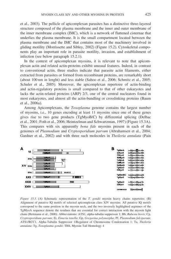
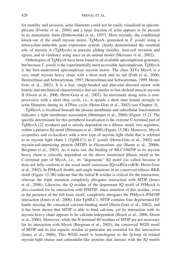
Among Apicomplexans, the Toxoplasma genome contains the largest numberof myosins, i.e., 10 genes encoding at least 11 myosins since one of these genesgives rise to two gene products (TgMyoB/C) by differential splicing (Delbacet al., 2001; Foth et al., 2006; Heintzelman and Schwartzman, 1997) (Figure 15.3A).This compares with six apparently bona fide myosins present in each of thegenomes of Plasmodium and Cryptosporidium parvum (Abrahamsen et al., 2004;Gardner et al., 2002) and with three such molecules in Theileria annulata (Pain
Figure 15.3. (A) Schematic representation of the T. gondii myosin heavy chains repertoire; (B)Alignment of putative IQ motifs of selected apicomplexan class XIV myosins. All putative IQ motifscorrespond to the same position in the myosin neck, and the two inversely highlighted arginines of theTgMyoA sequence denote the residues that are essential for correct interaction with the myosin lightchain (Hettmann et al., 2000). Abbreviations: ATS1, alpha-tubulin suppressor 1; Bb, Babesia bovis; Cp,Cryptosporidium parvum; Et, Eimeria tenella; Gp, Gregarina polymorpha; Pf, Plasmodium falciparum;ATS1/RCC1, Alpha-Tubulin Suppressor 1/Regulator of Chromosome Condensation 1; Ta, Theileriaannulata; Tg, Toxoplasma gondii; TH4, Myosin Tail Homology 4

426 FRENAL ET AL.
et al., 2005). Since researchers had commonly named new apicomplexan myosinsin the order of discovery (MyoA, MyoB, etc.) and usually independently foreach species (Chaparro-Olaya et al., 2005; Heintzelman, 2004; Heintzelman andSchwartzman, 1997; Heintzelman and Schwartzman, 2001; Hettmann et al., 2000;Lew et al., 2002; Matuschewski et al., 2001), the nomenclature of apicomplexanmyosins had at one point become rather confusing with non-orthologous sequencesbeing designated with the same letter, e.g., PfMyoC (PfM-C) and TgMyoC ofPlasmodium falciparum and T. gondii, respectively (Chaparro-Olaya et al., 2005).To clarify this situation, we recently proposed a systematic naming convention forapicomplexan myosin heavy chains with the aim to designate orthologous myosinsacross all apicomplexans by the same capital letter (e.g., TgMyoA, PfMyoA, etc.)(Foth et al., 2006). We based this naming system on homology to the T. gondiimyosins (TgMyoA to TgMyoK), simply because T. gondii has the largest knownmyosin repertoire of any apicomplexan for which significant genome data areavailable. Consequently, six apicomplexan myosins have been renamed (MyoC,MyoD, and MyoF of Plasmodium and Babesia were changed to MyoF, MyoJ, andMyoK, respectively, and MyoB of Babesia and Theileria was changed to MyoH)(Foth et al., 2006), while MyoB and MyoE found in the genus Plasmodium remainthe only exceptions to the general naming convention in that these myosins areapparently not orthologous to MyoB and MyoE of T. gondii. These two divergentPlasmodium myosins have retained, for now, their original nomenclature becauseof their uncertain phylogenetic association with other apicomplexan myosins (Fothet al., 2006).
In this chapter, we will focus on myosin class XIV, now subdivided into foursubclasses, and on the new classes XXI, XXII, XXIII, and XXIV, with particularemphasis on apicomplexan myosins. Of these only a few have been experimentallyinvestigated so far, so we will therefore discuss their organisation and possiblefunctions based on their classification and apparent protein domains.
15.2. CLASS XIV
Myosin class XIV had previously been described as exclusively comprisingapicomplexan myosins and had been divided into two subclasses (Chaparro-Olayaet al., 2005; Lew et al., 2002). But our recent phylogenetic analyses providedstrong support for a common origin of ciliate and apicomplexan class XIV myosins,which led to the inclusion of myosins from both apicomplexans and ciliates inthis class (Figure 15.1). Class XIV thus now includes 12 sequences from theciliate Tetrahymena thermophila that had been suggested previously to establishan independent class XX (Williams and Gavin, 2005). But apart from leading tothe inclusion of these ciliate myosins, our phylogenetic analyses of the conservedmyosin head domains also clearly suggested that numerous apicomplexan myosinshad to be excluded from class XIV. This novel overall class XIV classificationreceived further strong support from a particular amino acid polymorphism inthe otherwise highly conserved HYAG sequence (position 584 in chicken skeletal

MYOSIN CLASS XIV AND OTHER MYOSINS IN PROTISTS 427
muscle myosin II, GenBank accession no. AAB47555). At the second positionof this tetrapeptide, 220 (95.7%) of a collection of 230 non-class XIV myosinsequences (including several apicomplexan myosins) feature a tyrosine or pheny-lalanine, and five sequences (2.2%) an (iso)leucine. In contrast, all 37 myosins nowsuggested to comprise class XIV, including the 12 ciliate myosins, contain eithera serine or a threonine at this position, a character shared by only two non-classXIV myosins (human and mouse class XVIII) (Foth et al., 2006). Interestingly, thisconserved residue could be a site of phosphorylation and thus a potential regulatorysite for class XIV myosin heavy chains. Both phylogenetic analyses as well as theclearcut distribution of this particular polymorphism strongly support a commonevolutionary origin of all myosins now included in class XIV.
Class XIV is now divided into four subclasses (Figure 15.1). Subclass XIVacomprises myosin A, which is highly conserved in all apicomplexans, and thevery closely related myosin D (Hettmann et al., 2000) that has so far only beendetected in the coccidians T. gondii and Eimeria tenella. These two short myosinsexhibit the same structural organisation with a short neck and no tail. SubclassXIVb comprises myosin B/C, present in the genomes of T. gondii, E. tenella andGregarina polymorpha. Evolutionary origin and closer affiliation of PlasmodiumMyoB and MyoE, on the other hand, are uncertain. The new subclass XIVccomprises a well-resolved group of apicomplexan myosins, myosin H, found so farin all apicomplexan parasites except P. falciparum and G. polymorpha, whereasthe new subclass XIVd comprises 12 myosins from the ciliate T. thermophila (Fothet al., 2006).
15.2.1. Class XIVa: MyoA and MyoD
15.2.1.1. Myosin A: A molecular motor powering cell motility and a keycomponent for invasion
Host-cell penetration by apicomplexan parasites does not result from parasite-induced phagocytosis, but is an active parasite-based process driven by theactomyosin cytoskeleton of the parasite. This had been suggested early (Endoet al., 1988; King, 1981; King, 1988; Russell and Sinden, 1981; Schwartzmanand Pfefferkorn, 1983; Yasuda et al., 1988) and was later confirmed by theinability of several apicomplexan genera to glide and/or invade in the presenceof drugs that interfere with actin dynamics (cytochalasins, jasplakinolide, andlatrunculin B) or myosin ATPase function (2,3-butanedione monoxime or BDM)(Dobrowolski and Sibley, 1997; Dobrowolski and Sibley, 1996; Forney et al., 1998;Hakansson et al., 1999; Lew et al., 2002; Matuschewski et al., 2001; Poupel andTardieux, 1999; Schwartzman and Pfefferkorn, 1983; Shaw and Tilney, 1999;Siden-Kiamos et al., 2006; Wetzel et al., 2005). Elegant experiments with cytocha-lasin D-resistant actin have conclusively established that gliding motility and cellinvasion are critically dependent on actin filaments of the parasite and not ofthe host cell (Dobrowolski and Sibley, 1996). Actin polymerisation is apparentlytightly controlled in apicomplexan zoites. Although polymerised actin is crucial
428 FRENAL ET AL.
for motility and invasion, actin filaments could not be easily visualised in apicom-plexans (Fowler et al., 2004) and a large fraction of actin appears to be presentin its monomeric form (Dobrowolski et al., 1997). More recently, the conditionalknock-out of the small myosin motor, TgMyoA, generated in T. gondii using atetracycline-inducible gene expression system, clearly demonstrated the essentialrole of myosin A (TgMyoA) in parasite gliding motility, host-cell invasion andegress, and in virulence using mice as an animal model (Meissner et al., 2002).
Orthologues of TgMyoA have been found in all available apicomplexan genomes,but because T. gondii is the experimentally most accessible Apicomplexan, TgMyoAis the best-understood apicomplexan myosin motor. The class XIVa MyoA is avery small myosin heavy chain with a short neck and no tail (Foth et al., 2006;Heintzelman and Schwartzman, 1997; Heintzelman and Schwartzman, 1999; Herm-Gotz et al., 2002). It is a fast, single-headed and plus-end directed motor withkinetic and mechanical characteristics that are similar to fast skeletal muscle myosinII (Green et al., 2006; Herm-Gotz et al., 2002). Its movement along actin is non-processive with a short duty cycle, i.e., it spends a short time bound strongly toactin filaments during its ATPase cycle (Herm-Gotz et al., 2002) (see Chapter 3).
TgMyoA is localised beneath the plasma membrane and subcellular fractionationindicates a tight membrane association (Hettmann et al., 2000) (Figure 15.2). Thespecific determinant for this peripheral localisation is the extreme C-terminal part ofTgMyoA (22 residues) and is strictly dependent on a dibasic motif, two arginines,within a putative IQ motif (Hettmann et al., 2000) (Figure 15.3B). Moreover, MyoAco-purifies and co-localises with a new type of myosin light chain that is referredto as myosin light chain 1 (TgMLC1) in T. gondii (Herm-Gotz et al., 2002) andmyosin-tail-interacting protein (MTIP) in Plasmodium spp (Baum et al., 2006b;Bergman et al., 2003). As it turns out, the binding of MLC1/MTIP to its myosinheavy chain is critically dependent on the above mentioned dibasic motif in theC-terminal part of MyoA, i.e., its “degenerate” IQ motif (so called because itdoes not fully conform to the usual motif consensus IQxxxRGxxxR/K; Herm-Gotzet al., 2002). In PfMyoA double and single mutations in its conserved tribasic RKKmotif (Figure 15.3B) indicate that the initial R residue is critical for the interaction,whereas the triple mutation completely abrogates interaction with MTIP (Joneset al., 2006). Likewise, the Q residue of the degenerate IQ motif of PfMyoA isalso essential for its interaction with PfMTIP, since mutation of this residue, evenin the presence of the full basic motif, completely abrogates the PfMyoA–PfMTIPinteraction (Jones et al., 2006). Like TgMLC1, MTIP contains four degenerated EFhands missing the canonical calcium-binding motif (Herm-Gotz et al., 2002), andit has been shown that MTIP is able to bind calcium, yet its interaction with itsmyosin heavy chain appears to be calcium-independent (Bosch et al., 2006; Greenet al., 2006). Moreover, while the N-terminal 80 residues of MTIP are not necessaryfor its interaction with MyoA (Bergman et al., 2003), the conserved WGD motifof MTIP and its last aspartic residue in particular are essential for this interaction(Jones et al., 2006). This WGD motif is homologous to the Q-loop of relatedmyosin light chains and calmodulin-like proteins that interact with the IQ motifs

MYOSIN CLASS XIV AND OTHER MYOSINS IN PROTISTS 429
of myosin heavy chains (Terrak et al., 2003). It therefore appears that while MyoApossesses a highly degenerated IQ motif, this motif is nevertheless essential in theMyoA–MTIP interaction, thereby reflecting the broadly conserved nature of theinteraction between myosins and their associated light chains. Recently, the crystalstructure of the C-terminal part of MTIP from P. knowlesi solved in complex withthe tail of PyMyoA confirmed that the interaction occurs in a calcium-independentmanner and that it mainly involves the first and the third residues of the conservedtribasic motif of the MyoA IQ motif and the Q-loop of PkMTIP (Bosch et al., 2006).Interestingly, the N-terminal extension of TgMLC1 plays a crucial role in targetingTgMyoA to the inner membrane complex. This represents an unusual example of amyosin motor lacking a tail and using its light chain instead as a means of bringingit to its site of action (Frénal et al., unpublished).
Both MyoA and its light chain constitute, along with two inner membranecomplex (IMC) proteins, GAP45 on the outer leaflet and the integral membraneGAP50, the motor complex of the “glideosome”, the macromolecular complexthat integrates the actomyosin system with other elements of the cytoskeleton,that is responsible for the substrate-dependent gliding motility of apicomplexanparasites (Gaskins et al., 2004; Keeley and Soldati, 2004; Opitz and Soldati, 2002)(Figure 15.2). Although this motor complex has been studied most extensively inT. gondii, the proteins involved appear to be highly conserved across the phylumApicomplexa (Baum et al., 2006b). In T. gondii, it has been shown that this complexis assembled in two steps: MyoA, MLC1 and GAP45 are first assembled in thecytosol and then associate with GAP50 and become anchored in the IMC (Gaskinset al., 2004; Jones et al., 2006). Importantly, for gliding motility to occur, thecurrent model proposes that the MyoA motor complex is somehow firmly anchoredto the subpellicular microtubule cytoskeleton of the parasite (the details of thisinteraction are currently unknown), while actin is firmly attached to a solid substrateoutside the cell via adhesive proteins (e.g., microneme proteins like TgMIC2 orTRAP) and connecting linker proteins (e.g., aldolase) (Buscaglia et al., 2003; Jewettand Sibley, 2003; Soldati et al., 2004). Owing to this arrangement, movement ofMyoA relative to actin then leads to locomotion of the whole parasite. In a similarmanner, host-cell invasion is driven by the same or a similar molecular machinery,except that during invasion the interaction of MyoA with the adhesin/linker/actincomplex is concentrated in a ring-like structure around the circumference of theparasite (a “moving junction”) that forms a tight connection with the host cell beinginvaded. During invasion, this moving junction is formed between the apical tip ofthe parasite and the host cell plasma membrane, and movement of MyoA relativeto actin translocates it from the apical to the posterior end of the parasite, therebymechanically forcing the parasite into the host cell (Kappe et al., 2004; Keeley andSoldati, 2004; Soldati et al., 2004; Soldati and Meissner, 2004).
Both T. gondii and P. falciparum appear to abundantly express their respectiveMyoA throughout all life stages, underlining the essential function of this motorfor the parasite biology. But despite the critical importance of MyoA for parasitemotility and invasion capacity, it is still a possibility that other molecular motors

430 FRENAL ET AL.
might also contribute to cell motility – perhaps specifically in other stages of theparasite life cycle. Another open question relates to the complex motile capacitiesof T. gondii. This parasite generates three different types of motion, i.e., circulargliding, upright twirling, and helical rotation (Hakansson et al., 1999), but howthis is accomplished and controlled, and whether myosins other than MyoA areinvolved in making these movements possible, is currently unclear.
15.2.1.2. Myosin D
Myosin D has so far only been detected in the coccidian T. gondii and E. tenellaand experimental data are available only for TgMyoD (Herm-Gotz et al., 2006)(Table 15.1). TgMyoD is the smallest T. gondii myosin identified so far (91 kDa)with a short neck and no tail (Figure 15.3). It is structurally closely related toTgMyoA, with both motors sharing 55% and 70% amino-acid sequence identity and
Table 15.1. Overview of protist myosin heavy chains classes in the myosin superfamily
Myosin class Known organismal distribution IQ motifs Protein domains
I Fungi/Metazoa; Acanthamoeba,Antamoeba, Dictyostelium;Kinetoplastida (Li, Lm, Tb, Tc, Tv[Myo1])
0–2 Myosin tail2, SH3,FYVE, WW
VI Metazoa, Apicomplexa [MyoJ,MyoK]
0–3 SH3-like, coiled-coil
XIV XIVa Apicomplexa (all [MyoA]; Tg andEt [MyoD])
0–1 No tail
XIVb Apicomplexa (Tg, Et and Gp[MyoB/C]; Tg [MyoE])
0–1
XIVc Apicomplexa (Tg, Et, Cp, Ta, Bb[MyoH])
0–8 ATS1/RCC1-like,coiled-coil
XIVd Ciliates (Tt [Myo1 to Myo12]) 0–3 ATS1/RCC1-like,MyTH4, FERM,coiled-coil
XXI Kinetoplastida (Li, Lm, Tc, Tb[Myo2]; Tc [Myo3 to Myo7])
0–1 UBA, WW, FYVE
XXII Apicomplexan (all [MyoF]);Diatoms (Tpn)
3–6 SH3-like, WD40
XXIII Apicomplexa (Tg and Et [MyoG]) 1 MyTH4XXIV Apicomplexa (Tg and Cp [MyoI]) 2 SH3-like (N-terminal);
coiled-coil
Abbreviations and accession numbers (SMART, COG and Pfam databases): Bb, Babesia bovis;Cp, Cryptosporidium parvum; Et, Eimeria tenella; Gp, Gregarina polymorpha; Ta, Theileria annulata;Tg, Toxoplasma gondii; Tt, Tetrahymena thermophila; Tc, Trypanosoma cruzi; Tb, Trypanosomabrucei; Tv, Trypanosoma vivax; Li, Leishmania infantum ; Lm, Leishmania major; Tpn, Thalassiosirapseudonana; ATS1, Alpha-Tubulin Suppressor 1 COG5184; MyosinTail1, PF01576; MyTH4, MyosinTail Homology 4 SM00139; FERM, Band 4.1, Ezrin Radixin Moesin SM00295; FYVE, Zn fingerSM00064; RCC1, Regulator of Chromosome Condensation PF00415; SH3, Src homology 3 SM00326;UBA, Ubiquitin Associated; WD40, SM00165; WW, SM00456

MYOSIN CLASS XIV AND OTHER MYOSINS IN PROTISTS 431
similarity, respectively. This pronounced similarity as well as phylogenetic analysessuggest that MyoD has arisen from a gene duplication of MyoA in the Coccidia.Furthermore, its kinetic characteristics are also similar to those of TgMyoA andconventional muscle-type myosins (Class II). The interaction between MyoD andATP has been shown to occur relatively fast suggesting that these motors arelikely to be fast moving motors, not designed for efficient force-holding activity(Herm-Gotz et al., 2006; Herm-Gotz et al., 2002).
TgMyoD is predominantly expressed in the persistent bradyzoite stage and onlyweakly in the virulent tachyzoite stage (Delbac et al., 2001; Herm-Gotz et al., 2006).Like TgMyoA, TgMyoD is present in the pellicle of tachyzoites, as evidenced inimmunofluorescence analyses by a peripheral but more punctate staining patternthan TgMyoA. Fusing only the C-terminal domain of TgMyoD to GFP resulted intargeting of the reporter protein to the plasma membrane, whereas its truncationabolished the peripheral localization of the protein, indicating that the short basicC-terminus of TgMyoD is crucial for its correct localization (Hettmann et al., 2000).
In contrast to TgMyoA, a genetic knockout of TgMyoD generated by doublehomologous recombination in the RH strain of T. gondii showed no apparentphenotype compared to the wild type regarding tachyzoite motility, host cellinvasion, or virulence in mice (Herm-Gotz et al., 2006). Interestingly, the mutantalso showed no significant decrease in growth rate, thereby ruling out an essentialrole for TgMyoD in tachyzoite cell division (Herm-Gotz et al., 2006). SinceTgMyoD is mostly expressed in the bradyzoite stage, it is possible that TgMyoDfunctions mainly or exclusively during a non-tachyzoite life-cycle stage. Unfor-tunately, a potential functional role of TgMyoD in bradyzoites has not yet beenexperimentally investigated (Herm-Gotz et al., 2006), mostly because of the exper-imental difficulties associated with the cyst-forming strain Prugniaud and becausethe easily manipulatable RH strain does not differentiate into bradyzoites in vitro.
15.2.2. Class XIVb: MyoB/C and MyoE
Currently, the only known myosins belonging to subclass XIVb are from thecoccidians T. gondii and E. tenella as well as from Gregarina polymorpha, whereasthe two divergent myosin sequences MyoB and MyoE of the genus Plasmodiumappear not to belong to this subclass (Foth et al., 2006) (Figure 15.1).
15.2.2.1. Myosin B/C
In T. gondii, MyoB and MyoC are encoded by a single gene that gives rise to twoalternatively spliced mRNAs. TgMyoC is derived from a larger transcript encodedby one more exon than TgMyoB. Consequently, both myosins have identical headand neck domains and diverge only in the C-terminus of their tail, with only thelast 21 residues of TgMyoB and the last 118 residues of TgMyoC being specific tothe respective sequence (Delbac et al., 2001; Heintzelman and Schwartzman, 1999)(Figure 15.3). But despite being encoded by the same gene and sharing almostidentical primary sequences, TgMyoB and TgMyoC exhibit many differences in

432 FRENAL ET AL.
regard to subcellular localisation, solubility and expression patterns in tachy- andbradyzoites (Delbac et al., 2001). TgMyoC, which had previously been shown to bea classical myosin that binds F-actin in an ATP-sensitive fashion (Heintzelman andSchwartzman, 1999), features a very distinct subcellular localization at the anteriorand posterior polar rings of the inner membrane complex; whereas TgMyoB isfound associated with punctate structures in the cytoplasm. While TgMyoC is veryresistant to solubilisation even in presence of detergent (2% Triton X-100), TgMyoBis mostly soluble in PBS. And while TgMyoC expression is observed in both tachy-and bradyzoites, TgMyoB appears to be expressed only weakly in the bradyzoitestage and hardly at all in tachyzoites (Delbac et al., 2001). Not surprisingly, thediffering subcellular localisations and solubilities of these to myosins are due totheir specific tail domains (Delbac et al., 2001).
Transgenic T. gondii parasites over-expressing TgMyoB are not affected in host-cell invasion, but exhibit remarkable cell division-related phenomena: cytokinesis isslowed down, dividing parasites form abnormal residual bodies, the normally regulararrangement of parasites within a parasitophorous vacuole (rosette) is significantlydisturbed, and the epitope-tagged myosins are distributed very unevenly betweendaughter cells. Consequently, it was concluded that TgMyoB/C probably plays arole in cell division (Delbac et al., 2001). The almost exclusive localisation ofTgMyoC in dividing parasites at the posterior polar ring, one of the two ring-liketermination points of the IMC, and the onset of this association already at an earlystage of daughter cell formation further suggested that this myosin might fulfill adirect role in assembly and elongation of the IMC during budding of the daughtercells (Delbac et al., 2001). Support for such a function has recently emerged fromthe study of the protein MORN1 (Membrane Occupation and Recognition Nexus),which co-localises with TgMyoC in the posterior polar ring (Gubbels et al., 2006).Like TgMyoC, MORN1 can be seen to move as a ring with the leading edgeof the extending IMC along the length of the developing daughter cells duringendodyogeny. When MORN1 rings are disturbed by MORN1 over-expression, themyosin rings are also no longer detected and budding is inhibited suggesting thatMORN1 could act as a linker between the myosin and the posterior end of the IMCand that one function of TgMyoC may be to constrict the posterior polar ring at theend of cytokinesis (Gubbels et al., 2006).
Fewer data are available for the other apicomplexan members. It has been shownthat MyoB in G. polymorpha has the ability to bind to actin in an ATP-sensitivefashion (Heintzelman, 2004). Moreover, it is localised to the epicytic folds of theparasite (cell surface) and is in tight association with the membrane cytoskeletonas indicated by its resistance to extraction even in the presence of detergent and/orhigh salt (Heintzelman, 2004), but no functional data are available so far.
15.2.2.2. Myosin E
Like TgMyoA and TgMyoD, myosin E of T. gondii completely lacks a tail domain,but phylogenetic analyses clearly place it more closely together with TgMyoB/Cin subclass XIVb (Foth et al., 2006; Lew et al., 2002) (Figure 15.1). No clear

MYOSIN CLASS XIV AND OTHER MYOSINS IN PROTISTS 433
orthologues have so far been found in other Apicomplexans. TgMyoE features asingle putative, degenerated IQ motif at its C-terminus and represents the least-wellstudied class XIVa/b myosin of T. gondii. The only experimental data available thusfar indicate that TgMyoE is expressed in bradyzoites and not at all in tachyzoites(Delbac et al., 2001), but its function is currently completely unknown.
15.2.3. Class XIVc: MyoH
The recently described subclass XIVc comprises a well resolved group of apicom-plexan myosin H, found so far in the genomes of all apicomplexan parasitesexcept P. falciparum and G. polymorpha (Foth et al., 2006; Lew et al., 2002). Incontrast to all other class XIV myosins of apicomplexans, MyoH exhibit a longneck domain containing at least six predicted IQ motifs and within their tail adomain with similarity to ATS1 (Alpha Tubulin Suppressor) and the related RCC1(Regulator of Chromosome Condensation) proteins (Foth et al., 2006), which areboth believed to act as guanine-nucleotide exchange factors (Table 15.1). Theinteraction of mammalian RCC1 with chromatin is regulated by the small nuclearGTPase Ran known to have several cell division-related functions, e.g., in nucle-ocytoplasmic transport, mitotic spindle assembly, and nuclear envelope formation.The yeast protein ATS1 may participate in regulatory interactions between micro-tubules during the cell cycle (Hutchins et al., 2004; Kirkpatrick and Solomon, 1994;Shields et al., 2003; Zhang et al., 2002). Unfortunately, no experimental data areso far available for MyoH, therefore one can only speculate whether MyoH mightalso play a role during mitosis, cytokinesis or other cell-cycle events.
15.2.4. Class XIVd: Ciliates
The macronuclear genome of the ciliate T. thermophila encodes 13 myosin genes(Eisen et al., 2006) of which 12 (TtMyo1 to TtMyo12) had been suggested toestablish their own independent myosin class XX (Williams and Gavin, 2005).But our own phylogenetic analyses as well as the presence of a conserved aminoacid polymorphism characteristic of all members of myosin class XIV (see aboveintroduction to Section 15.2) suggest that these ciliate myosins constitute a groupingwithin class XIV and have been assigned to form the new subclass XIVd (Fothet al., 2006). One ciliate myosin, TtMyo13, remains unclassified (Figure 15.1).Between one and five putative IQ motifs are found in the neck domain of sixciliate myosins, whereas the others do not contain an apparent IQ motif (Williamsand Gavin, 2005). Four ciliate myosins of class XIVd apparently contain a domainwithin their tail with similarity to ATS1 and the related RCC1 proteins (Fothet al., 2006). This character is so far shared only with apicomplexan class XIVcmyosins, thus making these myosins the first examples of such ATS1/RCC1-likeprotein domains associated with myosin tails (Table 15.1). Moreover, seven classXIVd myosins contain a MyTH4 (Myosin Tail Homology 4 (Chen et al., 1996))and/or a FERM (Band 4.1, Ezrin, Radixin, Moesin (Chishti et al., 1998)) domain,
434 FRENAL ET AL.
which are otherwise found in the tail region of myosins of many different classes(IV, VII, X, XII, XIV, XV and XXIII) (Foth et al., 2006; Richards and Cavalier-Smith, 2005; Thompson and Langford, 2002).
The MyTH4 domain is predicted to be largely alpha-helical, interrupted bythree or four turns. It contains four highly conserved regions designated MGD(consensus L[K/R][F/Y]MGDhP), LRDE (consensus LRDEhYCQhhKQHxxxN),RGW (consensus RGWxLh), and ELEA (RxxPPSxhELEA), where h indicates ahydrophobic residue and x may stand for any amino acid (Chen et al., 2001).The FERM domain is found in a number of cytoskeleton-associated proteins thatinteract with various proteins at the interface between the plasma membrane and thecytoskeleton. It is a conserved domain of about 150 residues involved in the linkageof cytoplasmic proteins to the membrane (Chishti et al., 1998). Interestingly, ithas been shown that the Xenopus laevis myosin 10 (metazoan class X) associateswith microtubules in vivo and in vitro and that this interaction is mediated bythe MyTH4/FERM tandem domain (Weber et al., 2004), suggesting that othermyosins containing MyTH4/FERM domains may also interact with microtubulesand could thus provide an important link between myosin/actin and the microtubulecytoskeleton.
The only experimental data available for the class XIVd myosins are related toT. thermophila Myo1. Indeed, it has been shown by knock-out that this myosin,which contains both a MyTH4 and a FERM domain, is required for directed motilityof phagosomes and that it is also involved in nuclear elongation (Hosein et al., 2005;Smith et al., 2004; Williams et al, 2000).
15.3. CLASS XXI: TRYPANOSOMATID PROTOZOA
The Trypanosomatids encompass many parasitic flagellated protozoa. Among them,two genera, Trypanosoma and Leishmania, include important pathogens of humansand domestic animals. Trypanosoma brucei, T. cruzi, and Leishmania, e.g., arethe causative agents of the debilitating human diseases African sleeping sickness,Chagas disease and leishmaniasis, respectively.
The recent sequencing of their nuclear genomes revealed a number of previouslyunrecognised myosins (El-Sayed et al., 2005). T. cruzi, T. brucei, and L. majorall contain one class I myosin (Myo1) and one myosin (Myo2) belonging to thenewly establish class XXI. Five additional myosins found only in T. cruzi (TcMyo3to TcMyo7) also belong to this class and have no direct homologs in the otherkinetoplastids, while an eighth myosin of T. cruzi, TcMyo8, is currently unclassified(El-Sayed et al., 2005; Foth et al., 2006) (Figure 15.1). Interestingly, the tailsequence of kinetoplastid myosins contain protein domains previously unknown tobe associated with myosin heavy chains, i.e., a FYVE zinc finger, a WW domain,and a Ubiquitin associated-like (UBA) domain, but unfortunately no experimentaldata are available at present (Foth et al., 2006) (Table 15.1).

MYOSIN CLASS XIV AND OTHER MYOSINS IN PROTISTS 435
15.4. CLASS XXII: APICOMPLEXAN MYOF AND DIATOMS
Myosin F, like MyoA, has so far been found in all apicomplexan species forwhich significant genome data are available (Foth et al., 2006). The correspondingmyosin of P. falciparum (previously known as PfMyoC) had initially been proposedto belong to myosin class V because the compact arrangement of six IQ motifsobserved in its neck domain is a characteristic feature of this class (Vale, 2003).But while our recent phylogenetic analyses do indeed suggest a certain affiliationof this myosin with myosin class V and related class XI, it is evident that theapicomplexan myosin F sequences together with six myosins from the diatom algaThalassiosira pseudonana are sufficiently different to be assigned to their own newclass XXII (Foth et al., 2006) (Figure 15.1). This classification and the monophyleticnature of class XXII myosins are further supported by a particular amino-acidpolymorphism in an otherwise highly conserved region within the conserved myosinhead domain. In all 14 current members of class XXII, the fourth position in theLEKSR pentapeptide (position 271 in chicken skeletal muscle myosin) is valineor alanine. In contrast, in a collection of 253 non-class XXII myosins, a valine oralanine is observed in this position in only 3.6% of sequences, whereas 83.4% ofthese myosins feature a serine or threonine (Foth et al., 2006).
All apicomplexan class XXII sequences are predicted to feature an extended neckdomain with three to six IQ motifs and a tail region with four to six WD40 repeats(Table 15.1). The WD40 motif is known to adopt a beta-propeller fold (Wallet al., 1995) and has been implicated in a wide variety of crucial functions likesignal transduction and transcriptional regulation, where it is thought to be a site ofprotein–protein interaction (Smith et al., 1999; van Nocker and Ludwig, 2003). Yetpreviously the WD40 motif had never been described in association with myosinheavy chains. In addition, the N-terminus of the Theileria and Plasmodium MyoFtail domains is predicted to form coiled-coil structures suggesting that these twomyosins may be dimeric. The P. falciparum orthologue of TgMyoF appears to behighly expressed throughout the intra-erythrocytic life stages (Le Roch et al., 2004;Le Roch et al., 2003), but localisation and function of class XXII myosins arecurrently unknown.
15.5. CLASS XXIII: COCCIDIAN MYOG
Myosin G sequences of the coccidian T. gondii and E. tenella form a very wellsupported clade (Foth et al., 2006). Their neck region comprises one IQ motif and theirmyosin tail contains a single MyTH4 but no FERM domain. It is thus unclear if MyoG,like the metazoan class X myosin from X. laevis, is able to interact with micotubules(Weber et al., 2004). However, given that motility and invasion of coccidiansprobably also involve subpellicular and conoid microtubules, it would not befar-fetched to expect the existence of molecules linking actin filaments and micro-tubules in these parasites. Recently, several myosins have been found in the conoid-enriched material and thus could be potentially localised to the conoid of T. gondii,although final confirmation awaits direct experimental evidence (Hu et al., 2006).

436 FRENAL ET AL.
15.6. CLASS XXIV: TOXOPLASMA AND CRYPTOSPORIDIUMMYOI
The newly established class XXIV comprises myosin I from T. gondii and itshomolog from C. parvum (Foth et al., 2006). TgMyoI contains two IQ motifs,but no other recognisable protein domains are present in its long tail domain. Theonly apparent orthologue of this myosin, CpMyoI, is predicted to contain an N-terminal Src homology 3 or SH3-like domain (Table 15.1). The function of thisdomain is not well understood, but it may mediate many diverse processes suchas increasing local concentration of proteins, altering their subcellular location andmediating the assembly of large multiprotein complexes by binding to proline-rich peptides (Morton and Campbell, 1994). Recently, it has been shown thatthe SH3 domain of the human MYO1E (class I) interacts with two endocyticproteins (synaptojanin-1 and dynamin), suggesting that it may play a role in clathrin-dependent endocytosis (Krendel et al., 2007). Nevertheless, the function of thisdomain in the other myosins remains to be determined. CpMyoI also containscoiled-coil forming regions in its tail domain, indicating that this myosin may bedimeric. Again, localisation and biological function of these myosins are at presentunclear.
15.7. CONCLUDING REMARKS
The recent phylogenetic analyses of myosin heavy chains in apicomplexan parasitesand other protists revealed that class XIV is not restricted to Apicomplexans, butencompasses also sequences from the ciliate T. thermophila and is divided into foursubclasses. This class XIV is so far the only one to contain (in subclasses XIVc andXIVd) myosins having within their tail region a domain with similarities to ATS1-like/RRC1 proteins, and their functional characterisation may therefore reveal newroles for myosin heavy chains. The novel classification also established six newmyosin classes, one restricted so far to the kinetoplastid parasites Trypanosoma andLeishmania (class XXI), while three of them, class XXII, XXIII, and XXIV, containsequences from Chromalveolates (including the Apicomplexa and heterokonts likediatom algae and oomycetes). Interestingly, in classes XXI and XXII, the tail domainof several myosin heavy chains presents domains never found before in a myosin,e.g., WW, UBA-like, and WD40 domains. Moreover, the phylogenetic analyses ofprotist myosins revealed that T. gondii has the largest myosin repertoire of anyapicomplexan for which significant genome data are available and confirmed thatsome myosins are specific to Apicomplexans (like MyoF) or specific to coccidians(like MyoD and MyoG). This may suggest that other motors beside TgMyoA couldplay a critical role in motility, invasion and the establishment of infection, andthat other apicomplexan-specific myosins could fulfill further functions specificallyrelated to parasitism. Moreover, an extensive repertoire of putative myosin lightchains is present in the T. gondii genome, probably to accommodate the diversityof myosin heavy chains (Frénal et al., unpublished).

MYOSIN CLASS XIV AND OTHER MYOSINS IN PROTISTS 437
ACKNOWLEDGEMENTS
We thank Timothy Dowse for contributing to Figure 15.2. This work is supportedby the Swiss National Sciences Foundation and is part of the activities of theBioMalPar European Network of Excellence supported by a European grant (LSHP-CT-2004-503578) from the Priority 1 “Life Sciences, Genomics and Biotechnologyfor Health” in the 6th Framework Programme.
REFERENCES
Abrahamsen, M.S. et al. (2004). Complete genome sequence of the apicomplexan, Cryptosporidiumparvum. Science, 304(5669), 441–5.
Adl, S.M. et al. (2005). The new higher level classification of eukaryotes with emphasis on the taxonomyof protists. J Eukaryot Microbiol, 52(5), 399–451.
Barragan, A. and Sibley, L.D. (2003). Migration of Toxoplasma gondii across biological barriers. TrendsMicrobiol., 11(9), 426–30.
Baum, J., Papenfuss, A.T., Baum, B., Speed, T.P. and Cowman, A.F. (2006a). Regulation of apicom-plexan actin-based motility. Nat Rev Microbiol 4(8), 621–8.
Baum, J. et al. (2006b). A conserved molecular motor drives cell invasion and gliding motility acrossmalaria life cycle stages and other apicomplexan parasites. J Biol Chem 281(8), 5197–208.
Berg, J.S., Powell, B.C. and Cheney, R.E. (2001). A millennial myosin census. Mol Biol Cell 12(4),780–94.
Bergman, L.W. et al. (2003). Myosin A tail domain interacting protein (MTIP) localizes to the innermembrane complex of Plasmodium sporozoites. J Cell Sci 116(Pt 1), 39–49.
Bhattacharya, D., Yoon, H.S. and Hackett, J.D. (2004). Photosynthetic eukaryotes unite: endosymbiosisconnects the dots. Bioessays, 26(1), 50–60.
Bosch, J. et al. (2006). Structure of the MTIP–MyoA complex, a key component of the malaria parasiteinvasion motor. Proc Natl Acad Sci USA 103(13), 4852–7.
Buscaglia, C.A., Coppens, I., Hol, W.G. and Nussenzweig, V. (2003). Sites of interaction betweenaldolase and thrombospondin-related anonymous protein in plasmodium. Mol Biol Cell, 14(12),4947–57.
Cavalier-Smith, T. (2000). Membrane heredity and early chloroplast evolution. Trends Plant Sci 5(4),174–82.
Chaparro-Olaya, J. et al. (2005). Plasmodium falciparum myosins: transcription and translation duringasexual parasite development. Cell Motil Cytoskeleton 60(4), 200–13.
Chen, Z.Y. et al. (1996). Molecular cloning and domain structure of human myosin-VIIa, the geneproduct defective in Usher syndrome 1B. Genomics 36(3), 440–8.
Chen, Z.Y. et al. (2001). Myosin-VIIb, a novel unconventional myosin, is a constituent of microvilli intransporting epithelia. Genomics 72(3), 285–96.
Chishti, A.H. et al. (1998). The FERM domain: a unique module involved in the linkage of cytoplasmicproteins to the membrane. Trends Biochem Sci 23(8), 281–2.
Dawson, S.C. and Pace, N.R. (2002). Novel kingdom-level eukaryotic diversity in anoxic environments.Proc Natl Acad Sci USA 99(12), 8324–9.
Delbac, F. et al. (2001). Toxoplasma gondii myosins B/C: one gene, two tails, two localizations, and arole in parasite division. J Cell Biol 155(4), 613–23.
Dobrowolski, J. and Sibley, L.D. (1997). The role of the cytoskeleton in host cell invasion by Toxoplasmagondii. Behring Inst Mitt 99, 90–6.
Dobrowolski, J.M., Niesman, I.R. and Sibley, L.D. (1997). Actin in the parasite Toxoplasma gondii isencoded by a single copy gene, ACT1 and exists primarily in a globular form. Cell Motil Cytoskeleton37(3), 253–62.
Dobrowolski, J.M. and Sibley, L.D. (1996). Toxoplasma invasion of mammalian cells is powered bythe actin cytoskeleton of the parasite. Cell 84(6), 933–9.

438 FRENAL ET AL.
Eisen, J.A. et al. (2006). Macronuclear genome sequence of the ciliate Tetrahymena thermophila, amodel eukaryote. PLoS Biol 4(9), e286.
El-Sayed, N.M. et al. (2005). The genome sequence of Trypanosoma cruzi, etiologic agent of Chagasdisease. Science 309(5733), 409–15.
Endo, T., Yagita, K., Yasuda, T. and Nakamura, T. (1988). Detection and localization of actin inToxoplasma gondii. Parasitol Res 75(2), 102–6.
Forney, J.R., Vaughan, D.K., Yang, S. and Healey, M.C. (1998). Actin-dependent motility inCryptosporidium parvum sporozoites. J Parasitol 84(5), 908–13.
Foth, B.J., Goedecke, M.C. and Soldati, D. (2006). New insights into myosin evolution and classification.Proc Natl Acad Sci USA 103(10), 3681–6.
Fowler, R.E., Margos, G. and Mitchell, G.H. (2004). The cytoskeleton and motility in apicomplexaninvasion. Adv Parasitol 56, 213–63.
Gardner, M.J. et al. (2002). Genome sequence of the human malaria parasite Plasmodium falciparum.Nature 419(6906), 498–511.
Gaskins, E. et al. (2004). Identification of the membrane receptor of a class XIV myosin in Toxoplasmagondii. J Cell Biol 165(3), 383–93.
Goodson, H.V. and Dawson, S.C. (2006). Multiplying myosins. Proc Natl Acad Sci USA 103(10),3498–9.
Green, J.L. et al. (2006). The MTIP–myosin A complex in blood stage malaria parasites. J Mol Biol355(5), 933–41.
Gubbels, M.J., Vaishnava, S., Boot, N., Dubremetz, J.F. and Striepen, B. (2006). A MORN-repeat proteinis a dynamic component of the Toxoplasma gondii cell division apparatus. J Cell Sci 119(Pt 11),2236–45.
Hakansson, S., Morisaki, H., Heuser, J. and Sibley, L.D. (1999). Time-lapse video microscopy of glidingmotility in Toxoplasma gondii reveals a novel, biphasic mechanism of cell locomotion. Mol BiolCell 10(11), 3539–47.
Harper, J.T., Waanders, E. and Keeling, P.J. (2005). On the monophyly of chromalveolates using asix-protein phylogeny of eukaryotes. Int J Syst Evol Microbiol 55(Pt 1), 487–96.
Heintzelman, M.B. (2004). Actin and myosin in Gregarina polymorpha. Cell Motil Cytoskeleton 58(2),83–95.
Heintzelman, M.B. and Schwartzman, J.D. (1997). A novel class of unconventional myosins fromToxoplasma gondii. J Mol Biol 271(1), 139–46.
Heintzelman, M.B. and Schwartzman, J.D. (1999). Characterization of myosin-A and myosin-C: twoclass XIV unconventional myosins from Toxoplasma gondii. Cell Motil Cytoskeleton 44(1), 58–67.
Heintzelman, M.B. and Schwartzman, J.D. (2001). Myosin diversity in Apicomplexa. J Parasitol 87(2),429–32.
Herm-Gotz, A. et al. (2006). Functional and biophysical analyses of the class XIV Toxoplasma gondiiMyosin D. J Muscle Res Cell Motil 27(2), 139–51.
Herm-Gotz, A. et al. (2002). Toxoplasma gondii myosin A and its light chain: a fast, single-headed,plus-end-directed motor. Embo J 21(9), 2149–58.
Hettmann, C. et al. (2000). A dibasic motif in the tail of a class XIV apicomplexan myosin is an essentialdeterminant of plasma membrane localization. Mol Biol Cell 11(4), 1385–400.
Hosein, R.E., Williams, S.A. and Gavin, R.H. (2005). Directed motility of phagosomes in Tetrahymenathermophila requires actin and Myo1p, a novel unconventional myosin. Cell Motil Cytoskeleton61(1), 49–60.
Hu, K. et al. (2006). Cytoskeletal components of an invasion machine-the apical complex of Toxoplasmagondii. PLoS Pathog 2(2), e13.
Hutchins, J.R. et al. (2004). Phosphorylation regulates the dynamic interaction of RCC1 with chromo-somes during mitosis. Curr Biol 14(12), 1099–104.
Jewett, T.J. and Sibley, L.D. (2003). Aldolase forms a bridge between cell surface adhesins and theactin cytoskeleton in apicomplexan parasites. Mol Cell 11(4), 885–94.
Jones, M.L., Kitson, E.L. and Rayner, J.C. (2006). Plasmodium falciparum erythrocyte invasion: aconserved myosin associated complex. Mol Biochem Parasitol 147(1), 74–84.

MYOSIN CLASS XIV AND OTHER MYOSINS IN PROTISTS 439
Kappe, S.H., Buscaglia, C.A., Bergman, L.W., Coppens, I. and Nussenzweig, V. (2004). Apicomplexangliding motility and host cell invasion: overhauling the motor model. Trends Parasitol 20(1), 13–6.
Keeley, A. and Soldati, D. (2004). The glideosome: a molecular machine powering motility and host-cellinvasion by Apicomplexa. Trends Cell Biol 14(10), 528–32.
Keeling, P.J. et al. (2005). The tree of eukaryotes. Trends Ecol Evol 20(12), 670–6.King, C.A. (1981). Cell surface interaction of the protozoan Gregarina with concanavalin A beads -
implications for models of gregarine gliding. Cell Biol Int Rep 5(3), 297–305.King, C.A. (1988). Cell motility of sporozoan protozoa. Parasitol Today 4(11), 315–9.Kirkpatrick, D. and Solomon, F. (1994). Overexpression of yeast homologs of the mammalian checkpoint
gene RCC1 suppresses the class of alpha-tubulin mutations that arrest with excess microtubules.Genetics 137(2), 381–92.
Krendel, M., Osterweil, E.K. and Mooseker, M.S. (2007). Myosin 1E interacts with synaptojanin-1 anddynamin and is involved in endocytosis. FEBS Lett 581(4), 644–50.
Le Roch, K.G. et al. (2004). Global analysis of transcript and protein levels across the Plasmodiumfalciparum life cycle. Genome Res 14(11), 2308–18.
Le Roch, K.G. et al. (2003). Discovery of gene function by expression profiling of the malaria parasitelife cycle. Science 301(5639), 1503–8.
Lew, A.E., Dluzewski, A.R., Johnson, A.M. and Pinder, J.C. (2002). Myosins of Babesia bovis: molecularcharacterisation, erythrocyte invasion, and phylogeny. Cell Motil Cytoskeleton 52(4), 202–20.
Matuschewski, K., Mota, M.M., Pinder, J.C., Nussenzweig, V. and Kappe, S.H. (2001). Identificationof the class XIV myosins Pb-MyoA and Py-MyoA and expression in Plasmodium sporozoites. MolBiochem Parasitol 112(1), 157–61.
Meissner, M., Schluter, D. and Soldati, D. (2002). Role of Toxoplasma gondii myosin A in poweringparasite gliding and host cell invasion. Science 298(5594), 837–40.
Morrissette, N.S. and Sibley, L.D. (2002). Cytoskeleton of apicomplexan parasites. Microbiol Mol BiolRev 66(1), 21–38; table of contents.
Morton, C.J. and Campbell, I.D. (1994). SH3 domains. Molecular ’Velcro’. Curr Biol 4(7), 615–7.Oliver, T.N., Berg, J.S. and Cheney, R.E. (1999). Tails of unconventional myosins. Cell Mol Life Sci
56(3–4), 243–57.Opitz, C. and Soldati, D. (2002). ‘The glideosome’: a dynamic complex powering gliding motion and
host cell invasion by Toxoplasma gondii. Mol Microbiol 45(3), 597–604.Pain, A. et al. (2005). Genome of the host-cell transforming parasite Theileria annulata compared with
T. parva. Science 309(5731), 131–3.Poupel, O. and Tardieux, I. (1999). Toxoplasma gondii motility and host cell invasiveness are drastically
impaired by jasplakinolide, a cyclic peptide stabilizing F-actin. Microbes Infect 1(9), 653–62.Richards, T.A. and Cavalier-Smith, T. (2005). Myosin domain evolution and the primary divergence of
eukaryotes. Nature 436(7054), 1113–8.Russell, D.G. and Sinden, R.E. (1981). The role of the cytoskeleton in the motility of coccidian
sporozoites. J Cell Sci 50, 345–59.Sahoo, N., Beatty, W., Heuser, J., Sept, D. and Sibley, L.D. (2006). Unusual kinetic and structural
properties control rapid assembly and turnover of actin in the parasite Toxoplasma gondii. Mol BiolCell 17(2), 895–906.
Schmitz, S. et al. (2005). Malaria parasite actin filaments are very short. J Mol Biol 349(1), 113–25.Schuler, H., Mueller, A.K. and Matuschewski, K. (2005). Unusual properties of Plasmodium falciparum
actin: new insights into microfilament dynamics of apicomplexan parasites. FEBS Lett 579(3),655–60.
Schwartzman, J.D. and Pfefferkorn, E.R. (1983). Immunofluorescent localization of myosin at theanterior pole of the coccidian, Toxoplasma gondii. J Protozool 30(4), 657–61.
Sellers, J.R. (2000). Myosins: a diverse superfamily. Biochim Biophys Acta 1496(1), 3–22.Shaw, M.K. and Tilney, L.G. (1999). Induction of an acrosomal process in Toxoplasma gondii: visual-
ization of actin filaments in a protozoan parasite. Proc Natl Acad Sci USA 96(16), 9095–9.Shields, C.M. et al. (2003). Saccharomyces cerevisiae Ats1p interacts with Nap1p, a cytoplasmic protein
that controls bud morphogenesis. Curr Genet 44(4), 184–94.

440 FRENAL ET AL.
Siden-Kiamos, I., Pinder, J.C. and Louis, C. (2006). Involvement of actin and myosins in Plasmodiumberghei ookinete motility. Mol Biochem Parasitol 150(2), 308–17.
Smith, J.J., Yakisich, J.S., Kapler, G.M., Cole, E.S. and Romero, D.P., (2004). A beta-tubulin mutationselectively uncouples nuclear division and cytokinesis in Tetrahymena thermophila. Eukaryot Cell3(5), 1217–26.
Smith, T.F., Gaitatzes, C., Saxena, K. and Neer, E.J. (1999). The WD repeat: a common architecturefor diverse functions. Trends Biochem Sci 24(5), 181–5.
Soldati, D., Foth, B.J. and Cowman, A.F. (2004). Molecular and functional aspects of parasite invasion.Trends Parasitol 20(12), 567–74.
Soldati, D. and Meissner, M. (2004). Toxoplasma as a novel system for motility. Curr Opin Cell Biol16(1), 32–40.
Terrak, M., Wu, G., Stafford, W.F., Lu, R.C. and Dominguez, R. (2003). Two distinct myosin lightchain structures are induced by specific variations within the bound IQ motifs-functional implications.Embo J 22(3), 362–71.
Thompson, R.F. and Langford, G.M. (2002). Myosin superfamily evolutionary history. Anat Rec 268(3),276–89.
Vale, R.D. (2003). The molecular motor toolbox for intracellular transport. Cell 112(4), 467–80.van Nocker, S. and Ludwig, P. (2003). The WD-repeat protein superfamily in Arabidopsis: conservation
and divergence in structure and function. BMC Genomics 4(1), 50.Wall, M.A. et al. (1995). The structure of the G protein heterotrimer Gi alpha 1 beta 1 gamma 2. Cell
83(6), 1047–58.Weber, K.L., Sokac, A.M., Berg, J.S., Cheney, R.E. and Bement, W.M. (2004). A microtubule-binding
myosin required for nuclear anchoring and spindle assembly. Nature, 431(7006): 325–9.Wetzel, D.M., Hakansson, S., Hu, K., Roos, D. and Sibley, L.D. (2003). Actin filament polymerization
regulates gliding motility by apicomplexan parasites. Mol Biol Cell 14(2), 396–406.Wetzel, D.M., Schmidt, J., Kuhlenschmidt, M.S., Dubey, J.P. and Sibley, L.D. (2005). Gliding motility
leads to active cellular invasion by Cryptosporidium parvum sporozoites. Infect Immun 73(9),5379–87.
Williams, S.A. and Gavin, R.H. (2005). Myosin genes in Tetrahymena. Cell Motil Cytoskeleton 61(4),237–43.
Williams, S.A., Hosein, R.E., Garces, J.A. and Gavin, R.H. (2000). MYO1, a novel, unconven-tional myosin gene affects endocytosis and macronuclear elongation in Tetrahymena thermophila.J Eukaryot Microbiol 47(6), 561–8.
Yasuda, T., Yagita, K., Nakamura, T. and Endo, T. (1988). Immunocytochemical localization of actinin Toxoplasma gondii. Parasitol Res 75(2), 107–13.
Yoon, H.S., Hackett, J.D., Pinto, G. and Bhattacharya, D. (2002). The single, ancient origin of chromistplastids. Proc Natl Acad Sci USA 99(24), 15507–12.
Zhang, C., Goldberg, M.W., Moore, W.J., Allen, T.D. and Clarke, P.R. (2002). Concentration of Ran onchromatin induces decondensation, nuclear envelope formation and nuclear pore complex assembly.Eur J Cell Biol 81(11), 623–33.