Myosin Vb Is Associated with Plasma Membrane Recycling Systems
15
Molecular Biology of the Cell Vol. 12, 1843–1857, June 2001 Myosin Vb Is Associated with Plasma Membrane Recycling Systems Lynne A. Lapierre,* Ravindra Kumar,* Chadwick M. Hales,* Jennifer Navarre,* Sheela G. Bhartur,* Jason O. Burnette,* D. William Provance, Jr., ‡ John A. Mercer, ‡ Martin Ba ¨hler, § and James R. Goldenring* | *Departments of Medicine, Surgery, and Cellular Biology and Anatomy, Institute of Molecular Medicine and Genetics, Medical College of Georgia and the Augusta VA Medical Center, Augusta, Georgia 30912; ‡ McLaughlin Research Institute, Great Falls, Montana 59405; and § Institut fu ¨r Allgemeine Zoologie und Genetik, Westf. Wilhelms-Universitat, Muenster, Germany Submitted December 5, 2000; Revised February 22, 2001; Accepted April 2, 2001 Monitoring Editor: Guido Guidotti Myosin Va is associated with discrete vesicle populations in a number of cell types, but little is known of the function of myosin Vb. Yeast two-hybrid screening of a rabbit parietal cell cDNA library with dominant active Rab11a (Rab11aS20V) identified myosin Vb as an interacting protein for Rab11a, a marker for plasma membrane recycling systems. The isolated clone, corresponding to the carboxyl terminal 60 kDa of the myosin Vb tail, interacted with all members of the Rab11 family (Rab11a, Rab11b, and Rab25). GFP-myosin Vb and endogenous myosin Vb immunoreac- tivity codistributed with Rab11a in HeLa and Madin-Darby canine kidney (MDCK) cells. As with Rab11a in MDCK cells, the myosin Vb immunoreactivity was dispersed with nocodazole treat- ment and relocated to the apical corners of cells with taxol treatment. A green fluorescent protein (GFP)-myosin Vb tail chimera overexpressed in HeLa cells retarded transferrin recycling and caused accumulation of transferrin and the transferrin receptor in pericentrosomal vesicles. Expression of the myosin Vb tail chimera in polarized MDCK cells stably expressing the polymeric IgA receptor caused accumulation of basolaterally endocytosed polymeric IgA and the polymeric IgA receptor in the pericentrosomal region. The myosin Vb tail had no effects on transferrin trafficking in polarized MDCK cells. The GFP-myosin Va tail did not colocalize with Rab11a and had no effects on recycling system vesicle distribution in either HeLa or MDCK cells. The results indicate myosin Vb is associated with the plasma membrane recycling system in nonpolarized cells and the apical recycling system in polarized cells. The dominant negative effects of the myosin Vb tail chimera indicate that this unconventional myosin is required for transit out of plasma membrane recycling systems. INTRODUCTION The class V myosins are one of the best characterized groups of unconventional myosins (Reck-Peterson et al., 2000), and myosin Va has been implicated in the regulation of vesicle trafficking in neurons and melanocytes (Evans et al., 1998; Tabb et al., 1998; Wu et al., 1998). The dilute mutation in myosin Va leads to mental retardation and impaired mela- nocyte function (Griscelli syndrome in humans) (Mercer et al., 1991; Provance et al., 1996). Myosin Va regulates mela- nosome distribution along microfilaments (Provance et al., 1996; Nascimento et al., 1997; Rogers and Gelfand, 1998) and is also found in association with centrosomes (Bonafe and Sellers, 1998). In addition, myosin Va has been implicated in the regulation of endoplasmic reticulum-to-Golgi trafficking in neurons (Tabb et al., 1998). A number of studies suggest that class V myosins associate with membrane vesicles through their carboxyl-terminal tails (Catlett and Weisman, 1998; Schott et al., 1999; Tsakraklides et al., 2000). Miller and Sheetz (2000) have reported recently a stoichiometry of 100 myosin Va molecules per brain vesicle. Although the association of myosin Va with membrane- bounded organelles is well established, the precise function of myosin Va in regulating vesicle trafficking has been more obscure. Recently, Bretscher and colleagues (Schott et al., 1999) have suggested, based on genetic evidence, that the yeast Rab protein Sec4 interacts with the carboxyl terminus of Myo2p, a yeast class V myosin. These results suggested that the association of myosin V subclass family members ² These authors contributed equally to this work. \ Corresponding author. E-mail address: [email protected]. © 2001 by The American Society for Cell Biology 1843
-
Upload
independent -
Category
Documents
-
view
0 -
download
0
Transcript of Myosin Vb Is Associated with Plasma Membrane Recycling Systems

Molecular Biology of the CellVol. 12, 1843–1857, June 2001
Myosin Vb Is Associated with Plasma MembraneRecycling SystemsLynne A. Lapierre,*† Ravindra Kumar,*† Chadwick M. Hales,* JenniferNavarre,* Sheela G. Bhartur,* Jason O. Burnette,* D. William Provance, Jr.,‡John A. Mercer,‡ Martin Bahler,§ and James R. Goldenring*|
*Departments of Medicine, Surgery, and Cellular Biology and Anatomy, Institute of MolecularMedicine and Genetics, Medical College of Georgia and the Augusta VA Medical Center, Augusta,Georgia 30912; ‡McLaughlin Research Institute, Great Falls, Montana 59405; and §Institut furAllgemeine Zoologie und Genetik, Westf. Wilhelms-Universitat, Muenster, Germany
Submitted December 5, 2000; Revised February 22, 2001; Accepted April 2, 2001Monitoring Editor: Guido Guidotti
Myosin Va is associated with discrete vesicle populations in a number of cell types, but little isknown of the function of myosin Vb. Yeast two-hybrid screening of a rabbit parietal cell cDNAlibrary with dominant active Rab11a (Rab11aS20V) identified myosin Vb as an interacting proteinfor Rab11a, a marker for plasma membrane recycling systems. The isolated clone, correspondingto the carboxyl terminal 60 kDa of the myosin Vb tail, interacted with all members of the Rab11family (Rab11a, Rab11b, and Rab25). GFP-myosin Vb and endogenous myosin Vb immunoreac-tivity codistributed with Rab11a in HeLa and Madin-Darby canine kidney (MDCK) cells. As withRab11a in MDCK cells, the myosin Vb immunoreactivity was dispersed with nocodazole treat-ment and relocated to the apical corners of cells with taxol treatment. A green fluorescent protein(GFP)-myosin Vb tail chimera overexpressed in HeLa cells retarded transferrin recycling andcaused accumulation of transferrin and the transferrin receptor in pericentrosomal vesicles.Expression of the myosin Vb tail chimera in polarized MDCK cells stably expressing the polymericIgA receptor caused accumulation of basolaterally endocytosed polymeric IgA and the polymericIgA receptor in the pericentrosomal region. The myosin Vb tail had no effects on transferrintrafficking in polarized MDCK cells. The GFP-myosin Va tail did not colocalize with Rab11a andhad no effects on recycling system vesicle distribution in either HeLa or MDCK cells. The resultsindicate myosin Vb is associated with the plasma membrane recycling system in nonpolarizedcells and the apical recycling system in polarized cells. The dominant negative effects of themyosin Vb tail chimera indicate that this unconventional myosin is required for transit out ofplasma membrane recycling systems.
INTRODUCTION
The class V myosins are one of the best characterized groupsof unconventional myosins (Reck-Peterson et al., 2000), andmyosin Va has been implicated in the regulation of vesicletrafficking in neurons and melanocytes (Evans et al., 1998;Tabb et al., 1998; Wu et al., 1998). The dilute mutation inmyosin Va leads to mental retardation and impaired mela-nocyte function (Griscelli syndrome in humans) (Mercer etal., 1991; Provance et al., 1996). Myosin Va regulates mela-nosome distribution along microfilaments (Provance et al.,1996; Nascimento et al., 1997; Rogers and Gelfand, 1998) andis also found in association with centrosomes (Bonafe and
Sellers, 1998). In addition, myosin Va has been implicated inthe regulation of endoplasmic reticulum-to-Golgi traffickingin neurons (Tabb et al., 1998). A number of studies suggestthat class V myosins associate with membrane vesiclesthrough their carboxyl-terminal tails (Catlett and Weisman,1998; Schott et al., 1999; Tsakraklides et al., 2000). Miller andSheetz (2000) have reported recently a stoichiometry of 100myosin Va molecules per brain vesicle.
Although the association of myosin Va with membrane-bounded organelles is well established, the precise functionof myosin Va in regulating vesicle trafficking has been moreobscure. Recently, Bretscher and colleagues (Schott et al.,1999) have suggested, based on genetic evidence, that theyeast Rab protein Sec4 interacts with the carboxyl terminusof Myo2p, a yeast class V myosin. These results suggestedthat the association of myosin V subclass family members
† These authors contributed equally to this work.\ Corresponding author. E-mail address: [email protected].
© 2001 by The American Society for Cell Biology 1843

with discrete vesicle populations may be regulated by Rabsmall GTP-binding protein family members. Although it iswell-established that Rab proteins are localized to specificsubcellular vesicle populations, the identities of their down-stream effectors remain generally unknown.
Recycling of internalized plasma membrane receptors,pumps, and channels is fundamental to the maintenance ofnormal membrane composition in both nonpolarized andpolarized cells (Mostov et al., 1992). The Rab11 family ofsmall GTP-binding proteins in mammalian cells consists ofthree proteins: Rab11a (Kikuchi et al., 1988; Chavrier et al.,1990; Goldenring et al., 1994), Rab11b (Lai et al., 1994), andRab25 (Goldenring et al., 1993). A number of investigationsin both polarized and nonpolarized cells have demonstratedthat Rab11a is associated with plasma membrane recyclingsystems. In nonpolarized cells, transferrin trafficks intoRab11a-containing vesicles (Ullrich et al., 1996; Green et al.,1997) and a dominant-negative mutant (Rab11aS25N) inhib-its plasma membrane recycling (Ullrich et al., 1996; Ren et al.,1998). In polarized Madin-Darby canine kidney (MDCK)cells, Rab11a and Rab25 are both associated with the apicalrecycling system and overexpression of Rab25 inhibits trans-cytosis and apical recycling (Casanova et al., 1999). Gastricparietal cells appear to possess the highest endogenous lev-els of Rab11a of any eukaryotic cell (Goldenring et al., 1994).Rab11a is contained on tubulovesicles that mediate the reg-ulated apical recycling of the H/K-ATPase responsible forluminal acid secretion from gastric parietal cells (Calhounand Goldenring, 1997). Thus, the parietal cell represents anexample of an amplified apical recycling system (Forte andYao, 1996). Transfection of rabbit parietal cells with a dom-inant negative Rab11a inhibits regulated apical trafficking ofthe H/K-ATPase (Duman et al., 1999).
With the use of yeast two-hybrid screening of a parietalcell cDNA library, we have now identified myosin Vb as aputative regulator of Rab11a-containing plasma membranerecycling systems. Myosin Vb associates with plasma mem-brane recycling systems in both polarized and nonpolarizedcells. In addition, we have found that expression of thecarboxyl terminal 60 kDa of myosin Vb, lacking the motordomain, retards trafficking through plasma membrane recy-cling systems. These studies indicate that myosin Vb isinvolved in mediating transit out of the plasma membranerecycling system.
MATERIALS AND METHODS
Yeast Two-Hybrid ScreeningA rabbit gastric parietal cell cDNA library was constructed in pAD-GAL (Stratagene, La Jolla, CA) from polyA1 mRNA isolated from98% purity parietal cells. The library was constructed from cDNAsprimed with oligo-dT and the average clone length was 2.0 kb. TheRab11aS20V, Rab25S21V, Rab11aS25N, Rab11aT43A, Rab11aI44Nmutations were constructed by single base pair (bp) replacements inwild-type Rab11a or Rab25 cloned into pBDGAL(CaM) with the useof a two complementary oligonucleotide method with Pfu polymer-ase (Stratagene). Rab11aS20VDC, Rab25DC, rabbit Rab2, rat Rab3a,and murine Rab11b were amplified with Pfu polymerase with theuse of sense and antisense primers containing restriction endonu-clease sites and the cloned products were ligated into pBDGAL-(CaM). Ezrin was cloned into pBDGAL(CaM) as previously de-scribed (Bhartur and Goldenring, 1998). The veracity of allconstructs was confirmed by automated sequencing in the Medical
College of Georgia Molecular Biology Core Facility. The Y190 yeaststrain was transfected with pBDGAL(CaM)-Rab11aS20VDC and astable bait line was isolated on tryptophan-deficient media. The baitstrain was then transfected with 10 mg of library plasmid and platedon media deficient in leucine, tryptophan, and histidine with 2.5mM 3-aminotriazole. After 4 d of growth, colonies were lifted ontofilter paper and incubated with Xgal. Interactions were consideredpositive if blue b-galactosidase reaction product was evident after4 h of incubation at room temperature. Positive clones were re-grown on triple deficient media and beta-galactosidase activity wasreconfirmed. Plasmids were then rescued from yeast and trans-formed into XL-1 Blue bacteria with selection on ampicillin. Isolatedplasmids were sequenced by automated sequencing with the use offlanking vector sequences and primer walking.
For binary screening, the pADGAL-myosin Vb tail plasmid wasdual transfected into Y109 yeast with Rab bait plasmids. Yeast wereplated on leucine and tryptophan dual deficient media and allowedto grow for 4 d. Colonies were lifted onto filter paper and screenedas described above. Expression of bait proteins was confirmed inlysates of yeast proteins on Western blots with the use of antibodiesagainst the binding domain of GAL4 (CLONTECH, Palo Alto, CA).The paralogous region of chicken myosin Va (a gift of Dr. RichardCheney, University of North Carolina, Chapel Hill, NC) was ampli-fied with Pfu polymerase and cloned into pADGAL. The pADGAL-myosin Va tail plasmid was dual transfected into yeast with selectedpBDGAL(CaM)-Rab11 family member plasmids and interactionswere tested on filter paper lifts. For truncation of amino acids 1–100of the myosin Vb tail, pADGAL-myosin Vb was cut with PvuI andSmaI and religated. Similarly, amino acids 1–188 of the myosin Vbwere generated with SmaI and SfoI digestion of pADGAL-myosinVb and religation. All other myosin Vb truncations were con-structed by amplification of specific sequences with Pfu polymeraseand cloning into pADGAL. The veracity of all amplified sequenceswas confirmed by automated sequencing.
Targeting the Green Fluorescent Protein (GFP)-Myosin Vb Tail Construct and GFP-Myosin VbThe rabbit myosin Vb carboxyl-terminal coding sequence isolatedfrom the two-hybrid screen was amplified from the pADGAL plas-mid with the use of Pfu polymerase and was ligated into EGFP-C1plasmid (CLONTECH). The paralogous region of chicken myosinVa (a gift of Dr. R. Cheney) was similarly amplified and cloned intoEGFP-C2. The full-length rat myosin Vb cDNA was cloned into SacIIand SmaI sites of EGFP-C2. The GFP-myosin Vb and GFP-myosinVa tail plasmids were transfected transiently with the use of Effect-ene (Qiagen, Chatsworth, CA) into 50% confluent HeLa cells grownon glass coverslips. For HeLa cells, Effectene/DNA complexes (0.5mg of plasmid DNA per 25-mm coverslips in 6-well plates) wereincubated with cells for 4 h and the cells were then allowed torecover for 24 h in normal serum-containing medium at which timeGFP fluorescence was present in .80% of HeLa cells and .20% ofMDCK cells. Cells were fixed in 4% paraformaldehyde for 30 min at4°C. Cells were permeabilized and blocked with 5% donkey serum,0.3% Triton X-100 in phosphate-buffered saline for 30 min and thenincubated for 2 h at room temperature with primary antibodies.HeLa cells were stained with rabbit polyclonal anti-Rab11a (1:100;Zymed, South San Francisco, CA), monoclonal anti-human trans-ferrin receptor (1:25; Chemicon, Temecula, CA), monoclonal antip58(1:100; Sigma, St. Louis, MO), polyclonal anti-Rab4 (1:100; QCB,Hopkinton, MA) or polyclonal anti-Rab5 (1:100; QCB) for 2 h atroom temperature followed by incubation with multiply absorbedsecondary antibodies conjugated with either Cy3 or Cy5 (JacksonImmunoresearch, West Grove, PA). Coverslips were mounted withProlong antifade (Molecular Probes, Eugene, OR). Cells were visu-alized with a Zeiss Axiophot microscope outfitted with a Spotcamera or a Molecular Dynamics (Sunnyvale, CA) scanning confo-cal microscope.
For MDCK cell transfection, trypsinized cells in suspension weretransfected with Effectene as described above and then seeded onto
L.A. Lapierre et al.
Molecular Biology of the Cell1844

Transwell filters. At 24 h after transfection, the confluent monolay-ers were fixed in 4% paraformaldehyde. Cells were stained withpolyclonal anti-Rab11a and rat anti-ZO-1 (1:200; Chemicon), to vi-sualize the location of tight junctions, followed by secondary anti-bodies conjugated with Cy5 and Cy3, respectively. Cells were visu-alized with scanning confocal fluorescence microscopy (MolecularDynamics). For these and all other triple labeling experiments,images were collected as previously described (Casanova et al.,1999) with filters that abolish interchannel contamination. Thus, Cy2and Cy5 were determined simultaneously by excitation with dual488- and 647-nm laser wavelengths with fluorescence emmissiondetected through a 650-nm beamsplitter by a 530-nm DF30 filter forCy2 fluorescence and a 660-nm EFLP filter for Cy5. The cells werethen reimaged with excitation at 568 nm with fluorescence detectionthrough a 565-nm beamsplitter followed by a D605 nm/55 M filterfor Cy3. Thirty 0.29-mm optical sections were collected for all threelabels and section series were rendered with look-through projec-tions with the use of Imagespace software on a Silicon GraphicsIndy workstation (Medical College of Georgia Imaging Core Facil-ity, Augusta, GA).
Transferrin Trafficking in HeLa CellsTo assess trafficking in HeLa cells transiently transfected with theGFP-myosin Vb tail or GFP-myosin Va tail, cells grown on cover-slips were transfected as described above. Twenty-four hours aftertransfection, cells were serum starved in serum-free media contain-ing 0.6% bovine serum albumin for 30 min. Coverslips were thenincubated for 1 h with 10 mg/ml Cy3-human transferrin (a gift of Dr.James Casanova, Massachusetts General Hospital, Boston, MA) inserum-free media. Cells were then washed and fixed in 4% para-formaldehyde or allowed to recover for 4 h in media containing 10%fetal bovine serum before fixation. Fixed cells were stained witheither polyclonal anti-Rab11a or monoclonal anti-transferrin recep-tor and visualized by fluorescence microscopy as described above.
To assess quantitatively trafficking in HeLa cells, HeLa cellsgrown in 24-well plates were transiently transfected with eitherGFP-myosin Va tail or GFP-myosin Vb tail (40–50% transfection).Twenty-four hours after transfection, cells were serum starved for2 h in serum-free medium containing 1.0% bovine serum albumin(trafficking media) at 37°C. Cells were then loaded for 40 min with5 mg/ml biotinylated-transferrin (Sigma) at 37°C. Cells werewashed in four changes of trafficking media. Then, trafficking mediawith 0.1% bovine serum albumin and 5-mg/ml unlabeled humanholotransferrin (Sigma) was added and cells were allowed to com-plete internalization of transferrin for either 5 min at 37°C (baselinetransferrin uptake) or 20 min at 37°C (chase) in a water bath. At thetwo time points, media were removed, cells were rapidly washed inice cold phosphate-buffered saline, and then lysed in 1.5% SDScontaining buffer. 2-Mercaptoethanol was then added to the lysatesat a final concentration of 5% and samples were heated to 70°C for15 min. All determinations were performed in triplicate wells. Toquantitate the amount of transferrin within cells, 1/10 of each lysatewas resolved on 10% SDS-PAGE gels and transferred to Immo-bilon-P (Millipore, Bedford, MA). Blots were blocked in 5% nonfatdry milk in Tris-buffered saline with 0.05% Tween 20 and theamount of biotinylated transferrin was then determined by incuba-tion of blots with horseradish peroxidase-conjugated streptavidin(Jackson Immunoresearch) for 60 min at room temperature. Bindingwas visualized by enhanced chemiluminescence (Supersignal;Pierce, Rockford, IL). Multiple film exposures were done to ensurelinearity of signal. Quantification of films was performed by imagedigitization with analysis by IPLab Gel (Scanalytics, Fairfax, VA).Results were expressed as a percentage of transferrin retained incells after chase compared with the initial uptake at 5 min. Thevalues for transferrin uptake were compared with the use of aMann-Whitney test.
Trafficking in MDCK CellsTo assess trafficking of polymeric IgA in polarized MDCK cells,MDCK cells stably transfected with the polymeric IgA receptor(pWE) (Brown et al. 2000) were transfected with GFP-myosin Vb tailand seeded onto permeable filters as described above except regularTranswell filters were used instead of Transwell-clear filters. Cellswere grown at confluence for 4 d. Transwells were incubated withTexas Red-polymeric IgA in the basal media (20 mg/ml) for 60 min.After loading with IgA, the cells were either fixed or allowed torecover for 60 min in fresh media without IgA and then fixed. Cellswere stained with polyclonal anti-Rab11a and examined by confocalfluorescence microscopy. In some cases, polarized cells transfectedwith either GFP-myosin Va tail or GFP-myosin Vb tail were fixedand dual stained with anti-Rab11a and sheep anti-polymeric IgAreceptor (a gift of Dr. Curtis Okamoto, University of SouthernCalifornia, Los Angeles, CA).
To assess trafficking of transferrin in polarized MDCK cells,MDCK cells were transiently transfected and seeded onto Transwellfilters as described above. Cells were grown at confluence for 4 d.Cells were serum starved as described above for HeLa cells andthen loaded for 60 min with 50 mg/ml Cy3-human transferrin inbasolateral serum-free media. After loading, cells were fixed andthen stained with polyclonal Rab11a antibodies.
MDCK Cells Stably Transfected with GFP-Rab11aRabbit Rab11a was amplified with Pfu polymerase (Stratagene) andcloned into the EGFP-C2 vector. MDCK cells were transfected withEffectene as described above and plated on 110-mm dishes. Twodays after transfection, cells were trypsinized into solution, replatedwith serial dilutions in 110-mm dishes, and grown in media sup-plemented with G418. Cell colonies growing under selection after5–7 d were observed under fluorescence microscopy to identifycolonies expressing GFP-Rab11a constructs. Individual clones werethen transferred to 24-well plates and clones were expanded. Threeclones were isolated for expression of GFP-Rab11a. The distributionof GFP-chimeric protein was identical among individual cell lines.
Anti-myosin Vb AntibodiesPolyclonal antibodies against myosin Vb were raised in rabbitagainst the sequence SDSNYPSISTSEIGD with an additional termi-nal cysteine. This sequence from myosin Vb (myr 6) was not presentin the sequence of myosin Va. The peptide was coupled with MBSto keyhole lympet hemocyanin. Rabbits were injected and boostedthree times. The antiserum was affinity purified with the use of thepeptide coupled to Sulfo-Link resin (Pierce). Bound antibody waseluted with Tris-buffered saline/glycine pH 2.8 into equal volumesof 1 M Tris/HCl, pH 8.0, and dialyzed against Tris-buffered saline/2mM NaN3. This antibody recognized a single 200-kDa band onWestern blot (Huber et al., 2000) and showed a different tissuedistribution from myosin Va (Bahler, unpublished results).
For immunostaining, MDCK cells stably transfected with GFP-Rab11a were grown on permeable filters for 3 d. Cells were thenfixed in 4% paraformaldehyde after no treatment, treatment with 33mM nocodazole, or treatment with 5 mM taxol, as previously de-scribed (Casanova et al., 1999). The cells were then stained as de-scribed above with a combination of affinity-purified rabbit anti-myosin Vb antibodies (1:100) and rat anti ZO-1.
RESULTS
Identification of Myosin Vb as a Rab11a-interactingProteinWe have previously demonstrated that the Q70L mutationin Rab11a does not alter GTPase activity (Casanova et al.,1999). Such a mutation has been used for the study of
Myosin Vb in Membrane Recycling System
Vol. 12, June 2001 1845

dominant active mutations in a number of other small GTP-binding proteins (Hoffenberg et al., 1995). We have recentlydemonstrated that an S20V mutation in Rab11a inactivatesthe GTPase activity and functions as a dominant-active mu-tation (Wang et al., 2000). We therefore screened a parietalcell yeast two-hybrid cDNA library with Rab11aS20V trun-cated to remove the carboxyl-terminal cysteines(Rab11aS20VDC). In screening 5 3 105 clones, we obtainedtwo identical 1979 nucleotide clones for a cDNA sequencewith homology to the carboxyl-terminal tail of myosin Vb(Figure 1). The rabbit sequence exhibited 89% amino acidsequence identity with the rat myosin Vb sequence (myr 6)(Zhao et al., 1996). The rabbit myosin Vb cDNA fragment ismissing a 26 amino acid coding region that is also absent ina partial murine myosin Vb sequence originally isolated
erroneously as a putative glutamic acid decarboxylase se-quence (Huang et al. 1990). This difference is likely consistentwith alternative splice variants. The rabbit clone comprisesthe carboxyl-terminal 60 kDa of the myosin Vb tail regionand lacks both calmodulin-binding and motor domains.
Interaction of Myosin Vb Tail with Rab11 FamilyMembers and MutantsTo characterize the association of Rab11 family memberswith myosin Vb, we examined their interaction with therabbit myosin Vb tail clone with the use of the yeast two-hybrid assay (Table 1). Wild-type Rab11a, Rab11aS20V, andRab11aS20VDC all interacted with the myosin Vb tail se-quence. Rab11a and Rab11b differ only in their carboxyl-
Figure 1. Partial amino acid sequence of rabbitmyosin Vb compared with rat myosin Vb (myr 6), amurine myosin Vb fragment mistakenly identifiedas glutamate decarboxylase (GAD) and chicken my-osin Va. Underlined regions denote amino acid se-quences required for association with Rab11a (Fig-ure 2).
L.A. Lapierre et al.
Molecular Biology of the Cell1846

terminal hypervariable regions (Lai et al. 1994), and Rab11binteracted with the myosin Vb tail in the two-hybrid assay.Similarly, Rab25 and Rab25DC both interacted with myosinVb tail. In contrast, the myosin Vb tail failed to interact withwild-type Rab2 or Rab3A. The GTP-binding mutantRab11aS25N also failed to interact with the myosin Vb tail.Similarly, mutations in the Rab11a effector domain(Rab11aT43A and Rab11aI44N) blocked interaction withmyosin Vb tail. These results suggested that myosin Vbinteracts with the GTP-bound form of Rab11a. Interestingly,a chicken myosin Va construct corresponding to the paralo-gous region from myosin Vb failed to interact with any ofthe Rab11 family members (Table 1). Thus, Rab11a appearsto interact with only a single myosin V family member,myosin Vb.
Mapping of Requirements for Rab11a Interactionwith Myosin Vb TailAs Figure 1 shows, the tail regions of myosin Va and myosinVb show regions of extensive identity and similarity in theirtails. We have attempted to map the regions on myosin Vbthat interact with Rab11a with the use of amino and carbox-yl-terminal truncations (Figure 2). Deletion of the 35 carbox-yl-terminal amino acids, containing sequence unique to themyosin Vb tail, did not alter the interaction with Rab11a. Incontrast, truncation of the carboxyl-terminal region by afurther 15 amino acids eliminated interaction with Rab11a(Figures 1 and 2). Truncation of the amino terminal 138amino acids containing the major predicted coiled-coildimerization region (amino acids 82–124 in the rabbit clone)did not alter the interaction with Rab11a. However, trunca-tion of a further 22 amino acids eliminated binding toRab11a. Constructs containing up to 345 amino acids of theamino terminal region all failed to interact with Rab11a. Theresults therefore suggest that the binding site for Rab11a iscomplex and requires elements from both the amino andcarboxyl regions of the tail of myosin Vb.
Myosin Vb Localizes with Rab11aTo investigate the association of myosin Vb with Rab11ain situ, we transiently transfected nonpolarized HeLa cellsand MDCK cells polarized on permeable filters with aGFP chimera with full-length rat myosin Vb (GFP-myosinVb). In HeLa cells (Figure 3, a– c), GFP-myosin Vb local-ized to the perinuclear region of cells, colocalizing withendogenous Rab11a and endogenous transferrin receptor.In cells overexpressing the GFP-myosin Vb, immunostain-ing for both Rab11a and transferrin receptor appearedenhanced in the perinuclear region, although their overalldistribution throughout the cell was similar to that innontransfected cells (Figure 3). In addition, we observedbright staining of GFP-myosin Vb along with Rab11a andtransferrin receptor in projections from spreading cells(arrows, Figure 3, a– c). In polarized MDCK cells (Figure3, d–f), we also observed prominent overlap of GFP-myosin Vb with endogenous Rab11a. As in HeLa cells,overexpression of GFP-myosinVb elicited enhanced stain-
Table 1. Yeast two-hybrid interaction of Rab11 constructs withmyosin Vb and myosin VaA positive interaction was indicated by development of blue colorwithin 4 h of incubation with Xgal. Expression of noninteracting baitproteins was confirmed on Western blots of yeast lysates by usingantibodies directed against the GAL4 binding domain.
Targetconstruct
Interactionwith myosin Vb
Interactionwith myosin Va
Rab11aWT 1 2Rab11aS20V 1 2Rab11aS20VDC 1 2Rab11aS25N 2 2Rab11aT43A 2 2Rab11aI44N 2 2Rab11bWT 1 2Rab25WT 1 2Rab25DC 1 2Rab2 2 2Rab3a 2 2Ezrin 2
Figure 2. Rab11a interaction with truncated myosin Vb tail con-structs. The structure of the myosin Vb tail is shown schematicallyincluding a region of coiled-coil structure and the terminal globularregion. Amino acid numbers in parentheses correspond to the se-quence of murine myosin Vb (myr 6), whereas the nonbracketednumbers refer to the amino acid sequence of the rabbit myosin Vbtail clone shown in Figure 1. Truncations of the myosin Vb tailsequence were assembled in pADGAL and cotransfected into yeastwith either pBDGAL-Rab11a or pBDGAL-Rab11aS20V. Lifts ofyeast colonies were then exposed to Xgal and development of bluecolor was interpreted as a positive interaction (1). Results withRab11aS20V were identical to those with Rab11a wild type.
Myosin Vb in Membrane Recycling System
Vol. 12, June 2001 1847

ing of endogenous Rab11a without a change in the overalldistribution of Rab11a within the cell.
To confirm these findings, MDCK cells stably transfectedwith GFP-Rab11a were stained with an affinity-purified rab-bit polyclonal antibody raised against a peptide sequencespecific for myosin Vb and not contained in the sequence ofmyosin Va (Huber et al., 2000). As seen with the GFP-myosinVb chimera, myosin Vb immunoreactivity overlapped con-siderably with GFP-Rab11a (Figure 4, a–c). In MDCK cells,we have previously demonstrated that the microtubule de-polymerizing agent nocodazole disperses Rab11a-contain-ing vesicles (Casanova et al., 1999). Figure 4, d–f, demon-strates that treatment with nocodazole dispersed both GFP-Rab11a and myosin Vb immunoreactivity. After treatmentwith nocodazole, there was a loss of colocalization betweenRab11a and myosin Vb. Because microtubules are requiredfor entry into the recycling system (Apodaca et al., 1994),these results suggest that the association of myosin Vb withthe recycling system vesicles may require organized assem-bly of the tubulovesicular system.
In a previous investigation, we noted that the microtu-bule-stabilizing drug taxol causes relocation of Rab11a-con-taining vesicles to the perijunctional apical corners of polar-ized MDCK cells (Casanova et al., 1999). This effect of taxolis an important assay for proteins associated with the apicalrecycling system, because taxol did not have a similar effecton the distribution of early and late endosomal markers(Casanova et al., 1999). Taxol treatment of polarized MDCKcells stably transfected with GFP-Rab11a caused relocationof both GFP-Rab11a and myosin Vb to the apical corners ofcells (Figure 4, g–i). GFP-myosin Vb transiently transfectedinto MDCK cells also redistributed to the apical corners ofpolarized cells in response to taxol treatment (our unpub-lished results). These results suggest that myosin Vb is dy-namically associated with Rab11a containing vesicles.
Targeting of GFP-Myosin Vb Tail in HeLa CellsTo examine further the association of myosin Vb and Rab11ain situ, we transiently expressed a chimera of GFP and therabbit myosin Vb tail sequence (GFP-myosin Vb tail) in
HeLa cells. Because this construct does not contain a motordomain, we reasoned that it may act as a targeted dominant-negative inhibitor of vesicle movement. Figure 5 demon-strates that the GFP-myosin Vb tail completely colocalizedwith endogenous Rab11a. Interestingly, transfection of theGFP-myosin Vb tail caused a redistribution of Rab11a, re-sulting in compaction of staining in the perinuclear area anda relative loss of peripheral Rab11a-immunoreactive vesicles(Figure 5b). Transfection of the GFP-myosin Vb tail also ledto concentration of the endogenous transferrin receptor, co-localizing with both the GFP-myosin Vb tail and Rab11a inthe perinuclear region (Figure 5c). Similar to the patternobserved for Rab11a, GFP-myosin Vb tail transfection led toloss of peripheral transferrin receptor containing vesicles.
Because nocodazole had altered the distribution of endog-enous myosin Vb, we examined the effects of nocodazole onHeLa cells transfected with the GFP-myosin Vb tail chimera(Figure 5, d–f). Nocodazole caused a partial dispersal of theGFP-myosin Vb tail staining, although the resulting vesicleswere considerably larger than the fine vesicle pattern forendogenous Rab11a in nontransfected cells.
Whereas the GFP-myosin Vb tail altered the structure ofthe recycling system vesicles, there were no apparentchanges in the distribution or morphology of Golgi mem-branes (as assessed with either p58 immunostaining, Figure5h; or g-adaptin immunostaining, our unpublished results).In addition, we observed no overlap of GFP-myosin Vb tailwith Golgi markers. Transfection with GFP-myosin Vb tailalso had no effect on the distribution of endogenous Rab4,Rab5, Rab7, or Rab9 (our unpublished results).
GFP-Myosin Vb Tail Retards Exit of Transferrinfrom Recycling System in HeLa CellsAccumulation of both Rab11a and transferrin receptor stain-ing with the GFP-myosin Vb tail suggested that the carbox-yl-terminal myosin Vb tail construct was inhibiting traffick-ing through the plasma membrane recycling system. Wetherefore sought to assess transferrin trafficking by evaluat-ing the uptake of Cy3-transferrin into HeLa cells transiently
Figure 3. Localization of GFP-myo-sin Vb in transfected HeLa andMDCK cells. HeLa cells (a–c) weretransiently transfected with a full-length GFP-myosin Vb chimera.Cells were triple imaged for GFP-my-osin Vb (a), Rab11a (b), and trans-ferrin receptor (c). Arrowheads indi-cate the positions of cell-labeledextensions in transfected cells.MDCK cells (d–f) were transfectedwith the full-length GFP-myosin Vbchimera and grown at confluence for4 d on permeable filters. Cells weretriple imaged for GFP-myosin Vb (d),Rab11a (e), and ZO-1 (f). Images inMDCK cells represent projections as-sembled from confocal optical sec-tion series. The results are represen-tative of three separate experiments.Bar, 5 mm (a–c), 1 mm (d–f).
L.A. Lapierre et al.
Molecular Biology of the Cell1848

transfected with the GFP-myosin Vb tail. Serum-starvedcells were pulsed with Cy3-transferrin for 60 min and thenchased into complete serum-containing medium. Cy3-trans-ferrin was taken up by all cells and colocalized with bothendogenous transferrin receptor (Figure 6A) and endoge-nous Rab11a (our unpublished results). However, in GFP-myosin Vb tail-transfected cells, the transferrin was moreconcentrated in the perinuclear region (Figure 6A). Impor-tantly, although chasing of transferrin with serum-contain-ing media led to the loss of Cy3-transferrin in nontransfectedcells, in GFP-myosin Vb tail-transfected cells Cy3-transferrinwas retained in the perinuclear region at 2 h after addition ofserum-containing medium (Figure 6A). This pattern wasmaintained for at least 8 h after serum chase (our unpub-lished results). These studies indicate that, although trans-ferrin and transferrin receptor were able to enter the recy-
cling system in GFP-myosin Vb tail-transfected HeLa cells,expression of the myosin Vb tail chimera markedly retardedrecycling of transferrin receptor back to the plasma mem-brane.
In contrast with the results with myosin Vb tail, transfectionof GFP-myosin Va tail into HeLa cells produced a more diffusedistribution (Figure 6B). In addition, the GFP-myosin Va taildid not elicit the concentration of either transferrin receptor(Figure 6B) or Rab11a (our unpublished results). Additionally,cells transfected with the myosin Va tail displayed a normalpattern for transferrin distribution after loading with fluores-cent ligand. Finally, as seen in nontransfected cells, labeled-transferrin was completely chased from cells overexpressingthe myosin Va tail (Figure 6B). These results indicate that theeffects of the myosin Vb tail on transferrin receptor traffickingare specific among the myosin V classes.
Figure 4. Localization of endoge-nous myosin Vb in MDCK cells sta-bly expressing GFP-Rab11a. GFP-Rab11a/MDCK cells were grown atconfluence for 4 d on permeable fil-ters. Cells were fixed after no treat-ment (a–c), treatment with nocoda-zole (d–f), or treatment with taxol (g–i). Cells were triple imaged for GFP-Rab11a (a, d, and g), endogenousmyosin Vb immunostaining (b, e,and h), and ZO-1 immunostaining (c,f, and i). Each panel represents X-Yconfocal projection reconstructions.Arrowheads indicate the positionused for X-Z reconstructions shownin the bottom portion of each panel.The results are representative of twoseparate experiments. Bar, 1 mm (a–c), 2 mm (d–i).
Myosin Vb in Membrane Recycling System
Vol. 12, June 2001 1849

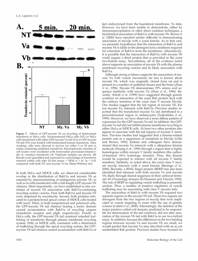
To evaluate more quantitatively the effects of myosin Vbtail expression on transferrin receptor recycling, we studiedthe recycling of biotinylated-transferrin in nontransfectedcells compared with those transiently transfected with eitherthe GFP-myosin Va tail or the GFP-myosin Vb tail. Figure 7demonstrates that although transfection of the GFP-myosinVa tail had no significant effect on the recycling of trans-ferrin, the GFP-myosin Vb tail caused a significant retentionof biotinylated transferrin within cells, consistent with theinhibition of recycling observed with the use of fluorescenttransferrin.
GFP-Myosin Vb Tail Alters Recycling System inPolarized MDCK CellsBecause Rab11a also regulates plasma membrane recyclingin polarized cells, we studied the effects of GFP-myosin Vbtail transfection in polarized MDCK cells (Figure 8). Similarto the pattern seen in HeLa cells, the GFP-myosin Vb tailcolocalized with endogenous Rab11a and elicited an accu-mulation of Rab11a in the pericentriolar region (Figure 8b).
We also studied the effects of nocodazole on transfectedGFP-myosin Vb tail in polarized MDCK cells. Figure 8ddemonstrates that, as in HeLa cells, nocodazole only par-tially dispersed the GFP-myosin Vb tail-labeled vesicles,compared with the effects seen for the endogenous Rab11ain nontransfected cells. These results are consistent with arole for microtubules in trafficking into the apical recyclingsystem. Taxol had no effect on the distribution of GFP-
myosin Vb tail in transiently transfected MDCK cells (ourunpublished results). Thus, the effects of taxol on the apicalrecycling system vesicles probably requires a myosin Vb-dependent process.
As in HeLa cells, GFP-myosin Vb tail transfection did notalter significantly the structure of the Golgi complex (Figure8h). Also as in HeLa cells, GFP-myosin Vb tail had no effecton the distribution of endogenous Rab4, Rab5, Rab7, or Rab9(our unpublished results). The GFP-myosin Vb tail had asimilar effect of concentrating Rab11a immunoreactivity inpolarized HCA-7 colonic adenocarcinoma cells (our unpub-lished results).
GFP-Myosin Vb Tail Alters Transcytosis inPolarized MDCK CellsBecause Rab11a is a critical regulator of transcytosis in po-larized epithelial cells, we sought to evaluate the effects ofthe GFP-myosin Vb tail chimera on transcytosis in MDCKcells stably transfected with the polymeric IgA receptor(pIgR). We first studied the effects of GFP-myosin Vb tail onthe distribution of the pIgR in these MDCK cells. Figure 9demonstrates that expression of GFP-myosin Vb tail causedan accumulation of pIgR in a pericentrosomal compartmentand this accumulation also colocalized with Rab11a (ourunpublished results). In contrast, transfection of GFP-myo-sin Va tail elicited a diffuse distribution in the MDCK cellsand had no effect on the distribution of either pIgR (Figure9) or Rab11a (our unpublished results). These results sug-
Figure 5. Morphological effects ofoverexpression of GFP-myosin Vbtail in HeLa cells. (a–f) HeLa cellswere transiently transfected withGFP-myosin Vb tail and allowed torecover for 24 h. Cells then receivedeither no treatment (a–c) or treat-ment with nocodazole (d–f). Cellswere then fixed and in addition toimaging GFP (a and d) cells werestained for endogenous transferrinreceptor (b and e) and endogenousRab11a (c and f). (g and h) HeLa cellswere transiently transfected withGFP-myosin Vb tail as describedabove. Fixed cells were triple imagedfor GFP (g), p58 immunostaining, aGolgi marker (h), and Rab11a immu-nostaining (i). Images in g–i repre-sent projections assembled from fif-teen 0.29-mm optical sectionsobtained in confocal microscopy. Theresults are representative of threeseparate experiments. Bar, 5 mm (a–f), 2 mm (g–i).
L.A. Lapierre et al.
Molecular Biology of the Cell1850

gested that the myosin Vb tail could retard traffickingthrough the apical recycling system of polarized MDCKcells.
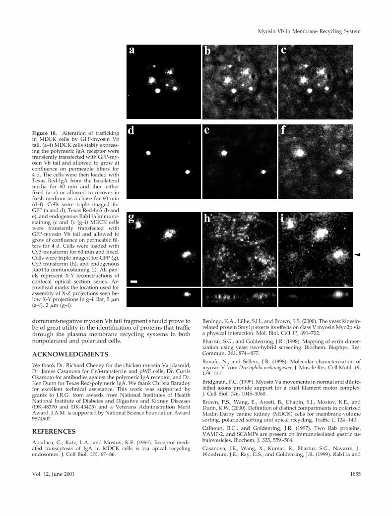
To evaluate trafficking in the polarized MDCK cells ex-pressing pIgR, cells were loaded with Texas Red-polymericIgA from the basolateral medium. In cells overexpressingthe GFP-myosin Vb tail, fluorescent IgA accumulated withthe GFP-myosin Vb tail and endogenous Rab11a (Figure 10,a–c). In addition, when cells were returned into fresh me-dium for 1 h, fluorescent IgA was chased out of nontrans-fected cells, whereas fluorescent IgA was retained in trans-fected cells (Figure 10e). Transfection of GFP-myosin Va tailhad no effect on fluorescent IgA trafficking (our unpublishedresults). In contrast with the results for IgA, when cells wereloaded with Cy3-transferrin from the basolateral medium,no accumulation of fluorescent transferrin was observed inassociation with GFP-myosin Vb tail (Figure 10h). Finally,we were unable to observe trafficking of Cy3-transferrin intoRab11a immunoreactive vesicles. These results are consis-tent with previous findings (Low et al., 1999; Brown et al.,2000) and support the conclusion that transferrin does not
enter the Rab11a-containing apical recycling system in po-larized MDCK cells.
DISCUSSION
Plasma membrane recycling is a fundamental cellular pro-cess regulating the trafficking of membrane receptors, chan-nels, and pumps. In nonpolarized cells, the transferrin re-ceptor is internalized and recycled to the plasma membranethrough a pericentrosomal vesicle system. In polarized cells,plasma membrane recycling is more complicated and a peri-centrosomal vesicular recycling system is thought to medi-ate both basolateral and apical membrane recycling as wellas coordinating the process of basolateral-to-apical transcy-tosis (Mostov et al., 1992; Apodaca et al., 1994). Studies overthe past several years have established that Rab11a is aubiquitous marker of plasma membrane recycling systems(Ullrich et al., 1996). The results presented here indicate thatmyosin Vb also is associated with the plasma membranerecycling system in both nonpolarized and polarized cells.
Figure 6. Effects of overexpressionof GFP-myosin Vb tail and GFP-my-osin Va tail on transferrin traffickingin HeLa cells. To assess the effects ontransferrin trafficking GFP-myosinVb tail (A) or GFP-myosin Va tail (B)were transiently transfected intoHeLa cells. Twenty-four hours aftertransfection, cells were serumstarved for 30 min and then allowedto endocytose Cy3-transferrin for 60min. After uptake, some cells wereallowed to recover in 10% fetal bo-vine serum-containing medium for2 h. Cells were imaged for GFP, Cy3-transferrin, and immunostaining forendogenous transferrin receptor. Theresults are representative of four sep-arate experiments. Bar, 5 mm.
Myosin Vb in Membrane Recycling System
Vol. 12, June 2001 1851
In both HeLa and MDCK cells, we observed considerableoverlap in the distribution of Rab11a and myosin Vb asassessed by immunostaining of endogenous myosin Vb, aswell as in cells transfected with a full-length GFP-myosin Vbchimera. Most importantly, we have established in situ cor-relates of myosin Vb association with Rab11a-containingrecycling system vesicles. First, both Rab11a and myosin Vbwere dispersed by nocodazole. Second, both proteins relo-cated to a perijunctional apical corner of MDCK cells treatedwith taxol. Third, in both nonpolarized and polarized cells,the GFP-myosin Vb tail chimera lacking a motor domaincaused accumulation with Rab11a of recycling cargoes(transferrin receptor and pIgR, respectively). Fourth, inHeLa cells, the GFP-myosin Vb tail construct retarded traf-ficking of transferrin through plasma membrane recyclingsystem. Fifth, in MDCK cells, consistent with a diminutionof trafficking through the apical recycling system, the GFP-myosin Vb tail chimera caused accumulation with Rab11a of
IgA endocytosed from the basolateral membrane. To date,however, we have been unable to demonstrate, either byimmunoprecipitation or other direct isolation techniques, abiochemical association of Rab11a with myosin Vb. Brown etal. (2000) have reported similar difficulty in demonstratingassociation of myo2p with a yeast kinesin. As in that case,we presently hypothesize that the interaction of Rab11a andmyosin Vb is labile in the detergent lysis conditions requiredfor extraction of Rab11a from the membrane. Alternatively,it is possible that the interaction of Rab11a with myosin Vbcould require a third protein that is provided in the yeasttwo-hybrid assay. Nevertheless, all of the evidence notedabove supports an association of myosin Vb with the plasmamembrane recycling vesicles and its likely association withRab11a.
Although strong evidence supports the association of my-osin Va with vesicle movement, far less is known aboutmyosin Vb, which was originally cloned from rat and ispresent in a number of epithelial tissues and the brain (Zhaoet al., 1996). Myosin Vb demonstrates 59% amino acid se-quence similarity with myosin Va (Zhao et al., 1996). Re-cently, Schott et al. (1999) have suggested through geneticevidence an interaction of the yeast Rab protein Sec4 withthe carboxy terminus of the yeast class V myosin Myo2p.Our studies suggest that the tail region of myosin Vb, butnot myosin Va, interacts with Rab11a. Previous studies re-ported that the transfected myosin Va tail distributed to apericentrosomal region in melancoytes (Tsakralides et al.,1999). However, we have observed a more diffuse pattern ofexpression for the GFP-myosin Va tail. In addition, the GFP-myosin Va tail did not inhibit plasma membrane recycling ineither HeLa or MDCK cells. At least two other proteins alsoappear to associate with the tail regions of myosin V mem-bers. Previous studies had suggested that a kinesin-relatedprotein acts as a suppressor of mutations in Myo2p (Lillieand Brown, 1992). Jenkins and colleagues have demon-strated that myosin Va interacts with a ubiquitous kinesinmolecule (Huang et al., 1999) through a region that is highlyhomologous within myosin V family members (the carbox-yl-terminal AF-6 homology domain). Thus, this kinesinwould be expected to interact with all myosin V familymembers. Similarly, as noted above, the yeast class V myo-sin myo2p interacts with a yeast kinesin (Beningo et al.,2000). Recently, a RING finger protein (BERP) has also beenidentified that interacts with both myosin Va and myosinVb, likely through shared sequences in their carboxyl-termi-nal AF-6 homology domain (El-Husseini and Vincent, 1999).The role of BERP in regulating vesicle trafficking is presentlyunclear. Thus, a number of putative regulators of vesicletrafficking may be associating with class V myosin tails.
The association of Rab11a with myosin Vb required twoseparate regions in the myosin Vb tail. These two regions aredivergent from the two regions of myo2p that were impli-cated in vesicle targeting in yeast with the use of geneticscreens (Catlett et al., 2000). Interestingly, the deletion of themajor putative coiled-coil domain, predicted to be responsi-ble for dimerization of the tail construct, did not alter asso-ciation of the myosin Vb tail with Rab11a in our two-hybridassay. In addition, because the differences in the two bindingregions between myosin Va and myosin Vb are small, wewould predict that myosin Va may also bind with an as yetunidentified Rab protein. Previous studies have focused on
Figure 7. Effects of GFP-myosin Vb on recycling of biotinylatedtransferrin in HeLa cells. Nontransfected HeLa cells (NT) or HeLacells transfected with either GFP-myosin Va tail (Va) or GFP-myosinVb tail (Vb) were loaded with biotinylated-human transferrin. Afterwashing, cells were allowed to recover for either 5 or 20 min inmedia containing unlabeled transferrin. Electrophoretic transfers ofcell lysates were incubated with horseradish peroxidase-streptavi-din to visualize transferrin (A) Triplicate samples are shown. (B)Results were quantified and expressed as a percentage of transferrinretained within cells after 20 min (mean 1 SEM; n 5 6). *p , 0.01compared with both NT and myosin Va by Mann-Whitney test.
L.A. Lapierre et al.
Molecular Biology of the Cell1852

the presence of mutations in myosin Va in mouse dilutemutants (Wu et al., 1998; Bridgman, 1999) and human Gris-celli syndrome patients. However, a number of recent inves-tigations have identified a mutation in Rab27a responsiblefor the phenotype of the ashen mouse (Wilson et al., 2000) aswell as in a subset of Griscelli syndrome patients (Menascheet al., 2000). The similarity of these phenotypes in the mousemutants and Griscelli syndrome patients suggest thatRab27a may be the specific binder for the myosin Va tail.Another unconventional myosin, myr4, has also recentlybeen implicated in the regulation of early steps in traffickingof recycling cargoes (Huber et al., 2000). Nevertheless, it isclear from the data presented here that the myosin Va tailhad no effect on recycling of either transferrin or IgA recep-tor in either nonpolarized or polarized cells. In contrast, themyosin Vb tail acted as a potent dominant-negative con-struct, causing pericentrosomal accumulation of transferrinreceptor in nonpolarized HeLa cells and of pIgR in polarizedMDCK cells.
As in the study of all Rab proteins, the identification ofupstream and downstream regulators is critical for an un-derstanding of the function of Rab11 family members. Twogroups (Mammoto et al., 1999; Zeng et al., 1999) have iden-tified a novel protein (Rab11-BP/Rabphillin-11), which in-
teracts with Rab11a as assessed by protein overlay. Al-though the protein does not strictly colocalize with Rab11a,transfection and overexpression of an amino or carboxyl-truncated protein alters transferrin trafficking in nonpolar-ized cells (Mammoto et al., 1999; Zeng et al., 1999). Likemyosin Vb, this protein appears to interact with Rab11a inits GTP-bound form. Also like myosin Vb, Rab11-BP did notassociate with Rab11a effector domain mutants (Mammotoet al., 1999; Zeng et al. 1999). These studies and the presentdata suggest that Rab11a may be interacting with multipleprotein effectors and modulators along the recycling path-way. Indeed, in further yeast two-hybrid screens with dif-ferent Rab11a and Rab25 baits, we have now identified fourfurther putative Rab11-interacting proteins (Goldenring, un-published data).
Previous studies have also established the importance ofthe microtubule cytoskeleton in the regulation of the plasmamembrane recycling system in both nonpolarized and po-larized cells. Depolymerization of microtubules causes dis-persal of the recycling system (Apodaca et al., 1994;Casanova et al., 1999). In MDCK cells, stabilization of micro-tubules with taxol causes relocation of the Rab11a-contain-ing recycling vesicles to a position adjacent to the junctionalcomplex (Casanova et al., 1999). Our results suggest that the
Figure 8. GFP-myosin Vb tail altersapical recycling system morphologyin MDCK cells. MDCK were tran-siently transfected with GFP-myosinVb tail and grown for 3 d at conflu-ence on permeable filters. (a–f) Cellswere then exposed to either no treat-ment (a–c) or treatment with nocoda-zole (d–f). Fixed cells were triple im-aged for GFP (a and d), endogenousRab11a (b and e), and ZO-1 (c and f).(g–i) GFP-myosin Vb tail transfectedMDCK cells were fixed and triple im-aged for GFP (g), the Golgi markerp58 (h), and ZO-1 (i). The results arerepresentative of three separate ex-periments. Bar, 5 mm.
Myosin Vb in Membrane Recycling System
Vol. 12, June 2001 1853

distribution of myosin Vb also is affected by the state ofmicrotubule polymerization. In MDCK cells, myosin Vbstaining dispersed along with GFP-Rab11a after treatmentwith nocodazole, and both colocalized to the apical cornersafter taxol treatment. Because rearrangements with taxol areespecially characteristic of Rab11a recycling system vesicles(Casanova et al., 1999), these results provide a functionalconfirmation of myosin Vb association with the recyclingsystem. Previous investigations in melanocytes have shownthat microtubule polymerization affects myosin Va distribu-tion (Tsakraklides et al., 2000). In these studies, as in ourown, an association of myosin V species with a kinesin toform a multifunctional motor complex (Huang et al., 1999;Beningo et al., 2000) would explain the dispersal of vesiclesand myosin Vb staining by nocodozole treatment.
The present investigations support the hypothesis thatmyosin Vb, likely through an interaction with Rab11a, isinvolved in exiting the recycling system. Consistent withthis hypothesis, the myosin Vb tail construct without a mo-tor domain acts as a dominant-negative protein, associatingwith Rab11a-containing recycling system vesicles, but pre-venting their interaction with cytoskeletal elements requiredfor transit out of the recycling system. Furthermore, nocoda-zole partially dispersed GFP-myosin Vb tail concentration ofthe recycling system in both HeLa and MDCK cells. Thus, it
is likely that microtubules are required for steps proximal tothe function of myosin Vb. This model would explain theaccumulation of cargoes trafficking through the recyclingsystem within GFP-myosin Vb tail containing vesicles. Re-cent studies have provided evidence that microfilamentsand Rac1 may be associated with the recycling system inMDCK cells (Jou and Nelson, 1998; Jou et al., 1998). Domi-nant active Rac1 colocalizes with Rab11a and inhibits apicalrecycling (Jou and Nelson, 1998; Jou et al., 1998). Indeed, thepresumption that myosin Vb also would interact with akinesin (Huang et al., 1999) suggests that a myosin Vb/kinesin complex could mediate trafficking through the recy-cling system. Thus, although the microtubule system is in-volved in trafficking of vesicles into the plasma membranerecycling systems, microfilaments and myosin Vb may beresponsible, at least in part, for trafficking of vesicles out ofthe recycling system. A similar mechanism may be involvedin Golgi trafficking where Goud and colleagues have re-cently identified another novel kinesin as an effector forRab6 (Echard et al., 1998).
In polarized cells, controversy continues to exist over therole of Rab11a in basolateral trafficking of transferrin recep-tor versus apical recycling and transcytosis. With the use ofelectron microscopic examination, Hopkins and colleagues(Hughson and Hopkins, 1990; Knight et al., 1995; Odorizzi etal., 1996) have noted colocalization of endocytosed trans-ferrin and polymeric IgA in tubular endosomes. However,in polarized MDCK cells, Dunn and colleagues (Brown et al.,2000) have recently reported that fluorescently labeled trans-ferrin and polymeric IgA were sorted away from each otherbefore the entry of IgA into a Rab11a-containing apicalrecycling compartment. In addition, we have demonstratedin polarized MDCK cells that dominant-negative Rab11amutants inhibit transcytosis of IgA, but have no effect on thebasolateral recycling of transferrin (Wang et al., 2000). Thestudies of Dunn (Brown et al., 2000) have established anorderly progression of sorting whereby transferrin and IgAendocytosed from the basolateral membrane enter a com-mon recycling endosome after segregation away from low-density lipoprotein receptors. This recycling endosome islikely the common recycling endosome originally describedby Hopkins and colleagues in MDCK and HeLa cells (Hugh-son and Hopkins, 1990; Knight et al., 1995; Odorizzi et al.,1996). From the recycling endosome, transferrin and IgA areseparated with transferrin receptor recycling to the basolat-eral membrane, whereas transcytosing IgA receptor movesinto an apical recycling endosome containing Rab11a. Wehave verified these findings here and, additionally, we havefound that transferrin was not trafficked into GFP-myosinVb tail containing vesicles in MDCK cells, as it was in HeLacells. These data support the hypothesis that, in polarizedepithelial cells, transcytosing cargoes are sorted away fromthe majority of transferrin receptor before entry into theRab11a-containing apical recycling system.
In summary, the results presented here demonstrate thatmyosin Vb associates with Rab11a-containing plasma mem-brane recycling systems in both nonpolarized and polarizedcells. The data indicate that myosin Vb is required for traf-ficking out of plasma membrane recycling systems. Becausemyosin Va does not interact with any Rab11 family member,further studies will be required to discern whether otherclasses of Rab proteins interact with the myosin Va tail. The
Figure 9. GFP-myosin Vb tails alters the distribution of polymericIgA receptor. MDCK cells were transiently transfected with eitherGFP-myosin Vb tail or GFP-myosin Va tail and grown for 3 d atconfluence on permeable filters. Cells were then fixed and dualimaged for GFP and pIgR immunostaining. The results are repre-sentative of three separate experiments. Bar, 5 mm.
L.A. Lapierre et al.
Molecular Biology of the Cell1854
dominant-negative myosin Vb tail fragment should prove tobe of great utility in the identification of proteins that trafficthrough the plasma membrane recycling systems in bothnonpolarized and polarized cells.
ACKNOWLEDGMENTS
We thank Dr. Richard Cheney for the chicken myosin Va plasmid,Dr. James Casanova for Cy3-transferrin and pWE cells, Dr. CurtisOkamoto for antibodies against the polymeric IgA receptor, and Dr.Ken Dunn for Texas Red-polymeric IgA. We thank Christa Baradoyfor excellent technical assistance. This work was supported bygrants to J.R.G. from awards from National Institutes of HealthNational Institute of Diabetes and Digestive and Kidney Diseases(DK-48370 and DK-43405) and a Veterans Administration MeritAward. J.A.M. is supported by National Science Foundation Award9874907.
REFERENCES
Apodaca, G., Katz, L.A., and Mostov, K.E. (1994). Receptor-medi-ated transcytosis of IgA in MDCK cells is via apical recyclingendosomes. J. Cell Biol. 125, 67–86.
Beningo, K.A., Lillie, S.H., and Brown, S.S. (2000). The yeast kinesin-related protein Smy1p exerts its effects on class V myosin Myo2p viaa physical interaction. Mol. Biol. Cell 11, 691–702.
Bhartur, S.G., and Goldenring, J.R. (1998). Mapping of ezrin dimer-ization using yeast two-hybrid screening. Biochem. Biophys. Res.Commun. 243, 874–877.
Bonafe, N., and Sellers, J.R. (1998). Molecular characterization ofmyosin V from Drosophila melanogaster. J. Muscle Res. Cell Motil. 19,129–141.
Bridgman, P.C. (1999). Myosin Va movements in normal and dilute-lethal axons provide support for a dual filament motor complex.J. Cell Biol. 146, 1045–1060.
Brown, P.S., Wang, E., Aroeti, B., Chapin, S.J., Mostov, K.E., andDunn, K.W. (2000). Definition of distinct compartments in polarizedMadin-Darby canine kidney (MDCK) cells for membrane-volumesorting, polarized sorting and apical recycling. Traffic 1, 124–140.
Calhoun, B.C., and Goldenring, J.R. (1997). Two Rab proteins,VAMP-2, and SCAMPs are present on immunoisolated gastric tu-bulovesicles. Biochem. J. 325, 559–564.
Casanova, J.E., Wang, X., Kumar, R., Bhartur, S.G., Navarre, J.,Woodrum, J.E., Ray, G.S., and Goldenring, J.R. (1999). Rab11a and
Figure 10. Alteration of traffickingin MDCK cells by GFP-myosin Vbtail. (a–f) MDCK cells stably express-ing the polymeric IgA receptor weretransiently transfected with GFP-my-osin Vb tail and allowed to grow atconfluence on permeable filters for4 d. The cells were then loaded withTexas Red-IgA from the basolateralmedia for 60 min and then eitherfixed (a–c) or allowed to recover infresh medium as a chase for 60 min(d–f). Cells were triple imaged forGFP (a and d), Texas Red-IgA (b ande), and endogenous Rab11a immuno-staining (c and f). (g–i) MDCK cellswere transiently transfected withGFP-myosin Vb tail and allowed togrow at confluence on permeable fil-ters for 4 d. Cells were loaded withCy3-transferrin for 60 min and fixed.Cells were triple imaged for GFP (g),Cy3-transferrin (h), and endogenousRab11a immunostaining (i). All pan-els represent X-Y reconstructions ofconfocal optical section series. Ar-rowhead marks the location used forassembly of X-Z projections seen be-low X-Y projections in g–i. Bar, 5 mm(a–f), 2 mm (g–i).
Myosin Vb in Membrane Recycling System
Vol. 12, June 2001 1855

Rab25 association with the apical recycling system of polarizedMDCK cells. Mol. Biol. Cell 10, 47–61.
Catlett, N.L., Duex, J.E., Tang, F., and Weisman, L.S. (2000). Twodistinct regions in a yeast myosin-V tail domain are required for themovement of different cargoes. J. Cell Biol. 150, 513–525.
Catlett, N.L., and Weisman, L.S. (1998). The terminal tail region of ayeast myosin-V mediates its attachment to vacuole membranes andsites of polarized growth. Proc. Natl. Acad. Sci. USA 95, 14799–14804.
Chavrier, P., Vingron, M., Sander, C., Simons, K., and Zerial, M.(1990). Molecular cloning of YPT1/SEC4-related cDNAs from anepithelial cell line. Mol. Cell. Biol. 10, 6578–6585.
Duman, J.G., Tyagarajan, K., Kolsi, M.S., Moore, H.H., and Forte,J.G. (1999). Expression of rab11a N124I in gastric parietal cellsinhibits stimulatory recruitment of the H1-K1-ATPase. Am. J.Physiol. 277, C361–C372.
Echard, A., Jollivet, F., Martinez, O., Lacapere, J.-J., Rousselet, A.,Janoueix-Lerosey, I., and Goud, B. (1998). Interaction of a Golgi-associated kinesin-like protein with Rab6. Science 279, 580–585.
El-Husseini, A.E., and Vincent, S.R. (1999). Cloning and character-ization of a novel RING finger protein that interacts with class Vmyosins. J. Biol. Chem. 274, 19771–19777.
Evans, L.L., Lee, A.J., Bridgman, P.C., and Mooseker, M.S. (1998).Vesicle-associated brain myosin-V can be activated to catalyze actin-based transport. J. Cell Sci. 111, 2055–2066.
Forte, J.G., and Yao, X. (1996). The membrane-recruitment-and-recycling hypothesis of gastric HCl secretion. Trends Cell Biol. 6,45–48.
Goldenring, J.R., Shen, K.R., Vaughan, H.D., and Modlin, I.M.(1993). Identification of a small GTP-binding protein, Rab25, ex-pressed in the gastrointestinal mucosa, kidney and lung. J. Biol.Chem. 268, 18419–18422.
Goldenring, J.R., Soroka, C.J., Shen, K.R., Tang, L.H., Rodriguez, W.,Vaughan, H.D., Stoch, S.A., and Modlin, I.M. (1994). Enrichment ofrab11, a small GTP-binding protein, in gastric parietal cells. Am. J.Physiol. 267, G187–G194.
Green, E.G., Ramm, E., Riley, N.M., Spiro, D.J., Goldenring, J.R., andWessling-Resnick, M. (1997). Rab11 is associated with transferrin-containing recycling compartments in K562 cells. Biochem. Biophys.Res. Commun. 239, 612–616.
Hoffenberg, S., Sanford, J.C., Liu, S., Daniel, D.S., Tuvin, M., Knoll,B.J., Wessling-Resnick, M., and Dickey, B.F. (1995). Biochemical andfunctional characterization of a recombinant GTPase, Rab5, and twoof its mutants. J. Biol. Chem. 270, 5048–5056.
Huang, J.-D., Brady, S.T., Richards, B.W., Stenoien, D., Resau, J.H.,Copeland, N.G., and Jenkins, N.A. (1999). Direct interaction of mi-crotubule- and actin-based transport motors. Nature 397, 267–270.
Huang, W.M., Reed-Fourquet, L., Wu, E., and Wu, J.Y. (1990).Molecular cloning and amino acid sequence of brain L-glutamatedecarboxylase. Proc. Natl. Acad. Sci. USA 87, 8491–8495.
Huber, L.A., Fialka, I., Paiha, K., Hunziker, W., Sacks, D.B., Bahler,M., Way, M., Gagescu, R., and Gruenberg, J. (2000). Both calmodulinand the unconventional myosin myr4 regulate membrane traffick-ing along the recycling pathway of MDCK cells. Traffic 1, 494–503.
Hughson, E.J., and Hopkins, C. (1990). Endocytic pathways in po-larized caco-2 cells: identification of an endosomal compartmentaccessible from both apical and basolateral surfaces. J. Cell Biol. 110,337–348.
Jou, T.-Z., and Nelson, J.W. (1998). Effects of regulated expression ofmutant RhoA and Rac1 small GTPases on the development ofepithelial (MDCK) cell polarity. J. Cell Biol. 142, 85–100.
Jou, T.S., Schneeberger, E.E., and Nelson, W.J. (1998). Structural andfunctional regulation of tight junctions by rhoA and rac1 smallGTPases. J. Cell Biol. 142, 101–115.
Kikuchi, A., Yamashita, T., Kawata, M., Yamamoto, K., Ikeda, K.,Tanimoto, T., and Takai, Y. (1988). Purification and characterizationof a novel GTP-binding protein with a molecular weight of 24,000from bovine brain membranes. J. Biol. Chem. 263, 2897–2904.
Knight, A., Hughson, E., Hopkins, C.R., and Cutler, D.F. (1995).Membrane protein trafficking through the common apical endo-some compartment of polarized Caco-2 cells. Mol. Biol. Cell 6,597–610.
Lai, F., Stubbs, L., and Artzt, K. (1994). Molecular analysis of mouseRab11b: a new type of mammalian YPT/Rab protein. Genomics 22,610–616.
Lillie, S.H., and Brown, S.S. (1992). Suppression of a myosin defectby a kinesin-related gene. Nature 356, 358–361.
Low, S.H., Chapin, S.J., Wimmer, C., Whiteheart, S.W., Komuves,L.G., Mostov, K.E., and Weimbs, T. (1999). The SNARE machinery isinvolved in apical plasma membrane trafficking in MDCK cells.J. Biol. Chem. 141, 1503–1513.
Mammoto, A., Ohtsuka, T., Hotta, I., Sasaki, T., and Takai, Y. (1999).Rab11BP/Rabphillin-11, a downstream target of Rab11 small Gprotein implicated in vesicle recycling. J. Biol. Chem. 274, 25517–25524.
Menasche, G., Pastural, E., Feldman, J., Certain, S., Ersoy, F., Du-puis, S., Wulffraat, N., Bianchi, D., Fischer, A., Le Deist, F., and deSaint Basile, G. (2000). Mutations in Rab27a cause Griscelli syn-drome associated with hemophagocytic syndrome. Nature Genet.25, 173–176.
Mercer, J.A., Seperack, P.K., Strobel, M.C., Copeland, N.G., andJenkins, N.A. (1991). Novel myosin heavy chain encoded by murinedilute coat color locus. Nature 349, 709–712.
Miller, K.E., and Sheetz, M.P. (2000). Characterization of myosin Vbinding to brain vesicles. J. Biol. Chem. 275, 2598–2606.
Mostov, K., Apodaca, G., Aroeti, B., and Okamoto, C. (1992). Plasmamembrane protein sorting in polarized epithelial cells. J. Cell Biol.116, 577–583.
Nascimento, A.A.C., Amaral, R.G., Bizario, J.C.S., Larson, R.E., andEspreafico, E.M. (1997). Subcellular localization of myosin-V in theB16 melanoma cells, a wild-type cell line for the dilute gene. Mol.Biol. Cell 8, 1971–1988.
Odorizzi, G., Pearse, A., Domingo, D., Trowbridge, I.S., and Hop-kins, C.R. (1996). Apical and basolateral nedosomes of MDCK cellsare interconnectedand contain a polarized sorting mechanism.J. Cell Biol. 135, 139–152.
Provance, D.W., Jr., Wei, M., Ipe, V., and Mercer, J.A. (1996). Cul-tured melanocytes from dilute mice exhibit dendritic morphologyand altered melanosome distribution. Proc. Natl. Acad. Sci. USA 93,14554–14558.
Reck-Peterson, S.L., Provance, D.W., Mooseker, M.S., and Mercer,J.A. (2000). Class V myosins. Biochem. Biophys. Acta 1496, 36–51.
Ren, X., Xu, G., Zeng, J., De Lemos-Chiarandini, C., Adesnik, M.,and Sabatini, D.D. (1998). Hydrolysis of GTP on rab11 is requiredfor the direct delivery of transferrin from pericentriolar recyclingcompartment to the cell surface but not from sorting endosomes.Proc. Natl. Acad. Sci. USA 95, 6187–6192.
Rogers, S.L., and Gelfand, V.I. (1998). Myosin cooperates with mi-crotubule motors during organelle transport in melanophores. Curr.Biol. 8, 161–164.
Schott, D., Ho, J., Pruyne, D., and Bretscher, A. (1999). The COOH-terminal domain of Myo2p, a yeast myosin V, has a direct role insecretory vesicle targeting. J. Cell Biol. 147, 791–807.
L.A. Lapierre et al.
Molecular Biology of the Cell1856
Tabb, J.S., Molyneaux, B.J., Cohen, D.L., Kuznetsov, S.A., and Lang-ford, G.M. (1998). Transport of ER vesicles on actin filaments inneurons by myosin V. J. Cell Sci. 111, 3221–3234.
Tsakraklides, V., Krogh, K., Bizario, J.C., Larson, R.E., Espreafico,E.M., and Wolenski, J.S. (2000). Subcellular localization of GFP-myosin V in live mouse melanocytes. J. Cell Sci. 112, 2853–2865.
Ullrich, O., Reinsch, S., Urbe, S., Zerial, M., and Parton, R.G. (1996).Rab11 regulates recycling through the pericentriolar recycling en-dosome. J. Cell Biol. 135, 913–924.
Wang, X., Kumar, R., Navarre, J., Casanova, J.E., and Goldenring,J.R. (2000). Regulation of vesicle trafficking in Madin-Darby CanineKidney cells by Rab11a and Rab25. J. Biol. Chem. 275, 29138–29146.
Wilson, S.M., Yip, R., Swing, D.A., O’Sullivan, N., Zhang, Y., Novak,E.K., Swank, R.T., Russell, L.B., Copeland, N.G., and Jenkins, N.A.
(2000). A mutation in Rab27a causes the vesicle transport defectsobserved in ashen mice. Proc. Natl. Acad. Sci. USA 97, 7933–7938.
Wu, X., Bowers, B., Rao, K., Wei, Q., and Hammer, J.A. (1998).Visualization of melanosome dynamics within wild-type and dilutemelanocytes suggests a paradigm for myosin V function In vivo.J. Cell Biol. 143, 1899–1918.
Zeng, X., Ren, M., Gravotta, D., De Lemos-Chiarandini, C., Lui, M.,Erdjument-Bromage, H., Tempst, P., Xu, G., Shen, T.H., Morimoto,T., Adesnick, M., and Sabatini, D.D. (1999). Identification of a pu-tative effector protein for rab11 that participates in transferrin recy-cling. Proc. Natl. Acad. Sci. USA 96, 2840–2845.
Zhao, L.P., Koslovsky, J.S., Reinhard, J., Bahler, M., Witt, A.E.,Provance, Jr., D.W., and Mercer, J.A. (1996). Cloning and character-ization of myr6, an unconventional myosin of the dilute/myosin-Vfamily. Proc. Natl. Acad. Sci., USA 93, 10826–10831.
Myosin Vb in Membrane Recycling System
Vol. 12, June 2001 1857