Identification of SH3 Domain Proteins Interacting with the Cytoplasmic Tail of the A Disintegrin and...
14
Identification of SH3 Domain Proteins Interacting with the Cytoplasmic Tail of the A Disintegrin and Metalloprotease 10 (ADAM10) Henriette Ebsen, Marcus Lettau, Dieter Kabelitz, Ottmar Janssen* University of Kiel, Molecular Immunology, Institute for Immunology, University Hospital Schleswig-Holstein Campus Kiel, Kiel, Germany Abstract The a disintegrin and metalloproteases (ADAMs) play a pivotal role in the control of development, adhesion, migration, inflammation and cancer. Although numerous substrates of ADAM10 have been identified, the regulation of its surface expression and proteolytic activity is still poorly defined. One current hypothesis is that both processes are in part modulated by protein-protein interactions mediated by the intracellular portion of the protease. For related proteases, especially proline-rich regions serving as docking sites for Src homology domain 3 (SH3) domain-containing proteins proved to be important for mediating regulatory interactions. In order to identify ADAM10-binding SH3 domain proteins, we screened the All SH3 Domain Phager library comprising 305 human SH3 domains using a GST fusion protein with the intracellular region of human ADAM10 as a bait for selection. Of a total of 291 analyzed phage clones, we found 38 SH3 domains that were precipitated with the ADAM10-derived fusion protein but not with GST. We verified the binding to the cytosolic portion of ADAM10 for several candidates by co-immunoprecipitation and/or pull down analyses. Intriguingly, several of the identified proteins have been implicated in regulating surface appearance and/or proteolytic activity of related ADAMs. Thus, it seems likely that they also play a role in ADAM10 biology. Citation: Ebsen H, Lettau M, Kabelitz D, Janssen O (2014) Identification of SH3 Domain Proteins Interacting with the Cytoplasmic Tail of the A Disintegrin and Metalloprotease 10 (ADAM10). PLoS ONE 9(7): e102899. doi:10.1371/journal.pone.0102899 Editor: Laszlo Buday, Hungarian Academy of Sciences, Hungary Received May 6, 2014; Accepted June 25, 2014; Published July 18, 2014 Copyright: ß 2014 Ebsen et al. This is an open-access article distributed under the terms of the Creative Commons Attribution License, which permits unrestricted use, distribution, and reproduction in any medium, provided the original author and source are credited. Data Availability: The authors confirm that all data underlying the findings are fully available without restriction. All relevant data are within the paper and its Supporting Information files. Funding: Funding for this study was provided by the German Research Foundation (DFG), www.dfg.de, DFG CRC 877 project B4 - OJ; DFG CRC 877 project A7 - DK; Individual funding DFG LE2571/3-1 - ML. The funders had no role in study design, data collection and analysis, decision to publish, or preparation of the manuscript. Competing Interests: The authors have declared that no competing interests exist. * Email: [email protected] Introduction The a disintegrin and metalloprotease (ADAM) proteins form a subgroup of the metzincin superfamily that also comprises matrix metalloproteinases (MMPs) and the ADAM proteases with thrombospondin motifs (ADAMTSs). ADAMs are glycosylated type I transmembrane proteins that are specialized for juxtamem- brane cleavage of spatially associated membrane proteins [1,2]. This ectodomain shedding results in the release of bioactive extracellular protein fragments. For example, TNF-a is liberated by the TNF-a converting enzyme (TACE, ADAM17) [3] and the Fas ligand (FasL, CD95L) is cleaved by ADAM10 [4–6]. Structurally, ADAMs contain several distinct subdomains including a signal peptide, a pro-domain that is cleaved off during maturation, a metalloprotease domain, a disintegrin domain, a cysteine-rich region, an EGF (epidermal growth factor)-like or membrane-proximal domain, a transmembrane domain and an intracellular region (Fig. 1). Since only 13 of the 21 or 22 presumed functional human ADAMs possess proteolytic activity [1,2], it is likely that other domains also contribute to the overall biological functions of ADAM proteins. For example, the disintegrin domains guide interactions with integrins and the cysteine-rich domains support cell adhesion by binding to syndecans or fibronectin or clustering with other ADAMs [7]. The C-terminal cytoplasmic tails vary in length between different members of the family and have been implicated in the regulation of ADAM maturation, activity and localization [8]. Different phosphorylation sites seem to be relevant for signal transduction in the context of ADAM mobilization or activity [1]. For some ADAMs, serine/threonine and/or tyrosine phosphorylation was reported [9,10], which might lead to the generation of inducible binding sites and/or protein complex formation, for example to facilitate Src homology 2 (SH2) domain protein binding upon tyrosine phosphorylation. In addition, most ADAM proteases contain one or more proline-rich stretches, that potentially enable interactions with Src homology 3 (SH3) domain-containing signaling molecules. As depicted in Fig. 1, except for one proline-to-glutamine exchange, the intracellular regions of ADAM10 are identical in mice and humans, suggesting an important and highly conserved regulatory function. In human ADAM10, the two prominent proline-rich regions (PRRs) comprise amino acids 708–717 (PKLPPPKPLP) and 722–728 (RRRPPQP), respectively. As mentioned, different ADAM family members vary in the number of intracellular PRRs. Whereas some ADAM proteins do not contain any classical SH3 domain binding site (e.g. ADAM28), other members contain only one or two (ADAM10, ADAM17) and some ADAMs (ADAM12, ADAM15) contain multiple SH3 binding motifs. This in turn PLOS ONE | www.plosone.org 1 July 2014 | Volume 9 | Issue 7 | e102899
Transcript of Identification of SH3 Domain Proteins Interacting with the Cytoplasmic Tail of the A Disintegrin and...

Identification of SH3 Domain Proteins Interacting withthe Cytoplasmic Tail of the A Disintegrin andMetalloprotease 10 (ADAM10)Henriette Ebsen, Marcus Lettau, Dieter Kabelitz, Ottmar Janssen*
University of Kiel, Molecular Immunology, Institute for Immunology, University Hospital Schleswig-Holstein Campus Kiel, Kiel, Germany
Abstract
The a disintegrin and metalloproteases (ADAMs) play a pivotal role in the control of development, adhesion, migration,inflammation and cancer. Although numerous substrates of ADAM10 have been identified, the regulation of its surfaceexpression and proteolytic activity is still poorly defined. One current hypothesis is that both processes are in partmodulated by protein-protein interactions mediated by the intracellular portion of the protease. For related proteases,especially proline-rich regions serving as docking sites for Src homology domain 3 (SH3) domain-containing proteins provedto be important for mediating regulatory interactions. In order to identify ADAM10-binding SH3 domain proteins, wescreened the All SH3 Domain Phager library comprising 305 human SH3 domains using a GST fusion protein with theintracellular region of human ADAM10 as a bait for selection. Of a total of 291 analyzed phage clones, we found 38 SH3domains that were precipitated with the ADAM10-derived fusion protein but not with GST. We verified the binding to thecytosolic portion of ADAM10 for several candidates by co-immunoprecipitation and/or pull down analyses. Intriguingly,several of the identified proteins have been implicated in regulating surface appearance and/or proteolytic activity ofrelated ADAMs. Thus, it seems likely that they also play a role in ADAM10 biology.
Citation: Ebsen H, Lettau M, Kabelitz D, Janssen O (2014) Identification of SH3 Domain Proteins Interacting with the Cytoplasmic Tail of the A Disintegrin andMetalloprotease 10 (ADAM10). PLoS ONE 9(7): e102899. doi:10.1371/journal.pone.0102899
Editor: Laszlo Buday, Hungarian Academy of Sciences, Hungary
Received May 6, 2014; Accepted June 25, 2014; Published July 18, 2014
Copyright: � 2014 Ebsen et al. This is an open-access article distributed under the terms of the Creative Commons Attribution License, which permitsunrestricted use, distribution, and reproduction in any medium, provided the original author and source are credited.
Data Availability: The authors confirm that all data underlying the findings are fully available without restriction. All relevant data are within the paper and itsSupporting Information files.
Funding: Funding for this study was provided by the German Research Foundation (DFG), www.dfg.de, DFG CRC 877 project B4 - OJ; DFG CRC 877 project A7 -DK; Individual funding DFG LE2571/3-1 - ML. The funders had no role in study design, data collection and analysis, decision to publish, or preparation of themanuscript.
Competing Interests: The authors have declared that no competing interests exist.
* Email: [email protected]
Introduction
The a disintegrin and metalloprotease (ADAM) proteins form a
subgroup of the metzincin superfamily that also comprises matrix
metalloproteinases (MMPs) and the ADAM proteases with
thrombospondin motifs (ADAMTSs). ADAMs are glycosylated
type I transmembrane proteins that are specialized for juxtamem-
brane cleavage of spatially associated membrane proteins [1,2].
This ectodomain shedding results in the release of bioactive
extracellular protein fragments. For example, TNF-a is liberated
by the TNF-a converting enzyme (TACE, ADAM17) [3] and the
Fas ligand (FasL, CD95L) is cleaved by ADAM10 [4–6].
Structurally, ADAMs contain several distinct subdomains
including a signal peptide, a pro-domain that is cleaved off during
maturation, a metalloprotease domain, a disintegrin domain, a
cysteine-rich region, an EGF (epidermal growth factor)-like or
membrane-proximal domain, a transmembrane domain and an
intracellular region (Fig. 1). Since only 13 of the 21 or 22
presumed functional human ADAMs possess proteolytic activity
[1,2], it is likely that other domains also contribute to the overall
biological functions of ADAM proteins. For example, the
disintegrin domains guide interactions with integrins and the
cysteine-rich domains support cell adhesion by binding to
syndecans or fibronectin or clustering with other ADAMs [7].
The C-terminal cytoplasmic tails vary in length between different
members of the family and have been implicated in the regulation
of ADAM maturation, activity and localization [8]. Different
phosphorylation sites seem to be relevant for signal transduction in
the context of ADAM mobilization or activity [1]. For some
ADAMs, serine/threonine and/or tyrosine phosphorylation was
reported [9,10], which might lead to the generation of inducible
binding sites and/or protein complex formation, for example to
facilitate Src homology 2 (SH2) domain protein binding upon
tyrosine phosphorylation.
In addition, most ADAM proteases contain one or more
proline-rich stretches, that potentially enable interactions with Src
homology 3 (SH3) domain-containing signaling molecules. As
depicted in Fig. 1, except for one proline-to-glutamine exchange,
the intracellular regions of ADAM10 are identical in mice and
humans, suggesting an important and highly conserved regulatory
function. In human ADAM10, the two prominent proline-rich
regions (PRRs) comprise amino acids 708–717 (PKLPPPKPLP)
and 722–728 (RRRPPQP), respectively. As mentioned, different
ADAM family members vary in the number of intracellular PRRs.
Whereas some ADAM proteins do not contain any classical SH3
domain binding site (e.g. ADAM28), other members contain only
one or two (ADAM10, ADAM17) and some ADAMs (ADAM12,
ADAM15) contain multiple SH3 binding motifs. This in turn
PLOS ONE | www.plosone.org 1 July 2014 | Volume 9 | Issue 7 | e102899

suggests that specific PRR-mediated protein-protein interactions
might exert characteristic functions to modulate transport,
localization and/or regulation of activity of individual ADAM
family members.
The first screening for ADAM-interacting SH3 domain proteins
was performed by Cousin and colleagues who described the
interaction of the protein kinase C and casein kinase substrate in
neurons 2 (PACSIN2) and ADAM13 in Xenopus embryos/cells.
Functionally, overexpression of X-PACSIN2 could rescue devel-
opmental alterations induced by overexpression of ADAM13 [11].
Given that individual SH3 domains of different proteins bind to
several similar PRRs, two or even more putative SH3 domain
binding sites indicate that individual ADAM proteases might
associate with a whole array of SH3 domain-containing proteins.
As an example, ADAM12 was reported to bind Src kinases (c-Src
and Yes), the growth factor receptor-bound protein 2 (Grb2)
[12,13], the p85a subunit of phosphatidylinositol 3 kinase (PI3K)
[14], the protein kinase C and casein kinase substrate in neurons 3
(PACSIN3) [15], a-Actinin-1 and -2 [16,17], the adaptor protein
TKS5 (Five SH3 domain-containing protein = FISH) [18] and
the SH3 domain-containing protein 19 (SH3D19, ADAM-binding
protein Eve-1, EEN-binding protein, EBP) [19]. Several of these
interactions have been further investigated in different cellular
systems. It turned out that PACSIN3 up-regulates ectodomain
shedding of the ADAM12 substrate heparin-binding epidermal
growth factor-like growth factor (HBEGF) [15], similar to what
was described shortly thereafter for the multidomain adaptor
protein Eve-1 by Tanaka and colleagues [19]. ADAM12-L, the
long transmembrane form of ADAM12, transiently interacts with
c-Src. When ADAM12-L gets phosphorylated by c-Src, it
redistributes from perinuclear regions to actin-associated regions
at the cell periphery. Interestingly, in response to external signals
Figure 1. Schematic representation of the domain structure of ADAM10. (A) The cytoplasmic tail at the C-terminus harbors two proline-richregions (PRR) that might enable binding to SH3 or WW domain-containing proteins. The intracellular parts of human and murine ADAM10 are highlyconserved and differ in only one amino acid. (B) Modular composition, maturation and processing of ADAM10. The N-terminal signal sequence of theprotease is needed for intracellular maturation (1R2). To generate an enzymatically active protease, the pro-domain has to be removed by a proteinconvertase such as furin (2R3). The catalytic metalloproteinase domain is the largest domain of ADAM10 and might be activated by different signalsincluding substrate-induced conformational changes at the plasma membrane (3R4). The membrane-proximal region is important for adhesion andsubstrate recognition and contains a disintegrin, a cysteine-rich and an EGF-like domain. ADAM10 itself is subjected to proteolysis by ADAM9 orADAM15 and the c–secretase complex (4R5). The fate and functions of the soluble ectodomain or the resulting C-terminal fragments are still unclear.Notably, intracellular regions at all stages might interact with individual SH3 domain-containing interaction partners.doi:10.1371/journal.pone.0102899.g001
SH3 Domain Binding Partners for ADAM10
PLOS ONE | www.plosone.org 2 July 2014 | Volume 9 | Issue 7 | e102899

such as integrin engagement, or by overexpression, ADAM12-L in
turn enhances Src kinase activity, most prominently in focal
adhesions [20]. The interaction of ADAM12 with Grb2 appar-
ently regulates internalization and recycling of the protease in a
clathrin-dependent manner, whereas it does not affect protease
activity [21]. For ADAM9 and ADAM15, Endophilin-A1 (EEN-
B1, SH3GL1) and sorting nexin 9 (SNX9, SH3PX1) have been
identified as putative interactors by yeast two-hybrid screens and
their association was confirmed by pull down assays and co-
immunoprecipitation [22]. Both Endophilin-A1 and SNX9 are
implicated in endocytosis and intracellular protein trafficking
processes [23–25]. ADAM15 also interacts with Grb2 and the Src
family tyrosine kinases Src, Lck and Hck. Upon phosphorylation,
ADAM15 shows an increased binding to Lck, Hck and Grb2 [10].
Moreover, an interaction of the SH3 domain-containing protein
synapse-associated protein 97 (SAP97) with the cytosolic portion of
ADAM10 was reported to modulate the localization and thereby
the activity of ADAM10 [26]. An interaction of SAP97 was also
shown before for ADAM17 [27]. These few examples illustrate the
importance, the complexity and maybe the dynamics of SH3
domain-associated signaling networks in the context of ADAM
proteases.
Given that ADAM10 and its closest relative ADAM17 are
meanwhile regarded as key regulators of cell adhesion, migration,
inflammation and cancer, they are also in focus as putative
therapeutic targets [28,29]. Thus, it is important to know how
these proteases are regulated on a molecular level. An insight into
their intracellular interactome might provide hints and ideas which
signaling proteins modulate ADAM protease expression and
activity. Based on analogy to other ADAM proteases, SH3
domain-mediated interactions with respective proline motifs
within the intracellular region attracted our attention. We
therefore used a library of human SH3 domain expressing phages
contained in the All SH3 Domain Phager to identify proteins that
potentially bind to the intracellular part of human ADAM10
expressed as a glutathione S-transferase (GST) fusion protein
(GST-hADAM10(697–748)). From 291 sequenced clones, a
surprising high number of 38 candidate proteins was identified
whose SH3 domains precipitated with GST-hADAM10(697–748)
but not with GST alone. Interestingly, many of the identified
proteins (i.e. endophilins, non-receptor protein tyrosine kinases
(PTK), Grb-2-related adaptor proteins, sorting nexins and
PACSINs) fit into the categories of proteins described as
interaction partners for other ADAM proteases such as ADAM9,
12 and 15. Of note, more or less all identified SH3 domain
proteins have in common that they are somehow involved in
membrane-to-cytoskeleton signaling, mostly affecting protein
trafficking or localization. We therefore assume that several
ADAM proteases are indeed regulated in a comparable manner
by similar subsets of SH3 domain-containing proteins.
Material and Methods
Production of recombinant GST fusion proteinsStandard RT-PCR-based cloning strategies were used for the
expression of the intracellular part of ADAM10 or ADAM17 as
glutathione S-transferase (GST) fusion proteins. Here, cDNA from
activated human T cell blasts served as a template. Primers for
ADAM10 (forward 59-TGGGATCCGGCAAGATATGCAGT-
GTTC-39 and reverse 59-TGGAATTCTTAG CGTCTCAT-
GTGTCCC-39) and ADAM17 (forward 59-CAGGATCCGG-
TGGTGTG GATAAGAAATTG-39 and reverse 59-GGAAT-
TCTTAGCACTCTGTTTCTTTGCTG-39) were designed ac-
cording to the published sequence (GenBank, accession number
AAC51766 and AAI36784) with adding flanking BamH1 and
EcoR1 restriction sites for unidirectional insertion of purified
fragments encoding hADAM10(697–748) and hADAM17(694–
824) into pGEX-2T (GE Healthcare, Freiburg, Germany). In
frame insertion was verified by sequencing before transforming
competent E. coli (DH5a). Individual clones were selected by
small scale protein expression and visualization by coomassie
staining after SDS-PAGE. Selected clones were then used for
medium scale purification from culture supernatants. To this end,
LB medium supplemented with ampicillin was inoculated with an
overnight culture of GST-hADAM10(697–748)-/GST-AD-
AM17(694–824)-transformed or GST-transformed bacteria. After
1.5 hours, protein expression was induced by adding 0.1–0.5 mM
isopropyl-b-D-1-thiogalactopyranoside (IPTG) for 4 hours. Bacte-
ria were pelleted, resuspended in cold PBS, sonicated and further
lysed by adding 1% (v/v) Triton X-100 in PBS. After centrifu-
gation, the supernatant was incubated with glutathione sepharose
4B beads (GE Healthcare, Freiburg, Germany) at 4uC. The bound
fusion protein was eluted with reduced glutathione and extensively
dialyzed in PBS and concentrated to 1 mg/ml by centrifugation
on Amicon filter units (Millipore, Billerica, USA). All other GST-
tagged fusion proteins used for pull down experiments were
produced and purified following the standard protocol as
described elsewhere [30–32].
Bacteria and phagesFor the identification of ADAM10-interacting SH3 proteins, a
commercially available M13 phage display library was used (All
SH3 Domain Phager Geneart AG, Regensburg, Germany). For
the screening, E. coli TG1 bacteria (Stratagene, La Jolla, USA)
were inoculated on M9 plates and expanded in M9-glu medium
with high glucose content overnight. Afterwards, an equivalent of
OD (l= 600 nm) = 0.05 was diluted in 50 ml sterile LB medium.
Following cultivation at 37uC, transduction competence of the
bacteria was checked by infecting the bacteria with M13K08
helper phages (New England Biolabs, Ipswich, USA). The
acquired kanamycin resistance revealed bacteria infected by
competent phages. For the subsequent production of recombinant
proteins, the heat shock competent E. coli (DH5a, Invitrogen/Life
Technologies, Carlsbad, CA, USA) were propagated according to
standard protocols.
Phage display library screeningGST-hADAM10(697–748) and GST as a control were cova-
lently bound to magnetic beads following the manufacturer’s
protocol (Dynabeads M-270 Epoxy, Invitrogen). Beads were first
washed three times with 100 ml sodium phosphate buffer. The
supernatant was carefully removed and 10 mg of the respective
fusion protein were dissolved in 100 ml of sodium phosphate buffer
and added to the beads. Following gentle shaking, 50 ml
ammonium phosphate buffer were added to the suspension and
incubated overnight at 4uC with slow rotation. The protein
solution was removed and 200 ml blocking buffer were added.
After incubation for 1 h at room temperature with gentle shaking,
the beads were washed three times with PBS and 200 ml phage
solution were added (100 ml blocking buffer plus 100 ml Phage
Display Library Solution (661010 plaque-forming units (PFU) per
ml)). The suspension was incubated at room temperature for 1.5 h.
The supernatant was removed and the beads were washed
thoroughly (ten times) with wash buffer containing 0.5% (v/v)
Tween-20. Elution of the phages was done by adding 150 ml
elution buffer and gentle rotation at room temperature for 15 min.
The supernatant was transferred and neutralized (1 M Tris,
pH 9.0). Phage eluates were serially diluted with sterile LB
SH3 Domain Binding Partners for ADAM10
PLOS ONE | www.plosone.org 3 July 2014 | Volume 9 | Issue 7 | e102899

medium. After 10 min of incubation at 37uC, 500 ml TG1 cells
were transduced with 5 ml of the respective dilution, gently mixed
and incubated for 30 min at 37uC without shaking. 100 ml of each
sample were plated on SOBAG plates containing ampicillin and
incubated overnight at 37uC. Transduced colonies were counted
in order to determine the phage titer.
Isolation of phagemid DNA and identification of boundSH3 domains
5 ml LB medium supplemented with ampicillin were inoculated
with an E. coli clone. Isolation of phagemid DNA was performed
using the QIAprep Spin Miniprep Kit (Qiagen, Hilden, Germany)
following the manufacturer’s protocol. Respective SH3 domain
coding regions were sequenced with the BigDye Terminator Kit
v1.1 Cycle Sequencing Kit (Applied Biosystems/Life Technolo-
gies, Carlsbad, California, USA) using the sequencing primers J-55
((59-CCTATTGCCTACGGCAGCC-39) and H-301 (59-CAGG-
GAGTCAAAGGCCGCTTTTGC-39). Samples were further
analyzed by capillary electrophoresis with the Applied Biosystems
Genetic Analyzer 3130 and sequence analysis was performed using
the VectorNTI 10 software suite (Life Technologies). After
translation of the sequences in silico, the SH3 domains were
identified via BLAST using the Swiss-Prot server. Of note, the
phage display screening was performed in two steps, using two
separate aliquots of the initially provided library.
Cell culture and transfectionPeripheral blood mononuclear cells (PBMCs) were isolated by
Ficoll density gradient centrifugation from leukocyte concentrates.
PHA blasts were generated by stimulating PBMCs with phytohe-
magglutinin (PHA, Murex Biotech, UK, 0.5 mg/ml) for three to
four days. Following Ficoll density gradient centrifugation to
remove dead cells, viable PHA blasts were expanded for 10–14
days in RPMI-1640 medium (Gibco, Carlsbad, USA) with 5% (v/
v) FCS, antibiotics and 10 U/ml recombinant IL-2 (Chiron,
Marburg, Germany). The cell line JFL39.1 was generated from
the apoptosis resistant Jurkat variant J16-Rapo by stable transfec-
tion with hFasL [32]. In addition, we used the human Jurkat cell
line JE6-1 (T cell leukemia, ATCC TIB-152). Both Jurkat cell lines
were cultivated in RPMI-1640 medium containing 10% (v/v) FCS
and antibiotics. HEK 293T cells (ACC 635, DMSZ) were
propagated in DMEM medium (PAA, Pasching, Austria) contain-
ing 10% (v/v) FCS, 1% (v/v) L-glutamine (200 mM) and
antibiotics. All cells were cultivated in a humidified atmosphere
with 5% (v/v) CO2 at 37uC.
Ethics statementLeukocyte concentrates of healthy blood donors were obtained
from the Institute for Transfusion Medicine of the University
Hospital Schleswig-Holstein Campus Lubeck. During enrollment,
all volunteers have given their written consent that leukocyte
concentrates may be used for research purposes. The respective
consent forms and the studies on human leukocyte populations
were approved by the Ethics Committee of the Medical Faculty of
the University of Kiel (file reference: D405/10).
Verification of selected ADAM10-interacting SH3 domainproteins: cell lysis, pull down analyses, co-immunoprecipitations and Western blotting
For the verification of putative interactors of the cytoplasmic tail
of ADAM10, pull down analyses and co-immunoprecipitation
experiments were performed from PHA-stimulated PBMCs,
Jurkat cells (JE6-1, JFL) or transfected HEK 293T cells. Cells
were washed once with ice-cold PBS and lysed in 1% NP-40 lysis
buffer containing 5 mM EDTA to prevent autoproteolysis of
metalloproteinases and protease and phosphatase inhibitors
(sodium orthovanadate, sodium fluoride, sodium pyrophosphate,
PMSF, aprotinin, leupeptin, and pepstatin A). Following incuba-
tion on ice for 20 min, cell debris was removed by centrifugation.
For pull down experiments, lysates were added to glutathione
sepharose beads and 10–25 mg of the respective fusion protein.
Immunoprecipitations were performed with Protein G sepharose
beads (GE Healthcare) and 1–2 mg of the respective antibody.
After rotation with the lysate for 2 h at 4uC, beads were washed
five times with NP-40 buffer and subjected to electrophoresis and
Western blot. Gel electrophoresis and Western blotting were
performed following standard protocols using either discontinuous
electrophoresis or Bis-Tris NuPAGE gels (Invitrogen). Following
protein transfer to nitrocellulose membranes (GE Healthcare),
these were blocked with 5% (v/v) BSA or dry milk powder in TBS-
T for 1 hour. Membranes were then incubated with primary
antibodies diluted in TBS-T for 1 hour at room temperature or
overnight at 4uC, washed three times with TBS-T and
subsequently incubated for 1 hour with the horseradish peroxi-
dase-conjugated secondary antibody at room temperature. After
three additional washes in TBS-T, blots were developed using
ECL reagents and films from GE Healthcare.
Antibodies and expression constructsThe monoclonal antibody clone 11G2, directed against the
extracellular part of ADAM10, was obtained from Diaclone
(Besancon, France). A polyclonal antiserum against ADAM10
(‘‘animal 1’’) was provided by Dr. Paul Saftig (Biochemical
Institute, CAU, Kiel). The polyclonal antibody against Grb2 (C-
23) was from Santa Cruz Biotechnology (Santa Cruz, CA, USA).
Monoclonal antibodies against Lck (clones 4/129, 4/215) were
produced in our laboratory after immunization of Balb/c mice
with respective GST-tagged SH3 domain fusion proteins [33].
The monoclonal antibody against the myc-tag (46–0603) was
purchased from Invitrogen. The rat anti-HA High Affinity
monoclonal antibody (3F10) was obtained from Roche (Gren-
zach-Wyhlen, Germany). The monoclonal anti-Endophilin-A2/
EEN antibody (clone 2F5) was from Abcam (Cambridge, UK).
For transient transfection of HEK 293T cells, 46106 cells were
transfected using calcium phosphate-mediated transfection ac-
cording to standard protocols with plasmids encoding for myc-
tagged pombe Cdc15 homology (PCH) protein family members
[32]. 18 h post transfection, cells were lysed for pull down and (co-
)immunoprecipitation analyses. To confirm the interaction of
ADAM10 with members of the sorting nexin family, HA-tagged
constructs, kindly provided by S. F. Lichtenthaler (Munich,
Germany), were transfected as described [34]. To verify the
interaction of ADAM10 with Endophilin-A2/EEN, murine
ADAM10 was transfected alone or in combination with a human
Endophilin-A2/EEN expression construct (SKU: SC118256)
obtained from OriGene Technologies (Rockville, MD, USA). All
verification experiments were performed at least three times,
mostly using lysates of different origin. Some experiments
unfortunately had to be repeated more often because of the
variable quality of some of the available antibodies.
Results and Discussion
Human and murine ADAM10 harbor two putative SH3
domain-binding motifs represented by the amino acid stretches
708–717 (709–718 in mice) (PKLPPPKPLP) and 722–728 (723–
729) (RRRPPQP), respectively (Fig. 1). In order to screen for
SH3 Domain Binding Partners for ADAM10
PLOS ONE | www.plosone.org 4 July 2014 | Volume 9 | Issue 7 | e102899

potentially interacting SH3 domains in an unbiased proteome-
wide approach, we used the All SH3 Domain Phager from
Geneart. The magnetic beads used for selection were coated with
GST as a control or with GST-hADAM10(697–748), a GST
fusion protein containing the complete intracellular region of
human ADAM10. The panning of the library was performed as
described [35] with high stringency conditions throughout sample
processing to reduce the number of false positive identifications.
The screening was done in two separate experiments and revealed
that out of the 305 human SH3 domains represented in the used
phage library, as many as 38 were precipitated using the GST-
hADAM10(697–748) fusion protein (Table 1). It should be noted,
however, that most candidates were identified only once (27) or
twice (8). The most frequent single hits were found for the SH3
domains of Endophilin-A2 (8 hits), Lck (5) and ZDHHC6 (3). Of
note, using this screening procedure, the number of clones does
not necessarily mirror the binding specificity of a given fusion
protein, because - although the library had been checked for equal
representation of individual phage clones according to the
manufacturer - there is no guarantee that all SH3 domains are
equally abundant in a given experiment. Even though using
stringent washing conditions, we still got a relatively high
background of GST-bound SH3 domains, which might be
explained due to the relative ratio of only 51 amino acids of the
intracellular part of ADAM10 compared to 234 amino acids of the
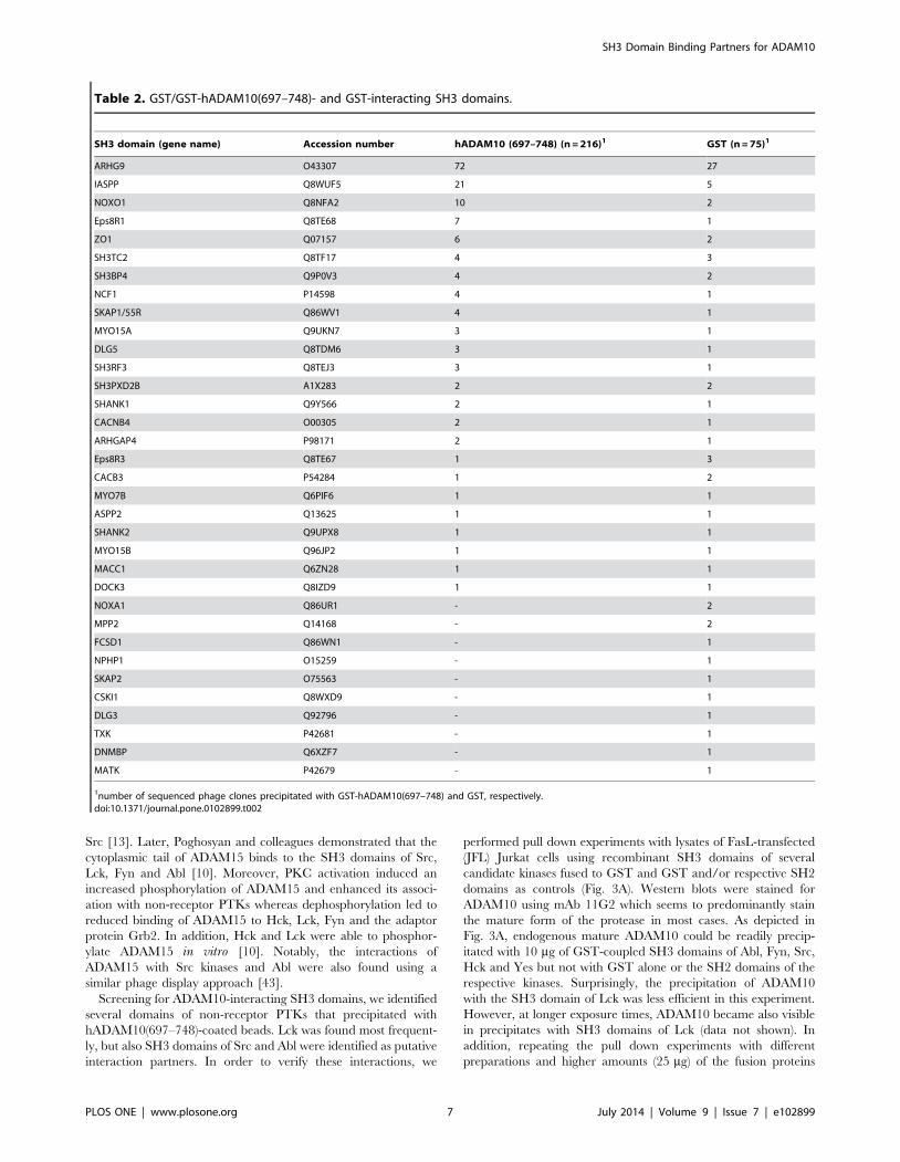
GST-tag. SH3 domains which were identified with the GST
control or with both GST and GST-hADAM10(697–748) are
listed in Table 2. For further analyses, we focused on the SH3
domain proteins that did not precipitate with GST. We do
nevertheless not exclude that other candidate binders are among
the SH3 domains that precipitated with both GST-hA-
DAM10(697–748) and GST.
The identified putative SH3 domain-containing interactors of
the intracellular domain of ADAM10 belong to different protein
families of adaptor proteins, non-receptor tyrosine kinases or
palmitoyltransferases. As listed in Table 1, many of the candidate
adaptor proteins are involved in membrane shaping and
trafficking (e.g. endophilins, sorting nexins (SNXs), PCH proteins)
or mediate cytosolic protein-protein interactions that are somehow
linked to cytoskeletal reorganization. Importantly, several of the
candidate interactors or related proteins have been identified as
binding partners for other ADAM family proteases including
ADAM9, 12, 13 or 15 (see below).
Before we started co-precipitation or pull down experiments to
investigate individual interactions, we verified the presence of
selected candidate proteins in PHA blasts, cloned T cells and
Jurkat cells by Western blot. Moreover, we performed a
comprehensive pull down screening from PHA blasts or Jurkat
cells to get additional information about possible interactions with
SH3 domains and/or full length proteins (supplementary Fig. S1–
S5). Being aware that functional data on the relevance of
individual interactions still have to be established, along with the
verification of ADAM10 binding, we discuss the potential role of
selected binding partners for ADAM10 biology and provide a
hypothetical model for their role in ADAM10 biology (Fig. S6).
EndophilinsDuring our screening, the SH3 domain of Endophilin-A2
(SH3GL1, EEN) was found eight times in GST-hADAM10(697–
748) precipitates but never with GST alone. Endophilins (encoded
by the EEN gene family) are cytosolic proteins with an N-terminal
Bin-Amphiphysin-Rvs (N-BAR) domain and a C-terminal SH3
domain. In general, endophilins contribute to the formation of
membrane tubules during endocytosis by inserting into the
membrane and inducing/changing membrane curvature [23,36–
38]. Whereas Endophilin-A2 is ubiquitously expressed, Endophi-
lin-A1 is prominently found in the brain and Endophilin-A3
mainly in testis, but also in liver and brain [37]. Notably, the close
relative Endophilin-A1 was identified in an earlier study as an
interaction partner for the precursor but not the processed forms
of ADAM9 and ADAM15 in a yeast two-hybrid screen [22].
These interactions were confirmed using fusion proteins and co-
precipitations from eukaryotic cells overexpressing both binding
partners. It was proposed that Endophilin-A1 may have a role in
regulating the function of ADAM9 and ADAM15 by modulating
their intracellular processing, transport, or final subcellular
localization. Like the other family members, Endophilin-A2
interacts with dynamin and synaptojanin via its C-terminal SH3
domain and is involved in endocytotic processes [37]. Further-
more, the SH3 domain of Endophilin-A2 was shown to bind the
proline-rich region of the Rho GTPase-activating protein
(RhoGAP) BPGAP1, leading to EGF-stimulated endocytosis of
the EGF receptor and ERK1/2 phosphorylation [39]. Notably,
due to the spatial aggregation of several endophilins at sites of
endocytosis, an individual SH3 interaction (e.g. to dynamin) of one
endophilin does not rule out that the neighboring molecule binds
to the PRR of another protein (e.g. ADAM10).
In order to verify the interaction of ADAM10 with Endophilin-
A2/EEN, murine HA-tagged ADAM10 and human Endophilin-
A2/EEN were transiently overexpressed in HEK 293T cells
(either alone or in combination). As mentioned, the intracellular
domains of murine and human ADAM10 differ only in one amino
acid outside the PRDs (Fig. 1). 18 hours post transfection, the cells
were lysed and precipitations were performed using antibodies
directed against HA or Endophilin-A2/EEN. Precipitating HA-
tagged ADAM10 led to co-precipitation of EEN (Fig. 2A, upper
panel). Moreover, immunoprecipitation of EEN resulted in co-
precipitated ADAM10, here detected with the anti-HA antibody
3F10 (Fig. 2A, lower panels). Of note, the anti-Endophilin-A2/
EEN monoclonal antibody 2F5 efficiently precipitated endogenous
EEN from untransfected cells (protein input ,2 mg per IP). At the
given short exposure time, endogenous EEN was not detected in
whole cell lysates (total protein input: 10 mg). The ADAM10/EEN
interaction was also investigated by pull down experiments using
the GST-tagged SH3 domain of EEN (EEN SH3) for precipitation
of endogenous ADAM10 from lysates of day 16 PHA blasts
(Fig. 2B). The immature form of ADAM10 (proADAM10,
82 kDa) was apparently precipitated more effectively with the
SH3 domain of EEN. Using mAb 11G2 for Western blot detection
of ADAM10, we did not detect equal amounts of mature
ADAM10 in EEN SH3 precipitates although this form was highly
abundant in the cellular lysate. Similar results were obtained in the
pull down screening (Fig. S2). In EEN SH3 precipitates, amongst
other bands, the polyclonal anti-ADAM10 antiserum stained a
protein band migrating at the height of the ADAM10 pro-form in
the corresponding immunoprecipitate. Thus, it might be interest-
ing in this context to further address the issue of whether
individual SH3 domain proteins preferentially associate with the
pro-form, the mature form or even the intracellular domain of
ADAM10 as it has been suggested for the interaction of
endophilins with ADAM9 and ADAM15.
Non-receptor protein tyrosine kinasesSH3 domains of several non-receptor protein tyrosine kinases
(PTKs) including Src family members and Abl were also identified
as putative interactors of ADAM10 in the phage screen. The Src
family of cytosolic non-receptor protein tyrosine kinases consists of
nine members (Blk, Fgr, Frk, Fyn, Hck, Lck, Lyn, Src and Yes) (see
SH3 Domain Binding Partners for ADAM10
PLOS ONE | www.plosone.org 5 July 2014 | Volume 9 | Issue 7 | e102899

[40] for a recent review). Src kinases initiate and direct numerous
signal transduction processes, including for example antigen
receptor-driven activation of T and B cells. The related Abl
tyrosine kinase has been implicated in cell migration, cell adhesion
and apoptosis. Moreover, Abl contributes to TCR signaling since
it is phosphorylated and activated by Src kinases and in turn
phosphorylates receptor tyrosine kinases, e.g. the epidermal
growth factor receptor (EGFR) to modulate receptor endocytosis
[41,42]. Interactions with non-receptor PTKs have been previ-
ously reported for several ADAM family members. It was shown
that the SH3 domain of Src interacts with the cytoplasmic domain
of ADAM12 [12,13] and that ADAM12 gets phosphorylated by v-
Table 1. GST-hADAM10(697-748)-precipitated SH3 domains: Function and localization of putative interaction partners accordingto the UniProt Protein Knowledgebase (UniprotKB).
SH3 domain protein gene name(s)Accessionnumber function localization hits
Endophilin-A2 SH3GL1 Q99961 endocytosis, podosome formation Cyt1, PM, Endo, Podo 8
Lck LCK P06239 Src-related tyrosine kinase Cyt, PM, LR 5
ZDHHC6 ZDHHC6 Q9H6R6 palmitoyltransferase ER 3
Growth factor receptor binding protein 2 GRB2, ASH P62993 adaptor (growth factorR Ras) Cyt, Endo, Nuc, Golgi 2
HS1/HCLS1/LckBP HCLS1 P14317 adaptor for Lck signaling PM, Cyt, Mito 2
SH3 domain protein 7, HIP-55 DBNL, SH3P7 Q9UJU6 adaptor, cytoskeleton, endocytosis Cyt, cell junction, PM,Golgi, ER, Podo
2
Otoraplin OTOR Q9NRC9 unknown secreted 2
Dedicator of cytokinesis protein 4 DOCK4 Q8N1I0 migration, GEF for Rap-1 EMS, Cyt 2
SH3 domain protein 21 SH3D21, C1orf113 A4FU49 unknown unknown 2
RUN and SH3 domain-containing protein 1 RUSC1 Q9BVN2 adaptor, cytoskeleton Cyt, Endo, Golgi 2
Rho GEF 38 ARHGEF38 Q9NXL2 GEF for Rho Cyt 2
PKC and CK substrate in neurons 3 PACSIN3 Q9UKS6 endocytosis Cyt, PM 1
GRB2-related adaptor protein 2 GRAP2, GADS O75791 adaptor (LAT, SLP-76) Cyt, Nuc, Endo 1
c-Src Src P12931 tyrosine kinase Cyt, PM, Nuc, Mito 1
c-Abl ABL1 P00519 tyrosine kinase, cytoskeleton Cyt, Nuc, Mito 1
Sorting nexin 18 SNX18 Q96RF0 endocytosis, vesicle transport PM, Endo, Cyt 1
Adaptor protein crk (c-Crk) Crk P46108 adaptor, actin cytoskeleton Cyt, PM 1
Rho GEF 19 ARHGEF19 Q8IW93 GEF for RhoA, cytoskeleton Cyt 1
Peroxin-13 PEX13 Q92968 peroxisomal import Peroxisome membrane 1
Ephexin-1 NGEF Q8N5V2 GEF for RhoA, Rac1, CDC42 Cyt, PM, growth cone 1
Rho GAP 32, RICS protein ARHGAP32 A7KAX9 GAP for RhoA, Rac1, CDC42 Cyt, cell junction, PM,Golgi, ER, Endo
1
ARHGEF16, Ephexin-4 ARHGEF16 Q5VV41 GEF for RhoG Cyt 1
Growth arrest-specific protein 7 GAS7 O60861 neuronal differentiation Cyt, ruffles 1
unconventional myosin 1-E MYO1E Q12965 cytoskeleton Cyt, cell junction 1
Rho GEF 4 ARHGEF4 Q9NR80 GEF for RhoA, Rac1, CDC42 Cyt, PM, ruffles 1
Vinexin SH3 #2 SORBS3, SCAM1 O60504 cytoskeleton Cyt, cell junction, Nuc 1
Vinexin SH3 #3 SORBS3, SCAM1 O60504 cytoskeleton Cyt, cell junction, Nuc 1
RIMS Binding protein 3A RIMBP3A Q9UFD9 unknown unknown 1
Rho GAP 33, Sorting nexin 26 ARHGAP33 O14559 protein transport Cyt, PM 1
LIM and SH3 domain protein 1 LASP1 Q14847 adhesion, cytoskeleton Cyt 1
unconventional myosin VIIa MYO7A Q13402 cytoskeleton, intracellular movement Cyt 1
Disks large homolog 1 DLG1 Q12959 scaffold protein ER, PM, cell junction 1
Erythroid a spectrin SPTA1 P02549 cell shape, actin cytoskeleton Cyt 1
non erythroid a spectrin (fodrin) SPTAN1 Q13813 Ca2+-dependent movement Cyt 1
SH3 domain-containing protein 19, Eve-1 SH3D19 Q5HYK7 cell morphology, cytoskeleton Cyt, Nuc 1
RIMS Binding protein 2 RIMB2 O15034 synaptic transmission PM, cell junction 1
peripheral PM protein CASK CASK O14936 calmodulin-dependentserine kinase
Nuc, Cyt, PM (bindsAPP, syndecan)
1
Dedicator of cytokinesis protein 2 DOCK2 Q92608 migration, GEF for Rac1,Rac2 EMS, PM, Cyt 1
1Abbreviations: Cyt, cytosol; EMS, endomembrane system; ER, endoplasmic reticulum; Endo, endosomes; LR, lipid rafts; Mito, Mitochondria; Nuc, Nucleus; PM, plasmamembrane; Podo, podosomes.doi:10.1371/journal.pone.0102899.t001
SH3 Domain Binding Partners for ADAM10
PLOS ONE | www.plosone.org 6 July 2014 | Volume 9 | Issue 7 | e102899
Src [13]. Later, Poghosyan and colleagues demonstrated that the
cytoplasmic tail of ADAM15 binds to the SH3 domains of Src,
Lck, Fyn and Abl [10]. Moreover, PKC activation induced an
increased phosphorylation of ADAM15 and enhanced its associ-
ation with non-receptor PTKs whereas dephosphorylation led to
reduced binding of ADAM15 to Hck, Lck, Fyn and the adaptor
protein Grb2. In addition, Hck and Lck were able to phosphor-
ylate ADAM15 in vitro [10]. Notably, the interactions of
ADAM15 with Src kinases and Abl were also found using a
similar phage display approach [43].
Screening for ADAM10-interacting SH3 domains, we identified
several domains of non-receptor PTKs that precipitated with
hADAM10(697–748)-coated beads. Lck was found most frequent-
ly, but also SH3 domains of Src and Abl were identified as putative
interaction partners. In order to verify these interactions, we
performed pull down experiments with lysates of FasL-transfected
(JFL) Jurkat cells using recombinant SH3 domains of several
candidate kinases fused to GST and GST and/or respective SH2
domains as controls (Fig. 3A). Western blots were stained for
ADAM10 using mAb 11G2 which seems to predominantly stain
the mature form of the protease in most cases. As depicted in
Fig. 3A, endogenous mature ADAM10 could be readily precip-
itated with 10 mg of GST-coupled SH3 domains of Abl, Fyn, Src,
Hck and Yes but not with GST alone or the SH2 domains of the
respective kinases. Surprisingly, the precipitation of ADAM10
with the SH3 domain of Lck was less efficient in this experiment.
However, at longer exposure times, ADAM10 became also visible
in precipitates with SH3 domains of Lck (data not shown). In
addition, repeating the pull down experiments with different
preparations and higher amounts (25 mg) of the fusion proteins
Table 2. GST/GST-hADAM10(697–748)- and GST-interacting SH3 domains.
SH3 domain (gene name) Accession number hADAM10 (697–748) (n = 216)1 GST (n = 75)1
ARHG9 O43307 72 27
IASPP Q8WUF5 21 5
NOXO1 Q8NFA2 10 2
Eps8R1 Q8TE68 7 1
ZO1 Q07157 6 2
SH3TC2 Q8TF17 4 3
SH3BP4 Q9P0V3 4 2
NCF1 P14598 4 1
SKAP1/55R Q86WV1 4 1
MYO15A Q9UKN7 3 1
DLG5 Q8TDM6 3 1
SH3RF3 Q8TEJ3 3 1
SH3PXD2B A1X283 2 2
SHANK1 Q9Y566 2 1
CACNB4 O00305 2 1
ARHGAP4 P98171 2 1
Eps8R3 Q8TE67 1 3
CACB3 P54284 1 2
MYO7B Q6PIF6 1 1
ASPP2 Q13625 1 1
SHANK2 Q9UPX8 1 1
MYO15B Q96JP2 1 1
MACC1 Q6ZN28 1 1
DOCK3 Q8IZD9 1 1
NOXA1 Q86UR1 - 2
MPP2 Q14168 - 2
FCSD1 Q86WN1 - 1
NPHP1 O15259 - 1
SKAP2 O75563 - 1
CSKI1 Q8WXD9 - 1
DLG3 Q92796 - 1
TXK P42681 - 1
DNMBP Q6XZF7 - 1
MATK P42679 - 1
1number of sequenced phage clones precipitated with GST-hADAM10(697–748) and GST, respectively.doi:10.1371/journal.pone.0102899.t002
SH3 Domain Binding Partners for ADAM10
PLOS ONE | www.plosone.org 7 July 2014 | Volume 9 | Issue 7 | e102899

confirmed the Lck interaction (supplementary Fig. S1). Under
these conditions, SH3 domains of different Src-related kinases (Fig.
S1), of Abl and of the Tec-kinase Itk (Fig. S4) apparently
preferentially precipitated different forms of ADAM10 from PHA
blasts or Jurkat cells as stained with the polyclonal anti-ADAM10
antiserum. It should be mentioned in this context that neither
GST nor any of the used SH2 domains precipitated proteins
detected by this anti-ADAM10 antiserum. As an additional proof
of the Lck/ADAM10 interaction, we performed precipitations
from Jurkat cells (JE6-1) using two monoclonal antibodies that we
had generated against Lck and co-precipitated mature (and pro-)
ADAM10 as visualized with mAb 11G2 on the Western blot
(Fig. 3B). As a positive control, we included an ADAM10 IP using
the mAb 11G2.
The palmitoyltransferase ZDHHC6The third most frequently identified SH3 domain was
annotated in the library data sheet as ‘homology to Ablphilin-2’
and might be better known as ‘zinc finger DHHC domain-
containing protein 6’ (ZDHHC6). ZDHHC6 belongs to the
DHHC family of palmitoyltransferases. Unfortunately, at present
there are hardly any reagents available to address the potential
interaction of ZDHHC6 with ADAM10. In general, transfer of
palmitoyl groups increases protein hydrophobicity favoring
membrane association. As a reversible posttranslational modifica-
tion, palmitoylation contributes to the intracellular trafficking of
proteins between membrane-shielded compartments and thereby
determines their intracellular localization and contributes to
protein clustering in platforms such as lipid rafts. As an example,
calnexin, a major ER chaperone involved in glycoprotein folding,
is modified by the ER-resident palmitoyltransferase DHHC6
leading to a preferential localization of calnexin to the perinuclear
rough ER [44]. Moreover, Ablphilin-2 (ZDHHC16, Aph2) was
shown to interact with ER-associated c-Abl and to promote ER-
stress and apoptosis [45]. The available information about other
DHHC proteins including ZDHHC6, however, is very limited.
Regarding ADAM family proteases, palmitoylation as a posttrans-
lational modification has not been described so far. However,
substrates of ADAM10 and ADAM17 are known to be
palmitoylated, leading to altered functionality and localization.
For example, palmitoylation of the Fas ligand (FasL, CD95L) is
essential for its killing capacity and also for its proteolytic
processing by ADAM10 and SPPL2a [46]. In contrast, palmitoy-
lation of TNF-a is involved in lipid raft positioning of the cytokine
and seems to interfere with the cleavage/degradation of TNF
intracellular fragments, but is not involved in the regulation of its
ectodomain shedding by ADAM17 [47]. Also, the shedding of the
NKG2D ligands MICA/B (MHC-class-I-related chain A/B) was
shown to be regulated by their localization in cholesterol-enriched
domains, while inhibition of palmitoylation by 2-bromopalmitate
led to reduced shedding [48,49]. With regard to regulation of
ADAM10, it was reported that only palmitoylated Tetraspanin12
might serve as a binding partner for ADAM10 to promote its
localization and maturation and thereby for instance facilitating
the ADAM10-dependent proteolysis of amyloid precursor protein
[50]. Thus, ZDHHC6 could not only modulate the palmitoylation
status of ADAM10, but also of certain substrates to alter their
localization, accessibility or processing and degradation for
example in tetraspanin or lipid raft platforms. These hypotheses
could be addressed in future experiments as soon as respective
reagents become available.
Grb2 family adaptor proteinsAdaptor proteins play a fundamental role in orchestrating
cellular signaling processes. They are composed of different
modules that either form interaction domains or binding sites for
Figure 2. Verification of the interaction between ADAM10 and EEN. (A) In order to verify the potential interaction of ADAM10 withEndophilin-A2/EEN, HEK 293T cells were either left untransfected or transfected with HA-tagged murine ADAM10 alone or in combination withhuman Endophilin-A2/EEN. 18 h later, the cells were lysed and immunoprecipitations (IPs) were performed with monoclonal antibodies directedagainst the HA-tag (clone 3F10) or EEN (clone 2F5), respectively. Protein input for IPs was 1.8 or 2 mg of protein, respectively. Of note: at theemployed exposure time, endogenous EEN is hardly detectable in the whole cell lysates containing a total of 10 mg of protein. (B) Pull down analyseswere performed from PHA blasts (day 16) using a GST fusion protein containing the SH3 domain of EEN coupled to GST (EEN SH3) and GST as acontrol. The subsequent Western blot was probed with anti-ADAM10 (clone 11G2).doi:10.1371/journal.pone.0102899.g002
SH3 Domain Binding Partners for ADAM10
PLOS ONE | www.plosone.org 8 July 2014 | Volume 9 | Issue 7 | e102899

constitutive or activation-dependent protein-protein interactions
[51]. The Grb2 family belongs to the cytosolic adaptor proteins
and consists of three members: the growth factor receptor-bound
protein 2 (Grb2) is widely expressed in different cell types and
tissues, whereas the Grb2-related adaptor protein (GRAP) and
Grb2-related adaptor protein 2 (GRAP2/MONA/GADS) are
more restricted to hematopoietic cells. All three proteins consist of
one SH2 domain flanked by two SH3 domains. This allows
simultaneous interactions with several proteins and the formation
of molecular networks [52].
In our screen, we found that SH3 domains of Grb2 and GRAP2
precipitated with the intracellular portion of ADAM10. To verify
the putative interaction of ADAM10 with Grb2, Jurkat T cells
(JE6-1) were lysed and a pull down using the GST-hA-
DAM10(697–748) fusion protein and an immunoprecipitation
with mAb 11G2 were performed as depicted in Fig. 3C. Western
blotting using a polyclonal anti-Grb2 antibody (C-23) revealed that
Grb2 was precipitated with the GST fusion protein containing the
intracellular domain of ADAM10, but not with GST. Further-
more, Grb2 was effectively co-precipitated with ADAM10
(Fig. 3C). Note that the blot was reprobed for ADAM10 using
Figure 3. Interactions between ADAM10 and non-receptor protein tyrosine kinases and adaptor proteins of the Grb2 family. (A)Lysates from Jurkat T cells (here JFL) were used for pull down analyses with SH2 or SH3 domain fusion proteins of non-receptor PTKs as indicated.GST served as a negative control. Protein input of the whole cell lysate was 15 mg. 10 mg of the respective fusion proteins were used for precipitationfrom 1 ml of cell lysate with 3.2 mg/ml of protein. MAb 11G2 was used to detect ADAM10 after Western blotting. (B) Immunoprecipitations wereperformed from Jurkat T cells (here JE6-1) using 2 mg of mAbs against Lck (clones 4/129 and 4/215) or ADAM10 (clone 11G2). Protein G beads servedas a control for unspecific binding. Input of the cellular lysate was 15 mg; precipitates were performed from 1 ml of lysate (1 mg/ml protein). MAb11G2 was used to detect ADAM10 by Western blotting. (C) Jurkat cells (JE6-1) were lysed and one ml of lysate containing 900 mg/ml protein wassubjected to precipitation using 10 mg GST or the GST fusion protein containing the intracellular part of human ADAM10 (hADAM10(697–748)). Inparallel, ADAM10 was precipitated using 2 mg/ml of mAb 11G2 with protein G beads serving as a control. Precipitated proteins were separated bySDS-PAGE and blotted on nitrocellulose. The blot was stained with a polyclonal anti-Grb2 antibody and re-probed with a polyclonal anti-ADAM10antiserum (‘‘animal 1’’). (D) C- and N-terminal SH3 domains of Grb2, GRAP and GRAP2 fused to GST (10 mg each) were used for precipitations fromJurkat T cells (here JFL; 2.2 mg/ml protein input per precipitation) with GST alone serving as a control. 15 mg protein of the whole cell lysate wereincluded as a reference. ADAM10 was detected with mAb 11G2.doi:10.1371/journal.pone.0102899.g003
SH3 Domain Binding Partners for ADAM10
PLOS ONE | www.plosone.org 9 July 2014 | Volume 9 | Issue 7 | e102899

the polyclonal antiserum (‘‘animal 1’’). In order to assess the
binding of other members of the protein family to ADAM10, we
used GST fusion proteins comprising the C- or N-terminal SH3
domains of Grb2, GRAP and GRAP2. Interestingly, as shown for
other interaction partners before [53], we found differential
binding of ADAM10 to the C- or N-terminal SH3 domains of all
three adaptor proteins. Using mAb 11G2, ADAM10 was more
prominently detected in the presence of C-terminal SH3 domains,
whereas less protein was observed when precipitated with the N-
terminal SH3 domains (Fig. 3D). Similar differences were also seen
in the pull down screen (supplementary Fig. S2). However,
although the polyclonal anti-ADAM10 antiserum used for staining
of ADAM10 detected only two bands in ADAM10 IPs performed
with mAb 11G2, the band patterns stained in SH3 domain
precipitates formed by Grb2, GRAP and GRAP2 were quite
heterogeneous.
Notably, also for Grb2 family members, interactions with
several ADAM family members were previously described. For
example, it was shown that immunoprecipitation of endogenous
Grb2 led to co-precipitation of ADAM12 [13]. Moreover,
internalization of ADAM12 was found to be dependent on Grb2
and knockdown of endogenous Grb2 resulted in reduced
internalization [21]. As mentioned before, ADAM15 also binds
to Grb2 and dephosphorylation of cellular extracts decreased this
interaction, possibly arguing for a cooperative SH2/SH3 domain
interaction network [10].
Figure 4. The intracellular domains of ADAM10 and ADAM17 interact with sorting nexins and PACSINs. (A) HEK 293T cells were eitherleft untransfected or were transfected with a control vector (p12linker) or with HA-tagged SNX9, SNX18 or SNX33. 18 h later, cells were lysed andprecipitations were performed with 25 mg of GST, GST-hADAM10(697–748) or GST-hADAM17(694–824), respectively. Western blots were developedusing mAb 3F10 directed against the HA-tag of the sorting nexins. Lysates (20 mg/lane) were stained as a control. (B) HEK 293T cells were leftuntransfected or transfected with control vector (pcDNA3.1) or with myc-tagged PACSIN1, PACSIN2 or PACSIN3. 18 h post transfection, cells werelysed and precipitations were performed with 10 mg of GST, GST-hADAM10(697–748) or GST-hADAM17(694–824). Precipitated proteins wereseparated by SDS-PAGE and transferred to nitrocellulose membranes. Blots were developed with mAb clone 46–0603 directed against the myc-tag.Of the whole cell lysates, 5 mg of protein were separated to verify efficient transfection. (C) Recombinant GST fusion proteins (15 mg) containingindividual SH3 domains of PACSIN1-3 were used to precipitate endogenous ADAM10 from lysates of Jurkat T cells (JFL, 1 ml each; 2.2 mg/ml). GSTserved as a negative control. 10 mg of whole cell lysate was used as a control to detect ADAM10 using mAb 11G2.doi:10.1371/journal.pone.0102899.g004
SH3 Domain Binding Partners for ADAM10
PLOS ONE | www.plosone.org 10 July 2014 | Volume 9 | Issue 7 | e102899

Sorting nexinsThe sorting nexins (SNXs) form a large family of ubiquitously
expressed proteins which also function as regulators of intracellular
trafficking, endocytosis and signal transduction. SNX9, SNX18
and SNX33 constitute a subfamily since they contain SH3
domains and display a high overall homology. Similar to
endophilins, sorting nexins contain an N-BAR domain and are
thus implicated in the modulation of membrane curvature and
tubulation in endosomal sorting or, more general, in endo- and
exocytosis [24,25]. As examples, SNX9 promotes the internaliza-
tion of the TNF receptor (TNFR) and influences the degradation
of the EGF receptor (EGFR) after EGF signaling [54]. Further-
more, SNX9 participates in the reorganization of the actin
cytoskeleton by promoting the activation of the Arp2/3 complex
[55].
SNX18 was identified as a putative SH3 domain binding
partner for ADAM10. In subsequent experiments, we found that
ADAM10 interacts with exogenously expressed SNX9, SNX18
and SNX33. To address this, HEK 293T cells were transiently
transfected with HA-tagged sorting nexin constructs and precip-
itates using GST or the GST-hADAM10(697–748) fusion protein
were formed and analyzed. As shown in Fig. 4A, SNX9, SNX18
and SNX33 were readily precipitated as revealed by Western blot
using an anti-HA antibody (clone 3F10). Here, the strongest
interaction was observed with SNX9 and SNX18 (Fig. 4A). In this
context, we additionally analyzed whether the SNXs also interact
with ADAM17 employing a hADAM17(694–824) fusion protein
containing the putative SH3 binding motif PAPQTPGR (amino
acids 731–738) [56]. In case of ADAM17, strongest binding was
again seen with SNX9 and SNX18 (Fig. 4A). Not unexpectedly,
sorting nexins (and especially SNX9) had also been identified as
interaction partners of other ADAM proteases (including ADAM9
and ADAM15). Howard and colleagues demonstrated binding of
SNX9 to the precursor forms of ADAM9 and ADAM15, but not
to the mature form, proposing a potential role of SNX9 in
regulating processing, function and localization of the metallopro-
teases [22]. ADAM15 was also reported to interact with SNX33 in
a phage display screen [43], showing preferential binding to
specific isoforms of ADAM15 containing the C-terminal proline-
rich region [57].
PACSINsThe SH3 domain of PACSIN3 (PKC and casein kinase
substrate in neurons 3) was identified as a putative interactor for
ADAM10. PACSIN3 and its close relatives PACSIN1 and 22
belong to the pombe Cdc15 homology (PCH) protein family, a
family of adaptor proteins involved in cytoskeleton-to-membrane
crosstalk. PCH proteins share a Fes-CIP4 homology (FCH)
domain within their F-BAR domain. Like other proteins with
BAR domains, PCH proteins change membrane curvature and
are involved in the regulation of endo- and exocytosis and
vesicular transport [58,59]. Whereas PACSIN1 is mainly ex-
pressed in neurons, PACSIN2 and PACSIN3 seem to be broadly
distributed [60–63]. Moreover, most PCH proteins contain
functional SH3 domains at the C-terminus. Also PACSIN proteins
were already shown to interact with cytosolic portions of other
ADAM proteins. As mentioned, Cousin and colleagues were the
first to describe a SH3 domain-mediated interaction of PACSIN2
and ADAM13 in Xenopus embryos and demonstrated that
overexpression of X-PACSIN2 could rescue developmental
alterations induced by overexpression of ADAM13 [11]. PAC-
SIN3 was identified as an interactor of ADAM12 in a yeast two-
hybrid screen and this interaction was further substantiated by pull
down and co-immunoprecipitation analyses. Overexpression of
PACSIN3 resulted in enhanced shedding of the ADAM12
substrate heparin-binding EGF-like growth factor (proHB-EGF)
upon stimulation, whereas knockdown reduced proteolysis [15].
Since PACSIN3 was already on the list of ADAM interactors,
we followed this in more detail. When we transiently overex-
pressed myc-tagged PACSINs in HEK 293T cells, we could
readily precipitate all PACSINs with the GST-hADAM10(697–
748) fusion protein but not with a GST control (Fig. 4B).
Interestingly, the association with PACSIN3 was more pro-
nounced compared to PACSIN1 and PACSIN2. Moreover,
GST fusion proteins containing the individual SH3 domains of
the three different PACSINs precipitated mature as well as
immature ADAM10 (Fig. 4C). The ADAM10 interaction was also
seen for all three PACSINs in the complementary pull down
screen (supplementary Fig. S3). In parallel, we investigated a
potential binding of PACSINs to ADAM17, using the GST-
hADAM17(694–824) fusion protein. All PACSINs were efficiently
precipitated, but the strongest binding was again observed for
PACSIN3 (Fig. 4B).
Drawbacks and other candidate interactorsAs detailed above, we tried to verify as many interactions as
possible using a variety of different approaches. However, co-
localization studies of endogenous (and also of overexpressed)
proteins (e.g. by confocal imaging) turned out to be undoable with
available anti-human ADAM10 antibodies, although ADAM10 is
expressed at high levels on T lymphocyte populations [6]. Also, co-
immunoprecipitation of endogenous proteins proved to be difficult
and Western blotting using different electrophoresis systems in
combination with either monoclonal antibodies or polyclonal
antisera were not always superimposable. In some cases,
interactions could only be verified by overexpression and co-
immunoprecipitation or pull down of tagged full-length proteins
using adequate controls. We therefore tested a series of GST
fusion proteins containing isolated SH3 domains, full length
proteins or SH2 domains (as controls) of candidate interactors in
pull down assays from T cell blasts or Jurkat cells to complement
the data obtained by screening the phage library. As mentioned,
most interactions that were identified based on the phage display
screen were also seen in pull down analyses. Although the band
patterns obtained after staining of Western blots with the
polyclonal anti-ADAM10 antiserum were occasionally somewhat
confusing, from this pull down experiments a few other interactors
might also be worth testing in future experiments. These include
the adaptor protein Nck (Fig. S3) which seems to interact with
ADAM10 with at least one of its three SH3 domains, the Tec-
kinase Itk mentioned before, the p85 subunit of PI3 kinase, NCF1
(p47phox, the 47-kilodalton cytosolic subunit of the NADPH
oxidase complex), and CD2BP1, a PACSIN-related member of
the PCH protein family (Fig. S4).
Shared interaction partners: ADAM10 and FasLInterestingly, over the last decade, we and others have shown
that the ADAM10 substrate FasL [4–6] interacts with a very
similar subset of SH3 domain proteins as now shown for
ADAM10. For example, FasL binds to non-receptor PTKs (Fgr,
Fyn, Lyn, Src, Tec and Yes), PI3 kinase, Nck, Grb2-related
proteins, NCF1 [30,35,53,64], sorting nexins [35,65] and PCH
proteins [32,53,65,66]. The functional implication of this surpris-
ing coincidence is presently not understood. In the case of FasL,
several interactors were meanwhile shown to regulate its
intracellular storage and its mobilization to the cell surface
[32,66–69]. Although FasL and ADAM10 are quite different
regarding their intracellular trafficking and membrane appear-
SH3 Domain Binding Partners for ADAM10
PLOS ONE | www.plosone.org 11 July 2014 | Volume 9 | Issue 7 | e102899

ance, it seems obvious that molecules regulating membrane-to-
cytoskeleton-signaling are coupled to both proteins. Moreover, we
also observed a differential interaction of SNXs and PCH proteins
with the full length form of the protein or its N-terminal fragments
generated by ADAM10-mediated proteolysis [34]. Since AD-
AM10 itself is subject to proteolytic cleavage by ADAM9,
ADAM15 and c-secretase [70], it might be also possible that
SH3 domain proteins differentially bind to the full length protease
(i.e. to the pro- or mature form) or to its processed fragments
(Fig. 1B and supplementary Fig. S6).
Provided that reliable tools for the intracellular or biochemical
detection of human ADAM10 (and/or ADAM17) become
available, based on the present findings, the functional relevance
of individual interactions for the regulation of ADAM10
expression and/or activity could be addressed using similar
approaches that have been successfully used for the characteriza-
tion of SH3 domain proteins in the context of other ADAM
proteases [20,21,71]. Such studies could be complemented using
inhibitors (e.g. for Src kinases) and knockdown approaches
targeting potential interactors by siRNA or expressing mutants
that prevent SH3 binding. A hypothetical scenario for the role of
individual SH3 domain interactors in the regulation of ADAM10
biology is depicted in Fig. S6. As discussed before, it seems likely
that individual interactors that turned out to have regulatory
functions in the context of related proteases also modulate
ADAM10 function. The different protein families that comprise
the pool of binding partners suggest a role in ADAM10 storage or
transport in membrane positioning (e.g. in plasma membrane
platforms), in substrate association, in enzymatic activity, in
recycling, or in translocation and/or degradation of proteolytically
processed ADAM10. Undoubtedly, the knowledge about the role
of individual intracellular interactors for the regulation of
ADAM10 expression and activity would also foster further
approaches to target the protease for therapeutic intervention [72].
Conclusions
Using a phage display library screen, we identified an array of
individual SH3 domains as putative interaction sites for the PRRs
of ADAM10. The protein families that comprise these SH3
domains include non-receptor tyrosine kinases and adaptor
proteins that mostly regulate endo- and exocytosis, membrane
trafficking, protein positioning and/or membrane-to-cytoskeleton
interactions. Based on the initial biochemically verification of
different putative interaction partners and based on the overlap
with reported functionally relevant interactions with other ADAM
proteases, we consider these proteins promising targets for further
analyses to address whether SH3 domain proteins that bind to the
two intracellular PRRs of ADAM10 also play a role for the
subcellular localization and activity of the protease.
Supporting Information
Figure S1 Immunoprecipitation and pull down fromhuman PHA blasts – Src related kinases. PHA-stimulated T
cells were lysed in NP40 lysis buffer containing EDTA and
protease and phosphatase inhibitors. Immunoprecipitations were
performed from 1 ml of cell lysate (equivalent to 506106 cells)
using 2 mg/ml of the indicated anti-ADAM10 or anti-ADAM17
antibodies. Precipitations with GST as a control or GST fusion
proteins containing SH2 and/or SH3 domains of the Src-related
kinases Fyn, Lck, Src, Hck and Yes were done using 25 mg/ml
lysate of the respective fusion proteins. (A) Ponceau S staining
following Western transfer. (B) ADAM10 immunoblot using the
polyclonal anti-ADAM10 antibody (‘‘animal 1’’) - short exposure
time. (C) ADAM10 immunoblot using the polyclonal anti-
ADAM10 antibody (‘‘animal 1’’) - long exposure time.
(TIF)
Figure S2 Immunoprecipitation and pull down fromhuman PHA blasts – Grb-2 related adaptor proteins,EEN and FBP17. PHA-stimulated T cells were lysed in NP40
lysis buffer containing EDTA and protease and phosphatase
inhibitors. Immunoprecipitations were performed from 1 ml cell
lysate (equivalent to 506106 cells) using 2 mg/ml of the indicated
anti-ADAM10 or anti-ADAM17 antibodies. Precipitations with
GST as a control or GST fusion proteins containing SH2 and/or
SH3 domains or full length proteins of Grb2, GRAP, GRAP2,
EEN and FBP17 were done using 25 mg/ml lysate of the
respective fusion proteins. (A) Ponceau S staining following
Western transfer. (B) ADAM10 immunoblot using the polyclonal
anti-ADAM10 antibody (‘‘animal 1’’) - short exposure time. (C)
ADAM10 immunoblot using the polyclonal anti-ADAM10
antibody (‘‘animal 1’’) - long exposure time.
(TIF)
Figure S3 Immunoprecipitation and pull down fromhuman Jurkat cells (JE6-1) – PACSINs and Nck1. Jurkat
cells were lysed in NP40 lysis buffer containing EDTA and
protease and phosphatase inhibitors. Immunoprecipitations were
performed from 1 ml cell lysate (equivalent to 506106 cells) using
2 mg/ml of the indicated anti-ADAM10 or anti-ADAM17
antibodies. Precipitations with GST as a control or GST fusion
proteins containing SH2 and/or SH3 domains or full length
proteins of PACSINs or Nck1 were done using 25 mg/ml lysate of
the respective fusion proteins. (A) Ponceau S staining following
Western transfer. (B) ADAM10 immunoblot using the polyclonal
anti-ADAM10 antibody (‘‘animal 1’’) - short exposure time. (C)
ADAM10 immunoblot using the polyclonal anti-ADAM10
antibody (‘‘animal 1’’) - long exposure time.
(TIF)
Figure S4 Immunoprecipitation and pull down fromhuman Jurkat cells (JE6-1) – Abl, Itk, PI 3K, phox47 andCD2BP1. Jurkat cells were lysed in NP40 lysis buffer containing
EDTA and protease and phosphatase inhibitors. Immunoprecip-
itations were performed from 1 ml cell lysate (equivalent to
506106 cells) using 2 mg/ml of the indicated anti-ADAM10 or
anti-ADAM17 antibodies. Precipitations with GST as a control or
GST fusion proteins containing SH2 and/or SH3 domains or full
length proteins of Abl, Itk, PI 3K, phox47 or CD2BP1 were done
using 25 mg/ml lysate of the respective fusion proteins. (A)
Ponceau S staining following Western transfer. (B) ADAM10
immunoblot using the polyclonal anti-ADAM10 antibody (‘‘ani-
mal 1’’) - short exposure time. (C) ADAM10 immunoblot using the
polyclonal anti-ADAM10 antibody (‘‘animal 1’’) - long exposure
time.
(TIF)
Figure S5 Coomassie blue staining of fusion proteinsused for pull down experiments. (A-C) Prior to the
precipitations from cell lysates as depicted in the supplementary
Figures S1–S4, 25 mg of all fusion proteins were checked after
separation by SDS-PAGE for degradation by in-gel-staining with
coomassie blue. (D–E) Potentially degraded (‘‘old’’) fusion proteins
were compared to freshly thawed material (‘‘fresh’’) and replaced if
necessary.
(TIF)
Figure S6 The complex ADAM biology – a hypotheticalmodel depicting putative regulation by SH3 domainproteins. ADAM proteases are synthesized in the rough
SH3 Domain Binding Partners for ADAM10
PLOS ONE | www.plosone.org 12 July 2014 | Volume 9 | Issue 7 | e102899

endoplasmatic reticulum (ER) with a signal peptide that is
removed when entering the trans-Golgi network (TGN). It is
believed that the pro-domain is cleaved off in the Golgi
compartment by protein convertases such as furin. In certain cell
types, ADAM10 activity has been associated with a lysosomal
compartment. It is, however, not clear whether this organelle
association is involved in storage or translocation of the protease or
whether the protease exerts intracellular activity in an endo-
lysosomal compartment. When ADAM10 is translocated to the
cell surface, it might interact with proteins on an adjacent cell (e.g.
integrins or syndecan). In analogy to related proteases, ADAM10
activity at the plasma membrane might be regulated by
intracellular interactors that induce phosphorylation or more
complex signaling alterations. Importantly, several ADAM
proteases (including ADAM10) apparently require positioning
into defined membrane platforms (e.g. lipid rafts or tetraspanin
platforms) to get into proximity to their substrates. If substrate
cleavage occurs, the released soluble ectodomain of the substrate
can act in an autocrine or paracrine fashion. Interestingly, some
ADAM proteases (e.g. ADAM12) are recycled in a clathrin-
dependent manner, supported by SH3 domain adaptors such as
Grb-2. Moreover, it was reported that ADAM10 itself is
proteolytically processed by ADAM9 and 15 and that the
remaining C-terminal fragments (CTFs) are subjected to intra-
membrane proteolysis by c-secretase releasing an isolated
intracellular domain (ICD) into the cytosol. Importantly, if
available for protein-protein interactions, the intracellular region
of ADAM10 containing the SH3 binding sites may affect all
different aspects from intracellular transport, via plasma mem-
brane positioning and activation to recycling or translocation and
degradation of the CTFs or ICDs. The presented model was
modified based on a cartoon by Seals and Courtneidge (Genes
Dev. 2003, 17:7–30) [ref. [8] of the main manuscript] to highlight
potential sites of action of SH3 domain interactors identified in the
present study. For more detailed information concerning the
putative function of individual binding partners, we refer to the
Results and Discussion part of the main manuscript.
(TIF)
Appendix S1 Pull down analyses.
(DOCX)
Acknowledgments
We thank Gudrun Scherer and Alyn Gerneth for expert technical
assistance. We also thank Profs P. Saftig (Biochemical Institute, CAU,
Kiel) and S. F. Lichtenthaler (TU Munich & DZNE Munich) for providing
antibodies and/or expression constructs. This work forms part of the PhD
thesis of HE.
Author Contributions
Conceived and designed the experiments: HE ML DK OJ. Performed the
experiments: HE ML OJ. Analyzed the data: HE ML OJ. Contributed
reagents/materials/analysis tools: HE ML DK OJ. Contributed to the
writing of the manuscript: HE ML DK OJ.
References
1. Edwards DR, Handsley MM, Pennington CJ (2008) The ADAM metallopro-teinases. Mol Aspects Med 29: 258–289.
2. Reiss K, Saftig P (2009) The ‘‘a disintegrin and metalloprotease’’ (ADAM) familyof sheddases: physiological and cellular functions. Semin Cell Dev Biol 20: 126–
137.
3. Moss ML, Jin SL, Becherer JD, Bickett DM, Burkhart W, et al. (1997) Structural
features and biochemical properties of TNF-alpha converting enzyme (TACE).J Neuroimmunol 72: 127–129.
4. Schulte M, Reiss K, Lettau M, Maretzky T, Ludwig A, et al. (2007) ADAM10
regulates FasL cell surface expression and modulates FasL-induced cytotoxicity
and activation-induced cell death. Cell Death Differ 14: 1040–1049.
5. Kirkin V, Cahuzac N, Guardiola-Serrano F, Huault S, Luckerath K, et al.(2007) The Fas ligand intracellular domain is released by ADAM10 and SPPL2a
cleavage in T-cells. Cell Death Differ 14: 1678–1687.
6. Ebsen H, Schroder A, Kabelitz D, Janssen O (2013) Differential surface
expression of ADAM10 and ADAM17 on human T lymphocytes and tumorcells. PLoS One 8: e76853. 10.1371/journal.pone.0076853
7. Kveiborg M, Albrechtsen R, Couchman JR, Wewer UM (2008) Cellular roles ofADAM12 in health and disease. Int J Biochem Cell Biol 40: 1685–1702.
8. Seals DF, Courtneidge SA (2003) The ADAMs family of metalloproteases:multidomain proteins with multiple functions. Genes Dev 17: 7–30.
9. Reddy P, Slack JL, Davis R, Cerretti DP, Kozlosky CJ, et al. (2000) Functional
analysis of the domain structure of tumor necrosis factor-alpha converting
enzyme. J Biol Chem 275: 14608–14614.
10. Poghosyan Z, Robbins SM, Houslay MD, Webster A, Murphy G, et al. (2002)Phosphorylation-dependent interactions between ADAM15 cytoplasmic domain
and Src family protein-tyrosine kinases. J Biol Chem 277: 4999–5007.
11. Cousin H, Gaultier A, Bleux C, Darribere T, Alfandari D (2000) PACSIN2 is a
regulator of the metalloprotease/disintegrin ADAM13. Dev Biol 227: 197–210.
12. Kang Q, Cao Y, Zolkiewska A (2000) Metalloprotease-disintegrin ADAM 12
binds to the SH3 domain of Src and activates Src tyrosine kinase in C2C12 cells.Biochem J 352 Pt 3: 883–892.
13. Suzuki A, Kadota N, Hara T, Nakagami Y, Izumi T, et al. (2000) Meltrin alpha
cytoplasmic domain interacts with SH3 domains of Src and Grb2 and is
phosphorylated by v-Src. Oncogene 19: 5842–5850.
14. Kang Q, Cao Y, Zolkiewska A (2001) Direct interaction between thecytoplasmic tail of ADAM 12 and the Src homology 3 domain of p85alpha
activates phosphatidylinositol 3-kinase in C2C12 cells. J Biol Chem 276: 24466–
24472.
15. Mori S, Tanaka M, Nanba D, Nishiwaki E, Ishiguro H, et al. (2003) PACSIN3binds ADAM12/meltrin alpha and up-regulates ectodomain shedding of
heparin-binding epidermal growth factor-like growth factor. J Biol Chem 278:
46029–46034.
16. Cao Y, Kang Q, Zolkiewska A (2001) Metalloprotease-disintegrin ADAM 12interacts with alpha-actinin-1. Biochem J 357: 353–361.
17. Galliano MF, Huet C, Frygelius J, Polgren A, Wewer UM, et al. (2000) Binding
of ADAM12, a marker of skeletal muscle regeneration, to the muscle-specific
actin-binding protein, alpha -actinin-2, is required for myoblast fusion. J Biol
Chem 275: 13933–13939.
18. Abram CL, Seals DF, Pass I, Salinsky D, Maurer L, et al. (2003) The adaptor
protein fish associates with members of the ADAMs family and localizes to
podosomes of Src-transformed cells. J Biol Chem 278: 16844–16851.
19. Tanaka M, Nanba D, Mori S, Shiba F, Ishiguro H, et al. (2004) ADAM binding
protein Eve-1 is required for ectodomain shedding of epidermal growth factor
receptor ligands. J Biol Chem 279: 41950–41959.
20. Stautz D, Sanjay A, Hansen MT, Albrechtsen R, Wewer UM, et al. (2010)
ADAM12 localizes with c-Src to actin-rich structures at the cell periphery and
regulates Src kinase activity. Exp Cell Res 316: 55–67.
21. Stautz D, Leyme A, Grandal MV, Albrechtsen R, van Deurs B, et al. (2012)
Cell-surface metalloprotease ADAM12 is internalized by a clathrin- and Grb2-
dependent mechanism. Traffic 13: 1532–1546.
22. Howard L, Nelson KK, Maciewicz RA, Blobel CP (1999) Interaction of the
metalloprotease disintegrins MDC9 and MDC15 with two SH3 domain-
containing proteins, endophilin I and SH3PX1. J Biol Chem 274: 31693–31699.
23. Peter BJ, Kent HM, Mills IG, Vallis Y, Butler PJ, et al. (2004) BAR domains as
sensors of membrane curvature: the amphiphysin BAR structure. Science 303:
495–499.
24. Cullen PJ (2008) Endosomal sorting and signalling: an emerging role for sorting
nexins. Nat Rev Mol Cell Biol 9: 574–582.
25. Cullen PJ, Korswagen HC (2012) Sorting nexins provide diversity for retromer-
dependent trafficking events. Nat Cell Biol 14: 29–37.
26. Marcello E, Gardoni F, Mauceri D, Romorini S, Jeromin A, et al. (2007)
Synapse-associated protein-97 mediates alpha-secretase ADAM10 trafficking
and promotes its activity. J Neurosci 27: 1682–1691.
27. Peiretti F, Deprez-Beauclair P, Bonardo B, Aubert H, Juhan-Vague I, et al.
(2003) Identification of SAP97 as an intracellular binding partner of TACE.
J Cell Sci 116: 1949–1957.
28. Duffy MJ, Mullooly M, O’Donovan N, Sukor S, Crown J, et al. (2011) The
ADAMs family of proteases: new biomarkers and therapeutic targets for cancer?
Clin Proteomics 8: 9.
29. Rose-John S (2013) ADAM17, shedding, TACE as therapeutic targets.
Pharmacol Res 71: 19–22.
30. Wenzel J, Sanzenbacher R, Ghadimi M, Lewitzky M, Zhou Q, et al. (2001)
Multiple interactions of the cytosolic polyproline region of the CD95 ligand:
hints for the reverse signal transduction capacity of a death factor. FEBS Lett
509: 255–262.
31. Linkermann A, Qian J, Lettau M, Kabelitz D, Janssen O (2005) Considering Fas
ligand as a target for therapy. Expert Opin Ther Targets 9: 119–134.
SH3 Domain Binding Partners for ADAM10
PLOS ONE | www.plosone.org 13 July 2014 | Volume 9 | Issue 7 | e102899

32. Qian J, Chen W, Lettau M, Podda G, Zornig M, et al. (2006) Regulation of FasL
expression: a SH3 domain containing protein family involved in the lysosomalassociation of FasL. Cell Signal 18: 1327–1337.
33. Sanzenbacher R, Kabelitz D, Janssen O (1999) SLP-76 binding to p56lck: a role
for SLP-76 in CD4-induced desensitization of the TCR/CD3 signaling complex.J Immunol 163: 3143–3152.
34. Lettau M, Voss M, Ebsen H, Kabelitz D, Janssen O (2014) Differential protein-protein interactions of full length human FasL and FasL fragments generated by
proteolysis. Exp Cell Res 320: 290–301
35. Voss M, Lettau M, Janssen O (2009) Identification of SH3 domain interactionpartners of human FasL (CD178) by phage display screening. BMC Immunol
10: 53.36. de Heuvel E, Bell AW, Ramjaun AR, Wong K, Sossin WS, et al. (1997)
Identification of the major synaptojanin-binding proteins in brain. J Biol Chem272: 8710–8716.
37. Ringstad N, Nemoto Y, De CP (1997) The SH3p4/Sh3p8/SH3p13 protein
family: binding partners for synaptojanin and dynamin via a Grb2-like Srchomology 3 domain. Proc Natl Acad Sci U S A 94: 8569–8574.
38. Gallop JL, Jao CC, Kent HM, Butler PJ, Evans PR, et al. (2006) Mechanism ofendophilin N-BAR domain-mediated membrane curvature. EMBO J 25: 2898–
2910.
39. Lua BL, Low BC (2005) Activation of EGF receptor endocytosis and ERK1/2signaling by BPGAP1 requires direct interaction with EEN/endophilin II and a
functional RhoGAP domain. J Cell Sci 118: 2707–2721.40. Lowell CA (2011) Src-family and Syk kinases in activating and inhibitory
pathways in innate immune cells: signaling cross talk. Cold Spring HarbPerspect Biol 3.
41. Tanos B, Pendergast AM (2006) Abl tyrosine kinase regulates endocytosis of the
epidermal growth factor receptor. J Biol Chem 281: 32714–32723.42. Jacob M, Todd LA, Majumdar RS, Li Y, Yamamoto K, et al. (2009)
Endogenous cAbl regulates receptor endocytosis. Cell Signal 21: 1308–1316.43. Karkkainen S, Hiipakka M, Wang JH, Kleino I, Vaha-Jaakkola M, et al. (2006)
Identification of preferred protein interactions by phage-display of the human
Src homology-3 proteome. EMBO Rep 7: 186–191.44. Lakkaraju AK, Abrami L, Lemmin T, Blaskovic S, Kunz B, et al. (2012)
Palmitoylated calnexin is a key component of the ribosome-translocon complex.EMBO J 31: 1823–1835.
45. Li B, Cong F, Tan CP, Wang SX, Goff SP (2002) Aph2, a protein with a zf-DHHC motif, interacts with c-Abl and has pro-apoptotic activity. J Biol Chem
277: 28870–28876.
46. Guardiola-Serrano F, Rossin A, Cahuzac N, Luckerath K, Melzer I, et al. (2010)Palmitoylation of human FasL modulates its cell death-inducing function. Cell
Death Dis 1: e88.47. Poggi M, Kara I, Brunel JM, Landrier JF, Govers R, et al. (2013) Palmitoylation
of TNF alpha is involved in the regulation of TNF receptor 1 signalling. Biochim
Biophys Acta 1833: 602–612.48. Boutet P, Aguera-Gonzalez S, Atkinson S, Pennington CJ, Edwards, et al. (2009)
Cutting edge: the metalloproteinase ADAM17/TNF-alpha-converting enzymeregulates proteolytic shedding of the MHC class I-related chain B protein.
J Immunol 182: 49–53.49. Aguera-Gonzalez S, Gross CC, Fernandez-Messina L, Ashiru O, Esteso G, et al.
(2011) Palmitoylation of MICA, a ligand for NKG2D, mediates its recruitment
to membrane microdomains and promotes its shedding. Eur J Immunol 41:3667–3676.
50. Xu D, Sharma C, Hemler ME (2009) Tetraspanin12 regulates ADAM10-dependent cleavage of amyloid precursor protein. FASEB J 23: 3674–3681.
51. Leo A, Wienands J, Baier G, Horejsi V, Schraven B (2002) Adapters in
lymphocyte signaling. J Clin Invest 109: 301–309.52. Chardin P, Cussac D, Maignan S, Ducruix A (1995) The Grb2 adaptor. FEBS
Lett 369: 47–51.
53. Ghadimi MP, Sanzenbacher R, Thiede B, Wenzel J, Qian J, et al. (2002)
Identification of interaction partners of the cytosolic polyproline region of CD95
ligand (CD178). FEBS Lett 519: 50–58.
54. Lin Q, Lo CG, Cerione RA, Yang W (2002) The Cdc42 target ACK2 interacts
with sorting nexin 9 (SH3PX1) to regulate epidermal growth factor receptor
degradation. J Biol Chem 277: 10134–10138.
55. Shin N, Ahn N, Chang-Ileto B, Park J, Takei K, et al. (2008) SNX9 regulates
tubular invagination of the plasma membrane through interaction with actin
cytoskeleton and dynamin 2. J Cell Sci 121: 1252–1263.
56. Black RA, Rauch CT, Kozlosky CJ, Peschon JJ, Slack JL, et al. (1997) A
metalloproteinase disintegrin that releases tumour-necrosis factor- alpha from
cells. Nature 385: 729–733.
57. Kleino I, Ortiz RM, Yritys M, Huovila AP, Saksela K (2009) Alternative splicing
of ADAM15 regulates its interactions with cellular SH3 proteins. J Cell Biochem
108: 877–885.
58. Chitu V, Stanley ER (2007) Pombe Cdc15 homology (PCH) proteins:
coordinators of membrane-cytoskeletal interactions. Trends Cell Biol 17: 145–
156.
59. Aspenstrom P (2009) Roles of F-BAR/PCH proteins in the regulation of
membrane dynamics and actin reorganization. Int Rev Cell Mol Biol 272: 1–31.
60. Plomann M, Lange R, Vopper G, Cremer H, Heinlein UA, et al. (1998)
PACSIN, a brain protein that is upregulated upon differentiation into neuronal
cells. Eur J Biochem 256: 201–211.
61. Ritter B, Modregger J, Paulsson M, Plomann M (1999) PACSIN 2, a novel
member of the PACSIN family of cytoplasmic adapter proteins. FEBS Lett 454:
356–362.
62. Modregger J, Ritter B, Witter B, Paulsson M, Plomann M (2000) All three
PACSIN isoforms bind to endocytic proteins and inhibit endocytosis. J Cell Sci
113 Pt 24: 4511–4521.
63. Qualmann B, Kelly RB (2000) Syndapin isoforms participate in receptor-
mediated endocytosis and actin organization. J Cell Biol 148: 1047–1062.
64. Zuccato E, Blott EJ, Holt O, Sigismund S, Shaw M, et al. (2007) Sorting of Fas
ligand to secretory lysosomes is regulated by mono-ubiquitylation and
phosphorylation. J Cell Sci 120: 191–199.
65. Thornhill PB, Cohn JB, Drury G, Stanford WL, Bernstein A, et al. (2007) A
proteomic screen reveals novel Fas ligand interacting proteins within nervous
system Schwann cells. FEBS Lett 581: 4455–4462.
66. Baum W, Kirkin V, Fernandez SB, Pick R, Lettau M, et al. (2005) Binding of the
intracellular Fas ligand (FasL) domain to the adaptor protein PSTPIP results in a
cytoplasmic localization of FasL. J Biol Chem 280: 40012–40024.
67. Qian J, Lettau M, Podda G, Janssen O (2005) FasL associated factors and their
potential role in the regulation of FasL expression. Signal Transduction 5: 195–
201.
68. Lettau M, Qian J, Linkermann A, Latreille M, Larose L, et al. (2006) The
adaptor protein Nck interacts with Fas ligand: Guiding the death factor to the
cytotoxic immunological synapse. Proc Natl Acad Sci U S A 103: 5911–5916.
69. Lettau M, Paulsen M, Schmidt H, Janssen O (2011) Insights into the molecular
regulation of FasL (CD178) biology. Eur J Cell Biol 90: 456–466.
70. Tousseyn T, Thathiah A, Jorissen E, Raemaekers T, Konietzko U, et al. (2009)
ADAM10, the rate-limiting protease of regulated intramembrane proteolysis of
Notch and other proteins, is processed by ADAMS-9, ADAMS-15, and the
gamma-secretase. J Biol Chem 284: 11738–11747.
71. Albrechtsen R, Stautz D, Sanjay A, Kveiborg M, Wewer UM (2011)
Extracellular engagement of ADAM12 induces clusters of invadopodia with
localized ectodomain shedding activity. Exp Cell Res 317: 195–209.
72. Saftig P, Reiss K (2011) The ‘‘A Disintegrin And Metalloproteases’’ ADAM10
and ADAM17: novel drug targets with therapeutic potential? Eur J Cell Biol 90:
527–535.
SH3 Domain Binding Partners for ADAM10
PLOS ONE | www.plosone.org 14 July 2014 | Volume 9 | Issue 7 | e102899