Secondary osteon size and collagen/lamellar organization (в ...

lable at ScienceDirect
Biochimie 91 (2009) 1045–1052
Contents lists avai
Biochimie
journal homepage: www.elsevier .com/locate/biochi
Research paper
Inhibition of platelets and tumor cell adhesion by the disintegrin domainof human ADAM9 to collagen I under dynamic flow conditions
Marcia R. Cominetti a,*, Ana Carolina B.M. Martin a, Juliana U. Ribeiro a, Ibtissem Djaafri b,Françoise Fauvel-Lafeve b, Michel Crepin b,c, Heloisa S. Selistre-de-Araujo a
a Departamento de Ciencias Fisiologicas, Universidade Federal de Sao Carlos, Rodovia Washington Luıs, Km 235, Sao Carlos, SP 13565-905, Brazilb INSERM, U553, Hopital St Louis, Paris 75010, Francec Universite Paris 13, Laboratoire de Pharmacologie Clinique et Experimentale, CNRS UMR 7033, Bobigny 93000, France
a r t i c l e i n f o
Article history:Received 13 January 2009Accepted 28 May 2009Available online 6 June 2009
Keywords:ADAMIntegrinDisintegrinCell adhesionCancerMetastasis
Abbreviations: ADAM, A disintegrin and metalloprodomain of human ADAM9; ATCC, American type cultureacid; CMFDA, 5-chloromethylfluorescein diacetate; ECglutathione S-transferase; IPTG, isopropyl thio-b-D-gmerase chain reaction; PRP, platelet-rich plasma; RGD, aSDS-PAGE, sodium dodecyl sulfate polyacrylamide gelvenom metallopeptidase.
* Corresponding author. Tel.: þ55 162608333; fax:E-mail addresses: [email protected], marciaco
Cominetti).
0300-9084/$ – see front matter � 2009 Elsevier Masdoi:10.1016/j.biochi.2009.05.012
a b s t r a c t
This work aimed to investigate the role of the disintegrin domain of the human ADAM9 (ADAM9D) onthe adhesion of breast tumor cells and platelets to collagen I, in a dynamic flow assay to simulate invivo shear conditions. Recombinant ADAM9D was able to support tumor cell adhesion through bindingto the b1 integrin subunit and also to inhibit the invasion through matrigel in vitro. In a dynamic flowassay ADAM9D inhibited about 75% and 65% of MDA-MB-231 tumor cells and platelet adhesion tocollagen I, respectively. In addition, it was demonstrated that aVb3 integrin is new interacting partnerfor ADAM9D. In conclusion, these results suggest a role for the disintegrin domain of ADAM9 in themetastatic process. Also, ADAM9D may be a tool for investigating the role of ADAMs in metastasis andcancer progression and for the design of selective inhibitors against the adhesion and extravasation ofcancer cells.
� 2009 Elsevier Masson SAS. All rights reserved.
1. Introduction
Metastasis is a key characteristic of malignant cells, which usethe bloodstream to colonize distant target organs. Tumor cell arrestwithin the vasculature is a prerequisite for metastasis and it can besupported by tumor cell interaction with activated platelets [1].Once in the circulation, tumor cells and platelets are exposed toshear stress generated by the dynamic flow conditions in thebloodstream [2,3]. Under these conditions, the adhesive propertiesof cells are pronouncedly different from those that mediate tumorcell attachment under static or in vitro conditions [4].
To form metastasis, circulating tumor cells must attach tovascular endothelial cells or components of the vessel wall andinvade through them until reach the target organs. This is mediatedby specific adhesive functions of tumor cell receptors, including the
teinase; ADAM9D, disintegrincollection; BCA, bicinchoninicM, extracellular matrix; GST,alactopyranoside; PCR, poly-rginine–glycine–aspartic acid;electrophoresis; SVMP, snake
þ55 [email protected] (M.R.
son SAS. All rights reserved.
integrins. Integrins are a family of cell-surface a/b heterodimerreceptors, which mediate adhesive interactions with multipleprotein ligands, mostly extracellular matrix (ECM) proteins [5].
On the other hand, the ADAM (A Disintegrin And Metal-lopeptidase) family of proteins comprises a group of multifunctionalproteins that play important roles in many biological processes, suchas cell fusion, cell adhesion, proteolysis, and in some diseases as well,including cancer [6]. These type I transmembrane proteins arecharacterized by the presence of a prodomain, an N-terminalmetallopeptidase domain, a disintegrin domain, a cysteine-richdomain, an EGF-like domain, a transmembrane region, and a cyto-plasmic tail with signaling properties [6–11]. The peptidase, dis-integrin and cysteine-rich domains of the ADAMs are homologous tothe snake venom metallopeptidase (SVMP) family of proteins [12].
Although various studies have focused on the proteolyticactivities of some ADAM members, the importance of thedisintegrin domain in cell adhesion and tumor progression has onlybeen recently demonstrated [6,13]. The disintegrin domain of manyADAMs has the ability to interact with integrins on the surface ofdifferent cell types, including normal and cancer cells [13,14]. Theinteraction of ADAM15, or human metargidin, is provided by itsRGD adhesive motif, which binds to aVb3 and a5b1 integrins onhematopoietic cells [13]. In fact, ADAM15 is the only member of theADAM family of proteins that contains the RGD sequence in thedisintegrin domain. Other ADAM members bind to cells via ECD or

M.R. Cominetti et al. / Biochimie 91 (2009) 1045–10521046
DCD motifs [7,13,15]. In the literature, the adhesive properties of thedifferent ADAMs’ domains are still a matter of controversy.Whereas some works attribute adhesive properties to the dis-integrin domain [16–18], others indicate the cysteine-rich domainas having adhesive functions [19]. The same controversy is alsoobserved for ADAM homologues, the SVMP proteins [20].
Among the 30 described members of the ADAM family, ADAM9or Meltrin-g, is a widely expressed, non-RGD-containing proteinthat has been shown to bind to the a6b1 integrin on fibroblasts [16],aVb5 on myeloma cells [21] as well as osteoblasts [17]. Further-more, the disintegrin domain of ADAM9 was demonstrated to binddirectly to a6b4 and a2b1 integrins on the surface of colon carci-noma cells [18]. After binding to these integrins, the disintegrindomain of ADAM9 was able to promote different cell signalingresponses, such as an increase in IL-6 production via the p38MAPKand cPLA2 pathways [17].
In the present work we demonstrated for the first time, theadhesive properties of the disintegrin domain of ADAM9 underflow conditions, used to simulate the shear conditions found in vivoin the bloodstream. ADAM9D inhibited the adhesion of both tumorcells and platelets to type I collagen, a major component of theendothelium. In addition, we demonstrated that aVb3 integrin isa new interacting partner for ADAM9D. Moreover, ADAM9Dinhibited the invasion of tumor cells through matrigel, stronglysuggesting a role for the ADAM9 disintegrin domain on the adhe-sive properties of this protein.
2. Materials and methods
2.1. Antibodies
For competition assays the monoclonal antibodies directedagainst human a2 (MAB1233), a6 (MAB13501), b1 (MAB17781) andaVb5 (MAB2528) integrins were obtained from R&D Systems(Minneapolis, MN, USA). Antibodies against aVb3 (MAB1976) anda3 (AB1920) integrins were from Chemicon (Billerica, MA, USA),and antibodies to the a4 integrin subunit (I6528) were from Sigma–Aldrich (St. Louis, MO, USA). Fluorescein isothiocyanate (FITC)-labeled anti-aVb3, anti-a2b1, anti-a2 and anti-aIIbb3 integrinantibodies and FITC-labeled rat IgG2a isotype control antibodies forflow cytometry analysis were from Becton–Dickinson Biosciences(Franklin Lakes, NJ, USA). Purified control IgG was purchased fromDako (Hamburg, Germany).
2.2. Cell line and culture
MDA-MB-231 human breast tumor cell line, obtained fromATCC, was maintained at 37 �C in 5% CO2 in Dulbecco’s modifiedEagle’s medium (DMEM, Cultilab – Campinas; SP, Brazil) containing10% FBS, penicillin (100 UI/ml), streptomycin (100 mg/ml) andL-glutamine (2 mM). Cell cultures and experiments were conductedin a humidified environment with 5% CO2 at 37 �C.
2.3. ADAM9 disintegrin domain cloning
Total RNA from a VMM12 human melanoma cell line was kindlydonated by Dr. Jay W. Fox from the University of Virginia HealthSystems (Charlottesville, VA, USA). After reverse transcription usingthe Superscript One-Step RT-PCR kit (Invitrogen – Carlsbad, CA,USA), the resulting cDNA was used for amplification of the dis-integrin domain of human ADAM9 (ADAM9D) (GenBank accessionno. NM003816) with the following oligonucleotides: sense 50-CATGGATCCGCTCCCTCCTGTGGTAATAAGTTG-30 and antisense 50-GTCGCTCGAG TTAATATCCATTCTGAATAAAAACATCTG-30. The PCRproduct for ADAM9D corresponds to the nucleotides 1318–1588
and generates an amplicon of 270 pb which results in a protein withtwo ECD domains (base pairs 1357–1365 and 1513–1521). Tofacilitate the DNA cloning, Bam HI and Xho I restriction sites wereadded to the primers (underlined regions). For ADAM9D expressionit was used the pGEX-4T-1 vector which is classically used toproduce GST fusion proteins. The PCR product and pGEX-4T-1vector (GE Healthcare, Little Chalfont, UK) were digested with thesame restriction enzymes, purified from 1% agarose gels, andligated using T4 DNA ligase (Invitrogen). After the transformation ofEscherichia coli DH5-a cells, the ampicillin-resistant recombinantplasmids were selected for restriction analysis and the positiveclones were automatically sequenced in an ABI Prism 377 DNASequencer (Perkin Elmer – Foster City, CA, USA). The confirmedrecombinant plasmids (pADAM9D) were used to transform the E.coli AD494(DE3) expression strain.
2.4. Protein expression, purification and characterization
Cultures of E. coli AD494(DE3)pADAM9D were induced forexpression by addition of 0.5 mM isopropyl thio-b-D-galactopyr-anoside (IPTG). Four hours after induction, the cells were harvestedby centrifugation (7000 rpm, 15 min, 4 �C) in a Sorvall refrigeratedcentrifuge. Cell pellet was suspended in PBS buffer (140 Mm NaCl,2.7 mM KCl, 10 mM Na2HPO4, 1.8 mM KH2PO4, pH 7.3) and lysed bysonication (5 times, 4 �C, 1 min interval). ADAM9D was releasedfrom the fusion protein (GST) by thrombin (GE Healthcare)cleavage. Thrombin was eliminated from samples containingADAM9D by purification in a Benzamidine-Sepharose 4B column(GE Healthcare). Fractions were analyzed by SDS-PAGE with Coo-massie brilliant blue staining and ADAM9D concentration wasdetermined by BCA protein assay (Pierce, Rockford, IL, USA). TheADAM9D expression yield is approximately 2.5 mg/l of culture.
2.5. Platelet aggregation
Platelet aggregation assays were performed in human platelet-rich plasma (PRP). Human blood was obtained from healthy donorsand an 8% sodium citrate solution was added into the blood at theproportion of 1/9 (v/v). The mixture was centrifuged at 500 � g for10 min and PRP was transferred into a clean tube. Differentamounts of ADAM9D were added to PRP and allowed to incubatefor 2 min, followed by the addition of collagen type I (finalconcentration of 10 mg/ml) or ADP (10 mM) to initiate aggregation.Platelet aggregation was measured in a Chronolog Aggregometer at37 �C with stirring (900 rpm). The maximum aggregation responseobtained from addition of collagen or ADP and in the absence ofADAM9D was given a value of 100% aggregation.
2.6. Inhibition of cell adhesion
Cell adhesion assays were performed as described earlier [22].Briefly, 80% confluent monolayer cultures of MDA-MB-231 breasttumor cells were detached from tissue flasks by a short periodincubation with 0.25% Trypsin–0.1% EDTA solution. Collagen type Ifrom calf skin (Sigma–Aldrich) in acetic acid (0.1%) was immobi-lized in 96 well plates (10 mg/well) by incubation at 4 �C overnight.Wells were blocked with 1% BSA in adhesion buffer (20 mM HEPES,150 mM NaCl, 5 mM KCl, 1 mM MgSO4, and 1 mM MnCl2, pH 7.4)(200 ml/well) at room temperature for 2 h. MDA-MB-231 cells(5 � 106cells/ml) were labeled by incubation with 12.5 mM CMFDA(5-chloromethylfluorescein diacetate) in adhesion buffer at 37 �Cfor 30 min. Unbound label was removed by washing with adhesionbuffer. Labeled cells (1 � 105 cells/well) were incubated withADAM9D at several concentrations before being transferred to theplate and incubated at 37 �C for 30 min. After washing to remove

M.R. Cominetti et al. / Biochimie 91 (2009) 1045–1052 1047
unbound cells, the remaining cells were lysed by the addition of0.5% Triton X-100. In parallel, a standard curve was prepared in thesame plate using known quantities of labeled cells. The plates wereread using a Spectra-Max Gemini XS fluorescence plate reader(Molecular Devices, Sunnyvale, CA, USA) with a 485-nm excitationand 530-nm emission filters.
2.7. Promotion of cell adhesion and antibody competition assays
For the promotion of cell adhesion assay, collagen type I (10 mg/well) (Sigma–Aldrich) or ADAM9D (1–100 mg/well) dissolved in0.1% acetic acid or adhesion buffer respectively, were immobilizedon a 96-well microtiter plate overnight at 4 �C. Labeled cells(1 � 105 cells/well) alone or previously incubated with anti-aVb3,anti-aVb5, anti-a2, anti-a3, anti-a4, anti-a6, and anti-b1 blockingantibodies were added to the wells for 30 min at 37 �C. Afterwashing with adhesion buffer to remove unbound cells, theremaining cells were lysed and the plate was read as describedabove.
2.8. Flow cytometry analysis
The integrin content on MDA-MB-231 breast tumor cells andplatelets was determined by flow cytometry analysis using specificanti-integrin antibodies. Harvested MDA-MB-231 breast tumorcells were washed in PBS containing 1% BSA and 5 � 105 cells wereincubated for 30 min at 4 �C with 10 ml FITC-anti-aVb3, anti-a2,anti-b1, anti-a3, or anti-aIIbb3 integrin antibodies. As negativecontrols, cells were incubated with FITC-isotype antibodies. Cellswere immediately analyzed with a FACscalibur flow cytometer(Becton–Dickinson Biosciences).
2.9. Matrigel invasion assay
To measure cell invasion BD BioCoat Matrigel Invasion Cham-bers (Becton–Dickinson Biosciences) were used. Eighty percentconfluent monolayer cultures of MDA-MB-231 breast tumor cellswere detached as described above and 5 � 104 cells/ml, previouslymixed or not (controls) with ADAM9D (1 mM) were placed in theupper well of rehydrate inserts. For the controls, the lower well wasfilled with 750 ml medium with (þC) or without (�C) 10% FBS. Cellswere allowed to invade for 22 h. Then the non-invading cells werescraped from the upper surface of the membrane and the cells onthe lower side were fixed with 100% methanol and stained with 1%Toluidine Blue in 1% Borax. Inserts were rinsed with water andmembranes were allowed to air dry. Twenty images covering themembrane were taken under microscope and the recovered surfaceof insert membranes was calculated using the software Image J.
2.10. Flow adhesion assay
The effects of ADAM9D on the adhesion of tumor cells andplatelets in vitro under flow conditions were analyzed usinga model proposed by Gomes [23]. MDA-MB-231 cells (106 cells/ml)labeled with cell tracker red CMTPX (Invitrogen) were incubated(20 min) with ADAM9D (5 and 10 mM, final concentrations) or PBS(control) at 37 �C. Blood obtained from consenting healthy humandonors was anticoagulated with standard heparin (13 IU/ml) andcentrifuged at 200 � g for 20 min to obtain PRP which was thenlabeled with 5 mg/ml cell trace calcein green AM (Invitrogen) for30 min at 37 �C. PRP was mixed with the remaining red blood cells(1:1.5, v/v) and further mixed with ADAM9D-treated MDA-MB-231labeled cells (1:10, v/v). The mixture was perfused (1500 s�1) ina perfusion chamber for 10 min covered by a bovine collagen typeI-coated coverslip. After washing with PBS for 5 min, twenty
different fields were captured using the software Scion VisiocaptureImage Acquisition Application and an epifluorescent microscopy(Nikon Eclipse TE 300) coupled to a photo machine. Recoveredsurface of adhered platelets and cells in each field was differentiallycounted using the software Image J.
2.11. Statistical analysis
Each experiment was repeated three times in triplicate anda standard error mean was calculated. The results were comparedstatistically with a one-way analysis of variance (ANOVA). Since theANOVA tests showed significant differences (acceptable plevel < 0.05) Dunnett’s significant difference post hoc analysis wasperformed to determine differences between simple main-effectmeans.
3. Results
3.1. ADAM9D cloning, expression, purification and characterization
A band of 270 bp was amplified from total RNA extracted fromhuman VMM12 cells using specific primers for the disintegrindomain of human ADAM9 (GenBank accession no. NM003816)(Fig. 1A). The PCR product and the vector pGEX-4T-1 were purifiedand ligated. The success of the cloning process was confirmed byrestriction analysis and sequencing (not shown). E. coli AD494(DE3)cells were transformed by pADAM9D and the ADAM9D wasproduced after IPTG induction (Fig. 1B, lanes 1 and 2). Most of therecombinant protein (60%) was found in the soluble extract (Fig. 1B,lane 3), yielding approximately 2.5 mg of ADAM9D per liter ofbacterial culture. However, a great quantity of protein wasproduced as inclusion bodies and was found in the pellet fraction(Fig. 1B, lane 4). The ADAM9D was released from the pGEX-4T-1fusion protein (GST) using thrombin cleavage. Fig. 1C represents thethrombin cleavage at 0 h at room temperature (lane 1) or 4 �C(lane 2) and after 2, 4, 6, 8, 10 and 12 h, under room temperature,respectively (Fig. 1C, lanes 3–8). Thrombin was eliminated fromsamples containing ADAM9D by purification in a Benzamidine-Sepharose 4B column and the fractions containing ADAM9D wereconcentrated (Fig. 1D) to be used in the biological assays. ADAM9Dwas submitted to N-terminal sequencing of its first 8 residues(Gly-Ser-Ala-Pro-Ser-Cys-Gly-Asn). The amino acid sequence had100% identity with the predicted sequence of the disintegrindomain of ADAM9 (bold residues after the thrombin cleavage site).
3.2. Platelet aggregation
Recombinant ADAM9D was tested on platelet aggregation usinghuman PRP and collagen type I or ADP as agonists for aggregation.Recombinant ADAM9D, at concentrations as high as 10 mM, wasunable to inhibit platelet aggregation induced by both agonists,collagen type I or ADP (results not shown).
3.3. Effects of ADAM9D on the adhesion of MDA-MB-231 cells understatic conditions
In a static model, ADAM9D inhibited the adhesion of MDA-MB-231 breast cancer cells to the collagen type I. This inhibition waspartially proportional to the concentration of ADAM9D used.Concentrations of 1 nM of ADAM9D inhibited 22.96 � 2.6%,whereas concentrations of 10, 100 and 1000 nM inhibited32.75 � 4.8%, 36.56 � 0.05% and 37.43 � 1.4%, respectively, theadhesion of MDA-MB-231 cancer cells compared to the positivecontrol, collagen type I, to which the adhesion was considered as100% (Fig. 2).

Fig. 1. Cloning, expression and purification of recombinant disintegrin domain of human ADAM9 (ADAM9D). (A) cDNA amplification of ADAM9D (270 bp) starting from total RNA ofVMM12 cell line (1% agarose gel). (B) Recombinant ADAM9D protein was expressed by E. coli AD494(DE3). Bacterial culture sample before IPTG induction (lane 1) or 4 h after IPTGinduction (lane 2) are represented. The protein content of the cell extract representing the soluble (lane 3) and insoluble (lane 4) fractions are also showed (SDS-PAGE in a Coo-massie brilliant blue-stained 15% gel). (C) Digestion of ADAM9D/GST recombinant protein after expression and purification on a Glutathione-Sepharose 4B resin, following 0 h ofthrombin incubation at room temperature (lane 1) or 4 �C (lane 2) and 2, 4, 6, 8, 10 and 12 h (lanes 3–8) of thrombin incubation at room temperature, respectively (15% SDS-PAGE).Samples containing ADAM9D were fractionated by application on a Benzamidine-Sepharose 4B column for thrombin elimination. (D) Concentrated fraction of ADAM9D (15% SDS-PAGE). M: molecular mass marker.
Fig. 2. ADAM9D inhibits the adhesion of MDA-MB-231 breast tumor cells to collagentype I. Ninety six well plates were coated with collagen I (10 mg/well) overnight at 4 �C.After blocking with 1% BSA, CMFDA-labeled MDA-MB-231 cells (1 � 105 cells/well)previously incubated for 30 min at 37 �C with different concentrations of ADAM9D(1–1000 nM), were seeded in the wells. After washing, remaining cells were lysed andthe plate was read for the release of fluorescence. Results are expressed as mean � SEMof three independent experiments. The results were normalized by the collagen valueswhich were considered as 100% adhesion. The p value was determined using the usingDunnett’s test comparing ADAM9D bars with the positive control bar (collagen type I)(*p < 0.05; **p < 0.01).
M.R. Cominetti et al. / Biochimie 91 (2009) 1045–10521048
To analyze whether the recombinant ADAM9D could supportthe adhesion of breast tumor cancer cells, the protein was immo-bilized on the wells of a 96-well plate. When coated on wells,ADAM9D significantly induced concentration-dependent adhesionof breast tumor MDA-MB-231 cells, working as an adhesionmolecule for this cell type (Fig. 3A). ADAM9D at quantities of 1, 10and 100 mg supported the adhesion of tumor cells by 67.7 � 6.2%,99.8 � 4.5% and 110 � 10.2%, respectively, when compared to theadhesion of collagen type I (Fig. 3A). MDA-MB-231 cells adheredvery poorly on BSA (11.5 � 1.8%), used as a negative control(Fig. 3A). The morphology of cells plated on different substrateswas also analyzed (Fig. 3B). After 6 h of incubation the majority ofcells plated on BSA appeared rounded, contrasting with cells platedon collagen type I, which spread out through the coating formingcell projections. A similar pattern was observed when cells wereplated on ADAM9D coating (10 mg, 6 h and 24 h) (Fig. 3B) sug-gesting that this protein can serve as a substrate for supporting celladhesion.
The integrin content on tumor MDA-MB-231 cell and plateletswere measured by flow cytometry using specific antibodiesagainst different integrins (Fig. 4). Cytometry analysis of MDA-MB-231 cells showed that they express high levels of aVb3, b1, a2and a3 and integrins, however they do not express the plateletaIIbb3 integrin on their surface (Fig. 4). For the surface of humanplatelets, the integrin profile is already well documented [24].They contain at least five members of integrin family proteins,including a collagen receptor, the a2b1 integrin, which is the mostwidely distributed collagen-binding integrin on platelets [24].

Fig. 3. ADAM9D supports the adhesion of MDA-MB-231 breast tumor cells. (A) 96 well plates were coated with ADAM9D (1–100 mg) or collagen I (10 mg/well) overnight at 4 �C.After blocking with 1% BSA, CMFDA-labeled MDA-MB-231 cells (1 �105 cells/well) were seeded in the wells. The plates were incubated at 37 �C for 30 min, washed, lysed and readas in Fig. 2. Results are expressed as mean � SEM of three independent experiments. The p value was determined using the using Dunnett’s test comparing ADAM9D bars withnegative control bar (BSA) (**p < 0.01; ***p < 0.001). (B) Morphology of cells plated on BSA, collagen type I (10 mg) or ADAM9D (10 mg) was analyzed by photomicrographs takenafter 6 and 24 h of incubation (bars 10 mm, upper panel; 25 mm, lower panel).
M.R. Cominetti et al. / Biochimie 91 (2009) 1045–1052 1049
Other integrin receptors in platelets include a fibrinogen receptor(aIIbb3), a vitronectin receptor (aVb3), a fibronectin receptor(a5b1), and a laminin receptor (a6b1) [24].
3.4. ADAM9D binds to MDA-MB-231 tumor breast cellsby b1, a3, aVb3, aVb5 and a2 integrins
To verify which integrin subunits of tumor cells were specific forthe binding of ADAM9D, we performed competition assays usingblocking anti-integrin antibodies (Fig. 5). MDA-MB-231 cells wereincubated with different inhibitory anti-integrin antibodies andthen applied on ADAM9D-coated wells. Antibodies directed againstthe b1 integrin subunit efficiently inhibited MDA-MB-231 celladhesion to ADAM9D (88.13 � 3.4%) compared to the control,where cells were incubated only with PBS and plated on ADAM9D(Fig. 5). Antibodies against a3, aVb3, aVb5, and a2 integrins alsoinhibited MDA-MB-231 cell adhesion to ADAM9D by 74 � 17.1%,55.1 �1.8%, 50.66 � 3.7% and 39.63 � 7.6%, respectively. This resultdemonstrates for the first time that the disintegrin domain ofADAM9 interacts with aVb3 integrin. On the other hand, antibodiesraised against a6 and a4 integrin subunits did not have a significant
effect on the inhibition of MDA-MB-231 cell adhesion to ADAM9D-coated wells (Fig. 5).
3.5. ADAM9D inhibits the matrigel invasion of MDA-MB-231breast tumor cells
ADAM9D (1 mM) inhibited the invasion of MDA-MB-231 breasttumor cells through the matrigel in an in vitro assay by 60 � 11.3%(Fig. 6). Complete medium was used as a chemoattractant and thenegative control was medium without serum.
3.6. ADAM9D inhibits the adhesion of MDA-MB-231 breasttumor cells and activated platelets to collagen type I underdynamic flow conditions
The effects of ADAM9D on the adhesion of MDA-MB-231 cells andactivated platelets to collagen type I under dynamic flow assayconditions were tested (Fig. 7). ADAM9D (5 mM) was able to inhibit theadhesion of platelets and tumor cells by 65.6�14.2% and 69.5�13.2%respectively, compared to the control (Fig. 7). Results obtained with10 mM ADAM9D were not significantly different from those with 5 mM(63.4 � 29.1% for platelets and 81.8� 17.2% for tumor cells).

Fig. 4. MDA-MB-231 cells express aVb3, b1, a2, and a3, but not aIIbb3 integrin receptors. The presence of integrin receptors on MDA-MB-231 cell surface was determined by flowcytometry with FITC-anti-aVb3, anti-a2, anti-b1, anti-a3, or anti-aIIbb3 integrin antibodies, as described in Section 2. Shaded curve represents background fluorescence in thepresence of isotype-matched antibody.
M.R. Cominetti et al. / Biochimie 91 (2009) 1045–10521050
4. Discussion
ADAM9D was produced in a bacterial system in an active form,which was demonstrated by several biological assays. Similarresults were previously described [17,21] suggesting that no post-translational modifications are needed for its activity. ADAM9Dinhibited MDA-MB-231 cell adhesion to collagen I and functionedas an adhesive protein itself. Inhibition of cell adhesion by ADAM9Dwas only partial probably by the presence of other collagen-bindingreceptors on MDA-MB-231 cells, such as a1b1 integrin [25–27].
The interaction of ADAM9D with tumor cells was mediatedmainly by b1 integrin subunits in these cells, as demonstrated byantibody competition assays. It has been previously demonstratedthat the recombinant disintegrin and cysteine-rich domains fromhuman ADAM9 mediate cellular adhesion through b1 integrins[19,28]. The disintegrin domain of human ADAM9 was alsodemonstrated to be a ligand for aVb5 integrin on myeloma cells[21]. Our data are in agreement with these results and moreoverindicate, for the first time, an interaction of ADAM9D with aVb3integrin on MDA-MB-231 breast tumor cells. Flow cytometryanalysis of this cell line confirmed the presence of aVb3 integrin.
Nath et al. [16] and Mazzocca et al. [18] have demonstrated thebinding of the ADAM9 recombinant ectodomain to fibrosarcoma orliver cells. In addition, Karadag et al. [17] have demonstrated thatthe recombinant disintegrin domain of human ADAM9 regulates
myeloma-cell-induced IL-6 production in osteoblasts, throughinteraction with aVb5 integrin. In agreement with these results,Mahimkar et al. [28] produced the stable expression of ADAM9constructs in HEK-293 cells and showed that the disintegrindomain (and not the cysteine-rich domain) is the responsible forthe competitive inhibition of theses cells to the ECM components,by binding to different b1 integrins (a1, a3, a6 and aV). On the otherhand, Zigrino et al. [19] demonstrated that ADAM9 functions asa cell adhesion molecule via disintegrin and cysteine-rich domains.Therefore, it seems that both disintegrin and the cysteine-richdomains have important and probably cooperative roles in proteininteraction and cell adhesion.
In this work we demonstrated that ADAM9D is able to inhibitthe matrigel invasion of MDA-MB-231 breast cancer cells. Ina model proposed by Mazzocca et al. [18], a form called ADAM9-S,composed by a prodomain, metallopeptidase, disintegrin andcysteine-rich domains, localized in a proteolytically active formproximal to the cell surface of invasive cells would regulate cellmotility, facilitating invasion. On the other hand, according to ourresults, the isolated disintegrin domain is blocking MDA-MB-231breast tumor cell invasion through matrigel and this result is inagreement with in-flow assays. It is important to mention thatADAM9-S is composed by other domains besides the disintegrindomain [18], and the presence of these additional domains prob-ably also contributes to invasion.
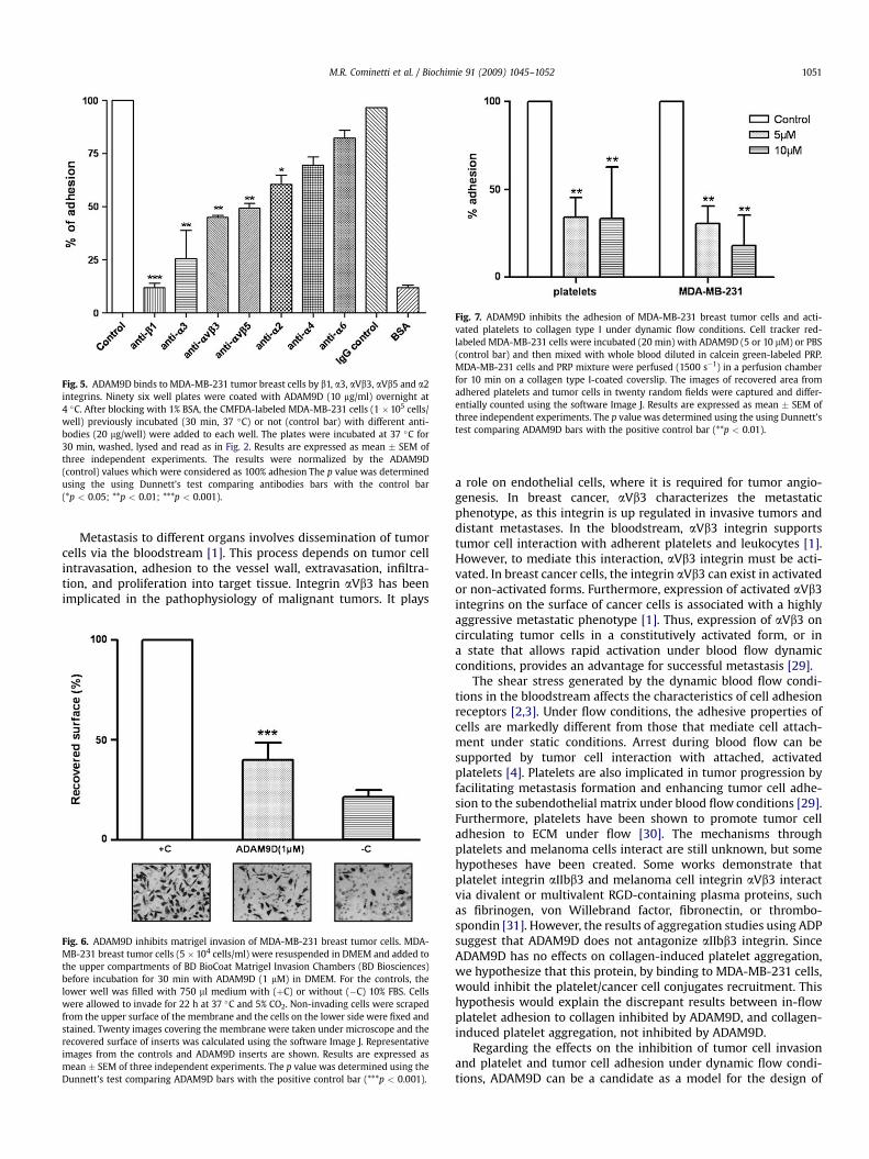
Fig. 5. ADAM9D binds to MDA-MB-231 tumor breast cells by b1, a3, aVb3, aVb5 and a2integrins. Ninety six well plates were coated with ADAM9D (10 mg/ml) overnight at4 �C. After blocking with 1% BSA, the CMFDA-labeled MDA-MB-231 cells (1 �105 cells/well) previously incubated (30 min, 37 �C) or not (control bar) with different anti-bodies (20 mg/well) were added to each well. The plates were incubated at 37 �C for30 min, washed, lysed and read as in Fig. 2. Results are expressed as mean � SEM ofthree independent experiments. The results were normalized by the ADAM9D(control) values which were considered as 100% adhesion The p value was determinedusing the using Dunnett’s test comparing antibodies bars with the control bar(*p < 0.05; **p < 0.01; ***p < 0.001).
Fig. 7. ADAM9D inhibits the adhesion of MDA-MB-231 breast tumor cells and acti-vated platelets to collagen type I under dynamic flow conditions. Cell tracker red-labeled MDA-MB-231 cells were incubated (20 min) with ADAM9D (5 or 10 mM) or PBS(control bar) and then mixed with whole blood diluted in calcein green-labeled PRP.MDA-MB-231 cells and PRP mixture were perfused (1500 s�1) in a perfusion chamberfor 10 min on a collagen type I-coated coverslip. The images of recovered area fromadhered platelets and tumor cells in twenty random fields were captured and differ-entially counted using the software Image J. Results are expressed as mean � SEM ofthree independent experiments. The p value was determined using the using Dunnett’stest comparing ADAM9D bars with the positive control bar (**p < 0.01).
M.R. Cominetti et al. / Biochimie 91 (2009) 1045–1052 1051
Metastasis to different organs involves dissemination of tumorcells via the bloodstream [1]. This process depends on tumor cellintravasation, adhesion to the vessel wall, extravasation, infiltra-tion, and proliferation into target tissue. Integrin aVb3 has beenimplicated in the pathophysiology of malignant tumors. It plays
Fig. 6. ADAM9D inhibits matrigel invasion of MDA-MB-231 breast tumor cells. MDA-MB-231 breast tumor cells (5 � 104 cells/ml) were resuspended in DMEM and added tothe upper compartments of BD BioCoat Matrigel Invasion Chambers (BD Biosciences)before incubation for 30 min with ADAM9D (1 mM) in DMEM. For the controls, thelower well was filled with 750 ml medium with (þC) or without (�C) 10% FBS. Cellswere allowed to invade for 22 h at 37 �C and 5% CO2. Non-invading cells were scrapedfrom the upper surface of the membrane and the cells on the lower side were fixed andstained. Twenty images covering the membrane were taken under microscope and therecovered surface of inserts was calculated using the software Image J. Representativeimages from the controls and ADAM9D inserts are shown. Results are expressed asmean � SEM of three independent experiments. The p value was determined using theDunnett’s test comparing ADAM9D bars with the positive control bar (***p < 0.001).
a role on endothelial cells, where it is required for tumor angio-genesis. In breast cancer, aVb3 characterizes the metastaticphenotype, as this integrin is up regulated in invasive tumors anddistant metastases. In the bloodstream, aVb3 integrin supportstumor cell interaction with adherent platelets and leukocytes [1].However, to mediate this interaction, aVb3 integrin must be acti-vated. In breast cancer cells, the integrin aVb3 can exist in activatedor non-activated forms. Furthermore, expression of activated aVb3integrins on the surface of cancer cells is associated with a highlyaggressive metastatic phenotype [1]. Thus, expression of aVb3 oncirculating tumor cells in a constitutively activated form, or ina state that allows rapid activation under blood flow dynamicconditions, provides an advantage for successful metastasis [29].
The shear stress generated by the dynamic blood flow condi-tions in the bloodstream affects the characteristics of cell adhesionreceptors [2,3]. Under flow conditions, the adhesive properties ofcells are markedly different from those that mediate cell attach-ment under static conditions. Arrest during blood flow can besupported by tumor cell interaction with attached, activatedplatelets [4]. Platelets are also implicated in tumor progression byfacilitating metastasis formation and enhancing tumor cell adhe-sion to the subendothelial matrix under blood flow conditions [29].Furthermore, platelets have been shown to promote tumor celladhesion to ECM under flow [30]. The mechanisms throughplatelets and melanoma cells interact are still unknown, but somehypotheses have been created. Some works demonstrate thatplatelet integrin aIIbb3 and melanoma cell integrin aVb3 interactvia divalent or multivalent RGD-containing plasma proteins, suchas fibrinogen, von Willebrand factor, fibronectin, or thrombo-spondin [31]. However, the results of aggregation studies using ADPsuggest that ADAM9D does not antagonize aIIbb3 integrin. SinceADAM9D has no effects on collagen-induced platelet aggregation,we hypothesize that this protein, by binding to MDA-MB-231 cells,would inhibit the platelet/cancer cell conjugates recruitment. Thishypothesis would explain the discrepant results between in-flowplatelet adhesion to collagen inhibited by ADAM9D, and collagen-induced platelet aggregation, not inhibited by ADAM9D.
Regarding the effects on the inhibition of tumor cell invasionand platelet and tumor cell adhesion under dynamic flow condi-tions, ADAM9D can be a candidate as a model for the design of

M.R. Cominetti et al. / Biochimie 91 (2009) 1045–10521052
selective inhibitors against the adhesion and extravasation ofcancer cells. Anti-adhesion therapy is a promising therapeuticconcept in oncology [32] and many pharmacological and toxico-logical studies indicate that antagonists of different integrins, suchas disintegrins, RGD peptides, small molecules, and blocking anti-bodies are well tolerated in different animal species, suggestingthat these molecules may be potentially effective and safe fortherapeutic purposes [33]. However, the secondary effects of tar-geting ADAM9D in tumor breast cancer remains to be identified.
In conclusion, we have demonstrated that ADAM9D is able toinhibit tumor and platelet cell adhesion under flow to simulate invivo conditions. Our results suggest a role for the disintegrindomain of ADAM9 in the metastatic process. In addition, ADAM9Dcan be used as a tool for investigating the role of ADAMs inmetastasis and cancer progression.
Acknowledgements
M.R. Cominetti, A.C.B.M. Martin and J.U. Ribeiro were therecipients of fellowships and research grants provided by FAPESP(Fundaçao de Amparo a Pesquisa do Estado de Sao Paulo, Brazil).This work was supported by travel grants provided in cooperationby INSERM and the Brazilian government (CNPq-INSERM andFAPESP-INSERM collaborations).
References
[1] B. Felding-Habermann, T.E. O’Toole, J.W. Smith, E. Fransvea, Z.M. Ruggeri,M.H. Ginsberg, P.E. Hughes, N. Pampori, S.J. Shattil, A. Saven, B.M. Mueller,Proc. Natl. Acad. Sci. U.S.A. 98 (2001) 1853–1858.
[2] A.B. Al-Mehdi, K. Tozawa, A.B. Fisher, L. Shientag, A. Lee, R.J. Muschel, Nat.Med. 6 (2000) 100–102.
[3] C.W. Wong, A. Lee, L. Shientag, J. Yu, Y. Dong, G. Kao, A.B. Al-Mehdi,E.J. Bernhard, R.J. Muschel, Cancer Res. 61 (2001) 333–338.
[4] J. Pilch, R. Habermann, B. Felding-Habermann, J. Biol. Chem. 277 (2002)21930–21938.
[5] M.J. Humphries, M.A. Travis, K. Clark, A.P. Mould, Biochem. Soc. Trans. 32(2004) 822–825.
[6] A.L. Stone, M. Kroeger, Q.X. Sang, J. Protein Chem. 18 (1999) 447–465.[7] J. Kratzschmar, L. Lum, C.P. Blobel, J. Biol. Chem. 271 (1996) 4593–4596.[8] C.P. Blobel, Cell 90 (1997) 589–592.[9] D.F. Seals, S.A. Courtneidge, Genes Dev. 17 (2003) 7–30.
[10] J.M. White, Curr. Opin. Cell Biol. 15 (2003) 598–606.[11] C.P. Blobel, Nat. Rev. Mol. Cell Biol. 6 (2005) 32–43.[12] L.G. Jia, K. Shimokawa, J.B. Bjarnason, J.W. Fox, Toxicon 34 (1996) 1269–1276.[13] D. Nath, P.M. Slocombe, P.E. Stephens, A. Warn, G.R. Hutchinson,
K.M. Yamada, A.J. Docherty, G. Murphy, J. Cell Sci. 112 (Pt 4) (1999) 579–587.[14] K. Reiss, A. Ludwig, P. Saftig, Pharmacol. Ther. 111 (2006) 985–1006.[15] X.P. Zhang, T. Kamata, K. Yokoyama, W. Puzon-McLaughlin, Y. Takada, J. Biol.
Chem. 273 (1998) 7345–7350.[16] D. Nath, P.M. Slocombe, A. Webster, P.E. Stephens, A.J. Docherty, G. Murphy,
J. Cell Sci. 113 (Pt 12) (2000) 2319–2328.[17] A. Karadag, M. Zhou, P.I. Croucher, Blood 107 (2006) 3271–3278.[18] A. Mazzocca, R. Coppari, R. De Franco, J.Y. Cho, T.A. Libermann, M. Pinzani,
A. Toker, Cancer Res. 65 (2005) 4728–4738.[19] P. Zigrino, J. Steiger, J.W. Fox, S. Loffek, A. Schild, R. Nischt, C. Mauch, J. Biol.
Chem. 282 (2007) 30785–30793.[20] J.M. Gutierrez, A. Rucavado, T. Escalante, C. Diaz, Toxicon 45 (2005) 997–1011.[21] M. Zhou, R. Graham, G. Russell, P.I. Croucher, Biochem. Biophys. Res. Com-
mun. 280 (2001) 574–580.[22] M.R. Cominetti, C.H. Terruggi, O.H. Ramos, J.W. Fox, A. Mariano-Oliveira,
M.S. De Freitas, C.C. Figueiredo, V. Morandi, H.S. Selistre-de-Araujo, J. Biol.Chem. 279 (2004) 18247–18255.
[23] N. Gomes, C. Legrand, F. Fauvel-Lafeve, Clin. Exp. Metastasis 22 (2005)215–223.
[24] B. Leitinger, E. Hohenester, Mammalian collagen receptors, Matrix Biol. 26(2007) 146–155.
[25] A.I. Roberts, R.E. Brolin, E.C. Ebert, Immunology 97 (1999) 679–685.[26] K. Herzhoff, S. Sollberg, C. Huerkamp, T. Krieg, B. Eckes, Br. J. Dermatol. 141
(1999) 218–223.[27] N.J. Tawil, M. Houde, R. Blacher, F. Esch, L.F. Reichardt, D.C. Turner,
S. Carbonetto, Biochemistry 29 (1990) 6540–6544.[28] R.M. Mahimkar, O. Visaya, A.S. Pollock, D.H. Lovett, Biochem. J. 385 (2005)
461–468.[29] R. Dardik, Y. Kaufmann, N. Savion, N. Rosenberg, B. Shenkman, D. Varon, Int.
J. Cancer 70 (1997) 201–207.[30] E. Bastida, L. Almirall, A. Ordinas, Thromb. Haemost. 61 (1989) 485–489.[31] B. Felding-Habermann, R. Habermann, E. Saldivar, Z.M. Ruggeri, J. Biol. Chem.
271 (1996) 5892–5900.[32] R. Schmidmaier, P. Baumann, Curr. Med. Chem. 15 (2008) 978–990.[33] S. Huveneers, H. Truong, H.J. Danen, Int. J. Radiat. Biol. 83 (2007) 743–751.
Copyright © 2022 FDOKUMEN