Environmentally cued parturition in a desert rattlesnake, Crotalus atrox
Upload
independentCategory
view
1download
0
CLONING, EXPRESSION, AND HEMOSTATIC ACTIVITIES OF ADISINTEGRIN, r-MOJASTIN 1, FROM THE MOHAVERATTLESNAKE (Crotalus scutulatus scutulatus)
Elda E. Sánchez1, Sara E. Lucena1, Steven Reyes1, Julio G. Soto2, Esteban Cantu1, JuanCarlos Lopez-Johnston1, Belsy Guerrero3, Ana Maria Salazar3, Alexis Rodríguez-Acosta4,Jacob A. Galán5, W. Andy Tao5, and John C. Pérez1,*
1Natural Toxins Research Center, College of Arts and Sciences, 975 W. Avenue B. MSC 158,Texas A&M University-Kingsville, Kingsville, TX 78363, U.S.A2Biological Sciences Department, San Jose State University, One Washington Square, San Jose,CA 95192-0100, USA3Instituto Venezolano de Investigaciones Cientificas (IVIC), Apartado 20632, Caracas 1020,Venezuela4Instituto de Medicina Tropical, Universidad Central de Venezuela, Apartado 47423, Caracas1041, Venezuela5Department of Biochemistry, Purdue University, West Lafayette, IN 47907
AbstractInteractions with exposed subendothelial extracellular proteins and cellular integrins (endothelialcells, platelets and lymphocytes) can cause alterations in the hemostatic system associated withatherothrombotic processes. Many molecules found in snake venoms induce pathophysiologicalchanges in humans, cause edema, hemorrhage, and necrosis. Disintegrins are low molecularweight, non-enzymatic proteins found in snake venom that mediate changes by binding tointegrins of platelets or other cells and prevent binding of the natural ligands such as fibrinogen,fibronectin or vitronectin. Disintegrins are of great biomedical importance due to their bindingaffinities resulting in the inhibition of platelet aggregation, adhesion of cancer cells, and inductionof signal transduction pathways. RT-PCR was used to obtain a 216 bp disintegrin cDNA from a C.s. scutulatus snake venom gland. The cloned recombinant disintegrin called r-mojastin 1 codes for71 amino acids, including 12 cysteines, and an RGD binding motif. r-Mojastin 1 inhibited plateletadhesion to fibronectin with an IC50 of 58.3 nM and ADP-induced platelet aggregation in wholeblood with an IC50 of 46 nM. r-Mojastin 1 was also tested for its ability to inhibit platelet ATPrelease using PRP resulting with an IC50 of 95.6 nM. MALDI-TOF mass spectrum analysisshowed that r-mojastin has a mass of 7.9509 kDa.
© 2010 Elsevier Ltd. All rights reserved.*Corresponding Author: JC Perez, [email protected].
Publisher's Disclaimer: This is a PDF file of an unedited manuscript that has been accepted for publication. As a service to ourcustomers we are providing this early version of the manuscript. The manuscript will undergo copyediting, typesetting, and review ofthe resulting proof before it is published in its final citable form. Please note that during the production process errors may bediscovered which could affect the content, and all legal disclaimers that apply to the journal pertain.
NIH Public AccessAuthor ManuscriptThromb Res. Author manuscript; available in PMC 2012 September 10.
Published in final edited form as:Thromb Res. 2010 September ; 126(3): e211–e219. doi:10.1016/j.thromres.2010.06.006.
NIH
-PA Author Manuscript
NIH
-PA Author Manuscript
NIH
-PA Author Manuscript

Keywordsr-Mojastin 1; Crotalus scutulatus scutulatus; recombinant disintegrins; hemostasis; plateletfunction
IntroductionDisintegrins are among a number of biomedically-important molecules in snake venoms thatare classified into five groups: short, medium, long, dimeric and the disintegrin domain ofthe PIII class of snake venom metalloproteanases [1]. Short disintegrins contain 41–51amino acids and 8 cysteines [2], medium are within the range of 70 amino acids and 12cysteines, and long usually with 84 amino acids and 14 cysteines [3]. Disintegrins aresynthesized from a metalloproteinase/disintegrin precursor and mature by cleavage from theprecursor molecule [4]. Disintegrins contain a conserved cysteine configuration within theirprimary structure, and their 3-D conformation is made stable by disulfide linkages. Theirbinding loops bind within the crevice of integrin receptors [5]. Most disintegrins posses anRGD motif located near the C-terminus; however, KGD, RTS, KTS, MGD, WGD, and ECDdomains have also been identified [6,7]. This “RGD” binding domain is found at the tip of aflexible hairpin loop of the disintegrin and is essential to the integrin-inhibitory activity[8,9]. Disintegrins inhibit platelet aggregation, and some can also inhibit cancer cell growth,and/or angiogenesis [10–14].
The expression of recombinant versions of interesting disintegrins has become essential,thus facilitating the maintenance of a continuous supply for drug development. Cloningmolecules from venomous snakes also has important conservation implications, as some ofthe snakes with promising biomedical venom components are in danger of extinction.Furthermore, cloning these biomedically-important molecules decreases the risk for thoseinvolved in the extraction of venom. Only a few disintegrins have been cloned andexpressed with activity, and some have been used to study anti-thrombotic and anti-tumoractivity [15–21].
A group of Crotalus scutulatus scutulatus (Mohave rattlesnake) identified in central Arizonacontained venom that was proteolytic, hemorrhagic, and had disintegrin activity [12]. Fromthat group, RGD containing disintegrins, mojastin 1 and 2 were isolated [12]. Mojastin 1 and2 were medium-sized disintegrins that inhibited APD-induced platelet aggregation in wholeblood. The goals of this study were to express a recombinant disintegrin from C. s.scutulatus snake in E. coli BL21 cells, and test its biological activities. A prokaryotic hostexpression system was used because they are less expensive than mammalian or insect cellexpression systems. Furthermore, a prokaryotic system yields a higher concentration ofprotein in less time. Even though prokaryotic systems do not have post-translationmodifications, active recombinant disintegrins have been expressed in bacterial cells [4–7,10–18]. The cloned disintegrin, named r-mojastin 1, was shown to be highly active ininhibiting APD-induced platelet aggregation using platelet-rich plasma and whole blood,platelet ATP release, and platelet adhesion to fibronectin.
Materials and methodsPCR amplification of Mojastin 1
A Mohave rattlesnake (C. s. scutulatus) from Arizona (Pinal Co., AVID#: 058-784-560;housed at the Natural Toxins Research Center Serpentarium.) that expressed disintegrinswas sacrificed and its venom gland excised and immediately frozen at −80°C. Poly (A)+
RNA was purified from the venom gland using Fast Tract 2.0 mRNA isolation Kit
Sánchez et al. Page 2
Thromb Res. Author manuscript; available in PMC 2012 September 10.
NIH
-PA Author Manuscript
NIH
-PA Author Manuscript
NIH
-PA Author Manuscript

(Invitrogen Life Technologies, USA). Using gene-specific primers, cDNA was synthesizedfrom the mRNA using the Promega Access RT-PCR system (Promega Corporation, USA).Disintegrin gene specific primers utilized in the RT-PCR method were designed fromconserved sequences found in the disintegrins of other snakes (Crotalus atrox [atrolysin e],Agkistrodon contortrix contortrix [contortrostatin and acostatin], Trimeresurusmucrosquamatus [Trimucin], Trimeresurus flavoviridis [flavostatin], and Gloydius halys[halystatin]). The forward primer: 5′-CCGGAATTCGGAGAAGAATGTGACTGTGGC-3′(EcoRI site is underlined) and reverse primer: 5′-ACGCCTCGAGCTGCCTGTTGCTGCAGACC-3′ (the XhoI site is underlined) wereutilized to obtain cDNA amplification products. RT-PCR conditions and cDNA analysiswere carried-out as previously described [22].
cDNA cloning of r-Mojastin 1The cDNA was ligated into the pGEX-4T-1 expression vector (GE Healthcare Lifesciences)and transformed into E. coli DH5α competent cells. Recombinant plasmids containing r-mojastin 1 were purified by the Wizard Plus Minipreps DNA Purification System (PromegaCorporation, USA), and sequenced with disintegrin-specific primers. The sequencing datawere analyzed with ClustalW DNA alignment program [23] in Biology Workbench [24].The MW/pI of the proteins was computed by Protein Identification and Analysis Tools onthe ExPASy Server.
Expression and purification of recombinant r-mojastin 1Once the sequence was obtained, in-frame r-mojastin 1-pGEX-4T-1 plasmids weretransformed into E. coli BL21 cells (Amersham Biosciences). Cultures were grown at 37 °Cto 0.6–0.8 A600. Induction was carried out by 0.5 mM (final concentration) isopropyl β-Dthiogalactoside (IPTG), (Amersham Biosciences) at 35 °C for 3 h. Bacterial cells werecentrifuged at 3,800 × g for 15 min (4 °C) and resuspended with 80 mL of ice cold 1X PBSbuffer pH 7.4. Bacterial cell disruption was conducted with a Branson Sonifier 450(Danbury, CT) with the output control setting at 1, a duty cycle setting of constant, and 6sonication pulses of 30 s per pulse. The cell debris was removed by centrifuging at 12,000 ×g for 10 min at 4 °C. Crude lysate was incubated with 2 mL 50% slurry glutathioneSepharose 4B (GS4B), (Amersham Biosciences), for 30 min at room temperature usinggentle agitation. r-Mojastin 1 proteins were cleaved and eluted from glutathione S-transferase (GST) bound to GS4B by thrombin cleavage, according to the GST Gene FusionSystem Handbook (Amersham Biosciences). Thrombin was removed from r-mojastin 1using a 1 mL HiTrap™ Benzamidine Sepharose 4 Fast Flow (high sub) column (AmershamBiosciences). The column was equilibrated with 5 column volumes of binding buffer (20mM sodium phosphate, 0.15M NaCl, pH 7.5). One milliliter of the sample was loaded intothe column and r-mojastin 1 protein was obtained by washing the column with a high saltbuffer (20 mM sodium phosphate, 1.0 M NaCl, pH 7.5). The column was finally washedwith 10 column volumes of elution buffer (10 mM HCl, 0.5 M NaCl, pH 2.0) to remove thethrombin bound to the column.
Isolation of native mojastin by high performance liquid chromatographyThe native mojastin disintegrin was isolated using the method of Sánchez et al., [12], whichconsisted of a combination of three chromatographic steps: reverse phase C18 (Vydac), sizeexclusion (WATERS Protein PAK60), and anion exchange (WATERS DEAE 5PW). Crudevenom was extracted [25] from an individual Mohave rattlesnake (Avid # 011-064-358)collected from Pinal Co., Arizona and kept at the Natural Toxins Research Center at TexasA&M University-Kingsville, Kingsville, TX.
Sánchez et al. Page 3
Thromb Res. Author manuscript; available in PMC 2012 September 10.
NIH
-PA Author Manuscript
NIH
-PA Author Manuscript
NIH
-PA Author Manuscript

Dialyzation and lyophilization of disintegrinsr-Mojastin 1, r-mojastin 1-GST, and native mojastin were dialyzed in a 2,000 MWCOdialyzing membrane (Spectrapore) against Milli-Q water at 4°C, overnight. The volumesand absorbances at 280 nm were measured. A total of 3.3 mg of r-mojastin 1 per 4 L ofbacteria was collected. The samples were frozen in liquid nitrogen and then lyophilizedusing a Labconco freeze dryer.
SDS Polyacrylamide Gel ElectrophoresisThe disintegrins were subjected to electrophoresis by using a pre-cast 10–20% Tricine gel[26] in an Xcell SureLock Mini-Cell (Invitrogen Life Technologies, USA). Gels werestained with 100 mL SimplyBlue SafeStain (Invitrogen Life Technologies, USA) anddestained overnight in Milli-Q water.
Mass Spectrometry Analysis (MALDI-TOF)The disintegrins were subjected to MALDI-TOF analysis according to the methods ofSalazar et al. [25]. A Matrix Assisted Laser Desorption/Ionization (MALDI) Time Of Flight(TOF) Mass Spectrometer (AUTOFLEX II TOF/TOF, Bruker Daltonics) was used.
LC-MS/MS and data analysis of native mojastinThe native mojastin disintegrin peptides were obtained according to the methods of Salazaret al. [25].
Blood donorsIRB approval and informed consent by the donors are acquired prior to blood draws.
Platelet adhesion assayPlatelet adhesion studies were done according to modified methods of Lucena et al. [27]. Todetermine the effect of the disintegrin on platelet adhesion to fibronectin, washed plateletswere used to eliminate plasma contaminants (procoagulant proteins), which could activatethe platelets, altering the assay results. The percentage of platelet adhesion was determinedby assigning 100% to the number of platelets adhered in the absence of the disintegrins. As anegative control, wells were coated with bovine serum albumin (2 mg/mL) to preventadhesion.
Platelet ATP releasePlatelet ATP release studies were done according to the Chrono-log manual for lumi-aggregometer protocol for PRP aggregation. A dual-channel Chrono-Log model 560 CAaggregometer (Havertown, USA) was used. In all instances of ATP release measurements,studies were carried out with a platelet count of 300,000 platelets/μL. Data were collectedusing the software Aggrolink v. 5.2.0.3 on a Pentium 4 computer containing Windows XP.The percentage of inhibition ATP release from platelet stimulated with ADP was calculatedby comparing luminescence of disintegrin to the control. The IC50 value was calculatedfrom a dose-dependent curve that is achieved from at least five different inhibitorconcentrations.
Platelet aggregation with whole blood and platelet rich plasma (PRP)A dual-channel Chrono-Log Whole-Blood Aggregometer [Ca+2] model 560 (Havertown,USA) was used to monitor platelet aggregation, by impedance and turbidimetry as describedpreviously [9, 28]. Briefly, different concentrations of recombinant disintegrins were addedto 10% citrated whole human blood or PRP, and pre-incubated at 37 °C for 2 and 4 min,
Sánchez et al. Page 4
Thromb Res. Author manuscript; available in PMC 2012 September 10.
NIH
-PA Author Manuscript
NIH
-PA Author Manuscript
NIH
-PA Author Manuscript
respectively. Platelet aggregation was initiated by 10 μM ADP. Light transmittancereflecting percentage aggregation was measured using PRP and percentage of impedancewas measured using whole blood. The maximal aggregation in the absence of recombinantdisintegrin was given as 100% aggregation. The IC50 values were calculated from a dose-dependent curve using Microsoft excel.
The inhibition of ADP-induced platelet aggregation by r-mojastin 1 in whole blood and PRPhaving the same platelet count was also performed. Whole blood and PRP were adjusted to aplatelet count of 230,000 platelets/μL, which corresponds to a hematocrit value of 30%.
Sonoclot® SignaturesActivated clot time (ACT), clot rate (CR) and platelet function (PF) were measured usingwhole human blood on a Sonoclot® Coagulation & Platelet Function Analyzer (SIENCO,Inc.) as described by Sanchez et al. [29]. Briefly, blood (10% citrated) was collected bygravity flow into a 50 mL test tube containing 3.2% sodium citrate using a 19G ¾Vacutainer needle with 12′ of tubing. A total of 13 μL of 0.25 M CaCl2 was added to oneside of a gbACT + KIT cuvette and then 10 μL of disintegrins at the same molarconcentrations or 0.85% saline were added to the opposite side of the cuvette. After bothsolutions were added, 360 μL of citrated blood were added to the cuvette and the analyzerwas activated. Data were collected by Signature Viewer™ program v. 3.1 on an iMaccomputer containing Mac OS X software. A p < 0.05 signifies a significant difference whencompared to the control values. P values were calculated using a t-test, two-tailed P value onGraphPad Prism 4 software. A total of four trials were performed for each sample.
ResultscDNA sequencing analysis
The cDNA obtained was a 216 bp long fragment coding for 71 amino acids. The deducedamino acid sequence also included twelve cysteines and an RGD-motif region (Fig. 1).NCBI protein BLAST analysis showed that the deduced amino acid sequence of the cloneddisintegrin (r-mojastin 1) was identical to the native disintegrin mojastin 1 isolated from thevenom of C. s. scutulatus [12].
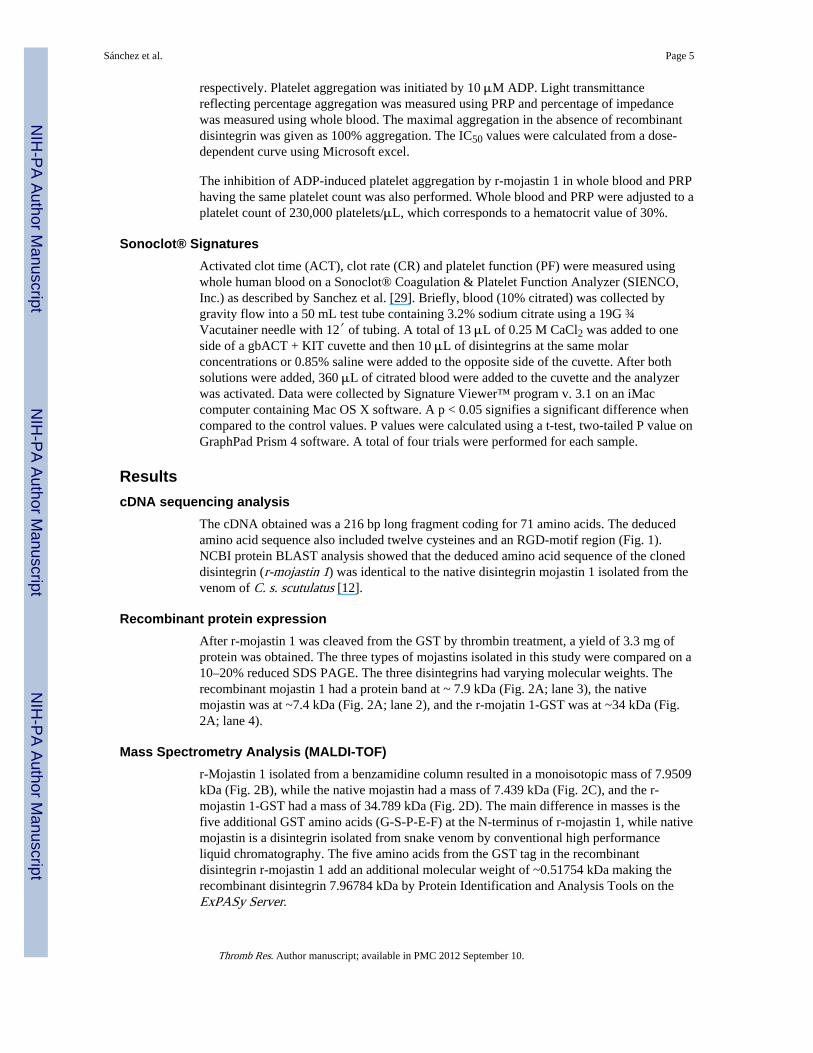
Recombinant protein expressionAfter r-mojastin 1 was cleaved from the GST by thrombin treatment, a yield of 3.3 mg ofprotein was obtained. The three types of mojastins isolated in this study were compared on a10–20% reduced SDS PAGE. The three disintegrins had varying molecular weights. Therecombinant mojastin 1 had a protein band at ~ 7.9 kDa (Fig. 2A; lane 3), the nativemojastin was at ~7.4 kDa (Fig. 2A; lane 2), and the r-mojatin 1-GST was at ~34 kDa (Fig.2A; lane 4).
Mass Spectrometry Analysis (MALDI-TOF)r-Mojastin 1 isolated from a benzamidine column resulted in a monoisotopic mass of 7.9509kDa (Fig. 2B), while the native mojastin had a mass of 7.439 kDa (Fig. 2C), and the r-mojastin 1-GST had a mass of 34.789 kDa (Fig. 2D). The main difference in masses is thefive additional GST amino acids (G-S-P-E-F) at the N-terminus of r-mojastin 1, while nativemojastin is a disintegrin isolated from snake venom by conventional high performanceliquid chromatography. The five amino acids from the GST tag in the recombinantdisintegrin r-mojastin 1 add an additional molecular weight of ~0.51754 kDa making therecombinant disintegrin 7.96784 kDa by Protein Identification and Analysis Tools on theExPASy Server.
Sánchez et al. Page 5
Thromb Res. Author manuscript; available in PMC 2012 September 10.
NIH
-PA Author Manuscript
NIH
-PA Author Manuscript
NIH
-PA Author Manuscript

LC-MS/MS of native mojastinIn order to verify that the protein isolated via chromatography from crude venom was adisintegrin similar to the r-mojastin 1, LC-MS/MS was conducted. Analysis of the nativemojastin by LC-MS/MS resulted in a total of 2 peptide fragments totaling 34 amino acids.Peptide GDWNDDTCTGQSADCPR MH+ 1954.7296 and peptideLRPGAQCADGLCCDQCR MH+ 2036.8523 were identified (Fig. 3). The native mojastinhad 47.2% coverage with the native disintegrins barbourin (P22827), cerastin (P31982),crotatroxin (P68520), durissin (P68521), horrdistatin-2 (P0C7X6), lutosin (P31986),mojastin-2 (P0C7X7), and tergeminin (P22828).
Platelet adhesion assayPlatelet adhesion promotes the formation of thrombus, arresting hemorrhage, and allowingwound healing. However, this essential hemostatic process can lead to diseases that result inarterial occlusion in vessels of the heart and brain. The r-mojastin 1 and native mojastinwere able to inhibit platelet adhesion to fibronectin with IC50s of 62.2 and 58.6 nM,respectively (Fig. 5).
Inhibition of platelet ATP secretionATP released from platelets is involved in platelet shape-change and helps to amplifyplatelet responses mediated by agonists such as ADP or collagen. ATP release is alsoinvolved in all of the sequential events involved in platelet function and hemostasis. Allmojastin disintegrins (r-mojastin 1-GST, r-mojastin 1, and native mojastin) inhibited ATPrelease from platelet induced by ADP with IC50s of 335, 95.6 and 19.5 nM on PRP,respectively (Table 1).
Inhibition of platelet aggregation with platelet rich plasma (PRP) and whole bloodThe studies of platelet aggregation inhibition are generally done with PRP. To determine ifwhole blood would be more efficient in inhibiting platelet aggregation, a parallel study wascarried out using both PRP and whole blood with r-mojastin 1, r-mojastin 1-GST and nativemojastin. The r-mojastin 1-GST, r-mojastin 1, and native mojastin had IC50s of 667.0, 119.7and 44.7 nM on PRP, respectively (Table 1). The r-mojastin 1-GST, r-mojastin 1 and nativemojastin had IC50s of 296.0, 46.0 and 19.3 nM on whole blood, respectively (Table 1). Withall three types of mojastin disintegrins, whole blood was more efficient when used as asubstrate of inhibiting platelet aggregation.
To insure that the most efficient IC50 using whole blood was not a factor of a lower plateletcount, inhibition of platelet aggregation by r-mojastin 1 was repeated using an equal plateletcount for both whole blood and PRP. Whole blood and PRP were both adjusted to plateletcounts of 230,000 platelets/μL. Thus, the inhibition of ADP-induced platelet aggregationIC50s values were 40 and 90 nM, respectively (Fig. 4). The results revealed that other factorsin whole blood could play a role in inhibiting platelet aggregation.
Sonoclot® SignaturesThe Sonoclot® signatures display the measurement of the blood’s activated clot time (ACT)in seconds, the clot rate (CR) in clot signals per minute and platelet function (PF) as afunction of clot retraction (Figs. 6A & B). The ACT is the time in which fibrin formationbegins, the CR is the kinetic measurement of fibrin formation and clot development, whichis the maximum slope of the Sonoclot Signature during initial fibrin polymerization and clotdevelopment, and PF is obtained from the timing and quality of the clot retraction. Thevalues for PF range from 0–5, where 0 represents no clot retraction and a flat SonoclotSignature as those observed for all three mojastins used in this study (Figs. 6A & B). A PF
Sánchez et al. Page 6
Thromb Res. Author manuscript; available in PMC 2012 September 10.
NIH
-PA Author Manuscript
NIH
-PA Author Manuscript
NIH
-PA Author Manuscript

higher than 1 represents normal clot retraction and varies from patient to patient. A normalPF contains a sharp peak in the Sonoclot Signature after fibrin formation, as seen on thecontrol sample in figure 6. The control blood, without disintegrins, had an average ACT of212.5, CR of 22.5 and PF of 2.7 (Table 2). A concentration of 409 nM of disintegrins inwhole blood had no significant effects on the ACTs and CRs; however, it did have asignificant impact on PF with all three disintegrins (Fig. 6A). In order to compare theactivity of all three disintegrins in regards to PF, a ½ dilution (204.5 nM) of the initialconcentration was used. This lower concentration did not have significant differences in theACTs and CRs (Table 1, Fig. 6B), which was expected. Furthermore, the PFs for r-mojastin1-GST and r-mojastin 1 were also not significantly different, while the PF for the nativemojastin was (p = 0.0052). Native mojastin proved to be more effective in inhibiting plateletfunction than its recombinant counterparts.
DiscussionDisintegrins found in venomous snakes can be expressed in E. coli cells and further purifiedby one step chromatography. This process bypasses the need for venom extraction fromsnakes, and the laborious chromatography procedures needed to obtain purified disintegrins.Once recombinant disintegrins are obtained [12,18–21], they can be tested for biomedicalapplications, such as inhibiting platelet aggregation for anti-thrombotic studies, andinhibition of cancer cell growth. Current disadvantages of cloning these proteins are thattheir activities may be less than those observed with native proteins, and snakes must besacrificed to obtain the venom gland. In this study, an expressed disintegrin gene Mojastin 1was isolated from the venom gland of C. s. scutulatus. The expressed r-mojastin 1 protein isidentical, in the active portion of the amino acid sequence, to the medium size nativedisintegrin mojastin 1 isolated from the venom of C. s. scutulatus [12].
The physiological activities of platelets undergo three sequential steps that can be studiedindependently of each other. These are: 1) adhesion, 2) activation, and 3) aggregation. Theparticipation of platelets in the process of hemostasis and thrombosis is well recognized.When a blood vessel is damaged, platelets adhere to the disrupted surface. The adherentplatelets subsequently liberate biologically active constituents and aggregates [30].
Disintegrins inhibit platelet adhesion to immobilized extracellular matrix blocking someintegrins [6.]. The majority of the disintegrin-primary hemostasis research is focused ontheir ability to inhibit platelet aggregation [6]. This is the first time that a single disintegrin(r-mojastin 1) has been reported to inhibit the three processes involved in platelet functions.Platelet adhesion is an important physiological response as a result of vascular lesionsamong others diseases. It is viewed as the first step in which platelets, through specificmembrane receptors, bind to cellular and extracellular matrix constituents of the vessel walland tissues [31]. This action promotes the formation of thrombus, arresting hemorrhage, andallowing wound healing. However, this essential hemostatic process can lead to diseases thatresult in arterial occlusion in vessels of the heart and brain [31]. In addition to suchpathological conditions, platelet adhesive properties are vital to many types ofpathophysiological processes that include inflammation, transplant rejection, and cancermetastasis [32]. In this study, the r-mojastin 1 and native mojastin disintegrins were able toinhibit platelet adhesion to fibronectin with IC50s of 62.2 and 58.6 nM (Fig. 5). Fibronectinis a major glycoprotein of the extracellular matrix and it is known to be involved in theattachment and spreading of many cell types. It binds to a number of biologically importantsubstrates including heparin, collagen/gelatin, and fibrin, as well as to cells through bothintegrin, and non-integrin receptors. The presence of fibronectin in the vessel wall isessential for platelet adhesion and greatly enhances thrombus formation [33]. For instance,jararhagin, a protein isolated of Bothrops jararaca venom, can interfere with platelet function
Sánchez et al. Page 7
Thromb Res. Author manuscript; available in PMC 2012 September 10.
NIH
-PA Author Manuscript
NIH
-PA Author Manuscript
NIH
-PA Author Manuscript

in two ways: first, by degradation of different platelet receptors and adhesive proteinsinvolved in hemostasis; and second, by a nonenzymatic interference (disintegrin-mediated)with the function of platelet adhesion receptors [34]. Furthermore, lonomin V, a serineprotease isolated from the haemolymph of the Lonomia achelous caterpillar, has also beendemonstrated to reduce platelet adhesion to fibronectin [27].
Sizeable amounts of adenosine triphosphate (ATP) and adenosine diphosphate (ADP) arefound in erythrocytes, platelets, and other cells and tissues and can depart the cells throughphysical damage or exocytosis [35]. ATP released from platelet-dense granules afteractivation is involved in platelet shape-change and assists to amplify platelet responsesmediated by agonists such as thrombin, ADP, adrenalin or collagen. Among other solublemediators, released ATP is involved in all of the sequential events involved in plateletfunction and hemostasis [36,37]. By using an ATP bioluminescence assay [38], the mojastindisintegrins were used to determine the inhibition of ATP release from platelets. In thequantitation of adenosine triphosphate (ATP) release using PRP, native mojastin was 5 timesmore efficient (19.5 nM) in inhibiting ATP secretion induced by ADP than r-mojastin 1(95.6 nM) (Table 1). This effect may be a consequence of disintegrin action on ADPreceptors, G protein-coupled P2Y1 and P2Y12 ADP receptors. ADP plays a crucial role inhaemostasis and thrombosis and its receptors are potential targets for antithrombotic drugs.[39].
Aggregation is initiated by the binding of agonists, such as thrombin, epinephrine, platelet-activating factor, collagen, or ADP to specific platelet membrane receptors [30]. Previousstudies involving inhibition of ADP-induced platelet aggregation performed withrecombinant disintegrins have demonstrated activity (Table 3). In our study, r-mojastin 1,using both PRP and whole blood, was more efficient in inhibiting ADP-induced plateletaggregation than the recombinant disintegrins listed in Table 3. Platelet aggregation studiesusing PRP with recombinant albolatin disintegrin obtained from Trimeresurus albolabrissnake venom showed that this protein significantly inhibited collagen-induced aggregation(IC50 value close to 1 μM), but had no effect on ADP-induced aggregation [40]. In addition,Marques et al. [41] reported that recombinant barbourin, a KGD-containing monomericdisintegrin, could inhibit ADP-induced platelet aggregation with IC50 values ranging from330 to 370nM. These results indicate that recombinant mojastatin 1 is a strong aggregationinhibitor and can be used as a useful tool for studies of integrin/ligand interaction.
When comparing the mojastin disintegrins, the native mojastin was always more effectivethan its recombinant counterpart in both PRP and whole blood (Table 1). The IC50s, forinhibiting ADP-induced platelet aggregation in whole blood, of the native mojastins were3.3 and 2.6 times more efficient than the r-mojastin 1. Similar results were observed usingPRP (Table 1). The difference between activities could be that r-mojastin 1 may not becompletely folding properly in the E. coli cells because disintegrins are rich in disulfidebonds [42].
When comparing the inhibition of platelet aggregation IC50s for whole blood and PRP,whole blood resulted more efficient in all 3 types of disintegrins used in this study (Table 1);and furthermore, all IC50s using whole blood were 2.2–2.6 times more efficient than theIC50s for PRP (Table 1). This finding is extremely important in drug discovery since wholeblood will always be a factor in drug dosing. Previous research done with disintegrins hasreported IC50 values for platelet rich plasma, which, in light of our findings, could beinterpreted to be lower for whole blood (Table 3). In order to eliminate the possibility thatthe higher dose of disintegrin needed to inhibit platelet aggregation was not due to the highernumber of platelets present in PRP, the platelet counts for PRP and whole blood equalized.The IC50 results (Fig. 4; 40 nM for whole blood and 90 nM for PRP) support the fact that
Sánchez et al. Page 8
Thromb Res. Author manuscript; available in PMC 2012 September 10.
NIH
-PA Author Manuscript
NIH
-PA Author Manuscript
NIH
-PA Author Manuscript

other components in whole blood play a role aiding in the inhibition of ADP-inducedplatelet aggregation. These results were similar, although slightly more efficient, to the IC50obtained by the standard methods (Table 1).
The differences between whole blood and platelet rich plasma are the presence of red bloodcells and leukocytes in whole blood, which are absent in platelet rich plasma [43].Leukocytes and platelets are the most important cellular constituents in hemostasis [44].Leukocytes are aggregated along with platelets having an influence on thrombi structure[44]. When leukocytes are activated, they secrete both serine and metalloproteinases thateffect fibrinolysis by direct digestion of fibrin, or indirectly by degradation of zymogens andinhibitors of coagulation and fibrinolytic proteinases [45,46]. For instance, matrixmetalloproteinase-2 (MMP-2) can cleave thrombin resulting in an enzyme void of clottingand platelet stimulating activity; and to further assist in the cause, a serine proteinase(elastase) can degrade factor XIII (the fibrin stabilizing factor) and inactivate factors VII,VIII, IX, and XII [47,48]. In addition, elastase is able to degrade fibrin directly along withstimulating an alternate pathway of plasminogen activation [49].
In addition to leukocytes, red blood cells (RBC) also play a role in influencing hemostasis[44]. RBCs help in the activation of the coagulation factor cascade by changing shape andserving as a procoagulant surface very similar to platelets [50]. Furthermore, RBCs have aninfluence on the ultimate physical properties of fibrin [51,52], forming larger pores in thepresence of these erythrocytes, which greatly affects the path of its dissolution. The rolesleukocytes and RBCs play in hemostasis, in concert with disintegrins, may very well becontributing to the efficiency of platelet aggregation inhibition that was determined in wholeblood as opposed to the less efficient activity detected by these disintegrins in PRP.
Furthermore, all three mojastins were analyzed using a Sonoclot® Coagulation & PlateletFunction Analyzer, in which the measurements are based on the detection of viscoelasticchanges of whole blood or plasma [53]. The Sonoclot® provides qualitative (SonoclotSignature graph) and quantitative (ACT, CR and PF) results on the entire hemostasisprocess. Disintegrins are non-enzymatic proteins that bind to receptors, such as αIIbβ3, onplatelets inhibiting platelet aggregation; and thus, in this particular assay, should only affectplatelet function (PF) maintaining normal ACT and CR values. There were no significantdifferences in the ACTs and CRs for the three mojastin disintegrins at concentrations of 409nM (Fig. 6A) and 204.5 nM (Fig. 6B & Table 2). However, there was a significantdifference in the PFs with the three mojastins when used at 409 nM. Although the Sonoclotanalysis is less sensitive than all the other assays used in this study, this assay can stillprovide additional information pertaining to platelet function and a global vision of thehemostatic system. For instance, the rapid evolution of hemostatic parameters can be easilymonitored using a Sonoclot in patients receiving anticoagulant treatments.
In conclusion, disintegrins can be cloned and purified through multidimensionalchromatographic steps, characterized through functional biological assays, and maintainstrong biological activities. Cloned disintegrins could also provide researchers continuousand effective material with which to conduct in-depth research that could someday be usedto detect, treat, and prevent a wide range of hemostatic diseases. Our data suggest thatmojastin may play a role in the tissue remodeling process occurring in pathologicalconditions. Although the focus of this study was primarily on the hemostatic system, r-mojastin 1 is currently being evaluated for its role in preventing cancer cell adhesion toextracellular matrices, tumor growth, and angiogenesis.
Sánchez et al. Page 9
Thromb Res. Author manuscript; available in PMC 2012 September 10.
NIH
-PA Author Manuscript
NIH
-PA Author Manuscript
NIH
-PA Author Manuscript

AcknowledgmentsFinancial support was obtained by Texas A&M University-Kingsville and NIH grants to the Natural ToxinsResearch Center at Texas A&M University-Kingsville: NIH/NCRR #1 P40 RR018300-05; NIH/SCORE # 2SO6GM008192 (to J.G. Soto), the Science and Technology Fund (FONACIT) programs (PG-2005000400;F-2005000212) and the Instituto Venezolano de Investigaciones Cientificas (IVIC) Caracas, Venezuela, NSFCAREER development award (to W.A.Tao) and NIH/NCI RO1-Minority Supplement #3R01CA115465-03S109.We are grateful for the venom extractions and gland extractions by Doug Hotle, Juan Salinas and Lucy Arispe ofthe NTRC. We thank Nora Diaz De Leon, Zoila Caravajal and Amparo Gil for their technical assistance. Specialthanks goes to Angela Wyro for her technical review of the manuscript.
References1. Calvete JJ, Moreno-Murciano MP, Theakston RD, Kisiel DG, Marcinkieicz C. Snake venom
disintegrins: novel dimeric disintegrins and structural diversification by disulphide bondengineering. Biochem J. 2003; 372(Pt 3):725–34. [PubMed: 12667142]
2. Olfa K, Jose L, Salma D, Amine B, Najet SA, Nicolas A, Maxime L, Raoudha Z, Kamel M, JacquesM, Jean-Marc S, Mohamed EA, Naziha M. Lebestatin, a disintegrin from Macrovipera venom,inhibits integrin-mediated cell adhesion, migration and angiogenesis. Laboratory Investigation.2005; 5:1507–16. [PubMed: 16200076]
3. Kim J, Hong S, Park H, Kim D, Lee W. Structure and function of RGD peptides derived fromdisintegrin proteins. Molecules and Cells. 2005; 19:205–11. [PubMed: 15879703]
4. Okuda D, Koike H, Morita T. A new disintegrin gene structure of the disintegrin family: A subunitof dimeric disintegrin has a short coding region. Biochemistry. 2002; 41:14248–54. [PubMed:12450389]
5. Fujii Y, Okuda D, Fujimoto Z, Horii K, Morita T, Mizuno H. Crystal structure of Trimestatin, adisintegrin containing a cell adhesion recognition motif RGD. J Mol Biol. 2003; 332:1115–22.[PubMed: 14499613]
6. McLane MA, Sánchez EE, Wong A, Paquette-Straub C, Pérez JC. Disintegrins. Curr Drug Targets-Cardiovas Haemat Dis. 2004; 4:327–355.
7. Calvete JJ. Structure-function correlations of snake venom disintegrins. Curr Pharm Des. 2005;11:829–35. [PubMed: 15777237]
8. Huang TF, Holt JC, Kirby EP, Niewiarowski S. Trigramin: primary structure and its inhibition ofvon Willebrand factor binding to glycoprotein IIb/IIIa comlex on human platelets. 1989; 28:661–6.
9. Kini RM, Evans HJ. Structural domains in venom proteins: Evidence that metalloproteinases andnonenzymatic platelet aggregation inhibitors (disintegrins) from snake venoms are derived byproteolysis from a common precursor. Toxicon. 1992; 30:265–293. [PubMed: 1529462]
10. Corrêa MC Jr, Maria DA, Moura-da-Silva AM, Pizzocaro KF, Ruiz IRG. Inhibition of melanomacells tumorigenicity by the snake venom toxin jararhagin. Toxicon. 2002; 40:739–48. [PubMed:12175610]
11. Moreno-Murciano MP, Monleón D, Calvete JJ, Celda B, Marcinkiewicz C. Amino acid sequenceand homology modeling of obtustatin, a novel non-RGD-containing short disintegrin isolated fromthe venom of Vipera lebetina obtusa. Protein Science. 2003; 12:366–71. [PubMed: 12538900]
12. Sánchez EE, Galán JA, Russell WK, Soto JG, Russell DH, Pérez JC. Isolation and characterizationof two disintegrins inhibitng ADP-induced human platelet aggregation from the venom of Crotalusscutulatus scutulatus (Mohave rattlesnake). Toxicology and Applied Pharmacology. 2006; 212:59–68. [PubMed: 16084550]
13. Yang R, Tang C, Chuang W, Huang T, Peng H, Huang T, Fu W. Inhibition of tumor formation bysnake venom disintegrin. Toxicon. 2005; 45:661–9. [PubMed: 15777962]
14. Sánchez EE, Rodríguez-Acosta A, Palomar R, Lucena SE, Bashir S, Soto JG, Pérez JC.Colombistatin: a disintegrin isolated from the venom of the South American snake (Bothropscolombiensis) that effectively inhibits platelet aggregation and SK-Mel-28 cell adhesion. ArchToxicol. 2009; 83:271–279. [PubMed: 18830584]
15. Assakura MT, Silva CA, Mentele R, Camargo ACM, Serrano SMT. Molecular cloning andexpression of structural domains of bothropasin, a P-III metalloproteinase from the venom ofBothrops jararaca. Toxicon. 2003; 41:217–27. [PubMed: 12565741]
Sánchez et al. Page 10
Thromb Res. Author manuscript; available in PMC 2012 September 10.
NIH
-PA Author Manuscript
NIH
-PA Author Manuscript
NIH
-PA Author Manuscript

16. Fernandez JH, Silva CA, Assakura MT, Camargo ACM, Serrano SMT. Molecular cloning,functional expression, and molecular modeling of bothrostatin, a new highly active disintegrinfrom Bothrops jararaca venom. Biochemical and Biophysical Research Communication. 2005;329:457–64.
17. Sanz L, Chen R, Pérez A, Hilario R, Juárez P, Marcinkiewicz C, Monleón D, Celda B, Xiong Y,Pérez-Payá E, Calvete JJ. cDNA cloning and functional expression of Jerdostatin, a novel RTS-disintegrin from Trimeresurus jerdonii and a specific antagonist of the α1β1 integrins. The Journalof Biological Chemistry. 2005; 280:40714–22. [PubMed: 16215260]
18. Wang JH, Wu Y, Ren F, Lu L, Zhao BC. Cloning and characterization of Adinbitor, a noveldisintegrin from the snake venom of Agkistrodon halys brevicaudus stejneger . Acta Biochemicaet Biophysical Sincia. 2004; 36:425–9.
19. Zhou Q, Nakada MT, Brooks PC, Swenson SD, Ritter MR, Argounova S, Arnold C, Markland FS.Contortrostatin, a homodimeric disintegrin, binds to Integrin αvβ5. Biochemical and BiophysicalResearch Communications. 2000; 276:350–5. [PubMed: 11006128]
20. Singhamatr P, Rojnuckarin P. Molecular cloning of albolatin, a novel snake venommetalloprotease from green pit viper (Trimeresurus albolabris), and expression of its disintegrindomain. Toxicon. 2007; 50:1192–200. [PubMed: 17870140]
21. Vija H, Samel M, Siigur E, Aaspõllu A, Tõnismägi K, Trummal K, Subbi J, Siigur J. VGD andMLD-motifs containing heterodimeric disintegrin viplebedin-2 from Vipera lebetina snake venom.Purification and cDNA cloning. Comp Biochem Physiol, Part B. 2009; 153:253–60.
22. Soto JG, White SA, Reyes SR, Regalado R, Sanchez EE, Perez JC. Molecular Evolution of PIII-SVMP and RGD disintegrin genes from the genus Crotalus. GENE. 2007; 389:66–72. [PubMed:17112685]
23. Thompson JD, Higgins DG, Gibson TJ. CLUSTAL W: improving the sensitivity of progressivemultiple alignment sequence through sequence weighting, positive-specific gap penalties andweight matrix choice. Nucleic Acids Res. 1994; 22:4673–80. [PubMed: 7984417]
24. Subramaniam S. The Biology Workbench—a seamless database and analysis environment for thebiologist. Proteins. 1998; 32:1–2. [PubMed: 9672036]
25. Salazar AM, Guerrero B, Cantu B, Cantu E, Rodríguez-Acosta A, Pérez JC, Galán JA, Tao A,Sánchez EE. Venom variation in hemostasis of the southern Pacific rattlesnake (Crotalus oreganushelleri): isolation of hellerase. Comp Biochem Physiol C Toxicol Pharmacol. 2009; 149:307–16.[PubMed: 18804187]
26. Schägger H, von Jagow G. Tricine-sodium dodeclyl sulfate-polyacrylamide gel electrophoresis forthe separation of proteins in the range of 1 to 100 kDa. Anal Biochem. 1987; 166:368–79.[PubMed: 2449095]
27. Lucena S, Salazar AM, Gil A, Arocha-Piñango CL, Guerrero B. The action of Lonomin V(Lonomia achelous) on fibronectin functional properties. Thromb Res. 2008; 121:653–61.[PubMed: 17997475]
28. da Silva M, Lucena S, Aguilar I, Rodríguez-Acosta A, Salazar AM, Sánchez EE, Girón ME,Caravajal Z, Arocha-Piñango C, Guerrero B. Anti-platelet effect of cumanastatin 1, a disintegrinisolated from venom of South American Crotalus rattlesnake. Thromb Res. 2009; 123:731–9.[PubMed: 18835011]
29. Sánchez EE, Galán JA, Perez JC, Rodríguez-Acosta A, Chase PB, Pérez JC. The efficacy of twoantivenoms against the venom of North American snakes. Toxicon. 2003; 41:357–65. [PubMed:12565759]
30. Clemetson KJ. Platelet activation: signal transduction via membrane receptors. Thromb Haemost.1995; 74:111–6. [PubMed: 8578442]
31. Ruggeri ZM, Mendolicchio GL. Adhesion mechanisms in platelet function. Circ Res. 2007;100:1673–85. [PubMed: 17585075]
32. Männel DN, Grau GE. Role of platelet adhesion in homeostasis and immunopathology. J ClinPathol: Mol Pathol. 1997; 50:175–85.
33. Houdijk WPM, Sixma JJ. Fibronectin in artery subendothelium is important for platelet adhesion.Blood. 1985; 65:598–604. [PubMed: 3882170]
Sánchez et al. Page 11
Thromb Res. Author manuscript; available in PMC 2012 September 10.
NIH
-PA Author Manuscript
NIH
-PA Author Manuscript
NIH
-PA Author Manuscript

34. Laing GD, Moura-da-Silva AM. Jararhagin and its multiple effects on hemostasis. Toxicon. 2005;45:987–96. [PubMed: 15922770]
35. Hoffbrand, AV.; Pettit, JE. Essential Haematology. 3. Oxford: Blackwell Science; 1999. Plateletsin blood coagulation and haemostasis; p. 209-305.
36. Hechler B, Cattaneo M, Gachet C. The P2 Receptors in Platelet Function. Semin ThrombosisHemost. 2005; 31:150–61.
37. Kahner BN, Shankar H, Murugappan S, Prasad GL, Kunapuli SP. Nucleotide receptor signaling inplatelets. J Thromb Haem. 2006; 4:2317–26.
38. Crouch SPM, Kozlowski R, Slater KJ, Fletcher J. The use of ATP bioluminescence as a measure ofcell proliferation and cytotoxicity. J Immunol Meth. 1993; 160:81–8.
39. Gachet C. ADP receptors of platelets and their inhibition. Thromb Haemost. 2001; 86:222–32.[PubMed: 11487010]
40. Pon S, Ponlapat R. Molecular cloning of albolatin, a novel snake venom metalloprotease fromgreen pit viper (Trimeresurus albolabris), and expression of its disintegrin domain. Toxicon. 2007;50:1192–200. [PubMed: 17870140]
41. Marques JA, George JK, Smith IJ, Bhakta V, Sheffield WP. A barbourin-albumin fusion proteinthat is slowly cleared in vivo retains the ability to inhibit platelet aggregation in vitro. ThrombHaemost. 2001; 86:902–8. [PubMed: 11583325]
42. Pan H, Du X, Yang G, Zhou Y, Wu X. cDNA cloning and expression of acutin. Biochem BiophysRes Comm. 1999; 255:412–5. [PubMed: 10049722]
43. Colman, RW.; Marder, VJ.; Clowes, AW.; George, JN.; Goldhaber, SZ. Hemostasis andThrombosis: Basic Principles and Clinical Practices. 5. Lippincott Williams & Wilkins; 2005.
44. Wohner N. Role of cellular elements in thrombus formation and dissolution. Cardiovasc HematolAgents Med Chem. 2008; 6:224–8. [PubMed: 18673236]
45. Brower MS, Walz DA, Garry KE, Fenton JW 2nd. Human neutrophil elastase alters human alpha-thrombin function: limited proteolysis near the gamma-cleavage site results in decreasedfibrinogen clotting and platelet-stimulatory activity. Blood. 1987; 69:813–9. [PubMed: 3101765]
46. Machovich R, Owen WG. The elastase-mediated pathway of fibrinolysis. Blood CoagulFibrinolysis. 1990; 1:79–90. [PubMed: 2151710]
47. Schmidt W, Egbring R, Havemann K. Effect of elastase-like and chymotrypsin-like neutralproteases from human granulocytes on isolated clotting factors. Thromb Res. 1975; 6:315–29.[PubMed: 1079636]
48. Henriksson P, Nilsson IM, Ohlsson K, Stenberg P. Granulocyte elastase activation and degradationof factor XIII. Thromb Res. 1980; 18:343–51. [PubMed: 6903021]
49. Komorowicz E, Kolev K, Léránt I, Machovich R. Flow rate-modulated dissolution of fibrin withclot-embedded and circulating proteases. Circ Res. 1998; 82:1102–8. [PubMed: 9622163]
50. Zwaal RF, Schroit AJ. Pathophysiologic implications of membrane phospholipids asymmetry inblood cells. Blood. 1997; 89:1121–32. [PubMed: 9028933]
51. Carr ME Jr, Hardin CL. Fibrin has larger pores when formed in the presence of erythrocytes. Am JPhysiol. 1987; 253:H1069–73. [PubMed: 3688251]
52. Weisel JW, Litvinov RI. The biochemical and physical process of fibrinolysis and effects of clotstructure and stability on the lysis rate. Cardiovasc Hematol Agents Med Chem. 2008; 6:161–80.[PubMed: 18673231]
53. Ganter MT, Hofer CK. Coagulation monitoring: Current techniques and clinical use of viscoelasticpoint-of-care coagulation devices International. Anesthesia Research Society. 2008; 106:1366–75.
54. Wierzbicka-Patynowski I, Niewiarowski S, Marcinkiewicz C, Calvete JJ, Marcinkiewicz MM,McLane MA. Stuctural requirements of echistatin for the recognition of alpha(v)beta(3) andalpha(5)beta(1) integrins. J Biol Chem. 199; 274:37809–14. [PubMed: 10608843]
55. McLane MA, Zhang X, Tian J, Zelinskas C, Srivastava A, Hensley B, Paquette-Straub C.Scratching below the surface: Wound healing and alanine mutagenesis provide unique insightsinto interactions between eristostatin, platelets and melanoma cells. Patholphysiol HaemostThromb. 2005; 34:164–8.
Sánchez et al. Page 12
Thromb Res. Author manuscript; available in PMC 2012 September 10.
NIH
-PA Author Manuscript
NIH
-PA Author Manuscript
NIH
-PA Author Manuscript

56. Park D, Kang I, Kim H, Chung K, Kim D-S, Yun Y. Cloning and characterization of noveldisintegrins from Agkistrodon halys venom. Mol Cells. 1998; 8:578–84. [PubMed: 9856345]
57. Kang I, Chung KH, Lee SJ, Yun Y, Moon HM, Kim DS. Purification and molecular cloning of aplatelet aggregation inhibitor from the snake (Agkistrodon halys brevicaudus) venom. ThrombosisRes. 1998; 91:65–73.
Sánchez et al. Page 13
Thromb Res. Author manuscript; available in PMC 2012 September 10.
NIH
-PA Author Manuscript
NIH
-PA Author Manuscript
NIH
-PA Author Manuscript

Fig. 1.cDNA sequence and deduced amino acid sequence of r-mojastin 1. The cDNA sequence isshown in the upper line. The deduced amino acid sequence (one letter abbreviation) isshown on the lower line. The RGD-motif is shaded in gray. The underlined amino acidscorrespond to the amino acids in the native mojastin identified by LC-MS/MS (Fig. 3).
Sánchez et al. Page 14
Thromb Res. Author manuscript; available in PMC 2012 September 10.
NIH
-PA Author Manuscript
NIH
-PA Author Manuscript
NIH
-PA Author Manuscript
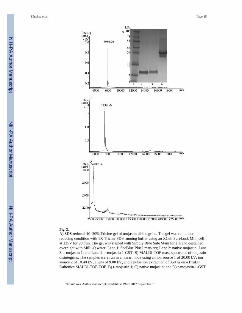
Fig. 2.A) SDS reduced 10–20% Tricine gel of mojastin disintegrins. The gel was run underreducing condition with 1X Tricine SDS running buffer using an XCell SureLock Mini cellat 125V for 90 min. The gel was stained with Simply Blue Safe Stain for 1 h and destainedovernight with Milli-Q water. Lane 1: SeeBlue Plus2 markers; Lane 2: native mojastin; Lane3: r-mojastin 1; and Lane 4: r-mojastin 1-GST. B) MALDI-TOF mass spectrums of mojastindisintegrins. The samples were run in a linear mode using an ion source 1 of 20.00 kV, ionsource 2 of 18.40 kV, a lens of 9.00 kV, and a pulse ion extraction of 350 ns on a BrukerDaltonics MALDI-TOF-TOF. B) r-mojastin 1; C) native mojastin; and D) r-mojastin 1-GST.
Sánchez et al. Page 15
Thromb Res. Author manuscript; available in PMC 2012 September 10.
NIH
-PA Author Manuscript
NIH
-PA Author Manuscript
NIH
-PA Author Manuscript

Fig. 3.LC-MS/MS of native mojastin A) peptide GDWNDDTCTGQSADCPRN, MH+ 1954.7296and B) peptide LRPGAQCADGLCCDQCR, MH+ 2036.8523. The characteristic peptidebond fragment ions, type b and y ions are labeled. Eight microliters of sample were injectedin an Agilent 1100 HPLC system using a reverse phase C18 liquid chromatography columnpacked with 5 μm C18 Magic beads (Michrom; 75 μm i.d. and 12 cm of bed length) on an1100 Agilent HPLC system coupled online with an LTQ Orbitrap hybrid mass spectrometer.The mass spectrometer was operated in the data-dependent mode, in which a full scan MSwas followed by MS/MS scans of the 3 most abundant ions with +2 to +3 charge states.
Sánchez et al. Page 16
Thromb Res. Author manuscript; available in PMC 2012 September 10.
NIH
-PA Author Manuscript
NIH
-PA Author Manuscript
NIH
-PA Author Manuscript

Fig. 4.Comparison of inhibition of platelet aggregation using whole blood and PRP with an equalplatelet count. A Chrono-Log aggregometer was used to measure ADP-induced plateletaggregation by impedance. A total of 10 μL of r-mojastin 1 at varying concentrations wasadded to whole blood and PRP both containing a platelet count of 230,000 platelets/μL andincubated for 4 min at 37°C prior to adding 10 μM of ADP. The IC50 was 40 nM for wholeblood and 90 nM for PRP. The vertical bars represent the standard deviation. n = 3.
Sánchez et al. Page 17
Thromb Res. Author manuscript; available in PMC 2012 September 10.
NIH
-PA Author Manuscript
NIH
-PA Author Manuscript
NIH
-PA Author Manuscript

Fig. 5.Inhibition of platelet adhesion to fibronectin by r-mojastin 1 and native mojastindisintegrins. A total of 100 μL of disintegrins at varying concentrations was added to 10 ×106 platelets and incubated for 1 h at 37°C prior to adding to fibronectin-coated wells. P-nitrophenyl phosphate (PNPP) is used as the substrate solution. The ability of plateletphosphatases to catalyze the hydrolysis of PNPP to p-nitrophenol (chromogenic product)was measured at 405 nm. The IC50s were 62.2 and 58.6 nM for r-mojastin 1 and nativemojastin, respectively. The vertical bars represent the standard deviation. n = 3.
Sánchez et al. Page 18
Thromb Res. Author manuscript; available in PMC 2012 September 10.
NIH
-PA Author Manuscript
NIH
-PA Author Manuscript
NIH
-PA Author Manuscript
Fig. 6.Sonoclot signatures of mojastin disintegrins using human whole blood. Two differentconcentrations of disintegrins A) 409 nM and B) 204.5 nM was added with whole bloodusing glass bead activated cuvettes (gbACT + KIT) on a Sonoclot® Analyzer System. Solidlines: control; long dashed lines: r-mojastin 1; medium dashed lines: r-mojastin 1-GST; andshort dashed lines: native mojastin. The data was obtained by the program Signature Viewerv. 3.1 on an iMac computer. ACT: Activated clot time; CR: Clot rate; and PF: Plateletfunction.
Sánchez et al. Page 19
Thromb Res. Author manuscript; available in PMC 2012 September 10.
NIH
-PA Author Manuscript
NIH
-PA Author Manuscript
NIH
-PA Author Manuscript

NIH
-PA Author Manuscript
NIH
-PA Author Manuscript
NIH
-PA Author Manuscript
Sánchez et al. Page 20
Table 1
IC50 of recombinant and native mojastin disintegrins.
Disintegrin Inhibition of Platelet AggregationPRP
Inhibition of Platelet Aggregation WholeBlood
Inhibition of ATP Release PRP
r-Mojastin1-GST 667 nM 296 nM 335 nM
r-Mojastin 1 119.7 nM 46 nM 95.6 nM
Native mojastin 44.7 nM 19.3 nM 19.54 nM
Thromb Res. Author manuscript; available in PMC 2012 September 10.

NIH
-PA Author Manuscript
NIH
-PA Author Manuscript
NIH
-PA Author Manuscript
Sánchez et al. Page 21
Tabl
e 2
Sono
clot
® a
naly
sis
of w
hole
blo
od c
oagu
latio
n an
d pl
atel
et r
etra
ctio
n us
ing
204.
5 nM
of
disi
nteg
rins
.
Sam
ple*
AC
TP
val
ueC
RP
val
ueP
FP
val
ue
Con
trol
212.
5±23
.122
.2±
4.0
2.7±
0.96
r-m
ojas
tin 1
-GST
208.
8±14
.7p
= 0
.798
223
.0±
1.8
p =
0.6
691
1.8±
0.47
p =
0.1
752
r-m
ojas
tin 1
197.
8±16
.6p
= 0
.349
823
.0±
0.8
p =
0.7
278
1.7±
0.53
p =
0.1
342
Nat
ive
moj
astin
201.
5±6.
4p
= 0
.408
621
.0±
3.7
p =
0.6
654
0.5±
0.29
p =
0.0
052
n= 4
* = T
ests
wer
e do
ne u
sing
gla
ss b
ead
activ
ated
cuv
ette
s (g
bAC
T +
KIT
ref
: 800
-041
2) b
y SI
EN
CO
®
Con
trol
= 1
0% c
itrat
ed w
hole
hum
an b
lood
AC
T=
Act
ivat
ed c
lot t
ime
(mea
sure
d in
sec
onds
)
CR
= C
lot r
ate
(mea
sure
d in
clo
t sig
nals
/min
)
PF=
Pla
tele
t fun
ctio
n (c
lot r
etra
ctio
n)
p <
0.0
5 si
gnif
y a
sign
ific
ant d
iffe
renc
e w
hen
com
pare
d to
the
cont
rol v
alue
s. P
val
ues
wer
e ca
lcul
ated
usi
ng a
t-te
st, t
wo-
taile
d P
valu
e on
Gra
phPa
d Pr
ism
4 s
oftw
are.
Thromb Res. Author manuscript; available in PMC 2012 September 10.

NIH
-PA Author Manuscript
NIH
-PA Author Manuscript
NIH
-PA Author Manuscript
Sánchez et al. Page 22
Table 3
Inhibition of ADP- induced platelet aggregation: IC50 comparisons of native and recombinant disintegrinsusing whole blood and PRP.
Disintegrins IC50 (nM) Media Ref.
Native mojastin 13.8 Whole Blood [12]
Native mojastin 19.29 Whole Blood This work
r-mojastin 1-GST 296 Whole Blood This work
r-mojastin 1 46 Whole Blood This work
r-mojastin 1 121.5 PRP This work
Native Mojastin 44.7 PRP This work
r-mojastin 1-GST 667 PRP This work
Native contortrostatin 60 PRP [19]
r-contortrostatin 250 PRP [19]
r-adinbitor 6000 PRP [18]
r-echistatin 126 PRP [54]
r-eristostatin >100 PRP [55]
r-viplebedin-2 480 PRP [21]
r-albolatin NA PRP [20]
r-salmosin1 2.0 Fibrinogen/αIIbβ3 [56]
Native salmosin1 2.2 Fibrinogen/αIIbβ3 [57]
Native salmosin1 131 PRP [57]
NA = No activity
Thromb Res. Author manuscript; available in PMC 2012 September 10.
Copyright © 2022 FDOKUMEN