Intraspecific venom variation in the medically significant Southern Pacific Rattlesnake (Crotalus...
16
Intraspecific venom variation in the medically significant Southern Pacific Rattlesnake (Crotalus oreganus helleri): Biodiscovery, clinical and evolutionary implications Kartik Sunagar a, b, 1 , Eivind A.B. Undheim c , d, 1 , Holger Scheib d, 1 , Eric C.K. Gren e , 1 , Chip Cochran e , 1 , Carl E. Person e , 1 , Ivan Koludarov c , Wayne Kelln e , William K. Hayes e , Glenn F. King d , Agosthino Antunes a, b , Bryan Grieg Fry c , d, ⁎ a Departamento de Biologia, Faculdade de Ciências, Universidade do Porto, Rua do Campo Alegre, 4169-007 Porto, Portugal b CIIMAR/CIMAR, Interdisciplinary Centre of Marine and Environmental Research, University of Porto, Rua dos Bragas 289, P 4050-123 Porto, Portugal c Venom Evolution Lab, School of Biological Sciences, University of Queensland, St. Lucia, Queensland, Australia d Institute for Molecular Bioscience, University of Queensland, St. Lucia, Queensland 4072, Australia e Department of Earth and Biological Sciences, Loma Linda University, Loma Linda, CA 92350, USA ARTICLE INFO ABSTRACT Article history: Received 6 September 2013 Accepted 13 January 2014 Due to the extreme variation of venom, which consequently results in drastically variable degrees of neutralization by CroFab antivenom, the management and treatment of envenoming by Crotalus oreganus helleri (the Southern Pacific Rattlesnake), one of the most medically significant snake species in all of North America, has been a clinician's nightmare. This snake has also been the subject of sensational news stories regarding supposed rapid (within the last few decades) evolution of its venom. This research demonstrates for the first time that variable evolutionary selection pressures sculpt the intraspecific molecular diversity of venom components in C. o. helleri. We show that myotoxic β-defensin peptides (aka: crotamines/small basic myotoxic peptides) are secreted in large amounts by all populations. However, the mature toxin-encoding nucleotide regions evolve under the constraints of negative selection, likely as a result of their non-specific mode of action which doesn't enforce them to follow the regime of the classic predator–prey chemical arms race. The hemorrhagic and tissue destroying snake venom metalloproteinases (SVMPs) were secreted in larger amounts by the Catalina Island and Phelan rattlesnake populations, in moderate amounts in the Loma Linda population and in only trace levels by the Idyllwild population. Only the Idyllwild population in the San Jacinto Mountains contained potent presynaptic neurotoxic phospholipase A 2 complex characteristic of Mohave Rattlesnake (Crotalus scutulatus) and Neotropical Rattlesnake (Crotalus durissus terrificus). The derived heterodimeric lectin toxins characteristic of viper venoms, which exhibit a diversity of biological activities, including anticoagulation, agonism/antagonism of platelet activation, or procoagulation, appear to have evolved under extremely variable selection pressures. While most lectin α- and β-chains evolved rapidly under the influence of positive Darwinian selection, the β-chain lectin of the Keywords: Venom Evolution Molecule Toxin Rattlesnake Crotalus JOURNAL OF PROTEOMICS 99 (2014) 68 – 83 ⁎ Corresponding author at: Venom Evolution Lab, School of Biological Sciences, University of Queensland, St. Lucia, Queensland, Australia. E-mail address: [email protected] (B.G. Fry). 1 Joint first-author. 1874-3919/$ – see front matter © 2014 Elsevier B.V. All rights reserved. http://dx.doi.org/10.1016/j.jprot.2014.01.013 Available online at www.sciencedirect.com ScienceDirect www.elsevier.com/locate/jprot
Transcript of Intraspecific venom variation in the medically significant Southern Pacific Rattlesnake (Crotalus...

J O U R N A L O F P R O T E O M I C S 9 9 ( 2 0 1 4 ) 6 8 – 8 3
Ava i l ab l e on l i ne a t www.sc i enced i r ec t . com
ScienceDirectwww.e l sev i e r . com/ loca te / j p ro t
Intraspecific venom variation in the medically
significant Southern Pacific Rattlesnake(Crotalus oreganus helleri): Biodiscovery,clinical and evolutionary implicationsKartik Sunagara,b,1, Eivind A.B. Undheimc,d,1, Holger Scheibd,1, Eric C.K. Grene,1,Chip Cochrane,1, Carl E. Persone,1, Ivan Koludarovc, Wayne Kellne, William K. Hayese,Glenn F. Kingd, Agosthino Antunesa,b, Bryan Grieg Fryc,d,⁎aDepartamento de Biologia, Faculdade de Ciências, Universidade do Porto, Rua do Campo Alegre, 4169-007 Porto, PortugalbCIIMAR/CIMAR, Interdisciplinary Centre of Marine and Environmental Research, University of Porto, Rua dos Bragas 289,P 4050-123 Porto, PortugalcVenom Evolution Lab, School of Biological Sciences, University of Queensland, St. Lucia, Queensland, AustraliadInstitute for Molecular Bioscience, University of Queensland, St. Lucia, Queensland 4072, AustraliaeDepartment of Earth and Biological Sciences, Loma Linda University, Loma Linda, CA 92350, USA
A R T I C L E I N F O
⁎ Corresponding author at: Venom Evolution LE-mail address: [email protected] (B.G. Fry
1 Joint first-author.
1874-3919/$ – see front matter © 2014 Elseviehttp://dx.doi.org/10.1016/j.jprot.2014.01.013
A B S T R A C T
Article history:Received 6 September 2013Accepted 13 January 2014
Due to the extreme variation of venom, which consequently results in drastically variabledegrees of neutralization by CroFab antivenom, the management and treatment ofenvenoming by Crotalus oreganus helleri (the Southern Pacific Rattlesnake), one of the mostmedically significant snake species in all of North America, has been a clinician's nightmare.This snake has also been the subject of sensational news stories regarding supposed rapid(within the last few decades) evolution of its venom. This research demonstrates for the firsttime that variable evolutionary selection pressures sculpt the intraspecificmolecular diversityof venom components in C. o. helleri. We show that myotoxic β-defensin peptides (aka:crotamines/small basic myotoxic peptides) are secreted in large amounts by all populations.However, the mature toxin-encoding nucleotide regions evolve under the constraints ofnegative selection, likely as a result of their non-specificmode of actionwhich doesn't enforcethem to follow the regime of the classic predator–prey chemical arms race. The hemorrhagicand tissue destroying snake venom metalloproteinases (SVMPs) were secreted in largeramounts by the Catalina Island and Phelan rattlesnake populations, in moderate amounts inthe Loma Linda population and in only trace levels by the Idyllwild population. Only theIdyllwild population in the San Jacinto Mountains contained potent presynaptic neurotoxicphospholipase A2 complex characteristic of Mohave Rattlesnake (Crotalus scutulatus) andNeotropical Rattlesnake (Crotalus durissus terrificus). The derived heterodimeric lectin toxinscharacteristic of viper venoms, which exhibit a diversity of biological activities, includinganticoagulation, agonism/antagonismof platelet activation, or procoagulation, appear to haveevolved under extremely variable selection pressures. While most lectin α- and β-chainsevolved rapidly under the influence of positive Darwinian selection, the β-chain lectin of the
Keywords:VenomEvolutionMoleculeToxinRattlesnakeCrotalus
ab, School of Biological Sciences, University of Queensland, St. Lucia, Queensland, Australia.).
r B.V. All rights reserved.

69J O U R N A L O F P R O T E O M I C S 9 9 ( 2 0 1 4 ) 6 8 – 8 3
Catalina Island population appears to have evolved under the constraint of negative selection.Both lectin chains were conspicuously absent in both the proteomics and transcriptomics ofthe Idyllwild population. Thus, we not only highlight the tremendous biochemical diversity inC. o. helleri's venom-arsenal, but we also show that they experience remarkably variablestrengths of evolutionary selection pressures, within each toxin class among populations andamong toxin classes within each population. The mapping of geographical venom variationnot only provides additional information regarding venom evolution, but also has directmedical implications by allowing prediction of the clinical effects of rattlesnake bites fromdifferent regions. Such information, however, also points to these highly variable venoms asbeing a rich source of novel toxinswhichmay ultimately prove to be useful in drug design anddevelopment.
Biological significance
• These results have direct implications for the treatment of envenomed patients.• The variable venom profile of Crotalus oreganus helleri underscores the biodiscovery
potential of novel snake venoms.
© 2014 Elsevier B.V. All rights reserved.
1. Introduction
Knowledge of venom composition has increased dramaticallywith improvements in technology and the advent of newtechniques, in particular the use of mass spectrometry invenom proteomics [1–15] and venom gland transcriptomeanalysis [16–27]. Snake venoms are complex secretions com-posed of numerous enzymes, toxins, peptides, small organicmolecules, and inorganic components that have diverse modesof action on both prey and human victims [28–32]. Snake venomserves both predatory and defensive purposes [28–30,33–38].Variation in venom profiles has been shown between specieswithin the same genus [5,11,12,15,27,39–44] and between indi-viduals within the same species, with the intraspecific differ-ences found among geographic locales [2,11,12,45–52], betweensexes [46,47,53] and between juveniles and adults [9,46,47,54,55].Venom variation has also been reported between venom glandsof a single individual [56]. Someauthors have argued that venomdiversity is the product of neutral evolutionary processes andnot subject to natural selection [57,58], whereas others haveargued that strong natural selection has driven adaptation toparticular prey species [12,30,31,40,46,47,59–63].
Venom in reptiles originated from a single early recruitmentevent approximately 180 million years ago (mya) during theearly Jurassic period and is a plesiotypic trait of the Toxicoferaclade [10,12,18,20–23,30,31,40,64]. New World pit vipers arethought to have descended from a single ancestral Asian pitviper species that colonized the New World via the Bering landbridge [65,66],with rattlesnakeshaving amid-Cenozoic origin intheMexican highlands [67–69]. The venomarsenals of Crotalinesnakes are characterized by a great diversity of venom-components; generalized venom “types” have been proposed,depending uponmetalloprotease activity and toxicity [70]. TypeI venoms possess high levels of metalloprotease activity andlower toxicity (>1.0 μg/g mouse body weight), whereas type IIvenoms have low metalloprotease activity and higher toxicity(<1.0 μg/g mouse body weight). The presence of these twovenom types in a diversity of well-defined species cladessuggests that it is not dependent upon phylogeny [49,52,70–72].
Crotalus oreganus helleri is a medium-sized rattlesnakeinhabiting Baja California northward through southern Califor-nia, and the Pacific islands of Santa Catalina (Los AngelesCounty, California) and Coronado Del Sur (Tijuana, Mexico) [67].Pronounced tectonic activity in the region has producedconsiderable variation in available habitat [73]. The speciesutilizes habitat ranging from sea level to >3000 m and preyencountered are highly varied. Significant regional variation invenom composition exists [51,74], with both type I and type IIvenoms identified in local populations [49]; however thisdichotomy of venom types fails to characterize the full extentof venom variability in the species. C. o. helleri is the mostmedically relevant species of the region and is responsiblefor the majority of severe envenomations in southern Califor-nia [29,75]. Therefore, determining intraspecific variation ofC. o. helleri venom components and the factors influencing theirmolecular evolution can yield important implications forclinical treatment of envenomation. Venomvariation also offerssubstantial potential for bioprospecting and pharmaceuticaldiscovery [8,18–23,30,40,76]. These variations have been thesubject of many popular press reports that grossly misattributethem to unparalleled recent diversification of the venom [77]and thus display a fundamental lack of understanding on howvenom evolves.
In this study, we investigated the diversity of toxins presentin C. o. helleri, across its geographic range, using a combinedproteomics–transcriptomics approach to investigate the rela-tive molecular evolution and diversification within a giventoxin type, and the relative expression levels of particular toxintypes.
2. Materials and methods
2.1. Sampling
We sampled four southern California populations of C. o. hellerifrom areas with pronounced geological, elevational, and floris-tic differences. Human envenomations from snakes in these

70 J O U R N A L O F P R O T E O M I C S 9 9 ( 2 0 1 4 ) 6 8 – 8 3
different regions have exhibited distinct symptoms rangingfromhemorrhage tomuscle fasciculations to paralysis. The fourpopulations chosen (Fig. 1) were: (1) Catalina Island, which isdominated by coastal sage scrub, interspersed with chaparraland oak woodland, has never been connected to the mainland[73] and has supported an isolated population since at least thePleistocene; (2) Idyllwild in the San Jacinto Mountains has highaltitude pine and cedar montane forests (elevation ~1600 m);(3) Loma Linda consists of low rolling hills covered with grassesand, on north facing slopes, Salvia mellifera and other shrubs;and (4) Phelan comprises a transition zone betweenHigh Desert(Mohave) and coastal mountain scrub. We sampled one snakefrom each region for transcriptome sequencing. We used thesame snake for proteome analysis of the Phelan and LomaLinda populations, and a separate individual of same sex andsize from the exact same locality for the other two locations inaddition to two more specimens for each location other thanLoma Linda, for which only one more specimen was obtaineddue to the rarity of C. o. helleri in this location. We used onlyadult specimens for venom analysis due to potential ontoge-netic shifts in venom composition [9,70].
2.2. Transcriptome sequencing, phylogenetics, selectionanalyses, and structural analyses
2.2.1. Transcriptome sequencingTotal RNA was extracted from venom glands using thestandard TRIzol Plus method (Invitrogen). Extracts wereenriched for mRNA using standard RNeasy mRNA mini kit(Qiagen) protocol. mRNA was reverse transcribed, fragmentedand ligated to a unique 10-base multiplex identifier (MID) tagprepared using standard protocols and applied to onePicoTitrePlate (PTP) for simultaneous amplification and se-quencing on a Roche 454 GS FLX + Titanium platform(Australian Genome Research Facility). An average of 50,000sequences were read for each library. Automated groupingand analysis of sample-specific MID reads informaticallyseparated sequences from the other transcriptomes on theplates, which were then post-processed to remove low qualitysequences before de novo assembly into contiguous se-quences (contigs) using v 3.4.0.1 of the MIRA softwareprogram. Assembly details for the transcriptomes are shown
Fig. 1 – C. o. helleri populations investigated.
in Supplementary Table 1. All raw reads have been depositedin the NCBI Sequence Read Archive (http://www.ncbi.nlm.nih.gov/sra/) with the accession numbers of: SRR871501 C. o. helleri(Catalina Island), SRR871502 C. o. helleri (Idyllwild), SRR871503C. o. helleri (Loma Linda), and SRR871504 C. o. helleri (Phelan).Assembled contigs were processed using CLC Main WorkBench (CLC-Bio) and Blast2GO bioinformatic suite to provideGene Ontology, BLAST and domain/Interpro annotation. Theabove analyses assisted in the rationalization of the largenumbers of assembled contigs into phylogenetic ‘groups’ fordetailed phylogenetic analyses outlined below.
2.2.2. Selection analysesTranslatednucleotide sequenceswere alignedusingMUSCLE 3.8[78] and the alignments were manually inspected to rectifyerrors. All nucleotide sequences and multiple sequence align-ments used for selection analyses are available as Supplemen-tary file 2 and Supplementary Figs. 1–4, respectively. In order toreconstruct gene phylogenies for selection assessments,maximum-likelihood method implemented in PhyML [79] wasemployed on the nucleotide datasets and node support wasevaluatedwith 1000 bootstrapping replicates. All themaximum-likelihood trees are provided as Supplementary Figs. 5–7, withthe results of branch-site REL testmapped onto them. In order todetect the nature of selection and its influence on variousvenom-encoding genes of C. o. helleri, we utilized maximum-likelihoodmodels implemented in Codeml of the PAML [80]. Weemployed site-specific models that estimate positive selectionstatistically as a non-synonymous-to-synonymous nucleotide-substitution rate ratio (ω) significantly greater than 1. Fortechnical details regarding models/methods see [20,81]. FUBAR[82] implemented in HyPhy [83] was employed to provideadditional support to the aforementioned analyses and to detectsites evolving under the influence of pervasive diversifying andpurifying selection pressures. Mixed Effects Model Evolution(MEME) [82] was also employed to efficiently detect episodicallydiversifying sites. To clearly depict the proportion of sites underdifferent regimes of selection, an evolutionary fingerprintanalysis was carried out using the evolutionary selectiondistance (ESD) algorithm implemented in Datamonkey [84]. Wefurther utilized the branch-site Random Effects Likelihood (REL)test [85] to identify lineages evolving under the influence ofepisodic diversifying selection pressures.
2.2.3. Structural analysesTo depict the natural selection pressures influencing theevolution of various C. o. helleri venom-components (only thosewith sufficient numbers of full-length sequenceswere analyzedin this regard: β-defensin, kallikrein and lectin), we mapped thesites under positive selection on the homology models createdusing Phyre 2 web server [86]. PyMOL 1.3 [87] was used tovisualize and generate the images of homologymodels. ConSurfweb server [88]was used formapping the evolutionary selectionpressures on the three-dimensional homology models.
Homology models of the presynaptic PLA2 complex fromC. o. helleri (Coh) (GenBank: GAKR01000015 [acid subunit] andGenBank: GAKR01000016 [basic subunit]) and the homologuefrom Crotalus scutulatus scutulatus (Css) (UniProt: P18998 [acidsubunit] andUniProt: P62023 [basic subunit]) were built using thecrystal structure of crotoxin from Crotalus durissus terrificus (Cdt)

71J O U R N A L O F P R O T E O M I C S 9 9 ( 2 0 1 4 ) 6 8 – 8 3
(PDB: 3R0L; UniProt: P08878 [acid subunit]; UniProt: P0CG56 [basicsubunit]) [89] as a template. Template to sequence alignmentswere generated using SPDBV [90,91] and exported as FASTA-formatted text. The 3R0L coordinates together with the align-ment file were used for comparative modeling using MODELLER[92]. Images of these homologymodelswere obtainedusingVMD[93] and Tachyon ray tracing. Charged surfaces were obtained byrunning the Adaptive Poisson–Boltzmann Solver APBS plug-in[94] to VMD. Representation in VMD was set to “orthographic”,depth cueingwas set to “off”, and rendermodewas set to “GLSL”.
2.3. Proteomics
2.3.1. HPLCLyophilized crude venom was diluted to a concentration of3 mg/mL in Buffer A (0.065% TFA, 2% acetonitrile in Nanopurewater) and centrifuged at 15,000 g for 10 min. The supernatant(100 μL) was fractionated on an ÄKTAmicro high-pressure liquidchromatography (HPLC) system (GE Healthcare Life Sciences,Piscataway, NJ, USA) fittedwith two reversed-phase (RP) columns(SOURCE 5RPC ST polystyrene/divinyl benzene, 4.6 × 150 mm; GEHealthcare) run in series at a flow rate of 0.5 mL/min, using alinear gradient of 0–100% Buffer B (0.05% TFA, 80% acetonitrile inNanopure water) over 40 column volumes. Protein elution wasmonitored at 214 nm using Unicorn 5.0 (GE Healthcare LifeSciences) software, and fractions were collected manually.
2.3.2. LC–MSEach fraction was subjected to reduction and alkylation prior toenzymatic digestion using dithiothreitol and iodoacetamide,respectively, following the protocol outlined by Matsudaira [95].
Table 1 – C. o. helleri intraspecific proteomic and transcriptomic
Toxin molecularscaffold type
Catalina Island Idyllwild
P T P
β-defensin Large amounts,medium complexity
✔ Large amounts,medium complex
CNP-BPP Medium amounts,low complexity
✔ Low amounts,low complexity
CRiSP Large amounts,medium complexity
✔ ✖
Hyaluronidase ✖ ✔ ✖
Kallikrein Medium amounts,low complexity
✔ Medium amountshigh complexity
Kunitz ✖ ✔ ✖
L-Amino acid oxidase Medium amounts,low complexity
✔ Medium amountslow complexity
Lectin Large amounts,medium complexity
✔ ✖
Nerve growth factor Low amounts, lowcomplexity
✔ ✖
Phospholipase A2 Medium amounts,medium complexity
✔ Large amounts,high complexity
Snake venommetalloprotease
Large amounts,medium complexity
✔ Not detected
Vascular endothelialgrowth factor
✖ ✔ ✖
Vespryn ✖ ✔ ✖
P = proteome.T = transcriptome.
Proteins were then digested with proteomics-grade porcinepancreatic trypsin (Sigma-Aldrich, St. Louis, MO, USA). Wedesalted samples using C18 ZipTips (EMD Millipore, Billerica,MA, USA) according to themanufacturer's protocol. The desaltedtryptic peptides were resuspended in mobile phase A (2%acetonitrile, 0.1% formic acid in water). Liquid chromatographywas conducted on a ThermoFinnigan LCQDeca XP spectrometer(ThermoFinnigan,Waltham,MA,USA) equippedwith a PicoView500 nanospray apparatus using Xcalibur software (ver. 1.3;ThermoFinnigan, Waltham, MA, USA) for instrument controland data acquisition. Separation was performed on a 10-cm ×75-μm-i.d. C18 BioBasic bead column (New Objective, Woburn,MA, USA) by injecting 20-μL samples. Mobile phase B consistedof 98% acetonitrile, 2% water, and 0.1% formic acid. Thegradient program was: 0% B at 0.18 mL/min for 7.5 min; 0% B at0.35 mL/min for 0.5 min; linear gradient to 20% B at 15 min at0.35 mL/min; linear gradient to 75% B at 55 min at 0.3 mL/min(flow rate constant for remainder of the program); linear gradientto 90% B at 60 min; hold at 90% B until 85 min; linear gradient to0%Bat 90 min; hold at 0%Buntil 120 min. Spectrawere acquiredin positive ion mode with a scan range of 300–1500 m/z. Weconverted MS/MS data into peak list files using ExtractMSnimplemented in BioWorks (version 3.1; ThermoFinnigan) withthe following parameters: peptide molecular weight range of300–3500, threshold of 100,000, precursor mass tolerance of 1.4,and minimum ion count of 35. We conducted MS/MS databasesearches using Mascot (licensed, version 2.2, Matrix Science,Boston, MA, USA) against the National Center for BiotechnologyInformation non-redundant (NCBInr) database in the taxonMetazoa with a parent tolerance of 1.20 Da, fragment toleranceof 0.60 Da, and two missed trypsin cleavages allowed. We
toxin presence.
Loma Linda Phelan
T P T P T
ity✔ Large amounts,
medium complexity✔ Large amounts,
medium complexity✔
✔ Large amounts,low complexity
✔ Large amounts,low complexity
✔
✔ Medium amounts,low complexity
✔ Medium amounts,medium complexity
✖
✔ ✖ ✔ ✖ ✔
, ✔ Large amounts,high complexity
Large amounts,high complexity
✔
✔ ✖ ✔ ✖ ✔
, ✔ Medium amounts,low complexity
✔ Medium amounts,low complexity
✔
✖ ✖ ✔ Low amounts,low complexity
✔
✔ Low amounts,low complexity
✔ ✖ ✖
✔ Low amounts,low complexity
✔ Low amounts,low complexity
✔
✔ Medium amounts,high complexity
✔ Large amounts,high complexity
✔
✔ Low amounts,low complexity
✔ Low amounts,low complexity
✔
✔ ✖ ✔ ✖ ✔

72 J O U R N A L O F P R O T E O M I C S 9 9 ( 2 0 1 4 ) 6 8 – 8 3
specified carbamidomethylation of cysteine and oxidation ofmethionine in Mascot as fixed and variable modifications,respectively.
2.3.3. MALDI ToF MS and MALDI ToF/ToF MS/MSRP-HPLC fractionswere submitted to the Institute for IntegratedResearch in Materials, Environments and Society at CaliforniaState University, Long Beach, to determine whole proteinmolecular masses and protein identification/similarity. ForMALDI ToF/ToF MS/MS analysis, tryptic peptides were mixedwith α-cyano-4-hydroxy cinnamic acid (CHCA) matrix anddirectly spotted onto MALDI plates. MS spectra were collectedusing 1000 laser shots/spectrum, and MS/MS spectra from 3000shots/spectrum. Peptides with signal-to-noise ratio above 15 inMSmodewere selected forMS/MS analysis,with amaximumof15 MS/MS spectra allowed per spot. Internal calibration wasachieved using ToF/ToF Calibration Mixture (AB SCIEX). Wesearched MS/MS data against the NCBInr database within
Fig. 2 – LC–MS/MS annotated RP-HPLC chromatograms from the
Metazoa using GPS Explorer, running Mascot (version 2.1)search engine with a peptide tolerance of 300 ppm, MS/MStolerance of 0.8 Da, and one missed cleavage allowed. Wespecified carbamidomethylation of cysteine as a fixed modifi-cation, and the following as variable modifications: carbamyl,Gln/pyro-Glu (N-term Q), and Glu/pyro-Glu (N-term E). Massspectrometry data for the peaks in Supplementary File 1 ispresented in Supplementary Spreadsheet 1.
2.3.4. Statistical analysesTo confirm that population differences existed among the 11snakes with the quantitative RP-HPLC data presented inSupplementary Spreadsheet 2, we subjected the percent proteinpresent in each of the 11 toxin families (area under the peaks) toa 4 × 11 (population × toxin family) analysis of variance (ANOVA[96]), treating population as a between-subjects factor and toxinfamily as a within-subjects factor. We rank-transformed thedata to avoid analysis of percentage data that summed to 100 for
four different C. o. helleri populations examined in this study.

73J O U R N A L O F P R O T E O M I C S 9 9 ( 2 0 1 4 ) 6 8 – 8 3
each individual. Although our samples were small and datawere somewhat non-normal and heteroscedastic, general linearmodels generally handle data well that fail to meet parametricassumptions and the resultswere extremely robust.We also ran
Fig. 3 – Sequence alignment of lectins from C. o. helleri: α) 1. GAKGAKQ01000016 CohCI-3, 4. GAKS01000016 CohPH-3, 5. GAKQ010CohLL-1, 8. GAKS01000014 CohPH-1, 9. GALC01000014 CohLL-2,GALC01000017 CohLL-5, 13. GALC01000016 CohLL-4; and β) 14. GGALC01000022 CohLL-5, 17. GAKS01000021 CohPH-4, 18. GAKS0GAKQ01000022 CohCI-4, 21. GAKQ01000021 CohCI-3, 22. GAKQ0GAKS01000019 CohPH-1, 25. GAKS01000020 CohPH-3, 26. GAKS0PH = Phelan. Signal peptide is shown in lowercase, cysteines are
a non-parametric Kruskal–Wallis ANOVA for each toxin familyto compare the populations, which allowed us to confirm theresults from the parametric ANOVA; this latter test requires noassumptions about data distribution [96]. We computed effect
Q01000018 CohCI-5, 2. GALC01000015 CohLL-3, 3.00015 CohCI-2, 6. GAKQ01000014 CohCI-1, 7. GALC0100001310. GAKQ01000017 CohCI-4, 11. GAKS01000017 CohPH-4, 12.ALC01000020 CohLL-3, 15. GALC01000018 CohLL-1, 16.
1000022 CohPH-5, 19. GALC01000021 CohLL-4, 20.1000020 CohCI-2, 23. GAKQ01000019 CohCI-1, 24.1000023 CohPH-6. CI = Catalina Island, LL = Loma Linda,highlighted in black.

74 J O U R N A L O F P R O T E O M I C S 9 9 ( 2 0 1 4 ) 6 8 – 8 3
sizes (approximate variance explained) as adjusted partialeta-squared (η2) for the parametric ANOVA and as η2 (computedas χ2 / [total N − 1]) for the Kruskal–Wallis ANOVAs [96,97].Eta-squared values ≥ 0.14 are generally deemed large [98]. Weconducted these analyses using SPSS 13.0 for Windows, withalpha = 0.05. Following Nakagawa [99], we did not applyBonferroni adjustments to multiple tests.
Fig. 4 – Comparative ribbon, surface charge and surfacehydrophobicity of the hetrodimeric presynaptic neurotoxicphospholipase A2 complex from the venoms of Crotalusdurissus terrificus (Cdt) (PDB: 3R0L; UniProt: P08878 [acidicsubunit]; UniProt: P0CG56 [basic subunit]), C. o. helleri (Coh)(GenBank: GAKR01000015 [acidic subunit] and GenBank:GAKR01000016 [basic subunit]) and Crotalus scutulatusscutulatus (Css) (UniProt: P18998 [acidic subunit] and UniProt:P62023 [basic subunit]). Cartoon images show helices inpurple, sheets in green and other structural regions inyellow. Surface charge potentials were mapped on surfacesallowing for color scale data range values of −10.00 to +10.00using the RWB coloring scheme. Surface residuehydrophicitymapping of residue-type surfaces depicts acidicresidues in red, basic residues in blue, polar residues inyellow, and nonpolar residues in silver.
3. Results and discussion
Random sequencing recovered sequences for 13 differentvenom protein encoding gene families (Table 1), with all butKunitz and Hyaluronidase recovered by both proteomics andtranscriptomics. The inability of our combined approach todetect these two venom-components in both result setsmay be due to a number of factors, such as, i) differentialtranscription/translation: not all toxins being replenished atequal stoichiometric rates or simultaneously; ii) technicallimitation: the relative separation ability of the HPLC columnutilized; iii) co-elution of toxins: one toxin type dominatinganother and thus obscuring the signal of a toxin present insignificantly lower amounts; iv) transcriptomics: the non-exhaustive random sampling procedure utilized which wouldstatistically be likely to recover the most abundant toxintypes, with lower-level expressed toxins not recovered; and/orv) microRNA silencing: whereby toxin coding regions undergotranscription but not translation [100]. Lectin toxins, however,were conspicuously absent in both the proteomics andtranscriptomics of the Idyllwild population. Sequences ana-lyzed in this study have the GenBank accession numbers of:C. o. helleri (Catalina Island) GAKQ01000001–GAKQ01000026;C. o. helleri (Idyllwild) GAKR01000001–GAKR01000018; C. o. helleri(Loma Linda) GALC01000001–GALC01000026; and C. o. helleri(Phelan) GAKS01000001–GAKS01000031. It must be noted that inaccordance with the new GenBank deposition rules to excludefragments of less than 200 base pairs, only the full lengthsequences were deposited. Thus 27 β-defensins were notdeposited, even though their processed and secreted toxinregions were sequenced (only regions of the signal peptide wereincomplete). Thus, while these sequences could not be depos-ited into GenBank, they were utilized in the analyses and areincluded in the Supplementary material.
Our proteomics analyses revealed significant differencesin the venoms of the four populations (Fig. 2), with venomRP-HPLC profiles within a population largely congruent amongindividuals (Supplementary Fig. 8; note: only two Loma Lindaspecimens were able to be analyzed due to the rarity ofC. o. helleri in this locality). The parametric ANOVA yielded ahighly significant interaction between population and toxinfamily (F9.8,22.9 = 13.15, P < 0.001, adjusted partial η2 = 0.31;Greenhouse–Geisser adjustment of degrees-of-freedom ap-plied), indicating that the distribution of toxins among thetoxin families differed significantly among thepopulations. TheKruskal–Wallis ANOVAs confirmed that toxin quantity variedsignificantly among populations for some (nerve growth factor,cysteine-rich secretory protein [CRiSP], lectin; all P = 0.21–0.35,η2 = 0.86–0.97) but not all toxin families. Five additional toxins(BPP, β-defensin, kallikrein, PLA2, SVMP) approached signifi-cance (P < 0.10) with exceptional effect sizes (η2 > 0.63). Thus,
the ANOVAs confirmed population differences despite thesmall sample sizes.
Some toxin types were notable for being either highlyconserved in their coding sequences (β-defensin, natriuretic),whereas others were extremely variable (kallikrein, lectin, PLA2,SVMP). While the β-defensins and bradykinin potentiatingpeptides (BPPs)were of low complexity, our proteomics analysesof the relative expression levels revealed that they are expressed

75J O U R N A L O F P R O T E O M I C S 9 9 ( 2 0 1 4 ) 6 8 – 8 3
in very high amounts in all populations, with β-defensin inparticular invariantly expressed in large quantities (Fig. 2). Themulti-product natriuretic/BPP precursor was invariant withinand between populations in both the plesiotypic natriureticpeptide domain and the apotypic (derived) BPP domains locatedwithin the propeptide region. In contrast, the lectin sequenceswerehighly variable, including the apotyposis of novel cysteineswhich may facilitate novel structural folding or unique subunitformation with lectins or other toxin types (Fig. 3). Consistentwith the proteomic results of this study and a previouslypublished study of San Jacinto Mountain specimens [49] aswell as observed notable clinical effects, only the Idyllwildpopulation contained both the acidic and basic subunits of theneurotoxic PLA2 complex type, with both chains virtuallyidentical to the well-characterized potent presynaptic neuro-toxins from C. d. terrificus and C. s. scutulatus (Fig. 4). It was also
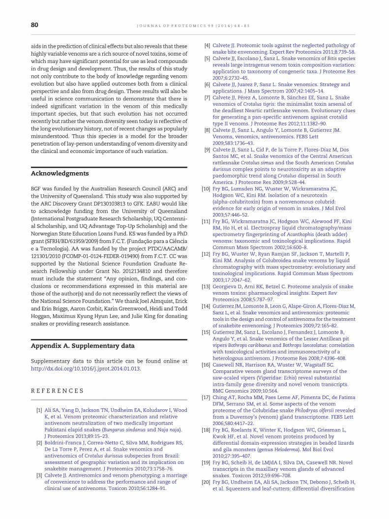
Fig. 5 – Molecular evolution of C. o. helleri β-defensins. Three-dim1Z99) of β-defensins with evolutionary conservation of amino acselected sites (in red) detected by site-model 8 (PP ≥ 0.95, BEB). Scthe locations of positively selected sites (red sticks) but also highα helices (purple) and β sheets (green), are also presented.
notable that the Idyllwild population secreted the lowestamount of SVMPs (Fig. 2), with only a single isoform obtainedin the transcriptome and only detectable in trace levels in theproteome. In contrast, the other populations secreted SVMPs inlarge amounts, with the Phelan having the greatest complexitywhile the Catalina Island population had less complexity but amuch higher relative expression level. This is consistent withthe pattern observed for C. s. scutulatus, that there is an inverserelationship between the relative amount of neurotoxic PLA2
and hemorrhagic SVMP [37,101,102]. Thus, it is quite evidenthow a biochemical arsenal with such variability in neurotoxic,hemotoxic and myotoxic venom-components can complicateclinical treatment of bite victims, not only through theproduction of highly variable clinical effects, but also as aconsequence the reciprocal variability in the efficacy of anti-venombinding. It shouldbenoted that the venomproteomics of
ensional homology models (built using the PDB templateids mapped onto them, depicting the locations of positivelyhematic representation of the models, which not only depictslights disulfide bonds (orange sticks),

76 J O U R N A L O F P R O T E O M I C S 9 9 ( 2 0 1 4 ) 6 8 – 8 3
multiple animals (n = 3; except Loma Linda population, wherethese animals are extremely rare) from the same region werefairly similar. Hence, it can be safely assumed that thevenom-gland transcriptomics of randomly chosen animalsrepresents the overall venomics (genetic makeup of the venomgland) of the representative population.
Understanding the nature and strength of natural selectionpressures,which sculpt genetic diversity, is the central themeofmolecular evolutionary studies. Since non-synonymous muta-tions aremore likely to influence the structure and function of aprotein and hence in turn influence the fitness of the organism,evaluating the rate of accumulation of non-synonymousmutations (dN) in genes, relative to synonymous mutations(dS), as a ratio known as ω (or dN/dS ratio), is essential. Weassessed the role of evolutionary selection pressures in shapingvarious venom proteins in different populations of C. o. helleri
Fig. 6 – Molecular evolution of C. o. helleri kallikreins. Three-dimusing the PDB template 1OP0; all others using 2AIQ) of kallikreinsthem, depicting the locations of positively selected sites (in red)Schematic representation of the models, which not only depictshighlights disulfide bonds (orange sticks), α helices (purple) and
using various state-of-art selection assessment methodologies.We detected a significant influence of positive Darwinianselection on the evolution of most venom protein encodinggenes in these snakes (Figs. 5–7; Tables 2–4; SupplementaryTables 2–5; Supplementary Figs. 1–7 and 9–11).
Site-specific selection assessments indicated thatβ-defensins, which were expressed in relatively large amountsby all C. o. helleri populations examined, followed a regime ofweak positive selection: Catalina Island: ω = 1.33 and 3 posi-tively selected (PS); Idyllwild:ω = 2.07 and 3 PS; Loma Linda:ω =1.14 and 2 PS; Phelan: ω = 1.31 and 5 PS; All: ω = 1.18 and 11 PS(Fig. 5; Table 2). However, the mapping of mutations ontosequence alignments indicated that most hypermutable sitesdetected by site-specific methods in β-defensins were concen-trated in thenon-secreted regions of the toxin that are not likelyto contribute in the envenoming process. It was also evident
ensional homology models (Loma Linda population modeledwith evolutionary conservation of amino acids mapped ontodetected by site-model 8 (PP ≥ 0.95, BEB) are presented.the locations of positively selected sites (red sticks) but alsoβ sheets (green), are also presented.

Fig. 7 – Molecular evolution of C. o. helleri lectin α- and β-chains. Three-dimensional homology models (α-chain lectins:Catalina Island population and the ‘combined set’ modeled using PDB template 1C3A; others using 1UMR; β-chain lectins:Loma Linda population modeled using the template 1V4L; all others using 1J34) of lectin α- and β-chains with evolutionaryconservation of amino acids mapped onto them, depicting the locations of positively selected sites (in red) detected bysite-model 8 (PP ≥ 0.95, BEB). Schematic representation of the models, which not only depicts the locations of positivelyselected sites (red sticks) but also highlights disulfide bonds (orange sticks), α helices (purple) and β sheets (green), are alsopresented.
77J O U R N A L O F P R O T E O M I C S 9 9 ( 2 0 1 4 ) 6 8 – 8 3
that the entire stretch of nucleotides encoding the secretedregion of β-defensins evolved under the extreme influence ofnegative selection, with 76% of residues being extremely wellconserved (percent identity ≥ 90%; Supplementary Fig. 1). Thiswas also supported by the results of MEME, an extremely
accurate method of detecting episodic bursts of adaptation,which detected fewer episodically diversifying sites inβ-defensins (Table 2). Mapping of variable sites on the structureof the β-defensin ‘crotamine’ from C. d. terrificus (PDB code: 1Z99[103]), which is homologous and thus structurally very similar

Table 2 – C. o. helleri intraspecific venom dynamics:β-defensins.
Population FUBARa MEMEsites b
BSRc PAMLd
M8 M2a
CI ω > 1e: 0ω < 1f: 1
0 1 3 3(2 + 1) (2 + 1)1.33 1.33
ID ω > 1e: 1ω < 1f: 0
0 2 3 3(2 + 1) (2 + 1)2.07 2.07
LL ω > 1e: 0ω < 1f: 0
1 1 2 0(0 + 2)1.14 1.14
PH ω > 1e: 1ω < 1f: 1
0 4 5 2(1 + 4) (1 + 1)1.31 1.31
Combined ω > 1e: 0ω < 1f: 0
4 6 11 6(5 + 6) (3 + 3)1.18 1.16
ω: mean dN/dS.Populations: CI = Catalina Island; ID = Idyllwild; LL = Loma Linda;PH = Phelan.a Fast Unconstrained Bayesian AppRoximation.b Sites detected as experiencing episodic diversifying selection(0.05 significance) by the Mixed Effects Model Evolution (MEME).c Number of branches detected by the branch-site REL (randomeffects likelihood) test as episodically diversifying.d Positively selected sites detected by the Bayes Empirical Bayesapproach implemented in M8 and M2a. Sites detected at 0.99 and0.95 significance are indicated in the parenthesis.e Number of sites under pervasive diversifying selection at theposterior probability ≥ 0.9 (FUBAR).f Number of sites under pervasive purifying selection at theposterior probability ≥ 0.9 (FUBAR).
Table 3 – C. o. helleri intraspecific venom dynamics:Kallikrein.
Population FUBARa MEMEsites b
BSRc PAMLd
M8 M2a
CI ω > 1e: 8ω < 1f: 5
6 4 7 7(6 + 1) (4 + 3)1.35 1.31
ID ω > 1e: 20ω < 1f: 4
6 6 11 11(2 + 9) (2 + 9)1.63 1.63
LL ω > 1e: 23ω < 1f: 8
9 9 24 15(8 + 16) (7 + 8)1.60 1.57
PH ω > 1e: 22ω < 1f: 7
15 5 24 15(10 + 14) (7 + 8)1.38 1.41
Combined ω > 1e: 27ω < 1f: 14
45 12 36 25(18 + 18) (13 + 11)1.36 1.38
ω: mean dN/dS.Populations: CI = Catalina Island; ID = Idyllwild; LL = Loma Linda;PH = Phelan.a Fast Unconstrained Bayesian AppRoximation.b Sites detected as experiencing episodic diversifying selection(0.05 significance) by the Mixed Effects Model Evolution (MEME).c Number of branches detected by the branch-site REL (randomeffects likelihood) test as episodically diversifying.d Positively selected sites detected by the Bayes Empirical Bayesapproach implemented in M8 and M2a. Sites detected at 0.99 and0.95 significance are indicated in the parenthesis.e Number of sites under pervasive diversifying selection at theposterior probability ≥ 0.9 (FUBAR).f Number of sites under pervasive purifying selection at theposterior probability ≥ 0.9 (FUBAR).
78 J O U R N A L O F P R O T E O M I C S 9 9 ( 2 0 1 4 ) 6 8 – 8 3
to β-defensin, revealed that the N-terminal positions 23 (Y insequence 1; Supplementary Fig. 1) and 25 (R in sequence 1;Supplementary Fig. 1) as well as the C-terminal residues inposition 61 (K in sequence 1; Supplementary Fig. 1), 62 (S insequence 1; Supplementary Fig. 1), and 63 (G in sequence 1;Supplementary Fig. 1) are structurally more flexible (for theremaining positively selected amino acids 3D-structure coordi-nates were not resolved). This can be explained by the fact thatthese locations in the protein structure fall outside the disulfidebond-stabilized core. Although the highly conserved positions,such as 28 (K), 53 (R), 54 (W), and 55 (R; all referred to in sequence1; Supplementary Fig. 1) were also solvent exposed, they werelocated inside the disulfide bridge-stabilized protein core andthus experienced heavy constraints of negative selection.However, the lack of variation in secreted regions ofβ-defensins may be indicative of their unique mode of action aspeptides non-specifically target and destabilize the negativelycharged microbial membranes using their cationic amino acidresidues, resulting in membrane permeabilization [104].Not-surprisingly, 29% of the residues in C. o. helleri β-defensinswere cationic (K, R and H) and were extremely well conserved(percent identity ≥ 80%; Supplementary Fig. 1). Hence, it isexpected that the evolutionary constraints favor the preserva-tionof cationic residues required for toxicity. Thebranch-site REL(BSR) test, which significantly identifies lineages that follow theregime of episodic diversification, clearly highlighted the
differences in strengths of evolutionary selection pressuresacting upon β-defensins in C. o. helleri populations (Supplemen-tary Fig. 5). In the Phelanpopulation this test detected asmanyasfour episodically diversifying branches in β-defensin genelineage, while detecting only one branch each in Catalina Islandand Loma Linda populations, and two branches in the Idyllwildpopulation (Supplementary Fig. 5).
While the kallikreins found in each of the C. o. helleripopulations examined were found to be rapidly evolving underthe influence of positive selection [Catalina Island: ω = 1.35 and7 PS; Idyllwild: ω = 1.63 and 11 PS; Loma Linda: ω = 1.60 and 24PS; Phelan: ω = 1.38 and 24 PS; All: ω = 1.36 and 36 PS] (Fig. 6;Supplementary Fig. 2), the number of positively selected sitesdetected by M8's Bayes empirical Bayes (BEB) approach, variedfrom 7 to 24, highlighting the differential rate of evolution ofkallikreins in various C. o. helleri populations (Table 3). Thenumber of branches detected by the BSR test as episodicallydiversifying in kallikrein encoding genes varied from 4 to 10 invarious populations (Supplementary Fig. 6), again highlightingthe differential role of selection in shaping these venom proteinencoding genes.
Lectin α-chain [Catalina Island: ω = 2.36 and 15 PS; LomaLinda:ω = 2.51 and 14 PS; Phelan:ω = 3.07 and 16 PS; All:ω = 2.23and 27 PS] and lectin β-chain [Catalina Island: ω = 0.46 and 0 PS;Loma Linda: ω = 2.73 and 11 PS; Phelan: ω = 2.34 and 28 PS; All:ω = 2.29 and 29 PS] were found to evolve under the significant

Table 4 – C. o. helleri intraspecific venomdynamics: Lectins.
Population FUBARa MEMEsites b
BSRc PAMLd
M8 M2a
α chainCI ω > 1e: 12
ω < 1f: 52 4 15 12
(8 + 7) (5 + 7)2.36 2.34
LL ω > 1e: 15ω < 1f: 2
1 3 14 12(7 + 7) (5 + 7)2.51 2.51
PH ω > 1e: 15ω < 1f: 4
0 4 16 13(8 + 8) (7 + 6)
Combined ω > 1e: 26ω < 1f: 6
7 7 27 23(14 + 13) (14 + 9)2.23 2.24
β chainCI ω > 1e: 0
ω < 1f: 10 0 0 0
0.46 0.46LL ω > 1e: 2
ω < 1f: 00 1 11 2
(0 + 11) (0 + 2)2.73 2.73
PH ω > 1e: 16ω < 1f: 2
5 6 28 22(12 + 16) (9 + 13)2.34 2.34
Combined ω > 1e: 22ω < 1f: 2
5 6 29 20(16 + 13) (11 + 9)2.29 2.31
ω: mean dN/dS.Populations: CI = Catalina Island; ID = Idyllwild; LL = Loma Linda;PH = Phelan.a Fast Unconstrained Bayesian AppRoximation.b Sites detected as experiencing episodic diversifying selection(0.05 significance) by the Mixed Effects Model Evolution (MEME).c Number of branches detected by the branch-site REL (randomeffects likelihood) test as episodically diversifying.d Positively selected sites detected by the Bayes Empirical Bayesapproach implemented in M8 and M2a. Sites detected at 0.99 and0.95 significance are indicated in the parenthesis.e Number of sites under pervasive diversifying selection at theposterior probability ≥ 0.9 (FUBAR).f Number of sites under pervasive purifying selection at theposterior probability ≥ 0.9 (FUBAR).
79J O U R N A L O F P R O T E O M I C S 9 9 ( 2 0 1 4 ) 6 8 – 8 3
influence of positive selection (Table 4; Supplementary Fig. 3).However, β-chain of the Catalina Island populationwas remark-ably revealed to have evolved under the influence of negativeselection (ω = 0.46, 0 PS; Table 4; Supplementary Fig. 4). Otherthan at position 56, amino acid residues in all other positionswere invariant (Supplementary Fig. 4). The rapid rate ofmolecular evolution observed in lectins is consistent with thegreat diversity of novel sequences recovered, including theapotyposis or the derivation of novel cysteine residues (Fig. 3).The rapid accumulation of hypermutable sites under theinfluence of positive selection in β-chain lectins from all C. o.helleri populations except those from Catalina Island, where thetoxin-encoding gene has evolved under strong negative selec-tion, is intriguing andwarrants further experimental evaluationstounderstand the stark differences in themagnitudeof selectionpressures. While the BSR test detected a few lineages asepisodically diversifying in the α-chain lectins of variouspopulations, the results of this test in the β-chain lectins wereparticularly interesting (Supplementary Fig. 7). This test failed to
detect any branch in the Catalina Island population, whiledetecting a single branch in Loma Linda population as episod-ically diversifying (Supplementary Fig. 7). In contrast, asmany as6 branches were detected as following the regime of episodicadaptation in β-chain lectins of the Phelan population (Supple-mentary Fig. 7). Similar to the results of all state-of-art selectionassessment methods outlined above, the evolutionary finger-prints of venom-encoding genes in C. o. helleri clearly depictedthe differential influence of natural selection on their evolution(Supplementary Figs. 9–11).
The structure and surface chemistry of the presynaptic PLA2
complex from C. o. helleri is verywell conservedwhen comparedto the homologues from C. d. terrificus and C. s. scutulatus (Fig. 4).Both amino acid type distribution on the protein surface aswellas studying surface charges and surface hydrophobicity of allthree PLA2 complexes revealed only minor differences. Whilethe positive andnegative charged patches in globowere locatedin the samepositions,minimal differenceswere observed in thesize and charge of these surface regions. Since the PLA2s ofC. d. terrificus and C. s. scutulatus are well-characterized to bepotent neurotoxins (cf. [89,105]), we conclude that the describedsimilarities ofC. o. helleri PLA2 to the former ones are responsiblefor neurotoxic effects of PLA2s observed in the C. o. helleripopulation. The precise evolutionary regimes followed by genesencoding PLA2 and SVMPs in these snakes remain to beelucidated.
Thus, it is evident that C. o. helleri venom-encoding geneshave experienced differential evolutionary selection pressures.Differential rate of molecular evolution or expression occurrednot only between toxin types within the venom of a particularpopulation, but also for the same toxin type between popula-tions. These results demonstrate that the different populationsof C. o. helleri follow distinct evolutionary trajectories, withthe differential venom profiles likely driven by variation inpredatory ecology. This is a reflection of the complex evolution-ary history of this species, which ranges from sea level to highmountain peaks and occupies a diverse range of habitats. Thesehabitats possess differing lizard andmammal prey assemblages[106–108], and evidence from other snakes suggests that strongnatural selection has driven venomadaptation to particular preyspecies [12,20,21,30,31,40,46,47,59,61–63,109]. Although climatemight be expected to influence venom composition, our datasuggest otherwise concerning the dichotomy of type I (proteo-lytic or “tenderizer”) versus type II (more toxic) venoms [70]. Ithas been suggested that snakes at higher elevation with thegreatest temperature fluctuations could be expected to possess atype I venom to facilitate digestion [70]. However, the populationthat faces the highest temperature fluctuations (Idyllwild)possesses a type II venom that lacks almost entirely themetalloproteases typical of type I venoms. These results alsoindicate significant differences in potential human envenom-ation profiles, consistent with the complex clinical picturepreviously observed, with some populations being hemorrhagicwhile others are neurotoxic. The exquisite diversity of venom-components highlighted in this study and the variation inintensity and the nature of natural selection shaping themolecular toxin scaffolds may not only result in distinctenvenoming profiles but may also induce variable responses toantivenom. Hence, understanding the true molecular diversityof venom and the evolutionary forces that shape them not only
80 J O U R N A L O F P R O T E O M I C S 9 9 ( 2 0 1 4 ) 6 8 – 8 3
aids in thepredictionof clinical effects but also reveals that thesehighly variable venoms are a rich source of novel toxins, some ofwhichmay have significant potential for use as lead compoundsin drug design and development. Thus, the results of this studynot only contribute to the body of knowledge regarding venomevolution but also have applied outcomes both from a clinicalperspective and also from drug design. These results will also beuseful in science communication to demonstrate that there isindeed significant variation in the venom of this medicallyimportant species, but that such evolution has not occurredrecently but rather the venomdiversity seen today is reflective ofthe long evolutionary history, not of recent changes as popularlymisunderstood. Thus this species is a model for the broaderpenetration of lay-person understanding of venomdiversity andthe clinical and economic importance of such variation.
Acknowledgments
BGF was funded by the Australian Research Council (ARC) andthe University of Queensland. This study was also supported bythe ARC Discovery Grant DP130103813 to GFK. EABU would liketo acknowledge funding from the University of Queensland(International Postgraduate Research Scholarship, UQ Centenni-al Scholarship, and UQ Advantage Top-Up Scholarship) and theNorwegian State Education Loans Fund. KSwas funded by a PhDgrant (SFRH/BD/61959/2009) fromF.C.T. (Fundação para aCiênciae a Tecnologia). AA was funded by the project PTDC/AACAMB/121301/2010 (FCOMP-01-0124-FEDER-019490) from F.C.T. CC wassupported by the National Science Foundation Graduate Re-search Fellowship under Grant No. 2012134810 and thereforemust include the statement “Any opinion, findings, and con-clusions or recommendations expressed in this material arethose of the author(s) and do not necessarily reflect the views ofthe National Science Foundation.”We thank Joel Almquist, Erickand Erin Briggs, Aaron Corbit, Karin Greenwood, Heidi and ToddHoggan, Maximus Kyung Hyun Lee, and Julie King for donatingsnakes or providing research assistance.
Appendix A. Supplementary data
Supplementary data to this article can be found online athttp://dx.doi.org/10.1016/j.jprot.2014.01.013.
R E F E R E N C E S
[1] Ali SA, Yang D, Jackson TN, Undheim EA, Koludarov I, WoodK, et al. Venom proteomic characterization and relativeantivenom neutralization of two medically importantPakistani elapid snakes (Bungarus sindanus and Naja naja).J Proteomics 2013;89:15–23.
[2] Boldrini-Franca J, Correa-Netto C, Silva MM, Rodrigues RS,De La Torre P, Perez A, et al. Snake venomics andantivenomics of Crotalus durissus subspecies from Brazil:assessment of geographic variation and its implication onsnakebite management. J Proteomics 2010;73:1758–76.
[3] Calvete JJ. Antivenomics and venom phenotyping: a marriageof convenience to address the performance and range ofclinical use of antivenoms. Toxicon 2010;56:1284–91.
[4] Calvete JJ. Proteomic tools against the neglected pathology ofsnake bite envenoming. Expert Rev Proteomics 2011;8:739–58.
[5] Calvete JJ, Escolano J, Sanz L. Snake venomics of Bitis speciesreveals large intragenus venom toxin composition variation:application to taxonomy of congeneric taxa. J Proteome Res2007;6:2732–45.
[6] Calvete JJ, Juarez P, Sanz L. Snake venomics. Strategy andapplications. J Mass Spectrom 2007;42:1405–14.
[7] Calvete JJ, Pérez A, Lomonte B, Sánchez EE, Sanz L. Snakevenomics of Crotalus tigris: the minimalist toxin arsenal ofthe deadliest Neartic rattlesnake venom. Evolutionary cluesfor generating a pan-specific antivenom against crotalidtype II venoms. J Proteome Res 2012;11:1382–90.
[8] Calvete JJ, Sanz L, Angulo Y, Lomonte B, Gutierrez JM.Venoms, venomics, antivenomics. FEBS Lett2009;583:1736–43.
[9] Calvete JJ, Sanz L, Cid P, de la Torre P, Flores-Díaz M, DosSantos MC, et al. Snake venomics of the Central Americanrattlesnake Crotalus simus and the South American Crotalusdurissus complex points to neurotoxicity as an adaptivepaedomorphic trend along Crotalus dispersal in SouthAmerica. J Proteome Res 2009;9:528–44.
[10] Fry BG, Lumsden NG, Wuster W, Wickramaratna JC,Hodgson WC, Kini RM. Isolation of a neurotoxin(alpha-colubritoxin) from a nonvenomous colubrid:evidence for early origin of venom in snakes. J Mol Evol2003;57:446–52.
[11] Fry BG, Wickramaratna JC, Hodgson WC, Alewood PF, KiniRM, Ho H, et al. Electrospray liquid chromatography/massspectrometry fingerprinting of Acanthophis (death adder)venoms: taxonomic and toxinological implications. RapidCommun Mass Spectrom 2002;16:600–8.
[12] Fry BG, Wuster W, Ryan Ramjan SF, Jackson T, Martelli P,Kini RM. Analysis of Colubroidea snake venoms by liquidchromatography with mass spectrometry: evolutionary andtoxinological implications. Rapid Commun Mass Spectrom2003;17:2047–62.
[13] Georgieva D, Arni RK, Betzel C. Proteome analysis of snakevenom toxins: pharmacological insights. Expert RevProteomics 2008;5:787–97.
[14] Gutierrez JM, Lomonte B, LeonG, Alape-GironA, Flores-DiazM,Sanz L, et al. Snake venomics and antivenomics: proteomictools in the design and control of antivenoms for the treatmentof snakebite envenoming. J Proteomics 2009;72:165–82.
[15] Gutierrez JM, Sanz L, Escolano J, Fernandez J, Lomonte B,Angulo Y, et al. Snake venomics of the Lesser Antillean pitvipers Bothrops caribbaeus and Bothrops lanceolatus: correlationwith toxicological activities and immunoreactivity of aheterologous antivenom. J Proteome Res 2008;7:4396–408.
[16] Casewell NR, Harrison RA, Wuster W, Wagstaff SC.Comparative venom gland transcriptome surveys of thesaw-scaled vipers (Viperidae: Echis) reveal substantialintra-family gene diversity and novel venom transcripts.BMC Genomics 2009;10:564.
[17] Ching AT, Rocha MM, Paes Leme AF, Pimenta DC, de FatimaDFM, Serrano SM, et al. Some aspects of the venomproteome of the Colubridae snake Philodryas olfersii revealedfrom a Duvernoy's (venom) gland transcriptome. FEBS Lett2006;580:4417–22.
[18] Fry BG, Roelants K, Winter K, Hodgson WC, Griesman L,Kwok HF, et al. Novel venom proteins produced bydifferential domain-expression strategies in beaded lizardsand gila monsters (genus Heloderma). Mol Biol Evol2010;27:395–407.
[19] Fry BG, Scheib H, de LMJdA I, Silva DA, Casewell NR. Noveltranscripts in the maxillary venom glands of advancedsnakes. Toxicon 2012;59:696–708.
[20] Fry BG, Undheim EA, Ali SA, Jackson TN, Debono J, Scheib H,et al. Squeezers and leaf-cutters: differential diversification

81J O U R N A L O F P R O T E O M I C S 9 9 ( 2 0 1 4 ) 6 8 – 8 3
and degeneration of the venom system in toxicoferanreptiles. Mol Cell Proteomics 2013;12:1881–99.
[21] Fry BG, Vidal N, Norman JA, Vonk FJ, Scheib H, Ramjan SF,et al. Early evolution of the venom system in lizards andsnakes. Nature 2006;439:584–8.
[22] Fry BG, Winter K, Norman JA, Roelants K, Nabuurs RJA, vanOsch MJP, et al. Functional and structural diversification ofthe Anguimorpha lizard venom system. Mol Cell Proteomics2010;9:2369–90.
[23] Fry BG, Wroe S, Teeuwisse W, van Osch MJ, Moreno K, IngleJ, et al. A central role for venom in predation by Varanuskomodoensis (Komodo dragon) and the extinct giant Varanus(Megalania) priscus. Proc Natl Acad Sci U S A2009;106:8969–74.
[24] RokytaD, LemmonA,MargresM, AronowK. The venom-glandtranscriptome of the eastern diamondback rattlesnake(Crotalus adamanteus). BMC Genomics 2012;13:312.
[25] Rokyta DR, Wray KP, Margres MJ. The genesis of anexceptionally lethal venom in the timber rattlesnake(Crotalus horridus) revealed through comparativevenom-gland transcriptomics. BMC Genomics 2013;14:394.
[26] Wagstaff SC, Harrison RA. Venom gland EST analysis of thesaw-scaled viper, Echis ocellatus, reveals novel alpha9beta1integrin-binding motifs in venom metalloproteinases and anew group of putative toxins, renin-like aspartic proteases.Gene 2006;377:21–32.
[27] Wagstaff SC, Sanz L, Juarez P, Harrison RA, Calvete JJ.Combined snake venomics and venom gland transcriptomicanalysis of the ocellated carpet viper, Echis ocellatus.J Proteomics 2009;71:609–23.
[28] Anaya M, Rael ED, Lieb CS, Perez JC, Salo RJ. Antibodydetection of venom protein variation within a population ofthe rattlesnake Crotalus v. viridis. J Herpetol 1992;26:473–82.
[29] Bush SP, Green SM, Moynihan JA, Hayes WK, Cardwell MD.Crotalidae polyvalent immune fab (ovine) antivenom isefficacious for envenomations by Southern Pacific rattlesnakes(Crotalus helleri). Ann Emerg Med 2002;40:619–24.
[30] Casewell NR, Wuster W, Vonk FJ, Harrison RA, Fry BG.Complex cocktails: the evolutionary novelty of venoms.Trends Ecol Evol 2013;28:219–29.
[31] Fry BG, Casewell NR, Wuster W, Vidal N, Young B, JacksonTN. The structural and functional diversification of theToxicofera reptile venom system. Toxicon 2012;60:434–48.
[32] Mackessy SP, Baxter LM. Bioweapons synthesis and storage:the venom gland of front-fanged snakes. Zool Anz2006;245:147–59.
[33] Chippaux J-P, Williams V, White J. Snake venom variability:methods of study, results and interpretation. Toxicon1991;29:1279–303.
[34] Chiszar DA, Walters A, Urbaniak J, Smith HM, Mackessy SP.Discrimination between envenomated and non-envenomated prey by western diamondback rattlesnakes(Crotalus atrox): chemosensory consequences of venom.Copeia 1999:640–8.
[35] Heatwole H, Poran NS. Resistances of sympatric andallopatric eels to sea-snake venoms. Copeia 1995:136–47.
[36] Jansa SA, Voss RS. Adaptive evolution of thevenom-targeted vWF protein in opossums that eat pitvipers.PLoS One 2011;6.
[37] Massey DJ, Calvete JJ, Sanchez EE, Sanz L, Richards K, CurtisR, et al. Venom variability and envenoming severityoutcomes of the Crotalus scutulatus scutulatus (Mojaverattlesnake) from Southern Arizona. J Proteomics2012;75:2576–87.
[38] Owings D, Coss R. Hunting California ground squirrels:constraints and opportunities for Northern Pacificrattlesnakes. In: Hayes WK, Cardwell MD, Beaman KR, BushSP, editors. Biology of the rattlesnakes. Loma Linda: LomaLinda University Press; 2008. p. 155–68.
[39] Angulo Y, Escolano J, Lomonte B, Gutierrez JM, Sanz L,Calvete JJ. Snake venomics of Central American pitvipers:clues for rationalizing the distinct envenomation profiles ofAtropoides nummifer and Atropoides picadoi. J Proteome Res2008;7:708–19.
[40] Fry BG, Scheib H, van der Weerd L, Young B, McNaughtan J,Ramjan SF, et al. Evolution of an arsenal: structural andfunctional diversification of the venom system in theadvanced snakes (Caenophidia). MCP 2008;7:215–46.
[41] Lomonte B, Escolano J, Fernandez J, Sanz L, Angulo Y,Gutierrez JM, et al. Snake venomics and antivenomics of thearboreal neotropical pitvipers Bothriechis lateralis andBothriechis schlegelii. J Proteome Res 2008;7:2445–57.
[42] Sanz L, Gibbs HL, Mackessy SP, Calvete JJ. Venom proteomesof closely related Sistrurus rattlesnakes with divergent diets.J Proteome Res 2006;5:2098–112.
[43] Tashima AK, Sanz L, Camargo AC, Serrano SM, Calvete JJ.Snake venomics of the Brazilian pitvipers Bothrops cotiaraand Bothrops fonsecai. Identification of taxonomy markers.J Proteomics 2008;71:473–85.
[44] van derWeyden L, Hains PG, Broady KW. Characterisation ofthe biochemical and biological variations from the venom ofthe death adder species (Acanthophis antarcticus,A. praelongus and A. pyrrhus). Toxicon 2000;38:1703–13.
[45] Castro EN, Lomonte B, Del Carmen Gutiérrez M, Alagón A,Gutiérrez JM. Intraspecies variation in the venom of therattlesnake Crotalus simus from Mexico: different expressionof crotoxin results in highly variable toxicity in the venomsof three subspecies. J Proteomics 2013;87:103–21.
[46] Daltry JC, Ponnudurai G, Shin CK, Tan NH, Thorpe RS,Wuster W. Electrophoretic profiles and biological activities:intraspecific variation in the venom of the Malayan pit viper(Calloselasma rhodostoma). Toxicon 1996;34:67–79.
[47] Daltry JC, Wuster W, Thorpe RS. Diet and snake venomevolution. Nature 1996;379:537–40.
[48] Forstner M, Hilsenbeck R, Scudday J. Geographic variation inwhole venom profiles from the mottled rock rattlesnake(Crotalus lepidus lepidus) in Texas. J Herpetol 1997:277–87.
[49] French WJ, Hayes WK, Bush SP, Cardwell MD, Bader JO, RaelED. Mojave toxin in venom of Crotalus helleri (SouthernPacific Rattlesnake): molecular and geographiccharacterization. Toxicon 2004;44:781–91.
[50] Mackessy SP. Evolutionary trends in venom composition inthe Western Rattlesnakes (Crotalus viridis sensu lato):toxicity vs. tenderizers. Toxicon 2010;55:1463–74.
[51] Salazar AM, Guerrero B, Cantu B, Cantu E, Rodríguez-AcostaA, Pérez JC, et al. Venom variation in hemostasis of theSouthern Pacific rattlesnake (Crotalus oreganus helleri):isolation of hellerase. Comp Biochem Physiol C: ToxicolPharmacol 2009;149:307–16.
[52] Wilkinson JA, Glenn JL, Straight RC, Sites Jr JW. Distributionand genetic variation in venom A and B populations of theMojave rattlesnake (Crotalus scutulatus scutulatus) in Arizona.Herpetol 1991:54–68.
[53] Menezes MC, Furtado MF, Travaglia-Cardoso SR, CamargoAC, Serrano SM. Sex-based individual variation of snakevenom proteome among eighteen Bothrops jararaca siblings.Toxicon 2006;47:304–12.
[54] Lopez-Lozano JL, de Sousa MV, Ricart CA, Chavez-OlorteguiC, Flores Sanchez E, Muniz EG, et al. Ontogenetic variation ofmetalloproteinases and plasma coagulant activity invenoms of wild Bothrops atrox specimens from Amazonianrain forest. Toxicon 2002;40:997–1006.
[55] Mackessy SP. Venom ontogeny in the Pacific rattlesnakesCrotalus viridis helleri and C. v. oreganus. Copeia 1988:92–101.
[56] Johnson EK, Kardong KV, Ownby CL. Observations onwhite and yellow venoms from an individual SouthernPacific rattlesnake (Crotalus viridis helleri). Toxicon1987;25:1169–80.

82 J O U R N A L O F P R O T E O M I C S 9 9 ( 2 0 1 4 ) 6 8 – 8 3
[57] Mebs D. Toxicity in animals. Trends in evolution? Toxicon2001;39:87–96.
[58] Sasa M. Diet and snake venom evolution: can local selectionalone explain intraspecific venom variation? Toxicon1999;37:249–52 [author reply 53–60].
[59] Aird SD. Ophidian envenomation strategies and the role ofpurines. Toxicon 2002;40:335–93.
[60] Brust A, Sunagar K, Undheim EA, Vetter I, Yang DC, CasewellNR, et al. Differential evolution and neofunctionalization ofsnake venom metalloprotease domains. MCP2013;12:651–63.
[61] Gibbs HL, Mackessy SP. Functional basis of a molecularadaptation: prey-specific toxic effects of venom fromSistrurus rattlesnakes. Toxicon 2009;53:672–9.
[62] Pawlak J, Mackessy SP, Fry BG, Bhatia M, Mourier G,Fruchart-Gaillard C, et al. Denmotoxin, a three-finger toxinfrom the colubrid snake Boiga dendrophila (mangrovecatsnake) with bird-specific activity. J Biol Chem2006;281:29030–41.
[63] Sunagar K, Johnson WE, O'Brien SJ, Vasconcelos V, AntunesA. Evolution of CRISPs associated with toxicoferan-reptilianvenom and mammalian reproduction. Mol Biol Evol2012;29:1807–22.
[64] Vidal N, Hedges SB. The phylogeny of squamate reptiles(lizards, snakes, and amphisbaenians) inferred from ninenuclear protein-coding genes. C R Biol 2005;328:1000–8.
[65] Parkinson CL. Molecular systematics and biogeographicalhistory of pitvipers as determined by mitochondrialribosomal DNA sequences. Copeia 1999:576–86.
[66] Parkinson CL, Campbell JA, Chippindale PT, Schuett G.Multigene phylogenetic analysis of pitvipers, withcomments on their biogeography. In: Schuett GW, HoggrenM, Douglas ME, Greene HW, editors. Biology of the vipers.Eagle Mountain Publishing; 2002. p. 93–110.
[67] Klauber LM. Rattlesnakes: their habits, life histories, andinfluence on mankind. Univ of California Press; 1997.
[68] Knight A, Styer D, Pelikan S, Campbell JA, Densmore LD,Mindell DP. Choosing among hypotheses of rattlesnakephylogeny: a best-fit rate test for DNA sequence data. SystBiol 1993;42:356–67.
[69] Kraus F, Mink DG, Brown WM. Crotaline intergenericrelationships based on mitochondrial DNA sequence data.Copeia 1996:763–73.
[70] Mackessy S. Venom composition in rattlesnakes: trends andbiological significance. In: Hayes WK, Cardwell MD, BeamanKR, Bush SP, editors. The biology of rattlesnakes. LomaLinda: Loma Linda University Press; 2008. p. 495–510.
[71] Pook CE, Wüster W, Thorpe RS. Historical biogeography ofthe western rattlesnake (Serpentes: Viperidae: Crotalusviridis), inferred from mitochondrial DNA sequenceinformation. Mol Phylogenet Evol 2000;15:269–82.
[72] Werman SD. Phylogeny and the evolution of β-neurotoxicphospholipases A2 (PLA2) in the venoms of rattlesnakes,Crotalus and Sistrurus (Serpentes: Viperidae). In: Hayes WK,Cardwell MD, Beaman KR, Bush SP, editors. The biology ofrattlesnakes. Loma Linda: Loma Linda University Press;2008. p. 511–36.
[73] Schoenherr AA. A natural history of California. Berkeley:University of California Press; 1992.
[74] Jurado JD, Rael ED, Lieb CS, Nakayasu E, Hayes WK, Bush SP,et al. Complement inactivating proteins and intraspeciesvenom variation in Crotalus oreganus helleri. Toxicon2007;49:339–50.
[75] Wasserberger J, Ordog G, Merkin TE. Southern PacificRattlesnake bite: a unique clinical challenge. J Emerg Med2006;31:263–6.
[76] Vonk FJ, Jackson K, Doley R, Madaras F, Mirtschin PJ, Vidal N.Snake venom: from fieldwork to the clinic: recent insightsinto snake biology, together with new technology allowing
high-throughput screening of venom, bring new hope fordrug discovery. Bioessays 2011;33(4):269–79.
[77] Hayes WK, Mackessy SP. Sensationalistic journalismand tales of snakebite: are rattlesnakes rapidlyevolving more toxic venom? Wilderness Environ Med2010;21:35–45.
[78] Edgar RC. MUSCLE: multiple sequence alignment with highaccuracy and high throughput. Nucleic Acids Res2004;32:1792–7.
[79] Guindon S, Dufayard JF, Lefort V, Anisimova M, Hordijk W,Gascuel O. New algorithms and methods to estimatemaximum-likelihood phylogenies: assessing theperformance of PhyML 3.0. Syst Biol 2010;59:307–21.
[80] Yang Z. PAML 4: phylogenetic analysis by maximumlikelihood. Mol Biol Evol 2007;24:1586–91.
[81] Low DH, Sunagar K, Undheim EA, Ali SA, Alagon AC, RuderT, et al. Dracula's children: molecular evolution of vampirebat venom. J Proteomics 2013;89:95–111.
[82] Murrell B, Wertheim JO, Moola S, Weighill T, Scheffler K,Kosakovsky Pond SL. Detecting individual sites subjectto episodic diversifying selection. PLoS Genet 2012;8:e1002764.
[83] Pond SLK, Frost SDW, Muse SV. HyPhy: hypothesis testingusing phylogenies. Bioinformatics 2005;21:676–9.
[84] Pond SL, Scheffler K, Gravenor MB, Poon AF, Frost SD.Evolutionary fingerprinting of genes. Mol Biol Evol2010;27:520–36.
[85] Pond SLK, Murrell B, Fourment M, Frost SD, Delport W,Scheffler K. A random effects branch-site model fordetecting episodic diversifying selection. Mol Biol Evol2011;28:3033–43.
[86] Kelley LA, Sternberg MJ. Protein structure prediction on theWeb: a case study using the Phyre server. Nat Protoc2009;4:363–71.
[87] DeLano WL. The PyMOL molecular graphics system.Scientific D; 2002 [San Carlos, CA].
[88] Armon A, Graur D, Ben-Tal N. ConSurf: an algorithmic toolfor the identification of functional regions in proteins bysurface mapping of phylogenetic information. J Mol Biol2001;307:447–63.
[89] Faure G, Xu H, Saul FA. Crystal structure of crotoxin revealskey residues involved in the stability and toxicity of thispotent heterodimeric beta-neurotoxin. J Mol Biol2011;412:176–91.
[90] Guex N, Peitsch MC, Schwede T. Automated comparativeprotein structure modeling with SWISS-MODEL andSwiss-PdbViewer: a historical perspective. Electrophoresis2009;30(Suppl. 1):S162–73.
[91] Guex N, PeitschMC. SWISS-MODEL and the Swiss-PdbViewer:an environment for comparative protein modeling.Electrophoresis 1997;18:2714–23.
[92] Sali A. Comparative protein modeling by satisfaction ofspatial restraints. Mol Med Today 1995;1:270–7.
[93] Humphrey W, Dalke A, Schulten K. VMD: visual moleculardynamics. J Mol Graph 1996;14(33-8):27–8.
[94] Baker NA, Sept D, Joseph S, Holst MJ, McCammon JA.Electrostatics of nanosystems: application to microtubulesand the ribosome. Proc Natl Acad Sci U S A 2001;98:10037–41.
[95] Matsudaira PT. A practical guide to protein and peptidepurification for microsequencing: access online via Elsevier;1993.
[96] Green SB, Salkind NJ. Using SPSS for Windows andMacintosh: analyzing and understanding data. 4th ed.Upper Saddle River, NJ, USA: Pearson Prentice Hall; 2005.
[97] Revell TK, Hayes WK. Desert iguanas (Dipsosaurus dorsalis)sleep less when in close proximity to a rattlesnake predator(Crotalus cerastes). J Herpetol 2009;43:29–37.
[98] Cohen J. Statistical power analysis for the behavioralsciences. 2nd ed. Hillsdale, New Jersey, USA: Erlbaum; 1988.

83J O U R N A L O F P R O T E O M I C S 9 9 ( 2 0 1 4 ) 6 8 – 8 3
[99] Nakagawa S. A farewell to Bonferroni: the problems of lowstatistical power and publication bias. Behav Ecol2004;15:1044–5.
[100] Durban J, Perez A, Sanz L, Gomez A, Bonilla F, Rodriguez S,et al. Integrated “omics” profiling indicates that miRNAs aremodulators of the ontogenetic venom composition shift inthe Central American rattlesnake, Crotalus simus simus. BMCGenomics 2013;14:234.
[101] Glenn JL, Straight R. Mojave rattlesnake Crotalus scutulatusscutulatus venom: variation in toxicity with geographicalorigin. Toxicon 1978;16:81–4.
[102] Glenn JL, Straight RC, Wolfe MC, Hardy DL. Geographicalvariation in Crotalus scutulatus scutulatus (Mojaverattlesnake) venom properties. Toxicon 1983;21:119–30.
[103] Fadel V, Bettendorff P, Herrmann T, de Azevedo Jr WF,Oliveira EB, Yamane T, et al. Automated NMR structuredetermination and disulfide bond identification of themyotoxin crotamine from Crotalus durissus terrificus. Toxicon2005;46:759–67.
[104] Radis-Baptista G, Kerkis I. Crotamine, a small basicpolypeptide myotoxin from rattlesnake venom withcell-penetrating properties. Curr Pharm Des2011;17:4351–61.
[105] Gopalakrishnakone P, Hawgood BJ, Holbrooke SE, Marsh NA,Santana De Sa S, Tu AT. Sites of action of Mojave toxinisolated from the venom of the Mojave rattlesnake. Br JPharmacol 1980;69:421–31.
[106] Dugan EA, Hayes WK. Diet and feeding ecology of the reddiamond rattlesnake, Crotalus ruber (Serpentes: Viperidae).Herpetol 2012;68:203–17.
[107] Peeters HJ. Mammals of California. University of CaliforniaPress; 2004.
[108] Stebbins RC. A field guide towestern reptiles and amphibians.Third Edition. Houghton Mifflin Company; 2003.
[109] Pawlak J, Mackessy SP, Sixberry NM, Stura EA, Le Du MH,Menez R, et al. Irditoxin, a novel covalently linkedheterodimeric three-finger toxin with high taxon-specificneurotoxicity. FASEB J 2009;23:534–45.