Expression in yeast and tobacco of plant cDNAs encoding acyl CoA:diacylglycerol acyltransferase
Upload
independentCategory
view
3download
0
ilable at ScienceDirect
Toxicon 54 (2009) 110–120
Contents lists ava
Toxicon
journal homepage: www.elsevier .com/locate/ toxicon
Cloning of serine protease cDNAs from Crotalus durissus terrificus venomgland and expression of a functional Gyroxin homologue in COS-7 cells
C.M. Yonamine a,*, A.R.B. Prieto-da-Silva b, G.S. Magalhaes c, G. Radis-Baptista d, L. Morganti a,F.C. Ambiel a, R.M. Chura-Chambi a, T. Yamane a, M.A.P. Camillo a
a Centro de Biotecnologia, Instituto de Pesquisas Energeticas e Nucleares, IPEN – CNEN, Cidade Universitaria, Av. Prof Lineu Prestes 2242, CEP 05508-000,Sao Paulo – SP, Brazilb Laboratorio de Herpetologia, Instituto Butantan, Sao Paulo – SP, Brazilc Laboratorio de Imunopatologia, Instituto Butantan, Sao Paulo – SP, Brazild Instituto de Ciencias do Mar, Universidade Federal do Ceara, Fortaleza – CE, Brazil
a r t i c l e i n f o
Article history:Received 22 September 2008Received in revised form 10 March 2009Accepted 23 March 2009Available online 31 March 2009
Keywords:GyroxinCrotalus durissusSerine proteaseCOS-7 cellsProtein expressionRecombinant toxin
* Corresponding author. Tel.: þ55 11 3133 9698; faE-mail address: [email protected] (C.M. Yonamin
0041-0101/$ – see front matter � 2009 Elsevier Ltddoi:10.1016/j.toxicon.2009.03.022
a b s t r a c t
Gyroxin is one of main serine proteases of Crotalus durissus terrificus venom, representingabout 2% of the protein content in the crude venom. It is a 33 kDa glycoprotein with 3.8%by weight of sugar moiety. This toxin induces hemotoxicity in mice and a neurologicalcondition called barrel rotation syndrome. In the present work, we report the molecularcloning of five new nucleotide sequences from a cDNA library of the venom glands ofa single specimen of C. d. terrificus. These sequences have been analyzed in silico withrespect to their cDNA organization and similarity with other snake venom serine proteases(SVSPs). We also describe a rapid and efficient method for screening vectors formammalian cell expression, based on the fact that SVSPs are difficult-to-express toxins dueto the presence of several disulfide bonds and glycosylation in their structures. Thus, one ofthe Gyroxin cDNAs was subcloned into pSectag2 HygroA and pED vectors and used totransfect COS-7 cells. Expression of the functional recombinant Gyroxin isoform wasachieved with this cell line with esterase activity in the conditioned culture medium, asrevealed by immunoblot of secreted protein and standard anti-crotalic serum fromButantan Institute.
� 2009 Elsevier Ltd. All rights reserved.
1. Introduction
Venoms from snake specimens belonging to the Crota-linae subfamily (pit vipers) are very rich in various types ofproteases, like metalloproteases and serine proteases. Suchenzymes constitute components of the venom that havebeen preserved throughout the snake evolution, since theyare found in species inhabiting all continents. This factdemonstrates the importance of the recruitment of theseenzymes for the survival of snakes, like self defense andfeeding, as well capture and digestion of preys.
x: þ55 11 3133 9709.e).
. All rights reserved.
The members of serine proteases family are character-ized by a common catalytic mechanism that includesa highly reactive serine residue, which plays a key role inthe formation of a transient acylenzyme complex, stabi-lized by the presence of histidine and aspartic acid residueswithin the active site (Serrano and Maroun, 2005).
The amino acid sequence identities of these enzymesrange from 50 to 80% and the intramolecular disulfidebonds are precisely conserved at the corresponding posi-tions. This similarity is also reflected in the 3D structure,that is folded into two six-stranded b barrels connected bytrans segments and two short a-helices (Parry et al., 1998).Despite such high similarities, every venom serine proteaseshows an exquisite specificity in its biological activity,which has motivated many studies on cloning, sequencing

C.M. Yonamine et al. / Toxicon 54 (2009) 110–120 111
and analysis of the structure–function relationships ofsnake venom serine proteases (SVSPs).
In this work we report the molecular cloning,sequencing and analysis of five new serine proteases cDNAsequences from the venom glands of one specimen ofCrotalus durissus terrificus.
Until now, only two serine proteases had been isolatedfrom the venom of this snake: Gyroxin (Barrabin et al.,1978) and Thrombin-like enzyme (Raw et al., 1986). Inaddition to this, Gyroxin had only its N-terminal sequencepartially determined (Da Silva et al., 1989) and the nucle-otide sequence has not yet been reported. Both enzymesaffect hemostasis; besides, Gyroxin has also a characteristicneurotoxic activity – the barrel rotation syndrome (Camilloet al., 2001). The structure–activity relationship as well asthe physio or pathological processes involved in thisbehavior are still unknown. For this reason, the preparationof recombinant Gyroxin constitutes an important approachto obtain homogeneous toxin samples with better batchreproducibility for careful biological assay.
There are few articles in the literature reporting theexpression of toxins in eukaryotic cells. The recombinantBatroxobin from Bothrops atrox moojeni venom –expressed in Pichia pastoris, was able to coagulate plasmain a dose dependent manner. However, the molecularweight was higher than the native protein, so it wassupposed that the recombinant Batroxobin might be con-taining the yeast-type carbohydrate structures (You et al.,2004). Another example of such scarce toxin expressed inmammalian cells is a C-type lectin from Bothrops jararaca.Bothrojaracin from B. jararaca venom was expressedtransiently in COS cells and the secreted toxin was able tobind to and inhibit thrombin (Arocas et al., 1997). Up tonow, none SVSPs has been expressed successfully inmammalian cell systems.
Fig. 1. Cloning strategy of Gyroxin-like sequences. PCR amplification of cDNAs cloneand T7 antisense, obtaining B2.1 clone, B – T3 sense and GYR AS antisense, to sequeand T7 antisense, obtaining B1.3, B1.4, B1.5 and B1.7 clones.
In this work, we present data concerning to molecularcloning of five novel cDNA precursor homologues of C. d.terrificus Gyroxin. In addition, we report the effectivetransfection of COS-7 cells with mammalian expressionvectors containing the cDNA sequence coding for one of theGyroxin-like homologues. The transient expression of suchGyroxin homologue in COS-7 cells proved to be a rapid andefficient method for screening plasmid constructs thatdrive high level expression of SVSPs in mammalian cells.Thus far, COS-7 cells transfected with either pSecTag2-Gyroor pED-Gyro vectors produced recombinant Gyroxin whichwas secreted into the medium with the expected molecularmass and preserved antigenic properties. Moreover, Gyro-xin was a fully active product as demonstrated by esteraseactivity.
2. Material and methods
2.1. Materials
The pED vector, kindly provided by Dr. R.J. Kauffman(Howard Hughes Medical Institute, University of Michigan).UniZAP XR expression vector was from Stratagene (LaJolla,CA, USA). pCR2.1 TOPO vector and Escherichia coli TOP 10competent cells; pSecTag2 HygroA vector; DMEM HighGlucose; Lipofectamine 2000 were from Invitrogen LifeSciences (CA-USA) and Penicillin, Streptomycin, and L-glutamine were from Gibco. Fetal bovine serum was fromCultilab (Campinas, SP, Brazil). T4 DNA ligase 6.2 U/mL,XhoI 14 U/mL, Chemiluminescence chemistry kit – ECL,Marker is Low-Range Rainbow Molecular Weight Marker(45 K to 2.5 KDa), Nitrocellulose membrane (Hybond-ECL)and Benzamidine Sepharose 6B resin were from GEHealthcare (Fairfield, CT USA). Toluil-arginine methyl ester(TAME) and Porcine pancreas Trypsin were from Sigma
s with three steps (A, B and C) using the following primers: A – GYR SE sensence the region of cDNA coding for signal peptide, C – SP signal peptide sense

Fig. 2. Similarity of cDNAs sequences of clones B2.1, B1.3, B1.4, B1.5 and B1.7.The table shows the similarity of percentage among the clones amplifiedfrom Crotalus durissus terrificus venom gland. Dendrogram was made fromthe alignment of cDNA sequences using PHYLYP 3.6 program.
C.M. Yonamine et al. / Toxicon 54 (2009) 110–120112
(St Louis, MO, USA). Other restriction endonucleases andmodifying enzymes were: Klenow DNA polymerase I 10 U/ml and SmaI 10 U/mL (Fermentas); Nhe I 10 U/ml and Xba I14 U/mL (USB); Not I 10 U/mL (Jena Bioscience); Spe I 10 U/mL(Q.Bio gene); Taq DNA polymerase Platinum 5 U/mL (Invi-trogen Life Sciences, CA, USA). C. d. terrificus venom andstandard anti-crotalic serum were kindly provided byButantan Institute (State Secretary of Health, Sao Paulo,Brazil). All the other reagents were of analytical or molec-ular biology grade.
2.2. Method
2.2.1. Molecular cloning of serine proteases and Gyroxin-likefrom cDNA library
The cDNA library of the venomous glands of a singlespecimen of C. d. terrificus was constructed by using theUniZAP XR expression vector (Radis-Baptista et al., 1999).The Gyroxin-like cDNAs were retrieved from this cDNAlibrary by (high fidelity) PCR with the primers GYR SE andT7 according to Fig. 1A. The primer GYR SE (50- GTCATTG-GAGGTG ATGAATGTAAC -30) is a sense primer that wassynthesized based on the N-terminal sequence of Gyroxinfrom C. d. terrificus venom (Da Silva et al., 1989), and theprimer T7 is a universal primer with antisense orientationdownstream to multiple cloning site (MCS) of UNIZAP XRvector.
In order to sequence the region of cDNA coding for thesignal peptide sequence of Gyroxin-like, primers T3 senseand GYR AS were used (Fig. 1B). T3 primer is a universalprimer with sense orientation that is complementary toupstream to (MCS) UNIZAP XR vector. Primer GYR AS(50- CGAGGGGCAATTCACAGT-30) is an antisense primercomplementary to the C-terminal region of Gyroxin-likeB2.1 clone.
For the purpose of cloning the full length cDNA ofGyroxin, the cDNA library served as a template for theamplification of Gyroxin sequences with the primers SPsignal peptide sense and T7 antisense (Fig. 1C). The signalpeptide-coding sequences of Gyroxin-like and other SVSPswere aligned and the primer SP coding for a region of signalpeptide was synthesized based on a conserved region of the6 first amino acids codons of signal peptide (50- ATGGTGCTGATCAGAGTG -30). The PCR product was cloned intopCR2.1 TOPO and the recombinant clones were sequencedusing DyEnamic ET Dye Terminator chemistry for Mega-BACE 1000, according to dideoxy chain terminationmethod (Sanger et al., 1977). The analyzed DNA sequencetraces of all clones had base calling PHRED quality scorehigher than 30.
2.2.2. Homology search and sequence comparisonSimilarity of cDNAs sequences was performed using the
program BLAST (Basic Local Alignment Search Tool). ThesecDNAs sequences and mature toxins of the snake venomserine proteases were aligned by using MACAW program,with the matrix Blosum 62 (Schuler et al., 1991). The codingregion for signal peptide, propetide and mature toxins wasanalyzed by SignalP 3.0 program (Bendtsen et al., 2004)and checked by alignment with precursors and maturetoxins.
The N-glycosylation sites of cDNAs sequences werepredicted by NetNGlyc Server, which examine the sequencecontext of Asn-Xaa-Ser/Thr sequons (Johansen et al., 2006).
2.2.3. Data analysis based on sequences and biologicalfunctions
The alignment of cDNAs sequences was used to calcu-late a distance matrix with DNAdist from PHYLIP 3.6program (Felsenstein, 1989), using the category model ofsubstitution F84. A dendrogram was drawn by a neighbor-joining method (Saitou and Nei, 1987) shown in Fig. 2.
The alignment of 34 snake venom serine proteasescorresponding to the mature form of toxins was used toconstruct a protein dendrogram by Neighbor-Joiningmethod with bootstrap test (Felsenstein, 1985) conductedin MEGA4 (Tamura et al., 2007) shown in Fig. 5.
2.2.4. Construction of expression vectorsThe mature toxin Gyroxin-like B2.1 (pCR2.1 Gyro) was
subcloned into the vector pSecTag2 HygroA containing thesequences of murine Igk-chain V-J2-C signal peptide.Purified pSecTag2 HygroA vector was digested with therestriction enzyme XhoI, by treatment with Klenow frag-ment of DNA polymerase I to create blunt end followed bydigestion with the enzyme NotI in order to obtain a cohe-sive end.
Purified pCR2.1 Gyro was digested with the restrictionenzyme SpeI, followed by treatment with Klenow fragmentof DNA polymerase I to create downstream blunt end andby digestion with the enzyme NotI. The DNA fragment(0.9 kb) containing the Gyroxin cDNA with upstreamcohesive end and a downstream blunt end, was cloned inpSecTag2 HygroA vector with the enzyme T4 DNA ligase.
Removal of nucleotides located between the leadersequence and the codon for the first amino acid of Gyroxin

C.M. Yonamine et al. / Toxicon 54 (2009) 110–120 113
was performed using site-specific mutagenesis by themethod of Kunkel (Kunkel, 1985). The 50-phosphorylatedoligonucleotide used in the mutagenesis was 50-CAT-CACCTCCAATGACGTCACCAGTGGAACC-30. The undigestedplasmids, thus lacking the sequences for the restrictionenzymes NotI and EcoRV, located in the mutated sequence,were sequenced and this vector was denominated pSec-Tag2-Gyro.
This vector pSecTag2-Gyro was used as a cassete donor togenerate a fragment Igk signal peptide plus Gyroxin-likecDNA to be subcloned in pED expression vector. To achievethat, the purified pSecTag2-Gyro vector was digested withthe restriction enzyme XhoI, followed by treatment withKlenow fragment of DNA polymerase I and by digestionwith the enzyme NheI, to obtain a DNA fragment (0.9 kb)containing the Igk-Gyroxin cDNAwith upstream cohesive endand downstream blunt end to be subcloned into pED vector.
The purified pED vector was digested with the restric-tion enzyme SmaI (that generates blunt end) and the XbaI(that generates cohesive end) which was compatible withcohesive end of the cassete Igk-Gyroxin was obtainedpreviously. The pED vector and the cassete Igk-Gyroxinwere ligated using the T4 DNA ligase enzyme.
The pED-Gyro construct containing the murine Igksignal peptide was transformed into E. coli TOP 10 and theplasmid construction was confirmed by restriction analysis.
2.2.5. Transfection of COS-7 cells and transient expression ofB2.1 Gyroxin
The COS-7 cells were maintained in DMEM culturemedium, supplemented with 50 U/mL penicillin, 50 mg/mLstreptomycin, 2 mM L-glutamine and 10% fetal bovineserum. Transfections were carried out with 20 mg of thepurified pSecTag-Gyro and pED-Gyro vectors, in 50 cm2
culture plates, containing 2�106 cells (85% of confluence),using the Lipofectamine 2000 reagent, according to themanufacturer.
Transfected COS-7 cells were propagated and theconditioned culture medium was collected after 24 h, har-vested and stored for further analysis. After 24 h of trans-fection, the cells were trypsinized and stored in 10% DMSO,under liquid nitrogen. As negative controls, the conditionedculture medium of untransfected COS-7 cells was used. Allsamples were stored at �80 �C.
Data of our laboratory shows that the native Gyroxin ata concentration of 120 mg/mL was shown to be not cyto-toxic for CHO-K1 cells in a cytotoxicity assay. The absence ofcytotoxicity enabled COS-7 cells to express recombinantGyroxin.
2.2.6. Gel electrophoresis and immunoblot analysisThe recombinant Gyroxin expressed by pED-Gyro and
pSecTag2-Gyro vectors in extract of COS-7 cells anduntransfected cells (negative control) was resolved by SDS-polyacrylamide gel electrophoresis (15% T/2.6% C), underreducing conditions (Laemmli, 1970). Native Gyroxin(0.5 mg and 5 mg) purified from C. d. terrificus venom wasused as a positive control.
Concomitantly, recombinant Gyroxin previously puri-fied from conditioned culture medium of COS-7 cellstransfected with pED-Gyro by affinity resin Benzamidine
Sepharose 6B was also run on electrophoresis and nativeGyroxin (0.05 mg and 0.5 mg) purified from C. d. terrificusvenom were used as a positive control.
The proteins in the gel were either stained with Coomassiebrilliant blue R-250 or transferred to nitrocellulosemembranes for immunoblotting. Thereafter, the membraneswere incubated for 12 h, at room temperature, with an aliquotof horse anti-crotalic serum from Butantan Institute diluted1:1000 in PBS containing 10% of defatted milk. The Gyroxinimmunocomplexes were detected with anti-horse conju-gated to horseradish peroxidase (1:2000 in PBS/10% defattedmilk), as secondary antibody, and with enhanced chem-iluminescence chemistry kit – ECL.
2.2.7. Purification of recombinant Gyroxin B2.1 and esteraseactivity assay
Recombinant B2.1 Gyroxin was purified by BenzamidineSepharose 6B affinity chromatography as previouslydescribed (Camillo et al., 2001). Benzamidine Sepharose 6Bresin was activated with 100 mM Tris–HCl, pH 8.5, 400 mMNaCl, and 100 mM Sodium Acetate, pH 4.5, 500 mM NaCl.
Basically, conditioned culture media over 24 h of COS-7cells transfected with pED-Gyro, supernatant of COS-7 cellslysate transfected with the same vector, conditionedculture medium of untransfected COS-7 cells and super-natant of untransfected COS-7 cells lysate (negativecontrols), were run on Benzamidine Sepharose 6B affinitypreviously activated. The incubation proceeded at 4 �C,overnight, followed by centrifugation (700� g, 4 �C, 5 min)of resin batches which were washed three times with50 mM Tris–HCl, pH 9.0, 400 mM NaCl. Gyroxin-like B2.1was eluted with small volumes of 100 mM AmmoniumAcetate, pH 5.0 and the purified recombinant toxin wasdried under vacuum.
The enzymatic activity of recombinant B2.1 Gyroxin wasdetermined by the method described by Nolan et al. (1976).Samples of 2 mg of purified toxin, as described previously,were solubilized in 80 mM Tris–HCl, pH 8.1, 20 mM CaCl2,and incubated with 1 mL of TAME substrate solution(2.5 mM). Porcine pancreas Trypsin and native C. d. terri-ficus Gyroxin (2 mg) were used as control. The enzymaticreaction proceeded for 5 min, at room temperature, and theabsorbance was measured at 247 nm. One unit of esteraseactivity was defined as 1 mmol of product formed perminute, at room temperature.
3. Results and discussion
3.1. Cloning of serine proteases and Gyroxin-like from cDNAlibrary
Five different cDNA precursors of C. d. terrificus Gyro-xin-like serine proteases were cloned, sequenced andanalyzed – clones B1.3, B1.4, B1.5 and B1.7 (GenBankaccession numbers EU360951; EU360952; EU360953; andEU360954, respectively). In case of clone B2.1, onlya partial sequence was obtained (GenBank accessionnumber AY954040). All clones were alignment as shownin Appendix.
The comparison of nucleotide sequences (Fig. 2) showedthat the highest similarity 99.55% is shared between B2.1

C.M. Yonamine et al. / Toxicon 54 (2009) 110–120114
and B1.3 clones. B1.4 and B1.5 clones, grouped in otherbranch of dendrogram, showed respectively 94.4% and94.86% of similarity with B2.1 clone. The B1.7 clone is moredivergent than the others, reaching 84.04% of similaritywith B2.1 clone. In spite of B1.5 clone showing 99.76% ofsimilarity with B1.4, it represents a truncated cDNAprecursor, with a deletion of 146 bases (corresponding toresidues 157–205 of Gyroxin-like B1.3) and this deletedregion corresponds to exon 4 of the Batroxobin gene (Itohet al., 1988). The end of exon 3 was probably joined to thebeginning of exon 5. Similar observation of the presence oftruncated or alternatively spliced cDNA species has alsobeen reported (Wang et al., 2001). In snake venom gland,this mRNA probably would not be translated in a functionalprotein and, accordingly, it was not analyzed in the func-tional dendrogram.
Fig. 3. Alignment of snake venom serine proteases. 1) Gyroxin-like B2.1 from Crotterrificus (B0FXM1), 3) Gyroxin-like B1.4 de Crotalus durissus terrificus (B0FXM2), 4B1.7 from Crotalus durissus terrificus (B0FXM3), 6) Bilineobin from Agkistrodon bil(Henschen-Edman et al., 1999), 8) Ancrod from Agkistrodon rhodostoma (P26324)(Magalhaes et al., 1993), 10) Acutobin from Agkistrodon acutus (Q9I8X2) (Wang et al.12) KN-BJ from Bothrops jararaca (O13069) (Serrano et al., 1998), 13) SVSP-1 VenomCrotalus atrox (Q8QHK3), 15) ACC-C Protein C activator from Agkistrodon contortrix ccaliginosus (O42207) (Hahn et al., 1998); 17) Dav-PA from Agkistrodon acutus (Q(Bjarnason et al., 1983), 19) PA-BJ from Bothrops jararaca (P81824) (Serrano et al., 199LV-PA from Lachesis muta muta (P84036) (Sanchez et al., 2000), 22) Batroxobin fromfrom Swissprot. The lines indicate the disulfide bonds and the catalytic triad (His,
The alignment with other SVSPs (Fig. 3) demonstratedthat the deduced amino acid sequences of clones B2.1, B1.3,B1.4, and B1.7 share the conserved catalytic triad formed bythe amino acids His, Asp and Ser and six disulfide bonds,except clone B1.5 because this clone is truncated by theinsertion of a stop codon in translated sequence at theposition 472 bp due to the joining of the exon 3 and exon 5.
In Fig. 4, the alignment of nucleotide sequences fromB1.3 and B2.1 clones revealed 3 mutations in nucleotidesresulting 3 amino acids substitutions in putative toxins.However, at the position 12 bp of B2.1 sequence a silentmutation (A / T) was introduced by the primer GYR SEsense used in PCR amplification.
Clone B1.3 contains two consensus motifs for hypo-thetical poly(Aþ) signals (50- AATAAA -30) at positions 929and 1380 bp, whereas the B2.1 sequence contains only the
alus durissus terrificus (Q58G94), 2) Gyroxin-like B1.3 from Crotalus durissus) Gyroxin-like B1.5 de Crotalus durissus terrificus (EU360953) 5) Gyroxin-likeineatus (Q9PSN3) (Nikai et al., 1995) 7) Crotalase from Crotalus adamanteus(Burkhart et al., 1992) 9) Gyroxin analog from Lachesis muta muta (P33589), 2001); 11) Calobin from Agkistrodon caliginosus (Q91053) (Hahn et al., 1996);serine proteinase from Crotalus adamanteus (Q8UUK2), 14) Catroxase-1 fromontortrix (P09872) (Mcmullen et al., 1989), 16) CPI enzyme from Agkistrodon
9I8X1) (Wang et al., 2001), 18) Catroxase-2 from Crotalus atrox (Q8QHK2)5), 20) TSV-PA from Trimeresurus stejnegeri (Q91516) (Zhang et al., 1995), 21)Bothrops atrox (P04971) (Itoh et al., 1987). Indicated accession numbers are
Asp and Ser) is represented by *.

Fig. 4. Alignment of nucleotide sequences from clones B1.3 (EU360951) and B2.1 (AY954040). Coding region for signal peptide and propeptide is indicated in darkgrey. Start codon and stop codons are in bold. The mature coding region is indicated in white. The mutations between B1.3 and B2.1 sequences are indicated bya box line and differences in nucleotides are in bold. Light grey encompasses the 30UTR. B1.3 hypothetical poly Aþ signal (929–934 bp), B1.3 poly Aþ signal (1380–1385 bp) and B2.1 poly Aþ signal (857–862 bp) are in bold. Dashes represent gaps introduced for optimal sequence alignment. The sequence of the primer GYR SEis underlined by an arrow.
C.M. Yonamine et al. / Toxicon 54 (2009) 110–120 115
first poly(Aþ) signal at the position 857 bp and has a shorter30UTR and poly(Aþ) tail.
The presence of short and long 30UTRs was alsodescribed for myogenin, XmyogU1 and XmyogU2 fromXenopus laevis (XmyogU2) (Charbonnier et al., 2002) thatcontains one and two consensus motifs for a poly(Aþ)signal, respectively. These results suggest the presence of atleast, two different poly(Aþ) signals in XmyogU2, gener-ating two transcripts with different 30 ends. Similarly, thepresence of two signals of polyadenylation in Gyroxin-likeB1.3 clone, suggests that two mRNAs could be transcriptedwith longer or smaller 30UTR.
3.2. Dendrogram of serine proteases based on mature toxinsequences
Some SVSPs affect pathways of the coagulation cascadeby activating specifically blood components involved inblood coagulation, fibrinolysis and platelet aggregation.These thrombin-like enzymes have both fibrinogenolyticand fibrinolytic activities, but many of them cleave onlyfibrinogen, releasing preferentially either FPA or FPB orboth FPA and FPB, what promotes coagulation (Serrano andMaroun, 2005).
In order to group the predicted Gyroxin-like serineproteases in relation to other SVSPs, a dendrogram wasgenerated based on the amino acid sequence and functionof each SVSPs shown in Fig. 5.
The group I, Thrombin-like enzymes group containsSVSPs that cleave fibrinogen and is divided into subgroups.The mature toxins encoded by Gyroxin-like B2.1, B1.3, B1.4homologues belong to subgroup Ia that contains most ofthe well-known venom clotting enzymes that convertfibrinogen into a fibrin clot, e.g. Batroxobin, Ancrod, Cro-talase and Acutobin. These toxins are characterized bybeing highly glycosylated Aa fibrinogenases.
The mature peptide codified by Gyroxin-like B1.7 clonebelongs to the subgroup Ib including toxins like CPI enzymeand Protein C. The CPI enzyme-2 (capillary permeability-increasing enzyme-2) was isolated from Agkistrodoncaliginosus venom and liberates permeability-increasingpeptides from rabbit fibrinogen (Shimokawa and Takaha-shi, 1997). Protein C was isolated from Agkistrodoncontortrix contortrix (Stocker et al., 1987) and denominatedACC-C (commercialized as Protac). Its activation wasmeasured by the prolongation of the activated partialthromboplastin time due to the proteolytic degradationfactors Va and VIIIa by activated protein C.
The subgroup Ic – comprises some kininogenases like KN-BJ that have been characterized as being capable of cleavingkininogen to produce bradykinin, leading to a hypotensiveeffect (Serrano et al., 1998). Halystase and Calobin had beendescribed as weakly clotting molecules, cleaving both the Aaand Bb fibrinogen chains (Wang et al., 2001).
The group II encompasses Plasminogen activators andcontains the TSV-PA toxin, which facilitate the solubiliza-tion of fibrin clots.
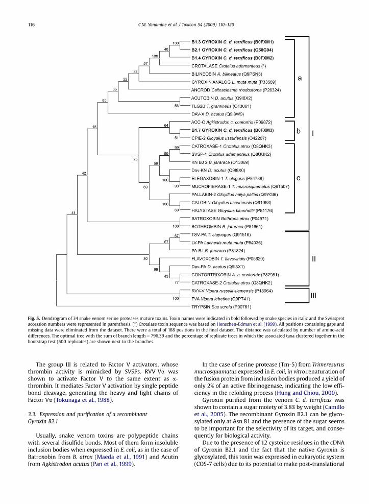
Fig. 5. Dendrogram of 34 snake venom serine proteases mature toxins. Toxin names were indicated in bold followed by snake species in italic and the Swissprotaccession numbers were represented in parenthesis. (*) Crotalase toxin sequence was based on Henschen-Edman et al. (1999). All positions containing gaps andmissing data were eliminated from the dataset. There were a total of 188 positions in the final dataset. The distance was calculated by number of amino-aciddifferences. The optimal tree with the sum of branch length¼ 796.39 and the percentage of replicate trees in which the associated taxa clustered together in thebootstrap test (500 replicates) are shown next to the branches.
C.M. Yonamine et al. / Toxicon 54 (2009) 110–120116
The group III is related to Factor V activators, whosethrombin activity is mimicked by SVSPs. RVV-Va wasshown to activate Factor V to the same extent as a-thrombin. It mediates Factor V activation by single peptidebond cleavage, generating the heavy and light chains ofFactor Va (Tokunaga et al., 1988).
3.3. Expression and purification of a recombinantGyroxin B2.1
Usually, snake venom toxins are polypeptide chainswith several disulfide bonds. Most of them form insolubleinclusion bodies when expressed in E. coli, as in the case ofBatroxobin from B. atrox (Maeda et al., 1991) and Acutinfrom Agkistrodon acutus (Pan et al., 1999).
In the case of serine protease (Tm-5) from Trimeresurusmucrosquamatus expressed in E. coli, in vitro renaturation ofthe fusion protein from inclusion bodies produced a yield ofonly 2% of an active fibrinogenase, indicating the low effi-ciency in the refolding process (Hung and Chiou, 2000).
Gyroxin purified from the venom C. d. terrificus wasshown to contain a sugar moiety of 3.8% by weight (Camilloet al., 2005). The recombinant Gyroxin B2.1 can be glyco-sylated only at Asn 81 and the presence of the sugar seemsto be important for the selectivity of its target, and conse-quently for biological activity.
Due to the presence of 12 cysteine residues in the cDNAof Gyroxin B2.1 and the fact that the native Gyroxin isglycosylated, this toxin was expressed in eukaryotic system(COS-7 cells) due to its potential to make post-translational

C.M. Yonamine et al. / Toxicon 54 (2009) 110–120 117
modification (PTM) like the proteolytic processing, glyco-sylation of precursors and secretion to the culture medium.
There are two SVSPs, AaV-SP-I and AaV-SP-II from thevenom of Agkistrodon acutus, that have putative glycosyl-ation sites at Asn 35. The proportions and types of carbo-hydrates within these two proteases are: 9% for AaV-SP-Iformed by trisaccharide NAG301-FUC302-NAG303 moleculeand 4% for AaV-SP-II formed by monosaccharide NAG301
molecule. It was seen that AaV-SP-I has higher amidolyticactivity than AaV-SP-II, and the distinct saccharides havedifferent influence on the structure of the active site (Zhuet al., 2005). Therefore, alteration of amidolytic activity canresult from different types and proportions of carbo-hydrates in the toxins.
The thrombin-like enzymes, Ancrod from the Agkis-trodon rhodostoma and Batroxobin from B. atrox moojenivenom, are N-glycosylated and contain about 31% and5.85% (by mass) in neutral carbohydrates respectively(Lochnit and Geyer, 1995). Interestingly, both toxins havehemotoxic effect, but are sold commercially as defib-rinogenolytic agents: Ancrod (Arvin�) and Batroxobin(Defibrase�) (Koh et al., 2006). However, differently fromBatroxobin, Ancrod induces the barrel rotation syndrome(Alexander et al., 1988).
Fig. 6. SDS-PAGE and Western blot of recombinant protein expressed by pED-Gyropurified by affinity with Benzamidine Sepharose 6B resin from conditioned. A – SDSCOS-7 cells total extract transfected with pSecTag2-Gyro; 3) COS-7 cells extract trcontrol); 6) empty; 7) 0.5 mg of Gyroxin purified from C. d. terrificus venom (positivecontrol). C – SDS-PAGE and D – Western blot of recombinant Gyroxin purified fro0.05 mg of Gyroxin purified from C. d. terrificus venom (positive control); 3) 0.5 mg oantibody was anti-crotalic serum from Butantan Institute and the reaction was det
Then, for the expression of recombinant SVSPs,eukaryotic systems should be the method of choice. Hence,we decided to express Gyroxin transiently in COS-7 cells inorder to select the vector construct which promotes thehighest level of production of this toxin. Gyroxin wascloned into pSecTag2 HygroA and pED vectors and trans-fected individually in COS-7 cells and recombinant Gyroxinwas detected by immunoblotting using anti-crotalic serumfrom Butantan Institute in transfected COS-7 cells totalextract and conditioned culture medium.
The Fig. 6A and B shows a western blot of COS-7 cellsextract and Fig. 6C and D shows a SDS-PAGE and respec-tively the western blot of secreted recombinant Gyroxinpurified by affinity with Benzamidine Sepharose 6B resinfrom conditioned culture medium over 24 h.
In Fig. 6A the lane 1 shows a Low-Range RainbowMolecular Weight Marker (45 K to 2.5 KDa), lane 2 containsthe COS-7 cells transfected with pSecTag2-Gyro totalextract and a recombinant protein band was detected in thewestern blot. The lane 3 shows the COS-7 cells transfectedwith pED-Gyro total extract and a correspondent band wasdetected in western blot. As one can see, COS-7 cellstransfected with pED-Gyro (lane 3) express Gyroxin moreefficiently than those transfected with pSecTag2-Gyro
and pSecTag2-Gyro in COS-7 cells and secreted in culture medium over 24 h-PAGE and B – Western blot of total extract COS-7 cells: 1) Marker (M.W); 2)ansfected with pED-Gyro; 4) empty; 5) untransfected COS-7 cells (negativecontrol); 8) 5 mg of Gyroxin purified from the C. d. terrificus venom (positive
m conditioned culture medium. 1) 3 mg of purified recombinant Gyroxin, 2)f Gyroxin purified from C. d. terrificus venom (positive control). The primaryected with secondary antibody conjugated to horseradish peroxidase.

Fig. 7. Esterase activity assay of recombinant Gyroxin purified by Benza-midine Sepharose from the supernatant of lysate and conditioned culturemedium over 24 h of transfected COS-7 cells with pED-Gyro. Supernatantlysate and conditioned culture medium of untransfected COS-7 cells wereused as negative controls. Trypsin and Gyroxin purified from the C. d. ter-rificus venom were used as positive controls.
C.M. Yonamine et al. / Toxicon 54 (2009) 110–120118
vector (lane 2). In the lane 5, untransfected COS-7 cells totalextract was applied as a negative control and this extractdoes not react with anti-crotalic serum from ButantanInstitute as expected.
The lanes 7 and 8 (6A) represent positive controls andcontain 0.5 mg and 5 mg of native Gyroxin respectively andthis protein was recognized by anti-crotalic serum inwestern blot (6B). Native Gyroxin is not well dyed by Coo-massie blue and requires at least 3 mg of the protein to bevisualized, so in the SDS-PAGEs it was not possible to visu-alize the native Gyroxin (positive control) at concentrationsof 0.5 mg (lane 7 – 6A, lane 3 – 6C) and 0.05 mg (lane 2 – 6C),but it was detected by immunoblottings (6B and 6D). In theSDS-PAGE (6C) lane 1 shows 3 mg of secreted recombinantGyroxin purified by affinity with Benzamidine Sepharose 6Bresin from conditioned culture medium over 24 h and isrecognized in western blot (lane 1 – 6D) by anti-crotalicserum from Butantan Institute.
The recombinant Gyroxin in total cell extracts and thesecreted recombinant Gyroxin in the culture mediumpurified by Benzamidine Sepharose shows the same elec-trophoretic pattern of the native Gyroxin purified from thevenom.
Transient expression offers a convenient means tocompare different vectors, by ensuring that an expressionplasmid is adequated to establish a stable expressing cellline. Thus the expression of Gyroxin, in a transient system,using COS-7 cells was a rapid way to choose the vector to beused in a future transfection for establishment of a stableGyroxin-expressing cell line. pED-Gyro is a dicistronicmRNA expression vector in which Gyroxin cDNA is locatedupstream of the amplifiable marker gene, dihydrofolatereductase (DHFR), which can be used for transfection ofdhfr deficient cells (CHO DX-B11 cells). Using this systemthere is a possibility of pED-Gyro amplification andconsequently of attaining higher level of Gyroxin expres-sion, as described for other proteins using such expressionsystem (Chura-Chambi et al., 2004; Peroni et al., 2002;Soares et al., 2000). Thus, stable transfection is being con-ducted to obtain higher and constitutive expression withCHO DX-B11 dhfr� cells.
Gyroxin cDNA was cloned in fusion with the murine Igksignal peptide nucleotide sequence, which allows itssecretion in the cell culture medium. Recombinant Gyroxinhas been purified using Benzamidine Sepharose 6B affinitychromatography, mainly from the conditioned mediumover 24 h from COS-7 cells transfected with pED-Gyro, in anrelative amount that is similar to one purified from crude C.d. terrificus venom. To confirm that recombinant Gyroxinwas fully active, an enzymatic assay using TAME assubstrate was done.
The esterase activity of conditioned culture mediumover 24 h transfected with pED-Gyro was higher thansupernatant of COS-7 cells lysate transfected with pED-Gyro (Fig. 7) indicating that the murine Igk signal peptidein this pED-Gyro was capable of driving active recombi-nant Gyroxin out of cell. The recombinant toxin showedesterase activity in the culture medium as indicated inFig. 7.
4. Conclusion
In the present work we described the cloning of fiveserine protease precursor genes from C. d. terrificus venomgland cDNA library: clones B2.1, B1.3, B1.4, B1.5 and B1.7.The dendrogram based on amino acid sequences and bio-logical activities showed that these clones belong to twosubgroups showing the divergence between clone B1.7(subgroup Ib) and the others belonging to subgroup Ia. Theclone B1.5 is a truncated form of cDNA precursor, witha deletion of 146 bp, which was not included in the func-tional dendrogram for analysis.
The transient expression of active Gyroxin was obtainedby transfecting COS-7 cells and this product was secretedinto the culture medium with an esterase activity, sug-gesting a correct folding of recombinant toxin. This rapidand direct methodological approach opens up new possi-bilities to express heterologous snake toxins in mammaliancells. Moreover, such strategy will allow one to proceed thecharacterization of their structure and activities, byobtaining the protein in the culture medium with high levelof purity and specificity.
Acknowledgements
We thank Juliana Branco Novo from BiotechnologyCentre of Butantan Institute – Sao Paulo for helpfuldiscussions. This research was supported in part by CNPqand FAPESP.
Conflict of interest
The authors declare that there are no conflicts ofinterest.
Appendix. Supplementary information
Supplementary data associated with this article can befound, in the online version, at doi: 10.1016/j.toxicon.2009.03.022.

C.M. Yonamine et al. / Toxicon 54 (2009) 110–120 119
References
Alexander, G., Grothusen, J., Zepeda, H., Schwartzman, R.J., 1988. Gyroxin,a toxin from the venom of Crotalus durissus terrificus is a thrombin-like enzyme. Toxicon 26 (10), 953–960.
Arocas, V., Castro, H.C., Zingali, R.B., Guillin, M.C., Jandrot-Perrus, M.,Bon, C., Wisner, A., 1997. Molecular cloning and expression ofbothrojaracin, a potent thrombin inhibitor from snake venom. Eur. J.Biochem. 248 (2), 550–557.
Barrabin, H., Martiarena, J.L., Vidal, J.C., Barrio, A., 1978. Isolation andcharacterization of Gyroxin from Crotalus durissus terrificus venom.In: Rosemberg, P. (Ed.), Toxins: Animals, Plant and Microbial. Perga-mon Press, New York, USA, pp. 113–133.
Bendtsen, J.D., Nielsen, H., Von Heijne, G., Brunak, S., 2004. Improvedprediction of signal peptides: SignalP 3.0. J. Mol. Biol. 340 (4), 783–795.
Bjarnason, J.B., Barish, A., Direnzo, G.S., Campbell, R., Fox, J.W., 1983.Kallikrein-like enzymes from Crotalus atrox venom. J. Biol. Chem. 258(20), 12566–12573.
Burkhart, W., Simth, G.F., Su, J.L., Parikh, I., LeVine, H.I.I.I., 1992. Amino acidsequence determination of ancrod, the thrombin-like alpha-fibrino-genase from the venom of Akistrodon rhodostoma. FEBS Lett. 297 (3),297–301.
Camillo, M.A.P., Alves da Silva, J.A., Yonamine, C.M., Hashizume, J.L.,Casare, M.S., Higa, O.Z., 2005. Biological activity and structural alter-ations in Gyroxin induced by gamma radiation. In: InternationalNuclear Atlantic Conference Proceedings of VII ENAN. BrazilianAssociation for Nuclear Energy – ABEN, Santos – SP, pp. 1845–1850.
Camillo, M.A.P., Arruda Paes, P.C., Troncone, L.R., Rogero, J.R., 2001.Gyroxin fails to modify in vitro release of labelled dopamine andacetylcholine from rat and mouse striatal tissue. Toxicon 39 (6),843–853.
Charbonnier, F., Gaspera, B.D., Armand, A.S., Van der Laarse, W.J.,Launay, T., Becker, C., Gallien, C.L., Chanoine, C., 2002. Two myogenin-related genes are differentially expressed in Xenopus laevis myo-genesis and differ in their ability to transactivate muscle structuralgenes. J. Biol. Chem. 277 (2), 1139–1147.
Chura-Chambi, R.M., Tornieri, P.H., Spencer, P.J., Nascimento, P.A.,Mathor, M.B., Morganti, L., 2004. High-level synthesis of recombinantmurine endostatin in Chinese hamster ovary cells. Protein. Expr. Purif.35 (1), 11–16.
Da Silva, N.J., Aird, S.D., Seebart, C., Kaiser, I.I., 1989. A analog from thevenom of the bushmaster (Lachesis muta muta). Toxicon 27 (7),763–771.
Felsenstein, J., 1985. Confidence limits on phylogenies: an approach usingthe bootstrap. Evolution 39 (4), 783–791.
Felsenstein, J., 1989. PHYLIP – Phylogeny Inference Package (Version 3.2).Cladistics 5, 164–166 (Felsenstein, J. 2005 PHYLIP – Phylogeny Infer-ence Package Version 3.6. Distributed by the author. Department ofGenome Science, University of Washington, Seatle).
Hahn, B.S., Baek, K., Kim, W.S., Lee, C.S., Chang, I.L., Kim, Y.S., 1998.Molecular cloning of capillary permeability-increasing enzyme-2from Agkistrodon caliginosus (Korean viper). Toxicon 36 (12), 1887–1893.
Hahn, B.S., Yang, K.Y., Park, E.M., Chang, I.M., Kim, Y.S., 1996. Purificationand molecular cloning of calobin, a thrombin-like enzyme fromAgkistrodon caliginosus (Korean viper). J. Biochem. 119 (5), 835–843.
Henschen-Edman, A.H., Theodor, I., Edwards, B.F., Pirkle, H., 1999. Crota-lase, a fibrinogen-clotting snake venom enzyme: primary structureand evidence for a fibrinogen recognition exosite different fromthrombin. Thromb. Haemost. 81 (1), 81–86.
Hung, C.C., Chiou, S.H., 2000. Expression of a kallikrein-like protease fromthe snake venom: engineering of autocatalytic site in the fusionprotein to facilitate protein refolding. Biochem. Biophys. Res. Com-mun. 275 (3), 924–930.
Itoh, N., Tanaka, N., Funakoshi, I., Kawasaki, T., Mihashi, S., Yamashina, I.,1988. The complete nucleotide sequence of the gene for batroxobin,a thrombin-like snake venom enzyme. Nucleic Acids Res. 16 (21),10377–10378.
Itoh, N., Tanaka, N., Mihashi, S., Yamashina, I., 1987. Molecular cloning andsequence analysis of cDNA for batroxobin, a thrombin-like snakevenom enzyme. J. Biol. Chem. 262 (7), 3132–3135.
Johansen, M.B., Kiemer, L., Brunak, S., 2006. Analysis and prediction ofmammalian protein glycation. Glycobiology 16 (9), 844–853.
Koh, D.C.I., Armugan, A., Jeyaseelan, K., 2006. Snake venom componentsand their applications in biomedicine. Cell. Mol. Life. Sci. 63 (24),3030–3041.
Kunkel, T.A., 1985. Rapid and efficient site-specific mutagenesis withoutphenotypic selection. Proc. Natl. Acad. Sci. USA 82 (2), 488–492.
Laemmli, U.K., 1970. Cleavage of structural proteins during the assembly ofthe head of bacteriophage T4. Nature 227 (5259), 680–685.
Lochnit, G., Geyer, R., 1995. Carbohydrate structure analysis of batroxobin,a thrombin-like serine protease from Bothrops moojeni venom. Eur. J.Biochem. 228 (3), 805–816.
Maeda, M., Satoh, S., Suzuki, S., Niwa, M., Itoh, N., Yamashina, I., 1991.Expression of cDNA for batroxobin, a thrombin-like enzyme. J. Bio-chem. 109 (4), 632–637.
Magalhaes, A., Da Fonseca, B.C.B., Diniz, C.R., Gilroy, J., Richardson, M.,1993. The complete amino acid sequence of a thrombin-like enzyme/gyroxin analogue from venom of the bushmaster snake (Lachesismuta muta). FEBS Lett. 329 (1–2), 116–120.
Mcmullen, B.A., Fujikawa, K., Kisiel, W., 1989. Primary structure ofa protein C activator from Agkistrodon contortrix contortrix venom.Biochemistry 28 (2), 674–679.
Nikai, T., Ohara, A., Komori, Y., Fox, J.W., Sugihara, H., 1995. Primarystructure of a coagulant enzyme, bilineobin, from Agkistrodon bili-neatus venom. Arch. Biochem. Biophys. 318 (1), 89–96.
Nolan, C., Hall, L.S., Barlow, G.H., 1976. Ancrod, the coagulating enzyme ofMalayan pitviper (Agkistrodon rhodostoma) venom. Meth. Enzymol.45, 205–213.
Pan, H., Du, X., Yang, G., Zhou, Y., Wu, X., 1999. cDNA cloning andexpression of acutin. Biochem. Biophys. Res. Commun. 255 (2),412–415.
Parry, M.A., Jacob, U., Huber, R., Wisner, A., Bon, C., Bode, W., 1998. Thecrystal structure of the novel snake venom plasminogen activatorTSV-PA: a prototype structure for snake venom serine proteinases.Structure 6 (9), 1195–1206.
Peroni, C.N., Soares, C.R.J., Gimbo, E., Morganti, L., Ribela, M.T.C.P.,Bartolini, P., 2002. High-level expression of human thyroid-stimu-lating hormone in Chinese hamster ovary cells by co-transfection ofdicistronic expression vectors followed by a dual-marker amplifica-tion strategy. Biotechnol. Appl. Biochem. 35 (Pt 1), 19–26.
Radis-Baptista, G., Oguiura, N., Hayashi, M.A., Camargo, M.E., Grego, K.F.,Oliveira, E.B., Yamane, T., 1999. Nucleotide sequence of crotamineisoform precursors from a single South American rattlesnake(Crotalus durissus terrificus). Toxicon 37 (7), 973–984.
Raw, I., Rocha, M.C., Esteves, M.I., Kamiguti, A.S., 1986. Isolation andcharacterization of a thrombin-like enzyme from the venom ofCrotalus durissus terrificus. Braz. J. Med. Biol. Res. 19 (3), 333–338.
Saitou, N., Nei, M., 1987. The neighbor-joining method: a new method forreconstructing phylogenetic trees. Mol. Biol. Evol. 4 (4), 406–425.
Sanchez, E.F., Santos, C.I., Magalhaes, A., Diniz, C.R., Figueiredo, S.,Gilroy, J., Richardson, M., 2000. Isolation of a proteinase withplasminogen-activating activity from Lachesis muta muta(bushmaster) snake venom. Arch. Biochem. Biophys. 378 (1), 131–141.
Sanger, N., Nicklen, J., Coulson, A.R., 1977. DNA sequencing with chaintermination inhibitors. Proc. Natl. Acad. Sci. 74 (12), 5463–5467.
Schuler, G.D., Altschul, S.F., Lipman, D.J., 1991. A workbench for multiplealignment construction and analysis. Proteins 9 (3), 180–190.
Serrano, S., Maroun, R.C., 2005. Snake venom serine proteinases:sequence homology vs. substrate specificity, a paradox to be solved.Toxicon 45 (8), 1115–1132.
Serrano, S.M., Hagiwara, Y., Murayama, N., Higuchi, S., Mentele, R.,Sampaio, C.A., Camargo, A.C., Fink, E., 1998. Purification and charac-terization of a kinin-releasing and fibrinogen-clotting serineproteinase (KN-BJ) from the venom of Bothrops jararaca and molec-ular cloning and sequence analysis of its cDNA. Eur. J. Biochem. 251(3), 845–853.
Serrano, S.M.T., Mentele, R., Sampaio, C.A.M., Fink, E., 1995. Purification,characterization, and amino acid sequence of a serine proteinase, PA-BJ, with platelet-aggregating activity from the venom of Bothropsjararaca. Biochemistry 34 (21), 7186–7193.
Shimokawa, K., Takahashi, H., 1997. Capillary permeability-increasingenzyme-2 from the venom of Agkistrodon caliginosus (kankoku-mamushi): activity resulting from the release of peptides fromfibrinogen. Toxicon 35 (4), 597–605.
Soares, C.R., Morganti, L., Miloux, B., Lupker, J.H., Ferrara, P., Bartolini, P.,2000. High-level synthesis of human prolactin in Chinese Hamsterovary cells. Biotechnol. Appl. Biochem. 32 (Pt 2), 127–135.
Stocker, K., Fischer, H., Meier, J., Brogli, M., Svendsen, L., 1987. Charac-terization of the protein C activator Protac from venom of thesouthern copperhead (Agkistrodon contortrix) snake. Toxicon 25 (3),239–252.
Tamura, K., Dudley, J., Nei, M., Kumar, S., 2007. MEGA4: MolecularEvolutionary Genetics Analysis (MEGA) software version 4.0. Mol.Biol. Evol. 24 (8), 1596–1599.
Tokunaga, F., Nagasawa, K., Tamura, S., Miyata, T., Iwanaga, S., Kisiel, W.,1988. The factor V-activating enzyme (RVV-V) from Russell’s viper

C.M. Yonamine et al. / Toxicon 54 (2009) 110–120120
venom. Identification of isoproteins RVV-V alpha, -V beta, and -Vgamma and their complete amino acid sequences. J. Biol. Chem. 263(33), 17471–17481.
Wang, Y.M., Wang, S.R., Tsai, I.H., 2001. Serine protease isoforms ofDeinagkistrodon acutus venom: cloning, sequencing and phylogeneticanalysis. Biochem. J. 354 (Pt 1), 161–168.
You, W.K., Choi, W.S., Koh, Y.S., Shin, H.C., Jang, Y., Chung, K.H., 2004.Functional characterization of recombinant batroxobin, a snake
venom thrombin-like enzyme, expressed from Pichia pastoris. FEBSLett. 571 (1–3), 67–73.
Zhang, Y., Wisner, A., Xiong, Y., Bon, C., 1995. A novel plasminogen acti-vator from snake venom. Purification, characterization, and molecularcloning. J. Biol. Chem. 270 (17), 10246–10255.
Zhu, Z., Liang, Z., Zhang, T., Zhu, Z., Xu, W., Teng, M., Niu, L., 2005. Crystalstructures and amidolytic activities of two glycosylated snake venomserine proteinases. J. Biol. Chem. 280 (11), 10524–10529.
Copyright © 2022 FDOKUMEN