Arterial acid–base status during digestion and following vascular infusion of NaHCO 3 and HCl in...
8
Arterial acid–base status during digestion and following vascular infusion of NaHCO 3 and HCl in the South American rattlesnake, Crotalus durissus Sine K. Arvedsen a,b , Johnnie B. Andersen a,b , Morten Zaar a,b , Denis Andrade b , Augusto S. Abe b , Tobias Wang a,b, * a Department of Zoophysiology, The University of Aarhus, Denmark b Departamento de Zoologia, Instituto de Biocie ˆncias, UNESP, Rio Claro, SP, Brazil Received 17 May 2005; received in revised form 30 September 2005; accepted 2 October 2005 Available online 10 November 2005 Abstract Digestion is associated with gastric secretion that leads to an alkalinisation of the blood, termed the ‘‘alkaline tide’’. Numerous studies on different reptiles and amphibians show that while plasma bicarbonate concentration ([HCO 3 ] pl ) increases substantially during digestion, arterial pH (pHa) remains virtually unchanged, due to a concurrent rise in arterial PCO 2 (PaCO 2 ) caused by a relative hypoventilation. This has led to the suggestion that postprandial amphibians and reptiles regulate pHa rather than PaCO 2 . Here we characterize blood gases in the South American rattlesnake (Crotalus durissus ) during digestion and following systemic infusions of NaHCO 3 and HCl in fasting animals to induce a metabolic alkalosis or acidosis in fasting animals. The magnitude of these acid – base disturbances were similar in magnitude to that mediated by digestion and exercise. Plasma [HCO 3 ] increased from 18.4 T 1.5 to 23.7 T 1.0 mmol L 1 during digestion and was accompanied by a respiratory compensation where PaCO 2 increased from 13.0 T 0.7 to 19.1 T 1.4 mm Hg at 24 h. As a result, pHa decreased slightly, but were significantly below fasting levels 36 h into digestion. Infusion of NaHCO 3 (7 mmol kg 1 ) resulted in a 10 mmol L 1 increase in plasma [HCO 3 ] within 1 h and was accompanied by a rapid elevation of pHa (from 7.58 T 0.01 to 7.78 T 0.02). PaCO 2 , however, did not change following HCO 3 infusion, which indicates a lack of respiratory compensation. Following infusion of HCl (4 mmol kg 1 ), plasma pHa decreased by 0.07 units and [HCO 3 ] pl was reduced by 4.6 mmol L 1 within the first 3 h. PaCO 2 , however, was not affected and there was no evidence for respiratory compensation. Our data show that digesting rattlesnakes exhibit respiratory compensations to the alkaline tide, whereas artificially induced metabolic acid – base disturbances of same magnitude remain uncompensated. It seems difficult to envision that the central and peripheral chemoreceptors would experience different stimuli during these conditions. One explanation for the different ventilatory responses could be that digestion induces a more relaxed state with low responsiveness to ventilatory stimuli. D 2005 Elsevier Inc. All rights reserved. Keywords: Arterial acid –base status; Vascular infusion; Crotalus durissus 1. Introduction Digestion causes metabolism to rise, the so-called specific dynamic action (SDA) of food, and induces gastric secretion that leads to an alkalinisation of the blood, the ‘‘alkaline tide’’ (e.g. McCorvie, 1925; Rune, 1965; Wang et al., 2001; Niv and Fraser, 2002; Wood et al., 2005). The SDA response and the alkaline tide are more pronounced in animals, such as reptiles, that ingest large meals relative to their own body mass (recently reviewed by Andrade et al., 2005; Wang et al., 2005). Numerous studies on different reptiles and amphibians show that plasma bicarbonate concentration ([HCO 3 ] pl ) may increase by up to 10 mmol L 1 during the alkaline tide concurrent with a rise in arterial PCO 2 (PaCO 2 ), so that arterial pH (pHa) remains virtually unchanged from that of fasting animals (Overgaard et al., 1999; Busk et al., 2000a,b; Wang et al., 2001, 2005; Andersen et al., 2003; Andersen and Wang, 2004; Andrade et al., 2004b). The rise in PaCO 2 is accomplished through a relative hypoven- tilation where ventilation does not increase proportionally to the rise in metabolism (Glass et al., 1979; Wang et al., 1995; Hicks et 1095-6433/$ - see front matter D 2005 Elsevier Inc. All rights reserved. doi:10.1016/j.cbpa.2005.10.001 * Corresponding author. Department of Zoophysiology, Building 131, Aarhus University, 8000 Aarhus C, Denmark. E-mail address: [email protected] (T. Wang). Comparative Biochemistry and Physiology, Part A 142 (2005) 495 – 502 www.elsevier.com/locate/cbpa
-
Upload
independent -
Category
Documents
-
view
0 -
download
0
Transcript of Arterial acid–base status during digestion and following vascular infusion of NaHCO 3 and HCl in...

.elsevier.com/locate/cbpa
Comparative Biochemistry and Physiolo
Arterial acid–base status during digestion and following vascular infusion of
NaHCO3 and HCl in the South American rattlesnake, Crotalus durissus
Sine K. Arvedsen a,b, Johnnie B. Andersen a,b, Morten Zaar a,b,
Denis Andrade b, Augusto S. Abe b, Tobias Wang a,b,*
a Department of Zoophysiology, The University of Aarhus, Denmarkb Departamento de Zoologia, Instituto de Biociencias, UNESP, Rio Claro, SP, Brazil
Received 17 May 2005; received in revised form 30 September 2005; accepted 2 October 2005
Available online 10 November 2005
Abstract
Digestion is associated with gastric secretion that leads to an alkalinisation of the blood, termed the ‘‘alkaline tide’’. Numerous studies on
different reptiles and amphibians show that while plasma bicarbonate concentration ([HCO3�]pl) increases substantially during digestion, arterial
pH (pHa) remains virtually unchanged, due to a concurrent rise in arterial PCO2 (PaCO2) caused by a relative hypoventilation. This has led to the
suggestion that postprandial amphibians and reptiles regulate pHa rather than PaCO2.
Here we characterize blood gases in the South American rattlesnake (Crotalus durissus) during digestion and following systemic infusions of
NaHCO3 and HCl in fasting animals to induce a metabolic alkalosis or acidosis in fasting animals. The magnitude of these acid–base disturbances
were similar in magnitude to that mediated by digestion and exercise. Plasma [HCO3�] increased from 18.4T1.5 to 23.7T1.0 mmol L�1 during
digestion and was accompanied by a respiratory compensation where PaCO2 increased from 13.0T0.7 to 19.1T1.4 mm Hg at 24 h. As a result,
pHa decreased slightly, but were significantly below fasting levels 36 h into digestion. Infusion of NaHCO3 (7 mmol kg�1) resulted in a 10 mmol
L�1 increase in plasma [HCO3�] within 1 h and was accompanied by a rapid elevation of pHa (from 7.58T0.01 to 7.78T0.02). PaCO2, however,
did not change following HCO3� infusion, which indicates a lack of respiratory compensation. Following infusion of HCl (4 mmol kg�1), plasma
pHa decreased by 0.07 units and [HCO3�]pl was reduced by 4.6 mmol L�1 within the first 3 h. PaCO2, however, was not affected and there was no
evidence for respiratory compensation.
Our data show that digesting rattlesnakes exhibit respiratory compensations to the alkaline tide, whereas artificially induced metabolic acid–
base disturbances of same magnitude remain uncompensated. It seems difficult to envision that the central and peripheral chemoreceptors would
experience different stimuli during these conditions. One explanation for the different ventilatory responses could be that digestion induces a more
relaxed state with low responsiveness to ventilatory stimuli.
D 2005 Elsevier Inc. All rights reserved.
Keywords: Arterial acid–base status; Vascular infusion; Crotalus durissus
1. Introduction
Digestion causes metabolism to rise, the so-called specific
dynamic action (SDA) of food, and induces gastric secretion
that leads to an alkalinisation of the blood, the ‘‘alkaline tide’’
(e.g. McCorvie, 1925; Rune, 1965; Wang et al., 2001; Niv and
Fraser, 2002; Wood et al., 2005). The SDA response and the
alkaline tide are more pronounced in animals, such as reptiles,
1095-6433/$ - see front matter D 2005 Elsevier Inc. All rights reserved.
doi:10.1016/j.cbpa.2005.10.001
* Corresponding author. Department of Zoophysiology, Building 131, Aarhus
University, 8000 Aarhus C, Denmark.
E-mail address: [email protected] (T. Wang).
that ingest large meals relative to their own body mass
(recently reviewed by Andrade et al., 2005; Wang et al., 2005).
Numerous studies on different reptiles and amphibians show
that plasma bicarbonate concentration ([HCO3�]pl) may increase
by up to 10 mmol L�1 during the alkaline tide concurrent with a
rise in arterial PCO2 (PaCO2), so that arterial pH (pHa) remains
virtually unchanged from that of fasting animals (Overgaard et
al., 1999; Busk et al., 2000a,b;Wang et al., 2001, 2005;Andersen
et al., 2003; Andersen and Wang, 2004; Andrade et al., 2004b).
The rise in PaCO2 is accomplished through a relative hypoven-
tilation where ventilation does not increase proportionally to the
rise in metabolism (Glass et al., 1979;Wang et al., 1995; Hicks et
gy, Part A 142 (2005) 495 – 502
www
S.K. Arvedsen et al. / Comparative Biochemistry and Physiology, Part A 142 (2005) 495–502496
al., 2000; Secor et al., 2000). Smaller, but qualitatively similar
respiratory compensations also occur in mammals (Higgens,
1914; Erdt, 1915; Van Slyke et al., 1917; Ou and Tenney, 1974;
Niv and Fraser, 2002; cf. Johnson et al., 1995).
The respiratory compensation of pH during digestion is likely
to represent a homeostatic response that prevents pH changes
from affecting enzyme function and metabolic processes.
However, the underlying regulation is not well understood and
the postprandial rise in PaCO2 may reflect an ineffective
ventilatory response to the increased metabolism during
digestion or digestion per se could induce a more relaxed state
with low responsiveness to ventilatory stimuli (e.g. Higgens,
1914). However, all animals studied exhibit a respiratory
compensation to the increased [HCO3�]pl during digestion, and
PaCO2 does not increase when gastric acid secretion is
pharmacologically inhibited, so that the alkaline tide is abolished
(Andersen et al., 2003; Andrade et al., 2004b). These observa-
tions have lead to the suggestion that postprandial amphibians
and reptiles in contrast to mammals regulate pHa rather than
PaCO2 (Wang et al., 2005).
Here we characterize blood gases during digestion in the
South American rattlesnake (Crotalus durissus). This species
occur widely across arid environments in tropical, subtropical
and temperate South America and exhibit pronounced metabolic
responses to digestion (Andrade et al., 1997). In addition, blood
gases have previously been characterised in this species (Wang
et al., 1998) and C. durissus exhibit marked ventilatory res-
ponses to hypercapnia (Andrade et al., 2004a). To gain further
insight into ventilatory compensation of arterial pH, we inves-
tigated whether respiratory compensations to metabolic acid–
base disturbances occur in fasting animals. This was achieved by
inducing a metabolic alkalosis and acidosis in fasting animals by
systemic injections of NaHCO3 and HCl, respectively. The
magnitude of these acid–base disturbances were similar to that
mediated by digestion (NaHCO3) and exercise (HCl).
2. Materials and methods
2.1. Animals
Thirty-one South American rattlesnakes (C. durissus) that
had been collected at several localities within the state of Sao
Paulo were obtained from the Butantan Institute (Sao Paulo,
Brazil), and transported to the Jacarezario, UNESP, Rio Claro,
SP, Brazil. Here the snakes were kept in separate containers
(20�30�25 cm) and maintained at a 12 h/12 h L/D cycle at a
temperature of 30 -C (T3 -C). The animals had free access to
water and were fed rodents on a weekly basis. At the time of
experimentation, the snakes weighed between 280 and 685 g
(390T100 g) and all appeared to be in good health. Food was
withheld for 2–3 weeks before commencing the experiments.
2.2. Surgical procedure
All snakes were instrumented with an arterial catheter for
blood sampling and infusions. To place the catheter, the snakes
were anaesthetised by inhalation of CO2 (Wang et al., 1993) until
they ceased to exhibit reflexes when pinched. A ventrolateral
incision was made about 3 cm cranial to the heart, so the
vertebral artery could be occlusively cannulated with PE60
catheter containing heparinized saline. The tip of the catheter
was pushed towards the right aortic arch and the catheter was
exteriorised through the back of the snake and secured with two
or three sutures. Then, the incision was closed and the snake
allowed to recover for at least 24 h. The surgery normally lasted
15–20 min, and most animals spontaneously resumed ventila-
tion immediately after termination of surgery.
2.3. Experimental protocols
Arterial blood gases, haematological parameters and plasma
concentrations of chloride ([Cl�]pl), potassium ([K+]pl) and
sodium ([Na+]pl) concentrations were measured in four
experimental groups: digesting snakes (N =5); HCO3� infused
(N =9); H+ infused (N =6); and a control group that was
injected with saline (N =3).
Blood samples were taken prior to feeding and at 12, 24, 36
and 48 h after the snake had voluntarily ingested a rat equal to
15T2% of body mass. HCO3Na infused animals were sampled
before and at 1, 3, 6, 12, 24, and 48 h after being infused with a
dose of 7 mmol NaHCO3 kg�1. Acid infused snakes received a
dose of 4 mmol HCl kg�1 over three infusions of 1.33 mmol
kg�1 in each. Each infusion was performed over 10 min with
20 min between infusions. To minimize the acute disturbance
during HCO3� and H+ infusions, 3 mL of blood was withdrawn
into the syringe, and mixed with the solution prior to injection.
To assess the possible effects of blood sampling, blood was
withdrawn three times at 24 h intervals in fasting (control)
snakes. All animals were kept in individual plastic boxes inside
a climatic chamber at 30 -C (T1 -C) during experimentation.
2.4. Measurements of blood gases and plasma ions
Arterial blood samples were drawn anaerobically and
analysed immediately after collection, except for plasma ion
samples which were frozen for subsequent measurement. Arte-
rial PO2 and pHwere measured using Radiometer (Copenhagen,
Denmark) electrodes mounted in a BMS Mk3 unit. Electrodes
were kept at 30 -C by a custom-made adaptation of the BMS
Mk3unit and electrodeswere calibrated before each sample anal-
ysis. Outputs from the electrodes were displayed on a Radio-
meter PHM73. Haematocrit was determined as the fractional red
cell volume after centrifugation (12,000 rpm for three min) and
monomeric haemoglobin concentration, [Hb], was measured
after conversion to cyanmethaemoglobin, applying a millimolar
extinction coefficient of 10.99 and measured at 540 nm (Zijlstra
et al., 1983). Arterial [O2] was measured as described by Tucker
(1967), with the correction pointed out by Bridges et al. (1979).
The Tucker chamber was thermostated at 40 -C. Haemoglobin
bound oxygen (HbO2) was calculated as [O2]� (aO2*PaO2
),
where aO2is the blood oxygen solubility (Christoforides and
Hedley-Whyte, 1969), and haemoglobin oxygen saturation was
subsequently calculated as: HbO2sat=HbO2/[Hb], under the
assumption that all Hb was functional.

pHa
7.45
7.50
7.55
7.60
7.65
7.70
7.75
7.80
7.85
PaC
O2
(mm
Hg)
10
12
14
16
18
20
22
Time (hours)
[HC
O3- ] p
l (m
mol
l-1)
66 12 24 36 48
6 12 24 36 48
6 12 24 36 48
9
12
15
18
21
24
27
30
**
C
A
B
*
**
**
*
*
*
* *
*
*
*
*
HCO3- infused
SDAHCl infused
Fig. 1. Arterial acid–base variables in South American rattlesnakes (Crotalus
durissus) in four experimental groups: digesting snakes (N =5); HCO3� infused
(N =9); HCl infused (N =6). Data at 0 h show fasting levels, values before
HCO3Na (7 mmol kg�1) or HCl (4 mmol kg�1 of HCl dispersed over three
infusions of 1.33 mmol kg�1) infusion. (A) Arterial pH, (B) plasma carbon
dioxide tension (PCO2), (C) plasma bicarbonate concentration ([HCO3�]pl). Data
are presented as meansTS.E.M. and values that are significantly different from 0
h levels are marked with an asterisk.
7.55 7.60 7.65 7.70 7.75 7.80 7.85
[HC
O3- ]
pl (
mm
ol l-
1 )
14
17
20
23
26
29
32
0h
1h3h
6h
12h
48h
24h
Arterial pH
7.40 7.45 7.50 7.55 7.60 7.65 7.70
[HC
O3- ]
pl (
mm
ol l-
1 )
8
10
12
14
16
18
20
48h
24h
6h
3h
0h
12
13
14
15
16
PC
O2
(mm
Hg)
PC
O2
(mm
Hg)
12
14161820
22 20 1826 242830
10
8
A
7.62 7.65 7.68 7.71 7.74 7.77 7.80
[HC
O3- ]
pl (
mm
ol l-1
)
16
18
20
22
24
26
12
13
14
151617
PC
O2 (
mm
Hg)
20 19 18
0h
12h
48h
24h
36h
A
A
A
B
C
Fig. 2. Davenport diagram, showing plasma bicarbonate concentration
([HCO3�]) and arterial pH in South American rattlesnakes (Crotalus
durissus). (A) Before and during digestion. (B) pH before and after infusion
of bicarbonate (7 mmol kg�1) (N =9). (C) Before and after infusion of HCl
(4 mmol kg�1 of HCl dispersed over three infusions of 1.33 mmol kg�1)
(N =7). The Davenport diagram includes carbon dioxide isoclines for the
partial CO2 tension/pressure in arterial blood (PaCO2, curved lines) and an in
vitro buffer-line (dotted lines, bnoncarb=�20.16) determined by Overgaard
and Wang (2002). Point A in A, B, and C illustrates the PaCO2 it would
require to fully compensate pH during alkaline tide, after HCO3Na and HCl
infusion. Data are presented as meansTS.E.M.
S.K. Arvedsen et al. / Comparative Biochemistry and Physiology, Part A 142 (2005) 495–502 497
Plasma carbon dioxide concentration ([CO2]) was measured
according to Cameron (1971) at 40 -C. PaCO2 was calculated
from pH and [CO2] of the plasma using the Henderson–
Hasselbalch equation, with the plasma solubility of CO2 (aCO2)of 0.0366 mmol L�1 (Heisler, 1984). The apparent pKVat 30 -Cwas taken from Overgaard and Wang (2002). Plasma chloride
was measured by colorimetric titration (Radiometer CMT 10),
while sodium and potassium was measured by flame photom-
etry (FLM 3 Flame Photometer, Radiometer). Osmolality was
determined by freezing point depression (Knauer semimicro
osmometer; Berlin, Germany).
2.5. Statistical analysis and data presentation
A one-way ANOVA for repeated measurements was
employed to test for significant differences in the measured

Table 1
Arterial oxygen levels and haematological variables in rattlesnakes prior to and
during digestion of a meal equal to 15% of their body mass (N =5)
Time (h)
0 12 24 36 48
PaO2 (mm Hg) 77.6T3.3 57.1T1.9* 58.9T2.0* 57.2T1.8* 55.0T2.8*
HbO2sat 0.95T0.02 1.03T0.05 0.97T0.05 0.98T0.05 0.88T0.01Haematocrit 0.18T0.01 0.18T0.00 0.16T0.01 0.16T0.01 0.15T0.01*
[Hb4]
(mmol L�1)
0.94T0.04 0.77T0.02* 0.65T0.04* 0.72T0.03* 0.71T0.04*
Hb (mmol L�1) 3.76T0.17 3.09T0.09* 2.60T0.17* 2.88T0.11* 2.86T0.16*MCHC
(mmol L�1)
5.16T0.11 4.37T0.17* 3.99T0.18* 4.41T0.15* 4.73T0.14
Values that are significantly different from 0 h levels are marked with an
asterisk.
S.K. Arvedsen et al. / Comparative Biochemistry and Physiology, Part A 142 (2005) 495–502498
parameters before and at different moments after feeding. A
Bonferroni post-hoc test was used to identify mean values that
differed from the control condition. Differences were consid-
ered statistically significant at the level of P�0.05, and all data
are presented as meanTS.E.M.
3. Results
3.1. Effects of digestion
Plasma bicarbonate concentration increased from a fasting
level of 18.4T1.5 mmol L�1 within 12 h after ingestion, and
reached the maximal level of 23.7T1.0 mmol L�1 by 24 h, and
Time (hours)
Na+
(m
mol
ml-1
)
1500 10 20 30 40 50
160
170
180
190HCO3
- infusedSDAHCl infused
Pla
sma
osm
olar
ity (
mO
sm)
340
350
360
370
380
390
* *
*
*
A
C
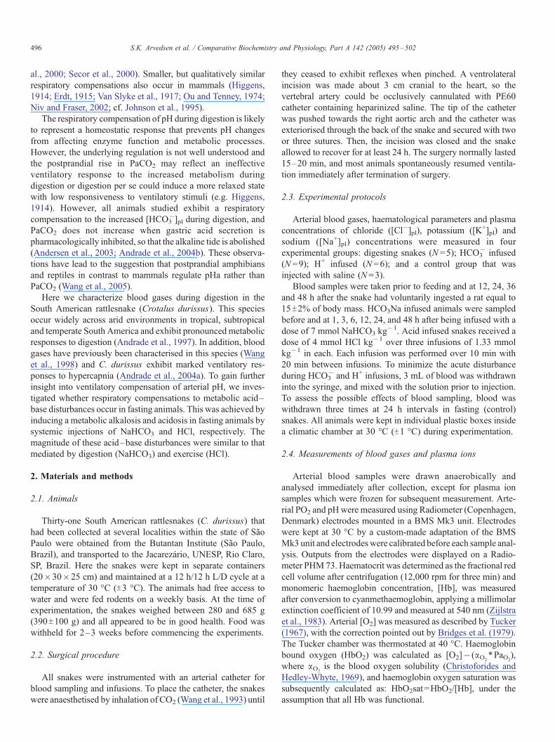
Fig. 3. Plasma ions and osmolality in South American rattlesnakes (Crotalus duri
(N =9); HCl infused (N =6); and a control group that was injected with saline (N =3
(4 mmol kg�1 of HCl dispersed over three infusions of 1.33 mmol kg�1) infusion.
concentration; (D) plasma Cl� concentration. The circles at 0 h show levels bef
meansTS.E.M. and values that are significantly different from fasting levels are m
then declined towards fasting level (Fig. 1A). This rise in
[HCO3�]pl was accompanied by a rise in PaCO2, which
increased from a fasting value of 13.0T0.7 to 19.1T1.4 mm
Hg at 24 h (Fig. 1B). As a result, pHa decreased slightly and was
significantly below fasting levels 36 h into digestion (Fig. 1C).
When depicted in a Davenport diagram (Fig. 2A), it is evident
that the metabolic alkalosis was slightly overcompensated by
the respiratory acidosis, i.e. pHa would have increased to 7.79 if
there had been no respiratory compensation, and plasma
[HCO3�] would have been 21.8 mmol L�1 (Fig. 2A). Thus,
the maximal change in ion difference, reflected maximal change
in plasma [HCO3�] at unchanged PaCO2, can be estimated to be
approximately 4 mmol L�1.
Arterial PO2 decreased significantly within 12 h after
ingestion and remained low throughout the experiment (Table
1). Both haematocrit and [HbO2]a decreased significantly within
24 h of digestion (Table 1). Plasma [Na+] increased significantly
after 24 h of digestion and persisted increased for the next 24 h.
On the other hand, feeding did not cause any significant changes
in plasma osmolality, [K+]pl or [Cl�]pl (Fig. 3).
3.2. Effects of HCO3� infusion
The vascular infusion of NaHCO3 in fasting rattlesnakes
caused plasma [HCO3�] to increase by approximately 10 mmol
L�1 within 1 h and was accompanied by a rapid elevation of
pHa from 7.58T0.01 to 7.78T0.02 (Fig. 1A). PaCO2, however,
did not change following HCO3� infusion, which indicates a
Time (hours)
0 10 20 30 40 50
[Cl -]a m
mol m
l -1
125
130
135
140
145
150
155
160
K+ (m
mol m
l -1)
2.0
2.4
2.8
3.2
3.6
4.0
*
*
*
*
B
D
ssus) in four experimental groups: digesting snakes (N =5); HCO3Na infused
). Data at 0 h show fasting levels, values before HCO3Na (7mmol kg�1) or HCl
(A) Plasma osmolality (mosM); (B) plasma K+ concentration; (C) plasma Na+
ore feeding, infusion of HCO3� and infusion of HCl. Data are presented as
arked with an asterisk.
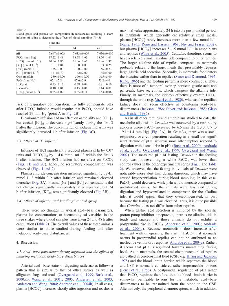
Table 2
Blood gases and plasma ion composition in rattlesnakes receiving a sham
infusion of saline to determine the effects of blood sampling (N =3)
Time (h)
0 24 48
pH 7.647T0.003 7.633T0.009 7.630T0.010
PCO2 (mm Hg) 17.22T1.49 18.78T1.63 18.70T1.61HCO3
� (mmol L�1) 20.04T1.86 21.06T1.67 20.86T1.97
[K+] (mmol L�1) 3.1T0.04 3.0T0.03 3.3T0.25
[Na+] (mmol L�1) 155T3.00 160T3.00 156T0.73
[Cl�] (mmol L�1) 141T0.70 142T2.00 145T5.00Osm (mosM) 380T10.00 370T10.00 365T5.00
PaO2 (mm Hg) 67.1T7.8 67.6T2.9 73.2T4.0
HbO2sat 0.73T0.13 0.78T0.04 0.81T0.10
Haematocrit 0.18T0.01 0.15T0.01 0.14T0.01[Hb4] (mmol L�1) 0.85T0.09 0.85T0.11 0.64T0.06
S.K. Arvedsen et al. / Comparative Biochemistry and Physiology, Part A 142 (2005) 495–502 499
lack of respiratory compensation. To fully compensate pHa
after HCO3� infusion would require that PaCO2 should have
reached 29 mm Hg (point A in Fig. 2B).
Bicarbonate infusion had no effect on osmolality and [Cl�]plbut caused [K+]pl to decrease significantly during the first 3
h after the infusion. The concentration of sodium in plasma was
significantly increased 1 h after infusion (Fig. 3C).
3.3. Effects of H+ infusion
Infusion of HCl significantly reduced plasma pHa by 0.07
units and [HCO3�]pl by ¨4.6 mmol mL�1 within the first 3
h after infusion. The HCl infusion had no effect on PaCO2
(Figs. 1B and 2C), hence, no respiratory compensation was
observed (Figs. 1 and 2).
Plasma chloride concentration increased significantly by 4.1
mmol L�1 within 3 h after infusion and remained elevated
thereafter (Fig. 3A). Plasma [Na+] and [K+] concentrations did
not change significantly immediately after injection, but 24
h after infusion, [K+]pl was significantly elevated (Fig. 3B).
3.4. Effects of infusion and handling: control group
There were no changes in arterial acid–base parameters,
plasma ion concentrations or haematological variables in the
three snakes where blood samples were taken 24 and 48 h after
cannulation (Table 2). The overall values of these three animals
were similar to those studied during feeding and after
metabolic acid–base disturbances.
4. Discussion
4.1. Acid–base parameters during digestion and the effects of
inducing metabolic acid–base disturbances
Arterial acid–base status of digesting rattlesnakes follows a
pattern that is similar to that of other snakes as well as
alligators, frogs and toads (Overgaard et al., 1999; Busk et al.,
2000a,b; Wang et al., 2001, 2005; Andersen et al., 2003;
Andersen and Wang, 2004; Andrade et al., 2004b). In all cases,
plasma [HCO3�] increases shortly after ingestion and reaches a
maximal value approximately 24 h into the postprandial period.
In mammals, which generally eat relatively small meals,
plasma [HCO3�] rarely increases more than a few mmol L�1
(Rune, 1965; Rune and Lassen, 1968; Niv and Fraser, 2002),
but plasma [HCO3�] increases 5–15 mmol L�1 in amphibians
and reptiles (Wang et al., 2005). Crotalus, therefore, seems to
have a relatively small alkaline tide compared to other reptiles.
The larger alkaline tide of reptiles compared to mammals
probably relates to the larger meals that presumably requires
large gastric acid secretion. Secondly, in mammals, food enters
the intestine earlier than in reptiles (Secor and Diamond, 1995;
Rune, 1965) and the feeding pattern is more continuous. Thus,
there is more of a temporal overlap between gastric acid and
pancreatic base secretions, which dampens the alkaline tide.
Finally, in mammals, the kidneys effectively excrete HCO3�
through the urine (e.g. Vaziri et al., 1980), whereas the reptilian
kidney does not seem effective in countering acid–base
disturbances (Jackson, 1986; Silver and Jackson, 1985; Glass
and Heisler, 1986).
As in all other reptiles and amphibians studied to date, the
metabolic alkalosis in Crotalus was countered by a respiratory
acidosis where PaCO2 increased by 6.5 mm Hg (13.0T0.7 to
19.1T1.4 mm Hg) (Fig. 2A). In Crotalus, there was a small
respiratory over-compensation resulting in a small but signif-
icant decline of pHa, whereas most other reptiles respond to
digestion with a small rise in pHa (Busk et al., 2000b; Andrade
et al., 2004b; Overgaard et al., 1999; Overgaard and Wang,
2002). The measured pHa of fasting Crotalus in the feeding
study was, however, higher while PaCO2 was lower than
control values in the other experimental series (Fig. 1 and Table
2). We observed that the fasting individuals of Crotalus were
noticeably more alert than during digestion, which may have
caused hyperventilation during blood sampling. In this case,
PaCO2 would decrease, while pHa would increase compared to
undisturbed levels. As the animals were less alert during
digestion and hypoventilated to compensate for the alkaline
tide, it would appear that they overcompensated, in part
because the fasting pHa was elevated. Thus, it is quite possible
that Crotalus does not differ from other reptiles.
When gastric acid secretion is inhibited by the specific
proton-pump inhibitor omeprazole, there is no alkaline tide in
toads and snakes and these animals do not exhibit a
postprandial rise in PaCO2 (Andersen et al., 2003; Andrade
et al., 2004a). Because metabolism does increase after
treatment with omeprazole, the rise in PaCO2 that normally
occurs in postprandial reptiles can not be attributed to an
ineffective ventilatory response (Andrade et al., 2004a). Rather,
it seems that pHa is regulated towards maintaining fasting
level. As in mammals, the central chemoreceptors of reptiles
are bathed in cerebrospinal fluid (CSF; e.g. Hitzig and Jackson,
1978) and the blood–brain barrier, which separates the blood
and CSF, is normally considered rather impermeable for ions
(Fencl et al., 1966). A postprandial regulation of pHa rather
than PaCO2 requires, therefore, that the blood–brain barrier is
somewhat permeable to ions for the metabolic acid–base
disturbances to be transmitted from the blood to the CSF.
Alternatively, the peripheral chemoreceptors, which in addition

S.K. Arvedsen et al. / Comparative Biochemistry and Physiology, Part A 142 (2005) 495–502500
to oxygen are sensitive to pH and PCO2, could exert a
dominating control of ventilation during the postprandial
period, but the central chemoreceptors are considered the
dominant regulator of ventilation in reptiles (e.g. Branco and
Wood, 1993).
If pHa is the regulated variable, the vascular infusion of
injection of HCO3� that resulted in plasma [HCO3
�] similar to
those observed during digestion should have been attended by
a respiratory compensation. Similarly, infusion of HCl causing
an increase in blood [H+] of same magnitude as during severe
exercise (e.g. Ruben, 1976) should have elicited a decline in
PaCO2. This was, however, not the case in Crotalus. PaCO2
actually tended to decrease after infusion of bicarbonate
( p =0.086) and pHa remained almost 0.2 pH units above the
control value for 48 h (Fig. 1). In the case of HCl infusion, the
immediate reduction in pHa after infusion was followed by an
initial decline of PaCO2, but returned to control levels within 6
h, and there was no respiratory compensation hereafter. The
observation that the metabolic acid–base remained uncompen-
sated for 24–48 h is consistent with previous studies on reptiles
and indicates that the kidney of Crotalus is unimportant in
acid–base regulation.
The lack of respiratory compensation to the metabolic acid–
base disturbances differs from several previous studies on
reptiles and amphibians. In toads, HCO3� infusion led to an
increase in PaCO2 that re-established pHa at the control level
within 24 h (Andersen et al., 2003). Similarly, daily gastric
infusions of either HCl or HCO3� in turtles resulted in a
decrease or increase of PaCO2, respectively (Jackson, 1969).
Lizards compensate in response to slow, but not fast, infusions
of lactic acid (Mitchell and Gleeson, 1985). Sturgeons, humans
and dogs also show respiratory compensation to HCl infusion
(Warren et al., 2003; Wasserman et al., 1975; Kaehny and
Jackson, 1979; Bainton, 1978). In the mammalian studies, the
acid load was administered over days, which may have allowed
for diffusion across the blood–brain barrier, so that the central
chemoreceptors could have been stimulated. The relative short
duration of the acidosis might, therefore, explain the lack of
compensation in Crotalus.
While the data in our study seems to differ from previous
studies that have induced metabolic acid–base disturbances, it
is clear that digesting rattlesnakes exhibit respiratory compen-
sations to the alkaline tide, whereas artificially induced
metabolic acid–base disturbances remain uncompensated. As
the magnitude and the time-course of plasma [HCO3�] were
similar during digestion and following HCO3� infusion, it
seems difficult to envisage that the central and peripheral
chemoreceptors would experience different stimuli during
these conditions. The explanation for the different ventilatory
responses must, therefore, reside elsewhere. It has been
suggested that digestion induces a more relaxed state with
low responsiveness to ventilatory stimuli, which could explain
PaCO2 to rise (Higgens, 1914; Rune and Lassen, 1968). Thus,
it is possible that digestion in Crotalus is associated with such a
change in state. In addition, while the lack of compensation
could reflect an ineffective ventilatory response to the
increased metabolism during digestion. This possibility, how-
ever, is not very likely since rattlesnakes can maintain low
PaCO2 during exercise where metabolism is elevated several
times (T. Wang and D. Andrade, unpublished).
The rattlesnakes did not tolerate the HCl infusions very well
and we were only able to obtain reliable measurements on the
animals within the first 24 h. When infused with higher
dosages of HCl, the animals died within 30 min. In two snakes
we observed a rise in PaCO2 and a decrease in pHa at 48 h, but
these responses may be attributed to the, at this time,
aggravated physical state with an insufficient breathing. It is
not clear why HCl infusions were not tolerated; the acid load
was not higher than seen in exercising Crotalus, where lactate
concentrations are up 7 mmol L�1 (Ruben, 1976 Kemper et al.,
2001). In dogs, however, HCl infusion causes haemorrhage in
the lungs, whilst similar infusion of lactate did not (Bainton,
1978). Vascular infusions of HCl have, however, been
performed in green sturgeon (0.45 mmol kg�1), humans and
dogs (4–7 mmol kg�1) without lethal or damaging effects (e.g.
Warren et al., 2003; Wasserman et al., 1975; Kaehny and
Jackson, 1979). We did not examine the dead snakes, but it is
possible that pulmonary haemorrhage caused by HCl could
explain the rise in PaCO2 at 48 h.
4.2. Plasma ions and osmolality during digestion
Plasma osmolarity tended to increase during digestion in the
rattlesnakes, but the change was not statistically significant
(Fig. 3A). In Bufo and some studies on Python, there was no
change in osmolality during digestion (Andersen et al., 2003;
Secor and Diamond, 1995). However, in other studies on
Python, Alligator and Rana plasma osmolality increased
during digestion (Overgaard et al., 1999; Coulson et al.,
1950; Coulson, 1985; Busk et al., 2000a). Also, as reported in
other studies, the plasma had a visually milky appearance
during digestion, which probably stems from fatty acids from
the food. Plasma [K+] has a tendency to decrease when the
alkaline tide is at its maximum (Fig. 3B). This has also been
found in Python (Overgaard and Wang, 2002) and may be
caused by K+ excretion by the kidneys in exchange of H+ to
compensate the alkalosis. Plasma [K+] also decreased signif-
icantly immediately after HCO3� infusion where [HCO3
�]pl was
maximal (Fig. 3B). Oppositely, [K+]pl increased after HCl (Fig.
3B), which may reflect exchange of H+ in the kidneys.
Moreover, the snakes were very irritable and easily began to
rattle after the HCl infusion, so part of the rise in [K+]pl could
stem from K+ loss from muscles participating in rattling.
An equimolar change in plasma HCO3� and Cl� levels is
expected from the stoichiometry of gastric acid secretion by the
H+–K+-ATPase in parietal cells located in the stomach lumen,
where K+ diffuses back in exchange for Cl�, so that the parietal
cells effectively secrete HCl (e.g. Hersey and Sachs, 1995). As
a result the increase in [HCO3�]pl after feeding is often mirrored
by a similar decrease in [Cl�]pl (Busk et al., 2000a,b; Wang et
al., 2005). Nonetheless, this was not apparent in Crotalus, and
although we do not have an explanation for the lack of
equimolar changes in plasma HCO3� and Cl�, the same was
observed in Bufo (Andersen and Wang, 2004).

S.K. Arvedsen et al. / Comparative Biochemistry and Physiology, Part A 142 (2005) 495–502 501
4.3. Arterial oxygen levels and haematological changes during
digestion and acid–base infusions
Arterial PO2 decreased during digestion inCrotalus (Table 1).
This response is different from other reptiles and amphibians
where PaO2 normally remains unchanged or increases slightly
above fasting levels (Andrade et al., 2004a; Overgaard et al.,
1999; Andersen and Wang, 2004; Wang et al., 1995; Busk et al.,
2000a,b). Because the fasting and alert snakes may have
hyperventilated during blood sampling, it is possible that PaO2
of fasting snakes is overestimated compared to truly resting
animals, and Wang et al. (1998) did indeed report slightly lower
values for PaO2 in the same species. Furthermore, it is possible
that Crotalus does not exhibit the decrease in right-to-left (R–L)
cardiac shunt during digestion that has been inferred for other
species of reptiles (Wang et al., 2001).
In some animals, increased oxygen demands are also meet
by a rise in haematocrit, which was not observed in this study.
The increase is very pronounced in Rana where haematocrit
increases with 60% following feeding (Busk et al., 2000a), but
much less pronounced in the toad Bufo marinus and dogs
(Andersen and Wang, 2004; Kurata et al., 1993). As in
rattlesnakes, haematocrit does not change in boas, pythons
and alligators (Andrade et al., 2004b; Overgaard and Wang,
2002; Busk et al., 2000b).
5. Conclusion
The arterial acid–base composition during digestion of
rattlesnakes resembles that of other reptiles and amphibians,
but magnitude of the alkaline tide is relatively small. There was
no compensation after systemic infusion of acid (HCl) or base
(NaHCO3�), which is in contrast with previous studies on
reptiles and amphibians. One explanation could be that
rattlesnakes diminish their ventilatory responsiveness during
digestion, which allows for PaCO2 to increase.
Acknowledgements
This study was supported by the Danish Research Council
and FAPESP.
References
Andersen, J.B., Wang, T., 2004. Cardio-respiratory effects of forced activity
and digestion in toads. Physiol. Biochem. Zool. 76, 459–470.
Andersen, J.B., Andrade, D.V., Wang, T., 2003. Effects of inhibition gastric
acid secretion on arterial acid–base status during digestion in the toad Bufo
marinus. Comp. Biochem. Physiol., A 135, 425–433.
Andrade, D., Cruz-Neto, A.P., Abe, A.S., 1997. Meal size and specific dynamic
action in the rattlesnake Crotalus durissus (serpents: Viperidae). Herpeto-
logica 53, 485–493.
Andrade, D.V., Tattersall, G.J., Brito, S.P., Soncini, R., Branco, L.G., Glass,
M.L., Abe, A.S., Milsom, W.K., 2004a. The ventilatory response to
environmental hypercarbia in the South American rattlesnake, Crotalus
durissus. J. Comp. Physiol., B 174, 281–291.
Andrade, D.V., Toledo, L.F., Abe, AbeWang, T., 2004b. Ventilatory compen-
sation of the alkaline tide during digestion in the snake Boa constrictor.
J. Exp. Biol. 207, 1379–1385.
Andrade, D.V., Abe, A.S., Cruz-Neto, A.P., Wang, T., 2005. Specific dynamic
action in ectothermic vertebrates: a general review on the determinants of
the metabolic responses to digestion in fish, amphibians and reptiles. In:
Starck, J.M., Wang, T. (Eds.), Adaptations in Food Processing and
Digestion in Vertebrates. Science Publishers Inc., pp. 305–324.
Bainton, C.R., 1978. Canine ventilation after acid–base infusions, exercise,
and carotid body denervation. J. Appl. Physiol. 44, 28–35.
Branco, L.G.S., Wood, S.C., 1993. Effects of temperature on central chemical
control of ventilation in the Alligator mississippiensis. J. Exp. Biol. 179,
261–272.
Bridges, C.R., Bicudo, J.E.P.W., Lykkeboe, G., 1979. Oxygen content
measurement on blood containing heamocyanin. Comp. Biochem. Physiol.
62, 457–462.
Busk, M., Jensen, F., Wang, T., 2000a. The effects of feeding on blood gases in
bullfrogs. Am. J. Physiol. 278, R185–R195.
Busk, M., Overgaard, J., Hicks, J.W., Bennett, A.F., Wang, T., 2000b. Effects of
feeding on arterial blood gases in the American Alligator Alligator
Mississippiensis. J. Exp. Biol. 203, 3117–3124.
Cameron, J.N., 1971. Rapid method for determination of total carbon dioxide in
small blood samples. J. Appl. Physiol. 31, 632–634.
Coulson, R.A., 1985. Delayed protein synthesis in the alligator following
carbonic anhydrase inhibition. Comp. Biochem. Physiol., A 82, 43–47.
Coulson, R.A., Hernandez, T., Dessauer, H.C., 1950. Alkaline tide in alligators.
Soc. Exp. Biol. Med. 74, 866–869.
Christoforides, C., Hedley-Whyte, J., 1969. Effect of temperature and
hemoglobin concentration on solubility of O2 in blood. J. Appl. Physiol.
27, 592–596.
Erdt, H., 1915. Die Tagensschwankungen der Kohlensaurespannung der
alveolarluft ihre Ursachen. Dtsch. Arch. Klin. Med. 117, 497–516.
Fencl, V., Miller, T.B., Pappenheimer, J.R., 1966. Studies on the respiratory res-
ponse to disturbances of acid–base balance, with deduction concerning the
ionic composition of cerebral interstitial fluid. Am. J. Physiol. 210, 459–472.
Glass, M.L., Heisler, N., 1986. The effect of hypercapnia on the arterial acid–
base status in the Tegu lizard, Tupinambis nigropunctatus. (spix). J. Exp.
Biol. 122, 13–24.
Glass, M.L., Wood, S.C., Hoyt, R.W., Johansen, K., 1979. Chemical control of
breathing in the lizard, Varanus exanthematicus. Comp. Biochem. Physiol.,
A 62, 999–1003.
Heisler, N., 1984. Acid–base regulation in fishes. Fish Physiol. 10A, 315–401.
Hersey, S.J., Sachs, G., 1995. Gastric-acid secretion. Physiol. Rev. 75, 155–189.
Hicks, J.W., Wang, T., Bennett, A.F., 2000. Patterns of cardiovascular and
ventilatory response to elevated metabolic states in the lizard, Varanus
exanthematicus. J. Exp. Biol. 203, 2437–2445.
Higgens, H.L., 1914. The influence of food, posture and other factors on
the alveolar carbon dioxide tension in man. Am. J. Physiol. 34, 114–126.
Hitzig, B.M., Jackson, D.C., 1978. Central chemical control of ventilation in
unanesthetizid Turtle. Am. J. Physiol. 235, 257–264.
Jackson, D.C., 1969. The response of the body fluids of the turtle to imposed
acid–base disturbances. Comp. Biochem. Physiol. 29, 1105–1110.
Jackson, D.C., 1986. Acid base regulation in reptiles. In: Heisler, N. (Ed.),
Acid–Base Regulation in Animals. Elsevier Science Publishers, Amster-
dam, pp. 235–263.
Johnson, C.D., Mole, D.R., Pestridge, A., 1995. Postprandial alkaline tide: does
it exist? Digestion 56, 100–106.
Kaehny, W.D., Jackson, J.T., 1979. Respiratory to HCL acidosis in dogs after
carotid body denervation. J. Appl. Physiol. 46, 1138–1142.
Kemper, W.F., Lindstedt, S.L., Hartzler, L.K., Hicks, J.W., Conley, K.E., 2001.
Shaking up glycolysis: sustained, high lactate flux during aerobic rattling.
Proc. Natl. Acad. Sci. U. S. A. 98, 723–728.
Kurata, M., Nakamura, H., Baba, Asano, T., Haruta, K., Takeda, K., Suzuki,
M., 1993. Postprandrial change in canine blood viscosity. Comp. Biochem.
Physiol., A 105, 587–592.
McCorvie, J.E., 1925. Studies on the morning alkaline tide of urine in normal
persons and in persons with Nephritis. J. Clin. Invest. 1, 35–66.
Mitchell, G.S., Gleeson, T.T., 1985. Acid–base balance during lactic acid
infusion in the lizard Varanus salvator. Respir. Physiol. 60, 253–266.
Niv, Y., Fraser, G.M., 2002. The alkaline tide phenomenon. J. Clin.
Gastroenterol. 35, 5–8.

S.K. Arvedsen et al. / Comparative Biochemistry and Physiology, Part A 142 (2005) 495–502502
Ou, L.C., Tenney, S.M., 1974. Post-prandial rise in alveolar CO2 and
ventilatory response in cats. Respir. Physiol. 22, 263–268.
Overgaard, J., Wang, T., 2002. Increased blood oxygen affinity during digestion
in the snake Python molurus. J. Exp. Biol. 205, 3327–3334.
Overgaard, J., Busk, M., Hicks, J.W., Jensen, F.B., Wang, T., 1999. Respiratory
consequences of feeding in the snake Python molorus. Comp. Biochem.
Physiol., A 124, 361–367.
Ruben, J.A., 1976. Aerobic and anaerobic metabolism during activity in snakes.
J. Comp. Physiol. 109, 147–157.
Rune, S.J., 1965. The metabolic alkalosis following aspiration of gastric
secretion. Scand. J. Clin. Lab. Invest. 17, 305–310.
Rune, S.J., Lassen, N.A., 1968. Diurnal variations in the acid base balance of
blood. Scand. J. Clin. Lab. Invest. 22, 151–156.
Secor, M.S., Diamond, J., 1995. Adaptive response to feeding in Burmese
pythons: pay before pumping. J. Exp. Biol. 198, 1313–1325.
Secor, S.M., Hicks, J.W., Bennett, A.F., 2000. Ventilatory and cardiovascular of
pythons (Python molurus) to exercise and digestion. J. Exp. Biol. 203,
2447–2454.
Silver, R.B., Jackson, D.C., 1985. Ventilatory and acid–base responses to long-
term hypercapnia in the freshwater turtle, Chrysemys picta bellii. J. Exp.
Biol. 114, 661–672.
Tucker, V.A., 1967. Method for oxygen content and dissociation curves on
microliter blood samples. J. Appl. Physiol. 23, 410–414.
Van Slyke, D.D., Stillman, E., Cullen, G.E., 1917. Alveolar carbon dioxide and
plasma bicarbonate in normal men during rest and activity. J. Biol. Chem.
30, 401–404.
Vaziri, N.D., Byrne, C., Ryan, G., Wilson, A., 1980. Preservation of urinary
postprandial alkaline tide despite inhibition of gastric acid secretion. Am. J.
Gastroenterol. 74, 328–331.
Wang, T., Fernandes, W., Abe, A.S., 1993. Blood pH and O2 homeostasis
upon CO2 anesthesia in the rattlesnake (Crotalus durissus). Snake 25,
21–26.
Wang, T., Burggren, W., Nobrega, E., 1995. Metabolic, ventilatory, and acid–
base responses associated with specific dynamic action in the toad Bufo
marinus. Physiol. Zool. 68, 192–205.
Wang, T., Abe, A.S., Glass, M.L., 1998. Effects of temperature on lung and
blood gases in the South American rattlesnake Crotalus durissus terrificus.
Comp. Biochem. Physiol., A 121, 7–11.
Wang, T., Busk, M., Overgaard, J., 2001. The respiratory consequences
of feeding in amphibians and reptiles. Comp. Biochem. Physiol., A 128,
533–547.
Wang, T., Andersen, J.B., Hicks, J.W., 2005. Effects of digestion on the
respiratory and cardiovascular physiology of amphibians and reptiles. In:
Starck, J.M., Wang, T. (Eds.), Adaptations in Food Processing and
Digestion in Vertebrates. Science Publishers Inc., pp. 279–303.
Warren, D.E., Matsumoto, S., Roessig, J.M., Cech, J.J., 2003. Cortisol response
of green sturgeon to acid-infusion stress. Comp. Biochem. Physiol., A 137,
611–618.
Wasserman, K.B., Whipp, J., Casaburi, R., Huntsman, D.J., Castagna, J.,
Lugliani, R., 1975. Regulation of arterial PCO2 during intravenous CO2
loading. J. Appl. Physiol. 38, 651–656.
Wood, C.M., Kajimura, M., Mommsen, T.P., Walsh, P.J., 2005. Alkaline tide
and nitrogen conservation after feeding in an elasmobranch (Squalus
acanthias). J. Exp. Biol. 208, 2693–2705.
Zijlstra, W.G., Buursma, A., Zwart, A., 1983. Molar absorptivities of human
hemoglobin in the visible spectral range. J. Appl. Physiol. 54, 1287–1291.