Inhibition of crotoxin binding to synaptosomes by a receptor-like protein from (the South American...
7
Inhibition of crotoxin binding to synaptosomes by a receptor-like protein from Crotalus durissus terrificus (the South American rattlesnake) B Roberta Ma ´rcia Marques dos Santos a,b , Leida Calega ´rio Oliveira c , Maria Ina ´cia Esteva ˜o-Costa a,b , Maria Elena de Lima b,c , Marcelo Matos Santoro b , Consuelo Latorre Fortes-Dias a, * a Centro de Pesquisa e Desenvolvimento, Fundac ¸a ˜o Ezequiel Dias (FUNED), R. Conde Pereira Carneiro 80, CEP 30510-010, Belo Horizonte, Minas Gerais, Brazil b Departamento de Bioquı ´mica e Imunologia, Universidade Federal de Minas Gerais, Av. Anto ˆnio Carlos 6627, CEP 31161-970, Belo Horizonte, Minas Gerais, Brazil c Departamento de Fisiologia e Biofı ´sica, Universidade Federal de Minas Gerais, Av. Anto ˆnio Carlos 6627, CEP 31161-970, Belo Horizonte, Minas Gerais, Brazil Received 5 April 2005; received in revised form 29 May 2005; accepted 17 June 2005 Available online 3 October 2005 Abstract Crotoxin (Ctx) is a potent neurotoxin of the venom of Crotalus durissus terrificus (the South American rattlesnake). Ctx is a heterodimer composed of CB, a toxic PLA 2 subunit, and CA, a non-toxic and non-enzymatic subunit, that potentiates the neurotoxicity of CB in vivo. The deleterious action of Ctx upon C. d. terrificus snakes themselves is known to be prevented by a PLA 2 inhibitor (CNF) present in their blood serum. CNF acts by replacing CA in Ctx, thus forming a new stable complex CNF – CB. This complex no longer interacts with the target receptor (TR) to deliver CB to cause its lethal effect. Furthermore, CNF – CB seems to be reminiscent of the interaction Ctx – TR at the pre-synaptic site. In the present work, the binding competition between rat brain synaptosomes (TR) and CNF for Ctx was investigated. Radiolabeled Ctx, made of CA and one isoform of CB (CA – 125 ICB 2 ), was used as ligand. The competition by unlabeled Ctx was taken as a reference. The potency of CNF as a competitor was evaluated under different incubation conditions with varying time scale addition of reagents (CA – 125 ICB 2 , synaptosomes and CA–CB 2 or CNF). CNF was able to inhibit the binding of the toxin to synaptosomes as well as to partially displace the toxin already bound to its membrane target. The mechanisms of competition involved were discussed and a previous schematic model of interactions between Ctx, TR and CNF was updated. D 2005 Elsevier B.V. All rights reserved. Keywords: Crotoxin; Phospholipase A 2 ; Phospholipase A 2 inhibitor; Synaptosome; CNF; Crotalus 1. Introduction Crotalus durissus terrificus (the South American rattle- snake) venom is mainly composed of a potent neurotoxic protein, crotoxin (Ctx), which is known to contain the bulk of the lethal toxicity of the crude venom. Crotoxin is a h- neurotoxin consisting of a heterodimer of a non-toxic, non- enzymatic acidic protein CA and a basic protein CB with phospholipase A 2 activity [1]. CA and CB are tightly linked in the Ctx complex by non-covalent, electrostatic forces [2]. The pharmacological action of Ctx is enhanced in vivo by CA that acts as a chaperone, thus preventing the non-specific adsorp- tion of CB to membrane sites other than its target receptor on pre-synaptic membranes [3]. Several isoforms of CA and CB have been isolated, each one displaying slightly different enzymatic and pharmacological activities [4,5]. The ultimate properties of Ctx are determined by the isoform of CB present in the heterodimeric complex [2]. It has long been noted that C. d. terrificus snakes, as well as other snake species, are resistant to envenomation by their own venom, due to the presence of toxin inhibitors in their blood. A primary function ascribed to these inhibitors has been the prevention of the deleterious action of toxins upon the snakes themselves, in case of an eventual leaking of the contents of the venom gland or a bite by another snake. In the case of C. d. terrificus , it was shown that the lethal activity of the crude venom and of Ctx in mice can be neutralized by a protein inhibitor present in the homologous blood. The whole C. d. 0005-2736/$ - see front matter D 2005 Elsevier B.V. All rights reserved. doi:10.1016/j.bbamem.2005.06.014 i This work is part of the Doctoral thesis of R.M.M. dos Santos. * Corresponding author. Fax: +55 31 3371 1753. E-mail address: [email protected] (C.L. Fortes-Dias). Biochimica et Biophysica Acta 1717 (2005) 27 – 33 http://www.elsevier.com/locate/bba
-
Upload
independent -
Category
Documents
-
view
3 -
download
0
Transcript of Inhibition of crotoxin binding to synaptosomes by a receptor-like protein from (the South American...

.elsevier.com/locate/bba
Biochimica et Biophysica Ac
Inhibition of crotoxin binding to synaptosomes by a receptor-like protein
from Crotalus durissus terrificus (the South American rattlesnake)B
Roberta Marcia Marques dos Santos a,b, Leida Calegario Oliveira c, Maria Inacia Estevao-Costa a,b,
Maria Elena de Lima b,c, Marcelo Matos Santoro b, Consuelo Latorre Fortes-Dias a,*
a Centro de Pesquisa e Desenvolvimento, Fundacao Ezequiel Dias (FUNED), R. Conde Pereira Carneiro 80, CEP 30510-010, Belo Horizonte, Minas Gerais, Brazilb Departamento de Bioquımica e Imunologia, Universidade Federal de Minas Gerais,
Av. Antonio Carlos 6627, CEP 31161-970, Belo Horizonte, Minas Gerais, Brazilc Departamento de Fisiologia e Biofısica, Universidade Federal de Minas Gerais, Av. Antonio Carlos 6627, CEP 31161-970, Belo Horizonte, Minas Gerais, Brazil
Received 5 April 2005; received in revised form 29 May 2005; accepted 17 June 2005
Available online 3 October 2005
Abstract
Crotoxin (Ctx) is a potent neurotoxin of the venom of Crotalus durissus terrificus (the South American rattlesnake). Ctx is a heterodimer
composed of CB, a toxic PLA2 subunit, and CA, a non-toxic and non-enzymatic subunit, that potentiates the neurotoxicity of CB in vivo. The
deleterious action of Ctx upon C. d. terrificus snakes themselves is known to be prevented by a PLA2 inhibitor (CNF) present in their blood
serum. CNF acts by replacing CA in Ctx, thus forming a new stable complex CNF–CB. This complex no longer interacts with the target receptor
(TR) to deliver CB to cause its lethal effect. Furthermore, CNF–CB seems to be reminiscent of the interaction Ctx–TR at the pre-synaptic site. In
the present work, the binding competition between rat brain synaptosomes (TR) and CNF for Ctx was investigated. Radiolabeled Ctx, made of CA
and one isoform of CB (CA–125ICB2), was used as ligand. The competition by unlabeled Ctx was taken as a reference. The potency of CNF as a
competitor was evaluated under different incubation conditions with varying time scale addition of reagents (CA–125ICB2, synaptosomes and
CA–CB2 or CNF). CNF was able to inhibit the binding of the toxin to synaptosomes as well as to partially displace the toxin already bound to its
membrane target. The mechanisms of competition involved were discussed and a previous schematic model of interactions between Ctx, TR and
CNF was updated.
D 2005 Elsevier B.V. All rights reserved.
Keywords: Crotoxin; Phospholipase A2; Phospholipase A2 inhibitor; Synaptosome; CNF; Crotalus
1. Introduction
Crotalus durissus terrificus (the South American rattle-
snake) venom is mainly composed of a potent neurotoxic
protein, crotoxin (Ctx), which is known to contain the bulk of
the lethal toxicity of the crude venom. Crotoxin is a h-neurotoxin consisting of a heterodimer of a non-toxic, non-
enzymatic acidic protein CA and a basic protein CB with
phospholipase A2 activity [1]. CA and CB are tightly linked in
the Ctx complex by non-covalent, electrostatic forces [2]. The
pharmacological action of Ctx is enhanced in vivo by CA that
acts as a chaperone, thus preventing the non-specific adsorp-
0005-2736/$ - see front matter D 2005 Elsevier B.V. All rights reserved.
doi:10.1016/j.bbamem.2005.06.014
i This work is part of the Doctoral thesis of R.M.M. dos Santos.
* Corresponding author. Fax: +55 31 3371 1753.
E-mail address: [email protected] (C.L. Fortes-Dias).
tion of CB to membrane sites other than its target receptor on
pre-synaptic membranes [3]. Several isoforms of CA and CB
have been isolated, each one displaying slightly different
enzymatic and pharmacological activities [4,5]. The ultimate
properties of Ctx are determined by the isoform of CB present
in the heterodimeric complex [2].
It has long been noted that C. d. terrificus snakes, as well as
other snake species, are resistant to envenomation by their own
venom, due to the presence of toxin inhibitors in their blood. A
primary function ascribed to these inhibitors has been the
prevention of the deleterious action of toxins upon the snakes
themselves, in case of an eventual leaking of the contents of the
venom gland or a bite by another snake. In the case of C. d.
terrificus, it was shown that the lethal activity of the crude
venom and of Ctx in mice can be neutralized by a protein
inhibitor present in the homologous blood. The whole C. d.
ta 1717 (2005) 27 – 33
http://www

R.M.M. dos Santos et al. / Biochimica et Biophysica Acta 1717 (2005) 27–3328
terrificus plasma inhibits the phospholipase A2 enzymatic
activity of Ctx in vitro as efficiently as a commercial anti-
serum, used in the treatment of victims of C. durissus
snakebites [6]. The antiserum is produced by hyper immuni-
zation of horses with the whole venom and needs further
processing aiming at the concentration of the immunoglobulin
fraction and, consequently, the enhancement of its neutralizing
potency. An acidic glycoprotein, present in the a1-globulin
fraction of the snake blood plasma, was purified and
characterized, later on, as a Ctx inhibitor [7]. The native
protein, named CNF [8] or CICS [9], exists as an oligomer of
molecular mass around 140 kDa. The aggregate is formed by 6
to 8 single-polypeptide-chain subunits of one type [8]. The
exact number of subunits in the oligomer has not been clearly
determined yet. The primary structure of the monomer was
deduced from the cDNA nucleotide sequence. It is composed
of 181 amino acid residues with a calculated mass of 20.06
kDa and contains a putative N-glycosylation site. When CNF is
incubated with Ctx, it displaces CA in the toxin complex and
binds to CB. The exchange reaction between CA and CNF
leads to the formation of a new stable complex, CNF–CB that
no longer delivers CB to its target receptor. Quantitative
analysis of the CNF–CB complex demonstrated that it is most
likely formed by one CB per subunit of CNF and it is
completely devoid of CA [8].
During the last 15 years, a series of natural PLA2 inhibitors
(PLIs) have been isolated and characterized from the blood
plasma of snakes from different families ([10–12] for reviews).
The increasing number of isolated PLIs led to the proposal of
three different structural classes of blood inhibitors (a, h and g)
[13]. More recently, the sub classification of the g-type PLIs in
two subclasses, I and II, according to their heteromeric or
homomeric character, respectively, was proposed [12]. CNF
was placed in the subclass II of the g-type inhibitors, which
comprises homomeric PLIs with two structural units of highly
conserved three-finger motifs. CNF inhibits PLA2s from other
snake venoms but no such effect was observed on the
mammalian PLA2 tested so far [14,15]. This property would
seem to be advantageous if CNF is considered as a model for
the development of alternative drugs for the treatment of snake
bites.
It has been suggested that the interaction between CNF and
CB may be reminiscent of the interaction of Ctx with its target
receptor (TR) at the neuromuscular transmission site in the
presynaptic cells [8,9]. Based on that, we decided to investigate
more closely the effect of CNF on the interaction between Ctx
and TR, here represented by rat brain synaptosomes. Particular
attention was devoted to this action on the Ctx already bound to
TR, a condition that simulates the human envenomation by C.
d. terrificus snake bite.
2. Materials and methods
2.1. Crotalus Neutralizing Factor (CNF)
CNF was purified from the plasma of C. d. terrificus snakes in a two step
procedure. First, a preparative isoelectric focusing using ampholytes within pH
range 3–10 was performed. CNF-containing fractions were then loaded on a
CB-agarose column, previously equilibrated with 0.1 M Tris–HCl/0.5 M NaCl
pH 8.0 buffer. This column was incubated overnight at 4 -C, with periodical
and automatic inversion, to assure the best contact between the matrix and the
sample solution. Then, the column was successively washed with 0.15 M
phosphate-citrate buffer of decreasing pH (7.0, 6.0, 5.0 and 4.0). Fractions (1.5
ml) containing CNF were eluted with the same buffer at pH 3.0 and collected in
tubes containing 150 Al of 1.0 M Tris–HCl pH 8.0 buffer. The final CNF
preparation was analyzed by SDS-PAGE in a 12.5% minigel (Bio-Rad
Laboratories, Inc.) according to Laemmli [16], before and after deglycosyla-
tion. Deglycosylation was performed by incubating 2.5 Ag of CNF with 1 mU
of PNGase F (2.5U/ml, Bio-Rad Laboratories, Inc.) in 50 mM Na2HPO4 buffer,
pH 7.5, at 37 -C for 24 h.
2.2. Crotoxin
C. d. terrificus snake venom was used for crotoxin purification [17,18].
Further fractionation into CA and CB isoforms was performed by reverse phase
chromatography [8].
Briefly, 0.5 mg of Ctx were loaded per run on a Sephasil Peptide C18 5A4.6/250 column (Pharmacia Biotech). The mobile phases were 0.1% TFA in
H2O (A) and 0.1% TFA in acetonitrile (B). Fractions of 0.5 ml were eluted with
a gradient from 25% to 35% of B in 40 ml. CA was used as a mixture of the
isoforms present in the preparation, eluted between 29.3 and 30% of B. Only
the isoform CB2 (34.1–34.3% of B) was employed in the experiments.
2.3. Iodination procedure
To avoid using of a mixture of CB isoforms or concomitant labeling of CA,
isolated CB2 was firstly radio iodinated using the chloramine-T method [19–
21]. Then, the Ctx complex was reformed by incubating 125ICB2 with the
corresponding amount of CA in the native complex, for 15 min at room
temperature. CA–125ICB2 was loaded onto a 0.8�18 cm Sephadex G-25
column, previously calibrated with the unlabeled CA–CB2 complex. Fractions
of 0.75 ml were eluted with 0.1% BSA in PBS and the radioactivity present in
aliquots of 5 Al each was counted in a Gamma Counter. Specific activities
ranged from 3700 to 4200 cpm/fmol of 125ICB2.
2.4. Synaptosomes
Synaptosomes were prepared as described [22]. Briefly, the cerebral cortex
of Wistar female rats was dissected and immersed in 0.32 mM sucrose, 1 mM
EDTA, 0.25 mM DTT, pH 7.4 at 10% w/v. The tissue was homogenized and
centrifuged at 1,000g for 10 min. Two ml of the supernatant were loaded onto
the top of a discontinuous Percoll gradient (23%, 15%, 10% and 3%) prepared
in the sucrose solution. After centrifugation at 32,500�g for 5 min, the
synaptosomal fraction was collected at the interface between 15 and 23% of
Percoll and diluted 4-fold in buffer A (25 mM HEPES, 10 mM glucose, 140
mM choline, 5.4 mM KCl, 0.8 mM MgSO4, 1.8 mM CaCl2, 0.2% BSA, pH
7.4). Synaptosomes were pelleted by centrifugation at 15,000�g for 15 min
and resuspended in about 8 ml of buffer A. This step was repeated until
complete removal of the Percoll in the sample was achieved. The final
protein concentration was determined using bovine serum albumin as a
standard [23].
2.5. General conditions for the binding assays
The binding experiments were performed based on Degn et al. [21].
Triplicates were used throughout and incubations were performed in a rocking
water bath at 37 -C for 1 h, unless otherwise stated. After the incubation period,
the samples were filtered through a GFB disc, previously soaked in buffer A
containing 20 mg/ml of BSA. The radioactivity on the filters was counted for 1
min in a Gamma Counter, following two washings with 5 ml of buffer B (5 mM
HEPES, 10 mM glucose, 140 mM choline, 5.4 mM KCl, 0.8 mM MgSO4, 1.8
mM CaCl2, 7.0% BSA, pH 7.4). Control samples were prepared with
CA–125ICB2 alone (total binding) and in the presence of a 1000-fold excess
of CA–CB2 (nonspecific binding). Values for nonspecific binding to the discs,
obtained by filtering equivalent concentrations of CA–125ICB2 alone, were
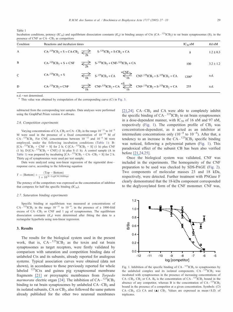
Table 1
Incubation conditions, potency (IC50) and equilibrium dissociation constants (Kd) in binding assays of Ctx (CA–125ICB2) to rat brain synaptosomes (S), in the
presence of CNF or CA–CB2 as competitors
n.d.=not determined.a This value was obtained by extrapolation of the corresponding curve (C) in Fig. 3.
Fig. 1. Inhibition of the specific binding of CA–125ICB2 to synaptosomes by
the unlabeled complex and its isolated components. CA– 125ICB2 was
incubated with synaptosomes in the presence of increasing concentrations of
CA–CB2, CB2 or CA. B0 is the concentration of CA–125ICB2 bound in the
absence of any competitor, whereas B is the concentration of CA–125ICB2
bound in the presence of a competitor at a given concentration. Symbols: (o)
CA–CB2, (g) CA and (r) CB2. Values are expressed as meanTS.D. of
triplicates.
R.M.M. dos Santos et al. / Biochimica et Biophysica Acta 1717 (2005) 27–33 29
subtracted from the corresponding test samples. Data analyses were performed
using the GraphPad Prism version 4 software.
2.6. Competition experiments
Varying concentrations of CA, CB2 or CA–CB2 in the range 10�11 to 10�6
M were used in the presence of a fixed concentration of 10�10 M of
CA–125ICB2. For CNF, concentrations between 10�12 and 10�6 M were
employed, under the following incubation conditions (Table 1): B-
[CA–125ICB2 + CNF + S] for 2 h; C-[CA–125ICB2 + S] (1 h) plus CNF
(1 h); D-[CA–125ICB2 + CNF] (1 h) plus S (1 h). A control sample (A in
Table 1) was prepared by incubating [CA–125ICB2 + CA–CB2 + S] for 2 h.
Thirty Ag of synaptosomes were used per test sample.
Data were analyzed using non-linear regression of the sigmoidal dose–
response curve, according to the following equation
Y ¼ Bottomð Þ þ Top� Bottomð Þ1þ 10 X�LogIC50ð Þ:HillSlope
The potency of the competitors was expressed as the concentration of inhibitor
that competes for half the specific binding (IC50).
2.7. Saturation binding experiments
Specific binding at equilibrium was measured at concentrations of
CA–125ICB2 in the range 10�11 to 10�9, in the presence of a 1000-fold
excess of CA–CB2 or CNF and 1 Ag of synaptosomes. The equilibrium
dissociation constants (Kd) were determined after fitting the data to a
rectangular hyperbola using non-linear regression.
3. Results
The results for the biological system used in the present
work, that is, CA–125ICB2 as the toxin and rat brain
synaptosomes as target receptors, were firstly validated by
comparison with saturation and competition binding data of
unlabeled Ctx and its subunits, already reported for analogous
systems. Typical association curves were obtained (data not
shown), in accordance to those previously reported for whole
labeled 125ICtx and guinea pig synaptosomal membrane
fragments [21] or presynaptic membranes from Torpedo
marmorata electric organ [24]. The inhibition of CA–125ICB2
binding to rat brain synaptosomes by unlabeled CA–CB2 and
its isolated subunits, CA or CB2, also followed the same pattern
already published for the other two neuronal membranes
[21,24]. CA–CB2 and CA were able to completely inhibit
the specific binding of CA–125ICB2 to rat brain synaptosomes
in a dose-dependent manner, with IC50 of 16 nM and 97 nM,
respectively (Fig. 1). The competition profile of CB2 was
concentration-dependent, as it acted as an inhibitor at
intermediate concentrations only (10�8 to 10�6). After that, a
tendency to an increase in the CA–125ICB2 specific binding
was noticed, following a polynomial pattern (Fig. 1). This
paradoxical effect of the subunit CB has been also verified
before [21,24,25].
Once the biological system was validated, CNF was
included in the experiments. The homogeneity of the CNF
preparation to be used was checked by SDS-PAGE (Fig. 2).
Two components of molecular masses 23 and 18 kDa,
respectively, were detected. Further treatment with PNGase F
clearly demonstrated that the 18-kDa component corresponded
to the deglycosylated form of the CNF monomer. CNF was,

Fig. 3. Inhibition of the specific binding of CA–125ICB2 (10� 10 M) to
synaptosomes (S) by CNF. B0 is the concentration of bound CA–125ICB2 in the
absence of any competitor, whereas B is the concentration of bound
CA–125ICB2 in the presence of a given concentration of a competitor
Unlabeled CA–CB2 was used as control. Order of addition of reagents and
incubation times (in brackets) were as follows: (n) A. [CA–125ICB2+ S+ CA–
CB2] (2 h)-reference curve; (o) B. [CA–125ICB2+ S+ CNF] (2 h); (r) C
[CA–125ICB2+ S] (1 h) plus CNF (1 h). Inset: D. (q) [CA–125ICB2+ CNF] (1
h) plus S (1 h). Values are expressed as meanTS.D. of triplicates.
Fig. 4. Saturation binding curves of CA–125ICB2 to rat brain synaptosomes, in
the presence of CA–CB2 (g) and CNF (.) as competitors. The values are
mean of triplicates. Data fitting to a rectangular hyperbole gave equilibrium
dissociation constants (Kd) of 1.2T0.3 nM and 3.2T1.2 nM for CA–CB2 and
CNF, respectively.
Fig. 2. SDS-PAGE (12.5%) of CNF (2.5 Ag per well), before and after
deglycosylation with PNGase F. The gel was silver impregnated. The molecular
masses of standard proteins (prestained broad range markers, Bio-Rad
Laboratories, Inc.) are indicated on the left side.
R.M.M. dos Santos et al. / Biochimica et Biophysica Acta 1717 (2005) 27–3330
then, used in the experiments as the natural mixture of
glycosylated and deglycosylated monomers.
Competition binding assays with synaptosomes were
performed with a fixed concentration of CA–125ICB2 and
increasing concentrations of CNF over six orders of magnitude.
The time scale of addition of reagents was varied as detailed in
Table 1 (B to D). Incubation periods of 2 h for one-step and 1
h each for two-step reactions were based on previous kinetic
experiments, where equilibrium plateaus were reached after 40
min of reaction, in typical hyperbolic curves for every case.
Competition by unlabeled CA–CB2 was run in parallel to be
taken as reference (Table 1A). Under all experimental condi-
tions, a dose-dependent inhibition was obtained for CNF (Fig.
3). When an excess of CNF was simultaneously incubated with
CA–125ICB2 and synaptosomes (Table 1B) the IC50 (100 nM)
was very close to that determined before for CA (97 nM,
Fig. 1). Pre-incubation of CA–125ICB2 and CNF followed
by the addition of synaptosomes (Table 1D) reduced the
initial ratio B/Bo to 60% (inset of Fig. 3). An IC50 of 25
nM was found. When the labeled toxin was already bound
to the synaptosomes (Table 1C), the displacement by CNF
reached a maximum of 45%, even with the highest dose of
CNF tested (1 AM) (Fig. 3). This percentage remained
unchanged even after prolonged incubation times up to 240
min (data not shown).
Saturation curves for the binding of labeled Ctx to
synaptosomes, in the presence of CA–CB2 or CNF as
competitors, are shown in Fig. 4. Equilibrium dissociation
constants (Kd) of 1.2T0.3 nM and 3.2T1.2 nM were
determined for CA–CB2 and CNF, respectively.
4. Discussion
The strategy of using a Ctx complex made of one single
isoform of CB and total CA (CA–CB2) was employed based
.
.
on the fact that CNF binds preferentially to CB2 [8].
Nevertheless, our results should agree with those performed
with mixtures of CB isoforms, considering that CB2 is the
major isoform in every venom of C. d. terrificus analyzed in
our laboratory, so far.
Although the activity of the Ctx complex was not directly
assayed after radio labeling, it was shown before that Ctx’s
structure and function were not greatly modified by the
iodination method used in the present work [21,24]. Besides,
the ready replacement of CA–125ICB2 by unlabeled Ctx

Fig. 5. Schematic representation of the interactions between Ctx, its target
receptor (TR) on pre-synaptic membranes and CNF [8], updated with the
inclusion of a new equilibrium step between CNF–CB and TR–CB.
R.M.M. dos Santos et al. / Biochimica et Biophysica Acta 1717 (2005) 27–33 31
offered indirect evidence that the procedure did not compro-
mise the binding properties of the toxin.
The presence of calcium ions in binding studies of toxins
with PLA2 activity has been a matter of discussion in the
literature. The kinetic parameters for Ctx binding to guinea pig
synaptosomes were almost unaffected by the presence of
calcium ions [21]. For Torpedo membranes, instead, concen-
trations between 1 and 10 mM CaCl2 have been considered
ideal for the specific binding of Ctx [26]. However, an increase
in the non-specific binding of Ctx was unexpectedly observed
after 10 min. This effect, absent when the calcium ions were
omitted, was attributed to the hydrolysis of the membranes due
to the addition of a large amount of PLA2 activity [26]. For rat
brain synaptosomes (present study), the non-specific binding of
CA–125ICB2 was stable up to 90 min in the presence of 1.8
mM CaCl2 (data not shown) and this concentration was
maintained throughout.
In an extensive study on the inhibition of Ctx binding to
guinea pig synaptosomal membrane fragments [21], a series of
competitors have been classified as strong, moderate, weak,
very weak and non-inhibitors, based on their strength of
inhibition. Native Ctx was among the strongest inhibitors
tested, with an IC50 around 10 nM [21,24]. A close value was
found here for rat brain synaptosomes (IC50 = 16 nM)
demonstrating an agreement in the results with both biological
preparations. As for CA, its potency seemed more dependent
on the biological membrane. CA was as potent as Ctx for
guinea pig synaptosomal membrane fragments [21] but its IC50
increased ten times for Torpedo membranes [24] and for rat
brain synaptosomes (present work). So, based on these IC50
values (around 100 nM), CA should not be considered as a
strong but a moderate inhibitor for the latter two target
membranes.
CNF was the first member of the g-type PLIs to be
purified and characterized as an oligomer of polypeptide
subunits of a single type, in its native form [8]. Later on,
when a g-PLI from Laticauda fasciata snakes was described
as composed of two different types of polypeptide subunits,
the homomeric nature of CNF was questioned [27]. At that
time, the conclusion was that a SDS-PAGE band of 20 kDa,
that appeared in the CNF preparation originally reported [7],
was most probably the second subunit of CNF. Presently, the
SDS-PAGE profiles obtained before and after deglycosylation
of CNF (Fig. 2), leave no doubt about the homomeric nature
of CNF. The positioning of CNF in the subclass II of g-type
PLIs can, thus, be no longer contested. Nevertheless, the
oligomeric aggregates of CNF are formed by a mixture of
glycosylated and deglycosylated monomers, the ratio of
which will vary among specimens of C. d. terrificus snakes
(unpublished results).
A direct competition between the target receptor and CNF
for 125ICB2 could be followed with the simultaneous incuba-
tion of CNF, CA–125ICB2 and synaptosomes (Table 1B, Fig.
3). Comparison of the IC50 value for CA–CB2 and CNF
indicates that CA–CB2 is about ten times more potent as a
competitor than CNF, under our experimental conditions (Table
1A and B). This finding would be expected, though, taking into
account the chaperone role of CA in the homologous
competition. An additional consideration is that, when CNF is
the competitor, part of 125ICB2 is sequestered from CA–125ICB2
to give rise to the CNF–125ICB2 complex (Table 1B). The
formation of this new complex, with CNF replacing CA in Ctx
[8], was more evident when CNF was pre-incubated with
CA–125ICB2, before the addition of synaptosomes (Table 1D).
Under this assay condition, the initial ratio B/Bo was reduced to
60%, due to the formation of that new stable complex in the first
incubation step. The real amount of CA–125ICB2 available to
synaptosomes in the second incubation period was, then, greatly
reduced (Table 1D, inset of Fig. 3).
Comparable IC50 values were found for CNF and CA as
competitors (100 and 97 nM, respectively). CA alone,
obviously, does not play its chaperone role. CNF can be
considered, then, as potent a competitor as CA for the Ctx
binding to rat brain synaptosomes. (Figs. 1 and 3).
Although unlabeled Ctx was taken as a reference for the
assays with CNF, the molecular models being dealt with are
quite different. The reference model comprises two ligands
(labeled and unlabeled Ctx) competing for the same receptor
site (S) on membranes, that is, two ligands versus one
receptor. Two new complexes, S-125ICB2 and S-CB2, will be
formed. As of CNF, it will not be competing for the receptor
site (S) but for the ligand (CB) instead, through two different
pathways. Firstly, the formation of the stable CNF–125ICB2
complex (Table 1B) will sequester part of 125ICB2, reducing
the amounts of the labeled ligand available to binding to the
synaptosomes. Secondly, CNF will act as an alternative
receptor for 125ICB2 already bound to the synaptosomes.
These characteristics make the competition by CNF a unique
and complex model.
In the absence of a more adequate mathematical treatment
to describe this peculiar competition, the equation of the law
of mass action, commonly employed for analysis of
radioligand binding experiments, was applied to our data.
The equilibrium dissociation constant (Kd) of S-125ICB2 was
determined as 1.2T0.3 nM in the presence of CA–CB2 as
competitor (Fig. 4, Table 1A). This value is very close to that

R.M.M. dos Santos et al. / Biochimica et Biophysica Acta 1717 (2005) 27–3332
reported for Ctx and guinea pig synaptosomes (Kd=2 nM)
[21]. When CA–CB2 was replaced by CNF, a Kd of 3.2T1.2nM was found. This increased value of Kd does not mean
that CNF is a better competitor than Ctx because the
mechanisms of competition in each case are different, as
already discussed. This Kd reflects an apparent increase in the
dissociation of S-125ICB2 due to a decrease in the real
concentration of 125ICB2 available for the membrane receptor
associated to a competition of CNF for 125ICB2 bound to the
synaptosomes.
The paradoxical concentration-dependent effect of CB for
guinea pig synaptosomal fragment [21] and for Torpedo
membranes [24] was confirmed for rat brain synaptosomes
(Fig. 1). The potentiation effect for concentrations of CB2
above 10�6 M could be possibly explained by its tendency to
form aggregates due to its highly basic character [28], its
binding to nonspecific sites on the membranes or both [25]. In
either case, the final result will be a decrease in the
concentration of free CB competing for the membrane receptor
and the induction of an increase in the ‘‘specific binding’’ of
labeled Ctx, as already suggested [24].
In conclusion, our results demonstrate that CNF is able to
totally inhibit the pharmacological action of Ctx and that this
action can be partially extended to the toxin already bound to
synaptosomes. The schematic model for the interaction of Ctx
with its target receptor and CNF proposed before [8] can thus
be updated with the inclusion of an additional equilibrium
step between CNF–CB and TR–CB, representing the
competition between TR and CNF for CB (Fig. 5). CNF, in
its natural form or modified, may constitute a useful tool to
investigate the mechanism of action and intoxication by Ctx
and other PLA2-toxins on targets in the nervous system, as
well as in the development of a future therapeutics for snake
bites.
Acknowledgements
We thank Ms. A.C. Valentim for technical assistance, Dr.
L.G.D. Heneine and Mrs. P. Cotta for their help in the
preparation of the affinity column and Dr. M. Richardson for
language review. This work was granted by FAPEMIG (EDT
24000/01; CBB 492/03) and by CNPq with fellowships to
R.M.M. dos Santos (141781/2000-9) and to C.L. Fortes-Dias
(300767/2003-0).
References
[1] R.A. Hendon, H. Fraenkel-Conrat, Biological roles of the two components
of crotoxin, Proc. Natl. Acad. Sci. U. S. A. 68 (1971) 1560–1563.
[2] K. Rubsamen, H. Breithaupt, E. Habermann, Biochemistry and pharma-
cology of the crotoxin complex, Naunyn-Schmiedeberg’s Arch. Pharma-
col. 270 (1971) 274–288.
[3] C. Bon, C. Bouchier, V. Choumet, G. Faure, M.S. Jiang, M.P.
Lambezat, F. Radvanyi, B. Saliou, B, Crotoxin, half-century of
investigations on a phospholipase A2 neurotoxin, Acta Physiol. Latinoam.
39 (1989) 439–448.
[4] G. Faure, C. Bon, Several isoforms of crotoxin are present in individual
venoms from the South American rattlesnake Crotalus durissus terrificus,
Toxicon 25 (1987) 229–234.
[5] G. Faure, C. Bon, Crotoxin, a phospholipase A2 neurotoxin from
the South American rattlesnake Crotalus durissus terrificus: purifi-
cation of several isoforms and comparison of their molecular
structure and of their biological activities, Biochemistry 27 (1988)
730–738.
[6] C.L. Fortes-Dias, C.R. Diniz, E. Kochva, Neutralization of Crotalus
durissus terrificus (South American rattlesnake) venom and crotoxin by
homologous plasma, Cienc. Cult. 42 (1990) 501–506.
[7] C.L. Fortes-Dias, B.C.B. Fonseca, E. Kochva, C.R. Diniz, Purification
and properties of an antivenom factor from the plasma of the South
American rattlesnake (Crotalus durissus terrificus), Toxicon 29 (1991)
997–1008.
[8] C.L. Fortes-Dias, Y. Lin, J. Ewell, C.R. Diniz, T.Y. Liu, A phospholipase
A2 inhibitor from the plasma of the South American rattlesnake (Crotalus
durissus terrificus). Protein structure, genomic structure, and mechanism
of action, J. Biol. Chem. 269 (1994) 15646–15651.
[9] J. Perales, C. Villela, G.B. Domont, V. Choumet, B. Saliou, H.
Moussatche, C. Bon, G. Faure, Molecular structure and mechanism of
action of the crotoxin inhibitor from Crotalus durissus terrificus serum,
Eur. J. Biochem. 227 (1995) 19–26.
[10] R.D. Dunn, K.W. Broady, Snake inhibitors of phospholipase A2 enzymes,
Biochim. Biophys. Acta 1533 (2001) 29–37.
[11] C.L. Fortes-Dias, Endogenous inhibitors of snake venom phospholipases
A2 in the blood plasma of snakes, Toxicon 40 (2002) 481–484.
[12] S. Lizano, G. Domont, J. Perales, Natural phospholipase A2 myotoxin
inhibitor proteins from snakes, mammals and plants, Toxicon 42 (2003)
963–977.
[13] N. Ohkura, H. Okuhara, S. Inoue, K. Ikeda, K. Hayashi, Purification and
characterization of three distinct types of phospholipase A2 inhibitors
from the blood plasma of the Chinese mamushi, Agkistrodon blomhoffii
siniticus, Biochem. J. 15 (1997) 527–531.
[14] C.L. Fortes-Dias, M.L.D. Jannotti, F.J.L. Franco, A. Magalhaes, C.R.
Diniz, Studies on the specificity of CNF, a phospholipase A2 inhibitor
isolated from the blood plasma of the South American rattlesnake
(Crotalus durissus terrificus): I. Interaction with PLA2 from Lachesis
muta muta snake venom, in the blood plasma of snakes, Toxicon 37
(1999) 1747–1759.
[15] G. Faure, C. Villela, J. Perales, C. Bon, Interaction of the neurotoxic and
nontoxic secretory phospholipases A2 with the crotoxin inhibitor from
Crotalus serum, Eur. J. Biochem. 267 (2000) 4799–4808.
[16] U.K. Laemmli, Cleavage of structural proteins during the assembly of the
head of bacteriophage T4, Nature 227 (1970) 680–685.
[17] K. Slotta, H. Fraenkel-Conrat, Snake venoms II. Nature of the sulphur
union, Ber. Dtsch. Chem. Ges. 71B (1938) 1076–1081.
[18] C. Seki, J.C. Vidal, A. Barrio, Purification of gyroxin from a South
American rattlesnake (Crotalus durissus terrificus), Toxicon 18 (1980)
235–247.
[19] F.C. Greenwood, W.M. Hunter, J.S. Glover, The preparation of 131I-
labelled human growth hormone of high specific radioactivity, Biochem.
J. 89 (1963) 114–123.
[20] A. Johnstone, R. Thorpe, Immunochemistry in Practice, Blackwell
Scientific Publications, Oxford, 1982, pp. 107–109.
[21] L.L. Degn, C.S. Seebart, I.I. Kaiser, Specific binding of crotoxin to
brain synaptosomes and synaptosomal membranes, Toxicon 29 (1991)
973–988.
[22] P.R. Dunkley, J.W. Heath, S.M. Harrison, P.E. Jarvie, P.J. Glenfield, J.A.
Rostas, A rapid Percoll gradient procedure for isolation of synaptosomes
directly from an S1 fraction: homogeneity and morphology of subcellular
fractions, Brain Res. 441 (1998) 59–71.
[23] O.H. Lowry, N.J. Rosebrough, A.L. Farral, R.J. Randall, Protein
measurement with the Folin phenol reagent, J. Biol. Chem. 193 (1951)
265–275.
[24] I. Kri”aj, G. Faure, F. Gubenek, C. Bon, Neurotoxic phospholipases A2
ammodytoxin and crotoxin bind to distinct high-affinity protein
acceptors in Torpedo marmorata electric organ, Biochemistry 36
(1997) 2779–2787.
[25] I. Kri”aj, G. Faure, F. Gubenxek, C. Bon, Re-examination of crotoxin–
membrane interactions, Toxicon 34 (1996) 1003–1009.

R.M.M. dos Santos et al. / Biochimica et Biophysica Acta 1717 (2005) 27–33 33
[26] E. Delot, C. Bon, Model for the interaction of crotoxin, a phospholipase
A2 neurotoxin, with presynaptic membranes, Biochemistry 32 (1993)
10708–10713.
[27] N. Ohkura, Y. Kitahara, S. Inoue, K. Ikeda, K. Hayashi, Isolation and
amino acid sequence of a phospholipase A2 inhibitor from the blood
plasma of the Sea Krait, Laticauda semifasciata, J. Biochem. 125 (1999)
375–382.
[28] E. Habermann, H. Breithaupt, Mini-review—The crotoxin complex, an
example of biochemical and pharmacological protein complementation,
Toxicon 16 (1978) 19–30.