Intermanual Differences in Movement-related Interhemispheric Inhibition
10
Intermanual Differences in Movement-related Interhemispheric Inhibition Julie Duque 1,2 , Nagako Murase 1,3 , Pablo Celnik 1 , Friedhelm Hummel 1 , Michelle Harris-Love 1 , Riccardo Mazzocchio 1,4 , Etienne Olivier 2 , and Leonardo G. Cohen 1 Abstract & Interhemispheric inhibition (IHI ) between motor cortical areas is thought to play a critical role in motor control and could influence manual dexterity. The purpose of this study was to investigate IHI preceding movements of the domi- nant and nondominant hands of healthy volunteers. Move- ment-related IHI was studied by means of a double-pulse transcranial magnetic stimulation protocol in right-handed in- dividuals in a simple reaction time paradigm. IHI targeting the motor cortex contralateral (IHI c ) and ipsilateral (IHI i ) to each moving finger was determined. IHI c was comparable after the go signal, a long time preceding movement onset, in both hands. Closer to movement onset, IHI c reversed into facili- tation for the right dominant hand but remained inhibitory for left nondominant hand movements. IHI i displayed a nearly constant inhibition with a trough early in the premovement period in both hands. In conclusion, our results unveil a more important modulation of interhemispheric interactions dur- ing generation of dominant than nondominant hand move- ments. This modulation essentially consisted of a shift from a balanced IHI at rest to an IHI predominantly directed toward the ipsilateral primary motor cortex at movement onset. Such a mechanism might release muscles from inhibition in the contralateral primary motor cortex while preventing the occur- rence of the mirror activity in ipsilateral primary motor cortex and could therefore contribute to intermanual differences in dexterity. & INTRODUCTION Behavioral studies of manual dexterity have reported a superior proficiency of the right hand under a variety of motor conditions (Roy, Bryden, & Cavill, 2003; Triggs, Calvanio, Levine, Heaton, & Heilman, 2000; Triggs, Calvanio, & Levine, 1997; Todor & Kyprie, 1980; Peters & Durding, 1978) in approximately 90% of the population (Coren & Porac, 1977). However, the neural bases of these intermanual differences in motor control remain unclear. One possibility is that this asymmetry in manual dexterity relates to differences in interhemispheric con- nectivity. Anatomical and physiological studies in ani- mals (Aboitiz & Montiel, 2003; Rouiller et al., 1994) and humans (Daskalakis, Christensen, Fitzgerald, Roshan, & Chen, 2002; Di Lazzaro et al., 1999; Gerloff et al., 1998; Meyer, Roricht, & Woiciechowsky, 1998; Thut et al., 1997; Meyer, Roricht, Grafin von Einsiedel, Kruggel, & Weindl, 1995; Netz, Ziemann, & Homberg, 1995; Ferbert et al., 1992) have documented interhemispheric inter- actions between both motor cortices. These interactions are predominantly inhibitory (interhemispheric inhibi- tion [IHI]) and can be studied by means of a double- pulse transcranial magnetic stimulation (TMS) protocol. The latter uses two magnetic stimulators to investigate the effect of a conditioning TMS pulse over one motor cortex on the amplitude of motor-evoked potentials (MEPs) elicited by another TMS pulse applied a few milliseconds later on the opposite motor cortex (Chen, 2004; Daskalakis et al., 2002; Ferbert et al., 1992). The strongest IHI is typically obtained when the conditioning stimulation (CS) precedes the test stimulation (TS) by about 10 msec. IHI is mediated predominantly through transcallosal connections (Daskalakis et al., 2002; Di Lazzaro et al., 1999; Meyer et al., 1995, 1998) with a probable subcortical contribution (Gerloff et al., 1998). The view that asymmetries in hand motor control could relate to hemispheric differences in IHI has motivated human double-pulse TMS investigations comparing IHI from dominant-to-nondominant and from nondominant- to-dominant hemispheres at rest (De Gennaro et al., 2004; Netz, 1999; Salerno & Georgesco, 1996; Netz et al., 1995). Although some studies reported a stronger dominant- to-nondominant hemisphere IHI (Netz, 1999; Netz et al., 1995), more detailed investigations testing a large range of CS–TS intervals and intensities revealed no differences (De Gennaro et al., 2004; Salerno & Georgesco, 1996). 1 National Institutes of Health, 2 Universite ´ catholique de Louvain, 3 Tokushima University, 4 Universita ´ di Siena D 2007 Massachusetts Institute of Technology Journal of Cognitive Neuroscience 19:2, pp. 204–213
-
Upload
independent -
Category
Documents
-
view
3 -
download
0
Transcript of Intermanual Differences in Movement-related Interhemispheric Inhibition

Intermanual Differences in Movement-relatedInterhemispheric Inhibition
Julie Duque1,2, Nagako Murase1,3, Pablo Celnik1,Friedhelm Hummel1, Michelle Harris-Love1, Riccardo Mazzocchio1,4,
Etienne Olivier2, and Leonardo G. Cohen1
Abstract
& Interhemispheric inhibition (IHI) between motor corticalareas is thought to play a critical role in motor control andcould influence manual dexterity. The purpose of this studywas to investigate IHI preceding movements of the domi-nant and nondominant hands of healthy volunteers. Move-ment-related IHI was studied by means of a double-pulsetranscranial magnetic stimulation protocol in right-handed in-dividuals in a simple reaction time paradigm. IHI targeting themotor cortex contralateral (IHIc) and ipsilateral (IHIi) to eachmoving finger was determined. IHIc was comparable after thego signal, a long time preceding movement onset, in bothhands. Closer to movement onset, IHIc reversed into facili-tation for the right dominant hand but remained inhibitory
for left nondominant hand movements. IHIi displayed a nearlyconstant inhibition with a trough early in the premovementperiod in both hands. In conclusion, our results unveil a moreimportant modulation of interhemispheric interactions dur-ing generation of dominant than nondominant hand move-ments. This modulation essentially consisted of a shift from abalanced IHI at rest to an IHI predominantly directed towardthe ipsilateral primary motor cortex at movement onset. Sucha mechanism might release muscles from inhibition in thecontralateral primary motor cortex while preventing the occur-rence of the mirror activity in ipsilateral primary motor cortexand could therefore contribute to intermanual differences indexterity. &
INTRODUCTION
Behavioral studies of manual dexterity have reported asuperior proficiency of the right hand under a variety ofmotor conditions (Roy, Bryden, & Cavill, 2003; Triggs,Calvanio, Levine, Heaton, & Heilman, 2000; Triggs,Calvanio, & Levine, 1997; Todor & Kyprie, 1980; Peters &Durding, 1978) in approximately 90% of the population(Coren & Porac, 1977). However, the neural bases of theseintermanual differences in motor control remain unclear.
One possibility is that this asymmetry in manualdexterity relates to differences in interhemispheric con-nectivity. Anatomical and physiological studies in ani-mals (Aboitiz & Montiel, 2003; Rouiller et al., 1994) andhumans (Daskalakis, Christensen, Fitzgerald, Roshan, &Chen, 2002; Di Lazzaro et al., 1999; Gerloff et al., 1998;Meyer, Roricht, & Woiciechowsky, 1998; Thut et al.,1997; Meyer, Roricht, Grafin von Einsiedel, Kruggel, &Weindl, 1995; Netz, Ziemann, & Homberg, 1995; Ferbertet al., 1992) have documented interhemispheric inter-actions between both motor cortices. These interactionsare predominantly inhibitory (interhemispheric inhibi-
tion [IHI]) and can be studied by means of a double-pulse transcranial magnetic stimulation (TMS) protocol.The latter uses two magnetic stimulators to investigatethe effect of a conditioning TMS pulse over one motorcortex on the amplitude of motor-evoked potentials(MEPs) elicited by another TMS pulse applied a fewmilliseconds later on the opposite motor cortex (Chen,2004; Daskalakis et al., 2002; Ferbert et al., 1992). Thestrongest IHI is typically obtained when the conditioningstimulation (CS) precedes the test stimulation (TS) byabout 10 msec. IHI is mediated predominantly throughtranscallosal connections (Daskalakis et al., 2002; DiLazzaro et al., 1999; Meyer et al., 1995, 1998) with aprobable subcortical contribution (Gerloff et al., 1998).
The view that asymmetries in hand motor control couldrelate to hemispheric differences in IHI has motivatedhuman double-pulse TMS investigations comparing IHIfrom dominant-to-nondominant and from nondominant-to-dominant hemispheres at rest (De Gennaro et al., 2004;Netz, 1999; Salerno & Georgesco, 1996; Netz et al., 1995).Although some studies reported a stronger dominant-to-nondominant hemisphere IHI (Netz, 1999; Netz et al.,1995), more detailed investigations testing a large rangeof CS–TS intervals and intensities revealed no differences(De Gennaro et al., 2004; Salerno & Georgesco, 1996).
1National Institutes of Health, 2Universite catholique de Louvain,3Tokushima University, 4Universita di Siena
D 2007 Massachusetts Institute of Technology Journal of Cognitive Neuroscience 19:2, pp. 204–213
One possible explanation for the lack of consistentevidence for brain asymmetries in IHI provided by thesestudies is that quantitative determinations of IHI testedat rest do not reflect IHI operating in association withperformance of voluntary movements. Therefore, it isconceivable that evaluation of IHI in the process of gen-eration of voluntary movements by either hand couldprovide additional insights on the contribution of IHIto intermanual asymmetries in dexterity.
We have previously shown that IHI targeting a motorcortex (M1) engaged in the preparation of a contralat-eral hand movement (IHIc) is profound immediately afterthe go signal in a reaction time (RT) paradigm (Duque,Hummel, et al., 2005; Murase, Duque, Mazzocchio, &Cohen, 2004). As movement onset approaches, IHIc isreleased substantially (Duque, Hummel, et al., 2005;Murase et al., 2004). In contrast, IHI targeting the ipsi-lateral M1 (IHIi) remains deep throughout the move-ment preparation. Abnormalities in these inhibitorymechanisms correlate with the magnitude of motorimpairment in the paretic hand of patients with chronicstroke (Murase et al., 2004), more prone to performmirror movements (Kim et al., 2003). It is then possiblethat proper movement-related modulation of IHIc andIHIi may contribute to the ability of healthy subjectsto perform unilateral finger motions without mirroring(Duque, Mazzocchio, et al., 2005; Swinnen, Dounskaia,& Duysens, 2002; Leocani, Cohen, Wassermann, Ikoma,& Hallett, 2000).
Here, we used a standard double-pulse TMS protocol(Di Lazzaro et al., 1999; Gerloff et al., 1998; Meyer et al.,1998; Ferbert et al., 1992) to study movement-relatedmodulation of IHI (IHIc and IHIi) for right and left handmovements in right-handed individuals. We found a fun-damental difference in the way IHI is modulated bymovements of the dominant and nondominant hands.
METHODS
Subjects
Thirteen healthy right-handed individuals (Oldfield,1971) participated in this study (53 ± 18.1 years old;six women and seven men). All subjects gave theirwritten informed consent, and the protocol was ap-proved by the National Institute of Neurological Disor-ders and Stroke Institutional Review Board.
Experimental Procedure
Each subject sat in front of a computer screen and per-formed voluntary index finger movements with eitherthe right or left hand. The task consisted of a simple RTparadigm in which they were instructed to performindex finger abduction motions as fast as possible inresponse to a go signal. After a short familiarization ses-sion, subjects participated in four separate sessions on
different days (two for each hand, see below) in whichIHI was tested using TMS at different time intervalspreceding movement onset (Duque, Hummel, et al.,2005; Murase et al., 2004). Practice trials at the beginningof each session were used to characterize each individ-ual’s RT in the absence of TMS. RT was defined as thetime between the go signal and the onset of the elec-tromyographic (EMG) response (at the time when it firstexceeded 100 AV peak to peak). Surface EMG was re-corded from the first dorsal interosseus (FDI) muscle ofboth hands in a time window that comprised 500 msecbefore and 1000 msec after the TS (see below). Afteramplification and bandpass filtering (50–2000 Hz) usinga Counterpoint electromyography machine (DantecElectronics, Skovlunde, Denmark), the signal was digi-tized (sampling rate 5000 Hz) and stored on a personalcomputer for off-line analysis using a data acquisition-analysis system written in LabView (Kaelin-Lang & Cohen,2000). To check whether the target muscles were at restat the time of TMS for each trial, the EMG traces wereinspected visually for 200 msec preceding stimulation.Trials with background EMG activity exceeding 100 AVin this 200-msec window were excluded from analysis.
Measurement of Task-relatedInterhemispheric Inhibition
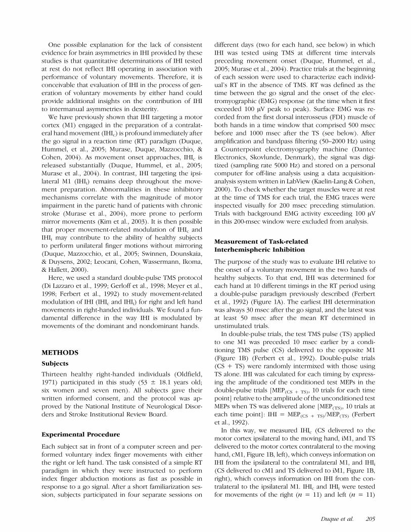
The purpose of the study was to evaluate IHI relative tothe onset of a voluntary movement in the two hands ofhealthy subjects. To that end, IHI was determined foreach hand at 10 different timings in the RT period usinga double-pulse paradigm previously described (Ferbertet al., 1992) (Figure 1A). The earliest IHI determinationwas always 30 msec after the go signal, and the latest wasat least 50 msec after the mean RT determined inunstimulated trials.
In double-pulse trials, the test TMS pulse (TS) appliedto one M1 was preceded 10 msec earlier by a condi-tioning TMS pulse (CS) delivered to the opposite M1(Figure 1B) (Ferbert et al., 1992). Double-pulse trials(CS + TS) were randomly intermixed with those usingTS alone. IHI was calculated for each timing by express-ing the amplitude of the conditioned test MEPs in thedouble-pulse trials [MEP(CS + TS), 10 trials for each timepoint] relative to the amplitude of the unconditioned testMEPs when TS was delivered alone [MEP(TS), 10 trials ateach time point]: IHI = MEP(CS + TS)/MEP(TS) (Ferbertet al., 1992).
In this way, we measured IHIc (CS delivered to themotor cortex ipsilateral to the moving hand, iM1, and TSdelivered to the motor cortex contralateral to the movinghand, cM1, Figure 1B, left), which conveys information onIHI from the ipsilateral to the contralateral M1, and IHIi
(CS delivered to cM1 and TS delivered to iM1, Figure 1B,right), which conveys information on IHI from the con-tralateral to the ipsilateral M1. IHIc and IHIi were testedfor movements of the right (n = 11) and left (n = 11)
Duque et al. 205

hands of healthy subjects in four sessions on separatedays. In most subjects, the right hand was tested beforethe left hand, but investigation of IHIc and IHIi wasrandomized in all subjects. Four subjects left the countrybefore the end of the study and were only tested for theirright (n = 2) or left hand (n = 2). As a result, statisticalcomparisons of IHI between the two hand movementswere performed on 9 subjects.
Transcranial Magnetic Stimulation
TMS was delivered from two Magstim 200 magneticstimulators (Magstim, Whitland, Dyfed, UK), eachconnected to a figure-eight coil. The coil for the TS(70-mm diameter) was placed tangentially over the M1
with the handle pointing backward and laterally at a 458angle away from the midline, approximately perpendic-ular to the central sulcus. The CS coil (35-mm internaldiameter) was oriented at 908 relative to the midsagittalline, a position thought to elicit predominantly D and I1waves (Sakai et al., 1997; Schnitzler, Kessler, & Benecke,1996; Werhahn et al., 1994). The orientation and smallsize of the CS coil were chosen because, in mostsubjects, it was not possible to place two large size coilsat 458 to the midsagittal line. This setup enabled us tooptimize baseline IHI determination in all subjects. Theoptimal scalp position for eliciting a contralateral MEPwas identified and marked on a head cap placed on thepatient’s scalp. Resting motor threshold (rMT) wasdetermined at this site and defined as the minimumstimulus intensity that produced MEPs � 50 AV peak topeak in at least 5 of 10 trials (Rossini et al., 1994).
For TS, we used the intensity of stimulation requiredto evoke MEP amplitudes of about 0.5 to 1.5 mV at rest(Boroojerdi, Diefenbach, & Ferbert, 1996; Ferbert et al.,1992), and for CS, we used the intensity required toevoke IHI of approximately 50% in double-pulse trials atrest (Murase et al., 2004). In this way, we ensured acomparable magnitude of IHI at rest in the four exper-imental sessions (see Results).
Determination of PremovementInterhemispheric Inhibition
To quantify IHI preceding movement onset, we usedthree measures (Duque, Hummel, et al., 2005; Muraseet al., 2004): (a) maximum IHI (IHImax), defined as themaximal (deepest) IHI identified in a given individual inany of the timings falling at least 70 msec after the gosignal in the RT paradigm. We chose this delay (70 msec)to make sure IHImax reflected processing within motorregions at a time they were most likely already involvedin movement preparation. (b) IHI immediately beforemovement onset (IHIbef-mvt) was defined by identifyingfirst the timings at which the TS fell after EMG onset in aproportion of up to 50% of the trials. We then includedin the analysis only the trials in which TS fell before EMGonset at the defined intervals. Finally, the last variablewas (c) IHI around movement onset (IHImvt-onset), de-fined as IHI determined at the timing immediatelypreceding (but at least 5 msec before) the mean RT inunstimulated trials; all trials at that particular interval,including those where TMS fell after EMG onset, wereconsidered for analysis. These three measures werecomputed for IHIc (IHIc max, IHIc bef-mvt, IHIc mvt-onset)and IHIi (IHIi max, IHIi bef-mvt, IHIi mvt-onset) for move-ments of the right and left hands of subjects.
To unveil a possible shift in the balance between theIHI exerted by each motor cortex concomitantly duringmovement preparation, we computed the differencebetween IHIi and IHIc at all timings (IHIi � c max, IHIi �
c bef-mvt, IHIi � c mvt-onset) for movements of the left and
Figure 1. (A) IHI was evaluated in the process of generation of a
voluntary movement by the right or left hand in a simple RT paradigm.
Patients responded with a brisk index finger abduction motion to a
go signal. The onset of the EMG response from the FDI musclecharacterized the RT. IHI was determined at 10 different timings
during the RT period. (B) IHIc (left) was studied by applying a CS to
the iM1 (M1 ipsilateral to the moving hand) 10 msec preceding a TSapplied to the cM1 (M1 contralateral to the moving hand). IHIi (right)
was studied by applying CS to the cM1 and TS to the iM1. IHIc and
IHIi were each measured for movements of the right and left hands
of subjects in a total of four separate sessions. See Methods for detailson the size and orientation of CS and TS coils. IHI was expressed as
the amplitude of the conditioned test MEPs [MEPs(CS + TS)] in the
double-pulse trials relative to the amplitude of the unconditioned
MEPs [MEPs(TS)] when TS was delivered alone for each IHIdetermination: MEP(CS + TS)/MEP(TS) (Ferbert et al., 1992).
206 Journal of Cognitive Neuroscience Volume 19, Number 2

right hands. An index close to 0 would reveal compara-ble levels of IHIc and IHIi (balanced IHI); positive values(IHIi �� IHIc) would reflect deeper IHIc than IHIi, andnegative values (IHIi �� IHIc) would reflect deeperIHIi than IHIc. As a consequence, a specific release inIHIc with respect to IHIi during movement preparationshould result in a progressive decrease in IHIi � c.
Statistical Analysis
Log transformations were applied when data were notnormally distributed (Kolmogorov-Smirnov tests). RTsand corticospinal excitability at rest were analyzedusing one-way repeated measures analyses of variance(ANOVAs) with hand (left, right) as factor. Separate re-peated measures ANOVAs with factors moving hand (left,right) and timing (IHIrest, IHImax, IHIbef-mvt, IHImvt-onset)were used to analyze premovement IHI targeting thecM1 (IHIc) and iM1 (IHIi), IHIi � c, and corresponding CSand TS MEP amplitudes. Post hoc pairwise comparisonswere performed using Tukey and paired t tests whenrequired. Data are expressed as mean ± SD. Because theage of subjects ranged from 27 to 79 years old, we couldinvestigate the effect of age on premovement IHI bymeans of the Pearson correlation procedure. Part ofthese data has served as control for previous studiesinvestigating the effect of IHI on motor function in elderpatients with chronic stroke (Duque, Hummel, et al.,2005; Murase et al., 2004).
RESULTS
Reaction Time, Corticospinal Excitability, andInterhemispheric Inhibition at Rest
There was no significant difference between RTs forthe left (199 ± 27.0 msec) and right hands (179 ±33.7 msec). In addition, the resting motor threshold was
found comparable in the right (43% ± 5.9%) and left M1(44% ± 6.9%). The intensity used for CS and TS was alsosimilar for the two hand movement conditions (seeTable 1; ANOVA, ns). Finally, as required by the exper-imental design (see Methods), the magnitude of IHIrest
was comparable across all sessions as well as wereMEP(TS) and MEP(CS) amplitudes (Tables 2 and 3).
Modulation of Interhemispheric InhibitionTargeting the Primary Motor Cortex Contralateralto the Moving Hand
IHIc conveys information on the IHI originating in iM1and targeting cM1, the motor cortex controlling the mov-ing hand. IHIc max, IHIc bef-mvt, and IHIc mvt-onset weremeasured 97 ± 20.9, 160 ± 28.3, and 159 ± 30.2 msec,respectively, after the go signal for movements of theright hand and 107 ± 23.1, 165 ± 28.2, 180 ± 24.1 msecfor movements of the left hand (ANOVA, ns). Whensubjects performed right index finger abductions, IHIc
was deep close to the go signal and changed intofacilitation around movement onset (Figure 2). Withleft index finger abduction, IHIc was also deep closeto the go signal but did not turn into facilitation atmovement onset (Figure 2). This is shown by a sig-nificant main effect of timing (F = 13.7, p < .001) anda Significant Timing � Moving Hand interaction (F =5.4, p = .01). Although IHIc max was comparable with
Table 1. CS and TS Intensities (Mean ± SD)
Moving Hand
Right Hand(n = 11)
Left Hand(n = 11)
p Value(Right/Left)
IHIc
CS intensity (% output) 58.8 ± 8.5 57.6 ± 10.9 ns
TS intensity (% output) 52.1 ± 10.3 49.9 ± 10.7 ns
IHIi
CS intensity (% output) 57.6 ± 12.2 54.9 ± 8.8 ns
TS intensity (% output) 49.6 ± 9.8 49.8 ± 8.1 ns
Four subjects were only tested for their left (n = 2) or right (n = 2)hand, and statistics are therefore reported for the nine subjects testedon both hands.
Table 2. IHI Targeting the cM1 (IHIc) and MEP Amplitudesof CS and TS (Mean ± SD)
Moving Hand
IHIc [MEP(CS + TS)/MEP(TS)]
Right Hand(n = 11)
Left Hand(n = 11)
p Value(Right/Left)
IHIc rest (mV) 0.63 ± 0.16 0.61 ± 0.17 ns
MEP(CS) 1.63 ± 1.03 2.39 ± 1.87 ns
MEP(TS) 1.06 ± 0.54 1.35 ± 0.49 ns
IHIc max (mV) 0.60 ± 0.18 0.59 ± 0.21 ns
MEP(CS) 1.73 ± 1.26 2.16 ± 1.88 ns
MEP(TS) 1.82 ± 0.77 1.79 ± 1.10 ns
IHIc bef-mvt (mV) 1.12 ± 0.25* 0.80 ± 0.23 ns (0.06)
MEP(CS) 1.89 ± 1.18 2.20 ± 2.13 ns
MEP(TS) 1.99 ± 0.82 2.32 ± 1.38 ns
IHIc mvt-onset (mV) 1.28 ± 0.43* 0.85 ± 0.21 <0.02
MEP(CS) 1.96 ± 1.26 2.74 ± 2.19 ns
MEP(TS) 2.51 ± 1.44 3.33 ± 1.69 ns
* = IHIc significantly different from rest; ns = non-significant. Notethat 4 subjects were only tested for their left (n = 2) or right (n = 2)hand and statistics are therefore reported for the 9 subjects tested onboth hands.
Duque et al. 207

movements of both hands, IHIc bef-mvt and IHIc mvt-onset
were deeper with left than with right hand movements( p = .06 and p < .02, respectively, see Table 2 andFigure 2). MEP(TS) and MEP(CS) amplitudes were simi-lar in left and right hands at any premovement timing(Figure 2, bottom).
Modulation of Interhemispheric InhibitionTargeting the Primary Motor Cortex Ipsilateralto the Moving Hand
IHIi conveys information on the IHI originating in cM1and targeting iM1, the motor cortex controlling theresting hand. IHIi max, IHIi bef-mvt, and IHIi mvt-onset
were measured 97 ± 18.4, 166 ± 27.4, and 163 ±29.3 msec, respectively, after the go signal for right handmovements and 99 ± 29.5, 164 ± 27.2, and 180 ±24.1 msec for movements of the left hand (ANOVA,ns). Premovement IHIi was characterized by a deepinhibition throughout movement preparation, whichwas indistinguishable for both hands (Figure 3). A two-way repeated measures ANOVA showed a significantmain effect of timing (F = 6.4, p = .003) withoutinteraction (F = 1.4, p = .3). IHIi max was stronger whencompared with rest or immediately preceding move-
Table 3. IHIi and MEP Amplitudes of CS and TS (Mean ± SD)
Moving Hand
IHIi [MEP(CS + TS)/MEP(TS)]
Right Hand(n = 11)
Left Hand(n = 11)
p Value(Right/Left)
IHIi rest (mV) 0.50 ± 0.19 0.53 ± 0.20 ns
MEP(CS) 1.37 ± 1.03 2.27 ± 1.86 ns
MEP(TS) 0.95 ± 0.39 1.10 ± 0.44 ns
IHIi max (mV) 0.30 ± 0.16* 0.40 ± 0.20* ns
MEP(CS) 1.65 ± 1.56 2.52 ± 1.63 ns
MEP(TS) 1.13 ± 0.94 1.46 ± 0.85 ns
IHIi bef-mvt (mV) 0.54 ± 0.30 0.64 ± 0.50 ns
MEP(CS) 2.26 ± 1.22 3.28 ± 1.69 ns
MEP(TS) 0.94 ± 0.60 1.72 ± 1.43 ns
IHIi mvt-onset (mV) 0.43 ± 0.25 0.57 ± 0.32 ns
MEP(CS) 2.62 ± 1.17 3.94 ± 1.85 ns
MEP(TS) 1.06 ± 0.68 1.56 ± 1.14 ns
* = IHIi significantly different from rest; ns = non-significant. Notethat 4 subjects were only tested for their left (n = 2) or right (n = 2)hand and statistics are therefore reported for the 9 subjects tested onboth hands.
Figure 2. Individual values of IHIc rest and in the process of generation
of voluntary index finger abduction motions with the right (n = 11)
or left (n = 11) hand. Note that four subjects were only tested fortheir left (n = 2) or right (n = 2) hand, and statistics are therefore
reported on the remaining nine subjects tested on both hands. IHIc max
depicts maximal inhibition soon after the go signal. IHIc max was
comparable to IHIc rest with movements of both hands. In contrast,premovement IHIc bef-mvt and IHIc mvt-onset were more prominent with
movements of the left than right hand ( p = .06 and p < .02,
respectively), in the presence of comparable MEP(TS) amplitudes.
Figure 3. Individual values of IHIi rest and in the process of
generation of voluntary index finger abduction motions with the
right (n = 11) or left (n = 11) hand. Note that four subjects wereonly tested for their left (n = 2) or right (n = 2) hand and statistics
are therefore reported on the remaining nine subjects tested on
both hands. IHIi max depicts maximal inhibition soon after the go
signal. IHIi max was deeper than IHIi rest or IHIi bef-mvt for movementsof both hands of subjects (*p < .05). IHIi and the amplitude of
MEP(TS) (elicited by stimulation of iM1) were comparable in both
hands of subjects at all premovement timings.
208 Journal of Cognitive Neuroscience Volume 19, Number 2
ments (IHIc bef-mvt) of either hand (Table 3 and Figure 3,all p < .05). MEP(TS) and MEP(CS) amplitudes weresimilar in left and right hands at any premovementtiming (Figure 3).
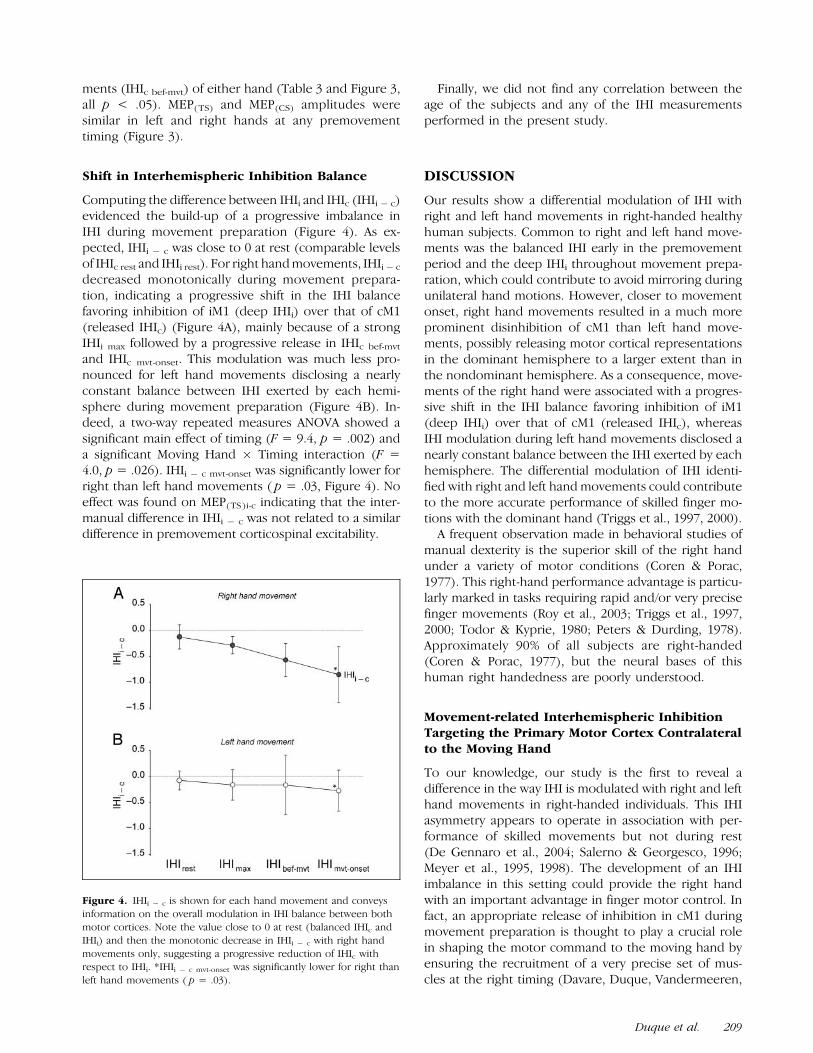
Shift in Interhemispheric Inhibition Balance
Computing the difference between IHIi and IHIc (IHIi � c)evidenced the build-up of a progressive imbalance inIHI during movement preparation (Figure 4). As ex-pected, IHIi � c was close to 0 at rest (comparable levelsof IHIc rest and IHIi rest). For right hand movements, IHIi � c
decreased monotonically during movement prepara-tion, indicating a progressive shift in the IHI balancefavoring inhibition of iM1 (deep IHIi) over that of cM1(released IHIc) (Figure 4A), mainly because of a strongIHIi max followed by a progressive release in IHIc bef-mvt
and IHIc mvt-onset. This modulation was much less pro-nounced for left hand movements disclosing a nearlyconstant balance between IHI exerted by each hemi-sphere during movement preparation (Figure 4B). In-deed, a two-way repeated measures ANOVA showed asignificant main effect of timing (F = 9.4, p = .002) anda significant Moving Hand � Timing interaction (F =4.0, p = .026). IHIi � c mvt-onset was significantly lower forright than left hand movements ( p = .03, Figure 4). Noeffect was found on MEP(TS)i-c indicating that the inter-manual difference in IHIi � c was not related to a similardifference in premovement corticospinal excitability.
Finally, we did not find any correlation between theage of the subjects and any of the IHI measurementsperformed in the present study.
DISCUSSION
Our results show a differential modulation of IHI withright and left hand movements in right-handed healthyhuman subjects. Common to right and left hand move-ments was the balanced IHI early in the premovementperiod and the deep IHIi throughout movement prepa-ration, which could contribute to avoid mirroring duringunilateral hand motions. However, closer to movementonset, right hand movements resulted in a much moreprominent disinhibition of cM1 than left hand move-ments, possibly releasing motor cortical representationsin the dominant hemisphere to a larger extent than inthe nondominant hemisphere. As a consequence, move-ments of the right hand were associated with a progres-sive shift in the IHI balance favoring inhibition of iM1(deep IHIi) over that of cM1 (released IHIc), whereasIHI modulation during left hand movements disclosed anearly constant balance between the IHI exerted by eachhemisphere. The differential modulation of IHI identi-fied with right and left hand movements could contributeto the more accurate performance of skilled finger mo-tions with the dominant hand (Triggs et al., 1997, 2000).
A frequent observation made in behavioral studies ofmanual dexterity is the superior skill of the right handunder a variety of motor conditions (Coren & Porac,1977). This right-hand performance advantage is particu-larly marked in tasks requiring rapid and/or very precisefinger movements (Roy et al., 2003; Triggs et al., 1997,2000; Todor & Kyprie, 1980; Peters & Durding, 1978).Approximately 90% of all subjects are right-handed(Coren & Porac, 1977), but the neural bases of thishuman right handedness are poorly understood.
Movement-related Interhemispheric InhibitionTargeting the Primary Motor Cortex Contralateralto the Moving Hand
To our knowledge, our study is the first to reveal adifference in the way IHI is modulated with right and lefthand movements in right-handed individuals. This IHIasymmetry appears to operate in association with per-formance of skilled movements but not during rest(De Gennaro et al., 2004; Salerno & Georgesco, 1996;Meyer et al., 1995, 1998). The development of an IHIimbalance in this setting could provide the right handwith an important advantage in finger motor control. Infact, an appropriate release of inhibition in cM1 duringmovement preparation is thought to play a crucial rolein shaping the motor command to the moving hand byensuring the recruitment of a very precise set of mus-cles at the right timing (Davare, Duque, Vandermeeren,
Figure 4. IHIi � c is shown for each hand movement and conveysinformation on the overall modulation in IHI balance between both
motor cortices. Note the value close to 0 at rest (balanced IHIc and
IHIi) and then the monotonic decrease in IHIi � c with right hand
movements only, suggesting a progressive reduction of IHIc withrespect to IHIi. *IHIi � c mvt-onset was significantly lower for right than
left hand movements ( p = .03).
Duque et al. 209
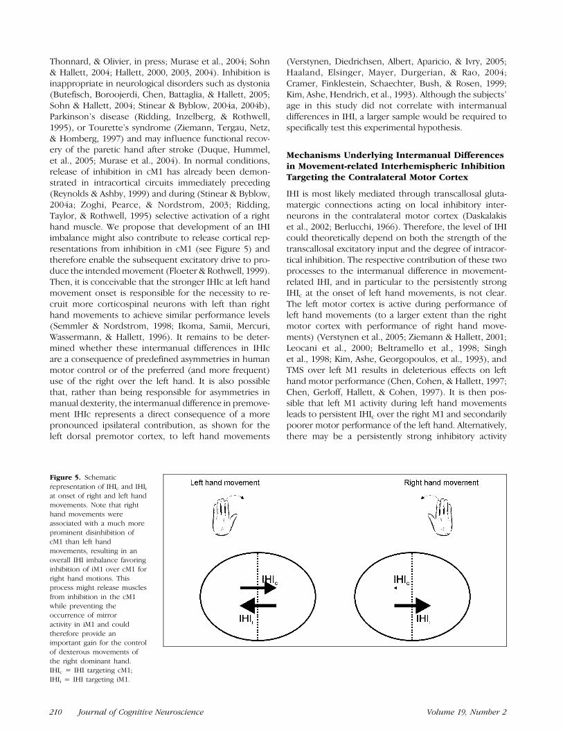
Thonnard, & Olivier, in press; Murase et al., 2004; Sohn& Hallett, 2004; Hallett, 2000, 2003, 2004). Inhibition isinappropriate in neurological disorders such as dystonia(Butefisch, Boroojerdi, Chen, Battaglia, & Hallett, 2005;Sohn & Hallett, 2004; Stinear & Byblow, 2004a, 2004b),Parkinson’s disease (Ridding, Inzelberg, & Rothwell,1995), or Tourette’s syndrome (Ziemann, Tergau, Netz,& Homberg, 1997) and may influence functional recov-ery of the paretic hand after stroke (Duque, Hummel,et al., 2005; Murase et al., 2004). In normal conditions,release of inhibition in cM1 has already been demon-strated in intracortical circuits immediately preceding(Reynolds & Ashby, 1999) and during (Stinear & Byblow,2004a; Zoghi, Pearce, & Nordstrom, 2003; Ridding,Taylor, & Rothwell, 1995) selective activation of a righthand muscle. We propose that development of an IHIimbalance might also contribute to release cortical rep-resentations from inhibition in cM1 (see Figure 5) andtherefore enable the subsequent excitatory drive to pro-duce the intended movement (Floeter & Rothwell, 1999).Then, it is conceivable that the stronger IHIc at left handmovement onset is responsible for the necessity to re-cruit more corticospinal neurons with left than righthand movements to achieve similar performance levels(Semmler & Nordstrom, 1998; Ikoma, Samii, Mercuri,Wassermann, & Hallett, 1996). It remains to be deter-mined whether these intermanual differences in IHIcare a consequence of predefined asymmetries in humanmotor control or of the preferred (and more frequent)use of the right over the left hand. It is also possiblethat, rather than being responsible for asymmetries inmanual dexterity, the intermanual difference in premove-ment IHIc represents a direct consequence of a morepronounced ipsilateral contribution, as shown for theleft dorsal premotor cortex, to left hand movements
(Verstynen, Diedrichsen, Albert, Aparicio, & Ivry, 2005;Haaland, Elsinger, Mayer, Durgerian, & Rao, 2004;Cramer, Finklestein, Schaechter, Bush, & Rosen, 1999;Kim, Ashe, Hendrich, et al., 1993). Although the subjects’age in this study did not correlate with intermanualdifferences in IHI, a larger sample would be required tospecifically test this experimental hypothesis.
Mechanisms Underlying Intermanual Differencesin Movement-related Interhemispheric InhibitionTargeting the Contralateral Motor Cortex
IHI is most likely mediated through transcallosal gluta-matergic connections acting on local inhibitory inter-neurons in the contralateral motor cortex (Daskalakiset al., 2002; Berlucchi, 1966). Therefore, the level of IHIcould theoretically depend on both the strength of thetranscallosal excitatory input and the degree of intracor-tical inhibition. The respective contribution of these twoprocesses to the intermanual difference in movement-related IHI, and in particular to the persistently strongIHIc at the onset of left hand movements, is not clear.The left motor cortex is active during performance ofleft hand movements (to a larger extent than the rightmotor cortex with performance of right hand move-ments) (Verstynen et al., 2005; Ziemann & Hallett, 2001;Leocani et al., 2000; Beltramello et al., 1998; Singhet al., 1998; Kim, Ashe, Georgopoulos, et al., 1993), andTMS over left M1 results in deleterious effects on lefthand motor performance (Chen, Cohen, & Hallett, 1997;Chen, Gerloff, Hallett, & Cohen, 1997). It is then pos-sible that left M1 activity during left hand movementsleads to persistent IHIc over the right M1 and secondarilypoorer motor performance of the left hand. Alternatively,there may be a persistently strong inhibitory activity
Figure 5. Schematic
representation of IHIc and IHIi
at onset of right and left handmovements. Note that right
hand movements were
associated with a much more
prominent disinhibition ofcM1 than left hand
movements, resulting in an
overall IHI imbalance favoringinhibition of iM1 over cM1 for
right hand motions. This
process might release muscles
from inhibition in the cM1while preventing the
occurrence of mirror
activity in iM1 and could
therefore provide animportant gain for the control
of dexterous movements of
the right dominant hand.IHIc = IHI targeting cM1;
IHIi = IHI targeting iM1.
210 Journal of Cognitive Neuroscience Volume 19, Number 2

within the right M1 when generating a left hand move-ment (Garry, Kamen, & Nordstrom, 2004). This higherinterneuronal inhibitory activity within the right M1 couldprevent it from being released from IHIc (Chen, 2004; Ilic,Jung, & Ziemann, 2004; Daskalakis et al., 2002; Banich &Brown, 2000; Priori et al., 1999; Cicinelli, Traversa, Bassi,Scivoletto, & Rossini, 1997; Triggs, Calvanio, Macdonell,Cros, & Chiappa, 1994; Berardelli, Inghilleri, Cruccu,Mercuri, & Manfredi, 1991). It is of note that althoughthere is substantial evidence that IHI is mediated throughtranscallosal connections (Daskalakis et al., 2002; DiLazzaro et al., 1999; Meyer et al., 1995, 1998), somesubcortical mechanisms might also be involved (Gerloffet al., 1998) and therefore contribute to some extent tothe intermanual difference in movement-related IHIc
evidenced in the present study.
Movement-related Interhemispheric InhibitionTargeting Primary Motor Cortex Ipsilateralto the Moving Hand
The finding of comparable inhibition of iM1 for move-ments of both hands suggests that the higher incidenceof mirroring with left than right hand movements(Liepert, Dettmers, Terborg, & Weiller, 2001; Armatas,Summers, & Bradshaw, 1994; Todor & Lazarus, 1986)cannot be accounted for by a differential efficiency ofIHIi. Rather, more frequent mirroring with left handmovements might relate to an increased contribution ofthe ipsilateral left premotor cortex when movementsare performed with the nondominant hand (Verstynenet al., 2005; Haaland et al., 2004; Cramer et al., 1999;Kim, Ashe, Hendrich, et al., 1993). A higher recruitmentof corticospinal neurons with nondominant hand move-ments (Semmler & Nordstrom, 1998; Ikoma et al., 1996)could also lead to an interhemispheric spread of fa-cilitation to the ipsilateral M1 (Baumer et al., 2006;Hanajima et al., 2001) and therefore elicit mirroring.Alternatively, the absence of movement-related modu-lation of IHI might alter the degree of intermanualindependence required to perform unilateral handmovements. In fact, because the default mode of acti-vation of the central nervous system seems to favor exe-cution of mirror movements (Swinnen, 2002; Serrien,Bogaerts, Suy, & Swinnen, 1999), it is possible that theability to execute strictly unilateral movements relies onan adequate movement-related modulation of IHI be-tween homonymous movement representations (Duque,Mazzocchio, et al., 2005; Liepert et al., 2001; Leocani et al.,2000; Chiarello & Maxfield, 1996; Schnitzler et al., 1996).
Conclusion
In conclusion, our results demonstrate a more promi-nent shift, from a balanced IHI at rest to an IHI pre-dominantly directed toward the ipsilateral M1, with rightthan left hand movements. Such movement-related
modulation of IHI might release movements performedwith the right hand from inhibition while preventing theoccurrence of mirror activity in the left hand, and couldtherefore provide an important gain for the control ofdexterous movements of the dominant hand (Triggset al., 1997, 2000).
Acknowledgments
J. Duque is a research assistant at the Belgian National Fundsfor Scientific Research (FNRS). P. Celnik is supported by agrant from the Rehabilitation Medicine Scientist Training Pro-gram (RMSTP Grant 5K12HD001097). F. Hummel is supportedby a grant of Alexander v. Humboldt-Foundation (Feodor-Lynen). This work was supported by the Intramural ResearchProgram of the National Institute of Neurological Disordersand Stroke, National Institutes of Health.
Reprint requests should be sent to Leonardo G. Cohen, HumanCortical Physiology Section, National Institute of NeurologicalDisorders and Stroke, National Institutes of Health, Bethesda,MD 20817, or via e-mail: [email protected].
REFERENCES
Aboitiz, F., & Montiel, J. (2003). One hundred million years ofinterhemispheric communication: The history of the corpuscallosum. Brazilian Journal of Medical and BiologicalResearch, 36, 409–420.
Armatas, C. A., Summers, J. J., & Bradshaw, J. L. (1994).Mirror movements in normal adult subjects. Journalof Clinical and Experimental Neuropsychology, 16,405–413.
Banich, M. T., & Brown, W. S. (2000). A life-span perspective oninteraction between the cerebral hemispheres.Developmental Neuropsychology, 18, 1–10.
Baumer, T., Bock, F., Koch, G., Lange, R., Rothwell, J. C.,Siebner, H. R., et al. (2006). Magnetic stimulation ofhuman premotor or motor cortex producesinterhemispheric facilitation through distinct pathways.Journal of Physiology, 572, 857–868.
Beltramello, A., Cerini, R., Puppini, G., El-Dalati, G., Viola, S.,Martone, E., et al. (1998). Motor representation of thehand in the human cortex: An f-MRI study with aconventional 1.5 T clinical unit. Italian Journal ofNeurological Sciences, 19, 277–284.
Berardelli, A., Inghilleri, M., Cruccu, G., Mercuri, B., &Manfredi, M. (1991). Electrical and magnetic transcranialstimulation in patients with corticospinal damage due tostroke or motor neurone disease. Electroencephalographyand Clinical Neurophysiology, 81, 389–396.
Berlucchi, G. (1966). Electroencephalographic studies in‘‘split brain’’ cats. Electroencephalography and ClinicalNeurophysiology, 20, 348–356.
Boroojerdi, B., Diefenbach, K., & Ferbert, A. (1996).Transcallosal inhibition in cortical and subcortical cerebralvascular lesions. Journal of Neurological Sciences, 144,160–170.
Butefisch, C. M., Boroojerdi, B., Chen, R., Battaglia, F., &Hallett, M. (2005). Task-dependent intracortical inhibitionis impaired in focal hand dystonia. Movement Disorders,20, 545–551.
Chen, R. (2004). Interactions between inhibitory and excitatorycircuits in the human motor cortex. Brain Research,Experimental Brain Research, 154, 1–10.
Duque et al. 211

Chen, R., Cohen, L. G., & Hallett, M. (1997). Role of theipsilateral motor cortex in voluntary movement. CanadianJournal of Neurological Sciences, 24, 284–291.
Chen, R., Gerloff, C., Hallett, M., & Cohen, L. G. (1997).Involvement of the ipsilateral motor cortex in fingermovements of different complexities. Annals of Neurology,41, 247–254.
Chiarello, C., & Maxfield, L. (1996). Varieties ofinterhemispheric inhibition, or how to keep a goodhemisphere down. Brain and Cognition, 30, 81–108.
Cicinelli, P., Traversa, R., Bassi, A., Scivoletto, G., &Rossini, P. M. (1997). Interhemispheric differences ofhand muscle representation in human motor cortex.Muscle and Nerve, 20, 535–542.
Coren, S., & Porac, C. (1977). Fifty centuries of right-handedness: The historical record. Science, 198,631–632.
Cramer, S. C., Finklestein, S. P., Schaechter, J. D., Bush, G.,& Rosen, B. R. (1999). Activation of distinct motor cortexregions during ipsilateral and contralateral fingermovements. Journal of Neurophysiology, 81, 383–387.
Daskalakis, Z. J., Christensen, B. K., Fitzgerald, P. B., Roshan, L.,& Chen, R. (2002). The mechanisms of interhemisphericinhibition in the human motor cortex. Journal ofPhysiology, 543, 317–326.
Davare, M., Duque, J., Vandermeeren, Y., Thonnard, J. L., &Olivier, E. (in press). Role of the ipsilateral primary motorcortex in controlling the timing of hand muscle recruitment.Cerebral Cortex.
De Gennaro, L., Bertini, M., Pauri, F., Cristiani, R., Curcio, G.,Ferrara, M., et al. (2004). Callosal effects of transcranialmagnetic stimulation (TMS): The influence of genderand stimulus parameters. Neuroscience Research, 48,129–137.
Di Lazzaro, V., Oliviero, A., Profice, P., Insola, A., Mazzone, P.,Tonali, P., et al. (1999). Direct demonstration ofinterhemispheric inhibition of the human motor cortexproduced by transcranial magnetic stimulation. BrainResearch, Experimental Brain Research, 124, 520–524.
Duque, J., Hummel, F., Celnik, P., Murase, N., Mazzocchio, R.,& Cohen, L. G. (2005). Transcallosal inhibition in chronicsubcortical stroke. Neuroimage, 28, 940–946.
Duque, J., Mazzocchio, R., Dambrosia, J., Murase, N., Olivier, E.,& Cohen, L. G. (2005). Kinematically specificinterhemispheric inhibition operating in the process ofgeneration of a voluntary movement. Cerebral Cortex, 15,588–593.
Ferbert, A., Priori, A., Rothwell, J. C., Day, B. L., Colebatch, J. G.,& Marsden, C. D. (1992). Interhemispheric inhibition ofthe human motor cortex. Journal of Physiology, 453,525–546.
Floeter, M. K., & Rothwell, J. C. (1999). Releasing the brakesbefore pressing the gas pedal. Neurology, 53, 664–665.
Garry, M. I., Kamen, G., & Nordstrom, M. A. (2004).Hemispheric differences in the relationship betweencorticomotor excitability changes following a fine-motortask and motor learning. Journal of Neurophysiology, 91,1570–1578.
Gerloff, C., Cohen, L. G., Floeter, M. K., Chen, R., Corwell, B.,& Hallett, M. (1998). Inhibitory influence of the ipsilateralmotor cortex on responses to stimulation of the humancortex and pyramidal tract. Journal of Physiology, 510,249–259.
Haaland, K. Y., Elsinger, C. L., Mayer, A. R., Durgerian, S.,& Rao, S. M. (2004). Motor sequence complexity andperforming hand produce differential patterns ofhemispheric lateralization. Journal of CognitiveNeuroscience, 16, 621–636.
Hallett, M. (2000). Disorder of movement preparation indystonia. Brain, 123, 1765–1766.
Hallett, M. (2003). Surround inhibition. Supplements toClinical Neurophysiology, 56, 153–159.
Hallett, M. (2004). Dystonia: Abnormal movements resultfrom loss of inhibition. Advances in Neurology, 94, 1–9.
Hanajima, R., Ugawa, Y., Machii, K., Mochizuki, H., Terao, Y.,Enomoto, H., et al. (2001). Interhemispheric facilitationof the hand motor area in humans. Journal of Physiology,531, 849–859.
Ikoma, K., Samii, A., Mercuri, B., Wassermann, E. M., &Hallett, M. (1996). Abnormal cortical motor excitability indystonia. Neurology, 46, 1371–1376.
Ilic, T. V., Jung, P., & Ziemann, U. (2004). Subtlehemispheric asymmetry of motor cortical inhibitorytone. Clinical Neurophysiology, 115, 330–340.
Kaelin-Lang, A., & Cohen, L. G. (2000). Enhancing thequality of studies using transcranial magnetic andelectrical stimulation with a new computer-controlledsystem. Journal of Neuroscience Methods, 102, 81–89.
Kim, S. G., Ashe, J., Georgopoulos, A. P., Merkle, H.,Ellermann, J. M., Menon, R. S., et al. (1993). Functionalimaging of human motor cortex at high magnetic field.Journal of Neurophysiology, 69, 297–302.
Kim, S. G., Ashe, J., Hendrich, K., Ellermann, J. M., Merkle, H.,Ugurbil, K., et al. (1993). Functional magnetic resonanceimaging of motor cortex: Hemispheric asymmetry andhandedness. Science, 261, 615–617.
Kim, Y. H., Jang, S. H., Chang, Y., Byun, W. M., Son, S., &Ahn, S. H. (2003). Bilateral primary sensori-motor cortexactivation of post-stroke mirror movements: An fMRI study.NeuroReport, 14, 1329–1332.
Leocani, L., Cohen, L. G., Wassermann, E. M., Ikoma, K., &Hallett, M. (2000). Human corticospinal excitabilityevaluated with transcranial magnetic stimulation duringdifferent reaction time paradigms. Brain, 123, 1161–1173.
Liepert, J., Dettmers, C., Terborg, C., & Weiller, C. (2001).Inhibition of ipsilateral motor cortex during phasic generationof low force. Clinical Neurophysiology, 112, 114–121.
Meyer, B. U., Roricht, S., Grafin von Einsiedel, H., Kruggel, F.,& Weindl, A. (1995). Inhibitory and excitatoryinterhemispheric transfers between motor cortical areas innormal humans and patients with abnormalities of thecorpus callosum. Brain, 118, 429–440.
Meyer, B. U., Roricht, S., & Woiciechowsky, C. (1998).Topography of fibers in the human corpus callosummediating interhemispheric inhibition between the motorcortices. Annals of Neurology, 43, 360–369.
Murase, N., Duque, J., Mazzocchio, R., & Cohen, L. G.(2004). Influence of interhemispheric interactions onmotor function in chronic stroke. Annals of Neurology,55, 400–409.
Netz, J. (1999). Asymmetry in transcallosal inhibition.Electroencephalography and Clinical Neurophysiology.Supplement, 51, 137–144.
Netz, J., Ziemann, U., & Homberg, V. (1995). Hemisphericasymmetry of transcallosal inhibition in man. BrainResearch, Experimental Brain Research, 104, 527–533.
Oldfield, R. C. (1971). The assessment and analysis ofhandedness: The Edinburgh inventory. Neuropsychologia,9, 97–113.
Peters, M., & Durding, B. M. (1978). Handedness measuredby finger tapping: A continuous variable. CanadianJournal of Psychology, 32, 257–261.
Priori, A., Oliviero, A., Donati, E., Callea, L., Bertolasi, L., &Rothwell, J. C. (1999). Human handedness and asymmetryof the motor cortical silent period. Brain Research,Experimental Brain Research, 128, 390–396.
212 Journal of Cognitive Neuroscience Volume 19, Number 2

Reynolds, C., & Ashby, P. (1999). Inhibition in the humanmotor cortex is reduced just before a voluntary contraction.Neurology, 53, 730–735.
Ridding, M. C, Inzelberg, R., & Rothwell, J. C. (1995). Changesin excitability of motor cortical circuitry in patients withParkinson’s disease. Annals of Neurology, 37, 181–188.
Ridding, M. C., Taylor, J. L., & Rothwell, J. C. (1995).The effect of voluntary contraction on cortico-corticalinhibition in human motor cortex. Journal of Physiology,487, 541–548.
Rossini, P. M., Barker, A. T., Berardelli, A., Caramia, M. D., Caruso,G., Cracco, R. Q., et al. (1994). Non-invasive electrical andmagnetic stimulation of the brain, spinal cord and roots: Basicprinciples and procedures for routine clinical application.Report of an IFCN committee. Electroencephalographyand Clinical Neurophysiology, 91, 79–92.
Rouiller, E. M., Babalian, A., Kazennikov, O., Moret, V., Yu,X. H., & Wiesendanger, M. (1994). Transcallosal connectionsof the distal forelimb representations of the primary andsupplementary motor cortical areas in macaque monkeys.Brain Research, Experimental Brain Research, 102,227–243.
Roy, E. A., Bryden, P., & Cavill, S. (2003). Hand differencesin pegboard performance through development. Brain andCognition, 53, 315–317.
Sakai, K., Ugawa, Y., Terao, Y., Hanajima, R., Furubayashi, T.,& Kanazawa, I. (1997). Preferential activation of differentI waves by transcranial magnetic stimulation with afigure-of-eight-shaped coil. Brain Research, ExperimentalBrain Research, 113, 24–32.
Salerno, A., & Georgesco, M. (1996). Interhemisphericfacilitation and inhibition studied in man with doublemagnetic stimulation. Electroencephalography andClinical Neurophysiology, 101, 395–403.
Schnitzler, A., Kessler, K. R., & Benecke, R. (1996).Transcallosally mediated inhibition of interneurons withinhuman primary motor cortex. Brain Research,Experimental Brain Research, 112, 381–391.
Semmler, J. G., & Nordstrom, M. A. (1998). Hemisphericdifferences in motor cortex excitability during a simpleindex finger abduction task in humans. Journal ofNeurophysiology, 79, 1246–1254.
Serrien, D. J., Bogaerts, H., Suy, E., & Swinnen, S. P. (1999).The identification of coordination constraints acrossplanes of motion. Brain Research, Experimental BrainResearch, 128, 250–255.
Singh, L. N., Higano, S., Takahashi, S., Kurihara, N., Furuta, S.,Tamura, H., et al. (1998). Comparison of ipsilateralactivation between right and left handers: A functionalMR imaging study. NeuroReport, 9, 1861–1866.
Sohn, Y. H., & Hallett, M. (2004). Disturbed surroundinhibition in focal hand dystonia. Annals of Neurology,56, 595–599.
Stinear, C. M., & Byblow, W. D. (2004a). Impaired modulationof intracortical inhibition in focal hand dystonia. CerebralCortex, 14, 555–561.
Stinear, C. M., & Byblow, W. D. (2004b). Elevated thresholdfor intracortical inhibition in focal hand dystonia. MovementDisorders, 19, 1312–1317.
Swinnen, S. P. (2002). Intermanual coordination: Frombehavioural principles to neural–network interactions.Nature Reviews Neuroscience, 3, 348–359.
Swinnen, S. P., Dounskaia, N., & Duysens, J. (2002). Patternsof bimanual interference reveal movement encodingwithin a radial egocentric reference frame. Journal ofCognitive Neuroscience, 14, 463–471.
Thut, G., Halsband, U., Regard, M., Mayer, E., Leenders, K. L.,& Landis, T. (1997). What is the role of the corpuscallosum in intermanual transfer of motor skills? A studyof three cases with callosal pathology. Brain Research,Experimental Brain Research, 113, 365–370.
Todor, J. I., & Kyprie, P. M. (1980). Hand differences in therate and variability of rapid tapping. Journal of MotorBehavior, 12, 57–62.
Todor, J. I., & Lazarus, J. A. (1986). Exertion level and theintensity of associated movements. DevelopmentalMedicine and Child Neurology, 28, 205–212.
Triggs, W. J., Calvanio, R., & Levine, M. (1997). Transcranialmagnetic stimulation reveals a hemispheric asymmetrycorrelate of intermanual differences in motor performance.Neuropsychologia, 35, 1355–1363.
Triggs, W. J., Calvanio, R., Levine, M., Heaton, R. K., &Heilman, K. M. (2000). Predicting hand preference withperformance on motor tasks. Cortex, 36, 679–689.
Triggs, W. J., Calvanio, R., Macdonell, R. A., Cros, D., &Chiappa, K. H. (1994). Physiological motor asymmetry inhuman handedness: Evidence from transcranial magneticstimulation. Brain Research, Experimental BrainResearch, 636, 270–276.
Verstynen, T., Diedrichsen, J., Albert, N., Aparicio, P., &Ivry, R. B. (2005). Ipsilateral motor cortex activity duringunimanual hand movements relates to task complexity.Journal of Neurophysiology, 93, 1209–1222.
Werhahn, K. J., Fong, J. K., Meyer, B. U., Priori, A., Rothwell,J. C., Day, B. L., et al. (1994). The effect of magnetic coilorientation on the latency of surface EMG and singlemotor unit responses in the first dorsal interosseousmuscle. Electroencephalography and ClinicalNeurophysiology, 93, 138–146.
Ziemann, U., & Hallett, M. (2001). Hemispheric asymmetryof ipsilateral motor cortex activation during unimanualmotor tasks: Further evidence for motor dominance.Clinical Neurophysiology, 112, 107–113.
Ziemann, U., Tergau, F., Netz, J., & Homberg, V. (1997). Delayin simple reaction time after focal transcranial magneticstimulation of the human brain occurs at the final motoroutput stage. Brain Research, Experimental BrainResearch, 744, 32–40.
Zoghi, M., Pearce, S. L., & Nordstrom, M. A. (2003).Differential modulation of intracortical inhibition in humanmotor cortex during selective activation of an intrinsichand muscle. Journal of Physiology, 550, 933–946.
Duque et al. 213