Quantum curcumin mediated inhibition of gingipains and ...
20
Quantum curcumin mediated inhibition of gingipains and mixed-biofilm of Porphyromonas gingivalis causing chronic periodontitis† Ashish Kumar Singh, ab Shivangi Yadav, a Kavanjali Sharma, c Zeba Firdaus, d Prerana Aditi, d Kaushik Neogi, e Monika Bansal, f Munesh Kumar Gupta, a Asheesh Shanker, g Rakesh Kumar Singh b and Pradyot Prakash * a Periodontitis is a biofilm-associated irreversible inflammation of the periodontal tissues. Reports suggest the role of Porphyromonas gingivalis specific Arg- and Lys-specific proteinases in the orchestration of the initiation and progression of periodontal diseases. These proteinases are precisely termed as gingipains R and K. Curcumin is an active polyphenol that is extracted from the rhizomes of Curcuma longa. However, the molecule curcumin owing to its high hydropathy index and poor stability has not been able to justify its role as frontline drug modality in the treatment of infectious and non-infectious diseases as claimed by several investigators. In the present study, at first, we synthesized and characterized quantum curcumin, and investigated its biocompatibility. This was subsequently followed by the evaluation of the role of quantum curcumin as an antimicrobial, anti-gingipains and antibiofilm agent against Porphyromonas gingivalis and select reference strains. We have successfully synthesized the quantum curcumin utilizing a top-down approach with the average size of 3.5 nm. Apart from its potent antimicrobial as well as antibiofilm properties, it also significantly inhibited the gingipains in a dose-dependent manner. At the minimal concentration of 17.826 mM, inhibition up to 98.7% and 89.4% was noted for gingipain R and K respectively. The data was also supported by the in silico docking experiments which revealed high exothermic enthalpies (7.01 and 7.02 cal mol 1 ). Besides, the inhibition constant was found to be 7.24 mM and 7.1 mM against gingipains R and K respectively. The results suggest that quantum curcumin is a potential drug candidate which needs further clinical validation. Introduction Periodontitis is a biolm-associated inammatory disease of the gingiva and the supporting structures of the periodontium (gingival, alveolar bone, periodontal ligament, and cementum) which has multiple etiologies; however, microbial and immunological factors are reported to be pivotal. 1,2 More precisely, periodontitis insinuates the plaque-induced irrevers- ible inammation of the periodontal tissues which ultimately obliterates the periodontal ligament and alveolar bone. 3 An estimate from World Health Organization reveals 10–15% global load of this disease among total adult population. 4 Pla- que is an established biolm which exemplies the socializa- tion wherein mixed consortia of bacteria co-exist which in turn determines the biolm matrix composition and mutual coop- erativity. 1,5 Aer microbial inhabitation, the immune system comes into play and the disease is aggravated further by the destructive host inammatory responses. 6–9 However, reports suggest the involvement of only a few sorts of bacteria inhab- iting the subgingival niche which orchestrate the initiation and progression of periodontal disease. 10 In this context, Porphyr- omonas gingivalis, a Gram-negative anaerobe becomes especially important because of its diverse armamentarium both struc- tural and functional, needed for its success (pathogenesis and progression of the inammatory events) in the said micro- environment. It seems to be the keystone species in the devel- opment of chronic periodontitis. 3,11 Datta et al. reported this bacterium in 85.75% of subgingival plaque samples from a Bacterial Biolm and Drug Resistance Research Laboratory, Department of Microbiology, Institute of Medical Sciences, Banaras Hindu University, Varanasi 221005, India. E-mail: [email protected] b Molecular Immunology Laboratory, Department of Biochemistry, Institute of Science, Banaras Hindu University, Varanasi 221005, India c Department of Pathology, Institute of Medical Sciences, Banaras Hindu University, Varanasi 221005, India d Department of Medicinal Chemistry, Institute of Medical Sciences, Banaras Hindu University, Varanasi 221005, India e Department of Pharmaceutical Engineering and Technology, Indian Institute of Technology, Banaras Hindu University, Varanasi 221005, India f Faculty of Dental Sciences, Institute of Medical Science, Banaras Hindu University, Varanasi 221005, India g Department of Bioinformatics, Central University of South Bihar, Gaya 824236, Bihar, India † Electronic supplementary information (ESI) available. See DOI: 10.1039/c8ra08435a Cite this: RSC Adv. , 2018, 8, 40426 Received 11th October 2018 Accepted 26th November 2018 DOI: 10.1039/c8ra08435a rsc.li/rsc-advances 40426 | RSC Adv. , 2018, 8, 40426–40445 This journal is © The Royal Society of Chemistry 2018 RSC Advances PAPER Open Access Article. Published on 05 December 2018. Downloaded on 2/7/2022 12:12:37 AM. This article is licensed under a Creative Commons Attribution-NonCommercial 3.0 Unported Licence. View Article Online View Journal | View Issue
-
Upload
khangminh22 -
Category
Documents
-
view
4 -
download
0
Transcript of Quantum curcumin mediated inhibition of gingipains and ...

RSC Advances
PAPER
Ope
n A
cces
s A
rtic
le. P
ublis
hed
on 0
5 D
ecem
ber
2018
. Dow
nloa
ded
on 2
/7/2
022
12:1
2:37
AM
. T
his
artic
le is
lice
nsed
und
er a
Cre
ativ
e C
omm
ons
Attr
ibut
ion-
Non
Com
mer
cial
3.0
Unp
orte
d L
icen
ce.
View Article OnlineView Journal | View Issue
Quantum curcum
aBacterial Biolm and Drug Resistance
Microbiology, Institute of Medical Science
221005, India. E-mail: pradyot_micro@bhubMolecular Immunology Laboratory, Departm
Banaras Hindu University, Varanasi 221005cDepartment of Pathology, Institute of Med
Varanasi 221005, IndiadDepartment of Medicinal Chemistry, Instit
University, Varanasi 221005, IndiaeDepartment of Pharmaceutical Engineerin
Technology, Banaras Hindu University, VarafFaculty of Dental Sciences, Institute of Me
Varanasi 221005, IndiagDepartment of Bioinformatics, Central Univ
India
† Electronic supplementary informa10.1039/c8ra08435a
Cite this: RSC Adv., 2018, 8, 40426
Received 11th October 2018Accepted 26th November 2018
DOI: 10.1039/c8ra08435a
rsc.li/rsc-advances
40426 | RSC Adv., 2018, 8, 40426–404
in mediated inhibition ofgingipains and mixed-biofilm of Porphyromonasgingivalis causing chronic periodontitis†
Ashish Kumar Singh,ab Shivangi Yadav,a Kavanjali Sharma,c Zeba Firdaus,d
Prerana Aditi,d Kaushik Neogi,e Monika Bansal,f Munesh Kumar Gupta,a
Asheesh Shanker,g Rakesh Kumar Singhb and Pradyot Prakash *a
Periodontitis is a biofilm-associated irreversible inflammation of the periodontal tissues. Reports suggest
the role of Porphyromonas gingivalis specific Arg- and Lys-specific proteinases in the orchestration of
the initiation and progression of periodontal diseases. These proteinases are precisely termed as
gingipains R and K. Curcumin is an active polyphenol that is extracted from the rhizomes of Curcuma
longa. However, the molecule curcumin owing to its high hydropathy index and poor stability has not
been able to justify its role as frontline drug modality in the treatment of infectious and non-infectious
diseases as claimed by several investigators. In the present study, at first, we synthesized and
characterized quantum curcumin, and investigated its biocompatibility. This was subsequently followed
by the evaluation of the role of quantum curcumin as an antimicrobial, anti-gingipains and antibiofilm
agent against Porphyromonas gingivalis and select reference strains. We have successfully synthesized
the quantum curcumin utilizing a top-down approach with the average size of 3.5 nm. Apart from its
potent antimicrobial as well as antibiofilm properties, it also significantly inhibited the gingipains in
a dose-dependent manner. At the minimal concentration of 17.826 mM, inhibition up to 98.7% and 89.4%
was noted for gingipain R and K respectively. The data was also supported by the in silico docking
experiments which revealed high exothermic enthalpies (�7.01 and �7.02 cal mol�1). Besides, the
inhibition constant was found to be 7.24 mM and 7.1 mM against gingipains R and K respectively. The
results suggest that quantum curcumin is a potential drug candidate which needs further clinical validation.
Introduction
Periodontitis is a biolm-associated inammatory disease ofthe gingiva and the supporting structures of the periodontium(gingival, alveolar bone, periodontal ligament, and cementum)which has multiple etiologies; however, microbial and
Research Laboratory, Department of
s, Banaras Hindu University, Varanasi
.ac.in
ent of Biochemistry, Institute of Science,
, India
ical Sciences, Banaras Hindu University,
ute of Medical Sciences, Banaras Hindu
g and Technology, Indian Institute of
nasi 221005, India
dical Science, Banaras Hindu University,
ersity of South Bihar, Gaya 824236, Bihar,
tion (ESI) available. See DOI:
45
immunological factors are reported to be pivotal.1,2 Moreprecisely, periodontitis insinuates the plaque-induced irrevers-ible inammation of the periodontal tissues which ultimatelyobliterates the periodontal ligament and alveolar bone.3 Anestimate from World Health Organization reveals 10–15%global load of this disease among total adult population.4 Pla-que is an established biolm which exemplies the socializa-tion wherein mixed consortia of bacteria co-exist which in turndetermines the biolm matrix composition and mutual coop-erativity.1,5 Aer microbial inhabitation, the immune systemcomes into play and the disease is aggravated further by thedestructive host inammatory responses.6–9 However, reportssuggest the involvement of only a few sorts of bacteria inhab-iting the subgingival niche which orchestrate the initiation andprogression of periodontal disease.10 In this context, Porphyr-omonas gingivalis, a Gram-negative anaerobe becomes especiallyimportant because of its diverse armamentarium both struc-tural and functional, needed for its success (pathogenesis andprogression of the inammatory events) in the said micro-environment. It seems to be the keystone species in the devel-opment of chronic periodontitis.3,11 Datta et al. reported thisbacterium in 85.75% of subgingival plaque samples from
This journal is © The Royal Society of Chemistry 2018

Paper RSC Advances
Ope
n A
cces
s A
rtic
le. P
ublis
hed
on 0
5 D
ecem
ber
2018
. Dow
nloa
ded
on 2
/7/2
022
12:1
2:37
AM
. T
his
artic
le is
lice
nsed
und
er a
Cre
ativ
e C
omm
ons
Attr
ibut
ion-
Non
Com
mer
cial
3.0
Unp
orte
d L
icen
ce.
View Article Online
patients with chronic periodontitis.12 Surprisingly, for itssurvival, this bacterium depends on the fermentation of aminoacids for energy in deep periodontal pockets where sugaravailability is scarce.13 A plethora of literature document theessentiality of exceedingly high concentrations of cysteineproteinases with trypsin-like activities in the adult-onset peri-odontitis.6,11,13–16 Besides, to initiate and prolong the inam-matory response during periodontitis, a number of biochemicalcascades are needed which require proteolytic activities. As perthe report of de Diego et al., gingipain R and K collectivelyaccount for 85% of the extracellular proteolytic activity of P.gingivalis at the site of infection.17 Gingipains orchestratediverse functions, which aid in bacterial survival. It activateskallikrein/kinin cascade, inactivates host proteinase inhibitors,foster dysregulation of coagulation and complement cascade,degrades immunoglobulins, bactericidal proteins, Fe-transporting proteins etc.3,14–16
The routine therapeutic modality for periodontitis manage-ment is scaling and root planing (SRP) wherein supra andsubgingival plaque and calculus are removed.18 However, therecalcitrance of biolm dwellers and recolonization at thetreated sites cannot be overruled.19 Possibly, owing to this, nowa day's application of nonspecic broad-spectrum antibioticslocally at sites are in vogue as an addendum to the mechanicaldebridement. Drugs like chlorhexidine, delmopinol, tetracy-cline, minocycline, doxycycline, metronidazole etc. in the formof mouthwash etc. are routinely used to curb the re-proliferationof periodontopathic bacteria following SRP.20 However, the pooreffectiveness of these drugs against the plaque biolms andbacterial stochastic variations demands new effective measuresfor its management. The lack of selectivity of these drugsfurther complicates the situation by affecting the benecialcommensals as well as impart undesired side effects such asvomiting, diarrhea, addiction, or teeth discoloration.18,20–22
The therapeutic inadequacies of the drugs are due to the lackof selectivity and upsurge of antimicrobial resistance within theplaque biolms.20 The situation becomes more complex whenmicrobial shiing occurs during the course of biolm devel-opment, especially during plaque formation and maturation.5 Areport by Marsh et al. has indicated the role of dysbiosis in theoral cavity that leads to periodontitis.23 A recent report byDewhirst et al. suggested the presence of over 700 bacterialspecies in the oral cavity.21 Interestingly, early bacterial colo-nizers in the oral cavity are reported to actively recruit otherbacteria as well. For instance, Streptococcus gordonii was foundto promote and help establish the biolm of Porphyromonasgingivalis via several genetic mechanisms.13 As periodontitisdevelops, the oral microbiota shis from the one consistinglargely of Gram-positive aerobes to one consisting predomi-nantly of Gram-negative anaerobes. This cooperative recruit-ment of metabolically and phylogenetically diverse bacterialeads to the overall functional heterogeneity which confersseveral advantages like metabolic cooperation, wherein thewaste of one bacterial species serves as the food for the other.5
One noteworthy illustration is the fermentation of sugars tolactic acid by Streptococci which is then disintegrated to propi-onate and acetate by Veillonella spp.5 Furthermore, bacterial
This journal is © The Royal Society of Chemistry 2018
cross-talk in the form of quorum sensing help them to regulatetheir social behaviour. Reports suggest that the oral bacteriaproduce and respond to the autoinducer-2 signaling.24 These allconducive underpinnings further tunes the matrix chemistryfor their survival, ultimately inuencing the therapeuticoutcomes. Therefore, it becomes prudent for the scienticcommunity to look for easily available, cost-effective therapeuticagent(s) having both antimicrobial and anti-biolm activitieswith no or minimal side-effects or toxicity.
Etiology suggests that the periodontitis can only besuccessfully tackled aer simultaneously addressing thebacterial and inammatory components responsible for thecondition. Curcumin ts in our requirement window. Aplethora of literature documents the anti-inammatory, anti-oxidant, anticancer, antibacterial and anti-fungal properties ofcurcumin, a hydrophobic curcuminoid, extracted from therhizomes of Curcuma longa.25,26 Curcumin was found to behighly effective against a wide array of microbial pathogensranging from Trichomonas vaginalis, Helicobacter pylori toCandida albicans and Paracoccidioides brasiliensis.26–29 Besides, itwas also reported to inhibit the biolm formation by Escherichiacoli, Pseudomonas aeruginosa, Klebsiella pneumonia, Staphylo-coccus aureus, and Staphylococcus epidermidis.25 Among the oralbacteria, curcumin was reported to inhibit sortase A of Strep-tococcus mutans, a major cariogenic bacteria.30
The scientic literature is inundated with the reports per-taining to the determination of minimum inhibitory concen-tration (MIC) of curcumin against various pathogens.31–34
However, we faced difficulties in determining and interpretingthe results due to its insolubility and instability. These factorsresulted in the colored precipitation which obscured the preciseand accurate MIC determination as per CLSI guidelines.Therefore, as a drug, the use of curcumin is limited due toseveral such constraints. Nevertheless, attempts have beenmade to increase its aqueous solubility and bioavailability bythe nanoparticle-based approach.33 For this, liposomes, poly-meric nanoparticles, and recently lipid-drug hybrid nano-particles have been extensively studied, where they have shownimproved efficacies.35–38 Nonetheless, the innate toxicity issuescannot be overlooked. Recently, the newer quantum approachhas also been utilized for its stability, solubility and therapeuticpotential by making quantum curcumin against aerobic path-ogens. However, its detailed toxicity evaluation is yet to bedone.25
In the present work, we synthesized, characterized and thenevaluated the antimicrobial and antibiolm potential ofCurQDs against representative periodontic bacterium Porphyr-omonas gingivalis along with other facilitating bacteria likeActinomycetemcomitans viscosus and Streptococcus mutans.Moreover, degradative effects of CurQDs on P. gingivalis and onmixed P. gingivalis–S. mutans–A. viscosus biolm formation werealso examined. Furthermore, we tried to elucidate the action ofquantum curcumin against the most signicant virulence factorof anaerobe Porphyromonas gingivalis: gingipain R and K whichwas further validated by docking experiments. These studieswere made aer the detailed investigations pertaining to thebiocompatibility of quantum curcumin which included in vitro
RSC Adv., 2018, 8, 40426–40445 | 40427

RSC Advances Paper
Ope
n A
cces
s A
rtic
le. P
ublis
hed
on 0
5 D
ecem
ber
2018
. Dow
nloa
ded
on 2
/7/2
022
12:1
2:37
AM
. T
his
artic
le is
lice
nsed
und
er a
Cre
ativ
e C
omm
ons
Attr
ibut
ion-
Non
Com
mer
cial
3.0
Unp
orte
d L
icen
ce.
View Article Online
cell viability assay by methylthiazoltetrazolium salt, haemo-compatibility assay, membrane integrity assay using lactatedehydrogenase asmarker and investigation of the production ofreactive oxygen species (ROS) using ow cytometry.
Materials and methodsChemicals
Curcumin (>95% pure) was procured from TCI chemicals,Japan. Phosphate Buffered Saline (PBS, pH 7.2), Tris–HCl buffer(pH 7.6) were prepared in-house while Dulbecco's modiedEagle's medium, DABCO and propidium iodide from Lifetechnologies, Invitrogen were used. Flat-bottom polystyrene 96-well tissue culture plates and 8-well chambered slides wereprocured from SPL biosciences, Korea. Penicillin, streptomycinand gentamicin were from SRL laboratories, India while iodo-nitrotetrazolium chloride, Hanks balanced salt solution (HBSS),heparin, sodium bicarbonate, dimethylsulphoxide (DMSO),benzoyl-L-arginine-4-methylcoumaryl-7-amide, t-butyl-oxy-carbonyl-L-valyl-L-leucyl-L-lysine-4-methylcoumaryl-7-amide, Na-p-tosyl-L-lysine chloromethyl ketone hydrochloride, and meth-ylthiazoltetrazolium salt were from Sigma-Aldrich, USA. Triso-dium citrate, calcium chloride (CaCl2), sodium chloride (NaCl),crystal violet (CV), dimethyl sulfoxide (DMSO) and acetone wereprocured fromMerck, USA while paraformaldehyde, brain heartinfusion (BHI) broth, Colombia blood agar, yeast extract,hemin, menadione, cysteine, and fetal bovine serum wereprocured from HiMedia laboratories, Mumbai.
Synthesis of quantum curcumin (CurQDs)
The synthesis of CurQDs was hierarchical and was done by thetwo-step top-down method as described earlier with modica-tions.25 Initially, a stock solution of 20 mM curcumin wasprepared in DMSO. A stock solution of 150 mM trisodiumcitrate was also prepared in deionized water. CurQDs formationwas investigated using different concentrations curcumin0.125 mM to 2.5 mM at different pH ranging from 4 to 7. Ina typical experiment, 0.1 mM of curcumin was taken in deion-ized water and its pH adjusted using trisodium citrate. Further,2.5 g of zirconia beads (0.1 mm in diameter) were put in the50 ml oak ridge tube and spun at 2500 rpm for 15 min with theprogressive increase in the drug loading up to 2.5 mM. Theresultant solution was siphoned out and dried using rotaryevaporator at 70 �C. From this, a stock solution of 1 mM wasprepared in acetone which was then added dropwise to hotwater (25 ml) at 75 � 5 �C at the rate of 0.2 ml min�1 withcontinuous ultra-sonication for about an hour with power inputof 750 W, frequency 20 kHz, and intensity 30 W cm�2.
The nal solution was kept at room temperature for 48 h andcentrifuged at a speed of 10 000 rpm for 15 min at 4 �C toremove any residual native curcumin if present. The suspensionwas nally dried and re-suspended in deionized water and usedfor further characterizations. For XRD and Raman spectro-scopic measurements, the isolated quantum curcumin werefreeze-dried to obtain a dry powder.
40428 | RSC Adv., 2018, 8, 40426–40445
Physical characterization
The prepared quantum curcumin was monitored spectroscop-ically at a wavelength range of 300–700 nm. All the absorbanceand uorescence readings were made in Lambda 25 UV-visiblespectrometer, Perkin Elmer and LS uorescence spectropho-tometer Perkin Elmer. Morphological features of quantumcurcumin were analyzed by electron microscopy. A drop of thesample was allowed to dry on carbon-coated copper grids in theair. For TEM analysis, the images were acquired through FEITecnai 200 TEM operated at 80 kV and high-resolution imagesin Tecnai transmission electron microscope at 300 kV. Thephase compositions and crystalline nature of CurQDs werefurther studied by XRD in Rigaku MINIFLEX 600, Japan usingCu radiation at k¼ 0.1546 nm at a voltage of 40 kV and a currentof 30 mA. Additionally, surface composition of the synthesizedquantum curcumin was investigated by high-resolution X-rayphotoelectron spectroscopy. Besides, Raman spectroscopy wasalso used to assess the phase variability and structure. TheRaman spectrum was obtained on Renishaw In-Via Ramanspectrometer using 50� objective from Lica. The sample wasexcited by 532 nm solid state diode laser and acquisition timefor each window was selected as 50 s. The zeta potential ofCurQDs was measured on Delsa Nano (Beckman-coulter,United Kingdom) utilizing zeta dip cells. For the measure-ment, the sample was prepared by mixing CurQDs in 10 mMNaCl in 1 : 10 proportion and then 1 ml of the diluted samplewas used.
Chemical characterization
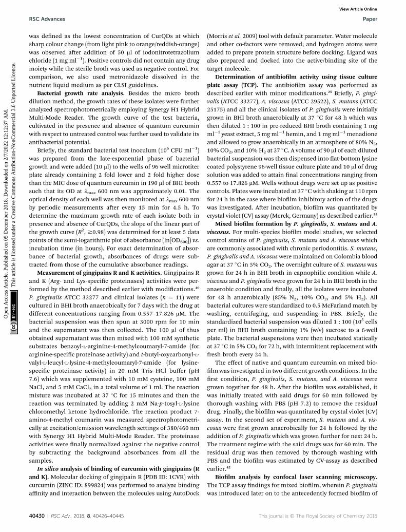
Nuclear magnetic resonance spectroscopy. The nuclearmagnetic resonance analysis (1H NMR and 13C NMR) wascarried out using JEOL spectrometer at 300 K at 500.13 MHzequipped with 5 mm broadband probe, using dried quantumcurcumin dissolved in 600 ml DMSO-d6.
1H NMRmeasurementswere made at 300 K, and the chemical shis (ppm) wereassigned. The spectral width was 10 330.578 Hz, and the digitalresolution was 0.157 Hz, with an acquisition time of 3.17 s. Atotal of 16 scans were taken. 13C NMRmeasurements were doneat 300 K at 500.13 MHz, and the chemical shis (ppm) wereassigned. The spectral width was 26 455.02 Hz with the digitalresolution of 1.61 Hz and acquisition time of 0.30 s. The spec-trum was obtained with 1024 scans.
Toxicity proling of quantum curcumin
Cell culture and treatment conditions. Vero cells (kidney cellline) were cultured in Dulbecco's modied Eagle's mediumsupplemented with 10% fetal bovine serum along with 100 Uml�1 penicillin and 100 mg ml�1 streptomycin in a 5% CO2
humidied atmosphere at 37 �C in a CO2 incubator. A 1 Msolution of quantum curcumin was prepared as extricatedabove and was stored as small aliquots at 4 �C and diluted twofolds in a different dose ranging from 10–70 mM in Dulbecco'smodied Eagle's medium. The cells were exposed to CurQDs for24 h.
This journal is © The Royal Society of Chemistry 2018
Paper RSC Advances
Ope
n A
cces
s A
rtic
le. P
ublis
hed
on 0
5 D
ecem
ber
2018
. Dow
nloa
ded
on 2
/7/2
022
12:1
2:37
AM
. T
his
artic
le is
lice
nsed
und
er a
Cre
ativ
e C
omm
ons
Attr
ibut
ion-
Non
Com
mer
cial
3.0
Unp
orte
d L
icen
ce.
View Article Online
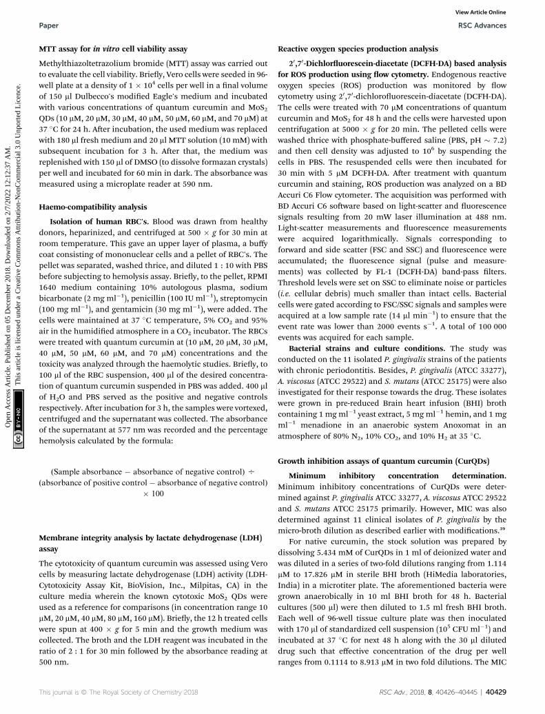
MTT assay for in vitro cell viability assay
Methylthiazoltetrazolium bromide (MTT) assay was carried outto evaluate the cell viability. Briey, Vero cells were seeded in 96-well plate at a density of 1 � 104 cells per well in a nal volumeof 150 ml Dulbecco's modied Eagle's medium and incubatedwith various concentrations of quantum curcumin and MoS2QDs (10 mM, 20 mM, 30 mM, 40 mM, 50 mM, 60 mM, and 70 mM) at37 �C for 24 h. Aer incubation, the used medium was replacedwith 180 ml fresh medium and 20 ml MTT solution (10 mM) withsubsequent incubation for 3 h. Aer that, the medium wasreplenished with 150 ml of DMSO (to dissolve formazan crystals)per well and incubated for 60 min in dark. The absorbance wasmeasured using a microplate reader at 590 nm.
Haemo-compatibility analysis
Isolation of human RBC's. Blood was drawn from healthydonors, heparinized, and centrifuged at 500 � g for 30 min atroom temperature. This gave an upper layer of plasma, a buffycoat consisting of mononuclear cells and a pellet of RBC's. Thepellet was separated, washed thrice, and diluted 1 : 10 with PBSbefore subjecting to hemolysis assay. Briey, to the pellet, RPMI1640 medium containing 10% autologous plasma, sodiumbicarbonate (2 mg ml�1), penicillin (100 IU ml�1), streptomycin(100 mg ml�1), and gentamicin (30 mg ml�1), were added. Thecells were maintained at 37 �C temperature, 5% CO2 and 95%air in the humidied atmosphere in a CO2 incubator. The RBCswere treated with quantum curcumin at (10 mM, 20 mM, 30 mM,40 mM, 50 mM, 60 mM, and 70 mM) concentrations and thetoxicity was analyzed through the haemolytic studies. Briey, to100 ml of the RBC suspension, 400 ml of the desired concentra-tion of quantum curcumin suspended in PBS was added. 400 mlof H2O and PBS served as the positive and negative controlsrespectively. Aer incubation for 3 h, the samples were vortexed,centrifuged and the supernatant was collected. The absorbanceof the supernatant at 577 nm was recorded and the percentagehemolysis calculated by the formula:
(Sample absorbance � absorbance of negative control) O
(absorbance of positive control � absorbance of negative control)
� 100
Membrane integrity analysis by lactate dehydrogenase (LDH)assay
The cytotoxicity of quantum curcumin was assessed using Verocells by measuring lactate dehydrogenase (LDH) activity (LDH-Cytotoxicity Assay Kit, BioVision, Inc., Milpitas, CA) in theculture media wherein the known cytotoxic MoS2 QDs wereused as a reference for comparisons (in concentration range 10mM, 20 mM, 40 mM, 80 mM, 160 mM). Briey, the 12 h treated cellswere spun at 400 � g for 5 min and the growth medium wascollected. The broth and the LDH reagent was incubated in theratio of 2 : 1 for 30 min followed by the absorbance reading at500 nm.
This journal is © The Royal Society of Chemistry 2018
Reactive oxygen species production analysis
20,70-Dichloruorescein-diacetate (DCFH-DA) based analysisfor ROS production using ow cytometry. Endogenous reactiveoxygen species (ROS) production was monitored by owcytometry using 20,70-dichlorouorescein-diacetate (DCFH-DA).The cells were treated with 70 mM concentrations of quantumcurcumin and MoS2 for 48 h and the cells were harvested uponcentrifugation at 5000 � g for 20 min. The pelleted cells werewashed thrice with phosphate-buffered saline (PBS, pH � 7.2)and then cell density was adjusted to 106 by suspending thecells in PBS. The resuspended cells were then incubated for30 min with 5 mM DCFH-DA. Aer treatment with quantumcurcumin and staining, ROS production was analyzed on a BDAccuri C6 Flow cytometer. The acquisition was performed withBD Accuri C6 soware based on light-scatter and uorescencesignals resulting from 20 mW laser illumination at 488 nm.Light-scatter measurements and uorescence measurementswere acquired logarithmically. Signals corresponding toforward and side scatter (FSC and SSC) and uorescence wereaccumulated; the uorescence signal (pulse and measure-ments) was collected by FL-1 (DCFH-DA) band-pass lters.Threshold levels were set on SSC to eliminate noise or particles(i.e. cellular debris) much smaller than intact cells. Bacterialcells were gated according to FSC/SSC signals and samples wereacquired at a low sample rate (14 ml min�1) to ensure that theevent rate was lower than 2000 events s�1. A total of 100 000events was acquired for each sample.
Bacterial strains and culture conditions. The study wasconducted on the 11 isolated P. gingivalis strains of the patientswith chronic periodontitis. Besides, P. gingivalis (ATCC 33277),A. viscosus (ATCC 29522) and S. mutans (ATCC 25175) were alsoinvestigated for their response towards the drug. These isolateswere grown in pre-reduced Brain heart infusion (BHI) brothcontaining 1 mg ml�1 yeast extract, 5 mg ml�1 hemin, and 1 mgml�1 menadione in an anaerobic system Anoxomat in anatmosphere of 80% N2, 10% CO2, and 10% H2 at 35 �C.
Growth inhibition assays of quantum curcumin (CurQDs)
Minimum inhibitory concentration determination.Minimum inhibitory concentrations of CurQDs were deter-mined against P. gingivalis ATCC 33277, A. viscosus ATCC 29522and S. mutans ATCC 25175 primarily. However, MIC was alsodetermined against 11 clinical isolates of P. gingivalis by themicro-broth dilution as described earlier with modications.39
For native curcumin, the stock solution was prepared bydissolving 5.434 mM of CurQDs in 1 ml of deionized water andwas diluted in a series of two-fold dilutions ranging from 1.114mM to 17.826 mM in sterile BHI broth (HiMedia laboratories,India) in a microtiter plate. The aforementioned bacteria weregrown anaerobically in 10 ml BHI broth for 48 h. Bacterialcultures (500 ml) were then diluted to 1.5 ml fresh BHI broth.Each well of 96-well tissue culture plate was then inoculatedwith 170 ml of standardized cell suspension (105 CFU ml�1) andincubated at 37 �C for next 48 h along with the 30 ml diluteddrug such that effective concentration of the drug per wellranges from 0.1114 to 8.913 mM in two fold dilutions. The MIC
RSC Adv., 2018, 8, 40426–40445 | 40429
RSC Advances Paper
Ope
n A
cces
s A
rtic
le. P
ublis
hed
on 0
5 D
ecem
ber
2018
. Dow
nloa
ded
on 2
/7/2
022
12:1
2:37
AM
. T
his
artic
le is
lice
nsed
und
er a
Cre
ativ
e C
omm
ons
Attr
ibut
ion-
Non
Com
mer
cial
3.0
Unp
orte
d L
icen
ce.
View Article Online
was dened as the lowest concentration of CurQDs at whichsharp colour change (from light pink to orange/reddish-orange)was observed aer addition of 50 ml of iodonitrotetrazoliumchloride (1 mg ml�1). Positive controls did not contain any drugmoiety while the sterile broth was used as negative control. Forcomparison, we also used metronidazole dissolved in thenutrient liquid medium as per CLSI guidelines.
Bacterial growth rate analysis. Besides the micro brothdilution method, the growth rates of these isolates were furtheranalyzed spectrophotometrically employing Synergy H1 HybridMulti-Mode Reader. The growth curve of the test bacteria,cultivated in the presence and absence of quantum curcuminwith respect to untreated control was further used to validate itsantibacterial potential.
Briey, the standard bacterial test inoculum (106 CFU ml�1)was prepared from the late-exponential phase of bacterialgrowth and were added (10 ml) to the wells of 96 well microtiterplate already containing 2 fold lower and 2 fold higher dosethan the MIC dose of quantum curcumin in 190 ml of BHI brothsuch that its OD at lmax 600 nm was approximately 0.01. Theoptical density of each well was then monitored at lmax 600 nmby periodic measurements aer every 15 min for 4.5 h. Todetermine the maximum growth rate of each isolate both inpresence and absence of CurQDs, the slope of the linear part ofthe growth curve (R2, $0.98) was determined for at least 5 datapoints of the semi-logarithmic plot of absorbance (ln[OD600]) vs.incubation time (in hours). For exact determination of absor-bance of bacterial growth, absorbances of drugs were sub-tracted from those of the cumulative absorbance readings.
Measurement of gingipains R and K activities. Gingipains Rand K (Arg- and Lys-specic proteinases) activities were per-formed by the method described earlier with modications.40
P. gingivalis ATCC 33277 and clinical isolates (n ¼ 11) werecultured in BHI broth anaerobically for 7 days with the drug atdifferent concentrations ranging from 0.557–17.826 mM. Thebacterial suspension was then spun at 3000 rpm for 10 minand the supernatant was then collected. The 100 ml of thusobtained supernatant was then mixed with 100 mM syntheticsubstrates benzoyl-L-arginine-4-methylcoumaryl-7-amide (forarginine-specic proteinase activity) and t-butyl-oxycarbonyl-L-valyl-L-leucyl-L-lysine-4-methylcoumaryl-7-amide (for lysine-specic proteinase activity) in 20 mM Tris–HCl buffer (pH7.6) which was supplemented with 10 mM cysteine, 100 mMNaCl, and 5 mM CaCl2 in a total volume of 1 ml. The reactionmixture was incubated at 37 �C for 15 minutes and then thereaction was terminated by adding 2 mM Na-p-tosyl-L-lysinechloromethyl ketone hydrochloride. The reaction product 7-amino-4-methyl coumarin was measured spectrophotometri-cally at excitation/emission wavelength settings of 380/460 nmwith Synergy H1 Hybrid Multi-Mode Reader. The proteinaseactivities were nally normalized against the negative controlby subtracting the background absorbances from all thesamples.
In silico analysis of binding of curcumin with gingipains (Rand K). Molecular docking of gingipain R (PDB ID: 1CVR) withcurcumin (ZINC ID: 899824) was performed to analyze bindingaffinity and interaction between the molecules using AutoDock
40430 | RSC Adv., 2018, 8, 40426–40445
(Morris et al. 2009) tool with default parameter. Water moleculeand other co-factors were removed; and hydrogen atoms wereadded to prepare protein structure before docking. Ligand wasalso prepared and docked into the active/binding site of thetarget molecule.
Determination of antibiolm activity using tissue cultureplate assay (TCP). The antibiolm assay was performed asdescribed earlier with minor modications.25 Briey, P. gingi-valis (ATCC 33277), A. viscosus (ATCC 29522), S. mutans (ATCC25175) and all the clinical isolates of P. gingivalis were initiallygrown in BHI broth anaerobically at 37 �C for 48 h which wasthen diluted 1 : 100 in pre-reduced BHI broth containing 1 mgml�1 yeast extract, 5 mgml�1 hemin, and 1mgml�1 menadioneand allowed to grow anaerobically in an atmosphere of 80% N2,10% CO2, and 10%H2 at 37 �C. A volume of 90 ml of each dilutedbacterial suspension was then dispensed into at-bottom lysinecoated polystyrene 96-well tissue culture plate and 10 ml of drugsolution was added to attain nal concentrations ranging from0.557 to 17.826 mM. Wells without drugs were set up as positivecontrols. Plates were incubated at 37 �C with shaking at 110 rpmfor 24 h in the case where biolm inhibitory action of the drugswas investigated. Aer incubation, biolm was quantitated bycrystal violet (CV) assay (Merck, Germany) as described earlier.25
Mixed biolm formation by P. gingivalis, S. mutans and A.viscosus. For multi-species biolm model studies, we selectedcontrol strains of P. gingivalis, S. mutans and A. viscosus whichare commonly associated with chronic periodontitis. S. mutans,P. gingivalis and A. viscosus were maintained on Colombia bloodagar at 37 �C in 5% CO2. The overnight culture of S. mutans wasgrown for 24 h in BHI broth in capnophilic condition while A.viscosus and P. gingivalis were grown for 24 h in BHI broth in theanaerobic condition and nally, all the isolates were incubatedfor 48 h anaerobically (85% N2, 10% CO2, and 5% H2). Allbacterial cultures were standardized to 0.5 McFarland match bywashing, centrifuging, and suspending in PBS. Briey, thestandardized bacterial suspension was diluted 1 : 100 (105 cellsper ml) in BHI broth containing 1% (w/v) sucrose to a 6-wellplate. The bacterial suspensions were then incubated staticallyat 37 �C in 5% CO2 for 72 h, with intermittent replacement withfresh broth every 24 h.
The effect of native and quantum curcumin on mixed bio-lm was investigated in two different growth conditions. In therst condition, P. gingivalis, S. mutans, and A. viscosus weregrown together for 48 h. Aer the biolm was established, itwas initially treated with said drugs for 60 min followed bythorough washing with PBS (pH 7.2) to remove the residualdrug. Finally, the biolm was quantitated by crystal violet (CV)assay. In the second set of experiment, S. mutans and A. vis-cosus were rst grown anaerobically for 24 h followed by theaddition of P. gingivalis which was grown further for next 24 h.The treatment regime with the said drugs was for 60 min. Theresidual drug was then removed by thorough washing withPBS and the biolm was estimated by CV-assay as describedearlier.42
Biolm analysis by confocal laser scanning microscopy.The TCP assay ndings for mixed biolm, wherein P. gingivaliswas introduced later on to the antecedently formed biolm of
This journal is © The Royal Society of Chemistry 2018

Fig. 1 TEM, HR-TEM, UV-visible absorption, and photo-luminescence (PL) spectra of quantum curcumin. (A) Transmission electron microscopy(TEM) images of CurQDs. Inset: the bar diagram of particle distribution fitted for a Gaussian function. (B) High-resolution transmission electronmicroscopy (HR-TEM) of CurQDs. Inset: shows an interlayer spacing of �0.63 nm corresponding to the (002) plane. (C) UV-visible absorptionspectrum of CurQDs. (D) PL spectrum of CurQDs excited at wavelength 265 nm.
Paper RSC Advances
Ope
n A
cces
s A
rtic
le. P
ublis
hed
on 0
5 D
ecem
ber
2018
. Dow
nloa
ded
on 2
/7/2
022
12:1
2:37
AM
. T
his
artic
le is
lice
nsed
und
er a
Cre
ativ
e C
omm
ons
Attr
ibut
ion-
Non
Com
mer
cial
3.0
Unp
orte
d L
icen
ce.
View Article Online
S. mutans and A. viscous, were then validated by confocal laserscanning microscopy (CLSM). For confocal analysis, biolmwas grown in a chambered slide as described earlier. Briey,fresh cultures of S. mutans and A. viscosus were rst grown incapnophilic conditions for 36 h while P. gingivalis (ATCC33277) was grown anaerobically in BHI broth for 36 h. All thebroth cultures were then diluted 1 : 100 with the fresh broth toOD at lmax 600 nm equivalent to 0.2. Fiy microliters ofa diluted suspension of representative bacterial isolates (S.mutans and A. viscosus) were then dispensed into chamberedslide containing 450 ml of BHI broth. The biolm was grownanaerobically with shaking 110 rpm for 36 h. This was followedby addition of 50 ml P. gingivalis (ATCC 33277) and was regrownanaerobically for the next 72 h. These established biolmswere then exposed to 4 different concentrations of quantumcurcumin (1.114–8.913 mM) for 45 min. Prior to staining, theresidual broth was aspirated out followed by three washes withphosphate buffer (pH 7.2). Biolm was xed using 4% (v/v)paraformaldehyde for 60 min and washed thrice with PBS(pH 7.2). Aer xation, concanavalin A, labelled withtetramethyl-rhodamine isothiocyanate (TRITC) (Life Technol-ogies, USA) was used to visualize the biolm matrix as it bindswith the sugars of the biolm. The stock (1 mg ml�1) of the
This journal is © The Royal Society of Chemistry 2018
conjugated lectin was prepared in 10 mM phosphate buffer(pH 7.5). For use, stock solutions were diluted again withphosphate buffer to a lectin concentration of 10 mg ml�1. Ten-microliters was the aliquot were added to the wells and incu-bated at room temperature for 20 min in the dark. Aerincubation, the residual dye was removed by washing with PBSthrice. The sections were then analyzed and images acquiredusing the Zeiss LSM 510 inverted confocal laser scanningmicroscope (Carl Zeiss, Jena, Germany). Optical sections ofuorescent specimens were obtained via a Plan-Neouar 40�/1.3 oil objective with a z-step of 2.0 mm using a HeNe laser (l ¼543 nm) and an argon laser (l ¼ 458 nm) using a pinhole of250. Each image was acquired within 2 min, to minimizephoto-degradation.
Statistical analysis. All the experiments were performed intriplicates and the data were based on the average of 3different experiments. The data were expressed as mean valueswith the corresponding standard deviations (SD). Statisticalsignicance between treated and control groups was analyzedby Mann–Whitney U test and student's t-test (two-tailed,unequal variance) using GraphPad Prism version 5.1 (Graph-Pad Soware, Inc., La Jolla, CA, USA). P-value of <0.05 wasconsidered statistically signicant.
RSC Adv., 2018, 8, 40426–40445 | 40431

RSC Advances Paper
Ope
n A
cces
s A
rtic
le. P
ublis
hed
on 0
5 D
ecem
ber
2018
. Dow
nloa
ded
on 2
/7/2
022
12:1
2:37
AM
. T
his
artic
le is
lice
nsed
und
er a
Cre
ativ
e C
omm
ons
Attr
ibut
ion-
Non
Com
mer
cial
3.0
Unp
orte
d L
icen
ce.
View Article Online
ResultsTEM, HR-TEM, UV-visible absorption and photo-luminescence (PL) spectra of CurQDs
The TEM micrograph of CurQDs is depicted in Fig. 1A CurQDsare encircled with red color. Inset of the Fig. 1A shows theparticle distribution estimated over 30–35 particles. The particledistribution bar graph has been tted for Gaussian functionand the average particle size is found to be 3.5 nm. The HR-TEMhas been shown in the Fig. 1B. Inset therein shows an interlayerspacing of �0.63 nm corresponding to the (002) plane ofcurcumin.
UV-visible absorption spectra of CurQDs were recorded inwater as depicted in Fig. 1C. One milliliter CurQDs suspensionwas used for photoluminescence studies using excitationwavelength 265 nm as represented in Fig. 1D. Three discreteabsorption bands were obtained for CurQDs: one in UV region(263 nm) and two in the visible region (425 and 512 nm).
X-ray photoelectron spectroscopy (XPS) analysis
XPS was used to study the chemical states and electronicstructure of quantum curcumin (Fig. 2A and B). HR-XPS spec-trum of C1s (Fig. 2A) was found deconvoluted into four peaks ofbinding energies �284.0 eV, 285.5 eV, 286.8 eV, and 288.8 eVrespectively. These four deconvoluted XPS peaks correspond tosp2 (C]C), sp3 (C–C & C–H), C–OH and C]O binding energiesrespectively. Presence of C–OH and C]O at binding energies�286.6 eV and 288.8 eV conrm the presence of alcoholic andcarbonyl (keto) functional groups of the CurQDs. HR-XPSspectrum of O1s is shown in the Fig. 2B where four deconvo-lutions appeared at positions �532 eV, 532.4 eV, 533.6 eV, and534.3 eV respectively. These peaks correspond to oxygen atoms,O]C and O–C & O–H respectively which again conrm the nochange in the chemistry of curcumin while being converted intoquantum form.
Raman spectroscopy
Raman spectrum of quantum curcumin is presented in Fig. 3A.The spectrum was similar to that of the native curcumin indi-cating no chemical and structural change during synthesis. Theabsence of other additional peaks indicated the purity of the
Fig. 2 High-resolution (A) C1s and (B) O1s XPS survey of quantum curc
40432 | RSC Adv., 2018, 8, 40426–40445
synthesis. In 1600–1800 cm�1 region, the peak at 1630 cm�1 isassigned to the vibration of w (C]O). The distal benzene ringsof the curcumin molecule remain intact as expected aerquantum dot formation as indicated by C–C stretching of thearomatic ring at 1603 cm�1. At 1428 cm�1 stretching of phenolicC–O is also observed. These are the signature peaks of curcuminwhich remained unaltered. At �1753 cm�1, a new broad-bandappears in CurQDs possibly owing to keto–enol tautomerism(due to w (C]O)). However, peaks at 1254 cm�1 and 1182 cm�1
were due to the stretchings of enolic C–OH and C–O–Cstretchings. Interestingly, a peak at 967 cm�1 is also noted thathas appeared due to the in-plane bending of C–H.
X-ray diffraction analysis
The core size of CurQDs was estimated by Scherrer formula:
D ¼ 0.9l/b cos q
where b is the full width at half maximum of the peak, q is theangle of diffraction, and l is the wavelength of the X-ray radia-tion. The diffraction pattern peaks at 17.2107, 21.118, 23.248,24.675, and 25.648 (2q) represent characteristics of the crystal-line structure of quantum curcumin (Fig. 3B).
Zeta potential
The zeta potential of CurQDs was found to be �24.25 mV(Fig. 3C).
Nuclear magnetic resonance spectroscopy1H NMR (500 MHz, DMSO-d6). d 3.82 (s, 6H), 6.04 (s, 1H),
6.72 (s, 1H), 6.75 (s, 1H), 6.81 (d, J ¼ 7.5 Hz, 2H), 7.14 (d, J ¼7.5 Hz, 2H), 7.31 (s, 2H), 7.54 (d, J ¼ 15.0 Hz, 3H), 9.65 (s, 2H)(Fig. 4A).
13C NMR (125 MHz, DMSO-d6). d 55.6, 100.8, 111.3, 115.6,121.0, 123.1, 126.3, 140.7, 147.9, 149.3, 183.2 ppm (Fig. 4B).
Biocompatibility assays
Haemo-compatibility analysis. The membrane stability ofthe erythrocytes indicates the effect of various in vitro damagelevied on it by the quantum dots under investigation. Therefore,in vitro haemo-compatibility assay becomes relevant and
umin.
This journal is © The Royal Society of Chemistry 2018

Fig. 3 Raman spectrum, X-ray diffraction (XRD) pattern, and zeta potential of quantum curcumin. (A) Raman spectrum of quantum curcuminrevealing signature peaks at 1630 cm�1 and 1603 cm�1 corresponding to vibration (w) of (C]O) and C–C stretching of the aromatic ring ofcurcumin. Besides, peak at�1753 cm�1 indicates w (C]O) possibly due to keto–enol tautomerism. (B) X-ray diffractogram of quantum curcumindepicts signature peaks at 2q � 17.2107, 23.248 and 24.675. (C) Zeta potential of quantum curcumin.
Paper RSC Advances
Ope
n A
cces
s A
rtic
le. P
ublis
hed
on 0
5 D
ecem
ber
2018
. Dow
nloa
ded
on 2
/7/2
022
12:1
2:37
AM
. T
his
artic
le is
lice
nsed
und
er a
Cre
ativ
e C
omm
ons
Attr
ibut
ion-
Non
Com
mer
cial
3.0
Unp
orte
d L
icen
ce.
View Article Online
reliable in ensuring the compatibility of drug particle forsystemic use. As can be seen in Fig. 5A, the hemolysis was foundto be 0.11 � 0.023, 1.26 � 0.440, 2.23 � 0.289, 3.789 � 0.316,and 6.34 � 0.441% (corresponding percent viability 99.89 �0.0833, 98.74 � 0.440, 97.77 � 0.289, 96.22 � 0.316, 93.66 �0.441%) in the presence of quantum curcumin at the muchhigher concentrations compared to MIC (10 mM, 20 mM, 40 mM,80 mM, and 160 mM respectively). Of note, no signicant hae-molysis was observed up to 100 mM concentration. These resultssuggest that the synthesized quantum curcumin are non-toxicto human erythrocytes.
Cell leakage study
LDH assay. Cell viability assays was further investigated usingLDH assay to assess the toxicity of quantum curcumin toeukaryotic cells. The structural damage to the cell, which may beinstigated either by apoptosis or necrosis or by any other agent
ðMean absorbance of treated cells� absorbance of
ðabsorbance of Triton X treated cell�
This journal is © The Royal Society of Chemistry 2018
that may adversely alter the cell membrane or the mitochondria,prompts the release of lactate dehydrogenase (a cytosolicenzyme) from leaky membranes into the culture medium.Therefore, we used lactate dehydrogenase (LDH), as the markerfor membrane integrity to indirectly assess cytotoxicity. Table 1shows the LDH activity status (in terms of mean absorbance) inpercentage aer 12 h exposure of the cells to the CurQDs andMoS2 QDs. At any given time, Vero cells incubated with CurQDsat various concentrations (10 mM, 20 mM, 40 mM, 80 mM, and 160mM) showed observably lower LDH activity than the group inoc-ulated with MoS2 QDs in the same concentration range,demonstrating that MoS2 QDs fostered cell leakage in a dose-dependent manner (Fig. 5B). This indicates a very goodbiocompatibility. In short, CurQDs does not foster cell leakageeven at the higher concentration which was in consonance withthe phase contrast micrographs.
The formula used for % cytotoxicity calculation is as follows:
medium� absorbance of cell and mediumÞabsorbanc of cell and mediumÞ � 100
RSC Adv., 2018, 8, 40426–40445 | 40433
Fig. 4 Nuclear magnetic resonance (NMR) spectra of quantum curcumin. (A) 1H NMR (500 MHz, DMSO-d6): d 3.82 (s, 6H), 6.04 (s, 1H), 6.72 (s,1H), 6.75 (s, 1H), 6.81 (d, J¼ 7.5 Hz, 2H), 7.14 (d, J¼ 7.5 Hz, 2H), 7.31 (s, 2H), 7.54 (d, J¼ 15.0 Hz, 3H), 9.65 (s, 2H). (B) 13C NMR (125MHz, DMSO-d6):d 55.6, 100.8, 111.3, 115.6, 121.0, 123.1, 126.3, 140.7, 147.9, 149.3, 183.2 ppm.
RSC Advances Paper
Ope
n A
cces
s A
rtic
le. P
ublis
hed
on 0
5 D
ecem
ber
2018
. Dow
nloa
ded
on 2
/7/2
022
12:1
2:37
AM
. T
his
artic
le is
lice
nsed
und
er a
Cre
ativ
e C
omm
ons
Attr
ibut
ion-
Non
Com
mer
cial
3.0
Unp
orte
d L
icen
ce.
View Article Online
MTT assay. The in vitro cellular viability in presence ofquantum curcumin was evaluated by MTT assay (Fig. 5C). Wecomparatively quantitated the amount of purple formazanformed in treated Vero cells and the control. This killingpotential of the drug can be reckoned effectively by this method.Incubation of Vero cells with CurQDs did not inuence cellviability in the tested range (10–160 mM) which can be inferredfrom the fact that upon exposure to 10 mMCurQDs, cell viabilitywas around 99% which remained 96.7% (almost unchanged)when exposed to 160 mM. Unlike quantum curcumin, the MoS2QDs adversely affected the cell viability in a concentration-dependent manner. For instance, at 10 mM concentration,around 96% cell viability was noted but as the concentrationwas raised to 160 mM; cell viability was reduced to <20%.Although, with escalated concentration, viability was reducedmore but no signicant linear relationship was documented.
Flow cytometry
The Vero cells were exposed to 160 mM concentrations ofquantum curcumin and MoS2 QDs, followed by evaluation ofROS generation by probing with 20,70-dichloruorescein-diacetate (DCFH-DA) using ow cytometry. We observedsignicant differences in the ROS generation prole of the twodiscrete QDs as evident from the increased intensity/count inthe DCF uorescence (Fig. 6) and increase in the cell count(those took up the dye) when exposed to MoS2 quantum dots.MoS2 QD treated cells exhibited a signicant increase in the
40434 | RSC Adv., 2018, 8, 40426–40445
DCF uorescence compared to the control, whereas no signi-cant increase in the uorescence was perceived when the cellswere treated with quantum curcumin. This showed thatCurQDs yielded signicantly low ROS in comparison to theMoS2 QDs. Interestingly; the administration of quantum cur-cumin has improved the cellular health as evidenced with therestoration and increase of cell population in the gated region(more granularity) under investigation. The results of owcytometry are in agreement with the results obtained frompreceding assays.
Antimicrobial assay of quantum curcumin against clinicalisolates of Porphyromonas gingivalis and select referencestrains
We tried to observe antibacterial activity of CurQDs over elevenclinical isolates of P. gingivalis isolated from the patients ofchronic periodontitis as well as antibiolm activity of the drugover the individual as well as mixed biolm of P. gingivalis(ATCC 33277), A. viscosus (ATCC 29522), and S. mutans (ATCC25175).
Determination of minimum inhibitory concentration.Quantum curcumin drastically deterred the growth of thebacteria under investigation. Whereas the MIC of native cur-cumin against tested periodontopathic bacteria was observed tobe beyond the tested range. The MIC and MBC of CurQDs for P.gingivalis (ATCC 33277), A. viscosus (ATCC 29522), and S. mutans(ATCC 25175) was found to be 1.114 mM, 1.114 mM and 2.228 mM
This journal is © The Royal Society of Chemistry 2018
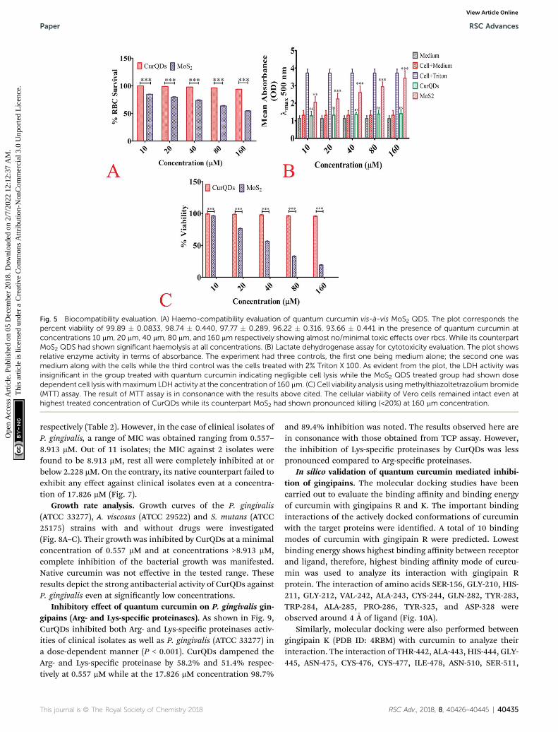
Fig. 5 Biocompatibility evaluation. (A) Haemo-compatibility evaluation of quantum curcumin vis-a-vis MoS2 QDS. The plot corresponds thepercent viability of 99.89 � 0.0833, 98.74 � 0.440, 97.77 � 0.289, 96.22 � 0.316, 93.66 � 0.441 in the presence of quantum curcumin atconcentrations 10 mm, 20 mm, 40 mm, 80 mm, and 160 mm respectively showing almost no/minimal toxic effects over rbcs. While its counterpartMoS2 QDS had shown significant haemolysis at all concentrations. (B) Lactate dehydrogenase assay for cytotoxicity evaluation. The plot showsrelative enzyme activity in terms of absorbance. The experiment had three controls, the first one being medium alone; the second one wasmedium along with the cells while the third control was the cells treated with 2% Triton X 100. As evident from the plot, the LDH activity wasinsignificant in the group treated with quantum curcumin indicating negligible cell lysis while the MoS2 QDS treated group had shown dosedependent cell lysis withmaximum LDH activity at the concentration of 160 mm. (C) Cell viability analysis usingmethylthiazoltetrazolium bromide(MTT) assay. The result of MTT assay is in consonance with the results above cited. The cellular viability of Vero cells remained intact even athighest treated concentration of CurQDs while its counterpart MoS2 had shown pronounced killing (<20%) at 160 mm concentration.
Paper RSC Advances
Ope
n A
cces
s A
rtic
le. P
ublis
hed
on 0
5 D
ecem
ber
2018
. Dow
nloa
ded
on 2
/7/2
022
12:1
2:37
AM
. T
his
artic
le is
lice
nsed
und
er a
Cre
ativ
e C
omm
ons
Attr
ibut
ion-
Non
Com
mer
cial
3.0
Unp
orte
d L
icen
ce.
View Article Online
respectively (Table 2). However, in the case of clinical isolates ofP. gingivalis, a range of MIC was obtained ranging from 0.557–8.913 mM. Out of 11 isolates; the MIC against 2 isolates werefound to be 8.913 mM, rest all were completely inhibited at orbelow 2.228 mM. On the contrary, its native counterpart failed toexhibit any effect against clinical isolates even at a concentra-tion of 17.826 mM (Fig. 7).
Growth rate analysis. Growth curves of the P. gingivalis(ATCC 33277), A. viscosus (ATCC 29522) and S. mutans (ATCC25175) strains with and without drugs were investigated(Fig. 8A–C). Their growth was inhibited by CurQDs at a minimalconcentration of 0.557 mM and at concentrations >8.913 mM,complete inhibition of the bacterial growth was manifested.Native curcumin was not effective in the tested range. Theseresults depict the strong antibacterial activity of CurQDs againstP. gingivalis even at signicantly low concentrations.
Inhibitory effect of quantum curcumin on P. gingivalis gin-gipains (Arg- and Lys-specic proteinases). As shown in Fig. 9,CurQDs inhibited both Arg- and Lys-specic proteinases activ-ities of clinical isolates as well as P. gingivalis (ATCC 33277) ina dose-dependent manner (P < 0.001). CurQDs dampened theArg- and Lys-specic proteinase by 58.2% and 51.4% respec-tively at 0.557 mM while at the 17.826 mM concentration 98.7%
This journal is © The Royal Society of Chemistry 2018
and 89.4% inhibition was noted. The results observed here arein consonance with those obtained from TCP assay. However,the inhibition of Lys-specic proteinases by CurQDs was lesspronounced compared to Arg-specic proteinases.
In silico validation of quantum curcumin mediated inhibi-tion of gingipains. The molecular docking studies have beencarried out to evaluate the binding affinity and binding energyof curcumin with gingipains R and K. The important bindinginteractions of the actively docked conformations of curcuminwith the target proteins were identied. A total of 10 bindingmodes of curcumin with gingipain R were predicted. Lowestbinding energy shows highest binding affinity between receptorand ligand, therefore, highest binding affinity mode of curcu-min was used to analyze its interaction with gingipain Rprotein. The interaction of amino acids SER-156, GLY-210, HIS-211, GLY-212, VAL-242, ALA-243, CYS-244, GLN-282, TYR-283,TRP-284, ALA-285, PRO-286, TYR-325, and ASP-328 wereobserved around 4 �A of ligand (Fig. 10A).
Similarly, molecular docking were also performed betweengingipain K (PDB ID: 4RBM) with curcumin to analyze theirinteraction. The interaction of THR-442, ALA-443, HIS-444, GLY-445, ASN-475, CYS-476, CYS-477, ILE-478, ASN-510, SER-511,
RSC Adv., 2018, 8, 40426–40445 | 40435

Tab
le1
LDH
cytotoxicity
assayag
ainst
Vero
(kidney)
cells
Med
ium
absorban
ce(l
max
500)
Cell+med
ium
absorban
ce(l
max
500)
Cell+2%
TritonX10
0ab
sorban
ce(l
max
500)
Con
centration
(mM)
CurQDstreatedcells
absorban
ce(l
max
500)
MoS
2treated
cells
absorban
ce(l
max
500)
%cytotoxicity
byMoS
2
1.12
�0.15
31.31
�0.26
43.71
2�
0.22
310
1.35
�0.25
12.45
1�
0.31
40.87
4220
1.47
�0.30
22.64
6�
0.32
8.99
240
1.59
�0.10
52.91
3�
0.36
720
.168
801.63
�0.21
33.42
�0.27
541
.215
160
1.78
�0.21
13.91
3�
0.41
361
.74
40436 | RSC Adv., 2018, 8, 40426–40445
RSC Advances Paper
Ope
n A
cces
s A
rtic
le. P
ublis
hed
on 0
5 D
ecem
ber
2018
. Dow
nloa
ded
on 2
/7/2
022
12:1
2:37
AM
. T
his
artic
le is
lice
nsed
und
er a
Cre
ativ
e C
omm
ons
Attr
ibut
ion-
Non
Com
mer
cial
3.0
Unp
orte
d L
icen
ce.
View Article Online
TYR-512, TRP-513, and ASP-516 amino acids were observedaround 4 �A of curcumin (Fig. 10B).
The analysis suggests that the curcumin interacts withalmost similar affinity with both gingipain R and gingipain Kprotein.
Analysis of the effect of CurQDs on biolm formation by TCPassay
Individual isolates & mixed. The biolm inhibition byquantum curcumin was concentration dependent and wasobserved to be variable among different oral pathogens. At theconcentration 0.557 mM, quantum curcumin was found toinhibit the biolm formation by 61% and 53% in Porphyr-omonas gingivalis and Actinomycetemcomitans viscosus respec-tively. Interestingly, CurQDs were found to be poorly effectiveagainst Streptococcus mutans biolms at the said concentrationwhere merely 35% inhibition was perceived. But, no sooner, theconcentration was escalated to 17.826 mM then, signicantinhibitions of 97%, 91%, and 89% were observed in individualbiolms of P. gingivalis, A. viscosus, and S. mutans respectively(Table 3) (Fig. 11). However, the trend of inhibition was found tobe different when quantum curcumin was tested against mixedbiolm of P. gingivalis, A. viscosus, and S. mutans. The twodistinctly grown mixed biolms responded differently. In thecase of mixed biolm of the three isolates grown simulta-neously, at 0.557 mM concentration, only 49% of biomassreduction was observed. In this condition also, the inhibitionwas found to be concentration dependent with maximuminhibition (87.28%) observed at 17.826 mM concentration. Thisis lesser than the inhibition of any individual biolm of the saidbacteria. However, in the case of second type of biolm whereinP. gingivalis was introduced later on in the antecedently formedbiolm of S. mutans and A. viscosus (24 h old), the quantumcurcumin was found to be comparatively less effective in inhi-bition, although the inhibition was concentration dependent.Unlike individually formed biolms and the previouslydescribed mixed biolm, at 0.557 mM and 17.826 mM concen-trations, only 28% and � 82% of inhibition were manifested.
Confocal microscopic evaluation of the effect of CurQDs onbiolm architecture
P. gingivalis, S. mutans, and A. viscosus isolates were selected andgrown simultaneously for the biolm study because of denserand relatively robust biolm forming potential together, whichis more closely associated with chronic periodontitis. Thedegradative effect of quantum curcumin on mixed biolm wasexamined using CLSM. As shown in Fig. 12A and B, CurQDscorroded the mixed biolm in a dose-dependent manner (P <0.001). With initial exposure to 0.557 mM CurQDs, the biolmmatrix slackened off (became more porous which indicates theloosening of the matrix material) which can be seen unambig-uously in Fig. 12A(B). This loosening intensied to its maximawhen the exposed concentration was escalated to 8.913 mM(Fig. 12A(D)). Interestingly, upon exposure of 17.826 mM, onecan note the scattered planktonic cells. These results are inconsonance with the observations of TCP assay.
This journal is © The Royal Society of Chemistry 2018

Fig. 6 Flow cytometry analyses of the cell cultures stained with DCFH-DA to probe ros generation in the groups treated with two discretequantum dots. The symbols, pattern and colours used here remained the same for every panel. (A) Total 100 000 cells were acquired for eachflow cytometry analysis. Histograms of untreated logarithmic vero cells labeled with DCFH-DA. The first panel shown herein at proximal leftdepicts the bivariate dot plot diagram of the intensity (height) of forward scatter versus area of forward scatter to select the singlet cells. Thesinglet cells were gated and its population was found to be 65.9%. Moving right to this panel (the middle one) is a plot diagram of the side-scatterversus forward scatter depicting the cellular granularity or internal complexity. At the distal most panel of A, intensity of untreated cell populationwas captured for further comparisons in FL1A channel. (B) Histograms of MoS2 QDS treated logarithmic Vero cells labeled with DCFH-DA. Thepanels show the increase in cell population (66.3%) that had taken up DCFH-DA indicating increased ros generation. Similarly, the distal mostpanel shows that themajority of cell population had shifted right with highly significant intensity, which embarks the pronounced ROS generationand damage. (C) Histograms of CurQDs treated logarithmic vero cells labeled with DCFH-DA. The panels show the decrease in cell population(47.4%) that had taken up DCFH-DA indicating increased ros generation. The distal most panel shows that the majority of cell population hadshifted right but the intensity was significantly low, which depicts the ROS quenching by the quantum curcumin.
Paper RSC Advances
Ope
n A
cces
s A
rtic
le. P
ublis
hed
on 0
5 D
ecem
ber
2018
. Dow
nloa
ded
on 2
/7/2
022
12:1
2:37
AM
. T
his
artic
le is
lice
nsed
und
er a
Cre
ativ
e C
omm
ons
Attr
ibut
ion-
Non
Com
mer
cial
3.0
Unp
orte
d L
icen
ce.
View Article Online
For the assessment of the nature of the interaction of thequantum curcumin and the matrix, we selectively studied(Fig. 12B) the panel D of Fig. 12A. One can note the ubiquitous
Table 2 Minimum inhibitory concentration of quantum curcumin
Bacterial isolate MIC (mM) MBIC50
Porphyromonas gingivalis (ATCC 33277) 1.114 0.557Actinomycetemcomitans viscosus (ATCC29522)
2.228 0.557
Streptococcus mutans (ATCC 25175) 8.913 1.14
This journal is © The Royal Society of Chemistry 2018
distribution of quantum curcumin (as green uorescentsignals, Fig. 12B(B)) in the biolm matrix (red uorescentsignals, Fig. 12B(A). Panel 12B(C) shows the intermediary
(mM) MBIC90 (mM)MIC (mM) formetronidazole
MBIC90 (mM)for metronidazole
17.826 1.46 93.4817.826 2.921 186.97
>17.826 11.68 373.94
RSC Adv., 2018, 8, 40426–40445 | 40437

Fig. 7 MIC determination of CurQDs against P. gingivalis (ATCC33277), S. mutans (ATCC 25175), and A. viscosus (ATCC 29522). Lanes1, 2 and 3 are respectively of P. gingivalis (ATCC 33277), S. mutans(ATCC 25175), and A. viscosus (ATCC 29522). The MIC was found to be1.114, 2.228, and 1.114 mm (the wells shown with blue arrows) while theMBC was found to be 2.228, 4.456 and 4.456 mm (the wells shown inblack arrows) against the said organisms respectively. MIC was definedwhere the colour first changed sharply from pink to brick red colourand the MBC was defined where the brick red colour first intensified.
RSC Advances Paper
Ope
n A
cces
s A
rtic
le. P
ublis
hed
on 0
5 D
ecem
ber
2018
. Dow
nloa
ded
on 2
/7/2
022
12:1
2:37
AM
. T
his
artic
le is
lice
nsed
und
er a
Cre
ativ
e C
omm
ons
Attr
ibut
ion-
Non
Com
mer
cial
3.0
Unp
orte
d L
icen
ce.
View Article Online
orange coloured signals which show the substantial interactionof quantum curcumin with the matrix materials. Panel 12C,a uorogram, portrays the co-localization of CurQDs and the
Fig. 8 Growth rate analysis curve of (A) P. gingivalis (ATCC 33277) (B) A
40438 | RSC Adv., 2018, 8, 40426–40445
matrix for the region where a line is arbitrarily drawn for thestability assessment of the nature of co-localization (Fig. 12C).The unvarying linearity was observed for the signals in the u-orogram that revealed the stable association/interaction.
Discussion
The primary objective of the present study was the synthesisand the appraisal of antibacterial and antibiolm potential ofquantum curcumin (CurQDs) on pathogens especially involvedin progression and aggravation of oral disease(s) such aschronic periodontitis (P. gingivalis ATCC 33277, A. viscosusATCC 29522 and S. mutans ATCC 25175). We further attemptedto observe the inhibitory effects of quantum curcumin over Pgingivalis specic gingipains (Arg- and Lys-specic proteinases).Besides, we also evaluated the biocompatibility of quantumcurcumin in detail.
Quantum dots (QDs) are the discrete class of uorescentparticles with smaller physical dimensions compared to theexciton Bohr radius. These QDs have high quantum efficiency,long-term photo-stability, narrow emission, and continuousabsorption spectra.43 Excitation–emission prole reveals that italways emits the same light irrespective of excitation wave-length. This simply implies that a single emitted colour can beseen throughout by using single laser excitation source. Thisemitted wavelength can further be used for imaging owing toexcellent photo-stability.44 Our synthesized quantum curcuminwas found to be green uorescent and this property wasexploited well in the confocal microscopy. No additional uo-rescent tag was needed for the evaluation of its biologicalactivity using CLSM or any other method that requires
. viscosus (ATCC 29522) and (C) S. mutans (ATCC 25175).
This journal is © The Royal Society of Chemistry 2018

Fig. 9 Inhibitory effect of quantum curcumin on P. gingivalis gingi-pains (Arg- and Lys-proteinases). The inhibition percentage was foundto be dose dependent with maximum inhibition of 98.7% and 89.4%respectively for Arg- and Lys-proteinases at the concentration of17.826 mm.Of note at all concentrations quantum curcuminwas foundto be more effective in inhibiting Arg-proteinase compared to Lys-proteinase.
Paper RSC Advances
Ope
n A
cces
s A
rtic
le. P
ublis
hed
on 0
5 D
ecem
ber
2018
. Dow
nloa
ded
on 2
/7/2
022
12:1
2:37
AM
. T
his
artic
le is
lice
nsed
und
er a
Cre
ativ
e C
omm
ons
Attr
ibut
ion-
Non
Com
mer
cial
3.0
Unp
orte
d L
icen
ce.
View Article Online
uorescent probing/tagging. In our present study, aersynthesizing CurQDs as per protocol from our previous study,we analysed the QDs particle on some additional parameterssuch as X-ray photoelectron spectroscopy, X-ray diffractionanalysis and NMR apart from the previously describedparameters.
Essentially, biomedical applications of QDs require theirhigh-quality and high aqueous solubility. Quantum dots couldbe made directly in water but they are polydisperse. They haveeither narrow available size ranges or wide size distribution.This obscures its use as imaging material in biologicalsystems.44–47 But bestows an additional advantage in trickling
Fig. 10 (A) Interaction of curcumin with gingipain R (B) interaction of cu
This journal is © The Royal Society of Chemistry 2018
down across the layers of biolm matrices with assorted poresizes. For imaging pursuits, monodisperse quantum dots canbe produced from high-temperature organic solvent syntheticstrategies possessing wide emission colours which may rangefrom ultraviolet to near infrared (300–2500 nm) by simplytuning its size, composition and/or structure. However, theseQDs are insoluble in water.48
QDs have unique physico-electro-optical properties due tothe combination of innate material characteristics & itsdimensionality. Our PL and UV-vis prole is in consonance withthe said characteristics. The rationale behind the strongeractivity of quantum curcumin is related to the reduction in itsparticle size in the range of 0.5 to 7 nm (average size 3.5 nm),which is far less than the size of native curcumin particles(average size 2350 nm). This may be the reason for betterpenetration and interaction with the biolm matrix and higheruptake by the bacterial cells.
The basic challenge of quantum dot synthesis is to control itsuniform growth using stabilizers to avoid agglomeration oraggregation that may culminate into the formation of micro-crystals. However, during our synthesis, when acetone dissolvedcurcumin was injected slowly at the rate of 0.2 ml min�1 in theaqueous phase at 75 � 5 �C with continuous ultra-sonication,the solubilized curcumin spread simultaneously with acetonewith rapid volatilization. As acetone is miscible with water withdensity almost half of it, its rapid volatilization in presence ofhigh energy sonication, resulted in sizing down of curcumin toquantum dot level, ruled out the need of addition of stabilizersto prevent conglomeration. The TEM prole revealed that theaverage size of the CurQDs was 3.5 nm. The HR-TEM shows aninterlayer spacing of�0.63 nm corresponding to the (002) planeof curcumin. The XRD data of quantum curcumin revealsnumber of peaks in the range of 2q (10–40�) implying its crys-talline nature. XRD was used to verify the crystalline content ofthe drug present in the formulation. Surface characteristics and
rcumin with gingipain K.
RSC Adv., 2018, 8, 40426–40445 | 40439

Fig. 11 Evaluation of the degradative effect of quantum curcumin onbiofilm formation by TCP assay. The inhibition was concentrationdependent in all the three bacteria. P. gingivalis biofilmwas found to bemore susceptible compared to those of A. viscosus and S. mutans.
Table 3 Comparison of biofilm degradative activity of CurQDs (mM) based on percent reduction in biofilm biomass in TCP assay
Bacterial strains Biolm inhibition as % decrease in biomass/matrix
Porphyromonas gingivalis, ATCC 33277 97.43 93.59 87.8 81.76 72.57 61.24Actinomycetemcomitans viscosus, ATCC29522
91.29 84.67 79.43 73.67 65.42 53.13
Streptococcus mutans, ATCC 25175 88.77 78.18 69.15 58.23 49.6 35.25Mixed biolm, P. gingivalis, S. mutansand A. viscosus together
93.28 88.54 79.67 71.41 60.87 49.32
Mixed biolm, S. mutans + A. viscosusthen P. gingivalis
81.65 72.88 60.37 51.25 39.76 28.26
Effective conc. (mM) 17.826 8.913 4.456 2.228 1.114 0.557
RSC Advances Paper
Ope
n A
cces
s A
rtic
le. P
ublis
hed
on 0
5 D
ecem
ber
2018
. Dow
nloa
ded
on 2
/7/2
022
12:1
2:37
AM
. T
his
artic
le is
lice
nsed
und
er a
Cre
ativ
e C
omm
ons
Attr
ibut
ion-
Non
Com
mer
cial
3.0
Unp
orte
d L
icen
ce.
View Article Online
charge of CurQDs were investigated by zeta potential analysisand was found to be �25.6 mV. This value provides indirectevidence of the potential stability of the quantum curcumin.The UV-vis spectra showed three bands at �265, 426 and512 nm. A broad absorption band was obtained at �426 nmalong with a shoulder at �512 nm extending to the visibleregion. The absorption band at 426 nm in the visible region canbe assigned to low energyp–p* transitions of curcumin in waterarising possibly due to change in the tautomeric form of theketo-enol-enolate group. The chemistry of the synthesizedquantum curcumin was further ascertained by using 1H and 13CNMR spectroscopy. Besides, Raman spectroscopy was also usedto assess the phase variability and the structure of the quantumcurcumin. The results clearly show the synthesis of curcuminquantum dots.
Various institutions across the globe have looked for itstoxicity prole. Curcumin has been assessed by the Joint FAO/WHO Expert Committee on Food Additives (JECFA) and theEU Scientic Committee for Food (SCF).49 The JECFA set anacceptable daily intake (ADI) value of 0–3 mg kg�1 body weightper day for curcumin in 2004. The ADI value of 3 mg kg�1 bodyweight per day was based on the NOAEL (no observed adverseeffect level) of 250–320 mg kg�1 body weight per day. The SCF,
40440 | RSC Adv., 2018, 8, 40426–40445
on the other hand, has not made any specication for the ADIvalue of curcumin.50 According to the directive 67/548/EWG asreported by Rimbach et al., curcumin neither poses a threat toman nor the environment.50 The JECFA evaluated four acuteoral toxicity studies in mice and rats and found LD50 values of 2to 10 g kg�1 body weight for mice and 5 to 10 g kg�1 body weightfor rats. In the present study, the in vitro toxicity proling wasdone in the range of 2–32 mg ml�1 and it qualied as non-toxicon all the concentrations.49 Therefore, the quantum curcumin(CurQDs) in the present study was found signicantly effectiveas both antibacterial and antibiolm agent that too in theconcentration as low as 0.089 mg ml�1 while it was found non-toxic even at the concentration of 1024 mg ml�1 which ismuch below themaximum non-toxic threshold level as reportedearlier.
To determine whether the synthesized CurQDs showed onlyanti-bacterial and antibiolm efficacy without imparting anytoxicity to healthy cells, its biocompatibility was evaluated. Forthis purpose, the morphology of Vero cells aer treatment for24 h of culture was monitored by phase contrast and uores-cence microscopy. Results clearly suggested that the Vero cellsproliferated over the time forming a cell layer on the cultureplate. Notably, the Vero cells displayed a more polygonalmorphology upon CurQDs treatment which is the normalphenotype behavior of healthy Vero cells, suggesting thatquantum curcumin restored/promoted Vero functionality.
The viability and proliferation rate of Vero cells were char-acterized by MTT assay, which was directly proportional to theoptical density of the reaction product from the MTT workingsolution with live cells. As seen, clearly in Fig. S1,† aer 3 days,the viability was signicantly higher indicating that quantumcurcumin has enhanced the cell viability.
Periodontitis continues to be a major health concern,particularly amid people with poor oral hygiene. Its develop-ment and progression involve multifactorial cascades and toa larger extent, it is dynamic in terms of its regulation andsocialization.7,8 The primary microbiological factor which issignicantly responsible for the disease progression is the shiin the oral microora.5 Virulence factors of P. gingivalis arediverse and can be broadly classied on functionality. Forinstance, while mbriae, hemagglutinins, outer membraneproteins, vesicles, and gingipains are mainly involved in colo-nization and attachment, proteinases (Arg- and Lys-gingipains),
This journal is © The Royal Society of Chemistry 2018
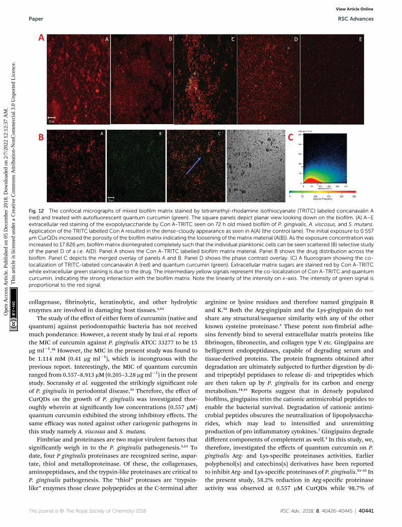
Fig. 12 The confocal micrographs of mixed biofilm matrix stained by tetramethyl-rhodamine isothiocyanate (TRITC) labeled concanavalin A(red) and treated with autofluorescent quantum curcumin (green). The square panels depict planar view looking down on the biofilm. (A) A–Eextracellular red staining of the exopolysaccharide by Con A-TRITC seen on 72 h old mixed biofilm of P. gingivalis, A. viscosus, and S. mutans.Application of the TRITC labelled Con A resulted in the dense-cloudy appearance as seen in A(A) (the control lane). The initial exposure to 0.557mmCurQDs increased the porosity of the biofilmmatrix indicating the loosening of the matrix material (A(B)). As the exposure concentration wasincreased to 17.826 mm, biofilmmatrix disintegrated completely such that the individual planktonic cells can be seen scattered (B) selective studyof the panel D of a i.e. A(D). Panel A shows the Con A-TRITC labelled biofilm matrix material. Panel B shows the drug distribution across thebiofilm. Panel C depicts the merged overlay of panels A and B. Panel D shows the phase contrast overlay. (C) A fluorogram showing the co-localization of TRITC-labeled concanavalin A (red) and quantum curcumin (green). Extracellular matrix sugars are stained red by Con A-TRITCwhile extracellular green staining is due to the drug. The intermediary yellow signals represent the co-localization of Con A-TRITC and quantumcurcumin, indicating the strong interaction with the biofilm matrix. Note the linearity of the intensity on x-axis. The intensity of green signal isproportional to the red signal.
Paper RSC Advances
Ope
n A
cces
s A
rtic
le. P
ublis
hed
on 0
5 D
ecem
ber
2018
. Dow
nloa
ded
on 2
/7/2
022
12:1
2:37
AM
. T
his
artic
le is
lice
nsed
und
er a
Cre
ativ
e C
omm
ons
Attr
ibut
ion-
Non
Com
mer
cial
3.0
Unp
orte
d L
icen
ce.
View Article Online
collagenase, brinolytic, keratinolytic, and other hydrolyticenzymes are involved in damaging host tissues.3,13
The study of the effect of either form of curcumin (native andquantum) against periodontopathic bacteria has not receivedmuch ponderance. However, a recent study by Izui et al. reportsthe MIC of curcumin against P. gingivalis ATCC 33277 to be 15mg ml�1.41 However, the MIC in the present study was found tobe 1.114 mM (0.41 mg ml�1), which is incongruous with theprevious report. Interestingly, the MIC of quantum curcuminranged from 0.557–8.913 mM (0.205–3.28 mgml�1) in the presentstudy. Socransky et al. suggested the strikingly signicant roleof P. gingivalis in periodontal disease.51 Therefore, the effect ofCurQDs on the growth of P. gingivalis was investigated thor-oughly wherein at signicantly low concentrations (0.557 mM)quantum curcumin exhibited the strong inhibitory effects. Thesame efficacy was noted against other cariogenic pathogens inthis study namely A. viscosus and S. mutans.
Fimbriae and proteinases are two major virulent factors thatsignicantly weigh in to the P. gingivalis pathogenesis.3,13 Todate, four P gingivalis proteinases are recognized serine, aspar-tate, thiol and metalloproteinase. Of these, the collagenases,aminopeptidases, and the trypsin-like proteinases are critical toP. gingivalis pathogenesis. The “thiol” proteases are “trypsin-like” enzymes those cleave polypeptides at the C-terminal aer
This journal is © The Royal Society of Chemistry 2018
arginine or lysine residues and therefore named gingipain Rand K.52 Both the Arg-gingipain and the Lys-gingipain do notshare any structural/sequence similarity with any of the otherknown cysteine proteinase.3 These potent non-mbrial adhe-sins fervently bind to several extracellular matrix proteins likebrinogen, bronectin, and collagen type V etc. Gingipains arebelligerent endopeptidases, capable of degrading serum andtissue-derived proteins. The protein fragments obtained aerdegradation are ultimately subjected to further digestion by di-and tripeptidyl peptidases to release di- and tripeptides whichare then taken up by P. gingivalis for its carbon and energymetabolism.14,15 Reports suggest that in densely populatedbiolms, gingipains trim the cationic antimicrobial peptides toenable the bacterial survival. Degradation of cationic antimi-crobial peptides obscures the neutralization of lipopolysaccha-rides, which may lead to intensied and unremittingproduction of pro inammatory cytokines.7 Gingipains degradedifferent components of complement as well.3 In this study, we,therefore, investigated the effects of quantum curcumin on P.gingivalis Arg- and Lys-specic proteinases activities. Earlierpolyphenol(s) and catechins(s) derivatives have been reportedto inhibit Arg- and Lys-specic proteinases of P. gingivalis.53–55 Inthe present study, 58.2% reduction in Arg-specic proteinaseactivity was observed at 0.557 mM CurQDs while 98.7% of
RSC Adv., 2018, 8, 40426–40445 | 40441
RSC Advances Paper
Ope
n A
cces
s A
rtic
le. P
ublis
hed
on 0
5 D
ecem
ber
2018
. Dow
nloa
ded
on 2
/7/2
022
12:1
2:37
AM
. T
his
artic
le is
lice
nsed
und
er a
Cre
ativ
e C
omm
ons
Attr
ibut
ion-
Non
Com
mer
cial
3.0
Unp
orte
d L
icen
ce.
View Article Online
reduction was seen at the concentration of 17.826 mM in bothATCC 33277 and clinical isolates of P. gingivalis. The similartrend was seen over Lys-specic proteinase activities with 51.4%and 89.4% reduction upon administration with 0.557 mM and17.826 mM CurQDs concentrations. The results obtained herewas then investigated by in silico docking analysis by AutoDock.The thermodynamic prole of the interaction of curcumin withgingipains R and K indicated that the binding process wascharacterized by large exothermic enthalpies (�7.01 and �7.02cal mol�1). Besides, the inhibition constant was found to be7.24 mM and 7.1 mM against gingipains R and K respectively. Inthe present study, curcumin was found adopting conformationssuitable for maximizing interactions with the gingipains. Thismay be due to its novel structural geometry and chemistrywhich include two hydrophobic phenyl domains connected bya exible methylene linker along with keto–enol tautomerismwhich provides a strong and directed electrostatic interaction toincrease favorable binding free energy.
Recently, the effect of native curcumin on mature, multi-species biolm was also reported wherein the multispeciesbiolm was allowed to mature for 7 days and then treated withcurcumin for 24 h. Aer treatment, the outre-structural changeswere observed using scanning electron microscopy.56,57
Curcumin-treated biolms displayed signicantly reducedmetabolic activity and no visible extracellular matrix. Althoughthe previous study investigated the effects of curcumin onmatured biolms, the current study investigates the effect ofcurcumin on the early as well as the late stages of biolmformed by P. gingivalis with S. mutans and A. viscosus,mimicking the in vivo condition of biolm associated lesion ofperiodontitis.
We observed when all the three bacteria were grownconcurrently and then the drug was administered, the effect ofthe drug was maximum, and at 8.913 mM concentration, biolmwas completely suppressed within 90 min. The said observationwas in consonance with the report which ascertains theinvolvement of P. gingivalis FimA and streptococcal cell surfaceglyceraldehyde-3-phosphate dehydrogenase (GAPDH) in theearly stages of biolm formation.58 Therefore, it can be con-jectured that quantum curcumin binds with high affinity to theP. gingivalis FimA which subsequently inhibits its binding to thestreptococcal GAPDH, culminating in the suppression of thebiolm. Moreover, in accordance with other reports, it may alsoinhibit auto-aggregation and interaction of FimA with salivaryproteins, resulting in suppression of P. gingivalis biolmformation. However, we cannot conrm whether the observedinhibition is owing to the different mechanism(s) or simply itrepresents multifactorial selection of targets depending uponthe site, ending up in biolm inhibition. However, we hypoth-esized the killing action of quantum curcumin by membranepermeabilization and subsequent lysis.
The TCP assay results and our hypothesis pertaining to themode of action was further validated by CLSM wherein P. gin-givalis (late colonizer of dental biolm) was added to anteced-ently formed biolm of S. mutans and A. viscosus (facilitator).We found CurQDs signicantly effective in degradation of thebiolmmatrix at the dose as low as 8.913 mM (3.28 mgml�1). The
40442 | RSC Adv., 2018, 8, 40426–40445
result revealed the interaction(s) between quantum curcuminand the biolm matrix which eventually culminated in theloosening and ultimately degradation of the biolm in theconcentration dependent manner. Quantum curcumin is thegreen uorescent moiety while concanavalin A (Con A), labelledwith tetramethyl-rhodamine isothiocyanate (TRITC) exhibitsred uorescence. Con A-TRITC binds with matrix sugars andhence gives a gross approximation related to biolm biomass.The colocalization map unfolds the juxtapositioning of Con A-TRITC and the quantum curcumin which is the indicative ofits higher affinity for the biolm matrix material. Accuratecolocalization analysis requires distinct uorescence emissionspectra. Both Con A-TRITC and quantum curcumin colours areeasily distinguishable and therefore yielded clear uorogramwith minimal bleed-through.
Reports pertaining to the maintenance of periodontal healthusing numerous natural products/phyto-extracts/plantsecondary metabolites are in abundance. They were screenedfor their potential to inhibit periodontitis.53–57,59,60 For instance,extracts from E. globules leaf, exhibited the antibacterial activitywith MIC values of 1–10 mg ml�1 against P. gingivalis and T.denticola, and was found to inhibit P. gingivalis Arg- and Lys-specic proteinase activities at 50–100 mg ml�1.55 Similarly,another controlled study showed that chewing gum containingeucalyptus extracts signicantly inhibited the plaque formationand gingival inammation.53 The MIC of CurQDs was some 244times lower (1.114 mM, 0.41 mg ml�1) than that of all testiedplant secondary metabolites against P. gingivalis. Besides,CurQDs also inhibited the growth of other anaerobes like A.viscosus as well and other cariogenic bacteria like S. mutanswhereas other plant secondary metabolites had insignicanteffects on the same. Recent reports suggest that green tea(Camellia sinensis) polyphenols, particularly epigallocatechingallate, inhibited the growth of P. gingivalis at concentrations of250–500 mg ml�1 and 50 mg ml�1.61,62 It is therefore, obviousthat compared to the quantum curcumin the efficacy of the saidphytochemicals are signicantly compromised.
Recently, therapeutic efficacy of curcumin for the treatmentof aggravated and chronic periodontitis was evaluated in a pilotstudy. In the study, it found that the patients who underwentscaling and root planing, followed by using curcumin mouth-wash (20%) for 21 days, showed signicant improvement in thegingival inammation than those who underwent only scalingand root planing. However, they found insignicant differencein their plaque indices.63
Conclusion
The molecule curcumin, the most active polyphenol present incurcuma, owing to its high hydropathy index and instability hasnot been able to justify its role as frontline drug modality in thetreatment of infectious and non-infectious diseases. The resultsof the current study indicate that quantum curcumin not onlyinhibited the growth of various periodontopathic bacteria butalso exhibited potent antibiolm activities against mixed bio-lm formed by P. gingivalis, A. viscosus, and S. mutans. Being anasaccharolytic bacterium, P. gingivalis solely relies for its carbon
This journal is © The Royal Society of Chemistry 2018
Paper RSC Advances
Ope
n A
cces
s A
rtic
le. P
ublis
hed
on 0
5 D
ecem
ber
2018
. Dow
nloa
ded
on 2
/7/2
022
12:1
2:37
AM
. T
his
artic
le is
lice
nsed
und
er a
Cre
ativ
e C
omm
ons
Attr
ibut
ion-
Non
Com
mer
cial
3.0
Unp
orte
d L
icen
ce.
View Article Online
and energy sources from proteins, which proves the inevitablerole of these gingipains R and K for the growth and multipli-cation in the host that help in continuing the vicious life cycleleading to the proliferation and aggravation in the lesion ofchronic periodontitis. Therefore, in this context, the activity ofquantum curcumin against P. gingivalis specic Arg- and Lys-proteinase indicates its usefulness as rst-line therapeuticmodality. The bioassay utilized herein for evaluating inhibitorsof P. gingivalis specic Arg- and Lys-specic proteinases may beused in future in screening of potential drugs candidatesagainst such pathogens. However, for pharmacological investi-gations one may utilize appropriate animal models such asnonhuman primates (like rhesus monkeys (Macaca mulatta),cynomolgus monkeys (Macaca fascicularis), and baboons (Papioanubis)), miniature pigs (Minnesota miniature pig), dogs,ferrets (Mustela putorius) and rodents (rice rat (Oryzomys pal-ustris), baker mouse) to substantiate the role of quantum cur-cumin in the management of chronic periodontitis. In all, theseresults add to the considerable body of evidence of antibacte-rial, antivirulence, antibiolm activities of quantum curcuminover P. gingivalis.
Author contributions
A. K. S. and P. P. conceived, designed and analyzed results of thestudy. A. K. S. and S. Y. designed the cultivation experiments,performed susceptibility experiments, bacterial growth rateanalysis, gingipains inhibition assay along with biolm degra-dation assay. A. K. S., Z. F., P. A. performed experiments onsynthesis, physical characterizations and analyzed data. A. K.S., M. B., M. K. G. performed confocal microcopy experimentsand analyzed the data. A. K. S., Z. F., P. A., K. N., and P. K. N.performed toxicity assay while A. K. S. and KS performed andanalyzed results with ow cytometry. A. K. S. wrote the manu-script which was endorsed by all the co-authors.
Conflicts of interest
There are no conicts to declare.
Acknowledgements
We sincerely acknowledge the nancial support from theDepartment of Science and Technology, Government of India[DST project (DST/TSG/NTS/2013/47) sanctioned to MB, and PP,contingency grant offered to AKS as JRF (2061430918 vide 22/06/2014(i) EU-V) by University Grants Commission, New Delhi andDST-PURSE grant sanctioned to Department of Microbiology,IMS, BHU.
References
1 R. C. Page and K. S. Kornman, The pathogenesis of humanperiodontitis: an introduction, Periodontol. 2000, 1997, 14,9–11.
2 R. C. Page, S. Offenbacher, H. E. Schroeder, G. J. Seymourand K. S. Kornman, Advances in the pathogenesis of
This journal is © The Royal Society of Chemistry 2018
periodontitis: summary of developments, clinicalimplications and future directions, Periodontol. 2000, 1997,14, 216–248.
3 K. Y. How, K. P. Song and K. G. Chan, Porphyromonasgingivalis: an overview of periodontopathic pathogen belowthe gum line, Front. Microbiol., 2016, 7, 53.
4 P. E. Petersen and H. Ogawa, The global burden ofperiodontal disease: towards integration with chronicdisease prevention and control, Periodontol. 2000, 2012, 60,15–39.
5 A. B. Berezow and R. P. Darveau, Microbial shi andperiodontitis, Periodontol. 2000, 2011, 55, 36–47.
6 M. Nakayama and N. Ohara, Molecular mechanisms ofPorphyromonas gingivalis-host cell interaction onperiodontal diseases, Jpn. Dent. Sci. Rev., 2017, 134–140.
7 G. Hajishengallis, Periodontitis: from microbial immunesubversion to systemic inammation, Nat. Rev. Immunol.,2015, 15, 30.
8 G. Hajishengallis, Immunomicrobial pathogenesis ofperiodontitis: keystones, pathobionts, and host response,Trends Immunol., 2014, 35, 3–11.
9 P. M. Bartold and T. E. Van Dyke, Periodontitis: a host-mediated disruption of microbial homeostasis. Unlearninglearned concepts, Periodontol. 2000, 2013, 62, 203–217.
10 A. C. R. Tanner, C. Haffer, G. T. Bratthall, R. A. Visconti andS. S. Socransky, A study of the bacteria associated withadvancing periodontitis in man, J. Clin. Periodontol., 1979,6, 278–307.
11 J. Mysak, S. Podzimek, P. Sommerova, Y. Lyuya-Mi,J. Bartova, T. Janatova, et al., Porphyromonas gingivalis:major periodontopathic pathogen overview, J. Immunol.Res., 2014, 2014, 1–8.
12 H. K. Datta, W. F. Ng, J. A. Walker, S. P. Tuck andS. S. Varanasi, The cell biology of bone metabolism, J. Clin.Pathol., 2008, 61, 577–587.
13 C. W. Cutler, J. R. Kalmar and C. A. Genco, Pathogenicstrategies of the oral anaerobe, Porphyromonas gingivalis,Trends Microbiol., 1995, 3, 45–51.
14 J. Travis, R. Pike, T. Imamura and J. Potempa,Porphyromonas gingivalis proteinases as virulence factors inthe development of periodontitis, J. Periodontal Res., 1997,32, 120–125.
15 J. Potempa, A. Sroka, T. Imamura and J. Travis, Gingipains,the major cysteine proteinases and virulence factors ofPorphyromonas gingivalis: structure, function and assemblyof multidomain protein complexes, Curr. Protein Pept. Sci.,2003, 4, 397–407.
16 T. Bengtsson, A. Khalaf and H. Khalaf, Secreted gingipainsfrom Porphyromonas gingivalis colonies exert potentimmunomodulatory effects on human gingival broblasts,Microbiol. Res., 2015, 178, 18–26.
17 I. de Diego, F. Veillard, M. Sztukowska, T. Guevara,B. Potempa, A. Pomowski, et al., Structure and mechanismof cysteine peptidase Kgp, a major virulence factor ofPorphyromonas gingivalis in periodontitis, J. Biol. Chem.,2014, DOI: 10.1074/jbc.M114.602052.
RSC Adv., 2018, 8, 40426–40445 | 40443
RSC Advances Paper
Ope
n A
cces
s A
rtic
le. P
ublis
hed
on 0
5 D
ecem
ber
2018
. Dow
nloa
ded
on 2
/7/2
022
12:1
2:37
AM
. T
his
artic
le is
lice
nsed
und
er a
Cre
ativ
e C
omm
ons
Attr
ibut
ion-
Non
Com
mer
cial
3.0
Unp
orte
d L
icen
ce.
View Article Online
18 A. D. Haffajee, M. A. Cugini, S. Dibart, C. Smith, R. L. Kent Jrand S. S. Socransky, Clinical and microbiological features ofsubjects with adult periodontitis who responded poorly toscaling and root planing, J. Clin. Periodontol., 1997, 24,767–776.
19 L. Sbordone, L. Ramaglia, E. Gulletta and V. Iacono,Recolonization of the subgingival microora aer scalingand root planing in human periodontitis, J. Periodontol.,1990, 61, 579–584.
20 D. Herrera, M. Sanz, S. Jepsen, I. Needleman and S. Roldan,A systematic review on the effect of systemic antimicrobialsas an adjunct to scaling and root planing in periodontitispatients, J. Clin. Periodontol., 2002, 29, 136–159.
21 A. P. V. Colombo, S. K. Boches, S. L. Cotton, J. M. Goodson,R. Kent, A. D. Haffajee, et al., Comparisons of subgingivalmicrobial proles of refractory periodontitis, severeperiodontitis, and periodontal health using the humanoral microbe identication microarray, J. Periodontol.,2009, 80, 1421–1432.
22 J. G. Caton, S. G. Ciancio, T. M. Blieden, M. Bradshaw,R. J. Crout, A. F. Hei, et al., Treatment withsubantimicrobial dose doxycycline improves the efficacy ofscaling and root planing in patients with adultperiodontitis, J. Periodontol., 2000, 71, 521–532.
23 P. D. Marsh, D. A. Head and D. A. Devine, Prospects of oraldisease control in the future–an opinion, J. Oral Microbiol.,2014, 6, 26176.
24 J. Frias, E. Olle and M. Alsina, Periodontal pathogensproduce quorum sensing signal molecules, Infect. Immun.,2001, 69, 3431–3434.
25 A. K. Singh, P. Prakash, R. Singh, N. Nandy, Z. Firdaus,M. Bansal, et al., Curcumin Quantum Dots MediatedDegradation of Bacterial Biolms, Front. Microbiol., 2017,8, 1517.
26 R. De, P. Kundu, S. Swarnakar, T. Ramamurthy,A. Chowdhury, G. B. Nair, et al., Antimicrobial activity ofcurcumin against Helicobacter pylori isolates from Indiaand during infections in mice, Antimicrob. AgentsChemother., 2009, 53, 1592–1597.
27 W. Jianhua and W. Hai, Antifungal susceptibility analysis ofberberine, baicalin, eugenol and curcumin on Candidaalbicans, J. Med. Coll. PLA, 2009, 24, 142–147.
28 E. Marini, M. Di Giulio, G. Magi, S. Di Lodovico,M. E. Cimarelli, A. Brenciani, et al., Curcumin, anantibiotic resistance breaker against a multiresistantclinical isolate of Mycobacterium abscessus, Phyther. Res.,2018, 32, 488–495.
29 B. Wachter, M. Syrowatka, A. Obwaller and J. Walochnik, Invitro efficacy of curcumin on Trichomonas vaginalis, Wien.Klin. Wochenschr., 2014, 126, 32–36.
30 P. Hu, P. Huang and M. W. Chen, Curcumin reducesStreptococcus mutans biolm formation by inhibitingsortase A activity, Arch. Oral Biol., 2013, 58, 1343–1348.
31 J. Song, B. Choi, E. J. Jin, Y. Yoon and K. H. Choi, Curcuminsuppresses Streptococcus mutans adherence to human toothsurfaces and extracellular matrix proteins, Eur. J. Clin.
40444 | RSC Adv., 2018, 8, 40426–40445
Microbiol. Infect. Dis., 2012, 31, 1347–1352, DOI: 10.1007/s10096-011-1448-y.
32 S.-H. Mun, S.-B. Kim, R. Kong, J.-G. Choi, Y.-C. Kim,D.-W. Shin, et al., Curcumin Reverse Methicillin Resistancein Staphylococcus aureus, Molecules, 2014, 19, 18283–18295,DOI: 10.3390/molecules191118283.
33 B. R. K. Bhawana, H. S. Buttar, V. K. Jain and N. Jain,Curcumin nanoparticles: preparation, characterization,and antimicrobial study, J. Agric. Food Chem., 2011, 59,2056–2061, DOI: 10.1021/jf104402t.
34 P. Tyagi, M. Singh, H. Kumari, A. Kumari andK. Mukhopadhyay, Bactericidal activity of curcumin I isassociated with damaging of bacterial membrane, PLoSOne, 2015, 10, 1–15, DOI: 10.1371/journal.pone.0121313.
35 H. K. Syed, M. A. Iqbal, R. A. Haque and K.-K. Peh, Synthesis,characterization and antibacterial activity of a curcumin–silver(I) complex, J. Coord. Chem., 2015, 68, 1088–1100,DOI: 10.1080/00958972.2014.1003051.
36 O. Naksuriya, S. Okonogi, R. M. Schiffelers andW. E. Hennink, Curcumin nanoformulations: a review ofpharmaceutical properties and preclinical studies andclinical data related to cancer treatment, Biomaterials,2014, 35, 3365–3383, DOI: 10.1016/j.biomaterials.2013.12.090.
37 Y. Liu, Y. Cai, X. Jiang, J. Wu and X. Le, Molecularinteractions, characterization and antimicrobial activity ofcurcumin–chitosan blend lms, Food Hydrocolloids, 2015,52, 564–572, DOI: 10.1016/j.foodhyd.2015.08.005.
38 R. Feng, Z. Song and G. Zhai, Preparation and in vivopharmacokinetics of curcumin-loaded PCL-PEG-PCLtriblock copolymeric nanoparticles, Int. J. Nanomed., 2012,7, 4089–4098, DOI: 10.2147/IJN.S33607.
39 J. N. Eloff, A sensitive and quick microplate method todetermine the minimal inhibitory concentration of plantextracts for bacteria, Planta Med., 1998, 64, 711–713.
40 R. Pike, W. McGraw, J. Potempa and J. Travis, Lysine-andarginine-specic proteinases from Porphyromonasgingivalis. Isolation, characterization, and evidence for theexistence of complexes with hemagglutinins, J. Biol. Chem.,1994, 269, 406–411.
41 S. Izui, S. Sekine, K. Maeda, M. Kuboniwa, A. Takada,A. Amano, et al., Antibacterial activity of curcumin againstperiodontopathic bacteria, J. Periodontol., 2016, 87, 83–90.
42 A. K. Singh, P. Prakash, A. Achra, G. P. Singh, A. Das andR. K. Singh, Standardization and classication of in vitrobiolm formation by clinical isolates of Staphylococcusaureus, J. Global Infect. Dis., 2017, 9, 93.
43 Y. Wang and L. Chen, Quantum dots, lighting up theresearch and development of nanomedicine, Nanomedicine,2011, 7, 385–402, DOI: 10.1016/j.nano.2010.12.006.
44 X. Michalet, L. a Bentolila, J. M. Tsay and S. Doose, QuantumDots for Live Cells in vivo Imaging, and Diagnostics, Science,2005, 307, 538–544, DOI: 10.1126/science.1104274.
45 K. Sun, M. Vasudev, H.-S. Jung, J. Yang, A. Kar, Y. Li, et al.,Applications of colloidal quantum dots, Microelectron. J.,2009, 40, 644–649, DOI: 10.1016/j.mejo.2008.06.033.
This journal is © The Royal Society of Chemistry 2018
Paper RSC Advances
Ope
n A
cces
s A
rtic
le. P
ublis
hed
on 0
5 D
ecem
ber
2018
. Dow
nloa
ded
on 2
/7/2
022
12:1
2:37
AM
. T
his
artic
le is
lice
nsed
und
er a
Cre
ativ
e C
omm
ons
Attr
ibut
ion-
Non
Com
mer
cial
3.0
Unp
orte
d L
icen
ce.
View Article Online
46 T. Jamieson, R. Bakhshi, D. Petrova, R. Pocock, M. Imani andA. M. Seifalian, Biological applications of quantum dots,Biomaterials, 2007, 28, 4717–4732, DOI: 10.1016/j.biomaterials.2007.07.014.
47 R. J. Byers and E. R. Hitchman, Quantum dots brightenbiological imaging, Prog. Histochem. Cytochem., 2011, 45,201–237, DOI: 10.1016/j.proghi.2010.11.001.
48 W. W. Yu, E. Chang, R. Drezek and V. L. Colvin, Water-soluble quantum dots for biomedical applications,Biochem. Biophys. Res. Commun., 2006, 348, 781–786, DOI:10.1016/j.bbrc.2006.07.160.
49 E. F. S. Authority, Rened exposure assessment for curcumin(E 100), EFSA J., 2014, 12, 3876.
50 T. Esatbeyoglu, P. Huebbe, I. M. A. Ernst, D. Chin,A. E. Wagner and G. Rimbach, Curcumin-from molecule tobiological function, Angew. Chem., Int. Ed., 2012, 51, 5308–5332, DOI: 10.1002/anie.201107724.
51 S. S. Socransky and A. D. Haffajee, Evidence of bacterialetiology: a historical perspective, Periodontol. 2000, 1994, 5,7–25.
52 S. C. Holt, L. Kesavalu, S. Walker and C. A. Genco, Virulencefactors of Porphyromonas gingivalis, Periodontol. 2000, 1999,20, 168–238.
53 H. Nagata, Y. Inagaki, M. Tanaka, M. Ojima, K. Kataoka,M. Kuboniwa, et al., Effect of eucalyptus extract chewinggum on periodontal health: a double-masked, randomizedtrial, J. Periodontol., 2008, 79, 1378–1385.
54 H. Nagata, Y. Inagaki, Y. Yamamoto, K. Maeda, K. Kataoka,K. Osawa, et al., Inhibitory effects of macrocarpals on thebiological activity of Porphyromonas gingivalis and otherperiodontopathic bacteria, Oral Microbiol. Immunol., 2006,21, 159–163.
55 K. Takahashi, M. Fukazawa, H. Motohira, K. Ochiai,H. Nishikawa and T. Miyata, A pilot study on antiplaque
This journal is © The Royal Society of Chemistry 2018
effects of mastic chewing gum in the oral cavity, J.Periodontol., 2003, 74, 501–505.
56 C. C. C. Quishida, E. G. D. O. Mima, J. H. Jorge, C. E. Vergani,V. S. Bagnato and A. C. Pavarina, Photodynamic inactivationof a multispecies biolm using curcumin and LED light,Lasers Med. Sci., 2016, 31, 997–1009.
57 N. C. Araujo, C. R. Fontana, V. S. Bagnato andM. E. M. Gerbi,Photodynamic effects of curcumin against cariogenicpathogens, Photomed. Laser Surg., 2012, 30, 393–399.
58 K. Maeda, H. Nagata, A. Nonaka, K. Kataoka, M. Tanaka andS. Shizukuishi, Oral streptococcal glyceraldehyde-3-phosphate dehydrogenase mediates interaction withPorphyromonas gingivalis mbriae, Microbes Infect., 2004, 6,1163–1170.
59 M. Shahzad, E. Millhouse, S. Culshaw, C. A. Edwards,G. Ramage and E. Combet, Selected dietary (poly) phenolsinhibit periodontal pathogen growth and biolmformation, Food Funct., 2015, 6, 719–729.
60 N. C. Araujo, C. R. Fontana, M. E. M. Gerbi and V. S. Bagnato,Overall-mouth disinfection by photodynamic therapy usingcurcumin, Photomed. Laser Surg., 2012, 30, 96–101.
61 A. Araghizadeh, J. Kohanteb and M. M. Fani, Inhibitoryactivity of green tea (Camellia sinensis) extract on someclinically isolated cariogenic and periodontopathicbacteria, Med. Princ. Pract., 2013, 22, 368–372.
62 S. Sakanaka, M. Aizawa, M. Kim and T. Yamamoto,Inhibitory effects of green tea polyphenols on growth andcellular adherence of an oral bacterium, Porphyromonasgingivalis, Biosci., Biotechnol., Biochem., 1996, 60, 745–749.
63 S. Muglikar, K. C. Patil, S. Shivswami and R. Hegde, Efficacyof curcumin in the treatment of chronic gingivitis: a pilotstudy, Oral Health Prev. Dent., 2013, 11, 81–86.
RSC Adv., 2018, 8, 40426–40445 | 40445