Nucleotide sequence of crotamine isoform precursors from a single South American rattlesnake (...
12
Nucleotide sequence of crotamine isoform precursors from a single South American rattlesnake (Crotalus durissus terrificus) G. Ra´dis-Baptista a , N. Oguiura a , M.A.F. Hayashi b , M.E. Camargo c , K.F. Grego d , E.B. Oliveira e , T. Yamane a, * a Biotechnology Center, Instituto Butantan, Av. Vital Brazil, 1500, 05503-900 Sa ˜o Paulo, Brazil b Biochemical and Biophysical Laboratory, Instituto Butantan, Av. Vital Brazil, 1500, 05503-900 Sa ˜o Paulo, Brazil c Department of Microbiology, Biomedical Sciences Institute, University of Sa ˜o Paulo, 05508-900 Sa ˜o Paulo, Brazil d Laboratory of Herpetology, Instituto Butantan, Av. Vital Brazil, 1500, 05503-900 Sa ˜o Paulo, Brazil e Department of Biochemistry, Faculty of Medicine of Ribeira ˜o Preto, University of Sa ˜o Paulo, Ribeira ˜o Preto, SP, Brazil Received 1 June 1998; accepted 15 October 1998 Abstract A cDNA phage library was constructed from venom glands of a single adult specimen of crotamine-plus Crotalus durissus terrificus (South American rattlesnake) captured in a known region. Fifteen crotamine positive clones were isolated using a PCR-based screening protocol and sequenced. These complete cDNAs clones were grouped for maximal alignment into six distinct nucleotide sequences. The crotamine cDNAs, with 340–360 bases, encompass open reading frame of 198 nucleotides with 5 0 and 3 0 untranslated regions of variable size, signal peptide sequence, one crotamine isoform message, and putative poly(A + ) signal. Of these six dierent crotamine cDNA precursors, two predict the identical amino acid sequence previously described by Laure (1975), and the other four a crotamine isoform precursor where the Leucine residue at position 19 is replaced by isoleucine by a single base change. On the other hand, nucleotide variation was observed in the 5 0 and 3 0 untranslated regions, with one interesting variant containing an 18 base pair deletion at the 5 0 untranslated region which results in the usual ATG initiator being replaced by the rarely used GUG start codon. Comparison by Northern blot analysis of poly(A + ) RNA from venom glands of a crotamine-plus specimen to total and poly(A + ) RNA from a crotamine-minus snake Toxicon 37 (1999) 973–984 0041-0101/99/$ - see front matter # 1999 Elsevier Science Ltd. All rights reserved. PII: S0041-0101(98)00226-8 PERGAMON * Corresponding author. Tel.: +55-11-212-1127; fax: +55-11-815-1505.
Transcript of Nucleotide sequence of crotamine isoform precursors from a single South American rattlesnake (...
Nucleotide sequence of crotamine isoformprecursors from a single South Americanrattlesnake (Crotalus durissus terri®cus)
G. Ra dis-Baptista a, N. Oguiura a, M.A.F. Hayashi b,M.E. Camargoc, K.F. Gregod, E.B. Oliveira e, T. Yamanea, *
aBiotechnology Center, Instituto Butantan, Av. Vital Brazil, 1500, 05503-900 SaÄo Paulo, BrazilbBiochemical and Biophysical Laboratory, Instituto Butantan, Av. Vital Brazil, 1500, 05503-900
SaÄo Paulo, BrazilcDepartment of Microbiology, Biomedical Sciences Institute, University of SaÄo Paulo, 05508-900
SaÄo Paulo, BrazildLaboratory of Herpetology, Instituto Butantan, Av. Vital Brazil, 1500, 05503-900 SaÄo Paulo, Brazil
eDepartment of Biochemistry, Faculty of Medicine of RibeiraÄo Preto, University of SaÄo Paulo, RibeiraÄo
Preto, SP, Brazil
Received 1 June 1998; accepted 15 October 1998
Abstract
A cDNA phage library was constructed from venom glands of a single adult specimen ofcrotamine-plus Crotalus durissus terri®cus (South American rattlesnake) captured in aknown region. Fifteen crotamine positive clones were isolated using a PCR-based screeningprotocol and sequenced. These complete cDNAs clones were grouped for maximal
alignment into six distinct nucleotide sequences. The crotamine cDNAs, with 340±360bases, encompass open reading frame of 198 nucleotides with 5 0 and 3 0 untranslated regionsof variable size, signal peptide sequence, one crotamine isoform message, and putative
poly(A+ ) signal. Of these six di�erent crotamine cDNA precursors, two predict theidentical amino acid sequence previously described by Laure (1975), and the other four acrotamine isoform precursor where the Leucine residue at position 19 is replaced by
isoleucine by a single base change. On the other hand, nucleotide variation was observed inthe 5 0 and 3 0 untranslated regions, with one interesting variant containing an 18 base pairdeletion at the 5 0 untranslated region which results in the usual ATG initiator beingreplaced by the rarely used GUG start codon.
Comparison by Northern blot analysis of poly(A+ ) RNA from venom glands of acrotamine-plus specimen to total and poly(A+ ) RNA from a crotamine-minus snake
Toxicon 37 (1999) 973±984
0041-0101/99/$ - see front matter # 1999 Elsevier Science Ltd. All rights reserved.
PII: S0041 -0101 (98)00226 -8
PERGAMON
* Corresponding author. Tel.: +55-11-212-1127; fax: +55-11-815-1505.
indicated that crotamine transcripts were not expressed in the crotamine-minus specimen.# 1999 Elsevier Science Ltd. All rights reserved.
1. Introduction
Crotamine, a myonecrotic polypeptide, is one of four main toxins in the venomof the South American rattlesnake, Crotalus durissus terri®cus. The others areconvulxin ± a convulsion-inducing component (Brazil, 1972; Lee, 1972), crotoxin ±a presynaptically acting neurotoxin (Slotta and Fraenkel-Conrat, 1938; Chang andLee, 1977; Hagwood and Santana de Sa , 1979), and gyrotoxin ± an enzyme withthrombin-like activity (Barrio, 1961; Alexander et al., 1988). Crotamine belongs toa group of closely related small, nonenzymatic, basic polypeptides which causemyonecrosis on snake envenomation (Bieber and Nedelkov, 1997). Crotamineshows a high degree of homology with other venom myotoxins, such as myotoxin-a from C. viridis viridis (Ownby et al., 1976; Fox et al., 1979), myotoxin I and IIfrom C. v. concolor (Engle et al., 1983; Bieber et al., 1987), peptide C from C. v.helleri (Maeda et al., 1978), CAM-toxin from C. adamanteus (Mebs andKornarlik, 1984; Samejima et al., 1987). Myotoxin isoforms have been observed inpooled venom as well as in the venom of a single adult Crotalus specimen (Gri�nand Aird, 1990; Aird et al., 1991). Furthermore, studies have shown geographicalvariation, independent of morphological di�erences, in the composition of snakevenom, with respect to crotamine (Schenberg, 1959a,b) and myotoxin-a (Boberet al., 1988; Straight et al., 1991). Thus, C. durissus terri®cus may be classi®ed ascrotamine-plus or crotamine-minus, depending on the region of capture(Schenberg, 1959a,b).
The mechanism by which crotamine exerts its toxicity is similar to that of itsmost studied counterpart, myotoxin-a (Cameron and Tu, 1978; Fletcher et al.,1996). It is known to cause reduction in resting membrane potential and increasein membrane conductance by a tetrodotoxin-sensitive, i.e., Na+ -channel mediatedmechanism (Hong and Chang, 1985; Brazil and Fontana, 1993). It also inducesCa2+ release from the heavy fraction of the sarcoplasmic reticulum, by interactingwith calsequestrin (Furukawa et al., 1994; Ohkura et al., 1994, 1995), resulting inswelling of the sarcoplasmic reticulum and necrosis of skeletal muscle.
Cloning and nucleotide sequence of crotamine genes of C. durissus terri®cus hasbeen reported by Smith and Schmidt (1990). Their data indicated the presence ofpolymorphic variants of crotamine. Among four clones analyzed by them, twocontained deletions in the 5 0 end and leader sequence of the genes, making itsmRNA's non-functional. One curious fact is that the coding regions of all fourclones encoded crotamine with amino acid sequences di�ering from each otherand also from those published by Laure (1975) and Dos Santos et al. (1993). Thebasis of this discrepancy is not clear but possible causes include the fact thatCrotalus specimens were obtained from a commercial serpentarium and the exactlocal of origin of the snakes used is not known, and/or the cDNA library was
G. RaÂdis-Baptista et al. / Toxicon 37 (1999) 973±984974

prepared from three specimens and it is not clear whether they were all crotamine-positive.
Because of regional variation in venom composition, the microheterogeneity ofmyotoxins in a single specimen and in the snake population as a whole, we reporthere the nucleotide sequence of crotamine isoform precursors in an individualspecimen of C. durissus terri®cus. The cDNA sequences described in this paperfrom clones KA19, MK9 and MK41 were submitted to GenBank under AccessNo. AF044674, AF053075 and AF055988.
2. Materials and methods
2.1. Venom glands
The venom glands were obtained from single adult specimens of Crotalusdurissus terri®cus crotamine-plus from Martino polis (SaÄ o Paulo, Brazil) andcrotamine-minus from SaÄ o Luis do Paraitinga (SaÄ o Paulo, Brazil), provided by theInstituto Butantan Serpentarium. The glands were removed three days aftervenom milking, when a maximum level of RNA synthesis is achieved (Rottenberget al., 1971), quickly frozen in liquid nitrogen and ®nely powdered in a mortarand pestle under liquid nitrogen.
2.2. Poly(A+) RNA isolation
Poly(A+) RNA from the powdered crotamine-plus glands was obtained usingthe Fast Track mRNA Isolation Kit (Invitrogen). Total RNA from the powderedcrotamine-minus glands was isolated by the single-step method extraction usingguanidine thiocyanate±phenol±chloroform mixture (Chomczynski and Sacchi,1987) and poly(A+) was prepared using the polyATract mRNA IsolationSystems (Promega) following the manufactured's instructions.
2.3. cDNA synthesis and cDNA library construction
cDNA synthesis was accomplished using the UniZAP XR Kit (Stratagene) from6.9 mg of crotamine-plus poly(A+) RNA. This cDNA was then ligated into theUniZAP XR vector ± a l phage derivative (Stratagene) and packaged using aReady-To-Go Packing Extract Kit (Pharmacia Biotech). The packaged cDNA wasthen titered, the primary library ampli®ed and stored at ÿ708C in 7% DMSO.
2.4. cDNA library screening
A PCR-based screening protocol was used (Israel, 1993, 1995) as follows: thecDNA phage library was subdivided into 64 aliquots of 1000 plaque forming units(pfu) per well in a microplate, and propagated for approximately 6 h. Wells that
G. RaÂdis-Baptista et al. / Toxicon 37 (1999) 973±984 975
contained a crotamine cDNA clone were identi®ed by the synthesis of a PCRproduct of 100 bp size that hybridized to an internal crotamine oligonucleotideprobe. Phages of the positive well were diluted to 25 pfu per well, reampli®ed andrescreened in the same way. By the third screening, randomly collected plaquesproved positive for crotamine cDNA.
2.5. PCR reaction
The speci®c oligunucleotides primers used for PCR-based cDNA libraryscreening were sense 5 0crot (5 0-CAGTGTCATTAAGAAAGGAGG-3 0) and anti-sense 3 0crot163 (5 0-ATGGACTGTCGATGGAGATG-3 0) (Gibco BRL). EachPCR reaction mixture (30 ml ®nal volume) contained 30 pmol of each speci®coligonucleotide primer, 0.58 U Taq polymerase (Gibco BRL, 5000 U/ml), 200 mMeach of dATP, dCTP, dGTP and dTTP, 3.5 mM MgCl2, 1�PCR bu�er (GibcoBRL), and 1 ml DNA template (recombinant phage stock). Each round ofscreening contained a tube without template (negative control), and a tubecontaining an aliquot of a myotoxin-a cDNA phage library (positive control), agift from Dr. A.A. Tu, Colorado State University. PCR was performed in athermal cycler (Perkin Elmer 9600) for 30 cycles, each consisting of denaturationat 948C for 1 min, annealing at 438C for 45 s, polymerization at 728C for 1 min,after the initial cycle of 948C for 4 min. At the end of all cycles, samples weremaintained a 728C for 7 min, then kept at 48C until gel analyses.
2.6. Analysis of PCR reaction products
PCR reaction products were analyzed by electrophoresis in 2% agarasoe/TAEgel, visualized with ethidium bromide, and transferred to a positively chargednylon membrane (GenScreen Plus, Du Pont) by downward alkaline elution (Zhouet al., 1994). After 2.5 h of transfer, the membrane was neutralized with 2�SSCand pre-hybridized at 428C for 2 h with 10 ml of a 6�SSC solution containing0.5% SDS, 5�Denhardt's, 0.05% PPi, 100 mg/ml of boiled herring sperm DNA.Hybridization was done at 428C, overnight, in a 6�SSC solution containing 0.5%SDS, 1�Denhardt's solution, 0.05% PPi, 100 mg/ml yeast tRNA, and the internaloligonucleotide HO7 (5 0-GGCCGCTCTAGAACTAG-3 0) (Gibco BRL) labeledwith DIG-dUTP (Boehringer-Mannheim) using a TdT enzyme (Terminaldeoxydinucleotidyl transferase, Gibco BRL) (Vincent et al., 1982; Ausubel et al.,1995). Hybridized bands which corresponded to a speci®c PCR product werevisualized by immunodetection with anti-DIG/alkaline phosphatase conjugate(Boehringer-Mannheim).
2.7. DNA sequencing
Phage inserts were excised from UniZap XR vector and recircularized in thepresence of ExAssist Helper phage (Stratagene) to form pBluescript phagemid.
G. RaÂdis-Baptista et al. / Toxicon 37 (1999) 973±984976
DNA sequencing was performed on both strands of the excised insert by thedideoxy chain termination (Sanger et al., 1977) using T7Sequencing Kit(Pharmacia Biotech) and external (SK, M13) and internal (5 0crot, 5crot 163)primers. Simultaneously, DNA sequencing was performed by the detection of¯uorescent labeled DNA (ABI Prism dye terminator cycle sequencing, PerkinElmer) in an automated sequencer (ABI Prism 377, Perkin Elmer). The cDNAsequences were translated using the CLONE program (Scienti®c and EducationalSoftware, USA), and aligned using MACAW (Multiple Alignment Constructionand Analysis Workbench) (Karlin and Altschul, 1990; Schuller et al., 1991;Lawrence et al., 1993). After translation into amino acid sequence, both proteinand cDNA were compared in OWL Composite Protein Sequence Database(University of Leeds, Leeds, UK) using the BLAST program (Altschul et al.,1990, 1997).
2.8. Northern blot analysis
Northern blot analysis was conducted on total and poly(A+) RNA fromglands of a single crotamine-minus C. durissus terri®cus specimen and on asample of poly(A+ ) RNA from crotamine-plus C. durissus terri®cus glandswhich have been used to construct the cDNA library. The RNAs (10 mg oftotal RNA and 0.5 mg each of crotamine-minus and crotamine-plus mRNA)were submitted to electrophoresis in a guanidine thiocyanate denaturing gel(Goda and Minton, 1995) and transferred to a nylon membrane (GenScreenPlus, Du Pont) by alkaline transfer (Zhou et al., 1994). The RNAs were ®xedby baking for 30 min at 808C under vacuum. The membrane was pre-hybridized at 428C for 2 h in 50% formamide, 5�SSC, 5�Denhardt's solution,0.1% n-lauroylsarcosine, 0.02% SDS, containing 100 mg/ml boiled herringsperm DNA. Hybridization was performed at 428C, overnight, by addition of250 ng of 350 bp crotamine cDNA labeled with DIG-dUTP by random primer(DIG DNA labeling Kit, Boehringer, Mannheim) to 5 ml of pre-hybridizationsolution. The membrane was washed using high stringency conditions and thehybridized bands were revealed with anti-DIG/alkaline phosphatase conjugate(Boehringer-Mannheim).
3. Results
3.1. Nucleotide sequence of crotamine precursors
To study the polymorphism of the crotamine gene, a cDNA phage library wasconstructed from the venom glands of a single specimen C. durissus terri®cus froma known geographical site.
For the library screening, a method based on a high stringency of PCR reactionwas used. The speci®city of PCR reaction was checked by hybridization of PCR
G. RaÂdis-Baptista et al. / Toxicon 37 (1999) 973±984 977

Fig. 1. Nucleotide and deduced amino acid sequences of crotamine isoforms precursors from six
distinct full-length cDNAs clones (KA13, KA19, MK24, MK41, MK9, and MK38). The cDNAs, 340±
360 nucleotides, are numbered at the right side of the sequences, encoding two crotamine isoforms
precursors. Signi®cant nucleotide changes are boxed and the putative poly(A) signal is accented.
Corresponded amino acids are shown under the nucleotide sequences and are denoted by one-letter
symbols. They include an 22 amino acids signal peptide, and 42 amino acids constituting mature
crotamine isoforms and a terminal lysine, which is post-translationally removed.
G. RaÂdis-Baptista et al. / Toxicon 37 (1999) 973±984978
products with an internal labeled primer (results not shown). Fifteen positivecrotamine clones were isolated and sequenced.
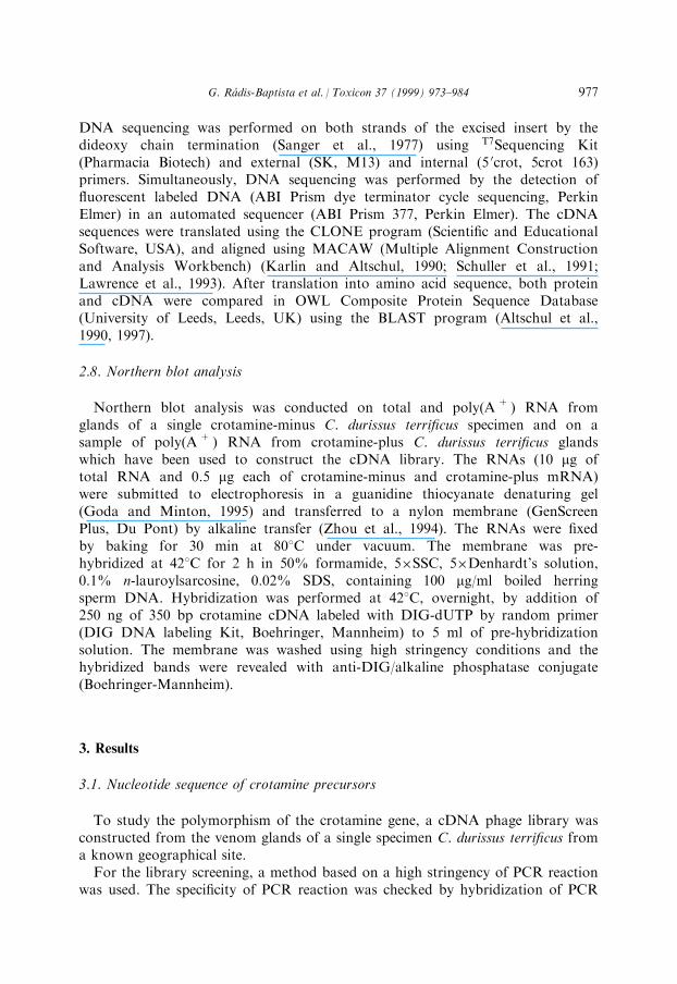
All sequenced cDNA clones, ranging from 340 to 360 nucleotides, have openreading frames (ORFs) of the same size of 198 nucleotides. Alignment of these 15sequences, using the MACAW program results in groupings of six types ofsequences (Fig. 1). All of them predict precursors of crotamine, when compared tosequences deposited in the OWL database using BLAST, with conserved cysteinesinvolved in disul®de bonds and conserved basic residues characteristic of smallbasic myotoxins (Fig. 2). These crotamine precursors have 63 amino acids, the®rst 22 correspond to signal peptide, the following amino acids (from 23 to 62)include a mature crotamine isoform and lysine, the last amino acid whichpresumably is removed in a post-translational process. Of these six distinctcDNAs, two encode crotamine with amino acid sequence identical to the onepublished by Laure (1975), and four code for a crotamine isoform with isoleucineat position 19 (crotamine-Ile 19).
All crotamine cDNAs present some base substitution or deletion at 5 0 and 3 0
untranslated regions (UTRs). One of them (clone MK41) shows a gross 18 basepair deletion at the 5 0 end including the ®rst base adenine of the initiation codon.In this way, the universal ATG start codon is changed to a rare GUG as theinitiation codon.
Fig. 2. Alignment of crotamine and myotoxins sequences from several Crotalus snakes. Shown above
the sequences is the myotoxins consensus pattern (signature), with alternative basic residues (R and K)
denoted as a `+ ' signal. Regions of amino acids variability are enclosed by rectangles. Sequences
2895610 and 2981144 ± crotamine isoform precursors from C. durissus terri®cus (this work);
MYX1_CRODU, MYX2_CRODU, MYX3_CRODU and MYX4_CRODU crotamine sequences from
C. durissus terri®cus (Smith and Schmidt, 1990); MYXC_CRODU ± crotamine from C. durissus
terri®cus (Laure, 1975); CRO_Ile-19 ± crotamine Ile-19 from C. durissus ruruima (Dos Santos et al.,
1993); MYX1_CROVV ± myotoxin-a from C. viridis virids (Fox et al., 1979); MYX_CROAD CAM-
toxin from C. adamanteus (Samejima et al., 1987); MYXC_CROVH ± peptide C from C. v. helleri
(Maeda et al., 1978); MYX1_CROVC and MYX2_CROVC ± myotoxin I and II from C. viridis
concolor (Bieber et al., 1987).
G. RaÂdis-Baptista et al. / Toxicon 37 (1999) 973±984 979
3.2. Northern blot analysis
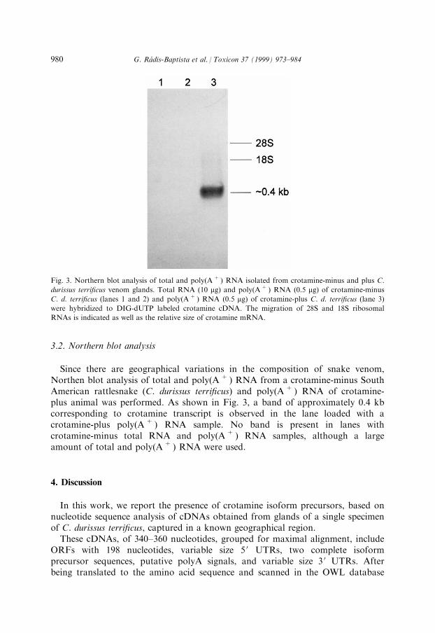
Since there are geographical variations in the composition of snake venom,Northen blot analysis of total and poly(A+) RNA from a crotamine-minus SouthAmerican rattlesnake (C. durissus terri®cus) and poly(A+) RNA of crotamine-plus animal was performed. As shown in Fig. 3, a band of approximately 0.4 kbcorresponding to crotamine transcript is observed in the lane loaded with acrotamine-plus poly(A+) RNA sample. No band is present in lanes withcrotamine-minus total RNA and poly(A+ ) RNA samples, although a largeamount of total and poly(A+) RNA were used.
4. Discussion
In this work, we report the presence of crotamine isoform precursors, based onnucleotide sequence analysis of cDNAs obtained from glands of a single specimenof C. durissus terri®cus, captured in a known geographical region.
These cDNAs, of 340±360 nucleotides, grouped for maximal alignment, includeORFs with 198 nucleotides, variable size 5 0 UTRs, two complete isoformprecursor sequences, putative polyA signals, and variable size 3 0 UTRs. Afterbeing translated to the amino acid sequence and scanned in the OWL database
Fig. 3. Northern blot analysis of total and poly(A+) RNA isolated from crotamine-minus and plus C.
durissus terri®cus venom glands. Total RNA (10 mg) and poly(A+ ) RNA (0.5 mg) of crotamine-minus
C. d. terri®cus (lanes 1 and 2) and poly(A+ ) RNA (0.5 mg) of crotamine-plus C. d. terri®cus (lane 3)
were hybridized to DIG-dUTP labeled crotamine cDNA. The migration of 28S and 18S ribosomal
RNAs is indicated as well as the relative size of crotamine mRNA.
G. RaÂdis-Baptista et al. / Toxicon 37 (1999) 973±984980

using the BLAST program, sequenced cDNAs and predicted amino acidssequences indicate that all code for a functional crotamine isoform, since theypresent the entire crotamine precursor sequence. Furthermore, they present theconsensus pattern KxCHxKx(2)HCx(2)Kx(3)Cx(8)Kx(2)Cx(2)[RK]xKCCKK ofother myotoxins with conserved cysteines involved in disul®de bonds andconserved basic amino acid residues (Gri�n and Aird, 1990). Although the 5 0 and3 0 UTRs of crotamine cDNAs show some nucleotide variation, one observes herea certain degree of similarity with 5 0 and 3 0 UTRs of myotoxin-a from C. viridisviridis (Norris et al., 1997).
One crotamine isoform described here has an amino acid sequence identical tothat described by Laure (1975), while the other predicted isoform has theisoleucine residue at position 19 (crotamine Ile-19). Interestingly, the existence ofthe crotamine Ile-19 has been reported only in the venom of C. durissus ruruima(Dos Santos et al., 1993) ± a subspecies of C. durissus that inhabits the northenregion of Brazil, while the subspecies terri®cus is found in the central andsouthern parts of the country.
The precise locality of capture is very important since the venom compositionshows regional variation and it is not correlated with the snake's philogeny(Daltry et al., 1996). It is known that the South American rattlesnake C. d.terri®cus can be additionally classi®ed as crotamine-plus and crotamine-minusdepending on the presence or absence of crotamine in the venom (Gonc° alves,1956; Schenberg, 1959a,b). The reason for this di�erence is not yet clear but,basically, it should be at either the mRNA or DNA level.
To examine if the absence of crotamine in the venom of crotamine-minussnakes is due to the lack of crotamine mRNA in the venom glands, a northernblot analysis of total and poly(A+ ) RNA from a crotamine-minus, and ofpoly(A+) RNA from a crotamine-plus C. d. terri®cus glands was performed,using the entire crotamine cDNA precursor as the probe. Preliminary resultsindicate the presence of crotamine transcripts of approximately 0.4 kb incrotamine-plus C. d. terri®cus venom glands, but absence in crotamine-minus C. d.terri®cus snakes. These results suggest that the lack of crotamine expression in thevenom of a crotamine-minus specimen operates at the DNA level.
Given our present data, some comments should be made on the work of Smithand Schmidt (1990). (1) They analyzed four clones and although the nucleotidesequence analysis predicted the existence of multiple variants of the crotamine,only two of these four had an intact ATG, thus coding for functional precursors.(2) These multiple variants showed 5±6 amino acid di�erences with published datafrom Laure (1975) and Dos Santos et al. (1993), a rather high substitution rate fora peptide of 42 amino acids, although this can happen. (3) Since their cDNAlibrary was constructed from three C. d. terri®cus specimens of unknown origin, itis di�cult to discuss the number of isoforms in one animal. (4) It is not clearwhether those three snake samples were all crotamine positive.
Interestingly, in the present work a critical change was observed in thecrotamine cDNA precursor involving the initiation codon. Deletion of 18nucleotides of the 5 0 UTR end of the clone MK41 results in juxtaposition of a G
G. RaÂdis-Baptista et al. / Toxicon 37 (1999) 973±984 981

to the remaining UG of initiation codon. A similar phenomenon has beenobserved in the gene of human hypoxanthine-guanine phosphoribosyltransferase(HRPT), where a 13 base pair deletion in exon 1 of HRPT forms a functionalGUG initiation codon (Davidson et al., 1994). Initiation of protein synthesis atthe non-AUG start codon is not rare and several examples have been foundrecently in eukaryotes cellular genes. Thus proto-oncogenes and gene encoding®broblast growth factor initiate translation at CUG (Hann et al., 1988; Prats et al.,1989), while Drosophila choline acetyltransferase (Sugihara et al., 1990) andhuman parain¯uenza virus type 1 (hPIV1 P/C) gene (Boeck et al., 1992) initiate atGUG. There are several more examples, but the majority of these genes areinvolved in highly regulated metabolic pathways. This kind of regulation is relatedto the low e�ciency of translation at non-AUG codons (Peabody, 1989; Sugiharaet al., 1990). Although initiation at non-AUG codons is accomplished at lowe�ciency, according to Kozak (1990, 1997), it seems to show the same strongdependence on the mRNA features that bene®ts AUG recognition.
Venom composition shows a regional variation and there are C. durissusterri®cus with a low concentration of crotamine. Whether crotamine genes withGUG start codon play any special role in these cases is under investigation.
Acknowledgements
This work was supported by FAPESP, Fundac° aÄ o de Amparo aÁ Pesquisa doEstado de SaÄ o Paulo, and CNPq, Conselho Nacional de DesenvolvimentoCientõ ®co e Tecnolo gico. We are thankful to Marilyn Kozak and to Alvaro Pietroda Silva for helpful discussions.
References
Aird, S.D., Kruggel, W.G., Kaiser, I.I., 1991. Multiple myotoxin sequences from the venom of a single
prairie rattlesnake (Crotalus viridis viridis). Toxicon 29, 265±268.
Alexander, A., Grothusen, J., Zepeda, H., Schwartzman, R.J., 1988. Gyrotoxin, a toxin from the venom of
Crotalus durissus terri®cus, is a thrombin-like ennzyme. Toxicon 26, 953±960.
Altschul, S.F., Gish, W., Miller, W., Myers, E.W., Lipman, D.L., 1990. Basic local alignment search tool.
J. Mol. Evol. 215, 403±410.
Altschul, S.F., Madden, T.L., SchaÈ �er, A.A., Zhang, J., Zhang, Z., Miller, W., Lipman, D.J., 1997.
Gapped BLAST and PSI-BLAST: a new generation of protein database search programs. Nucl. Ac.
Res. 25, 3389±3402.
Ausubel, F.M., Brent, R., Kingston, R.E., Moore, D.D., Seidman, J.G., Smith, J.A., Struhl, K., 1995.
Current Protocols in Molecular Biology. Wiley Interscience, New York.
Barrio, A., 1961. Gyrotoxin, a new neurotoxin of Crotalus durissus terri®cus venom. Acta Physiol.
Latinoamericana 11, 224.
Bieber, A.L., Nedelkov, D., 1997. Structural, biological and biochemical studies of myotoxin a and hom-
ologous myotoxin. J. Toxicol.-Toxin Rev. 16, 33±52.
Bieber, A.L., McParland, R.H., Becker, R.R., 1987. Amino acid sequences of myotoxins from Crotalus
viridis concolor venom. Toxicon 25, 677±680.
G. RaÂdis-Baptista et al. / Toxicon 37 (1999) 973±984982

Bober, M.A., Glenn, J.L., Straight, R.C., Ownby, C.L., 1988. Detection of myotoxin-a like proteins in var-
ious snake venoms. Toxicon 26, 665±673.
Boeck, R., Curran, J., Matsuoka, Y., Compans, R., Kolakofsky, D., 1992. The parain¯uenza virus type 1
P/C uses a very e�cient GUG codon to start its C 0 protein. J. Virol. 66, 1765±1768.Brazil, O.V., 1972. Neurotoxins from the South American rattlesnake venom. J. Formosan. Med. Assoc.
71, 394±400.
Brazil, O.V., Fontana, M.D., 1993. Toxins as tools in the study of sodium channel distribution in the
muscle ®bre membrane. Toxicon 31, 1085±1098.
Cameron, D.L., Tu, A.T., 1978. Chemical and functional homology of myotoxin a from prairie rattlesnake
venom and crotamine from South American rattlesnake venom. Biochem. Biophys. Acta 532, 147±154.
Chang, C.C., Lee, J.D., 1977. Crotoxin, the neurotoxin of South American rattlesnake venom, is a presyn-
aptic toxin acting like b-bungarotoxin. Naunyn-Schmiedebergs Arch. Pharmacol. 296, 159±168.
Chomczynski, P., Sacchi, N., 1987. Single-step method of RNA isolation by acid guanidinium thiocya-
nate±phenol±chlorofom extraction. Anal. Biochem. 162, 156±159.
Daltry, A.A., WuÈ ster, W., Thorpe, R.S., 1996. Diet and snake venom evolution. Nature 379, 537±540.
Davidson, B.L., Golovoy, N., Roessler, B.J., 1994. A 13 base pair deletion in exon 1 of HRPTIllinois forms
a functional GUG initiation codon. Human Gen. 93, 300±304.
Dos Santos, M.C., Morhy, L., Ferreira, L.C.L., Oliveira, E.B., 1993. Puri®cation and properties of a cro-
tamine analog from Crotalus durissus ruruima venom. Toxicon 31, 166.
Engle, C.M., Becker, R.R., Bailey, T., Bieber, A.L., 1983. Characterization of two myotoxin proteins from
venom of Crotalus viridis viridis. J. Toxicol.-Toxin Rev. 2, 267±283.
Fletcher, J.E., Hubert, M., Wieland, S.J., Gong, Q., Jiang, M., 1996. Similarities and di�erences in mech-
anisms of cardiotoxins, melittin and other myotoxins. Toxicon 34, 1301±1311.
Fox, J.W., Elzinga, M., Tu, A.T., 1979. Amino acid sequence and disul®de bond assignment of myotoxin a
isolated from the venom of prairie rattlesnake (Crotalus viridis viridis). Biochemistry 18, 678±684.
Furukawa, K., Funayama, K., Ohkura, M., Oshima, Y., Tu, A.T., Ohizumi, Y., 1994. Ca2+ release
induced by myotoxin a, a radio-labellable probe having novel Ca2+ release properties in sarcoplasmatic
reticulum. Br. J. Pharmacol. 113, 233±239.
Goda, S.K., Minton, N.P., 1995. A simple procedure for gel electrophoresis and northern blotting of
RNA. Nucl. Ac. Res. 23, 3357±3358.
Gonc° alves, J.M., 1956. Estudos sobre venenos de serpentes brasileiras. II ± Crotalus terri®cus crotaminicus,
subspe cie biolo gica). An. Acad. Brasileira Cieà ncias 28, 365±367.
Gri�n, P.R., Aird, S.D., 1990. A new small myotoxin from the venom of the prairie rattlesnake (Crotalus
viridis viridis). FEBS Lett. 274, 43±47.
Hagwood, B.J., Santana De Sa , S., 1979. Changes in spontaneous and evoked release of transmitters
induced by the crotoxin complex and its component phospholipase A2 at the frog neuromuscular junc-
tion. Neuroscience (Oxford) 4, 293±303.
Hann, S.R., King, M.W., Bentley, D.L., Anderson, C.W., Eisenman, R.N., 1988. A non-AUG transla-
tional initiation in c-myc exon 1 generates an N-terminally distinct protein whose synthesis is disrupted
in Burkitt's lymphomas. Cell 52, 185±195.
Hong, S.J., Chang, C., 1985. Electrophysiological studies of myotoxin a isolated from prairie rattlesnake
(Crotalus viridis viridis) venom on murine skeletal muscle. Toxicon 23, 927±937.
Israel, D.I., 1993. A PCR-based method for high stringency screening of DNA libraries. Nucl. Ac. Res. 21,
2627±2631.
Israel, D.I., 1995. A PCR-based method for screening DNA libraries. In: Di�enbach, C.W., Dveksler, G.S.
(Eds.), PCR Primer. A Laboratory Manual. Cold Spring Harbor Laboratory Press, New York.
Karlin, S., Altschul, S.F., 1990. Methods for assessing the statistical signi®cance of molecular sequence
features by using general scoring schemes. Proc. Nat. Acad. Sci. USA 87, 2264±2268.
Kozak, M., 1990. Dowstream secondary structure facilitates recognition of initiator codons by eucaryotic
ribosomes. Proc. Nat. Acad. Sci. USA 87, 8301±8305.
Kozak, M., 1997. Recognition of AUG and alternative initiator codons is augmented by G in position +4
but is not generally a�ected by the nucleotides in positions +5 and +6. EMBO J. 16, 2482±2492.
Laure, C.J., 1975. Die PrimaÈ rstruktur des Crotamines. Hoppe-Seyler's Z. physiol. Chem. 356, 213±215.
G. RaÂdis-Baptista et al. / Toxicon 37 (1999) 973±984 983

Lawrence, C.E., Altschul, S.F., Boguski, M.S., Liu, S.J., Neuwad, A.F., Wooton, J.C., 1993. Detecting
subtle sequence signals: a Gibbs sampling strategy for multiply alignment. Science 262, 208±214.
Lee, C.Y., 1972. Chemistry and pharmacology of polypeptide toxins in snake venoms. A. Rev. Pharmacol.
12, 265±286.
Maeda, N., Tamya, N., Pattabhiraman, T.R., Russel, F.E., 1978. Some chemical properties of the venom
of the rattlesnake, Crotalus viridis helleri. Toxicon 16, 431±441.
Mebs, D., Kornarlik, F., 1984. Intraspeci®c variation in content of a basic toxin in eastern diamond rat-
tlesnake (Crotalus adamanteus) venom. Toxicon 22, 831±833.
Norris, J.W., Fry, R.M., Tu, A.T., 1997. The nucleotide sequence of the translated and untranslated
regions of a cDNA for myotoxin a from the venom of prairie rattlesnake (Crotalus viridid viridis).
Biochem. Biophys. Res. Comm. 230, 607±610.
Ohkura, M., Furukawa, K., Tu, A.T., Ohizumi, Y., 1994. Calsequestrin is a major binding protein of
myotoxin a and an endogenous Ca2+ releaser in sarcoplasmatic reticulum. Eur. J. Pharmacol. 268,
R1±R2.
Ohkura, M., Furukawa, K., Oikawa, K., Ohizumi, Y., 1995. The properties of speci®c binding site of 125I-
radioiodinated myotoxin a, a novel Ca2+ releasing agent, in skeletal muscle sarcoplasmatic reticulum. J.
Pharmacol. Exp. Ther. 273, 934±939.
Ownby, C.L., Cameron, M.S., Tu, A.T., 1976. Isolation of myotoxin component from rattlesnake
(Crotalus viridis viridis) venom. Am. J. Pathol. 85, 149±166.
Peabody, D.S., 1989. Translation initiation at non-AUG triplets in mammalian cells. J. Biol. Chem. 264,
5031±5035.
Prats, H., Kaghad, M., Prats, A.C., Klagsbrun, M.L., Liazurn, P., Chalon, P., 1989. High molecular mass
forms of basic ®broblast growth factor are initiated by alternative CUG codons. Proc. Nat. Acad. Sci.
USA 80, 1836±1840.
Rottenberg, D., Bamberger, E.S., Kochva, E., 1971. Studies on ribonucleic acid synthesis in the venom
glands of Vipera palaestinae (Ophidiae, Reptilia). Biochem. J. 121, 609±612.
Samejima, Y., Aoki, Y., Mebs, D., 1987. Amino acids sequence of a myotoxin from venom of the eastern
diamondback rattlesnake (Crotalus adamanteus). Toxicon 29, 461±468.
Sanger, F., Nicklen, S., Coulson, A.R., 1977. DNA sequencing with chain-terminating inhibitors. Proc.
Nat. Acad. Sci. USA 74, 5463±5467.
Schenberg, S., 1959a. Ana lise da crotamina no veneno individual de cascave is recebidas pelo Instituto
Butantan. Mem. Inst. Butantan 29, 213±226.
Schenberg, S., 1959b. Geographical pattern of crotamine distribution in the same ratllesnake subspecies.
Science 129, 1361±1363.
Schuller, G.D., Altschul, S.F., Lipman, D.J., 1991. Workbench for multiple alignment construction and
analysis. Proteins Structure Functions Genetics 9, 180±190.
Slotta, K.H., Fraenkel-Conrat, H.L., 1938. Schlangengifte. III. Mitteil.: Reinigung und Kristallisation des
Klapperschlangengiftes. Ber. Deutsch. Chem. 71, 1076±1081.
Smith, L.A., Schmidt, J.J., 1990. Cloning and nucleotide sequences of crotamine genes. Toxicon 28, 575±
585.
Straight, R.C., Glenn, J.L., Wolt, T.B., Wolfe, M.C., 1991. Reginal di�erences in content of small basic
peptide toxins in the venom of Crotalus adamanteus and Crotalus horridus. Comp. Biochem. Physiol.
100B, 51±58.
Sugihara, H., Andrisani, V., Salvaterra, P.M., 1990. Drosophila choline acetyltranferase uses a non-AUG
initiation codon and full length RNA is ine�ciently translated. J. Biol. Chem. 265, 21714±21719.
Vincent, C., Tchen, P., Cohen-Solal, M., Kourilsky, P., 1982. Synthesis of 8-(2-4 dinitrophenyl 2-6 amino-
hexyl) amino-adenosine 5 0 triphosphate: biological properties and potential uses. Nucl. Ac. Res. 10,
6787±6796.
Zhou, M.Y., Xue, D., Gomez-Sanchez, E.P., Gomez-Sanchez, E., 1994. Improved downward cappillary
transfer for blotting of DNA and RNA. BioTechniques 16, 58±59.
G. RaÂdis-Baptista et al. / Toxicon 37 (1999) 973±984984
All in-text references underlined in blue are linked to publications on ResearchGate, letting you access and read them immediately.