Pael receptor induces death of dopaminergic neurons in the substantia nigra via endoplasmic...
11
Pael receptor induces death of dopaminergic neurons in the substantia nigra via endoplasmic reticulum stress and dopamine toxicity, which is enhanced under condition of parkin inactivation Yasuko Kitao 1, * ,{ , Yuzuru Imai 2,{ , Kentaro Ozawa 1 , Ayane Kataoka 2 , Toshio Ikeda 3 , Mariko Soda 2 , Kazuhiko Nakimawa 4 , Hiroshi Kiyama 4 , David M. Stern 7 , Osamu Hori 1 , Kazumasa Wakamatsu 6 , Shosuke Ito 6 , Shigeyoshi Itohara 3 , Ryosuke Takahashi 2,5,{ and Satoshi Ogawa 1,{ 1 Department of Neuroanatomy, Kanazawa University Medical School, 13-1, Takara-machi, Kanazawa City, 920-8640 Ishikawa, Japan, 2 Laboratory for Motor System Neurodegeneration and 3 Laboratory for Behavioral Genetics and RIKEN Brain Science Institute (BSI), Saitama 351-0198, Japan, 4 Department of Anatomy and Neurobiology, Osaka City University, Graduate School of Medicine, Osaka, Japan, 5 Department of Neurology, Kyoto University Medical School, Kyoto, Japan, 6 Department of Chemistry, Fujita Health University School of Health Sciences, Aichi 470-1192, Japan and 7 Dean’s Office, College of Medicine, University of Cincinnati, Cincinnati, OH 45267, USA Received August 2, 2006; Revised October 19, 2006; Accepted November 13, 2006 Selective loss of dopaminergic neurons is the final common pathway in Parkinson’s disease. Expression of Parkin associated endothelin-receptor like receptor (Pael-R) in mouse brain was achieved by injecting ade- noviral vectors carrying a modified neuron-specific promoter and Cre recombinase into the striatum. Upregulation of Pael-R in the substantia nigra pars compacta of mice by retrograde infection induced endo- plasmic reticulum (ER) stress leads to death of dopaminergic neurons. The role of ER stress in dopaminergic neuronal vulnerability was highlighted by their decreased survival in mice deficient in the ubiquitin-protein ligase Parkin and the ER chaperone ORP150 (150 kDa oxygen-regulated protein). Dopamine-related toxicity was also a key factor, as a dopamine synthesis inhibitor blocked neuronal death in parkin null mice. These data suggest a model in which ER- and dopamine-related stress are major contributors to decreased viability of dopaminergic neurons in a setting relevant to Parkinson’s disease. INTRODUCTION Though Parkinson’s disease (PD) is a major contributor to dis- ability and death in the aging population, the molecular basis of selective dopaminergic neuronal toxicity is still under investigation. Progression of the clinical syndrome associated with PD, which includes a well-characterized movement dis- order, correlates closely with inexorable loss of neurons in the substantia nigra pars compacta (SNpc) (1). Mutations in the Parkin gene (2) have been identified in the autosomal recessive form of PD (AR-JP), a major cause of juvenile PD. Parkin is a 465 amino acid polypeptide with properties of an ubiquitin-protein ligase (E3) whose N-terminus displays homology to ubiquitin and C-terminus is comprised of two RING fingers flanking a cysteine-rich domain, termed in between RING fingers (IBR) (3–5). Con- sistent with this view, AR-JP-linked parkin mutants are defec- tive in E3 activity (6–8). A putative G protein-coupled transmembrane polypeptide, Pael Receptor [Parkin-associated endothelin-receptor like recep- tor (Pael-R)], has been identified as a Parkin substrate (9). Expression of Pael-R in cultured cells results in accumulation # 2006 The Author(s) This is an Open Access article distributed under the terms of the Creative Commons Attribution Non-Commercial License (http://creativecommons.org/ licenses/by-nc/2.0/uk/) which permits unrestricted non-commercial use, distribution, and reproduction in any medium, provided the original work is properly cited. *To whom correspondence should be addressed. Tel: þ81 762652162; Fax: þ81 762344222; Email: [email protected] { These authors equally contributed to this work. Human Molecular Genetics, 2007, Vol. 16, No. 1 50–60 doi:10.1093/hmg/ddl439 Advance Access published on November 20, 2006 by guest on April 5, 2016 http://hmg.oxfordjournals.org/ Downloaded from
-
Upload
independent -
Category
Documents
-
view
0 -
download
0
Transcript of Pael receptor induces death of dopaminergic neurons in the substantia nigra via endoplasmic...

Pael receptor induces death of dopaminergicneurons in the substantia nigra via endoplasmicreticulum stress and dopamine toxicity, which isenhanced under condition of parkin inactivation
Yasuko Kitao1,*,{, Yuzuru Imai2,{, Kentaro Ozawa1, Ayane Kataoka2, Toshio Ikeda3,
Mariko Soda2, Kazuhiko Nakimawa4, Hiroshi Kiyama4, David M. Stern7, Osamu Hori1,
Kazumasa Wakamatsu6, Shosuke Ito6, Shigeyoshi Itohara3, Ryosuke Takahashi2,5,{
and Satoshi Ogawa1,{
1Department of Neuroanatomy, Kanazawa University Medical School, 13-1, Takara-machi, Kanazawa City, 920-8640
Ishikawa, Japan, 2Laboratory for Motor System Neurodegeneration and 3Laboratory for Behavioral Genetics and
RIKEN Brain Science Institute (BSI), Saitama 351-0198, Japan, 4Department of Anatomy and Neurobiology, Osaka
City University, Graduate School of Medicine, Osaka, Japan, 5Department of Neurology, Kyoto University Medical
School, Kyoto, Japan, 6Department of Chemistry, Fujita Health University School of Health Sciences, Aichi 470-1192,
Japan and 7Dean’s Office, College of Medicine, University of Cincinnati, Cincinnati, OH 45267, USA
Received August 2, 2006; Revised October 19, 2006; Accepted November 13, 2006
Selective loss of dopaminergic neurons is the final common pathway in Parkinson’s disease. Expression ofParkin associated endothelin-receptor like receptor (Pael-R) in mouse brain was achieved by injecting ade-noviral vectors carrying a modified neuron-specific promoter and Cre recombinase into the striatum.Upregulation of Pael-R in the substantia nigra pars compacta of mice by retrograde infection induced endo-plasmic reticulum (ER) stress leads to death of dopaminergic neurons. The role of ER stress in dopaminergicneuronal vulnerability was highlighted by their decreased survival in mice deficient in the ubiquitin-proteinligase Parkin and the ER chaperone ORP150 (150 kDa oxygen-regulated protein). Dopamine-related toxicitywas also a key factor, as a dopamine synthesis inhibitor blocked neuronal death in parkin null mice.These data suggest a model in which ER- and dopamine-related stress are major contributors to decreasedviability of dopaminergic neurons in a setting relevant to Parkinson’s disease.
INTRODUCTION
Though Parkinson’s disease (PD) is a major contributor to dis-ability and death in the aging population, the molecular basisof selective dopaminergic neuronal toxicity is still underinvestigation. Progression of the clinical syndrome associatedwith PD, which includes a well-characterized movement dis-order, correlates closely with inexorable loss of neurons inthe substantia nigra pars compacta (SNpc) (1).
Mutations in the Parkin gene (2) have been identified in theautosomal recessive form of PD (AR-JP), a major cause of
juvenile PD. Parkin is a 465 amino acid polypeptide withproperties of an ubiquitin-protein ligase (E3) whoseN-terminus displays homology to ubiquitin and C-terminusis comprised of two RING fingers flanking a cysteine-richdomain, termed in between RING fingers (IBR) (3–5). Con-sistent with this view, AR-JP-linked parkin mutants are defec-tive in E3 activity (6–8).
A putative G protein-coupled transmembrane polypeptide,Pael Receptor [Parkin-associated endothelin-receptor like recep-tor (Pael-R)], has been identified as a Parkin substrate (9).Expression of Pael-R in cultured cells results in accumulation
# 2006 The Author(s)This is an Open Access article distributed under the terms of the Creative Commons Attribution Non-Commercial License (http://creativecommons.org/licenses/by-nc/2.0/uk/) which permits unrestricted non-commercial use, distribution, and reproduction in any medium, provided the original work isproperly cited.
*To whom correspondence should be addressed. Tel: þ81 762652162; Fax: þ81 762344222; Email: [email protected]{These authors equally contributed to this work.
Human Molecular Genetics, 2007, Vol. 16, No. 1 50–60doi:10.1093/hmg/ddl439Advance Access published on November 20, 2006
by guest on April 5, 2016
http://hmg.oxfordjournals.org/
Dow
nloaded from

of unfolded, insoluble and ubiquinated Pael-R in the ER, even-tuating in ER stress, as indicated by upregulation of chaper-ones, such as GRP78/BiP, and subsequent neuronal death(10). Overexpression of Parkin in this in vitro model resultedin removal/degradation of accumulated Pael-R and increasedcell viability. The relevance of ER stress in the centralnervous system to pathologic situations and normal neuronaldevelopment is suggested by induction of the unfoldedprotein response (UPR) in cell stress associated with cerebralischemia (11,12) and exposure to excitatory amino acids (13),as well as during rapid neuronal growth in the neonatal period(14). Salient features of the UPR include upregulation of ERchaperones, suppression of general translation, and activationof the upiquitin-proteasome pathway.
A key facet of the pathology of PD is limitation of cell lossto dopaminergic neurons, especially in the SNpc. In thiscontext, studies of Pael-R might be especially relevant. Pan-neuronal expression of Pael-R in Drosophila brain causedselective, age-dependent degeneration of dopaminergicneurons (10). Thus, increased levels of Pael-R render dopa-minergic neurons vulnerable to cell death. In the currentstudy, we have found that increased expression of Pael-R inmice in the SNpc results in neuronal death which is accentu-ated by suppression of Parkin (in Parkin2/2 mice) or an ERchaperone (in Orp150þ/2 mice). In contrast, a dopamine(DA) synthesis inhibitor had neuroprotective properties inthis system. Thus, we propose a unified model for cytotoxicityin PD through which a combination of ER stress and dopa-mine toxicity, potentially acting in concert with mitochondrialdysfunction, result in an ascending cycle of cellular pertur-bation and, ultimately, death of dopaminergic neurons.
RESULTS
Retrograde and neuron-specific gene expressionby adenoviral vectors in the SNpc
It was essential to develop a system with cell-specific proteinexpression which could be targeted to the SNpc. For thispurpose, we established two adenoviral vectors in whichnuclear Cre-recombinase was driven by cell-specific promo-ters; for neuronal expression, we employed AxS2NPNCre(abbreviated as S2NPNCre) with a modified SCG10 promoter(superior cervical ganglia neural-specific 10 protein), and forglial-specific expression, we utilized AxGFAPNCre (abbre-viated as GFAPNCre) with the glial fibrillary acidic proteinpromoter (Fig. 1A). When either of these adenoviral vectorswas co-infected with AxCALNLEGFP (abbreviated asLoxEGFP), specific expression of EGFP occurred in neurons(with S2NPNCre) or astrocytes (with GFAPNCre) in a cellculture system (not shown). In vivoexpression studies wereperformed in mice by co-infecting either S2NPNCre withLoxEGFP, or GFAPNCre with LoxEGFP. Spatial limitationof gene expression was achieved by injecting viral vectorsinto the striatum (Fig. 1B), resulting in expression of EGFPprotein in the ipsilateral SNpc (Fig. 1C, top panel). WhenS2NPNCre was injected with LoxEGFP, expression ofEGFP (marking neurons) and tyrosine hydroxylase [TH;marking dopaminergic neurons in the SNpc (15)] overlapped(Fig. 1C; middle set of panels); quantitation of this overlap
by image analysis, based on studying multiple fields, con-firmed extensive coexpression of EGFP with TH (Fig. 1D).In contrast, there was no overlap of EGFP, following injectionof S2NPNCre with LoxEGFP, when immunostaining was per-formed to visualize GFAP (Fig. 1C, lowest set of panels andFig. 1D). These data indicate that co-infection of the striatumwith recombinant adenoviral vectors encoding cell-specificCre recombinase and EGFP (or other transgenes) flanked bylox P sites, leads to retrograde transport of adenovirus in thenigrostriatal system resulting in gene expression in the SNpc.
Expression of Pael-R in the SNpc activates the unfoldedprotein response
Using this adenoviral system, we sought to express Pael-R inthe SNpc. For this purpose, an adenoviral vector was madewith expression of Pael-R under control of the SCG10 promo-ter regulated by the LoxP system (16), AxCALNLPael-R(abbreviated as LoxPael-R; Fig. 2A). Co-injection of the twoadenoviral vectors, S2NPNCre and LoxPael-R, into the stria-tum increased expression of Pael-R in the SNpc (Fig. 2B,referred to as ipsilateral side). Enhanced expression ofPael-R required both vectors to be co-injected, and wasspecific for neurons.
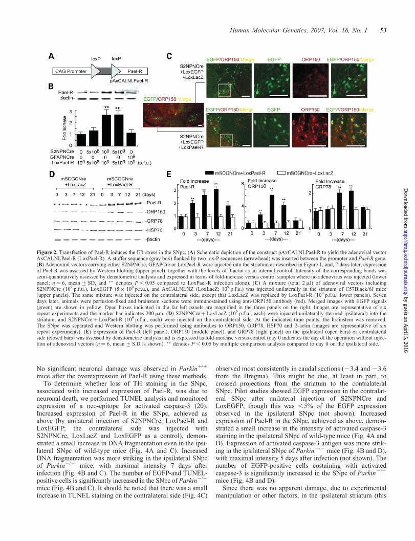
Based on previous in vitro findings, increased expression ofPael-R in dopaminergic neurons might result in ER stress anddiminished cell viability (9). Control experiments were per-formed by co-injecting three adenoviral vectors, S2NPNCre,LoxEGFP and LoxLacZ, into the striatum on one side of thebrain, referred to as contralateral (Fig. 2C, upper panels).Expression of an ER chaperone, ORP150 (oxygen-regulatedprotein 150), known to increase with ER stress (11), wasassessed in neurons expressing EGFP (Fig. 2C, upperpanels). ORP150 levels, assessed by Western blotting, wereunchanged in the SNpc following injection of this combi-nation of adenoviral vectors (Fig. 2D, left portion andFig. 2E, middle panel, open bars). Faint ORP150 stainingwas demonstrated in multiple cells and a much strongersignal for EGFP was observed (Fig. 2C, upper panels).ORP150 has a putative ATPase domain and protein bindingsite, suggesting that it may have a chaperone-like role in main-taining ER function under stress [(17), and see below]. In thiscontext, we have demonstrated that overexpression of ORP150rescues neurons from cell death mediated by ischemia (11,12)and excitatory amino acids (13). When S2NPNCre, LoxEGFPand LoxPael-R were injected into the striatum on the ipsilat-eral side of the brain (the vectors with LoxLacZ in place ofLoxPael-R were injected on the contralateral side), a promi-nent increase in Pael-R in the SNpc (Fig, 2D, right portion)was accompanied by increased expression of ORP150(Fig. 2C, lower panels) in Pael-R expressing neuron (labeledwith EGFP). Western blotting indicated that increasedexpression of ER chaperones, GRP78 and ORP150, reacheda maximum by 7–12 days after infection (Fig, 2D, rightportion). In contrast, there was no increase in these chaperoneson the contralateral side (i.e. the side where the LoxPael-Radenoviral vector was replace by a LoxLacZ vector; Fig. 2Dand E). Levels of a cytoplasmic chaperone, the 70 kDa heatshock protein, remained unchanged on both sides of thebrain (Fig. 2D). These data indicate that co-infection of
Human Molecular Genetics, 2007, Vol. 16, No. 1 51
by guest on April 5, 2016
http://hmg.oxfordjournals.org/
Dow
nloaded from

LoxPael-R and S2NPNCre in the striatum caused expressionof Pael-R in SNpc neurons, and triggered the ER stress.
Expression of Pael-R in SNpc of parkin2/2 miceresulted in neuronal death
Parkin null mice (Parkin2/2) were generated by replacing theproximal exon 3 with a neo cassette (PGK-neo) and two lox Psites (Supplementary Material, Fig. S1A). Southern blottingrevealed homologous recombination of the mutant allele(Supplementary Material, Fig. S1B). Reverse transcriptase–polymerase chain reaction (RT–PCR) to detect Parkintranscripts confirmed the absence of normal transcripts in homo-zygous mutant mice (Supplementary Material, Fig. S1C).Sequencing of RT–PCR products confirmed complete deletionof exon 3 and a frame-shift downstream of exon 2 in mutantmice (Supplementary Material, Fig. S1D). Consistent withthese data, western blotting with anti-Parkin antibodiesshowed the absence of Parkin antigen in brain samples(Supplementary Material, Fig. S1E). Parkin homozygousmutant mice were born at the expected mendelian ratio,showed no overt abnormalities (except a slight decrease in
body weight) and had normal lifespans (18,19). Moreover,Parkin2/2 mice were comparable, in terms of the number ofTH-positive neurons in the SNpc, compared to wild-type litter-mates. However, Parkin2/2 animals displayed a slight increasein striatal dopamine content, compared with wild-type litter-mates (not shown).
Parkin has been shown to ubiquinate Pael-R, thereby pro-moting degradation of insoluble and toxic Pael-R overex-pressed in cell culture systems (6). Thus, we hypothesizedthat similar overexpression of Pael-R in Parkin2/2 micemight result in severe ER stress and cell death in the SNpc.Using Parkinþ/þ and Parkin2/2 mice, LoxPael-R wasco-injected unilaterally into the striatum with LoxEGFP (asa neuronal marker) and S2NPNCre. Whereas up-regulationof Pael-R was confirmed by immunohistochemical analysisin the ipsilateral SNpc of both types of mice (white arrow-heads in the lower panels of Fig. 3A indicate neurons positivewith both TH and Pael-R), a marked decrease in the numberof Nissl-positive neurons was demonstrated in Parkin2/2
mice, (Fig. 3A and B). Neurodegeneration in the SNpc wasaccompanied by loss of TH immunointensity anddecreased dopamine content in the striatum (Fig. 3C and D).
Figure 1. Neuron-specific expression of a transgene in the SNpc after adenoviral infection of the striatum. Two adenoviral vectors were constructed to drive celltype-specific expression of Cre recombinase in neurons (AxS2NPNCre also termed S2NPNCre) and astrocytes (AxGFAPNCre also termed GFAPNCre) (A).S2NPNCre was injected unilaterally along with LoxEGFP into the striatum (109 p.f.u., in each case) as shown (B, upper panel). 7 days after injection, brainslices corresponding to the striatum (B, upper panel) or SNpc (B, lower panel) were analyzed by Nissl staining. On the contralateral side, GFAPNCre wasinjected along with LoxEGFP (109 p.f.u., in each case). SNpc sections were analyzed by fluorescence microscopy using antibodies to TH or GFAP (C).Merged images are shown on the top and the far right (marker bar corresponds to 200 mm; note that magnification is greater in the lower panels comparedwith the top panel), and are representative of six repeat experiments, obtained at 23.1 mM from the Bregma. (D) Combinations of adenoviral vectors at theindicated concentrations were injected into the striatum of wild-type mice, and, 7 days later, animals were sacrificed and sections of SNpc were analyzed atfive different levels (23.16, 23.28, 23.40, 23.52 and 23.64 mM from the Bregma). Sections were subjected to image analysis in order to determinethe area of TH-positivity [TH(þ)] as a percentage of the area of EGFP-positivity [%TH(þ)/EGFP(þ)]. The mean + SD is shown (n ¼ 6), and �� denotesP, 0.01, compared to S2NPNCre infection alone.
52 Human Molecular Genetics, 2007, Vol. 16, No. 1
by guest on April 5, 2016
http://hmg.oxfordjournals.org/
Dow
nloaded from
No significant neuronal damage was observed in Parkinþ/þ
mice after the overexpression of Pael-R using these methods.To determine whether loss of TH staining in the SNpc,
associated with increased expression of Pael-R, was due toneuronal death, we performed TUNEL analysis and monitoredexpression of a neo-epitope for activated caspase-3 (20).Increased expression of Pael-R in the SNpc, achieved asabove (by unilateral injection of S2NPNCre, LoxPael-R andLoxEGFP; the contralateral side was injected withS2NPNCre, LoxLacZ and LoxEGFP as a control), demon-strated a small increase in DNA fragmentation even in the ipsi-lateral SNpc of wild-type mice (Fig. 4A and C). IncreasedDNA fragmentation was more striking in the ipsilateral SNpcof Parkin2/2 mice, with maximal intensity 7 days afterinfection (Fig. 4B and C). The number of EGFP-and TUNEL-positive cells is significantly increased in the SNpc of Parkin2/2
mice (Fig. 4B and C). It should be noted that there was a smallincrease in TUNEL staining on the contralateral side (Fig. 4C)
observed most consistently in caudal sections (23.4 and 23.6from the Bregma). This might be due, at least in part, tocrossed projections from the striatum to the contralateralSNpc. Pilot studies showed EGFP expression in the contralat-eral SNpc after unilateral injection of S2NPNCre andLoxEGFP, though this was ,5% of the EGFP expressionobserved in the ipsilateral SNpc (not shown). Increasedexpression of Pael-R in the SNpc, achieved as above, demon-strated a small increase in the intensity of activated caspase-3staining in the ipsilateral SNpc of wild-type mice (Fig. 4A andD). Expression of activated caspase-3 antigen was more strik-ing in the ipsilateral SNpc of Parkin2/2 mice (Fig. 4B and D),with maximal intensity 5 days after infection (not shown). Thenumber of EGFP-positive cells costaining with activatedcaspase-3 is significantly increased in the SNpc of Parkin2/2
mice (Fig. 4B and D).Since there was no apparent damage, due to experimental
manipulation or other factors, in the ipsilateral striatum (this
Figure 2. Transfection of Pael-R induces the ER stress in the SNpc. (A) Schematic depiction of the construct pAxCALNLPael-R to yield the adenoviral vectorAxCALNLPael-R (LoxPael-R). A stuffer sequence (gray box) flanked by two lox-P sequences (arrowhead) was inserted between the promoter and Pael-R gene.(B) Adenoviral vectors carrying either S2NPNCre, GFAPCre or LoxPael-R were injected into the striatum as described in Figure 1, and, 7 days later, expressionof Pael-R was assessed by Western blotting (upper panel), together with the levels of ß-actin as an internal control. Intensity of the corresponding bands wassemi-quantitatively assessed by densitometric analysis and expressed in terms of fold-increase versus control samples where no adenovirus was injected (lowerpanel; n ¼ 6, mean + SD, and �� denotes P , 0.05 compared to LoxPael-R infection alone). (C) A mixture (total 2 ml) of adenoviral vectors includingS2NPNCre (109 p.f.u.), LoxEGFP (5 � 108 p.f.u.), and AxCALNLNZ (LoxLacZ; 109 p.f.u.) was injected unilaterally in the striatum of C57Black/6J mice(upper panels). The same mixture was injected on the contralateral side, except that LoxLacZ was replaced by LoxPael-R (109 p.f.u.; lower panels). Sevendays later, animals were perfusion-fixed and brainstem sections were immunostained using anti-ORP150 antibody (red). Merged images with EGFP signals(green) are shown in yellow. Open boxes indicated in the far left panels are magnified in the three panels on the right. Images are representative of sixrepeat experiments and the marker bar indicates 200 mm. (D) S2NPNCre þ LoxLacZ (109 p.f.u., each) were injected unilaterally (termed ipsilateral) into thestriatum, and S2NPNCre þ LoxPael-R (109 p.f.u., each) were injected on the contralateral side. At the indicated time points, the brainstem was removed.The SNpc was separated and Western blotting was performed using antibodies to ORP150, GRP78, HSP70 and b-actin (images are representative of sixrepeat experiments). (E) Expression of Pael-R (left panel), ORP150 (middle panel), and GRP78 (right panel) on the ipsilateral (open bars) or contralateralside (closed bars) was assessed by densitometric analysis and is expressed as fold-increase versus control (day 0 indicates the day of the operation without injec-tion of adenoviral vectors (n ¼ 6, mean + S.D is shown). �� denotes P , 0.05 by multiple comparison analysis compared to day 0 on the ipsilateral side.
Human Molecular Genetics, 2007, Vol. 16, No. 1 53
by guest on April 5, 2016
http://hmg.oxfordjournals.org/
Dow
nloaded from

side received injection of S2NPNCre, LoxPael-R andLoxEGFP) based on EGFP fluorescence or Nissl staining(Supplementary Material, Fig. S2B), diminished dopaminecontent most likely reflects loss of functional dopaminergicneurons in the ipsilateral SNpc. Moreover, injection ofS2NPNCre and LoxPael-R resulted in the expression ofPael-R (marked by expression of EGFP) also in the motorcortex by retrograde infection (Supplementary Material,Fig. S2A). In contrast to the SNpc, no neuronal death wasobserved in either motor cortex or striatum (Supplementary
Material, Fig. S2C). In other brain sublesions which havethe connection to the striatum (i.e. the thalamus, the interpe-duncular nucleus, the locus coeruleus and the raphenucleus), no EGFP signals were detected, which may be dueto the fewer communication to the striatum (not shown).The loss of TH immunointensity in the SNpc mainly occurredin neurons expressing Pael-R (Supplementary Material,Fig. S3A–C). These data suggest that Pael-R overexpressioncaused neuronal cell death, rather selectively in dopaminergicneurons in the SNpc.
Figure 3. Enhanced neuronal death in the SNpc of parkin2/2 mice by the up-regulation of Pael-R. (A) Adenoviral vectors (2 ml), including LoxEGFP(5 � 108 p.f.u.), S2NPNCre (109 p.f.u.) and LoxPael-R (109 p.f.u.) were injected unilaterally in the striatum of either parkinþ/þ or parkin2/2 mice, as describedin Figure 1. As a control, LoxPael-R was replaced by LoxLacZ (109 p.f.u.), and S2NPNCre þ LoxLacZ was injected on the contralateral side. Brains of animalswere then perfusion-fixed 7 days later, and midbrain sections were stained using the Nissl method (upper two panels). One of consecutive sections was alsoimmunostained with anti-Pael-R antibody (lower panels). Images at 23.52 mM from the Bregma are shown. The open boxes in the upper panel are magnifiedin lower two panels. Typical examples of neurons positive with both TH and Pael-R are indicated by white arrowheads in the lower panels. Typical examples ofdegenerating neurons are identified by the open arrowheads in the middle panels. (B) Nissl positive neurons were counted on the ipsilateral (closed bars) andcontralateral sides (open bars) as described in the text, 7 and 14 days after the injection. In each case, n ¼ 6, and the mean + SD is shown. �� denotes P, 0.01compared to parkinþ/þ mice injected with LoxLacZ. (C) 10 days after the injection, midbrain sections were stained with anti-TH antibody as described in text.Images at 23.28 mM (upper panels) and 23.52 mM from the Bregma (lower panels) are shown. (D) TH positive neurons were counted 7 days after the injection(left panel). DA content was also measured by the HPLC-EC method 7 days after the injection (right panel). In each case, closed and open bars correspond to theipsilateral (closed bars) or contralateral striatum (open bars) of parkin2/2 or parkinþ/þ mice, respectively. In each case, n ¼ 6, and the mean + SD is shown. ��
denotes P , 0.01 compared to parkinþ/þ mice injected with LoxLacZ.
54 Human Molecular Genetics, 2007, Vol. 16, No. 1
by guest on April 5, 2016
http://hmg.oxfordjournals.org/
Dow
nloaded from

ORP150 suppresses Pael-R-mediated neuronal cell death
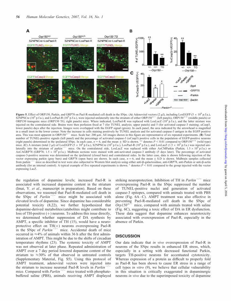
These observations led us to hypothesize that ER stress mighttrigger loss of dopaminergic neurons due to accumulation oftoxic Pael-R. To gain further insight into mechanisms under-lying this observation, we focused on the function of an ERchaperone, ORP150, which promotes protein folding/degra-dation (17). We reasoned that if the ER stress is the causeof Pael-R-mediated dopaminergic neuron death, even in thepresence of wild-type Parkin, decreased levels of ORP150should accentuate Pael-R-induced neuronal death in theSNpc, whereas overexpression of ORP150 might be protec-tive. Using targeted injection of adenoviral vectors (asabove), Pael-R was overexpressed in the SNpc of either het-erozygous Orp150 truncation mutants [Orp150þ/2 mice;note, homozygous Orp1502/2 mice have a developmentallethal phenotype 13)], strain-matched controls (Orp150þ/þ
mice) or Orp150 overexpressing wild-type transgenics[Orp150 TG mice, driven by pCAGGS promoter (21)]. Elev-ated levels of ORP150 were confirmed in SNpc neurons ofOrp150 TG mice by immunohistochemical analysis (Sup-plementary Material, Fig. S4 upper panels; levels ofORP150 in strain-matched normal animals are also shown inlower panels). Degeneration of dopaminergic neurons wasassessed by TUNEL staining and expression of activatedcaspase-3 (Fig. 5A and B), since these methods appeared tohave adequate sensitivity, as shown in Figure 4. A genedosage effect was observed; increased levels of ORP150afforded protection to dopaminergic neurons. Orp150þ/2
mice, with the lowest levels of functional ORP150, displayedexaggerated damage to dopaminergic neurons; TUNEL stain-ing and activated caspase-3 were enhanced on the ipsilateralSNpc (the side in which injection of adenoviral vectors
resulted in over-expression of Pael-R; Fig. 5A and B),whereas no significant cell death was observed on the contral-ateral side where LoxLacZ was expressed (data not shown).Each of these indices of neuronal stress/toxicity in thissetting was decreased, in a manner dependent on the ‘dose’of Orp150, when the same experiment was performed in wild-type (Orp150þ/þ) or mice overexpressing ORP150 (Orp150TG mice) (Fig. 5A and B).
These data suggest an essential contribution of ER functionin protecting neurons from lethal toxicity when Pael-R is over-expressed. According to this concept, we further reasoned thatsuch neurons in Parkin2/2 mice might be rescued by eitherexpression of Parkin or ER chaperones capable of promotingprotein folding/renaturation, such as GRP78. Though adeno-viral expression of LacZ in neurons failed to rescue SNpcneurons from Pael-R-mediated cell death, overexpression ofParkin minimized neuronal damage (Fig. 5C). Similarly, over-expression of GRP78 could substitute for Parkin in preventingPael-R-mediated neuronal death in Parkin2/2 mice. Westernblot analysis of brain stem samples confirmed the expressionof transfected gene products (Fig. 5C, right panel). Thesedata indicate that the ER chaperones, such as GRP78 andORP150, have the capacity to relieve ER stress due toincreased expression of Pael-R, thereby exerting a protectiveeffect on dopaminergic neurons in the SNpc.
Suppression of Pael-R-mediated cell death byinhibition of dopamine synthesis
Increased dopamine content in the striatum of Parkin2/2 micehas been noted by some investigators, though the increase issmall (18,19). Furthermore, Pael-R has been implicated in
Figure 4. Assessment of neuronal cell death in parkin2/2 mice (A,B) After infection of adenoviral vectors by the same protocol as used in Figure 3, brains ofanimals were perfusion-fixed 7 (upper panels for TUNEL) and 5 (lower panels for activated caspase-3 staining) days later, and midbrain sections were stainedusing either the TUNEL method or anti-activated caspase-3 antibody (aCasp3). Images at 23.52 mM from the Bregma were obtained to visualize the indicatedantibody and the EGFP signal (green). In each panel, areas indicated by arrowheads are magnified in the insets at the lower corner of the panel. Note thatTUNEL-positive and activated caspase-3-positive cells are increased in the EGFP positive area. Scale bar: 200 mm. Images shown in this figure (panelsA,B) are representative of six repeated experiments. (C,D) Quantitation of TUNEL-positive signals (number of positive signals) 7 days after injection (panelC) or the percentage of cells positive for activated caspase-3 (aCasp3) in the population of EGFP-positive neurons 5 days after injection (panel D) on the ipsi-lateral (closed bars) or contralateral SNpc (open bars) of either parkin2/2 or parkinþ/þ mice. In each case, n ¼ 6, and the mean + SD is shown. �� denotesP, 0.01 compared to parkinþ/þ mice injected with LoxLacZ.
Human Molecular Genetics, 2007, Vol. 16, No. 1 55
by guest on April 5, 2016
http://hmg.oxfordjournals.org/
Dow
nloaded from
the regulation of dopamine levels; increased Pael-R isassociated with increased dopamine content in the striatum(Imai, Y. et al., manuscript in preparation). Based on theseobservations, we reasoned that Pael-R-mediated cell death inthe SNpc of Parkin2/2 mice might be associated withelevated levels of dopamine. Since dopamine has considerablepotential toxicity (8,22), we further hypothesized thatdopamine-derived metabolites/catabolites might contribute toloss of TH-positive (þ) neurons. To address this issue directly,we determined whether suppression of DA synthesis byAMPT, a specific inhibitor of TH (15), would have a neuro-protective effect on TH(þ) neurons overexpressing Pael-Rin the SNpc of Parkin2/2 mice. Accidental death of miceoccurred in �4% of animals within 36 h after the first admin-istration of AMPT. This might be due to the shifts of circadiantemperature rhythms (23). The systemic toxicity of AMPTwas not observed at later phase. Repeated administration ofAMPT over a 7 day period lowered dopamine content of thestriatum to �30% of that observed in untreated controls(Supplementary Material, Fig. S5). Using this protocol ofAMPT treatment, adenoviral vectors were injected intothe striatum to increase neuronal Pael-R levels in Parkin2/2
mice. Compared with Parkin2/2 mice treated with phosphate-buffered saline (PBS), animals receiving AMPT displayed
striking neuroprotection. Inhibition of TH in Parkin2/2 miceoverexpressing Pael-R in the SNpc suppressed the numberof TUNEL-positive nuclei and generation of activatedcaspase-3 epitopes, compared with animals treated with PBSalone (Fig. 6A–C). AMPT treatment was also effective inpreventing Pael-R-mediated cell death in the SNpc ofOrp150þ/2 mice, compared with animals treated with saline(Fig. 6C), suggesting a toxic effect of DA in ER dysfunction.These data suggest that dopamine enhances neurotoxicityassociated with overexpression of Pael-R, especially in theabsence of Parkin.
DISCUSSION
Our data indicate that in vivo overexpression of Pael-R inneurons of the SNpc results in enhanced ER stress, which,especially in a setting with decreased functional Parkin,targets TH-positive neurons for accentuated cytotoxicity.Whereas expression of a protein as difficult to properly foldas Pael-R has been shown to cause ER stress in a range ofcell types in vitro (9), we believe that cellular vulnerabilityin this situation is critically exaggerated in dopaminergicneurons in vivo due to the superimposed toxicity of dopamine
Figure 5. Effect of ORP150, Parkin, and GRP78 on Pael-R-mediated cell death in the SNpc. (A) Adenoviral vectors (2 ml), including LoxEGFP (5 � 108 p.f.u.),S2NPNCre (109 p.f.u.), and LoxPael-R (109 p.f.u.), were injected unilaterally into the striatum of either ORP150þ/2 (left panels), ORP150þ/þ (middle panels) orORP150 transgenic mice (ORP150 TG; right panels) mice. Where indicated, LoxPael-R was replaced with LoxLacZ (109 p.f.u.), and the latter mixture wasinjected on the contralateral side. Brains were then perfusion fixed at 7 (for TUNEL analysis; upper panels) and 5 (for activated caspase-3 staining; aCasp3,lower panels) days after the injection. Images were overlapped with the EGFP signal (green). In each panel, the area indicated by the arrowhead is magnifiedin a small inset in the lower corner. Note the increase in cells staining positively by TUNEL analysis and for activated caspase-3 antigen in the EGFP positivearea. This was most apparent in ORP150þ/2 mice. Scale bar: 200 mm. All images shown in this figure are representative of six repeated experiments. (B) Totalnumber of TUNEL-positive signals (left panel) and the percentage of activated caspase-3 (aCasp3) positive cells in the population of EGFP-positive neurons(right panels) determined in the ipsilateral SNpc. In each case, n ¼ 6, and the mean + SD is shown. �� denotes P , 0.01 compared to ORP150þ/þ (wild type)mice. (C) A mixture (total 2 ml) of LoxEGFP (5 � 108 p.f.u.), S2NPNCre (109 p.f.u.), LoxPael-R (109 p.f.u.), and LoxLacZ (1.5 � 109 p.f.u.) was injected uni-laterally into the striatum of parkin2/2 mice. On the contralateral side, LoxLacZ was replaced with either AxCMParkin (Parkin; 1.5 � 109 p.f.u.) orAxCAGRP78 (GRP78; 1.5 � 109 p.f.u.). Midbrain sections were stained with anti-activated caspase-3 antibody (5 days later). The percentage of activatedcaspase-3-positive neurons was determined on the ipsilateral (closed bars) and contralateral sides. In the latter case, data is shown following injection of thevector expressing parkin (gray bars) and GRP78 (open bars) are shown. In each case, n ¼ 6, and the mean + S.D is shown. Midbrain samples collectedfrom parkin2/2 mice as described in text were also subjected to Western blot analysis using either anti-b-galactosidase, anti-GRP78, anti-Parkin or anti-b-actinantibody (for an internal control). A typical example of five repeated experiments is shown. � denotes P, 0.01 compared to the group injected with the vectorexpressing LacZ.
56 Human Molecular Genetics, 2007, Vol. 16, No. 1
by guest on April 5, 2016
http://hmg.oxfordjournals.org/
Dow
nloaded from

(DA) itself. These data also emphasize the potential relevanceof parkin targets, such as Pael-R, in addition to theaminoacyl-tRNA synthetase cofactor p38, to neurotoxicity indopaminergic neurons (24).
The technical approach employed in our experiments, injec-tion of adenovirus under stereotactic guidance into the stria-tum with selective neuronal expression of gene products, isquite unique. First, such results have been difficult toachieve in the mouse because of anatomic limitations.Though adenovirus vectors are more immunogenic than ade-novirus associated virus, they still have greater merits in retro-grade transfection (25). We have taken this advantage into ourexperimental system, to achieve an efficient gene transfer toSNpc, where direct injection of viral vectors will be inapplic-able because of anatomical limitations. Second, adenovirusinfection predominately affects glia, rather than neurons.Modification of the SCG10 (superior cervical ganglion) pro-moter by tandem insertion of two neuron-restrictive silencersproduced almost 100% expression of transgenes in neuronalcultures, and considerably lower expression in astrocytes(not shown). Coinjection of S2NPNCre with LoxEGFP intothe striatum resulted in expression of transgenes in neuronsof the mouse SNpc. This approach allowed us to obtainneuron-specific expression of Pael-R enabling study of itseffect(s) on neuronal physiology in vivo. The proximal resultof such Pael-R expression included upregulation of ER cha-perones, such as GRP78 and ORP150, whereas the distalresult was loss of TH-positive neurons, especially inParkin2/2 mice. In contrast, expression of Pael-R had noeffect on the constitutively expressed form of HSP70, import-ant for housekeeping functions in the cytoplasm (Fig. 2D).
Several observations are consistent with the importance ofER stress as a mechanism underlying Pael-R-induced cellulartoxicity. In previous studies using cultured neuroblastomacells, Pael-R has been identified as a substrate of Parkin, anE3 ubiquitin ligase (6). Thus, in the absence of Parkin,Pael-R may accumulate since it is not subject to efficientremoval. In contrast, no detectable change of Pael-R levelshas been reported in SNpc of Parkin null mice (24). Sincethere is no evidence of progressive neuronal cell death inParkin null mice, there would appear to be redundant mechan-isms able to compensate for loss of Parkin under physiologicconditions. The ability of neurons to withstand ER stressimposed by expression of Pael-R is likely to be dependenton the effectiveness of the ER-stress response; higher levelsof Pael-R would require a facile ER-stress response (i.e. ade-quate or increased levels of parkin, ORP150, GRP78 etc),whereas diminished levels of ORP150 or GRP78 wouldrender neurons vulnerable to toxicity because of a compro-mised stress response. These predictions have been borneout by our experimental results. Therefore, the possibletoxicity of Pael-R cannot be completely ignored, especiallyin pathological conditions. Increasing expression of GRP78had a protective effect in Parkin2/2 mice overexpressingPael-R. Furthermore, expression of ORP150, an importantfactor modulating ER stress even in the presence of Parkin,modulated the toxicity of Pael-R for dopaminergic neurons;increased levels of ORP150 in transgenic mice were neuropro-tective, whereas diminished levels in ORP150 in Orp150þ/2
mice resulted in enhanced cell death (i.e. increased TUNELstaining and immunoreactivity for activated caspase-3 in theSNpc).
Figure 6. Suppression of neuronal death in the SNpc of mice overexpressing Pael-R by inhibition of dopamine synthetase using AMPT. (A) Parkin2/2 mice weretreated with either PBS or AMPT up to 7 days after unilateral injection of adenoviral vectors, including LoxEGFP (5 � 108 p.f.u.), S2NPNCre (109 p.f.u.) andLoxPael-R (109 p.f.u.). As a control, the same vectors, with LoxPael-R replaced by LoxLacZ (109 p.f.u.), were injected on the contralateral side. Brains wereperfusion-fixed at 7 (upper panels; for TUNEL analysis) and 5 (lower panels; staining for activated caspase-3, aCasp3) days after injection of the vectors, andwere then subjected to histochemical analysis as described above. The above images were overlapped with EGFP (green). Scale bar: 200 mm. All imagesshown in this figure are representative of six repetitions of the experimental protocol. In each panel, the area indicated by the arrowhead is magnified in asmall inset in the lower corner. Note that TUNEL-positive and activated caspase-3-positive cells were diminished by AMPT treatment (i.e. in the presence ofthe latter, the area/level of apoptotic cells approximated that observed in controls overexpressing LacZ). (B,C) Statistical analysis was performed as describedabove, in either Parkin2/2 mice (B) or Orp150þ/2 mice (C). The number of TUNEL-positive signals (left panels), and % of activated caspase-3-positive cellsin the population of EGFP-positive neurons (right panels) in the ipsilateral SNpc are shown (n ¼ 6; the mean+ SD). P-values,obtained by Student’s t-analysis,are shown in each panel.
Human Molecular Genetics, 2007, Vol. 16, No. 1 57
by guest on April 5, 2016
http://hmg.oxfordjournals.org/
Dow
nloaded from

Our experimental system produced Pael-R overexpressionand subsequent cell death in the SNpc of mouse, whereas noapparent cell death occurred either in the striatum or in themotor cortex (Supplementary Material, Fig. S2C). Further-more, we observed a neuroprotective effect of a TH inhibitorin the SNpc of mouse, suggesting that DA also contributes toPael-R-induced cell death of dopaminergic neurons. Consist-ent with these results in our mouse models, studies in trans-genic flies overexpressing Pael-R in most of neuronalpopulations also showed selective loss of dopaminergicneurons, though the mechanism of cellular degeneration isnot fully understood (10). One plausible explanation for focus-ing toxicity on dopaminergic neurons is the oxidative stresscaused by the presence of DA and its derivatives (1).Another possibility that DA might compromise the survivalof dopaminergic neurons is a chemical inactivation of cellularproteins by addition of DA to sulfhydryl group of proteins.Recently, it has been reported that dopamine covalently mod-ifies Parkin, a protein rich in sulfhydryl group, in dopamin-ergic cells, leading to increased Parkin insolubility andinactivation of its E3 function (22). Vulnerability of Parkinto modification by DA further impairs degradation ofPael-R. Thus, even in sporadic PD, DA might interfere inthe degradation of certain proteins, such as Pael-R, by theinactivation of Parkin. Collectively, these data propose amodel in which a combination of ER stress and DA-relatedstress plays an important role in degeneration of dopaminergicneurons in sporadic PD as well as PD caused by parkinmutations.
MATERIALS AND METHODS
Targeted disruption of the mouse parkin gene
Parkin2/2 mice were generated using standard gene targetingtechniques (26). A targeting vector was constructed with a15.7 kb genomic DNA fragment containing exon 3 of theparkin gene (Supplementary Material, Fig. S1). The regioncontaining exon 3 was replaced with a Floxed pgk-neo cas-sette. A DT-ApA cassette was flanked at the 50-end of thehomologous arm for negative selection (27). The linearizedtargeting vector was transfected into E14 (129sv) ES cells.Positive clones were selected by Southern blotting, and theninjected into C57Bl/6J (B6) blastocysts. Offspring harboringthe targeted allele were generated by crossing chimeric micewith B6 mice. Results of such crosses were confirmed bySouthern analysis.
Reverse transcription–polymerase chain reactionanalysis of parkin null mice
Total RNA was extracted from whole brain tissue using Isogen(Nippon gene). RT reactions containing 1 mg of total RNAwere performed using the SuperScript II First-Strand Syn-thesis System for RT–PCR (Invitrogen). The resultingcDNA was added to PCR reactions containing 1 unit of ExTaq DNA Polymerase (Takara) for 35 PCR cycles. PCR pro-ducts were separated on a 1% agarose gel. Primers were asfollows: RT primer, 50-agt ttc cct tga ggt tgt gc; Primer A,50-cgt agg tcc ttc tcg acc; Primer B, 50-ttg agg ttg tgc gtc
cag g; Primer C, 50-acc tca gag ggc tcc ata tg; and, PrimerD, 50-ctc tct cta cac gtc aaa cca gtg.
Construction of adenoviral vectors
Modified SCG10 (S2NP10) promoter [2 kb (28)] and mouseGFAP promoter region (2.5 kb; kindly provided by DrIkenaka, National Institute for Physiological Science) werecloned into pAxAwNCre (kindly provided by Dr Saito,Tokyo University), a promoter-less cosmid vector for prepar-ing cell type-specific Cre-recombinase expressing adenovirus(Fig. 2A; AxS2NPNCre and AxGFAPNCre). Human Pael-R(9) was cloned into pAxCALNLw (Takara Bio Inc., Shiga,Japan) in order to control Pael-R expression using Cre recon-binase (AxLNLPael-R). The Pael-R gene is silenced becauseof the presence of the staffer of the neo-resistant gene, andis activated by Cre-mediated excisional deletion of thestuffer when a sufficient amount of Cre recombinase isexpressed (Fig. 3). To prepare an adenoviral vector ofparkin (AxCMParkin), the human parkin gene was clonedinto pAxCMwt. Recombinant adenovirus was generatingusing the COS-terminal protein complex (TPC) method andthe Takara adenovirus expression kit (Takara Bio Inc.).AxLNLNZ (Takara Bio Inc.), which overexpresses LacZwith a nuclear localization signal mediated by Cre recombi-nase, was used for control experiments. Cre-mediatedEGFP-expressing adenovirus, AxLNLEGFP, was kindly pro-vided by Dr Okado (Tokyo Metropolitan Institute of Neuro-science). An adenoviral vector for overexpression of GRP78/Bip (AxCAGRP78) was generously provided by DrsS. Tanaka and T. Koike [(29) Graduate School of Science,Hokkaido University]. Each adenovirus was amplified inHEK293 cells and purified using VIRAPREP AdenovirusPurification Kit (Virapur LLC., San Diego, CA, USA). Viraltiters were determined by a plaque-forming assay inHEK293 cells.
Western blotting
Levels of Pael-R in tissue extracts were determined byWestern blotting as described (9). Mouse brain was quicklyremoved and placed on a cold plate. Brain slices (200 mm)were obtained at 23.5 mM from the Bregma on a vibratome.Substantia nigra was then carefully removed under guidanceof a stereoscopic microscope according to the mouse brainatlas (Paxinos and Franklin, Academic Press, 1997, SanDiego) using the cerebral peduncle and medial lemniscus aslandmarks. Tissue extracts were prepared from SNpc in PBScontaining NP-40 (1%). Proteins were separated bySDS-PAGE, and transferred to PVDF. PVDF was then incu-bated with antibody to either human Pael-R or ß-actin, thelatter as an internal control (1000 � dilution, Sigma,St Louis, MO, USA). Levels of chaperones in tissue extractswere determined by Western blotting, as described (30).PVDF was incubated with either anti-human ORP150 anti-body (1 mg/ml), anti-GRP78 monoclonal antibody (Stressgen,Canada; 0.2 mg/ml), or anti-HSP73 antibody (0.1 mg/ml;Stressgen). Where indicated, either anti-b-galactosideasantibody (1000 � dilution, Sigma) or anti-Parkin antibody(1000 � dilution, Cell Signaling Technologies Inc.) was
58 Human Molecular Genetics, 2007, Vol. 16, No. 1
by guest on April 5, 2016
http://hmg.oxfordjournals.org/
Dow
nloaded from

used to access the level of these proteins. Images were furthersubjected to densitometric analysis using NIH image software.
Injection of adenoviral vectors into the striatum andtreatment of animals
Animals were housed and treated according to institutionaland national guidelines. Mice were stereotactically positionedunder deep anesthesia, and the indicated combination ofadenovirus vectors was injected unilaterally (a differentcombination, including a LacZ control, was injected on thecontralateral side) into the striatum at six points (AP/ML/DV/ ¼ 0.8/1.2/22.5, 0.8/1.2/23.2, 0.8/1.7/22.5, 0.8/1.7/23.2, 0.8/2.2/22.5 and 0.8/2.2/23.2; units are mm), followedby free access to food and water. Retrograde passage andinfection of adenoviruses was confirmed 5–12 days after theinjection by detection of a green fluorescent signal in theSNpc by fluorescence microscopy. Where indicated, animalswere pretreated with a-methyl-DL-Tyrosine (AMPT) tosuppress DA synthesis. AMPT-HCl (150 mg/kg, Sigma) wasintra-peritoneally administrated twice per day for 5 daysbefore injection of adenoviral vectors, as well as after thelatter and until the day of sacrifice.
Assessment of neuronal death in vivo
At the indicated time points, animals were perfused with 4%paraformaldehyde under deep anesthesia, the brain wasexcised and coronal brain sections (14 mm) were cut on acryostat. Sections were processed for cresyl violet stainingor immunohistochemistry using either mouse anti-tyrosinehydroxylase antibody (TH; 1 mg/ml, Sigma), rabbit anti-human ORP 150 antibody [5 mg/ml (30)], rabbit anti-activatedcasepase-3 antibody(0.1 mg/ml, Genzyme/Teche), rabbitanti-Pael-R antibody [10 mg/ml, (9)] or goat anti-GFAP anti-body (4 mg/ml. Santa Cruz Biotechnology, Santa Cruz, CA).Sections were also subjected to TUNEL staining using a com-mercially available kit (ApopTag, Intergen, Purchase, NY). Toevaluate neuronal death in the SNpc, either Nissl or EGFPpositive cells were counted in each coronal slice obtained atfive different levels (23.16, 23.28, 23.40, 23.52 and23.64 mM from the Bregma), as described (31) by acquiringdigital images using a CCD camera (Nikon, Coolscope).Cell death was semi-quantitatively assessed by countingTUNEL-positive cells/nuclei in the EGFP-positive area, andevaluating the percentage of activated caspase-3-positivecells in the population of EGFP-positive cells. Sections wereanalyzed using a laser scanning confocal microscope system(Leica, TCS SP2). In each case, two observers without knowl-edge of the experimental protocol evaluated sections andexperiments were repeated at least three times.
DA content in the striatum was measured in the SNpc asdescribed (32). In brief, striatal tissue was homogenized insolution H (0.4 M HClO4 containing 4 mM Na2S2O5, 4 mM
diethylenetriaminepentaacetic acid, and 5 mM 1,4-dithiothreitol). Crude tissue lysate was separated on a C-18reversed-phase column (MCM HPLC, 4.6 mM � 15 cm,5 ml, ESA, Chelmsford, MA, USA), followed byelectrochemical detection (Coulochem III, ESA).
Statistical analysis
Data shown represent the mean +SD Multiple group compari-sons were performed by one-way ANOVA, followed byNewman-Kuels test as a post hoc analysis. Comparisonbetween two groups was analyzed by two-tailed Student’st-test.
SUPPLEMENTARY MATERIAL
Supplementary Material is available at HMG Online.
ACKNOWLEDGEMENTS
We thank Dr Martin Hooper (Western General Hospital,Edinburgh) for generous contribution of E14 cell line, andDrs Yumi Onodera, Yoshikazu Saito, Hitomi Suzuki(RIKEN, Wako, Japan) for technical assistance for establishingchimera mice. The authors also appreciate the generous gifts ofAxCALNLEGFP, GAG promoter and Ax1CAGRP78 from DrsHaruo Okado (Tokyo Metropolitan Institute for Neuroscience),Jun-Ichi Miyazaki (Osaka University Medical School), andShuuitsu Tanaka and Tetsuro Koike (Hokkaido University),respectively. This work was partially supported by the Ministryof Education, Science, Sports and Culture, a Grant-in-Aid forScientific Research on Priority Areas—Advanced BrainScience Project—#15016120 to R.T., for Scientific Research(A) #14207032 to R.T., and for Young Scientists (A)#15680011 to Y.I. and a grant from the Special PostdoctoralResearcher Program of RIKEN to Y.I.
Conflict of Interest statement. None declared.
REFERENCES
1. Blum, D., Torch, S., Lambeng, N., Nissou, M., Benabid, A.L., Sadoul, R.and Verna, J.M. (1999) Molecular pathways involved in the neurotoxicityof 6-OHDA, dopamine and MPTP: contribution to the apoptotic theory inParkinson’s disease. Trends. Biochem. Sci., 24, 135–172.
2. Kitada, T., Asakawa, S., Hattori, N., Matsumine, H., Yamamura, Y.,Minoshima, S., Yokochi, M., Mizuno, Y. and Shimizu, N. (1998)Mutations in the parkin gene cause autosomal recessive juvenileparkinsonism. Nature, 392, 605–608.
3. Morett, E. and Bork, P. (1999) A novel transactivation domain in parkin.Trends. Biochem. Sci., 24, 229–231.
4. Jackson, P.K., Eldridge, A.G., Freed, E., Furstenthal, L., Hsu, J.Y., Kaiser,B.K. and Reimann, J.D. (2000) The lore of the RINGs: substraterecognition and catalysis by ubiquitin ligases. Trends. Cell. Biol., 10,429–439.
5. Takahashi, R., Imai, Y., Hattori, N. and Mizuno, Y. (2003) Parkin andendoplasmic reticulum stress. Ann. N.Y. Acad. Sci., 991, 101–106.
6. Imai, Y., Soda, M. and Takahashi, R. (2000) Parkin suppresses unfoldedprotein stress-induced cell death through its E3 ubiquitin-protein ligaseactivity. J. Biol. Chem., 275, 35661–36664.
7. Shimura, H., Hattori, N., Kubo, S., Mizuno, Y., Asakawa, S., Minoshima, S.,Shimizu, N., Iwai, K., Chiba, T., Tanaka, K. and Suzuki, T. (2000) FamilialParkinson disease gene product, parkin, is a ubiquitin-protein ligase. Nat.Genet., 25, 302–305.
8. Tanaka, K., Suzuki, T., Hattori, N. and Mizuno, Y. (2004) Ubiquitin,proteasome and parkin. Biochim. Biophys. Acta., 29, 235–247.
9. Imai, Y., Soda, M., Inoue, H., Hattori, N., Mizuno, Y. and Takahashi, R.(2001) An unfolded putative transmembrane polypeptide, which can leadto endoplasmic reticulum stress, is a substrate of Parkin. Cell., 105,891–902.
Human Molecular Genetics, 2007, Vol. 16, No. 1 59
by guest on April 5, 2016
http://hmg.oxfordjournals.org/
Dow
nloaded from

10. Yang, Y., Nishimura, I., Imai, Y., Takahashi, R. and Lu, B. (2003) Parkinsuppresses dopaminergic neuron-selective neurotoxicity induced byPael-R in drosophila. Neuron, 27, 911–924.
11. Tamatani, M., Matsuyama, T., Yamaguchi, A., Mitsuda, N., Tsukamoto,Y., Taniguchi, T., Che, Y.H., Ozawa, K., Hori, O., Nishimura, H. et al.(2001) ORP150 protects against hypoxia/ischemia-induced neuronaldeath. Nat. Med., 7, 317–323.
12. Miyazaki, M., Ozawa, K., Hori, O., Kitao, Y., Matsushita, K., Ogawa, S.and Matsuyama, T. (2002) Expression of ORP150 (150 kDa oxygenregulated protein) in the hippocampus suppresses delayed neuronal celldeath. J. Cereb. Blood Flow Metab., 22, 979–987.
13. Kitao, Y., Ozawa, K., Miyazaki, M., Tamatani, M., Kobayashi, T.,Yanagi, H., Okabe, M., Ikawa, M., Yamashima, T., Tohyama, M. et al.(2001) Expression of 150 kDa Oxygen Regulated Protein (ORP150), amolecular chaperone in the endoplasmic reticulum, rescues hippocampalneurons from glutamate toxicity. J. Clin. Invest., 108, 1439–1450.
14. Kitao, Y., Hashimoto, K., Matsuyama, T., Iso, H., Tamatani, T., Hori, O.,Stern, D.M., Kano, M., Ozawa, K. and Ogawa, S. (2004) ORP150/HSP12A regulates Purkinje cell survival: a role for endoplasmic reticulumstress in cerebellar development. J Neurosci., 24, 1486–1496.
15. Dong, Z., Ferger, B., Paterna, J.C., Vogel, D., Furler, S., Osinde, M.,Feldon, J. and Bueler, H. (2003) Dopamine-dependent neurodegenerationin rats induced by viral vector-mediated overexpression of the parkintarget protein, CDCrel-1. Proc. Natl Acad. Sci. U.S.A., 100,12438–12443.
16. Kanegae, Y., Lee, G., Sato, Y., Tanaka, M., Nakai, M., Sakaki, T.,Sugano, S. and Saito, I. (1995) Efficient gene activation in mammaliancells by using recombinant adenovirus expressing site-specific Crerecombinase. Nucleic Acids Res., 23, 3816–3821.
17. Bando, Y., Ogawa, S., Yamaguchi, A., Kuwabara, K., Ozawa, K.,Hori, O., Yanagi, H., Tamatani, M. and Tohyama, M. (2000) The 150 kDaOxygen Regulated Protein (ORP150) functions as a novel molecularchaperone in the protein transport of the MDCK cells. Am. J. Physiol.(Cell Physiol.), 278, C1172–C1182.
18. Itier, J.M., Ibanez, P., Mena, M.A., Abbas, N., Cohen-Salmon, C., Bohme,G.A., Laville, M., Pratt, J., Corti, O., Pradier, L. et al. (2003) Parkin geneinactivation alters behaviour and dopamine neurotransmission in themouse. Hum. Mol. Genet, 12, 2277–2291.
19. Goldberg, M.S., Fleming, S.M., Palacino, J.J., Cepeda, C., Lam, H.A.,Bhatnagar, A., Meloni, E.G., Wu, N., Ackerson, L.C., Klapstein, G.J.et al. (2003) Parkin-deficient mice exhibit nigrostriatal deficits but not lossof dopaminergic neurons. J. Biol. Chem, 278, 43628–43635.
20. Selimi, F., Doughty, M., Delhaye-Bouchaud, N. and Mariani, J. (2000)Target-related and intrinsic neuronal death in lurcher mutant mice areboth mediated by caspaea-3 actvation. J. Neurosci., 20, 992–1000.
21. Bando, Y., Tsukamoto, Y., Katayama, T., Ozawa, K., Kitao, Y., Hori, O.,Stern, D.M., Yamauchi, A. and Ogawa, S. (2004) ORP150/HSP12Aprotects renal tubular epithelium from ischemia-induced cell death.FASEB J., 18, 1401–1403.
22. Lavoie, M.J., Ostaszewski, B.L., Weihofen, A., Schlossmacher, M.G. andSelkoe, D.J. (2005) Dopamine covalently modifies and functionallyinactivates parkin. Nat. Med., 11, 1214–1221.
23. Cahill, A.L. and Ehret, C.F. (1982) Alpha-methyl-p-tyrosine shiftscircadian temperature rhythms. Am. J. Physiol., 243, R218–R222.
24. Ko, H.S., vonCoelln, R., Sriram, S.R., Kim, S.W., Chung, K.K.,Pletnikova, O., Troncoso, J., Johnson, B., Saffary, R., Goh, E.L. et al.(2005) Accumulation of the authentic parkin substrate aminoacyl-tRNAsynthetase cofactor, p38/JTV-1, leads to catecholaminergic cell death.J. Neurosci., 25, 7968–7978.
25. Barkats, M., Bilang-Bleuel, A., Buc-Caron, M.H., Castel-Barthe, M.N.,Corti, O., Finiels, F., Horellou, P., Revah, F., Sabate, O. and Mallet,J. (1998) Adenovirus in the brain: recent advances of gene therapy forneurodegenerative diseases. Prog. Neurobiol., 55, 333–341.
26. Gomi, H., Yokoyama, T., Fujimoto, K., Ikeda, T., Katoh, A., Itoh, T. andItohara, S. (1995) Mice devoid of the glial fibrillary acidic protein developnormally and are susceptible to scrapie prions. Neuron, 14, 29–41.
27. Yanagawa, Y., Kobayashi, T., Ohnishi, M., Kobayashi, T., Tamura, S.,Tsuzuki, T., Sanbo, M., Yagi, T., Tashiro, F. and Miyazaki, J. (1999)Enrichment and efficient screening of ES cells containing a targetedmutation: the use of DT-A gene with the polyadenylation signal as anegative selection maker. Transgenic Res., 8, 215–221.
28. Namikawa, K., Murakami, K., Okamoto, T., Okado, H. and Kiyama, H.(2006) A newly modified SCG10 promoter and Cre/loxP-mediated geneamplification system achieve highly specific neuronal expression inanimal brains. Gene Ther., 13, 1244–1250.
29. Satoh, T., Furuta, K., Tomokiyo, K., Nakatsuka, D., Tanikawa, M.,Nakanishi, M., Miura, M., Tanaka, S., Koike, T., Hatanaka, H. et al.(2000) Facilitatory roles of novel compounds designed fromcyclopentenone prostaglandins on neurite outgrowth-promoting activitiesof nerve growth factor. J. Neurochem., 75, 1092–1102.
30. Kuwabara, K., Matsumoto, M., Ikeda, J., Hori, O., Ogawa, S., Maeda, Y.,Kitagawa, K., Imuta, N., Kinoshita, K., Stern, D. et al. (1996)Purufucation and characterization of a novel stress protein, the 150 kDaoxygen regulated protein (ORP150), from cultured rat astrocytes, and itsexpression in ischemic mouse brain. J. Biol. Chem., 279, 5025–5032.
31. Kuhn, K., Wellen, J., Link, N., Maskri, L., Lubbert, H. and Stichel, C.C.(2003) The mouse MPTP model: gene expression changes indopaminergic neurons. Eur. J. Neurosci., 17, 1–12.
32. Itoh, S., Katsuura, G. and Takashima, A. (1988) Effect of vasoactiveintestinal peptide on dopaminergic system in the rat brain. Peptides, 9,315–317.
60 Human Molecular Genetics, 2007, Vol. 16, No. 1
by guest on April 5, 2016
http://hmg.oxfordjournals.org/
Dow
nloaded from