Sleep problems in Parkinson’s disease: a community-based study in Norway
Upload
independentCategory
view
3download
0
OPEN
Parkin loss of function contributes to RTP801 elevationand neurodegeneration in Parkinson’s disease
J Romanı-Aumedes1, M Canal1, N Martın-Flores1, X Sun2, V Perez-Fernandez1, S Wewering3, R Fernandez-Santiago4,5, M Ezquerra4,5,C Pont-Sunyer4, A Lafuente1,5, J Alberch5,6, H Luebbert3, E Tolosa4,5, OA Levy7, LA Greene2 and C Malagelada*,1
Mutations in the PARK2 gene are associated with an autosomal recessive form of juvenile parkinsonism (AR-JP).These mutations affect parkin solubility and impair its E3 ligase activity, leading to a toxic accumulation of proteins withinsusceptible neurons that results in a slow but progressive neuronal degeneration and cell death. Here, we report that RTP801/REDD1, a pro-apoptotic negative regulator of survival kinases mTOR and Akt, is one of such parkin substrates. We observed thatparkin knockdown elevated RTP801 in sympathetic neurons and neuronal PC12 cells, whereas ectopic parkin enhanced RTP801poly-ubiquitination and proteasomal degradation. In parkin knockout mouse brains and in human fibroblasts from AR-JPpatients with parkin mutations, RTP801 levels were elevated. Moreover, in human postmortem PD brains with mutated parkin,nigral neurons were highly positive for RTP801. Further consistent with the idea that RTP801 is a substrate for parkin, the twoendogenous proteins interacted in reciprocal co-immunoprecipitates of cell lysates. A potential physiological role forparkin-mediated RTP801 degradation is indicated by observations that parkin protects neuronal cells from death caused byRTP801 overexpression by mediating its degradation, whereas parkin knockdown exacerbates such death. Similarly, parkinknockdown enhanced RTP801 induction in neuronal cells exposed to the Parkinson’s disease mimetic 6-hydroxydopamine andincreased sensitivity to this toxin. This response to parkin loss of function appeared to be mediated by RTP801 as it wasabolished by RTP801 knockdown. Taken together these results indicate that RTP801 is a novel parkin substrate that maycontribute to neurodegeneration caused by loss of parkin expression or activity.Cell Death and Disease (2014) 5, e1364; doi:10.1038/cddis.2014.333; published online 7 August 2014
Parkinson’s disease (PD) is among the most frequentneurodegenerative disorders, characterized by loss of spe-cific populations of neurons in both the central and peripheralnervous systems, including those in the substantia nigra parscompacta (SNpc) and sympathetic ganglia.1–3 Althoughtreatments to ameliorate clinical manifestations of PD arecommon, there are no pharmacological therapies to suppressneuron degeneration and death.4
The PARK2 gene encodes for parkin protein. Parkin is anE3 ligase and genetic mutations impair its enzymatic activityand solubility. These PARK2 mutations are linked to theappearance of an autosomal recessive form of juvenileparkinsonism (AR-JP).5,6
Apart from mutations, parkin E3 ligase activity can beinactivated both in vitro and in vivo by S-nitrosylation,7
oxidative stress8 and dopaminergic stress.9 The combinationof these stresses plus heterozygous parkin mutations canalso lead to earlier manifestations of parkinsonism.10
AR-JP symptomatology resembles sporadic PD, with loss of
neuromelanin positive (NMþ ) catecholaminergic neurons inthe SNpc and locus coeruleus.
Parkin overexpression or restoration of parkin activity inculture or in animal models protects from various neurode-generative conditions including mutant alpha synuclein,11
kainic acid12 and 6-hydroxydopamine (6-OHDA) toxicity.13,14
In Drosophila, parkin has been linked to protein translation byinteracting with the TSC/TOR/4EBP pathway.15 Oneupstream regulator of the TSC/TOR/4EBP pathway is DDIT4,a stress-regulated gene that encodes a protein designatedRTP801/REDD1 that negatively regulates the mechanistictarget of rapamycin (mTOR).16,17 In cellular models of PD,DDIT4 was the most highly upregulated transcript (98-fold)and its encoded protein, RTP801, was significantly induced.18
Moreover, RTP801 was upregulated in animal models of PDand was elevated in NMþ neurons in the SNpc of idiopathicPD patients in comparison with non-PD controls.19
RTP801 is both sufficient and necessary to mediate neurondeath in in vitro and in vivo models of PD.19 This involves a
1Department of Pathological Anatomy, Pharmacology and Microbiology, Faculty of Medicine, Universitat de Barcelona, Barcelona, Catalonia, Spain; 2Department ofPathology and Cell Biology, Columbia University, New York, NY, USA; 3Department of Animal Physiology, Faculty for Biology, Ruhr University, Bochum, Germany;4Laboratory of Neurodegenerative Disorders and Department of Neurology, Institut Clınic de Neurociencies, Hospital Clınic de Barcelona, Department of Medicine,Universitat de Barcelona, Barcelona, Catalonia, Spain; 5IDIBAPS-Institut d’Investigacions Biomediques August Pi i Sunyer, Centro de Investigacion Biomedica en Redsobre Enfermedades Neurodegenerativas (CIBERNED), Barcelona, Catalonia, Spain; 6Department of Cell Biology, Immunology and Neurosciences, Faculty ofMedicine, Universitat de Barcelona, Barcelona, Catalonia, Spain and 7Department of Neurology Columbia University, New York, NY, USA*Corresponding author: C Malagelada, Department of Pathological Anatomy, Pharmacology and Microbiology, Faculty of Medicine, Universitat de Barcelona, CarrerCasanova 143, Barcelona, Catalonia 08036, Spain. Tel: þ 34 93 4024526; Fax: þ 34 93 4035881; E-mail: [email protected]
Received 11.4.14; revised 12.6.14; accepted 03.7.14; Edited by D Bano
Abbreviations: 6-OHDA, 6-hydroxydopamine; AR-JP, autosomal recessive juvenile parkinsonism; DSP, dithiobis succinimidyl propionate; EGFP, enhanced greenfluorescent protein; HMW, high molecular weight; IP, immunoprecipitation; mTOR, mechanistic target of rapamycin; NGF, nerve growth factor; NMþ , neuromelaninpositive; PaKO, parkin knockout; PKm, parkin mutant; PD, Parkinson’s disease; shRNA, short hairpin RNA; SPD, sporadic Parkinson’s disease; SNpc, substantia nigrapars compacta; UPS, ubiquitin proteasome system; WB, western immunoblotting; WT, wild type
Citation: Cell Death and Disease (2014) 5, e1364; doi:10.1038/cddis.2014.333& 2014 Macmillan Publishers Limited All rights reserved 2041-4889/14
www.nature.com/cddis
sequential mechanism in which it first blocks mTOR activationand then, as a consequence, leads to the inactivation of theneuronal survival kinase Akt, which is also an mTORsubstrate.20
As RTP801 protein has a very short cellular half-life(2–5 min)21–23 and is subject to fine-tuned regulation,we investigated whether parkin contributes to RTP801degradation. We first explored whether RTP801 is a parkinsubstrate, and second, whether parkin loss of function leadsto a toxic accumulation of RTP801 that could contribute toneurodegeneration.
Results
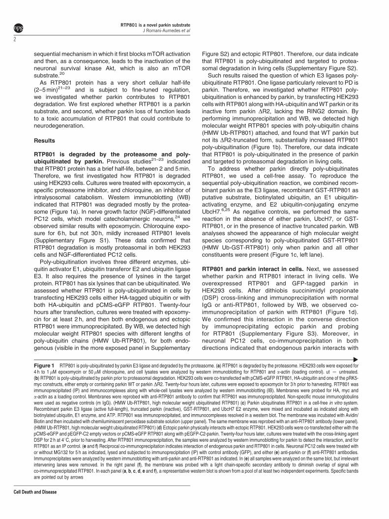
RTP801 is degraded by the proteasome and poly-ubiquitinated by parkin. Previous studies21–23 indicatedthat RTP801 protein has a brief half-life, between 2 and 5 min.Therefore, we first investigated how RTP801 is degradedusing HEK293 cells. Cultures were treated with epoxomycin, aspecific proteasome inhibitor, and chloroquine, an inhibitor ofintralysosomal catabolism. Western immunoblotting (WB)indicated that RTP801 was degraded mostly by the protea-some (Figure 1a). In nerve growth factor (NGF)-differentiatedPC12 cells, which model catecholaminergic neurons,24 weobserved similar results with epoxomycin. Chloroquine expo-sure for 6 h, but not 30 h, mildly increased RTP801 levels(Supplementary Figure S1). These data confirmed thatRTP801 degradation is mostly proteasomal in both HEK293cells and NGF-differentiated PC12 cells.
Poly-ubiquitination involves three different enzymes, ubi-quitin activator E1, ubiquitin transferor E2 and ubiquitin ligaseE3. It also requires the presence of lysines in the targetprotein. RTP801 has six lysines that can be ubiquitinated. Weassessed whether RTP801 is poly-ubiquitinated in cells bytransfecting HEK293 cells either HA-tagged ubiquitin or withboth HA-ubiquitin and pCMS-eGFP RTP801. Twenty-fourhours after transfection, cultures were treated with epoxomy-cin for at least 2 h, and then both endogenous and ectopicRTP801 were immunoprecipitated. By WB, we detected highmolecular weight RTP801 species with different lengths ofpoly-ubiquitin chains (HMW Ub-RTP801), for both endo-genous (visible in the more exposed panel in Supplementary
Figure S2) and ectopic RTP801. Therefore, our data indicatethat RTP801 is poly-ubiquitinated and targeted to protea-somal degradation in living cells (Supplementary Figure S2).
Such results raised the question of which E3 ligases poly-ubiquitinate RTP801. One ligase particularly relevant to PD isparkin. Therefore, we investigated whether RTP801 poly-ubiquitination is enhanced by parkin, by transfecting HEK293cells with RTP801 along with HA-ubiquitin and WT parkin or itsinactive form parkin DR2, lacking the RING2 domain. Byperforming immunoprecipitation and WB, we detected highmolecular weight RTP801 species with poly-ubiquitin chains(HMW Ub-RTP801) attached, and found that WT parkin butnot its DR2-truncated form, substantially increased RTP801poly-ubiquitination (Figure 1b). Therefore, our data indicatethat RTP801 is poly-ubiquitinated in the presence of parkinand targeted to proteasomal degradation in living cells.
To address whether parkin directly poly-ubiquitinatesRTP801, we used a cell-free assay. To reproduce thesequential poly-ubiquitination reaction, we combined recom-binant parkin as the E3 ligase, recombinant GST-RTP801 asputative substrate, biotinylated ubiquitin, an E1 ubiquitin-activating enzyme, and E2 ubiquitin-conjugating enzymeUbcH7.6,25 As negative controls, we performed the samereaction in the absence of either parkin, UbcH7, or GST-RTP801, or in the presence of inactive truncated parkin. WBanalyses showed the appearance of high molecular weightspecies corresponding to poly-ubiquitinated GST-RTP801(HMW Ub-GST-RTP801) only when parkin and all otherconstituents were present (Figure 1c, left lane).
RTP801 and parkin interact in cells. Next, we assessedwhether parkin and RTP801 interact in living cells. Weoverexpressed RTP801 and GFP-tagged parkin inHEK293 cells. After dithiobis succinimidyl propionate(DSP) cross-linking and immunoprecipitation with normalIgG or anti-RTP801, followed by WB, we observed co-immunoprecipitation of parkin with RTP801 (Figure 1d).We confirmed this interaction in the converse directionby immunoprecipitating ectopic parkin and probingfor RTP801 (Supplementary Figure S3). Moreover, inneuronal PC12 cells, co-immunoprecipitation in bothdirections indicated that endogenous parkin interacts with
Figure 1 RTP801 is poly-ubiquitinated by parkin E3 ligase and degraded by the proteasome. (a) RTP801 is degraded by the proteasome. HEK293 cells were exposed for4 h to 1 mM epoxomycin or 50mM chloroquine, and cell lysates were analyzed by western immunoblotting for RTP801 and a-actin (loading control). ut ¼ untreated.(b) RTP801 is poly-ubiquitinated by parkin prior to proteasomal degradation. HEK293 cells were co-transfected with pCMS-eGFP RTP801, HA-ubiquitin and one of the pRK5-myc constructs, either empty or containing parkin WT or parkin DR2. Twenty-four hours later, cultures were exposed to epoxomycin for 3 h prior to harvesting. RTP801 wasimmunoprecipitated (IP) and immunocomplexes along with whole-cell lysates were analyzed by western immunoblotting (IB). Membranes were probed for HA, myc anda-actin as a loading control. Membranes were reprobed with anti-RTP801 antibody to confirm that RTP801 was immunoprecipitated. Non-specific mouse immunoglobulinswere used as negative controls (m IgG). (HMW Ub-RTP801, high molecular weight ubiquitinated RTP801) (c) Parkin ubiquitinates RTP801 in a cell-free in vitro system.Recombinant parkin E3 ligase (active full-length), truncated parkin (inactive), GST-RTP801, and UbcH7 E2 enzyme, were mixed and incubated as indicated along withbiotinylated ubiquitin, E1 enzyme, and ATP. RTP801 was immunoprecipitated, and immunocomplexes resolved in a western blot. The membrane was incubated with Avidin/Biotin and then incubated with chemiluminiscent peroxidase substrate solution (upper panel). The same membrane was reprobed with an anti-RTP801 antibody (lower panel).(HMW Ub-RTP801, high molecular weight ubiquitinated RTP801) (d) Ectopic parkin physically interacts with ectopic RTP801. HEK293 cells were co-transfected either with thepCMS-eGFP and pEGFP-C2 empty vectors or pCMS-eGFP RTP801 along with pEGFP-C2-parkin. Twenty-four hours later, cultures were treated with the cross-linking agentDSP for 2 h at 41C, prior to harvesting. After RTP801 immunoprecipitation, the samples were analyzed by western immunoblotting for parkin to detect the interaction, and forRTP801 as an IP control. (e and f) Reciprocal co-immunoprecipitation indicates interaction of endogenous parkin and RTP801 in cells. Neuronal PC12 cells were treated withor without MG132 for 5 h as indicated, lysed and subjected to immunoprecipitation (IP) with control antibody (GFP), and either (e) anti-parkin or (f) anti-RTP801 antibodies.Immunoprecipitates were analyzed by western immunoblotting with anti-parkin and anti-RTP801 as indicated. In (e) all samples were analyzed on the same blot, but irrelevantintervening lanes were removed. In the right panel (f), the membrane was probed with a light chain-specific secondary antibody to diminish overlap of signal withco-immunoprecipitated RTP801. In each panel (a, b, c, d, e and f), a representative western blot is shown from a pool of at least two independent experiments. Specific bandsare pointed out by arrows
RTP801 is a novel parkin substrateJ Romanı-Aumedes et al
2
Cell Death and Disease

endogenous RTP801 (Figures 1e and f). This interactionwas observed when RTP801 was immunoprecipitated withor without pretreatment with the proteasome inhibitor
MG132 (Figure 1f). Taken together, our results indicatethat RTP801 interacts with parkin and that parkin promotesRTP801 poly-ubiquitination.
RTP801 is a novel parkin substrateJ Romanı-Aumedes et al
3
Cell Death and Disease
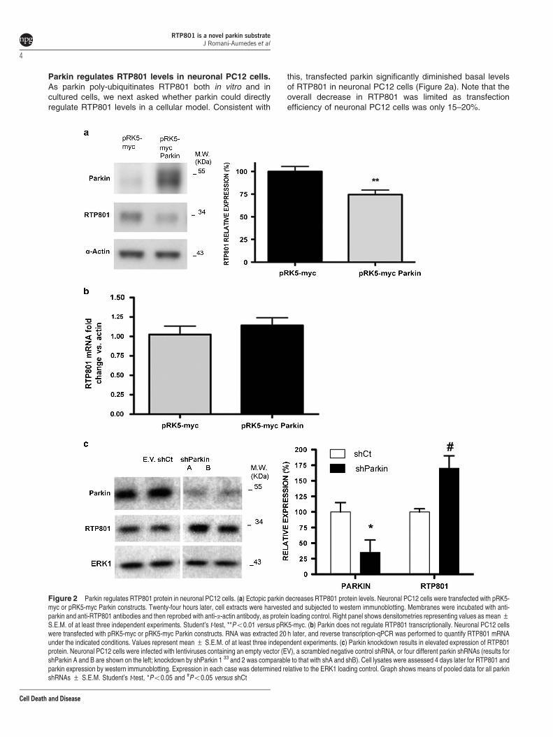
Parkin regulates RTP801 levels in neuronal PC12 cells.As parkin poly-ubiquitinates RTP801 both in vitro and incultured cells, we next asked whether parkin could directlyregulate RTP801 levels in a cellular model. Consistent with
this, transfected parkin significantly diminished basal levelsof RTP801 in neuronal PC12 cells (Figure 2a). Note that theoverall decrease in RTP801 was limited as transfectionefficiency of neuronal PC12 cells was only 15–20%.
Figure 2 Parkin regulates RTP801 protein in neuronal PC12 cells. (a) Ectopic parkin decreases RTP801 protein levels. Neuronal PC12 cells were transfected with pRK5-myc or pRK5-myc Parkin constructs. Twenty-four hours later, cell extracts were harvested and subjected to western immunoblotting. Membranes were incubated with anti-parkin and anti-RTP801 antibodies and then reprobed with anti-a-actin antibody, as protein loading control. Right panel shows densitometries representing values as mean ±S.E.M. of at least three independent experiments. Student’s t-test, **Po0.01 versus pRK5-myc. (b) Parkin does not regulate RTP801 transcriptionally. Neuronal PC12 cellswere transfected with pRK5-myc or pRK5-myc Parkin constructs. RNA was extracted 20 h later, and reverse transcription-qPCR was performed to quantify RTP801 mRNAunder the indicated conditions. Values represent mean ± S.E.M. of at least three independent experiments. (c) Parkin knockdown results in elevated expression of RTP801protein. Neuronal PC12 cells were infected with lentiviruses containing an empty vector (EV), a scrambled negative control shRNA, or four different parkin shRNAs (results forshParkin A and B are shown on the left; knockdown by shParkin 1 33 and 2 was comparable to that with shA and shB). Cell lysates were assessed 4 days later for RTP801 andparkin expression by western immunoblotting. Expression in each case was determined relative to the ERK1 loading control. Graph shows means of pooled data for all parkinshRNAs ± S.E.M. Student’s t-test, *Po0.05 and #Po0.05 versus shCt
RTP801 is a novel parkin substrateJ Romanı-Aumedes et al
4
Cell Death and Disease
Several studies indicate that parkin has DNA-binding andtranscriptional activities.26,27 However, ectopic parkin did notaffect RTP801 mRNA levels in neuronal PC12 cells,discounting the possibility that parkin regulates RTP801 at atranscriptional level (Figure 2b).
To further assess the role of parkin in RTP801 expression,we next diminished endogenous parkin expression withspecific shRNAs delivered by lentiviral infection. In neuronalPC12 cells, shParkin elevated levels of RTP801 in compar-ison with empty vector or scrambled control shRNA by about65–70% (Figure 2c).
We also confirmed the effect of shParkin sequences A andB on RTP801 expression by transient transfection in naivePC12 cells by WB (Supplementary Figure S4a). Additionally,NGF-differentiated PC12 cells transfected with these shRNAswere analyzed by immunofluorescence (SupplementaryFigure S4b and c). Parkin knockdown (30–40%) significantlyelevated RTP801 immunostaining (25–35%), although not asmarkedly as with lentiviral infection, owing to the lowerefficiency of transfection (Supplementary Figure S4).
Taken together, these results indicate that parkin regulatesendogenous RTP801 protein levels in neural cells and thatparkin knockdown leads to an increase in RTP801 proteinexpression.
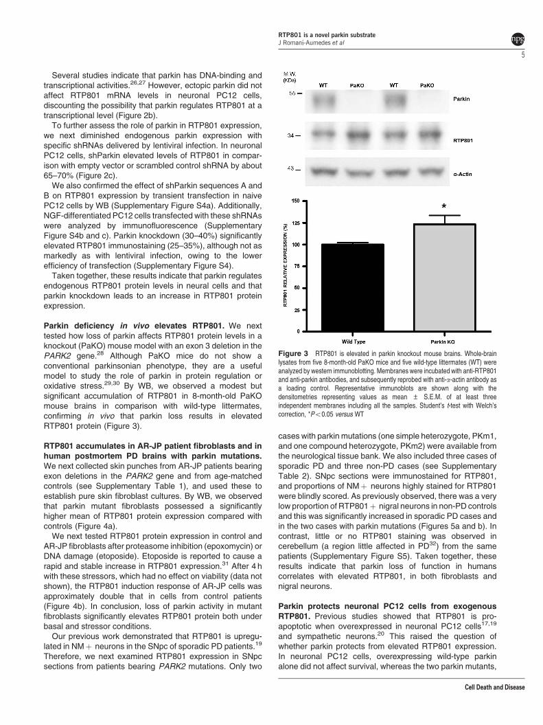
Parkin deficiency in vivo elevates RTP801. We nexttested how loss of parkin affects RTP801 protein levels in aknockout (PaKO) mouse model with an exon 3 deletion in thePARK2 gene.28 Although PaKO mice do not show aconventional parkinsonian phenotype, they are a usefulmodel to study the role of parkin in protein regulation oroxidative stress.29,30 By WB, we observed a modest butsignificant accumulation of RTP801 in 8-month-old PaKOmouse brains in comparison with wild-type littermates,confirming in vivo that parkin loss results in elevatedRTP801 protein (Figure 3).
RTP801 accumulates in AR-JP patient fibroblasts and inhuman postmortem PD brains with parkin mutations.We next collected skin punches from AR-JP patients bearingexon deletions in the PARK2 gene and from age-matchedcontrols (see Supplementary Table 1), and used these toestablish pure skin fibroblast cultures. By WB, we observedthat parkin mutant fibroblasts possessed a significantlyhigher mean of RTP801 protein expression compared withcontrols (Figure 4a).
We next tested RTP801 protein expression in control andAR-JP fibroblasts after proteasome inhibition (epoxomycin) orDNA damage (etoposide). Etoposide is reported to cause arapid and stable increase in RTP801 expression.31 After 4 hwith these stressors, which had no effect on viability (data notshown), the RTP801 induction response of AR-JP cells wasapproximately double that in cells from control patients(Figure 4b). In conclusion, loss of parkin activity in mutantfibroblasts significantly elevates RTP801 protein both underbasal and stressor conditions.
Our previous work demonstrated that RTP801 is upregu-lated in NMþ neurons in the SNpc of sporadic PD patients.19
Therefore, we next examined RTP801 expression in SNpcsections from patients bearing PARK2 mutations. Only two
cases with parkin mutations (one simple heterozygote, PKm1,and one compound heterozygote, PKm2) were available fromthe neurological tissue bank. We also included three cases ofsporadic PD and three non-PD cases (see SupplementaryTable 2). SNpc sections were immunostained for RTP801,and proportions of NMþ neurons highly stained for RTP801were blindly scored. As previously observed, there was a verylow proportion of RTP801þ nigral neurons in non-PD controlsand this was significantly increased in sporadic PD cases andin the two cases with parkin mutations (Figures 5a and b). Incontrast, little or no RTP801 staining was observed incerebellum (a region little affected in PD32) from the samepatients (Supplementary Figure S5). Taken together, theseresults indicate that parkin loss of function in humanscorrelates with elevated RTP801, in both fibroblasts andnigral neurons.
Parkin protects neuronal PC12 cells from exogenousRTP801. Previous studies showed that RTP801 is pro-apoptotic when overexpressed in neuronal PC12 cells17,19
and sympathetic neurons.20 This raised the question ofwhether parkin protects from elevated RTP801 expression.In neuronal PC12 cells, overexpressing wild-type parkinalone did not affect survival, whereas the two parkin mutants,
Figure 3 RTP801 is elevated in parkin knockout mouse brains. Whole-brainlysates from five 8-month-old PaKO mice and five wild-type littermates (WT) wereanalyzed by western immunoblotting. Membranes were incubated with anti-RTP801and anti-parkin antibodies, and subsequently reprobed with anti-a-actin antibody asa loading control. Representative immunoblots are shown along with thedensitometries representing values as mean ± S.E.M. of at least threeindependent membranes including all the samples. Student’s t-test with Welch’scorrection, *Po0.05 versus WT
RTP801 is a novel parkin substrateJ Romanı-Aumedes et al
5
Cell Death and Disease

R42P (mutated in the UBL domain) or P437L (mutated in theRING2 domain), with severely compromised E3 ligaseactivity, showed a slight, but not significant decrease inviability. When RTP801 was overexpressed alone, around60% of the cells died, as previously reported.17,19 However,ectopic wild-type parkin, but not the mutated parkin forms,counteracted RTP801-induced cell death (Figure 6a).
To confirm that parkin protects from ectopic RTP801 by poly-ubiquitinating it, we generated a non-ubiquitinable form ofRTP801 (RTP801 K-R), in which we substituted its six lysines(K) with arginines (R). In neuronal PC12 cells, overexpressedRTP801 K-R accumulated to a significantly higher degree thanoverexpressed WT RTP801 (Supplementary Figure S6), whichis consistent with the role of ubiquitination and proteasomaldegradation in regulating RTP801 expression. Both WT andRTP801 K-R induced similar levels of cell death (Figure 6b).However, when these vectors were overexpressed along with
parkin, in contrast to its protection from WT RTP801, ectopicparkin was incapable of preventing cell death induced byRTP801 K-R (Figure 6b). These results are consistent with theidea that RTP801 ubiquitination by parkin is necessary forinhibiting RTP801 toxicity.
We next investigated whether parkin knockdown enhancesectopic RTP801 toxicity. In NGF-differentiated PC12 cells,parkin knockdown alone induced around 30–35% cell death(Figure 6c). Ectopic RTP801 alone caused 50% cell loss,whereas, interestingly, parkin knockdown significantlyenhanced such cell death to about 70% (Figure 6c).
Parkin diminishes RTP801 elevation and protectsagainst 6-OHDA. Parkin is protective in a variety ofneurodegeneration models, including the 6-OHDA model ofPD.13,14 Our previous studies indicate that induced RTP801mediates neurotoxin-promoted death of dopaminergic
Figure 4 RTP801 is elevated in human fibroblasts derived from AR-JP patients with parkin mutations. (a) RTP801 is elevated in fibroblasts from parkin mutant AR-JPpatients. Extracts from human primary fibroblasts derived from six diagnosed parkin mutant AR-JP patients (PKm AR-JP) and six control individuals (Ct) were subjected towestern immunoblotting. Membranes were probed with antibodies against parkin and RTP801 and reprobed for a-actin (loading control). Representative immunoblots areshown along with the densitometries for RTP801 signals from at least three independent membranes including all the samples. Student’s t-test, *Po0.05 versus controlindividuals. (b) Parkin mutant fibroblasts exposed to epoxomycin or etoposide show elevated RTP801 accumulation. The same human primary fibroblasts were treated for 4 hwith 1 mM epoxomycin or 25mM etoposide before being harvested. The resulting extracts were analyzed by western immunoblotting. Membranes were incubated withantibodies against parkin and RTP801, and subsequently reprobed with anti-a-actin antibody as a loading control. The graph shows densitometries for RTP801 signals from atleast three independent membranes. Student’s t-test, *Po0.05 versus control untreated, þPo0.05 versus control epoxomycin, $Po0.05 versus control etoposide; ANOVAwith Bonferroni’s multiple comparison test, #Po0.05 and ##Po0.01 versus control untreated. ut ¼ untreated
RTP801 is a novel parkin substrateJ Romanı-Aumedes et al
6
Cell Death and Disease

neurons in vitro and in vivo.19,20,23 Therefore, we nextdetermined whether there is a relationship between parkinand RTP801 levels in the neuronal PC12 cell 6-OHDAmodel. 6-OHDA significantly induced endogenous RTP801protein19 and endogenous parkin levels were diminishedby nearly half33 (Figure 7a). Moreover, we found that
transfecting neuronal PC12 cells with WT parkin, but notthe R42P and P437L parkin mutants, significantly decreasedelevation of RTP801 protein induced by 6-OHDA (Figure 7b).
To verify our findings with primary neurons, we turned tosympathetic neurons, a population that degenerates in PD.We observed parkin knockdown by about 50% in cultured rat
Figure 5 RTP801 is elevated in parkin mutant NMþ nigral neurons. Paraffin-embedded sections of SNpc from control individuals (CT1, CT2 and CT3), parkin simpleheterozygous mutant patient (PKm1), parkin compound heterozygous mutant (PKm2) and sporadic PD patients (SPD1, SPD2 and SPD3) were immunostained for RTP801(grey-blue). Note the presence of neuromelanin granules (brown) in the somas of dopaminergic neurons. Arrows point to highly stained NMþ nigral neurons. The percentageof NMþ and RTP801þ neurons versus total NMþ neurons was scored for each case, and results are represented in the graph. Student’s t-test, *Po0.05 versus controlindividuals. In total, 150–500 neurons were evaluated per section
RTP801 is a novel parkin substrateJ Romanı-Aumedes et al
7
Cell Death and Disease

Figure 6 Parkin protects from RTP801-induced cell death. (a) Ectopic parkin WT protects from the cell death caused by RTP801. Neuronal PC12 cells were co-transfectedwith pRK5-myc, pRK5-myc Parkin WT, pRK5-myc Parkin R42P or pRK5-myc Parkin P437L constructs, along with either pCMS-eGFP or pCMS-eGFP RTP801 constructs.Twenty-four hours later, eGFPþ surviving cells were scored under fluorescence microscopy. Values represented as mean ± S.E.M. of at least three independentexperiments in triplicate in each condition. ANOVA Bonferroni’s multiple comparison test, ***Po0.001 versus pRK5-myc/pCMS-eGFP, ###Po0.001 versus pRK5-myc/pCMS-eGFP RTP801. (b) Parkin protects neuronal PC12 cells from RTP801-induced cell death by mediating its ubiquitination. In NGF-differentiated PC12 cells, we co-transfected pRK5-myc or pRK5-myc Parkin, along with one of the pCMS-eGFP constructs, either empty or containing RTP801 or RTP801 K-R. Twenty-four hours later, cellswere fixed and immunostained, and eGFPþ surviving cells were scored under fluorescence microscopy. Values represented as mean ± S.E.M. of at least threeindependent experiments in triplicate in each condition. ANOVA Bonferroni’s multiple comparison test, ***Po0.001 versus pRK5-myc/pCMS-eGFP, ###Po0.001 versuspRK5-myc/pCMS-eGFP RTP801. (c) Knocking down parkin exacerbates RTP801-mediated cell death. PC12 cells differentiated with NGF were first transfected with ascrambled negative control shRNA (shCt) or two different parkin shRNAs (shParkin A or B), and after 72 h, cultures were transfected with pCMS-eGFP or pCMS-eGFPRTP801 constructs. Twenty-four hours after the second transfection, ZsGreenþ /eGFPþ surviving cells were scored under fluorescence microscopy. Values represented asmean ± S.E.M. of at least three independent experiments in triplicate in each condition. ANOVA with Bonferroni’s multiple comparison test, ***Po0.001 versus shCt/pCMS-eGFP, ###Po0.001 versus shCt/pCMS-eGFP RTP801. In each panel, values represent mean ± S.E.M. of, at least, three independent experiments with four replicates percondition
RTP801 is a novel parkin substrateJ Romanı-Aumedes et al
8
Cell Death and Disease

sympathetic neurons with lentivirally expressed shRNAsagainst parkin. This raised basal RTP801 levels by aboutthe same magnitude (Figure 7c). Moreover, parkin knock-down in these neurons significantly increased RTP801 proteininduction by 6-OHDA (Figure 7c). These results support ourobservations in neuronal PC12 cells that RTP801 knockdownincreases RTP801 levels and extend them to show that parkinknockdown exacerbates the RTP801 response to 6-OHDA inneurons (see Figure 2c).
We next examined whether ectopic parkin, whichdecreases endogenous RTP801 upregulation (Figures 7aand b), is neuroprotective in the 6-OHDA model. In neuronalPC12 cells, overexpressed WT parkin, but not the two parkinmutants, significantly reduced 6-OHDA-induced death(Figure 7d). Note that the parkin P437L mutant decreasedcell viability after 6-OHDA exposure, possibly by acting as adominant-negative.
Finally we explored whether parkin knockdown sensitizesneuronal PC12 cells to 6-OHDA toxicity and whether thisdepends on RTP801. Parkin knockdown induced cell death inuntreated cultures (see also Figure 6c) and enhanced6-OHDA-promoted toxicity (Figure 7e and SupplementaryFigure S7). When we simultaneously knocked down parkinand RTP801, we prevented parkin knockdown-induced celldeath and its sensitization to 6-OHDA. These findings indicatethat the toxic effects of parkin loss of function require thepresence of RTP801.
Taken together, these results indicate that in cellularmodels of PD, parkin protects, at least in part, by preventingRTP801 elevation.
Discussion
Here, we identified a new substrate of parkin. We show thatparkin mediates RTP801/REDD1 poly-ubiquitination, facil-itates its proteasomal degradation and that endogenousparkin and RTP801 interact in living cells. We also show thatparkin loss of function elevates RTP801 protein in cellular andanimal models. This is supported by observations of elevatedRTP801 in both postmortem PD brains and fibroblasts frompatients with parkin mutations. Furthermore, overexpressionof WT parkin, but not its inactive mutated forms, protectsneuronal cells from death when RTP801 is overexpressed orinduced with 6-OHDA. We thus propose that one mechanismby which parkin exerts neuronal protection is by preventingaccumulation of lethal RTP801 protein levels.
Our data indicate that RTP801 is poly-ubiquitinated andtargeted to the ubiquitin proteasome system (UPS). Theseresults agree with others that RTP801 degradation dependson the UPS.21–23 UPS alterations are a common feature incellular and animal models of PD and in the disease itself.34,35
Based on our results, UPS impairment associated with PDcould contribute, in part, to RTP801 elevation in both sporadicPD and AR-JP.
We showed that parkin poly-ubiquitinates RTP801, bothin vitro and in vivo. The only presently described E3 ligase forREDD1/RTP801 is CUL4A-DDB1 in HEK293 and MCF-7cells.21 However, Zhao et al.36 could not confirm this result.Whether Cul4A-DDB1 is relevant in the context of PD ispresently unknown.
Parkin and its ligase activity have been linked to bothfamilial and sporadic PD. Mutations in PARK2,5,6 S-nitrosyla-tion7 and oxidative8 or dopaminergic stress9 critically com-promise parkin solubility and E3 ligase activity. Parkinenzymatic impairment has been proposed to produce toxicaccumulation of its substrate proteins, which in turn causesneurodegeneration.
Many proteins have been proposed as parkin substrates;however only a few have been confirmed. Dawson et al.37
proposed that a ‘true’ parkin substrate should accumulate in bothAR-JP and sporadic PD as well as in animal models of parkininactivation such as PaKO mice and the MPTP toxin model.37
Our previous19,23 and present work appears to fulfill theserequirements37 for RTP801. Additional parkin substrates havebeen reported38 and these, along with RTP801, could representaccumulated proteins that contribute to neuron degenerationassociated with parkin loss of expression or function.
We found a relatively modest, but significant, increase ofRTP801 in PaKO mice. These animals have motor and non-motor behavioral impairments,39 deficient synaptic transmis-sion,40 reduced mitochondrial respiration and increasedoxidative damage,41 and loss of DA neurons after 2 years ofage.42,43 The PaKO mice examined in our study were only 8-month old. Our data raise the possibility that early RTP801accumulation could contribute to the impairments cited aboveand to neuron death at an older age. Also, in younger PaKOmice, other ligases may compensate for parkin loss andmaintain levels of substrate proteins, such as RTP801. Therelatively small increase in RTP801 seen in 8-month-oldPaKO mice is also consistent with, and could potentiallyaccount for, the lack of neuron death at this age.
In postmortem sections from two PD cases with PARK2gene mutations, NMþ neurons showed higher levels ofRTP801 expression. One case involved compound hetero-zygous mutations and behaved as a homozygous parkinmutant, with early onset of disease. However, the secondcase had a simple heterozygous mutation in the parkin gene,with late disease onset. Both cases showed elevated RTP801levels as in sporadic cases. This could reflect findings that lossof only one copy of the parkin gene can be a risk factor forPD.10,44 To extend our results, fibroblasts from six AR-JPpatients with PARK2 gene mutations were assessed. Cul-tured skin fibroblasts are very useful to study genetic PDmechanisms.45 We reasoned that mutations in the PARK2gene should influence RTP801 turnover in cells in addition toneurons. We observed that RTP801 protein levels approxi-mately doubled in AR-JP patient fibroblasts compared withcontrols, and that the RTP801 response to etoposide orepoxomycin was exaggerated in AR-JP fibroblasts. This isconsistent with the idea that parkin loss of function elevatesRTP801 in both neuronal and non-neuronal cells. However,as fibroblasts are proliferative, RTP801 does not induce theircell death as it does in non-dividing neurons.17,19
Our previous work demonstrated that RTP801 wasupregulated in cellular and animal models of PD and inNMþ nigral neurons of sporadic PD patients. It also showedthat RTP801 elevation is sufficient to mediate neuron celldeath, and that RTP801 is required for neuron death in severalPD models.19 Hence, as parkin loss of function leads toRTP801 elevation, it is logical to suggest that this effect
RTP801 is a novel parkin substrateJ Romanı-Aumedes et al
9
Cell Death and Disease

contributes to neuronal degeneration and cell death in thecontext of PD. RTP801 protein elevation can be the end pointof two different processes; one being gene activation by PD-associated neuronal stress,18,19 and the other, defectiveRTP801 degradation due to impaired parkin activity. RTP801may therefore contribute, with other parkin substrates, toneuron death and degeneration in both sporadic and parkin-associated PD.
Parkin protects from neuron toxicity associated withmultiple stressors including mutant alpha synuclein,46 kainateacid excitotoxicity12 as well as dopamine47 and 6-OHDA.14
In consonance with this, we observed that WT parkin, but notits mutated forms, decreased RTP801-induced cell death ofneuronal PC12 cells. Significantly, the neuroprotective effect
of parkin was lost for non-ubiquitinable form of RTP801.Ectopic parkin protected against 6-OHDA, and this protectionwas accompanied by a decrease in RTP801 protein. More-over, parkin knockdown diminished cell survival, and sensi-tized cells against 6-OHDA. Interestingly, RTP801knockdown abrogated this sensitization and restored cellsurvival. These results strongly suggest that parkin isprotective, at least in part, by mediating RTP801 proteasomaldegradation. Neuroprotective effects of parkin appear due, atleast in part, to its effects on mitochondria, by regulatingcytochrome c release and apoptosis.48 A portion of RTP801protein is also translocated to mitochondria.49 This suggeststhat clearance of RTP801 by parkin may have direct effects onmitochondrial function as well as on mTOR/Akt signaling.
RTP801 is a novel parkin substrateJ Romanı-Aumedes et al
10
Cell Death and Disease

Abrogation of RTP801 expression with shRNAs or phar-macologic agents has been demonstrated to be a usefulstrategy for neuroprotection. In cellular models of PD,RTP801 shRNAs protected both sympathetic neurons andneuronal PC12 cells from neurotoxins.19 RTP801 knockdownalso has been successful in treating ocular diseases likemacular degeneration.50 Pharmacologically, rapamycin, bypartially inhibiting mTOR, interferes with RTP801 translationand is protective in cellular and animal models of PD.23 Ourdata indicate that parkin contributes to decreasing cellularRTP801 protein levels. Therefore, means to elevate parkinlevels or activity in neurons would be anticipated to provideneuroprotection, at least, in part, via degradation of RTP801.
In conclusion, our work in cellular and animal models and inhuman samples strongly indicates that RTP801 is a substrateof parkin and that RTP801 elevation due to parkin loss offunction in both AR-JP and sporadic PD may contribute toneurodegeneration. Any genetic conditions or stress situa-tions that compromise parkin activity have the potential toproduce a toxic accumulation of RTP801. This is relevant todesign of therapeutic approaches to promote neuron survivaland block neurodegeneration in both sporadic and AR-JP.
Materials and methodsAntibodies, plasmids and materials. Rabbit polyclonal anti-RTP801antibody (used for WB and immunohistochemistry) was purchased fromProteintech Group (Chicago, IL, USA). Mouse monoclonal anti-RTP801 (usedfor immunoprecipitation) was purchased from Bethyl Laboratories (Montgomery,TX, USA). Monoclonal mouse antibody anti-parkin used for WB andimmunohistochemistry was purchased from Abcam (Cambridge, UK). Monoclonalmouse anti-parkin antibody (used for immunoprecipitation), polyclonal rabbit anti-myc (used for WB), monoclonal mouse anti-myc (used for immunoprecipitation)and anti-HA tag were obtained from Cell Signaling Technology (Danvers, MA,USA). Mouse monoclonal antibody against GFP and rabbit polyclonal antibodiesagainst parkin and ERK1 were obtained from Santa Cruz Biotechnology (Dallas,TX, USA). Anti-a-actin antibody was purchased from MP Biomedicals (Santa Ana,CA, USA). Goat anti-mouse or anti-rabbit secondary antibodies conjugated tohorseradish peroxidase were obtained from Pierce Thermo Fisher Scientific(Rockford, IL, USA). Goat anti-mouse or anti-rabbit secondary antibodiesconjugated with Alexa 488 or Alexa 568 were purchased from Life Technologies(Grand Island, NY, USA). Peroxidase-conjugated goat anti-IgG light chain-specific
secondary antibody was obtained from Jackson ImmunoResearch Laboratories(West Grove, PA, USA).
The pCMS-eGFP RTP801 construct was generated as previously described.19
The pcDNA3 HA-ubiquitin construct was purchased from Addgene (Cambridge,MA, USA). The constructs pEGFP-C2, pEGFP-C2-parkin, pRK5-myc, pRK5-mycParkin and the parkin point mutation pRK5-myc Parkin P437L were a kind gift fromDr. Serge Przedborski (Columbia University, New York, NY, USA). The parkin pointmutant pRK5-myc Parkin R42P and the parkin deletion pRK5-myc Parkin DR2 werekindly provided by Dr. Leonidas Stefanis (Academy of Athens, Biomedical ResearchFoundation, Athens, Greece). All constructs were verified by DNA sequencing.Epoxomycin, MG132 and etoposide were purchased from Calbiochem MerckMillipore (Billerica, MA, USA), Chloroquine and CHAPS were obtained fromSigma-Aldrich (St. Louis, MO, USA) and 6-OHDA was purchased from TocrisBioscience (Bristol, UK).
Directed mutagenesis. The pCMS-eGFP RTP801 K-R construct wasobtained from the pCMS-eGFP RTP801 construct by mutating all the lysines (K) toarginines (R) using the QuickChange Lightning II multi site-directed mutagenesis(Agilent Technologies, Santa Clara, CA, USA), with the following primers: K126R,50-GAGCCAGGTGGGCAGGGAACTCCTGC-30; K152R, 50-GTGTGGAGCAAGGCAGGAGCTGCCATAGTGT-30; K185R, 50-GCCTCTGGCCCAGGATCCAGGGCCT-30; K215R-K216R-K217R, 50-CACCGGCTTCAGAGTCATCAGAAGGAGACTCTACAGCTCCGAG-30 All new constructs were verified by DNA sequencing.
shRNA production. shG-scrambled control RNA and shRTP801 weregenerated as previously described.51 Two different parkin shRNAs were preparedin a ZsGreen expressing pSIREN vector using the Knockout RNAi Systems(Clontech Laboratories, Mountain View, CA, USA) according to the manufacturer’sinstructions, based on the following sequences: shParkin A, 50-AACAACAGAGTATCGTTCACA-30; shParkin B, 50-ATCGTTCACATAGTACAGAGA-30; shParkin1, 50-AGCTCCATCACTTCAGGATCC-30 (as described in Sun et al.33); shParkin 2,50-ATCACCTGACAGTACAGAACT-30. The scrambled negative control shRNA(shCt) was provided by the same kit with the sequence: 50-GTGCGTTGCTAGTACCAAC-30. shParkins A, B and 2 were specific for rattus norvegicus parkin,whereas shParkin 1 was specific for rat and mouse.
Lentivirus preparation. Viral particles for neuronal PC12 infection wereprepared as previously described.33
Cell culture. PC12 cells were cultured and treated with NGF as describedpreviously.52 For NGF treatment, the cells were cultured in RPMI 1640 medium (GibcoLife Technologies, Grand Island, NY, USA) supplemented with 1% heat-inactivatedhorse serum (Sigma-Aldrich), penicillin/streptomycin, and 50 ng/ml recombinant humanb-NGF (Alomone Labs, Jerusalem, Israel; or kindly provided by Genentech, SouthSan Francisco, CA, USA) for 7–8 days, in a 7.5% CO2 atmosphere at 37 1C.
Figure 7 Parkin decreases elevation of RTP801 and protects from 6-OHDA. (a) RTP801 and parkin protein levels after 6-OHDA exposure. Neuronal PC12 cells wereexposed to 50 or 100mM 6-OHDA for 16 h. Cell extracts were subjected to western immunoblotting. Membranes were probed with antibodies against parkin and RTP801, andthen reprobed for a-actin as protein loading control. Right panels represent quantification of parkin and RTP801 densitometric values (mean ± S.E.M.) normalized to a-actin,of three independent experiments. ANOVA with Bonferroni’s multiple comparison test, ***Po0.001 versus untreated control cultures. (b) Ectopic parkin decreases 6-OHDA-induced RTP801 protein. Neuronal PC12 cells were transfected with pRK5-myc, pRK5-myc Parkin WT, pRK5-myc Parkin R42P or pRK5-myc Parkin P437L plasmids. Forty-eight hours later, cultures were exposed to 100mM 6-OHDA for 16 h. Cell extracts were harvested and subjected to western immunoblotting. Membranes were incubated withanti-parkin and anti-RTP801 antibodies. The same membranes were then reprobed with anti-a-actin antibody as loading control. Upper panel shows RTP801 densitometriesrepresenting values as mean ± S.E.M. of at least three independent experiments. ANOVA with Bonferroni’s multiple comparison test, *Po0.05 versus untreated pRK5-myc,##Po0.01 versus 6-OHDA-treated pRK5-myc. (c) Parkin knockdown increases RTP801 levels induced by 6-OHDA. Rat sympathetic neurons were infected with lentivirusescontaining either a scrambled negative control shRNA (shCT) or a pool of three different shRNA constructs against parkin (shParkin). Six days later, cultures were treated with5mM 6-OHDA for 16 h. Cell extracts were harvested and subjected to western immunoblotting. Membranes were probed with antibodies against parkin, RTP801 and a-actinas a loading control. Upper panel shows RTP801 quantification (mean ± S.E.M.) normalized to a-actin, of three independent experiments. ANOVA with Bonferroni’s multiplecomparison test, **Po0.01 versus untreated shCT, #Po0.05 versus 6-OHDA-treated shCT. (d) Parkin protects from 6-OHDA-induced cell death. Neuronal PC12 cells wereco-transfected with pRK5-myc/pCMS-eGFP, pRK5-myc Parkin WT/pCMS-eGFP, pRK5-myc Parkin R42P/pCMS-eGFP or pRK5-myc Parkin P437L/pCMS-eGFP constructs(4:1) and 48 h later cultures were treated with 100 mM 6-OHDA. Twenty-four hours after 6-OHDA exposure, eGFPþ surviving cells were scored under fluorescencemicroscopy. Values represent mean ± S.E.M. of at least three experiments with six replicates in each condition. ANOVA with Bonferroni’s multiple comparison test,***Po0.001 versus untreated pRK5-myc/pCMS-eGFP, ##Po0.01 and ###Po0.001 versus 6-OHDA-treated pRK5-myc/pCMS-eGFP. (e) Parkin knockdown sensitizes to6-OHDA-induced cell death in an RTP801-dependent manner. Neuronal PC12 cells were transfected with parkin shRNA (shParkin B) or its scrambled negative control (shCt),along with a shRNA sequence against RTP801 (shRTP801) or its corresponding shRNA scrambled control sequence (shG). After 72 h of transfection, cultures were treatedwith 100mM 6-OHDA. Twenty-four hours after treatment cells were fixed and ZsGreenþ /eGFPþ surviving cells were scored under fluorescence microscopy. Valuesrepresent mean ± S.E.M. of at least three experiments with three replicates in each condition. ANOVA with Bonferroni’s multiple comparison test, ***Po0.001 versusuntreated shCt/shG, ###Po0.001 versus untreated shParkin B/shG, þ þ þPo0.001 versus 6-OHDA-treated shCt/shG and $$$Po0.001 versus 6-OHDA-treated shParkinB/shG. ut ¼ untreated
RTP801 is a novel parkin substrateJ Romanı-Aumedes et al
11
Cell Death and Disease

Neonatal rat superior cervical ganglion sympathetic neurons were cultured aspreviously described.19,53
HEK293 cells were maintained in DMEM medium (Gibco Life Technologies)supplemented with 10% fetal calf serum (Gibco Life Technologies) and penicillin/streptomycin (Gibco Life Technologies) in a 5% CO2 atmosphere at 37 1C.
Human fibroblasts derived from six diagnosed AR-JP patients with PARK2dysfunctional mutations and six control individuals were cultured in DMEM mediumsupplemented with 10% fetal calf serum and penicillin/streptomycin in a 5% CO2
atmosphere at 37 1C. These fibroblasts were obtained from the Neurology Service-Movement Disorders Biorepository (IDIBAPS, Hospital Clinic, Universitat deBarcelona, Barcelona, Catalonia, Spain). Fibroblasts were used between 5 and 6passages in vitro.
Transfection, treatments and viral infection. Neuronal PC12 andHEK293 cells were transfected with Lipofectamine 2000 (Invitrogen LifeTechnologies, Grand Island, NY, USA) according to the manufacturer’s instructions.
6-OHDA treatments were performed after 6–7 days of culture in NGF-differentiated PC12, and at day in vitro (DIV) 9 in primary rat sympathetic neuronalcultures. Medium was changed right before treatments.
Neuronal PC12 cells were infected at DIV3 and at a multiplicity of infection (MOI) of 3.Primary neonatal rat SCG sympathetic neurons were infected at DIV 3 and at a MOI of2, with lentiviral particles containing a pool of three different shRNAs constructs againstrat parkin (shParkin) or a scrambled control sequence (shCT), both obtained from SantaCruz Biotechnologies. After 18–20 h of infection, medium containing remaining lentiviralparticles was removed and replaced by fresh medium.
Quantitative reverse transcription-PCR. Each sample of total RNAwas isolated from neuronal PC12 cells by using the High Pure RNA Isolation Kit(Roche Diagnostics Corporation, Indianapolis, IN, USA). cDNA was transcribedfrom total RNA with the Transcriptor First Strand cDNA Synthesis Kit RocheDiagnostics Corporation. Specific primers used for quantitative PCR amplificationwere as follows: RTP801 forward primer, 50-GCTCTGGACCCCAGTCTAGT-30;RTP801 reverse primer, 50-GGGACAGTCCTTCAGTCCTT-30; a-actin forwardprimer, 50-GGGTATGGGTCAGAAGGACT-30; a-actin reverse primer, 50-GAGGCATACAGGGACAACAC-30. Quantitative PCR was performed with a 7500 RealTime PCR System (Applied Biosystems, Foster City, CA, USA) using equalamounts of cDNA template for quantitative PCR analysis of RTP801, normalizedby a-actin. The genes analyzed in this study were examined by the relativeexpression by means of DDCT.
WB analysis. Whole-cell extracts or 8-month-old PaKO mouse total brainlysates were collected, prepared and analyzed, as previously described.19,28
Chemoluminiscent images were acquired with a LAS-3000 Imager (Fujifilm,Valhalla, NY, USA).
Immunoprecipitation. In the experiments where we immunoprecipitatedectopic RTP801 and parkin, cell extracts were collected with cell lysis buffer (CellSignaling Technology) and pre-cleared with protein A/G sepharose beads(Immunoprecipitation Starter Pack; GE Healthcare Bio-Science, Pittsburgh, PA,USA) for 1 hour at 4 1C. Pre-cleared cell lysates were incubated overnight at 4 1Cin rotation, with the corresponding antibody or a normal immunoglobulin (SantaCruz Biotechnology) as a negative control. Immunocomplexes were incubated withBSA-blocked protein A/G sepharose beads for 4 h at 4 1C in rotation. Then thebeads were centrifuged and washed five times with CHAPS buffer (Tris 50 mM; pH7.4, NaCl 150 mM, MgCl2 10 mM, CHAPS 0.4%), and the immunocomplexes werecollected and subjected to WB. The cross-linking agent DSP (Gibco LifeTechnologies) was used according to the manufacturer’s instructions. In order toimmunoprecipitate endogenous RTP801 and parkin, cell extracts were collectedwith cell lysis buffer and incubated overnight at 41C in rotation, with thecorresponding antibody or a control antibody (GFP) as a negative control.Immunocomplexes were incubated with protein A (parkin) or protein G (RTP801)for 2 h at 4 1C in rotation. Then the beads were centrifuged and washed four timeswith cell lysis buffer, and the immunocomplexes were resolved in a western blot.
In vitro ubiquitination. The assay was performed by using theUbiquitinylation Kit according to the manufacturer’s instructions (Enzo LifeSciences, Farmingdale, NY, USA) with slight modifications. Briefly, (20 mM Tris-HCl; pH 7.5, 1 mM dithiothreitol, 20 U/ml inorganic pyrophosphatase, 5 mM Mg-ATP, 0.1mM His6-tagged recombinant human ubiquitin-activating enzyme E1,
2.5mM biotinylated ubiquitin), His6-tagged recombinant human ubiquitin-conjugat-ing enzyme UbcH7 E2 (Enzo Life Biosciences), His6-tagged recombinant humanparkin full-lenght (Merck Millipore), N-terminal GST-tagged recombinant humanRTP801 full-length (Novus Biologicals, Cambridge, UK), and GST-taggedrecombinant human truncated and inactive parkin (1-387) (Novus Biologicals),were added to the reaction buffer, as referred in each condition, and thenincubated at 37 1C for 90 min. RTP801 was immunoprecipitated as describedabove, and equal volumes of each sample were subjected to WB. The blottedmembranes were probed for biotinylated ubiquitin using Avidin/Biotin-HorseradishPeroxidase (Ultra-sensitive ABC staining kit; Thermo Fisher Scientific, Waltham,MA, USA) or by anti-RTP801 antibody (Proteintech Group).
Immunofluorescence. Neuronal PC12 cells were fixed with 4% parafor-maldehyde, stained and observed under fluorescence microscopy, as previouslydescribed.20 Fluorescence from stained cells was quantified with ImageJ software(NIH). Fluorescence for parkin staining (in red) or for RTP801 staining (in red) ineach single transfected cell (ZsGreenþ ) with shParkin A and B was quantifiedand normalized with the fluorescence signal from non-transfected cells, andpercentages were compared with the ones obtained with shControl (shCt). Cellswere counter-stained with Hoescht 33342 (Invitrogen Life Technologies) tovisualize the nucleus. In survival assays, transfected viable cells were scored bystrip counting, as previously described.19
Immunohistochemistry of human sections. Paraffin-embedded sec-tions of postmortem human SNpc and cerebellum from control individuals, parkinheterozygous mutant PD patients, and sporadic PD patients, were obtained fromthe Neurological Tissue Bank (Biobank-HC-IDIBAPS) and stained as previouslydescribed.19
Statistics. All experiments were performed at least in triplicate, and results arereported as means±S.E.M. Student’s t-test was performed as appropriate andindicated in the figure legends, mostly as unpaired, two-tailed sets of arrays andpresented as probability P-values, or otherwise stated in the figure legends. One-way or two-way ANOVA with Bonferroni’s multiple comparison test wereperformed to compare multiple groups.
Conflict of InterestThe authors declare no conflict of interest.
Acknowledgements. Supported in part by grants from the Parkinson’sDisease Foundation, Spanish Ministry of Science and Innovation (SAF2010-21058),the PEOPLE Programme – Marie Curie Actions, European Community (PIRG08-GA-2010-276957) and National Institutes of Health Morris K. Udall Centers ofExcellence in Parkinson’s Research Grant P50 NS038370. Supported also by thecrowd funding campaign ‘SOS recerca en Parkinson’ via Goteo.org, Portal d’Avall,S.L. and ‘Mememtum: early detection of neurological disorders’. The constructspEGFP-C2-parkin, pRK5-myc, pRK5-myc Parkin and the parkin point mutant pRK5-myc Parkin P437L were a kind gift from Dr. Serge Przedborski (Columbia University,New York, (NY) USA). The parkin point mutant pRK5-myc Parkin R42P wasgenerously provided by Dr. Leonidas Stefanis (Academy of Athens, BiomedicalResearch Foundation, Athens, Greece). Parkin knockout mice whole-brain lysateswere provided by the laboratory of Dr. Hermann Luebbert (Ruhr University,Germany). We thank Dr. Bernat Crosas for helpful advice with the ubiquitinationassays. Early passages of human skin fibroblasts (controls and mutant parkin) wereobtained from the Movement Disorders Biorepository, provided by Dr. EduardTolosa (IDIBAPS, Hospital Clınic, Universitat de Barcelona, Barcelona). We thankDr. Esther Perez-Navarro for helpful discussion. We are indebted to theNeurological Tissue Bank (Biobank-Hospital Clınic-IDIBAPS) and to Dr. EllenGelpi, for the human postmortem brain samples and data procurement.
1. Fahn S. Medical treatment of Parkinson’s disease. J Neurol 1998; 245(11 Suppl 3):P15–P24.
2. Dauer W, Przedborski S. Parkinson’s disease: mechanisms and models. Neuron 2003; 39:889–909.
3. Marras C, Lang A. Invited article: changing concepts in Parkinson disease: moving beyondthe decade of the brain. Neurology 2008; 70: 1996–2003.
4. Levy OA, Malagelada C, Greene LA. Cell death pathways in Parkinson’s disease: proximaltriggers, distal effectors, and final steps. Apoptosis 2009; 14: 478–500.
RTP801 is a novel parkin substrateJ Romanı-Aumedes et al
12
Cell Death and Disease

5. Kitada T, Asakawa S, Hattori N, Matsumine H, Yamamura Y, Minoshima S et al. Mutationsin the parkin gene cause autosomal recessive juvenile parkinsonism. Nature 1998; 392:605–608.
6. Shimura H, Hattori N, Kubo S, Mizuno Y, Asakawa S, Minoshima S et al. Familial Parkinsondisease gene product, parkin, is a ubiquitin-protein ligase. Nat Genet 2000; 25: 302–305.
7. Chung KK, Thomas B, Li X, Pletnikova O, Troncoso JC, Marsh L et al. S-nitrosylation ofparkin regulates ubiquitination and compromises parkin’s protective function. Science2004; 304: 1328–1331.
8. Winklhofer KF, Henn IH, Kay-Jackson PC, Heller U, Tatzelt J. Inactivation of parkin byoxidative stress and C-terminal truncations: a protective role of molecular chaperones.J Biol Chem 2003; 278: 47199–47208.
9. LaVoie MJ, Ostaszewski BL, Weihofen A, Schlossmacher MG, Selkoe DJ. Dopaminecovalently modifies and functionally inactivates parkin. Nat Med 2005; 11: 1214–1221.
10. Klein C, Lohmann-Hedrich K, Rogaeva E, Schlossmacher MG, Lang AE. Deciphering therole of heterozygous mutations in genes associated with parkinsonism. Lancet Neurol2007; 6: 652–662.
11. Petrucelli L, O’Farrell C, Lockhart PJ, Baptista M, Kehoe K, Vink L et al. Parkin protectsagainst the toxicity associated with mutant alpha-synuclein: proteasome dysfunctionselectively affects catecholaminergic neurons. Neuron 2002; 36: 1007–1019.
12. Staropoli JF, McDermott C, Martinat C, Schulman B, Demireva E, Abeliovich A. Parkin is acomponent of an SCF-like ubiquitin ligase complex and protects postmitotic neurons fromkainate excitotoxicity. Neuron 2003; 37: 735–749.
13. Manfredsson FP, Burger C, Sullivan LF, Muzyczka N, Lewin AS, Mandel RJ. rAAV-mediatednigral human parkin over-expression partially ameliorates motor deficits via enhanced dopamineneurotransmission in a rat model of Parkinson’s disease. Exp Neurol 2007; 207: 289–301.
14. Vercammen L, Van der Perren A, Vaudano E, Gijsbers R, Debyser Z, Van den Haute C et al.Parkin protects against neurotoxicity in the 6-hydroxydopamine rat model for Parkinson’sdisease. Mol Ther 2006; 14: 716–723.
15. Tain LS, Mortiboys H, Tao RN, Ziviani E, Bandmann O, Whitworth AJ. Rapamycinactivation of 4E-BP prevents parkinsonian dopaminergic neuron loss. Nat Neurosci 2009;12: 1129–1135.
16. Ellisen LW, Ramsayer KD, Johannessen CM, Yang A, Beppu H, Minda K et al. REDD1, adevelopmentally regulated transcriptional target of p63 and p53, links p63 to regulation ofreactive oxygen species. Mol Cell 2002; 10: 995–1005.
17. Shoshani T, Faerman A, Mett I, Zelin E, Tenne T, Gorodin S et al. Identification of a novelhypoxia-inducible factor 1-responsive gene, RTP801, involved in apoptosis. Mol Cell Biol2002; 22: 2283–2293.
18. Ryu EJ, Angelastro JM, Greene LA. Analysis of gene expression changes in a cellularmodel of Parkinson disease. Neurobiol Dis 2005; 18: 54–74.
19. Malagelada C, Ryu EJ, Biswas SC, Jackson-Lewis V, Greene LA. RTP801 is elevated inParkinson brain substantia nigral neurons and mediates death in cellular models ofParkinson’s disease by a mechanism involving mammalian target of rapamycininactivation. J Neurosci 2006; 26: 9996–10005.
20. Malagelada C, Jin ZH, Greene LA. RTP801 is induced in Parkinson’s disease andmediates neuron death by inhibiting Akt phosphorylation/activation. J Neurosci 2008; 28:14363–14371.
21. Katiyar S, Liu E, Knutzen CA, Lang ES, Lombardo CR, Sankar S et al. REDD1, an inhibitorof mTOR signalling, is regulated by the CUL4A-DDB1 ubiquitin ligase. EMBO Rep 2009;10: 866–872.
22. Kimball SR, Do AN, Kutzler L, Cavener DR, Jefferson LS. Rapid turnover of the mTORcomplex 1 (mTORC1) repressor REDD1 and activation of mTORC1 signaling followinginhibition of protein synthesis. J Biol Chem 2008; 283: 3465–3475.
23. Malagelada C, Jin ZH, Jackson-Lewis V, Przedborski S, Greene LA. Rapamycin protectsagainst neuron death in in vitro and in vivo models of Parkinson’s disease. J Neurosci 2010;30: 1166–1175.
24. Malagelada C, Greene L.A. PC12 cells as a model for Parkinson’s disease research. In:Przedborski S, Nass R (eds) Parkinson’s Disease: Molecular and Therapeutic Insights fromExperimental Models. Elsevier, 2008, pp 375–389.
25. Imai Y, Soda M, Takahashi R. Parkin suppresses unfolded protein stress-induced celldeath through its E3 ubiquitin-protein ligase activity. J Biol Chem 2000; 275: 35661–35664.
26. da Costa CA, Sunyach C, Giaime E, West A, Corti O, Brice A et al. Transcriptionalrepression of p53 by parkin and impairment by mutations associated with autosomalrecessive juvenile Parkinson’s disease. Nat Cell Biol 2009; 11: 1370–1375.
27. Unschuld PG, Dachsel J, Darios F, Kohlmann A, Casademunt E, Lehmann-Horn K et al.Parkin modulates gene expression in control and ceramide-treated PC12 cells. Mol BiolRep 2006; 33: 13–32.
28. Stichel CC, Zhu XR, Bader V, Linnartz B, Schmidt S, Lubbert H. Mono- and double-mutantmouse models of Parkinson’s disease display severe mitochondrial damage. Hum MolGenet 2007; 16: 2377–2393.
29. Harvey BK, Wang Y, Hoffer BJ. Transgenic rodent models of Parkinson’s disease. ActaNeurochirurgica Supplement 2008; 101: 89–92.
30. Periquet M, Corti O, Jacquier S, Brice A. Proteomic analysis of parkin knockout mice: alterations inenergy metabolism, protein handling and synaptic function. J Neurochem 2005; 95: 1259–1276.
31. Wang Z, Malone MH, Thomenius MJ, Zhong F, Xu F, Distelhorst CW. Dexamethasone-induced gene 2 (dig2) is a novel pro-survival stress gene induced rapidly by diverseapoptotic signals. J Biol Chem 2003; 278: 27053–27058.
32. Wu T, Hallett M. The cerebellum in Parkinson’s disease. Brain 2013; 136(Pt 3): 696–709.33. Sun X, Liu J, Crary J.F, Malagelada C, Sulzer D, Greene L.A, Levy O.A. ATF4 protects
against neuronal death in cellular PD models by maintaining levels of parkin. J Neurosci2013; 33: 2398–2407.
34. McNaught KS, Belizaire R, Isacson O, Jenner P, Olanow CW. Altered proteasomal functionin sporadic Parkinson’s disease. Exp Neurol 2003; 179: 38–46.
35. Ebrahimi-Fakhari D, McLean PJ, Unni VK. Alpha-synuclein’s degradation in vivo: opening anew (cranial) window on the roles of degradation pathways in Parkinson disease.Autophagy 2012; 8: 281–283.
36. Zhao Y, Xiong X, Jia L, Sun Y. Targeting Cullin-RING ligases by MLN4924 inducesautophagy via modulating the HIF1-REDD1-TSC1-mTORC1-DEPTOR axis. Cell DeathDis 2012; 3: e386.
37. Dawson TM, Dawson VL. The role of parkin in familial and sporadic Parkinson’s disease.Mov Disord 2010; 25(Suppl 1): S32–S39.
38. Sandebring A, Cedazo-Mınguez A. Parkin- An E3 Ubiquitin Ligase with MultipleSubstrates. J Alzheimers Dis Parkinsonism 2012; 2: S10.
39. Zhu XR, Maskri L, Herold C, Bader V, Stichel CC, Gunturkun O et al. Non-motorbehavioural impairments in parkin-deficient mice. Eur J Neurosci 2007; 26: 1902–1911.
40. Kitada T, Pisani A, Karouani M, Haburcak M, Martella G, Tscherter A et al. Impaireddopamine release and synaptic plasticity in the striatum of parkin-/- mice. J Neurochem2009; 110: 613–621.
41. Palacino JJ, Sagi D, Goldberg MS, Krauss S, Motz C, Wacker M et al. Mitochondrial dysfunctionand oxidative damage in parkin-deficient mice. J Biol Chem 2004; 279: 18614–18622.
42. Goldberg MS, Fleming SM, Palacino JJ, Cepeda C, Lam HA, Bhatnagar A et al.Parkin-deficient mice exhibit nigrostriatal deficits but not loss of dopaminergic neurons.J Biol Chem 2003; 278: 43628–43635.
43. Goldberg MS, Pisani A, Haburcak M, Vortherms TA, Kitada T, Costa C et al. Nigrostriataldopaminergic deficits and hypokinesia caused by inactivation of the familial Parkinsonism-linked gene DJ-1. Neuron 2005; 45: 489–496.
44. Farrer M, Chan P, Chen R, Tan L, Lincoln S, Hernandez D et al. Lewy bodies andparkinsonism in families with parkin mutations. Ann Neurol 2001; 50: 293–300.
45. Auburger G, Klinkenberg M, Drost J, Marcus K, Morales-Gordo B, Kunz WS et al. Primaryskin fibroblasts as a model of Parkinson’s disease. Mol Neurobiol 2012; 46: 20–27.
46. Lo Bianco C, Schneider BL, Bauer M, Sajadi A, Brice A, Iwatsubo T et al. Lentiviral vectordelivery of parkin prevents dopaminergic degeneration in an alpha-synuclein rat model ofParkinson’s disease. Proc Natl Acad Sci USA 2004; 101: 17510–17515.
47. Jiang H, Ren Y, Yuen EY, Zhong P, Ghaedi M, Hu Z et al. Parkin controls dopamineutilization in human midbrain dopaminergic neurons derived from induced pluripotent stemcells. Nature Commun 2012; 3: 668.
48. Berger AK, Cortese GP, Amodeo KD, Weihofen A, Letai A, LaVoie MJ. Parkin selectivelyalters the intrinsic threshold for mitochondrial cytochrome c release. Hum Mol Genet 2009;18: 4317–4328.
49. Horak P, Crawford AR, Vadysirisack DD, Nash ZM, DeYoung MP, Sgroi D et al. Negativefeedback control of HIF-1 through REDD1-regulated ROS suppresses tumorigenesis. ProcNatl Acad Sci USA 2010; 107: 4675–4680.
50. Nguyen QD, Schachar RA, Nduaka CI, Sperling M, Basile AS, Klamerus KJ et al. Phase 1dose-escalation study of a siRNA targeting the RTP801 gene in age-related maculardegeneration patients. Eye 2012; 26: 1099–1105.
51. Malagelada C, Lopez-Toledano MA, Willett RT, Jin ZH, Shelanski ML, Greene LA.RTP801/REDD1 regulates the timing of cortical neurogenesis and neuron migration.J Neurosci 2011; 31: 3186–3196.
52. Greene LA, Tischler AS. Establishment of a noradrenergic clonal line of rat adrenalpheochromocytoma cells which respond to nerve growth factor. Proc Natl Acad Sci USA1976; 73: 2424–2428.
53. Ryu EJ, Harding HP, Angelastro JM, Vitolo OV, Ron D, Greene LA. Endoplasmic reticulumstress and the unfolded protein response in cellular models of Parkinson’s disease.J Neurosci 2002; 22: 10690–10698.
Cell Death and Disease is an open-access journalpublished by Nature Publishing Group. This work is
licensed under a Creative Commons Attribution 3.0 Unported License.The images or other third party material in this article are included inthe article’s Creative Commons license, unless indicated otherwise inthe credit line; if the material is not included under the CreativeCommons license, users will need to obtain permission from thelicense holder to reproduce the material. To view a copy of this license,visit http://creativecommons.org/licenses/by/3.0/
Supplementary Information accompanies this paper on Cell Death and Disease website (http://www.nature.com/cddis)
RTP801 is a novel parkin substrateJ Romanı-Aumedes et al
13
Cell Death and Disease
Copyright © 2022 FDOKUMEN