Electronic Excitation Processes in Single-Strand and Double-Strand DNA: A Computational Approach
37
Top Curr Chem (2014) DOI: 10.1007/128_2013_517 # Springer-Verlag Berlin Heidelberg 2014 Electronic Excitation Processes in Single-Strand and Double-Strand DNA: A Computational Approach Felix Plasser, Ade ´lia J.A. Aquino, Hans Lischka, and Dana Nachtigallova ´ Abstract Absorption of UV light by nucleic acids can lead to damaging photore- actions, which may ultimately lead to mutations of the genetic code. The complex- ity of the photodynamical behavior of nucleobases in the DNA double-helix provides a great challenge to both experimental and computational chemists study- ing these processes. Starting from the initially excited states, the main question regards the understanding of the subsequent relaxation processes, which can either utilize monomer-like deactivation pathways or lead to excitonic or charge transfer species with new relaxation dynamics. After a review of photophysical processes in single nucleobases we outline the theoretical background relevant for interacting chromophores and assess a large variety of computational approaches relevant for the understanding of the nature and dynamics of excited states of DNA. The discussion continues with the analysis of calculations on excitonic and charge transfer states followed by the presentation of the dynamics of excited-state processes in DNA. The review is concluded by topics on proton transfer in DNA and photochemical dimer formation of nucleobases. F. Plasser Interdisciplinary Center for Scientific Computing, Ruprecht-Karls-University, Im Neuenheimer Feld 368, 69120, Heidelberg, Germany A.J.A. Aquino and H. Lischka (*) Department of Chemistry and Biochemistry, Texas Tech University, Lubbock, TX 79409-1061, USA Institute for Theoretical Chemistry, University of Vienna, Wa ¨hringerstr. 17, 1090 Vienna, Austria e-mail: [email protected] D. Nachtigallova ´ Institute of Organic Chemistry and Biochemistry AS CR, Flemingovo nam. 2, Prague, Czech Republic e-mail: [email protected]
-
Upload
independent -
Category
Documents
-
view
0 -
download
0
Transcript of Electronic Excitation Processes in Single-Strand and Double-Strand DNA: A Computational Approach
Top Curr Chem (2014)DOI: 10.1007/128_2013_517# Springer-Verlag Berlin Heidelberg 2014
Electronic Excitation Processesin Single-Strand and Double-Strand DNA:A Computational Approach
Felix Plasser, Adelia J.A. Aquino, Hans Lischka, and Dana Nachtigallova
Abstract Absorption of UV light by nucleic acids can lead to damaging photore-
actions, which may ultimately lead to mutations of the genetic code. The complex-
ity of the photodynamical behavior of nucleobases in the DNA double-helix
provides a great challenge to both experimental and computational chemists study-
ing these processes. Starting from the initially excited states, the main question
regards the understanding of the subsequent relaxation processes, which can either
utilize monomer-like deactivation pathways or lead to excitonic or charge transfer
species with new relaxation dynamics. After a review of photophysical processes in
single nucleobases we outline the theoretical background relevant for interacting
chromophores and assess a large variety of computational approaches relevant for
the understanding of the nature and dynamics of excited states of DNA. The
discussion continues with the analysis of calculations on excitonic and charge
transfer states followed by the presentation of the dynamics of excited-state
processes in DNA. The review is concluded by topics on proton transfer in DNA
and photochemical dimer formation of nucleobases.
F. Plasser
Interdisciplinary Center for Scientific Computing, Ruprecht-Karls-University, Im
Neuenheimer Feld 368, 69120, Heidelberg, Germany
A.J.A. Aquino and H. Lischka (*)
Department of Chemistry and Biochemistry, Texas Tech University, Lubbock, TX 79409-1061,
USA
Institute for Theoretical Chemistry, University of Vienna, Wahringerstr. 17, 1090 Vienna,
Austria
e-mail: [email protected]
D. Nachtigallova
Institute of Organic Chemistry and Biochemistry AS CR, Flemingovo nam. 2, Prague, Czech
Republic
e-mail: [email protected]

Keywords Ab initio calculations � Charge transfer excited states � Excitonic
states � Interaction of excited state nucleic acid bases � Photodynamics � UV
absorption spectra
Contents
1 Introduction
1.1 Ultrafast Deactivation of Single Nucleobases
1.2 Survey of Experimental Studies of Interacting Nucleobases
2 Description of Electronic Coupling
2.1 Excimers/Exciplexes and Excitons
3 Assessment of Computational Methods
3.1 Electronic Structure Methods
3.2 Environmental Models, Sampling, and Dynamics
4 UV Absorption
4.1 Excitonic Delocalization
4.2 Charge Transfer States
5 Excited State Processes
5.1 Sterical Hindrances and Electrostatic Interactions
5.2 Excimer Formation and Excitation Energy Transfer
5.3 Proton Transfer Processes
5.4 Photochemical Processes
6 Conclusions
References
1 Introduction
Nucleic acids play a central role in biology as carriers of the genetic code. All
nucleic acid bases are strongly absorbing species in the ultraviolet (UV) region and
their excitation can result in production of harmful photoproducts leading to, e.g.,
mutations [1–3]. A well known example is the dimerization of pyrimidine type
bases, i.e., formation of thymine (T<>T), cytosine (C<>C), and thymine–cytosine
(C<>T) dimers [4–7]. This process is formally analogous to the dimerization of
two ethylene molecules to cyclobutane, a textbook example of a reaction which is
prohibited in the ground state but allowed in the excited state according to the
Woodward–Hoffmann rules. Fortunately, the formation of such photoproducts
occurs very rarely. The nucleic acid bases themselves show a high degree of
photostability (see, e.g., [8–11]). The decay mechanisms of isolated nucleobases
have been investigated by ultrafast time resolved spectroscopic techniques [11–15]
and extended computational methods [16–28]. Most of the general features and
many details are largely understood by now. It is well established [11, 29] that
ultrafast (on the time scale of a few picoseconds) internal conversion is responsible
for the relaxation of the system into the ground state without changing its chemical
identity. This process occurs at structures near the crossing seam of excited-state
and ground-state potential energy surfaces (PES) under conditions of a strong
F. Plasser et al.

nonadiabatic coupling. Due to this mechanism nucleobases are photostable which
in turn protects them against radiative damage.
The situation becomes much more complex when a nucleobase is interacting
with other bases within the nucleic acid polymer. The structure of nucleic acids
allows for interaction within the same strand via stacking and, in the case of double-
stranded DNA, also for interactions between two strands via hydrogen bonding as
illustrated in Fig. 1. These interactions are further affected by the conformation of
DNA (e.g., differences can be expected in B-DNA and RNA-like A-DNA confor-
mations) and the flexibility of the sugar–phosphate backbone which changes the
mutual orientation of adjacent nucleobases and thus their interaction. This complex
picture of photodynamics of nucleic acids provides a great challenge to both
experimental and computational chemists.
In the present contribution the current knowledge of the photodynamics of
isolated nucleobases and the survey of experimental observations and their inter-
pretations will be provided in Sect. 1. Only a brief discussion will be given since the
scope of the paper is laid on the computational approaches to the UV absorption
characteristics and photodynamics of nucleic acids. The theoretical concept of
electronic coupling is introduced in Sect. 2. Computational methods and their
reliability for the description of excited states in DNA are discussed in Sect. 3.
Section 4 discusses the interpretation of the experimentally observed absorption
Fig. 1 Structure of Gua and
Cyt allowing for inter-
strand hydrogen bonding (a)and intra-strand stacking (b)interactions in the DNA
double-helix
Electronic Excitation Processes in Single-Strand and Double-Strand DNA. . .

spectra of DNA and its model systems. In Sect. 5 state-of-the-art computational
studies on excited states processes, including the effect of DNA environment on the
photophysics of nucleobases, excimer formation, excitation energy and proton
transfer, and photochemical reactions are reported. The discussion on the latter is
limited to formation of cyclobutane pyrimidine dimers as the most studied process.
1.1 Ultrafast Deactivation of Single Nucleobases
The understanding of the photodeactivation mechanism of individual nucleobases is
important for analysis of the more complex deactivation patterns of nucleobases
embedded in DNA or RNA. It is interesting to note that all five nucleobases (adenine,
guanine, thymine, cytosine, and uracil; Scheme 1) show the property of photostability
due to their ultrafast, radiationless deactivation to the ground state within a few
picoseconds [8, 10]. This decay provides the possibility for the nucleobase to transfer
quickly the harmful excess energy accumulated by photoabsorption of UV light into
heat, which further on can be dissipated to the environment. These ultrafast processes
observed for all five naturally occurring nucleobases contrast with the much longer
lifetimes for nucleobase analogues not found in DNA or RNA [10, 30], indicating an
evolutionary pressure in early biotic ages.
The photodeactivation of nucleobases in the gas phase has been studied in detail
by means of ultrafast time-resolved spectroscopy [8, 10, 11, 17]. These investiga-
tions provide the main source for relaxation times which are obtained by fitting the
measured deactivation curves in terms of pump-probe delay times. Information on
Scheme 1 The numbering
scheme of nucleic acid
bases
F. Plasser et al.
the structural changes responsible for the photostability are not or barely available
from these data. The ultrafast and radiationless character of these processes points to
internal conversion governed by conical intersections located on the crossing
seam between different energy surfaces. This hypothesis has been substantiated in
great detail by many theoretical investigations. Starting from the Franck–Condon
(FC) region, deactivation paths have been computed leading to characteristic
crossing points which are usually chosen as the minima on the intersection seam.
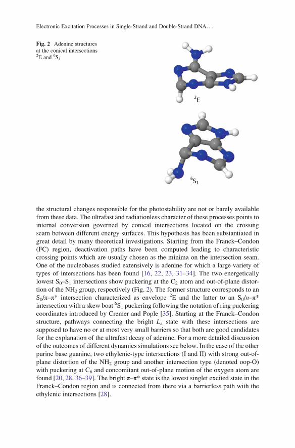
One of the nucleobases studied extensively is adenine for which a large variety of
types of intersections has been found [16, 22, 23, 31–34]. The two energetically
lowest S0–S1 intersections show puckering at the C2 atom and out-of-plane distor-
tion of the NH2 group, respectively (Fig. 2). The former structure corresponds to an
S0/π–π* intersection characterized as envelope 2E and the latter to an S0/n–π*intersection with a skew boat 6S1 puckering following the notation of ring puckering
coordinates introduced by Cremer and Pople [35]. Starting at the Franck–Condon
structure, pathways connecting the bright La state with these intersections are
supposed to have no or at most very small barriers so that both are good candidates
for the explanation of the ultrafast decay of adenine. For a more detailed discussion
of the outcomes of different dynamics simulations see below. In the case of the other
purine base guanine, two ethylenic-type intersections (I and II) with strong out-of-
plane distortion of the NH2 group and another intersection type (denoted oop-O)
with puckering at C6 and concomitant out-of-plane motion of the oxygen atom are
found [20, 28, 36–39]. The bright π–π* state is the lowest singlet excited state in theFranck–Condon region and is connected from there via a barrierless path with the
ethylenic intersections [28].
Fig. 2 Adenine structures
at the conical intersections2E and 6S1
Electronic Excitation Processes in Single-Strand and Double-Strand DNA. . .

In comparison to the purine bases, the pyrimidine bases are characterized by a
more complex system of conical intersections and reaction paths. In the case of
cytosine, three major conical intersections have been determined. The first [25] is
characterized as semi-planar. It occurs in a region of mixing of the nO–π*, π–π* andclosed shell states, which leads to a triple degeneracy as reported in [40, 41]. The
second intersection leads to a puckering at N3 connected with an out-of-plane
distortion of the amino group. The third intersection involves puckering at C6 (for
more details see [42–44]). Reaction paths computed in [44] connecting the Franck–
Condon region with the above-mentioned conical intersections show that these
intersections are all energetically accessible and display similar qualitative features
so that no a priori decision can be made concerning specific photodynamical
reaction mechanisms.
Concerning theoretical investigations on the remaining nucleobases (see, e.g.,
[17, 21, 24, 27]), it should be mentioned at this point that not only the ring
puckering motions are of relevance for the explanation of the ultrafast decay of
the nucleobases but also that NH dissociation is of interest since it has been shown
that this process leads to conical intersections [32] even though they are supposed to
be accessible only by higher excitation energies beyond the first absorption band.
The above discussion concentrates on the singlet excited states. It should be
noted that several studies discussing the triplet excited states appeared in the
literature (see, e.g., [45–50]).
The large manifold of conical intersections and their reaction paths give a good
picture of the different deactivation possibilities but makes conclusive predictions
about the real deactivation mechanisms difficult if not impossible. Dynamics
simulations have been performed to find the actual reaction paths and to obtain
information about the characteristic times connected with the different processes.
Owing to the strongly coupled motion involving many internal degrees of freedom,
Tully surface hopping [51] and ab initio multiple spawning (AIMS) [52] appear to
be methods of choice since all internal degrees of freedom are directly included in
these dynamics simulations (see also [53] for a general discussion on the options
and outlook of photodynamical simulations). The main bottleneck in these calcu-
lations is the necessity of performing a full quantum chemical calculation in each
time step using extended methods able to describe several electronic states and their
nonadiabatic coupling. Under these circumstances, mostly complete active space
self-consistent field (CASSCF) or multireference configuration interaction with
single excitations (MRCIS) approaches have been used [17, 19, 33, 38, 44,
54–56]. An overview of respective simulations of the photodynamics of
nucleobases is available, e.g., in [18]. As an interesting alternative to the compu-
tationally expensive ab initio approaches, semi-empirical multireference methods
based on the orthogonalization model 2 Hamiltonian (OM2) [34, 57, 58] and the
fractional orbital occupation/AM1model [59] have also been applied extensively in
surface hopping dynamics. The performance of TDDFT using a larger variety of
functionals has been investigated [60], and time-dependent tight binding density
functional theory (TD-DFTB) [61] and DFTB mean field simulations [62] have
been used as well. The restricted open shell Kohn–Sham (ROKS) method within the
F. Plasser et al.

framework of Car–Parinello dynamics [63] and quantum dynamical simulations
have also been performed [64].
From this multitude of widely differing approaches, we wanted to discuss the
photodynamics of adenine and cytosine in more detail. Surface hopping dynam-
ics simulations using an MRCIS approach [33] have been performed for adenine,
starting the dynamics in the La π–π*(S3) state. Already after 25 fs this state is
completely depopulated and practically all trajectories have arrived in the S1state after ~60 fs. By fitting an exponential function containing two decay
constant, values of 22 and 538 fs were found, in good agreement with those of
<50 and 750 fs measured by Ullrich et al. [9]. The analysis of the trajectories
showed that almost exclusively the 2E with puckering at C2 was accessed. In
contrast, OM2 photodynamics [34] shows a strong predominance of the NH2
out-of-plane conical intersection (1S6). In a recent study [60] the energy profiles
along the reaction coordinates leading to both mentioned intersections were
computed using several methods (OM2/MRCI, MRCIS, complete active space
perturbation theory to second order (CASPT2), approximate coupled cluster
singles- and doubles method (CC2), and a variety of different TDDFT methods).
These profiles show a preference toward the 2E intersection in the case of
MRCIS, CASPT2, and CC2 whereas the opposite trend is found for OM2. It
was further shown that the MRCIS calculations using one lone pair (n) orbital inthe active space were subject to a certain bias toward 2E which was partially
lifted by including two n orbitals into the active space [65]. Thus, according to
current understanding, the real dynamics should still be dominated by the 2E
pathway but significant participation of the 1S6 should be expected as well. The
investigations in [60] also show a severe failure of the TDDFT and TD-DFTB
approaches since an insufficient number of hoppings to the ground state were
observed. Inclusion of range-separated functionals did not improve the situation
significantly. For more details see [60].
In the case of cytosine, a more complex dynamics than that described for adenine
is observed [18]. All three intersections described above for cytosine participate in
the dynamics. Initially, cytosine relaxes along the π–π* pathway to a region of
strong mixture of π–π*, n–π*, and closed shell character, where in a first approach
about 16% of trajectories switch to the ground state with a time constant of 13 fs. It
should be noted that this semi-instantaneous deactivation of cytosine through the
semi-planar intersection was also found in AIMS simulations [19]. In the remaining
84% of cases, cytosine quickly relaxes to the n–π* state from where it either can
deactivate via the semi-planar intersection to the ground state or can overcome the
barrier to the π–π* state and subsequently switch to the ground state. In contrast,
CASSCF(2,2) AIMS [19] and OM2 [57] simulations do not show any significant
deactivation in this region. The results of surface hopping dynamics at CASSCF
(12,9) level [66] are similar to those found in [18] and show predominance of the
semi-planar conical intersection. Due to this complex set of events it has to be
expected that even rather subtle changes in the relative positions of the energy
surfaces can lead to significant modifications of the photodynamical mechanisms
and all discussed results have to be regarded with care. It can be expected that
Electronic Excitation Processes in Single-Strand and Double-Strand DNA. . .

further quantitative changes will occur when more sophisticated quantum chemical
methods will become available for photodynamical simulations.
Notable changes in the dynamics also have to be expected when it is performed
in aqueous solution. Several computations were performed to elucidate the effect of
solvent on the excited states of uracil [49, 67–78], thymine [68, 76, 77, 79, 80],
guanine [81–84], cytosine [75, 77, 85, 86], and adenine and its model systems
[87–89]. The aqueous solvent was shown to modify the photodynamics of
nucleobases by changing relative positions of excited states of different characters
in the FC region, changing the heights of reaction barriers on the paths leading to
conical intersections and relative energies of excited state minima. The conse-
quences such as a blue shift of the n–π* states of uracil (see, e.g., [68, 70, 71, 73,
76], stabilization of a polar S2(π–π*) [32] and π–σ* [87] states of adenine, and
geometry changes of excited state minima of guanine [82] on photodynamics were
discussed. Dynamics studies performed on adenine [89] have shown that the out-of-
plane motion of the amino group is more pronounced in water, which can explain
the faster decay of the S1 state compared to the gas phase. The overall relaxation
mechanism was however the same as observed in the gas phase. This finding is in
contrast to guanine [84] where the pathway via a conical intersection with out-of-
plane distortion of the carbonyl oxygen becomes dominant in water solvent.
It should be noted that in extension of the above-described dynamics simulations
restricted to the singlet manifold, first surface hopping dynamics simulations have
been performed for cytosine combining nonadiabatic and spin-orbit effects [90, 91].
At first sight it seems that each of the nucleobases possesses its own character-
istics. A general picture emerging from the dynamics simulations [18] can be given,
however, by the observation that the purine bases adenine and guanine have quite a
simple deactivation pattern following basically one excited state to the intersection
with the ground state whereas, in the case of the pyrimidine bases cytosine, thymine,
and uracil, a significantly more complex picture appears with the participation of
several excited states and a significantly more complex deactivation pattern.
1.2 Survey of Experimental Studies of InteractingNucleobases
The question of the nature of excited states of DNAwas first addressed in the 1960s.
Based on theoretical models, Tinoco et al. [92] invoked a delocalized character of
excited states of nucleic acids in the FC region to explain the hypochromism
observed in absorption spectra of polynucleotides. In contrast to this prediction,
Eisinger et al. [93] proposed that the UV photon is absorbed by a single base. This
prediction was made on the basis of comparison of absorption spectra of DNA with
corresponding pyrimidine and purine bases. The DNA absorption spectra closely
F. Plasser et al.

resembled the sum of the monomer spectra and showed theoretically predicted
spectral shifts and splitting of the UV band around 260 nm. A discussion on this
issue based on molecular dynamics simulations and quantum chemical calculations
will be given later in this chapter.
Following the prediction of the locally excited states red-shifted and broadened
fluorescence spectra of di- and polynucleotides were attributed to excimers formed
from these bases [5]. More pronounced occurrences of excimers observed at lower
temperatures are explained by a higher degree of stacking as compared to more
disordered structures at higher temperatures. Increased charge transfer
(CT) contributions in these excimers were also predicted by these authors [94, 95].
In contrast to the above-discussed local character of absorption, the excitonic
character in absorption spectra of di- and polynucleotides of adenine and cytosine
was predicted by Kononov et al. [7], with the exciton limited to two bases. The
excitonic features were, however, not observed in absorption spectra of guanine and
thymine.
The experiments performed during the last two decades utilizing femtosecond
time-resolved spectroscopy stimulated lively discussion on the nature of excited
states of DNA. In addition to the ultrafast components decaying on the order of
picoseconds, transients with lifetimes of 10–100 ps and even nanosecond time scale
were observed for single- and double-stranded oligonucleotides [14, 96–103]. The
existence of large decay times occurring for single-stranded oligonucleotides [98]
demonstrates that stacking interactions are of primary importance for the explana-
tion of differences in the complex decay dynamics in DNA as opposed to that
observed for individual nucleic acid bases.
The interpretation of the ultrafast component observed in the experiments
mentioned in the previous paragraph was based on the existence of bases undergo-
ing monomer-like photo decay in the disordered parts of the oligo- and polymers.
Different hypotheses were used to explain the slow-decay component: (1) the
excitation in the FC region is localized on a single base and the interaction with
an adjacent base leads to the formation of excimers [98, 101] several picoseconds
after the excitation and (2) the excitation is already delocalized during the initial FC
excitation, resulting in the formation of excitonic states [14, 96, 104–106]. In the
latter interpretation partial emission from localized bases was suggested as well
[105, 107]. Beyond the above question of the delocalization, the importance of
charge transfer states is being discussed as an important issue (see, e.g., [108–113]).
A special challenge lies in interpreting and reconciling the large number of
different decay times reported in the different experiments (see, e.g., [97–99, 101,
102, 114–121]). The results depend not only on the general character of the
experimental technique (i.e., time-dependent absorption or emission spectroscopy)
but also on the details of the preparation of the DNA samples and the base sequence,
both playing an important role [118, 122]. In light of all these challenges with
respect to interpreting experimental results, computational investigations play an
important role in providing complementary information, shedding new light on the
complex photodynamical behavior of DNA.
Electronic Excitation Processes in Single-Strand and Double-Strand DNA. . .

The extent of exciton delocalization is another issue discussed intensively. For
example, Kadhane et al. suggested that excitons are delocalized over no more than
two bases [123] and Bucharov et al. [14] suggested delocalization extending over
three bases. However, delocalization over six and more bases was also considered
[124]. Extended work concerning this issue is based mainly on dynamics simula-
tions in connection with excitonic models or quantum chemical calculations, and
will be discussed later in the chapter.
According to current evidence, inter-strand hydrogen bonding may also play an
important role and add to the complexity of the photodynamical behavior of nucleic
acids. A unique excited state behavior of Watson–Crick (WC) base pairs was shown
in a resonant multi-photon ionization experiment performed in the gas phase
[125]. Faster fluorescence decay of base pairs compared to isolated bases was
also observed by other authors as shown in [98, 126–128]. Furthermore, interplay
between intra-strand CT and inter-strand proton transfer has been suggested [128].
2 Description of Electronic Coupling
Before discussing the character of excited state interactions between nucleobases,
the underlying theory will be briefly discussed. Scheme 2 describes the simplest
situation of the electronic interactions of two identical chromophores (A and B) interms of locally and charge transfer excited states. At infinite separation the locally
Scheme 2 Schematic illustration of the electronic interactions of identical chromophores A and
B in terms of localized and charge transfer excited states
F. Plasser et al.
excited states (described by Ψ(A*B) and Ψ(AB*)) are degenerate. At finite
separation, in an adiabatic representation the degeneracy is removed and the energy
splitting may be directly used to measure the excited state interaction
(electronic coupling). The situation is similar for the charge transfer states described
by Ψ(A.�B.+) and Ψ(A.+B�.) [129–131]. For non-symmetrical arrangements this
approach is not so straightforward. Inside the polymer the environment of two bases
is strongly non-symmetrical due to thermal fluctuations. In such a case the splitting
will also reflect the difference in the environment of each chromophore, which
causes a shift of the orbital and excitation energies that are not directly related to
the electronic coupling. In the case of a stacked nucleobase pair with nucleobases
mutually orientated as in a B-DNA structure, the changes in excitation energies
observed during dimerization caused by non-symmetric effects are an order of
magnitude larger than those caused by the ‘pure’ electronic interaction [132, 133].
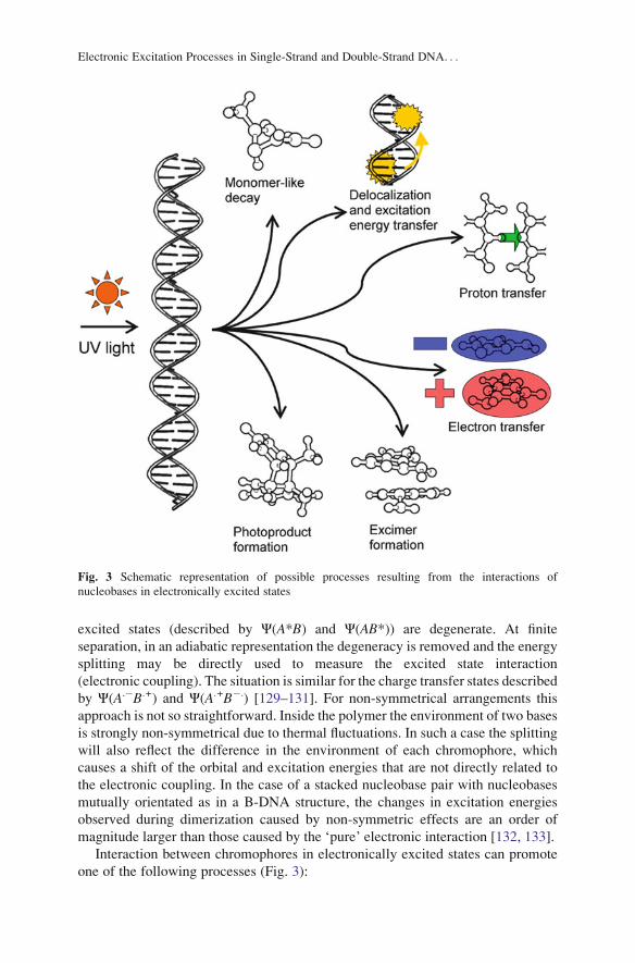
Interaction between chromophores in electronically excited states can promote
one of the following processes (Fig. 3):
Fig. 3 Schematic representation of possible processes resulting from the interactions of
nucleobases in electronically excited states
Electronic Excitation Processes in Single-Strand and Double-Strand DNA. . .

1. Delocalization of the excited states which results in exciton formation.
2. Electronic excitation energy transfer.
3. Formation of a strongly interacting complex – an excimer – which results from
the orbital overlap.
4. Formation of a new chemical species – a photochemical product.
5. Inter- and/or intra-strand electron or proton transfer.
6. Localization of the electronic excitation on a single base followed by a
monomer-like relaxation.
The interactions during electronic excitations are governed by two main contri-
butions: (1) Coulombic interactions which are often approximated by the interac-
tions of transition dipole moments and higher multipole interaction terms and
(2) short-range interactions, which depend on the orbital overlap between two
chromophores [129–131].
While the former interactions operate over larger through-space separation R,and depend asymptotically on R�3, the latter attenuate exponentially with R being
unimportant for the chromophore separation larger than approximately 6 A´. Since
these interactions depend on the orbital overlap they are greatly dependent on the
mutual orientation of the chromophores. Thus, significantly larger short-range
interactions were found for the nucleobases in the B-DNA-like orientation with
almost parallel stacking as compared to A-DNA-like orientation with disordered
stacking [132]. The character of the excited states also largely influences the
resulting electronic interactions. For example, these interactions between the states
of (n�π*) character with negligible transition dipole moments and small orbital
overlaps are much smaller in relation to states of (π–π*) character which possess a
larger transition dipole moment and a larger overlap of interacting orbitals [132].
2.1 Excimers/Exciplexes and Excitons
As already mentioned, the photoabsorption of two (or more) nucleobases within the
nucleic acid structure can result in excimers or exciplexes being formed by two
identical or different nucleobases, respectively. It should be emphasized that the
excimers/exciplexes are not necessarily formed from one molecule in the ground
state and the second from the excited state. They can be formed from different
initial states, including excitons.
The wavefunction has the following general form [129, 134, 135]:
Ψ Exciplexð Þ ¼ c1Ψ A�Bð Þ þ c2Ψ AB�ð Þ þ c3Ψ A:�B:þð Þ þ c4Ψ A:þB:�ð Þ, ð1Þ
where A ¼ B, c1 ¼ c2, and c3 ¼ c4 for excimers.
The first two terms correspond to locally excited states and their interaction
results in exciton states (see below). At intermolecular separations below 5–6 Å,orbital interactions come into play [129, 134] mediating a mixing of the locally
F. Plasser et al.

excited states with the charge transfer or radical-ion-pair states, described by the
third and fourth terms [135]. Stabilization of the exciplex can not only occur
through excitonic coupling and Coulombic interaction, which are directly derived
from the excitonic and CT contributions. It has been pointed out that with decreas-
ing separation distance a new type of strong interaction may come into play that can
be identified as a quasi-bond in the geminate radical ion pair [136]. Such quasi-
bonds may go along with strong structural distortions, and experimental evidence
shows that electronic and steric effects play an important role [137].
In Fig. 4, excimer formation in the face-to-face stacked naphthalene dimer
computed at the ab initio algebraic diagrammatic construction to second order
(ADC(2)) level is illustrated as a representative example (see [138] for more
details). While the ground state potential is only weakly attractive with a shallow
minimum around 3.65 Å intermolecular separation, the excited state is strongly
bound with a minimum at 3.2 Å and stabilization energy of 1.38 eV with respect to
infinite separation. Aside from the energy, a measure for charge transfer,
representing the weight of the c3 and c4 coefficients in the equation (1), is shown
in Fig. 4 (bottom). At separations >5.0 Å the CT value is approximately zero,
showing that the excited state corresponds to a pure Frenkel exciton. When the
molecules are moved closer together, orbital interactions are important (cf. [129])
and the locally excited states mix with the CT states, giving a gradual increase of
the CT character. At the excimer minimum the CT character reaches 50%, showing
that the excited state is now an even mixture between Frenkel excitonic and charge
resonance contributions. This state is accessed by a single transition between
delocalized orbitals, which corresponds to the situation of a coherent homogeneous
state extended over the whole complex (cf. [138, 139]).
Fig. 4 Excited state
energies of the S0 and S1states (top) and charge
transfer character of the S1state (bottom) of a face-to-face stacked naphthalene
dimer (see [138] for details)
Electronic Excitation Processes in Single-Strand and Double-Strand DNA. . .

In extended quasi-periodic systems excited states can be delocalized over a
number of chromophores. Such states are usually called excitons. In this context
it is important to distinguish whether the exciton can be described as a linear
combination of locally excited states, forming a Frenkel exciton, or whether CT
configurations also play a role.
The first case can be understood in terms of Frenkel exciton theory [140, 141]. In
this framework a model Hamiltonian H is written as the sum of isolated chromo-
phores Ha and a coupling term Vab:
H ¼X
aHa þ
Xa
Xb>a
Vab: ð2Þ
The singly excited states are described by the term
Φia ¼ �1ð ÞaΦi
a
Ya 6¼b
Φb, ð3Þ
where Φia corresponds to the wavefunction of the state where chromophore a is in
its excited state, while others are in their-ground state. The wavefunction of the
excitonic state is then written as a linear combination of the wavefunction with
locally excited states:
Ψk Excitonð Þ ¼X
ackaΦi
a: ð4Þ
The diagonal and off-diagonal terms of the exciton matrix correspond to the
excitation energies of the chromophore a and the exciton coupling terms, respec-
tively. In this picture the excitation within some energy range populates a number
of excited states delocalized over several nucleobases. Furthermore, if the initial
electronic wavepacket is prepared as a non-stationary starting state, such a Hamil-
tonian automatically leads to excitation energy transfer. If in addition vibronic
contributions are considered, the system can reach the lower part of the emission
band via internal conversion (intra-band scattering) [118].
When there are strong orbital interactions between the different fragments,
Frenkel exciton theory is no longer sufficient and it is necessary to include explic-
itly charge transfer configurations into the calculation. The theoretical treatment in
such a case is more involved considering that a larger number of parameters are
needed [142–144]. However, the advantage of such an approach is that charge
separation processes can be easily modeled.
3 Assessment of Computational Methods
A wide range of computational methods have been applied to the description of
electronically excited states of DNA fragments. In this section we review these
approaches shortly. Unfortunately, no perfect method exists because of the diffi-
culties in computing excited states and the relatively large sizes of the molecular
F. Plasser et al.

systems involved. In the following we will characterize the main features of the
individual methods, including their strong and weak points, in an attempt to aid in
assessing the substantial amount of available computational literature and to
explain some of the discrepancies obtained in the different studies. This section
will be strongly focused on DNA fragments; for a more general overview we refer
the reader to [53].
3.1 Electronic Structure Methods
In this section, the different electronic structure methods that were used to describe
DNA fragments will be considered. The main focus will be laid on the computation
of excited states, and additionally methods for a phenomenological analysis of
excited states and the computation of interaction potentials will be discussed. The
main challenge relates to the large system sizes that have to be considered.
Therefore, exciton models are a logical choice and were applied by several groups
[142, 145–147]. In such an approach the excited states on the different molecules
and their interactions are treated by a model Hamiltonian. This Hamiltonian can be
easily extended to large system sizes, allowing the study of long range effects and
potentially extended delocalization. However, a major challenge in such an
approach is a proper parameterization. In particular the electronic couplings are
of concern and a number of different methods for estimating them were used.
Usually only the Coulombic contribution is computed while the direct orbital
term is assumed to be smaller. The simplest approach for the estimation of this
interaction is to consider only dipole–dipole interactions. This approximation was
used, e.g., by Georghiou et al. [95] and Bittner [142] to estimate the efficiency of
energy transfer along the DNA helix. This approach is suitable for systems in which
the separation of the two chromophores is significantly greater than the dimension
of the molecule. Its validity is however questionable in the case of nucleobases,
because their molecular dimension is of the same order as the intermolecular
distance of a neighboring nucleobase pair. Several approximations can help to
overcome this difficulty using, e.g., the atomic-transition charge distribution [148,
149] or transition-density-cube models [144]. Comparison of the electronic cou-
plings calculated using dipole–dipole and transition-density-cube approaches show
that the former significantly overestimate the interactions at shorter distances
[144]. Alternatively, the hybrid-multipole model which represents a combination
of a truncated-multipole and the extended-dipole model [148, 150] was used to
estimate Coulombic interactions [132]. Within this hybrid model, the multipole
interaction up to R�5 is covered exactly; all terms with higher orders are considered
if they arise from the multipole expansion of the extended dipole. The Coulomb
part is usually the major component of the interaction potential, but a complete
description also requires consideration of the direct orbital contributions. While it
was shown from explicit quantum chemical calculations that at the ground state
equilibrium geometry these terms only made a small contribution (<100 cm) [132],
the involvement of such terms could in fact be identified through a mixing of
Electronic Excitation Processes in Single-Strand and Double-Strand DNA. . .

Frenkel excitonic and CT states [113]. An ad hoc addition of orbital overlap terms
in a Frenkel exciton model is difficult as it has already been pointed out that a
modest additional coupling of 100 cm had a dramatic effect by doubling the
delocalization length [149]. A more extended treatment of orbital overlap and
resulting charge separation is possible but requires a more involved formalism
and a larger number of parameters [142, 151].
Considering the structural flexibility of DNA, an atomistic description is in many
cases highly desirable. As one option semi-empirical methods were used [59, 152–
155]. Due to the fact that they allow an efficient description of quite large systems
they were used for extended dynamics simulations, providing interesting insights.
However, similar to exciton models, semi-empirical methods rely on careful param-
eterization, a problem which can be avoided by the use of ab initio methods. Time-
dependent density functional theory (TDDFT), which offers efficient excited state
computations while relying on no or relatively few empirical parameters, has been
used by a number of groups [109, 110, 156]. However, a major challenge for the
application of TDDFT is the overstabilization of charge transfer states occurring
when local functionals are used [157]. This problem can be overcome by using range
separated hybrid functionals or functionals containing an overall high amount of
Hartree–Fock exchange but in such cases the energy of CT states crucially depends
on the parameters determining the admixture of Hartree–Fock exchange [110,
112]. For a comparison of the performance of different density functionals when
applied to DNA stacks see, e.g., [158]. Finally, a large number of wavefunction-based
ab initio calculations was performed using single- and multi-reference methods. In
the first case, in particular efficient second order models like the approximate coupled
cluster method CC2 [159] and the algebraic diagrammatic construction ADC
(2) [160] in connection with the resolution of the identity approximation [161]
were applied [112, 113, 132]. However, computationally more demanding models
like the equation-of-motion coupled-cluster for excitation energies (EOM-EE-CC)
with double excitations and even perturbative triple excitations have also been used
[162–165]. In the case of strongly distorted structures and intersections between
different states, multi-reference methods are needed to provide a reliable description.
For this purpose the complete-active space self-consistent field (CASSCF) method,
second order perturbation theory on this reference (CASPT2) [166], and multi-
reference configuration interaction with single and double excitations (MR-CISD)
[167] were performed [168]. While wavefunction-based methods offer the attractive
property of systematic improvability toward the exact solution, they suffer from high
computational demands. Unless massively parallel computer systems are available
heavy truncations have to be carried out as far as the excitation level and/or the one
electron basis set are concerned. For this purpose wavefunction-based ab initio
methods also require careful testing and analysis before they can be successfully
applied.
After the computation of excitation energies the next decisive task is to get a
maximum of information for interpretive and phenomenological models. In
quantum chemical calculations on DNA fragments this task may be quite
challenging in many cases due to many interacting configurations, partially
F. Plasser et al.
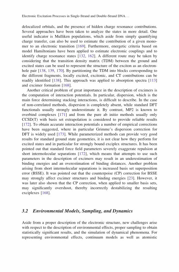
delocalized orbitals, and the presence of hidden charge resonance contributions.
Several approaches have been taken to analyze the states in more detail. One
useful indicator is Mulliken populations, which aside from simply quantifying
charge transfer, can also be used to estimate the contribution of a given mono-
mer to an electronic transition [169]. Furthermore, energetic criteria based on
model Hamiltonians have been applied to estimate electronic couplings and to
identify charge resonance states [132, 162]. A different route may be taken by
considering that the transition density matrix (TDM) between the ground and
excited states can be used to represent the structure of the exciton as an electron-
hole pair [138, 139, 170]. By partitioning the TDM into blocks corresponding to
the different fragments, locally excited, excitonic, and CT contributions can be
readily identified [138]. This approach was applied to absorption spectra [113]
and excimer formation [168].
Another critical problem of great importance in the description of excimers is
the computation of interaction potentials. In particular, dispersion, which is the
main force determining stacking interactions, is difficult to describe. In the case
of non-correlated methods, dispersion is completely absent, while standard DFT
functionals usually strongly underestimate it. By contrast, MP2 is known to
overbind complexes [171] and from the pure ab initio methods usually only
CCSD(T) with basis set extrapolation is considered to provide reliable results
[172]. To obtain accurate interaction potentials a number of empirical corrections
have been suggested, where in particular Grimme’s dispersion correction for
DFT is widely used [173]. While parameterized methods can provide very good
results for standard ground state geometries, it is not clear how they perform for
excited states and in particular for strongly bound exciplex structures. It has been
pointed out that standard force field parameters severely exaggerate repulsion at
short intermolecular separations [172], which means that attempts to use such
parameters in the description of excimers may result in an underestimation of
binding energies and an overestimation of binding distances. Another problem
arising from short intermolecular separations is increased basis set superposition
error (BSSE). It was pointed out that the counterpoise (CP) correction for BSSE
may strongly affect excimer structures and binding energies [23]. However, it
was later also shown that the CP correction, when applied to smaller basis sets,
may significantly overshoot, thereby incorrectly destabilizing the resulting
exciplexes [168].
3.2 Environmental Models, Sampling, and Dynamics
Aside from a proper description of the electronic structure, new challenges arise
with respect to the description of environmental effects, proper sampling to obtain
statistically significant results, and the simulation of dynamical phenomena. For
representing environmental effects, continuum models as well as atomistic
Electronic Excitation Processes in Single-Strand and Double-Strand DNA. . .

descriptions have been applied. The former type of approach offers a simple well-
defined way to include the main effects of environmental polarization where even a
differentiation between slow and fast polarization effects is easily achieved. In
particular the polarizable continuum model (PCM) [174] has been applied, e.g., in
[109, 112, 175]. There are only a few adjustable parameters, most importantly the
dielectric constant and the time-regime (equilibrium or non-equilibrium). However,
there is some arbitrariness when choosing an effective dielectric constant to repre-
sent the effect of the heterogeneous and anisotropic surroundings (i.e., the other
DNA bases, the backbone, the surrounding water molecules and counterions). To
overcome this problem, quantum mechanics/molecular mechanics (QM/MM) cou-
pling schemes for an atomistic description of the environment have been widely
used [110, 113, 176, 177]. Usually the effects of the environment are considered at
the level of electrostatic embedding only, and electronic polarization of the envi-
ronment is neglected (see [178] for a definition of these terms). This should have an
effect on vertical excitation energies, while in dynamics simulations the main
solvent response can be included through orientational polarization. A critical
observation, made from the PCM computations, is that the environment may have
a strong impact on the stability of charge transfer states and that, aside from the
time-regime (equilibrium or non-equilibrium), even the precise PCM implementa-
tion (linear-response [179] or state-specific [180]) makes a crucial difference
[181]. A similar sensitivity to the environmental models is probably also present
in a QM/MM framework. Thus, in summary, the importance of an accurate
description of the environment should be highlighted.
The most straightforward way for sampling ground state DNA structures is by
classical molecular dynamics (MD) using standard biomolecular force fields and
such an approach has been taken by a number of groups [144, 149, 156, 176]. The
situation becomes more complicated when effects of zero-point vibrations should
also be included. For this purpose a hybrid approach based on mixed initial
conditions was introduced: Large scale motions and solvent degrees of freedom
are properly treated by MD while the Wigner distribution of the vibrational ground
state is used to represent the central molecule of interest [182]. This approach was
applied to produce initial conditions for QM/MM dynamics simulations of a DNA
base [177], and a slight extension considering several active molecules was used for
spectra simulations [113]. QM/MM geometry optimizations can be performed by
using an averaged solvent electrostatic potential generated from MD sampling of
the environmental motions [183]. This method was applied to excited state optimi-
zations of the adenine dinucleotide [168].
To go beyond static calculations, a number of excited state dynamics studies
have also been performed. A particular focus was placed on the description of
non-adiabatic effects, which are essential for understanding, e.g., internal conver-
sion, excitation energy transfer, and charge separation processes. Dynamics have
been performed using exciton models [143] and wave packet propagation on
parameterized surfaces [184] but in most cases on-the-fly surface hopping dynam-
ics were applied [59, 154, 155, 176, 177]. In the latter case a QM/MM approach was
often chosen to allow a real time polarization response of the environment. While
F. Plasser et al.

the studies presented above considered only the singlet manifold, intersystem
crossings to the triplet were also already considered in surface hopping
dynamics [90].
4 UV Absorption
The nature of excited states in the Franck–Condon region significantly affects the
excited state behavior of nucleic acids. Thus, understanding the character of
absorption spectra is crucial for the further evaluation of the photodynamics of
nucleic acids. As already mentioned in the Introduction, there is still controversy as
to whether during photon absorption the nucleic acids form exciton or charge
transfer states or whether they remain localized. In this chapter the survey of
theoretical works suggesting both possibilities of delocalization is treated
separately.
4.1 Excitonic Delocalization
The concept of excitons in nucleic acids was first introduced by Tinoco
et al. [92, 185] and Rhodes [186]. However, a localized character of excited
states then dominated the discussions for several decades. The question of
exciton character was opened again by Bouvier et al. [148]. In this work the
homogeneous (dA)20.(dT)20 and alternating (dAdT)10.(dAdT)10 oligonucleotides
in idealized B-DNA geometry are investigated. The two lowest excited states of
adenine and one excited state of thymine are considered. The interaction between
the states is described by the atomic transition charges model [187] in which the
transition dipoles are decomposed onto atomic orbitals. This procedure results in
transition charges located on each atom. Using this approach, delocalization of
the excited states was found for both oligomers. In (dA)20.(dT)20 the intra-strand
coupling dominates with a strength of about 250 cm�1. In the alternating
(dAdT)10.(dAdT)10 the inter-strand coupling is more important, the estimated
value of the coupling being approximately 100 cm�1. In the following contri-
bution [104] the influence of structure fluctuations on the character of the Frank–
Condon excited states is analyzed for oligonucleotides (dA)10.(dT)10 and
(dAdT)5.(dAdT)5. A ground-state molecular dynamics simulation to scan possi-
ble DNA structures resulting from the plasticity of the sugar-phosphate helix.
Importantly, the diagonal energies of monomer excited states are not affected by
structural fluctuations, with the change being smaller than 10 cm�1. Relatively
large fluctuations of the off-diagonal terms for these structures were found, with
the amplitude of the variations 35% and 45% for (dA)10.(dT)10 and (dAdT)5.
(dAdT)5, respectively. The distribution of the oscillator strengths, on the other
hand, is not significantly affected by the structural dynamics for the (dA)10.
Electronic Excitation Processes in Single-Strand and Double-Strand DNA. . .

(dT)10 oligonucleotide. In fact, 90% of the oscillator strength remains concen-
trated on the same eigenstates. In the case of the alternating (dAdT)5.(dAdT)5oligonucleotide the structural disorder results in a larger spreading of the oscil-
lator strength among eigenstates. Studies performed on (dCdG)5.(dCdG)5 [149]
resulted in similar conclusions as for the afore-mentioned (dAdT)5.(dAdT)5 case:
small fluctuations of monomer excitation energies, less than 15 cm�1, and a
large perturbation of dipolar coupling due to the structural disorder. In all the
cases mentioned above a delocalization of the excited states over at least two
nucleobases was observed. Delocalization over three bases was found in the
calculations of the absorption spectra using the TDDFT method in combination
with ground-state MD simulations for single-stranded oligomers of adenine [156]
and (adenine–thymine) oligomers [188]. Formation of delocalized exciton states
was further investigated in studies which combine ground-state molecular
dynamics and the evaluation of the exciton model for the poly(dA).poly
(dT) [143] and (dA)12.(dT)12 [144]. The lattice model and the transition density
cube model, based on the transition model calculated using the TDDFT method,
were used in the former and later investigations, respectively. In agreement with
the work of Bouvier et al. [104] and Emanuele et al. [149], changes in electronic
coupling due to structural fluctuation were found. The results indicated that
during the absorption process the electronic excitation is delocalized over at
least six nucleobases and localizes upon relaxation on four nucleobases. Impor-
tantly, these authors predicted the presence of charge transfer excitons in their
model [144]. Charge transfer character of excitons was also suggested by
Starikov et al. [189]. The character of exciton states due to structural fluctuations
of B-DNA conformations was investigated in short (dA)n.(dT)n and (dC)n.(dG)noligomers (with n ¼ 3,4) obtained by means of ground-state molecular dynamics
[152]. The effect of different conformational modes was evaluated, among which
the twist is predicted as the most powerful regulator of the exciton character. It
was also shown that the effect of the twist angle on the localization of electronic
states is stronger in poly(dG)-poly(dC) than in poly(dA)-poly(dT) [190]. A new
study based on ab initio calculations of alternating duplexes after extended
sampling [113] presented a picture of rather localized states, which were situated
on one or at most two bases. Considering poly(dAdT)-poly(dAdT) and poly
(dGdC)-poly(dGdC) duplexes with four stacked bases in the QM region and
the remaining part of the system treated at the MM level, only about a third of
the states are delocalized over 1.5 bases or more. These are, however, respon-
sible for somewhat more than 50% of the intensity at the absorption maximum
due to higher than average oscillator strengths. Intramolecular vibrations were
identified as the main factor responsible for spectral broadening and for causing
disorder resulting in localization. This strong coupling of intramolecular modes
to the excited states suggests that these are also active in the early excited state
dynamics. The picture of rather localized states was also drawn from another
recent study that compared measured and computed circular dichroism spectra in
(dA)n.(dT)n hairpins [147]. The TDDFT study on adenine stacks pointed out that
F. Plasser et al.

the A5 spectrum is almost identical to that of A4 [191], i.e., there are no relevant
effects either of excitonic or indirect nature which go beyond four bases. While
most studies focused on singlet states, triplet excitations were studied in poly
(dA)-poly(dT) sequences, and it was reported that they are confined to single
nucleobases [192, 193].
4.2 Charge Transfer States
Compared to the studies performed within the framework of exciton models where
the charge transfer configurations are usually not included, supermolecular quan-
tum chemical calculations automatically provide a simultaneous treatment of
excitonic and the charge transfer states. The interpretation of the absorption spectra
in terms of delocalized, charge transfer and localized characters of excited states
became a matter of discussion due to the performance of different methods.
Suitability of various methods to describe long-range charge transfer states between
nucleic acids has been tested in recent years (see, for example, [108, 110–112, 169,
194–196]).
In the text below the calculations of the character of excited states observed at
the ground state geometries, i.e., upon UV absorption, as affected by base pairing
and stacking interactions will be reviewed with the emphasis on a formation of
charge transfer states. In initial ab initio calculations the effect of base pairing was
studied [42, 197–199]. Although charge transfer states were detected at higher
energies when using the CIS method [197], these were the lowest in calculations
at the TDDFT level employing the LDA functional [198]. Stacking interactions
were considered for the first time in calculations of Varsano et al. [200]. Other
calculations studying the effects of stacking on excited states formed upon UV
absorption in the gas phase have been reported, e.g., in [132, 133, 162, 201–
203]. The difficulties of a correct description of charge transfer states in the
absorption spectra is demonstrated in the case of homologous oligomers of adenine,
both single-stranded and in double stranded A–T sequences, as well as alternating
A–T sequences.
Santoro et al. [109] performed the first study on the excited states of stacked
nucleobases in a water environment. In this contribution the absorption spectra
of the adenine dimer are studied employing TDDFT with the PBE0 functional
for the (9-Met-A)2 and (dA)2 models using a polarizable continuum model for
solvent effects. The experimentally observed features when going from mono-
mers to multimers [97, 105, 118], i.e., a blue-shift of the maximum, a red-shift
of the lower-energy part, and hypochromic effect of the absorption spectra are
already reproduced in the calculations of the former model. These effects reflect
the coupling due to the orbital overlap in the stacking configuration of
nucleobases. The symmetric combination of monomer bright transitions gives
rise to the maximum of the absorption band. The character of the states respon-
sible for low-lying energy transitions changes with the mutual orientation of the
Electronic Excitation Processes in Single-Strand and Double-Strand DNA. . .

adjacent adenines, i.e., an increasing intra-strand charge transfer character of the
first excited state was found in less symmetrical arrangements in (dA)2 in
comparison to (9-Met-A)2 [109]. The same change of the maximum absorption
peak, i.e., blue shift and decrease in the intensity with increasing number of
stacked nucleobases, was already observed in the gas phase in calculations of
Lange et al. [110] performed with a long-range-corrected PBE0 functional
(LRC-ω-PBE0) on the homologous (A)n and (T)n, (n ¼ 1–4). In contrast to the
calculations performed with a non-corrected density functional [109], charge
transfer character was not observed in the states responsible for the red tail of
the absorption spectra. The effect of the solvent is studied by means of a
QM/MM approach with water molecules in the first solvation shell (within
2.5 A´) treated at the QM level. Depending on the solvent configuration the
energies of charge transfer states span more than 1 eV and for some configura-
tions they overlap with bright ππ* states. The average stabilization of the charge
transfer states by the solvent is 0.1 eV.
A blue shift of the maximum absorption band is also observed for double-
stranded (A)2.(T)2 with localization on the (A)2 dimer while excited states
localized on the (T)2 dimer are responsible for the low energy part of the
spectrum [169]. The calculations performed with the M052X functional place
the intra-strand (A ! A) and inter-strand (A ! T) charge transfer states in the
range of bright excited states of the adenine dimer. Note that in the PBE0
calculations the charge transfer states are the most stable. As in the case of
single-stranded oligomers, the gas phase calculations performed with the
LRC-ω-PBE0 functional [110] place the intra-strand charge transfer states
above the bright states. The inclusion of solvent causes a stabilization of these
states by about 0.1 eV on the average.
For a single-strand with an alternating sequence of adenine and thymine
studied with the LRC-ω-PBE0 functional in the gas phase, Lange et al. [110]
found that, due to a mismatch of ππ* state energies of these nucleobases, the
intra-strand excitonic delocalization is missing and, consequently, the bright
states are localized on a single base. In contrast to homologous oligomers, the
CT states become resonant with the ππ* states of adenine and thymine. When
the second strand is included, the intra-strand CT states are placed about 0.4 eV
below the bright absorption peak of adenine, while the inter-strand CT states
appear about 0.7 eV above this bright peak. Note that the ADC(2) calculations
performed by Aquino et al. [112] placed the lowest intra-strand CT state 0.5 and
0.3 eV above the lowest ππ* states localized on adenine and thymine,
respectively.
Recently Plasser et al. [113] reported the results of ADC(2) calculations of
alternating oligomer duplexes in both gas phase and embedded in DNA envi-
ronment employing a QM/MM scheme, together with a detailed analysis of the
excited states. For both systems considered, poly(dAdT)-poly(dAdT) and poly
(dGdC)-poly(dGdC), a similar picture was drawn: CT states were found ener-
getically well above the bright states. Due to their low intensity they do not
significantly contribute to the absorption spectra even when the DNA
F. Plasser et al.

environment is involved. A statistical treatment of the QM/MM simulations
shows that only about half of the states exhibit Frenkel exciton character
(a CT contribution below 0.1 e) while the remaining states show a
non-negligible admixture of CT contributions, which means that they may be
misrepresented in a pure Frenkel exciton model. About 15% of the states
considered show significant charge separation (>0.5 e).
5 Excited State Processes
A number of factors may play a role in DNA photoactivity. These may be divided
into external factors like sterical hindrances and electrostatic interactions on the one
hand, and electronic interactions on the other. From a computational point of view
the significance is that for the former class it may be sufficient to treat only one base
at the QM level and the environment at a lower, e.g., molecular mechanics, level
(QM/MM method). By contrast, for the second class it is indispensable to consider
several bases simultaneously at the QM level, which imposes restrictions on the
available approaches. In this chapter, studies concerned with external interactions
will be reviewed first. Then direct electronic interactions will be considered, which
may occur between stacked bases (leading to excitons, CT states, and exciplexes) as
well as hydrogen bonded bases (where proton coupled electron transfer processes
are of special interest). Finally, further reactions determining the photochemistry of
DNA will be discussed.
5.1 Sterical Hindrances and Electrostatic Interactions
The calculations of the excited state relaxation of adenine [204] performed at the
CASPT2/CASSCF/AMBER level assumed that the main reaction channel involves
the 2E conical intersection for the adenine molecule (see above, Fig. 2) in both
vacuo and solvated (dA)10 · (dT)10 duplex. In the latter case the reaction path is
flatter and features a small barrier of about 0.2 eV. These characteristics are
suggested to be responsible for the slow decay component (>100 ps) observed in
single and double-stranded systems with stacked adenine, thus questioning the
importance of delocalized excited states. Nonadiabatic dynamics simulations
employing the QM/MM method with semi-empirical treatment of the QM part
(using OM2/MRCI) [58, 155] performed on an adenine molecule embedded in
(dA)10 and (dA)10 · (dT)10 in water predict an elongation of the excited state
lifetime of adenine in comparison with the gas phase but the decay still occurs
within 10 ps. In particular, decay times of 5.7 and 4.1 ps for (dA)10 and (dA)10 ·
(dT)10, respectively, were predicted. In the single-stranded system relaxation pro-
ceeds mainly via the 6S1 conical intersection but the 2E pathway coexists, while in
Electronic Excitation Processes in Single-Strand and Double-Strand DNA. . .

double-stranded DNA the 6S1 conical intersection is blocked due to inter-strand
hydrogen bonding and only the 2E intersection is accessed.
Surface hopping dynamics simulations using the QM/MM approach with
CASSCF (cytosine) and MR-CIS (guanine) wavefunction for the QM part were
performed for these two nucleobases, each embedded individually in a DNA double
strand helix [177]. The restraining influence of the inter-strand hydrogen bonding
on the structural deformations necessary to reach conical intersections was studied.
In the case of photoexcited cytosine the isolated molecule shows relatively small
puckering of the structure at the conical intersections populated during the excited
state relaxation. The geometrical restrictions exerted by the hydrogen bonds of the
DNA environment thus do not inhibit the photodeactivation of cytosine and con-
sequently its excited state lifetime. In contrast to this, the isolated guanine relaxes to
the ground state with strong out-of-plane motions of the NH2 group. This motion is
significantly restrained by inter-strand hydrogen bonds which results in a consid-
erable elongation of the relaxation time.
The effect of the intra-strand interaction on the photodecay of adenine in a
single-stranded DNA was studied in nonadiabatic dynamics simulations using
4-aminopyrimidine (4AP) [205] as a model for adenine. In these QM/MM calcu-
lations 4AP was treated at the CASSCF level. Comparison with the previously
investigated dynamics of isolated 4AP [33] shows a very similar relaxation mech-
anism and only slight elongation of the excited state lifetime. During the dynamics
of embedded 4AP the dynamical formation and breaking of intra-strand hydrogen
bonds was observed. Interestingly, these bonds contribute to a faster decay com-
ponent by enhancing the out-of-plane motion of the amino-group in relevant
conical intersections.
5.2 Excimer Formation and Excitation Energy Transfer
An initial study used static calculations of stacked adenine dimers and trimers at the
TDDFT/PBE0 level with PCM solvation to discuss the effect of stacking interac-
tions on their excited state behavior [109]. This study describes a process starting
from rather delocalized absorbing states, with a subsequent localization on one base
on a subpicosecond time scale. After this, the system could either deactivate to the
ground state or reach a low energy CT minimum on the S1 surface, which could act
as a trapping site. A quantum dynamical study performed at the same computational
level indicated that charge separation could be fast and effective [108]. Subsequent
studies on a number of similar cases used, aside from PBE0, the range-separated
CAM-B3LYP and the M05-2X functional containing a large percentage of
non-local exchange. Consideration of the (dA)2 · (dT)2 system showed that the
interaction between stacked bases was more important than between hydrogen
bonded bases. Again, charge transfer excimers were found. A subsequent study
on AG and GA stacks also emphasized the importance of charge transfer in these
systems [181]. Furthermore, efficient coupling of the CT states to the locally
F. Plasser et al.

excited states was reported. Later, excited state relaxation in (dA)4 was considered
[206]. In this investigation several excited state minima were found pertaining to
localized excited states, neutral excimer states, and a CT minimum. The CT
minimum was identified as the most stable one. These studies used the newly
implemented state-specific PCM formalism [180] to describe the solvent response
to the excitation. In these calculations it was shown that this new PCM formalism
led to results significantly different from the standard linear-response approach, in
particular stabilizing the CT states. Similar results were also found in a more recent
combined experimental and theoretical paper [191].
In contrast to the TDDFT studies, a CASPT2/CASSCF work described the
formation of neutral excimers, which were especially stabilized in maximum-
overlap face-to-face configurations, whereas charge transfer excimers played only
a secondary role [175]. Similar to the above analysis, a bifurcation of the initial
wavepacket was considered leading either to ultrafast monomer-like decay or
toward the formation of excimer states. A dynamics survey using semi-empirical
methods [154] described the formation of a long-lived excimer between two
adenine molecules, which was structurally characterized by a short intermolecular
separation of the C2 atoms (cf. Scheme 1) to an average distance of 2.2 Å. A new
decay channel of this system was found, which occurred through an additional
shortening of the C2–C2 distance to 1.8 Å and concurrent ring deformations. A
subsequent study by this group, using dynamics at the DFTB level [207], even
described the formation of a chemical bond between two adenine molecules after
excitation, which was stable for about 2 ps. Whereas similar processes in pyrimi-
dine bases can lead to photodimerization (see below), a non-reactive deactivation
was observed in these dynamics investigations. Recent ab initio work using the
ADC(2) and MR-CISD methods in a QM/MM framework found several minima on
the S1 surface, which possessed different degrees of delocalization and charge
transfer (see Fig. 5) [168]. The lowest energy minimum was of exciplex type
stabilized neither by pure excitonic interactions nor charge transfer, but by direct
orbital interactions, which were mediated by a close approach of the adenine C6
atoms (down to about 2.0 Å) and a concurrent strong distortion of the molecular
structures. Indications for various decay channels were found, which were related to
either a further approach of the two molecules (as in [154]) or a restoration of
monomer-like excited states and decay through the 6S1 intersection. Partial charge
transfer was observed in response to solvent polarization but it did not play a
dominant role. A similar conclusion was also drawn from a recent study using the
RI-CIS(D) method [208] presenting a bonded exciplex mediated by an approach of
two C6 atoms. In that case no excited state minimum but only an intersection was
found. However, it was pointed out that the solvent effects may destabilize this
intersection. These newer results may be rationalized with the bonded exciplex
model [136] (see also Sect. 2.1). Experimental evidence for this type of mechanism
may be found in the unexpected fact of negative fluorescence anisotropy recorded
for (dA)20, which was taken as evidence of an out-of-plane polarization of the
emitting states [191], a phenomenon which is forbidden in planar molecules by
Electronic Excitation Processes in Single-Strand and Double-Strand DNA. . .

symmetry selection rules. Strong orbital overlap, combining two bases to one
effective chromophore, can lift this selection rule, and a transition dipole moment
with a strong out-of-plane component was indeed found for the exciplex [168].
Aside from explicit quantum chemical calculations, exciton models have also
been used to describe excited state dynamics in DNA. The poly(dA).poly
(dT) duplex was studied using a model considering both Frenkel excitonic and
charge-transfer interactions [143]. The dynamics in this study were dominated by
base-stacking whereas inter-strand interactions only played a minor role. It
was found that on the adenine side the exciton remained in a cohesive bound
state (i.e., no charge transfer occurred between stacked adenine molecules). By
contrast, electron-hole separation into mobile charge carriers was reported on the
thymine side. It was hypothesized that such charge separation and subsequent
recombination could lead to triplet formation.
While the above studies considered adenine and thymine molecules, stacking
interactions between two cytosine molecules were also investigated at the CASPT2
level [209] showing an attractive potential between the two cytosine molecules
identified in the excited state, which could lead to red-shifted fluorescence. The
strength of this interaction experienced strong dependence on BSSE. Using the CP
Fig. 5 Excited state
minima of ApA localized on
the S1 surface: excitation
localized on a single Ade
(top), excitation from
localized n orbital to
delocalized π* orbital
(middle), excitation from
delocalized π to delocalized
π* orbital (see [168] for
details)
F. Plasser et al.
correction, an intermolecular separation of 3.08Å and an excimer binding energy of
0.58 eV was obtained.
5.3 Proton Transfer Processes
The initial interest in the proton transfer process dates back to Watson and Crick
pointing out that the occurrence of rare tautomeric forms of the nucleobases may
lead to spontaneous mutations [210] and to Lowdin discussing that such tautomers
may be formed through double proton tunneling in the DNA structure [211]. It has
been pointed out by Guallar et al. [212] that such double proton transfer is not
feasible in the ground state but that such a pathway could be accessed in the excited
state. However, the computations suggested that it is unlikely that the rare tauto-
mers would persist long enough to perturb the duplication of the genetic code
[212]. Gorb et al. [213] reported that while the AT double proton transfer was
completely inaccessible, the corresponding GC structure would show minimal
population (with an equilibrium constant of about 10�6). The same group also
reported excited state investigations on the AT and GC pairs [197], as well as the
AU pair [197]. In both cases the presence of locally excited states and higher lying
charge transfer states was reported.
In 2004 Sobolewski and Domcke put forward the hypothesis that a proton
coupled electron transfer (PCET) along the central hydrogen bond in the Watson–
Crick base pair of guanine and cytosine could serve as an efficient decay channel to
the ground state [42]. This hypothesis was based on CASSCF/CASPT2 calculations
on structures and reaction paths optimized at the CIS level. It was found that in the
vicinity of the FC region a stable charge transfer state existed, and that starting in
this state a transfer of the central hydrogen bonding proton (yielding in total a
PCET) could neutralize the system and stabilize this state. Potential curves revealed
a weakly avoided crossing between the CT and ground states, which might provide
an efficient channel for the relaxation. The study also found a locally excited path
for cytosine to the ethylenic intersection, which was only later described for the
isolated molecule (see, e.g., [44, 214]). An experimental work on GC dimers in the
gas phase later found that the Watson–Crick bound base pair did indeed possess a
much broader UV spectrum as opposed to other structures of this system
[125]. These results were rationalized by computations at the CC2 level, where
the three lowest energy conformers of the GC system were considered [215]. In all
three cases a proton transfer path could be identified. However, this path was only
favored in the WC case, whereas for the other conformers significant barriers would
have to be overcome due to the fact that the CT states were higher in energy for
these structures. Subsequently, time-resolved fluorescence experiments in chloro-
form were carried out, which supported the existence of such a decay channel by
showing a short lifetime of about 0.36 ps for the GC base pair [127]. However,
transient absorption experiments in chloroform in connection with TDDFT
Electronic Excitation Processes in Single-Strand and Double-Strand DNA. . .
calculations in PCM solvent contested this claim [126]. Regarding GC duplexes, it
was pointed out from experiment that PCET may play a role in the case of
alternating but not in homopolymeric sequences [128]. This idea was supported
computationally by comparing TDDFT calculations of the GGG · CCC and GCG ·
CGC hexamer stacks [216].
Aside from static calculations, non-adiabatic dynamics simulations were also
performed to get a deeper mechanistic insight into a potential proton transfer in GC
and why it should or should not be effective. In particular, it is of interest to
understand the coupling between the electronic and nuclear motion during the
PCET process. In this regard the GC pair was studied by CASSCF using an
approximate diabatic surface hopping method [176]. The simulations were
performed in vacuo and embedded in a DNA double helix solvated in water using
a QM/MM scheme. Dynamics were started in the S1 (CT) state and the processes
leading to the ground state decay were monitored. After a quick initial proton
transfer, the back transfer was briefly inhibited by a diabatic trapping situation
leading to several recrossings before reaching the closed shell configuration on a
100 fs time scale. This phenomenon was explained by an unusual intersection
topology, possessing effectively only a one-dimensional branching space. The
authors showed that the inclusion of the environment could speed up the decay
through a stabilization of the charge transfer state. At this point it should be
mentioned that the intersection topology described in [176] may be interpreted as
a signature of an electron transfer process, which necessarily occurs when
switching from the charge transfer state to the neutral state. In fact, similar surface
topologies and diabatic trapping dynamics were also obtained in electron transfer
model systems [217], further supporting this interpretation. The PCET deactivation
process in the WC paired GC system was also studied using non-adiabatic dynam-
ics at the restricted open shell Kohn–Sham (ROKS) level, considering the system in
vacuo as well as in water solvation [218]. A steering technique was used to
accelerate the forward PCET. In this study a fast deactivation on the order of
100 fs was also reported. To date, no study has been performed attempting to
describe the dynamics starting from the bright ππ* state to the CT state and to give a
non-adiabatic description of the forward PCET process, considering that this would
pose special challenges as a larger number of excited states would have to be
described concurrently. However, a similar process was studied in the 2-pyridone
dimer model system, using static calculations [219] and non-adiabatic dynamics,
both performed at the TDDFT/BHLYP level [220] following time-resolved exper-
iments [221]. The dynamics study highlighted that the PCET was not only governed
by the energetics describing the proton transfer but also that non-adiabatic transi-
tion probabilities for the electron transfer (which were manifested by temporary
hoppings into the adiabatic S2 state) played a decisive role. This observation may
provide also an explanation for the discrepancies between the dynamics studies and
the above-mentioned experiments on the GC base pair [101, 126]. An efficient
deactivation channel may indeed exist once the proton is transferred from G to
C. However, it is not clear whether the initial proton transfer occurs after excitation
into one of the bright states.
F. Plasser et al.

While most studies focused on the GC pair, proton transfer in the AT pair was
also studied at the CC2 level [222]. Similar to [215], it was also found for AT that
the WC structure possessed an efficient decay channel related to proton transfer.
While the CT state was 1.5 eV above the lowest bright transition at the FC region,
this state was strongly stabilized by proton transfer. Barrierless deactivation could
occur through the CT state by a sequence of conical intersections. Internal conver-
sion to the nπ* state could serve as a competitive channel. As opposed to the WC
structure no such efficient decay path was present in the most stable hydrogen
bonded conformer of AT as the CT state was located at higher energy with respect
to the minimum of the locally excited state. A TDDFT study of the WC bonded AT
pair [223] highlighted the importance of solvent effects. In particular it was pointed
out that a polar solvent could strongly stabilize the inter-strand CT state, thus
disfavoring the subsequent proton transfer. The coupling via a πσ*state explains
the time-resolved photoionization spectroscopy of adenine and the adenine dimer.
In adenine this path is not dominating although it does play a role as indicated by
the H-atom detection in nanosecond spectroscopic experiments. In the adenine
dimer an increased population of πσ*transfer is observed. It is caused by stabiliza-
tion of the relevant state in the dimer [87].
5.4 Photochemical Processes
The major photoproducts observed upon UV-irradiation of DNA include dimer or
adduct formations of adjacent pyrimidine nucleobases, particularly cyclobutane
pyrimidine dimers (CPDs) and the pyrimidine(6-4) pyrimidone dimers ((6-4)PP).
The former products are observed more frequently (for review of experimental
investigations see [224] and references therein). The dimerization proceeds via
[2+2] cycloaddition of the C5–C6 double bonds of adjacent pyrimidine nucleobases
and can be formed via both the singlet and triplet manifolds [224–227]. The
thymine dimer (T<>T) is the major photoproduct occurring after DNA irradiation
by UV light. The cytosine dimer (C<>C) on the other hand is more likely to induce
mutations once it is formed [225, 228].
In the last decade, several results of experimental studies were reported,
discussing the mechanism of CPD formation [226, 229–236]. For example,
Schreier et al. showed that the T<>T is formed on the picosecond time scale
which suggests that the formation proceeds in the singlet excited state [230]. The
quantum yield of this reaction is, however, very low, of the order of 10�2. Based on
time-resolved femtosecond transient absorption spectroscopy, Kwok et al. [232]
suggested that both pathways might simultaneously contribute to the dimerization.
In this contribution the role of other excited states for this process, in particular the
role of the nπ* state, is discussed in the intersystem crossing to the triplet state.
The theoretical investigations focusing on the mechanism of the more frequently
formed T<>T photoproduct considered both triplet and singlet pathways of the
dimerization process. In the triplet state the stepwise mechanism was suggested
Electronic Excitation Processes in Single-Strand and Double-Strand DNA. . .

based on the results of the TDDFT [237] and CASPT2 [238] calculations. The
singlet pathway was proposed to proceed via a concerted mechanism [202, 203,
236, 237, 239–241] mediated via S1/S0 conical intersections leading to either the
original ground state or to T<>T. Blancafort and Migani [240] suggested coexis-
tence of the alternative singlet pathway where the excitation is first localized on a
single thymine with a subsequent formation of quasi-minimum of the S1 state
localized energetically above the relevant conical intersection. Based on the
recently published joint experimental and theoretical investigations Banyasz
et al. [236] put forward that the [2+2] cycloaddition occurs from the lowest
excitonic state and follows a barrierless path. No CT states were identified in the
reaction mechanism, in contrast to the formation of (6-4)PP adducts (for more
detailed discussion on the mechanism of (6-4)PP formation see [242, 243]). The
absence of charge transfer character in the former reaction might explain a decrease
of the T<>T production in the systems with purine residues flanking the TT
sequences by a formation of charge transfer purine-pyrimidine(T) exciplexes and
subsequent decrease of the TT exciton population observed in the experiment [244].
Much effort has been made to explain the origin of very low quantum yield of the
T<>T dimerization [229, 244–250]. It is now generally accepted that the ground
state conformation of adjacent thymine controls the efficiency of this reaction with
inter-molecular T. . .T distance and torsional angle between two double bonds
(C5–C6 bond) on each thymine moiety being identified as the most important.
While the agreement on the criteria for the former parameter was achieved
[246–248, 250], the importance of the latter parameter is not clarified yet [246,
247]. The influence of the sugar conformation on the T<>T dimerization process
was discussed as well [245, 250–252]. In a recently published contribution Improta
suggested that a barrierless path from the delocalized exciton state towards T<>T
is most effective when both sugars have C3-endo puckered conformation since the
electronic coupling between thymines is larger compared to other sugar
conformations [203].
The explanation of the experimentally observed lower yields of C<>C photo-
product as compared to T<>T [201, 239, 241, 253] has been the subject of several
theoretical investigations. Results of these studies reveal a unified mechanism, i.e.,
concerted photocycloaddition of the BPD formation for both sequences. Possible
competing reaction channels include monomer-like relaxation and excimer forma-
tion. The energy position of the conical intersection which mediates the cycload-
dition (CIdimer (S0/S1)) with respect to stable excimer structures and CI
corresponding to monomer-like relaxations determines the efficiency of the BPD
formation. While CIdimer (S0/S1) is the lowest-energy structure in the case of
thymine, it is comparable to the energy of the excimer in the case of cytosine and
the monomer-like CI, opening other reaction funnels.
F. Plasser et al.

6 Conclusions
The area of light absorption by DNA and subsequent photophysical and photo-
chemical processes is a fascinating research field with many important practical
implications for everyday life. It is also an outstanding example for the interaction
and influences between theory and experiment in an intensity which is very unique.
In writing this chapter we have made an attempt to document the large variety of
theoretical approaches and different, sometimes opposing, viewpoints to the pho-
todynamics of DNA. Looking back on the history one can observe enormous effort
and progress made in experiments and in theoretical simulations by collecting many
facts and putting them in the proper perspective. However, the story is certainly not
over yet and many interesting and fundamental insights are still waiting to be
discovered.
Acknowledgments This work was supported by the Austrian Science Fund within the framework
of the Special Research Program F41, Vienna Computational Materials Laboratory (ViCoM). We
also acknowledge technical support from and computer time at the Vienna Scientific Cluster
(Projects 70019 and 70151). Support was also provided by the Robert A. Welch Foundation under
Grant No. D-0005. FP is a recipient of a research fellowship by the Alexander von Humboldt
Foundation. This work has been supported by the grants of the Grant Agency of the Czech
Republic (P208/12/1318) and the grant of the Czech Ministry of Education, Youth and Sport
(LH11021). The research at IOCB was part of the project RVO:61388963.
References
1. Pfeifer GP, You YH, Besaratinia A (2005) Mutat Res Fund Mol M 571:19
2. Coulondre C, Miller JH (1977) J Mol Biol 117:577
3. Brash DE, Rudolph JA, Simon JA, Lin A, McKenna GJ, Baden HP, Halperin AJ, Ponten J
(1991) Proc Natl Acad Sci U S A 88:10124
4. Becker MM, Wang Z (1989) J Mol Biol 210:429
5. Rochette PJ, Therrien JP, Drouin R, Perdiz D, Bastien N, Drobetsky EA, Sage E (2003)
Nucleic Acids Res 31:2786
6. Beletskii A, Bhagwat AS (2001) J Bacteriol 183:6491
7. Douki T (2006) J Photoch Photobio B Biol 82:45
8. Ullrich S, Schultz T, Zgierski MZ, Stollow A (2004) Phys Chem Chem Phys 6:2796
9. Ullrich S, Schultz T, Zgierski MZ, Stolow A (2004) J Am Chem Soc 126:2262
10. Canuel C, Mons M, Piuzzi F, Tardivel B, Dimicoli I, Elhanine M (2005) J Chem Phys
122:074316
11. Crespo-Hernandez CE, Cohen B, Hare PM, Kohler B (2004) Chem Rev 104:1977
12. Doorley GW, McGovern DA, George MW, Towrie M, Parker AW, Kelly JM, Quinn SJ
(2009) Angew Chem Int Ed 48:123
13. Peon J, Zewail AH (2001) Chem Phys Lett 348:255
14. Buchvarov I, Wang Q, Raytchev M, Trifonov A, Fiebig T (2007) Proc Natl Acad Sci U S A
104:4794
15. Markovitsi D, Sharonov A, Onidas D, Gustavsson T (2003) ChemPhysChem 4:303
16. Marian CM (2005) J Chem Phys 122:104314
Electronic Excitation Processes in Single-Strand and Double-Strand DNA. . .
17. Hudock HR, Levine BG, Thompson AL, Satzger H, Townsend D, Gador N, Ullrich S,
Stolow A, Martinez TJ (2007) J Phys Chem A 111:8500
18. Barbatti M, Aquino AJA, Szymczak JJ, Nachtigallova D, Hobza P, Lischka H (2010) Proc
Natl Acad Sci U S A 107:21453
19. Hudock HR, Martinez TJ (2008) ChemPhysChem 9:2486
20. Chen H, Li SH (2006) J Chem Phys 124:154315
21. Perun S, Sobolewski AL, Domcke W (2006) J Phys Chem A 110:13238
22. Perun S, Sobolewski AL, Domcke W (2005) J Am Chem Soc 127:6257
23. Serrano-Andres L, Merchan M, Borin AC (2006) Proc Natl Acad Sci U S A 103:8691
24. Matsika S (2004) J Phys Chem A 108:7584
25. Ismail N, Blancafort L, Olivucci M, Kohler B, Robb MA (2002) J Am Chem Soc 124:6818
26. Sobolewski AL, Domcke W (2002) Eur Phys J D 20:369
27. Epifanovsky E, Kowalski K, Fan PD, Valiev M, Matsika S, Krylov AI (2008) J Phys Chem A
112:9983
28. Serrano-Andres L, Merchan M, Borin AC (2008) J Am Chem Soc 130:2473
29. Hare PM, Crespo-Hernandez CE, Kohler B (2007) Proc Natl Acad Sci U S A 104:435
30. Blancafort L, Cohen B, Hare PM, Kohler B, Robb MA (2005) J Phys Chem A 109:4431
31. Chen H, Lis S (2005) J Phys Chem A 109:8443
32. Perun S, Sobolewski AL, Domcke W (2005) Chem Phys 313:107
33. Barbatti M, Lischka H (2008) J Am Chem Soc 130:6831
34. Fabiano E, Thiel W (2008) J Phys Chem A 112:6859
35. Cremer D, Pople JA (1975) J Am Chem Soc 97:1354
36. Marian CM (2007) J Phys Chem A 111:1545
37. Lan ZG, Fabiano E, Thiel W (2009) ChemPhysChem 10:1225
38. Barbatti M, Szymczak JJ, Aquino AJA, Nachtigallova D, Lischka H (2011) J Chem Phys
134:014304
39. Yamazaki S, Domcke W, Sobolewski AL (2008) J Phys Chem A 112:11965
40. Kistler KA, Matsika S (2008) J Chem Phys 128:215102
41. Blancafort L, Robb MA (2004) J Phys Chem A 108:10609
42. Sobolewski AL, Domcke W (2004) Phys Chem Chem Phys 6:2763
43. Zgierski MZ, Patchkovskii S, Fujiwara T, Lim EC (2005) J Phys Chem A 109:9384
44. Barbatti M, Aquino AJA, Szymczak JJ, Nachtigallova D, Lischka H (2011) Phys Chem Chem
Phys 13:6145
45. Merchan M, Serrano-Andres L, Robb MA, Blancafort L (2005) J Am Chem Soc 127:1820
46. Gonzalez-Luque R, Climent T, Gonzalez-Ramirez I, Merchan M, Serrano-Andres L (2010) J
Chem Theory Comput 6:2103
47. Etinski M, Fleig T, Marian CA (2009) J Phys Chem A 113:11809
48. Serrano-Perez JJ, Gonzalez-Luque R, Merchan M, Serrano-Andres L (2007) J Phys Chem B
111:11880
49. Marian CM, Schneider F, Kleinschmidt M, Tatchen J (2002) Eur Phys J D 20:357
50. Climent T, Gonzalez-Luque R, Merchan M, Serrano-Andres L (2007) Chem Phys Lett
441:327
51. Tully JC (1990) J Chem Phys 93:1061
52. Ben-Nun M, Martinez TJ (2002) Adv Chem Phys 121:439
53. Plasser F, Barbatti M, Aquino AJA, Lischka H (2012) Theor Chem Acc 131
54. Asturiol D, Lasorne B, Robb MA, Blancafort L (2009) J Phys Chem A 113:10211
55. Szymczak JJ, Barbatti M, Hoo JTS, Adkins JA, Windus TL, Nachtigallova D, Lischka H
(2009) J Phys Chem A 113:12686
56. Nachtigallova D, Aquino AJA, Szymczak JJ, Barbatti M, Hobza P, Lischka H (2011) J Phys
Chem A 115:5247
57. Lan ZG, Fabiano E, Thiel W (2009) J Phys Chem B 113:3548
58. Lu Y, Lan ZG, Thiel W (2012) J Comput Chem 33:1225
59. Alexandrova AN, Tully JC, Granucci G (2010) J Phys Chem B 114:12116
F. Plasser et al.

60. Barbatti M, Lan ZG, Crespo-Otero R, Szymczak JJ, Lischka H, Thiel W (2012) J Chem Phys
137:22A503
61. Mitric R, Werner U, Wohlgemuth M, Seifert G, Bonacic-Koutecky V (2009) J Phys Chem A
113:12700
62. Lei YB, Yuan SA, Dou YS, Wang YB, Wen ZY (2008) J Phys Chem A 112:8497
63. Langer H, Doltsinis NL, Marx D (2005) ChemPhysChem 6:1734
64. Picconi D, Barone V, Lami A, Santoro F, Improta R (2011) ChemPhysChem 12:1957
65. Szymczak JJ, Barbatti M, Lischka H (2011) Int J Quantum Chem 111:3307
66. Gonzalez-Vazquez J, Gonzalez L (2010) ChemPhysChem 11:3617
67. DeFusco A, Minezawa N, Slipchenko LV, Zahariev F, Gordon MS (2011) J Phys Chem Lett
2:2184
68. Gustavsson T, Banyasz A, Lazzarotto E, Markovitsi D, Scalmani G, Frisch MJ, Barone V,
Improta R (2006) J Am Chem Soc 128:607
69. Gustavsson T, Sarkar N, Banyasz A, Markovitsi D, Improta R (2007) Photochem Photobiol
83:595
70. Improta R, Barone V (2004) J Am Chem Soc126:14320
71. Santoro F, Barone V, Gustavsson T, Improta R (2006) J Am Chem Soc 128:16312
72. Gustavsson T, Improta R, Markovitsi D (2010) J Phys Chem Lett 1:2025
73. Yoshikawa A, Matsika S (2008) Chem Phys 347:393
74. Mercier Y, Santoro F, Reguero M, Improta R (2008) J Phys Chem B 112:10769
75. Kistler KA, Matsika S (2009) J Phys Chem A 113:12396
76. Etinski M, Marian CM (2010) Phys Chem Chem Phys 12:4915
77. Rasmussen AM, Lind MC, Kim S, Schaefer HF (2010) J Chem Theory Comput 6:930
78. Olsen JM, Aidas K, Mikkelsen KV, Kongsted J (2010) J Chem Theory Comput 6:249
79. Nosenko Y, Kunitski M, Brutschy B (2011) J Phys Chem A 115:9429
80. Busker M, Nispel M, Haber T, Kleinermanns K, Etinski M, Fleig T (2008) ChemPhysChem
9:1570
81. Shukla MK, Leszczynski J (2008) J Phys Chem B 112:5139
82. Shukla MK, Leszczynski J (2009) Chem Phys Lett 478:254
83. Shukla MK, Leszczynski J (2010) Int J Quantum Chem 110:3027
84. Heggen B, Lan ZG, Thiel W (2012) Phys Chem Chem Phys 14:8137
85. Kistler KA, Matsika S (2010) Phys Chem Chem Phys 12:5024
86. Blancafort L, Migani A (2007) J Photochem Photobio A Chem 190:283
87. Ritze HH, Lippert H, Samoylova E, Smith VR, Hertel IV, Radloff W, Schultz T (2005) J
Chem Phys 122:224320
88. Szymczak JJ, Muller T, Lischka H (2010) Chem Phys 375:110
89. Lan ZG, Lu Y, Fabiano E, Thiel W (2011) ChemPhysChem 12:1989
90. Richter M, Marquetand P, Gonzalez-Vazquez J, Sola I, Gonzalez L (2012) J Phys Chem Lett
3:3090
91. Mai S, Marquetand P, Richter M, Gonzales-Vazquez J, Gonzales L (2013) ChemPhysChem
14:1
92. Tinoco IJ (1960) J Am Chem Soc 82:4785
93. Eisinger J, Shulman RG (1968) Science 161:1311
94. Gueron M, Shulman RG, Eisinger J (1966) Proc Natl Acad Sci U S A 56:814
95. Georghiou S, Zhu S, Weidner R, Huang CR, Ge G (1990) J Biomol Struct Dyn 8:657
96. Markovitsi D, Gustavsson T, Sharonov A (2004) Photochem Photobiol 79:526
97. Crespo-Hernandez CE, Kohler B (2004) J Phys Chem B 108:11182
98. Crespo-Hernandez CE, Cohen B, Kohler B (2005) Nature 436:1141
99. Crespo-Hernandez CE, de La Harpe K, Kohler B (2008) J Am Chem Soc 130:10844
100. Kwok WM, Ma CS, Phillips DL (2006) J Am Chem Soc 128:11894
101. Middleton CT, de La Harpe K, Su C, Law YK, Crespo-Hernandez CE, Kohler B (2009) Annu
Rev Phys Chem 60:217
102. Vaya I, Miannay FA, Gustavsson T, Markovitsi D (2010) ChemPhysChem 11:987
Electronic Excitation Processes in Single-Strand and Double-Strand DNA. . .

103. Plessow R, Brockhinke A, Eimer W, Kohse-Hoinghaus (2000) J Phys Chem B 104:3695
104. Bouvier B, Dognon JP, Lavery R, Markovitsi D, Millie P, Onidas D, Zakrzewska K (2003) J
Phys Chem B 107:13512
105. Onidas D, Gustavsson T, Lazzarotto E, Markovitsi D (2007) J Phys Chem B 111:9644
106. Markovitsi D, Onidas D, Gustavsson T, Talbot F, Lazzarotto E (2005) J Am Chem Soc
127:17130
107. Onidas D, Gustavsson T, Lazzarotto E, Markovitsi D (2007) Phys Chem Chem Phys 9:5143
108. Santoro F, Barone V, Improta R (2009) J Am Chem Soc 131:15232
109. Santoro F, Barone V, Improta R (2007) Proc Natl Acad Sci U S A 104:9931
110. Lange AW, Herbert JM (2009) J Am Chem Soc 131:3913
111. Lange AW, Rohrdanz MA, Herbert JM (2008) J J Phys Chem B 112:6304
112. Aquino AJA, Nachtigallova D, Hobza P, Truhlar DG, Hattig C, Lischka H (2011) J Comp
Chem 32:1217
113. Plasser F, Aquino AJA, Hase WL, Lischka H (2012) J Phys Chem A 116:11151
114. Crespo-Hernandez CE, Cohen B, Kohler B (2006) Nature 441:E8
115. Takaya T, Su C, de La Harpe K, Crespo-Hernandez CE, Kohler B (2008) Proc Natl Acad Sci
U S A 105:10285
116. de La Harpe K, Crespo-Hernandez CE, Kohler B (2009) ChemPhysChem 10:1421
117. Vaya I, Gustavsson T, Miannay FA, Douki T, Markovitsi D (2010) J Am Chem Soc
132:11834
118. Markovitsi D, Gustavsson T, Talbot F (2007) Photochem Photobiol Sci 6:717
119. Markovitsi D, Gustavsson T, Vaya I (2010) J Phys Chem Lett 1:3271
120. Onidas D, Gustavsson T, Lazzarotto E, Markovitsi D (2007) Phys Chem Chem Phys 9:1
121. Vaya I, Changenet-Barret P, Gustavsson T, Zikich D, Kotlyar AB, Markovitsi D (2010)
Photochem Photobiol Sci 9:1193
122. Schwalb NK, Temps F (2008) Science 322:243
123. Kadhane U, Holm AIS, Hoffmann SV, Nielsen SB (2008) Phys Rev E 77:021901
124. Markovitsi D, Gustavsson T, Banyasz A (2010) Mutat Res Rev Mutat 704:21
125. Abo-Riziq A, Grace L, Nir E, Kabelac M, Hobza P, de Vries MS (2005) Proc Natl Acad Sci
U S A 102:20
126. Biemann L, Kovalenko SA, Kleinermanns K, Mahrwald R, Markert M, Improta R (2011) J
Am Chem Soc 133:19664
127. Schwalb NK, Temps F (2007) J Am Chem Soc 129:9272
128. de La Harpe K, Crespo-Hernandez CE, Kohler B (2009) J Am Chem Soc 131:17557
129. Scholes GD, Ghiggino KP (1994) J Phys Chem 98:4580
130. Scholes GD (1996) J Phys Chem 100:18731
131. Scholes GD (1999) J Phys Chem B 103:2543
132. Nachtigallova D, Hobza P, Ritze HH (2008) Phys Chem Chem Phys 10:5689
133. Ritze HH, Hobza P, Nachtigallova D (2007) Phys Chem Chem Phys 9:1672
134. East ALL, Lim EC (2000) J Chem Phys 113:8981
135. Klessinger M, Michl J (1995) Excited states and photochemistry of organic molecules.
Wiley-VCH, New York
136. Wang YS, Haze O, Dinnocenzo JP, Farid S, Farid RS, Gould IR (2007) J Org Chem 72:6970
137. Wang YS, Haze O, Dinnocenzo JP, Farid S, Farid RS, Gould IR (2008) J Phys Chem A
112:13088
138. Plasser F, Lischka H (2012) J Chem Theory Comput 8:2777
139. Tretiak S, Mukamel S (2002) Chem Rev 102:3171
140. Frenkel J (1931) Phys Rev 37:1276
141. Davydov AS (1971) Theory of molecular excitons. McGraw-Hill, New York
142. Bittner ER (2006) J Chem Phys 125:094909
143. Bittner ER (2007) J Photochem Photobio A Chem 190:328
144. Czader A, Bittner ER (2008) J Chem Phys 128:035101
145. Emanuele E, Markovitsi D, Millie P, Zakrzewska K (2005) ChemPhysChem 6:1387
F. Plasser et al.

146. Conwell EM, Bloch SM, McLaughlin PM, Basko DM (2007) J Am Chem Soc 129:9175
147. Patwardhan S, Tonzani S, Lewis FD, Siebbeles LDA, Schatz GC, Grozema FC (2012) J Phys
Chem B 116:11447
148. Bouvier B, Gustavsson T, Markovitsi D, Millie P (2002) Chem Phys 275:75
149. Emanuele E, Zakrzewska K, Markovitsi D, Lavery R, Millie P (2005) J Phys Chem B
109:16109
150. Czikklely V, Forsterling HD, Kuhn H (1970) Chem Phys Lett 6:207
151. Conwell EM, McLaughlin PM, Bloch SM (2008) J Phys Chem B 112:2268
152. Starikov EB, Cuniberti G, Tanaka S (2009) J Phys Chem B 113:10428
153. Granucci G, Persico M (2007) J Chem Phys 126:134114
154. Zhang W, Yuan S, Whang Z, Qi Z, ZhaO J, Dou Y, Lo GV (2011) Chem Phys Lett 506:303
155. Lu Y, Lan ZG, Thiel W (2011) Angew Chem Int Ed 50:6864
156. Tonzani S, Schatz GC (2008) J Am Chem Soc 130:7607
157. Dreuw A, Weisman JL, Head-Gordon M (2003) J Chem Phys 119:2943
158. Improta R (2008) Phys Chem Chem Phys 10:2656
159. Christiansen O, Koch H, Jorgensen P (1995) Chem Phys Lett 243:409
160. Trofimov AB, Schirmer J (1995) J Phys B At Mol Opt 28:2299
161. Hattig C, Weigend F (2000) J Chem Phys 113:5154
162. Kozak CR, Kistler KA, Lu Z, Matsika S (2010) J Phys Chem B 114:1674
163. Szalay PG, Watson T, Perera A, Lotrich V, Fogarasi G, Bartlett RJ (2012) J Phys Chem A
116:8851
164. Szalay PG, Watson T, Perera A, Lotrich V, Bartlett RJ (2013) J Phys Chem A 117:3149
165. Szalay PG (2013) Int J Quantum Chem 113:1821
166. Andersson K, Malmqvist PA, Roos BO (1992) J Chem Phys 96:1218
167. Szalay PG, Muller T, Gidofalvi G, Lischka H, Shepard R (2012) Chem Rev 112:108
168. Plasser F, Lischka H (2013) Photochem Photobiol Sci 12:1440
169. Santoro F, Barone V, Improta R (2008) ChemPhysChem 9:2531
170. Luzanov AV, Zhikol OA (2010) Int J Quantum Chem 110:902
171. Riley KE, Platts JA, Rezac J, Hobza P, Hill JG (2012) J Phys Chem A 116:4159
172. Morgado CA, Jurecka P, Svozil D, Hobza P, Sponer J (2010) Phys Chem Chem Phys 12:3522
173. Grimme S (2004) J Comput Chem 25:1463
174. Cossi M, Barone V, Cammi R, Tomasi J (1996) Chem Phys Lett 255:327
175. Olaso-Gonzalez G, Merchan M, Serrano-Andres L (2009) J Am Chem Soc 131:4368
176. Groenhof G, Schafer LV, Boggio-Pasqua M, Goette M, Grubmuller H, Robb MA (2007) J
Am Chem Soc 129:6812
177. Zeleny T, Ruckenbauer M, Aquino AJA, Muller T, Lankas F, Drsata T, Hase WL,
Nachtigallova D, Lischka H (2012) J Am Chem Soc 134:13662
178. Bakowies D, Thiel W (1996) J Phys Chem 100:10580
179. Cossi M, Barone V (2001) J Chem Phys 115:4708
180. Improta R, Scalmani G, Frisch MJ, Barone V (2007) J Chem Phys 127
181. Santoro F, Barone V, Lami A, Improta R (2010) Phys Chem Chem Phys 12:4934
182. Ruckenbauer M, Barbatti M, Muller T, Lischka H (2010) J Phys Chem A 114:6757
183. Galvan IF, Sanchez ML, Martin ME, del Valle FJO, Aguilar MA (2003) J Chem Phys
118:255
184. Improta R, Santoro F, Barone V, Lami A (2009) J Phys Chem A 113:15346
185. Tinoco I, Bradley DF, Woody RW (1963) J Chem Phys 38:1317
186. Rhodes W (1961) J Am Chem Soc 83:3609
187. Claverie P (1978) In: Pullman B (ed) Intramolecular interactions – from diatomics to bio-
polymers. Wiley, New York, p 69
188. Voityuk AA (2013) Photochem Photobiol Sci 12:1303
189. Starikov EB, Lewis JP, Sankey OF (2005) Int J Mod Phys B 19:4331
190. Yamada H, Starikov EB, Hennig D, Archilla JFR (2005) Eur Phys J E 17:149
Electronic Excitation Processes in Single-Strand and Double-Strand DNA. . .
191. Banyasz A, Gustavsson T, Onidas D, Changenet-Barret P, Markovitsi D, Improta R (2013)
Chem Eur J 19:3762
192. Curutchet C, Voityuk AA (2011) Chem Phys Lett 512:118
193. Curutchet C, Voityuk AA (2011) Angew Chem Int Ed 50:1820
194. Jensen L, Govind N (2009) J Phys Chem A 113:9761
195. Shukla MK, Leszczynski J (2010) Mol Phys 108:3131
196. Santoro F, Barone V, Improta R (2008) J Comput Chem 29:957
197. Shukla MK, Leszczynski J (2002) J Phys Chem A 106:1011
198. Tsokalidis A, Kaxiras E (2005) J Phys Chem A 109:2373
199. Wesolowski TA (2004) J Am Chem Soc 126:11444
200. Varsano D, Di Felice R, Marques MAL, Rubio A (2006) J Phys Chem B 110:7129
201. Roca-Sanjuan D, Olaso-Gonzalez G, Gonzalez-Ramirez I, Serrano-Andres L, Merchan M
(2008) J Am Chem Soc 130:10768
202. Boggio-Pasqua M, Groenhof G, Schafer LV, Grubmuller H, Robb MA (2007) J Am Chem
Soc 129:10996
203. Improta R (2012) J Phys Chem B 116:14261
204. Conti I, Altoe P, Stenta M, Garavelli M, Orlandi G (2010) Phys Chem Chem Phys 12:5016
205. Nachtigallova D, Zeleny T, Ruckenbauer M, Muller T, Barbatti M, Hobza P, Lischka H
(2010) J Am Chem Soc 132:8261
206. Improta R, Barone V (2011) Angew Chem Int Ed 50:12016
207. Dou YS, Liu ZC, Yuan S, Zhang WY, Tang H, Zhao JS, Fang WH, Lo GV (2013) Int J Biol
Macromol 52:358
208. Spata VA, Matsika S (2013) J Phys Chem A 117. doi:10.1021/jp4033194
209. Olaso-Gonzales G, Roca-Sanjuan D, Serrano-Andres L, Merchan M (2006) J Chem Phys A
125:231102
210. Watson JD, Crick FHC (1953) Nature 171:737
211. Lowdin PO (1963) Rev Mod Phys 35:724
212. Guallar V, Douhal A, Moreno M, Lluch JM (1999) J Phys Chem A 103:6251
213. Gorb L, Podolyan Y, Dziekonski P, Sokalski WA, Leszczynski J (2004) J Am Chem Soc
126:10119
214. Blancafort L (2007) Photochem Photobiol 83:603
215. Sobolewski AL, Domcke W, Hattig C (2005) Proc Natl Acad Sci U S A 102:17903
216. Ko C, Hammes-Schiffer S (2013) J Phys Chem Lett 4:2540
217. Plasser F, Lischka H (2011) J Chem Phys 134:034309
218. Markwick PRL, Doltsinis NL (2007) J Chem Phys 126:175102
219. Sagvolden E, Furche F (2010) J Phys Chem A 114:6897
220. Plasser F, Granucci G, Pittner J, Barbatti M, Persico M, Lischka H (2012) J Chem Phys
137:22A514
221. Muller A, Talbot F, Leutwyler S (2002) J Chem Phys116:2836
222. Perun S, Sobolewski AL, Domcke W (2006) J Phys Chem A 110:9031
223. Dargiewicz M, Biczysko M, Improta R, Barone V (2012) Phys Chem Chem Phys 14:8981
224. Cadet J, Mouret S, Ravanat JL, Douki T (2012) Photochem Photobiol 88:1048
225. Ravanat JL, Douki T, Cadet J (2001) J Photochem Photobio B Biol 63:88
226. Cuquerella MC, Lhiaubet-Vallet V, Bosca F, Miranda MA (2011) Chem Sci 2:1219
227. Bosca F, Lhiaubet-Vallet V, Cuquerella MC, Castell JV, Miranda MA (2006) J Am Chem
Soc 128:6318
228. Lee DH, Pfeifer GP (2003) J Biol Chem 278:10314
229. Schreier WJ, Schrader TE, Koller FO, Gilch P, Crespo-Hernandez CE, Swaminathan VN,
Carell T, Zinth W, Kohler B (2007) Science 315:625
230. Schreier WJ, Kubon J, Regner N, Haiser K, Schrader TE, Zinth W, Clivio P, Gilch P (2009) J
Am Chem Soc 131:5038
231. Marguet S, Markovitsi D (2005) J Am Chem Soc 127:5780
232. Kwok WM, Ma C, Phillips DL (2008) J Am Chem Soc 130:5131
F. Plasser et al.

233. Douki T, Cadet J (2003) Photochem Photobiol Sci 2:433
234. Mouret S, Philippe C, Gracia-Chantegrel J, Banyasz A, Karpati S, Markovitsi D, Douki T
(2010) Org Biomol Chem 8:1706
235. Tommasi S, Denissenko MF, Pfeifer GP (1997) Cancer Res 57:4727
236. Banyasz A, Douki T, Improta R, Gustavsson T, Onidas D, Vaya I, Perron M, Markovitsi D
(2012) J Am Chem Soc 134:14834
237. Zhang RB, Eriksson LA (2006) J Phys Chem B 110:7556
238. Climent T, Gonzalez-Ramirez I, Gonzalez-Luque R, Merchan M, Serrano-Andres L (2010) J
Phys Chem Lett 1:2072
239. Gonzalez-Ramirez I, Roca-Sanjuan D, Climent T, Serrano-Perez JJ, Merchan M, Serrano-
Andres L (2011) Theor Chem Acc 128:705
240. Blancafort L, Migani A (2007) J Am Chem Soc 129:14540
241. Serrano-Perez JJ, Gonzalez-Ramirez I, Coto PB, Merchan M, Serrano-Andres L (2008) J
Phys Chem B 112:14096
242. Labet V, Jorge N, Morell C, Douki T, Grand A, Cadet J, Eriksson LA (2013) Photochem
Photobiol Sci 12:1509
243. Giussani A, Serrano-Andres L, Merchan M, Roca-Sanjuan D, Garavelli M (2013) J Phys
Chem B 117:1999
244. Pan ZZ, Hariharan M, Arkin JD, Jalilov AS, McCullagh M, Schatz GC, Lewis FD (2011) J
Am Chem Soc 134:3611
245. Santini GPH, Pakleza C, Auffinger P, Moriou C, Favre A, Clivio P, Cognet JAH (2007) J
Phys Chem B 111:9400
246. Law YK, Azadi J, Crespo-Hernandez CE, Olmon E, Kohler B (2008) Biophys J 94:3590
247. Johnson AT, Wiest O (2007) J Phys Chem B 111:14398
248. McCullagh M, Hariharan M, Lewis FD, Markovitsi D, Douki T, Schatz GC (2010) J Phys
Chem B 114:5215
249. Pan Z, McCullagh M, Schatz GC, Lewis FD (2011) J Phys Chem Lett 2:1432
250. Hariharan M, McCullagh M, Schatz GC, Lewis FD (2010) J Am Chem Soc 132:12856
251. Desnous C, Babu BR, McIriou C, Mayo JUO, Favre A,Wengel J, Clivio P (2008) J Am Chem
Soc 130:30
252. Ostrowski T, Maurizot JC, Adeline MT, Fourrey JL, Clivio P (2003) J Org Chem 68:6502
253. Yuan SA, Zhang WY, Liu LH, Dou YS, Fang WH, Lo GV (2011) J Phys Chem A 115:13291
Electronic Excitation Processes in Single-Strand and Double-Strand DNA. . .