The Role of Staphylococcus aureus Adhesins in the Pathogenesis of Ventricular Assist...
11
S. aureus Adhesins in VAD Infections • JID 2006:193 (15 April) • 1109 MAJOR ARTICLE The Role of Staphylococcus aureus Adhesins in the Pathogenesis of Ventricular Assist Device–Related Infections Carlos Arrecubieta, 1 Tomohiro Asai, 4,a Manuel Bayern, 2 Anthony Loughman, 5 J. Ross Fitzgerald, 5,a Corbett E. Shelton, 1 Helen M. Baron, 2,a Nicholas C. Dang, 4 Mario C. Deng, 2 Yoshifumi Naka, 4 Timothy J. Foster, 5 and Franklin D. Lowy 1,3 Divisions of 1 Infectious Diseases and 2 Cardiology, Department of Medicine, and Departments of 3 Pathology and 4 Surgery, College of Physicians and Surgeons, Columbia University, New York, New York; 5 Department of Microbiology, Moyne Institute of Preventive Medicine, Trinity College, Dublin, Ireland Ventricular assist devices (VADs) are an important form of therapy for end-stage congestive heart failure. However, infection of the VAD, which is often caused by Staphylococcus aureus, poses a major threat to survival. Using a novel in vitro binding assay with VAD membranes and a heterologous lactococcal system of expression, we identify 3 S. aureus proteins—clumping factor A (ClfA) and fibronectin binding proteins A and B (FnBPA and FnBPB) as the main factors involved in adherence to VAD polyurethane membranes. Adherence is greatly diminished by long implantation times, reflecting a change in topological features of the VAD membrane, and is primarily mediated by the FnBPA domains in the staphylococcal proteins. We also compare the adherence of S. aureus mutant strains and show that other staphylococcal components appear to be involved in adherence to VAD membranes. Finally, we demonstrate that ClfA, FnBPA, and FnBPB mediate bacterial infection of implanted murine intra-aortic polyurethane patches. The ventricular assist device (VAD) is an important form of therapy for patients with congestive heart fail- ure. Originally introduced as a bridge to cardiac trans- plantation, it is increasingly also used for “destination therapy” [1]. A serious limitation to the long-term use Received 20 July 2005; accepted 11 November 2005; electronically published 2 March 2006. Presented in part: 104th American Society for Microbiology general meeting, New Orleans, Louisiana, 23–24 May 2004; 2005 Gordon Conference on Staphylococcal Diseases, Newport, Rhode Island, August 2005. Potential conflicts of interest: F.D.L. was a consultant for Inhibitex and was involved in an Inhibitex multicenter clinical trial. All other authors report no conflicts. Financial support: National Heart, Lung, and Blood Institute–Specialized Center for Clinically Oriented Research (grant HL 077096-01); Health Research Board (program grant PRO 09/2002). a Present affiliations: Department of Cardiovascular Surgery, Shin-Tokyo Hospital, Japan (T.A.); Centre for Infectious Diseases, New Royal Infirmary, University of Edinburgh, Edinburgh, Scotland (J.R.F.); Clinical Development, XDX, San Francisco, California (H.M.B). Reprints or correspondence: Dr. Franklin D. Lowy, Depts. of Medicine and Pathology, Div. of Infectious Diseases, Dept. of Medicine, College of Physicians and Surgeons, Columbia University, 630 W. 168th St., 9-458, New York, NY 10032 (fl[email protected]). The Journal of Infectious Diseases 2006; 193:1109–19 2006 by the Infectious Diseases Society of America. All rights reserved. 0022-1899/2006/19308-0008$15.00 of VADs has been the high incidence of device-related infections, which occur in 28%–48% of patients [2–5]. These infections, which are often caused by Staphylo- coccus aureus, pose a major threat to survival, because eradication of the infection usually requires removal of the device [2, 6, 7]. Although the clinical features of VAD-related infec- tions caused by S. aureus are well described, little is known about their pathogenetic processes [2, 5–7]. The microenvironment of the VAD surface is dynamic, with constant remodeling of the neointimal surface, be- ginning with platelet deposition and formation of a fibrin scaffold after which a variety of cell types attach and proliferate [8, 9]. As a result of this process, the bacterial– neointimal surface interactions change over time. Despite the increasing biological relevance of these interactions, the complex interplay of bacterial adhesins and the pros- thetic neointimal surface has received limited attention. In particular, the contribution of proteins belonging to the family of microbial surface components recognizing adhesive matrix molecules (MSCRAMMs), which have been demonstrated to play a role in other S. aureus in- by guest on July 17, 2016 http://jid.oxfordjournals.org/ Downloaded from
-
Upload
independent -
Category
Documents
-
view
5 -
download
0
Transcript of The Role of Staphylococcus aureus Adhesins in the Pathogenesis of Ventricular Assist...
S. aureus Adhesins in VAD Infections • JID 2006:193 (15 April) • 1109
M A J O R A R T I C L E
The Role of Staphylococcus aureus Adhesinsin the Pathogenesis of Ventricular AssistDevice–Related Infections
Carlos Arrecubieta,1 Tomohiro Asai,4,a Manuel Bayern,2 Anthony Loughman,5 J. Ross Fitzgerald,5,a Corbett E. Shelton,1
Helen M. Baron,2,a Nicholas C. Dang,4 Mario C. Deng,2 Yoshifumi Naka,4 Timothy J. Foster,5 and Franklin D. Lowy1,3
Divisions of 1Infectious Diseases and 2Cardiology, Department of Medicine, and Departments of 3Pathology and 4Surgery, College of Physiciansand Surgeons, Columbia University, New York, New York; 5Department of Microbiology, Moyne Institute of Preventive Medicine, Trinity College,Dublin, Ireland
Ventricular assist devices (VADs) are an important form of therapy for end-stage congestive heart failure.However, infection of the VAD, which is often caused by Staphylococcus aureus, poses a major threat tosurvival. Using a novel in vitro binding assay with VAD membranes and a heterologous lactococcal systemof expression, we identify 3 S. aureus proteins—clumping factor A (ClfA) and fibronectin binding proteins Aand B (FnBPA and FnBPB) as the main factors involved in adherence to VAD polyurethane membranes.Adherence is greatly diminished by long implantation times, reflecting a change in topological features of theVAD membrane, and is primarily mediated by the FnBPA domains in the staphylococcal proteins. We alsocompare the adherence of S. aureus mutant strains and show that other staphylococcal components appearto be involved in adherence to VAD membranes. Finally, we demonstrate that ClfA, FnBPA, and FnBPB mediatebacterial infection of implanted murine intra-aortic polyurethane patches.
The ventricular assist device (VAD) is an important
form of therapy for patients with congestive heart fail-
ure. Originally introduced as a bridge to cardiac trans-
plantation, it is increasingly also used for “destination
therapy” [1]. A serious limitation to the long-term use
Received 20 July 2005; accepted 11 November 2005; electronically published2 March 2006.
Presented in part: 104th American Society for Microbiology general meeting, NewOrleans, Louisiana, 23–24 May 2004; 2005 Gordon Conference on StaphylococcalDiseases, Newport, Rhode Island, August 2005.
Potential conflicts of interest: F.D.L. was a consultant for Inhibitex and wasinvolved in an Inhibitex multicenter clinical trial. All other authors report noconflicts.
Financial support: National Heart, Lung, and Blood Institute–Specialized Centerfor Clinically Oriented Research (grant HL 077096-01); Health Research Board(program grant PRO 09/2002).
a Present affiliations: Department of Cardiovascular Surgery, Shin-Tokyo Hospital,Japan (T.A.); Centre for Infectious Diseases, New Royal Infirmary, University ofEdinburgh, Edinburgh, Scotland (J.R.F.); Clinical Development, XDX, San Francisco,California (H.M.B).
Reprints or correspondence: Dr. Franklin D. Lowy, Depts. of Medicine andPathology, Div. of Infectious Diseases, Dept. of Medicine, College of Physiciansand Surgeons, Columbia University, 630 W. 168th St., 9-458, New York, NY 10032([email protected]).
The Journal of Infectious Diseases 2006; 193:1109–19� 2006 by the Infectious Diseases Society of America. All rights reserved.0022-1899/2006/19308-0008$15.00
of VADs has been the high incidence of device-related
infections, which occur in 28%–48% of patients [2–5].
These infections, which are often caused by Staphylo-
coccus aureus, pose a major threat to survival, because
eradication of the infection usually requires removal of
the device [2, 6, 7].
Although the clinical features of VAD-related infec-
tions caused by S. aureus are well described, little is
known about their pathogenetic processes [2, 5–7].
The microenvironment of the VAD surface is dynamic,
with constant remodeling of the neointimal surface, be-
ginning with platelet deposition and formation of a fibrin
scaffold after which a variety of cell types attach and
proliferate [8, 9]. As a result of this process, the bacterial–
neointimal surface interactions change over time. Despite
the increasing biological relevance of these interactions,
the complex interplay of bacterial adhesins and the pros-
thetic neointimal surface has received limited attention.
In particular, the contribution of proteins belonging to
the family of microbial surface components recognizing
adhesive matrix molecules (MSCRAMMs), which have
been demonstrated to play a role in other S. aureus in-
by guest on July 17, 2016http://jid.oxfordjournals.org/
Dow
nloaded from

1110 • JID 2006:193 (15 April) • Arrecubieta et al.
Table 1. Plasmids used in the present study.
PlasmidProtein
encodedGene
expression Source
pKS80 None N/A [16]pKS80::clfA ClfA Constitutive [17]pKS80::clfB ClfB Constitutive [17]pKSS80::spa SpA Constitutive [17]pKS80::sdrC SdrC Constitutive [17]pKS80::sdrD SdrD Constitutive [17]pKS80::sdrE SdrE Constitutive [17]pKS80::sdrG SdrG Constitutive [16]pKS80::sasI SasI Constitutive Present studypKS80::sasF SasF Constitutive Present studypKS80::ebpS EbpS Constitutive Present studypKS80::eap Eap (Map) Constitutive [17]pNZ8037 None N/A [18]pNZ8037::fnbA FnBPA Nisin inducible [15]pNZ8037::fnbB FnBPB Nisin inducible [15]
NOTE. Clf, clumping factor; Eap, extracellular adherence protein; EbpS,elastin-binding protein S; FnBP, fibronectin binding protein; Map, major histo-compatibility complex class II analogous protein; N/A, not applicable; Sas,Staphylococcus aureas surface; Sdr, serine-aspartate repeat; SpA, staphylo-coccal protein A.
fections [10], to the development of VAD-related infections has
not been previously investigated.
The overall goal of the present study was to characterize the
interaction between S. aureus adhesins and the neointimal sur-
face formed on textured membranes typified by pulsatile VADs.
A novel in vitro binding assay combined with a heterologous
protein expression system was developed to identify these ad-
hesins. We provide the first information, to our knowledge, on
the nature of S. aureus–VAD membrane interactions, including
the topological features of the neointimal surface and the effect
of implantation time on adherence.
MATERIALS AND METHODS
Bacterial strains and plasmids. Lactococcus lactis strains
MG1363 [11] and NZ9800 [12] were routinely grown at 30�C
in medium composed of M17 medium (Difco) supplemented
with 0.5% (wt/vol) glucose (GM17) with or without 1% agar. S.
aureus strains SKM3 [13], LS-1 [14], LS-1 DclfA, LS-1 DclfA clfB:
:ermC, LS-1 fnbA::tetK fnbB::ermC, and LS-1 DclfA clfB::ermC
fnbA::tetK fnbB::ermC were grown at 37�C in Todd-Hewitt broth
(BD Biosciences) with or without 1% agar. A sortase A–deficient
(SrtA�) LS-1 strain was constructed by phage 85 transduction
[15] of the srtA::ermC cassette from SKM3 into LS-1. Trans-
ductants were selected in erythromycin-containing plates (9.5
mmol/L) and screened for SrtA production by Western blotting
with polyclonal antibodies to SrtA (gift from O. Schneewind,
University of Chicago). The presence of ermC insertion in srtA
was assessed by polymerase chain reaction (PCR) amplification
of genomic DNA, as described elsewhere [13].
The majority of the plasmids that were used in the present
study were described elsewhere (table 1). Strains expressing sasI,
sasF, and ebpS in pKS80 were obtained using similar protocols
for generation of NcoI restriction sites by PCR gene ampli-
fication and cloning into the NcoI site of the vector.
When necessary, culture medium was supplemented with the
following antibiotics (Sigma): erythromycin (6.8 mmol/L for L.
lactis and 9.5 mmol/L for S. aureus), chloramphenicol (30.9
mmol/L), tetracycline (5 nmol/L), or kanamycin (85.8 mmol/
L). The nisin concentration used for expression in pNZ8037
was 3.5–4.0 ng/mL.
Construction of mutant strains. Mutants of strain LS-1
that were defective in 1 surface protein or more were con-
structed as follows. Initially a frameshift mutation clfA5 (5 bases
between positions 110 and 114 were replaced with a HindIII
site) was isolated in each host by allele replacement using tem-
perature-sensitive vector [15]. The clfB::lacZ[ermC] [19] muta-
tion was transduced into the clfA5 mutants. The linked fnbA::
Tcr fnbB::ermC [20] mutations were cotransduced into LS-1 and
into the clfA clfB::lacZ[Emr] mutants of both strains. Each trans-
ductant was validated by PCR, Southern hybridization, and West-
ern blotting using specific antibodies recognizing the unique A
domains of each protein.
Antibodies. Anti–clumping factor (Clf) A is a murine mono-
clonal IgG1 antibody directed against the recombinant (r) A
domain (anti-rAClfA; gift from John Vernachio, Inhibitex, At-
lanta, GA). Anti-ClfB and the antibodies against the rA domains
of fibronectin binding protein (FnBP) A (anti-rAFnBPA) and
FnBPB (anti-rAFnBPB) have been described elsewhere [21, 22].
Isotypic control antibodies were murine or rabbit IgG1 (Sigma).
F(ab′)2 fractions were prepared using either the ImmunoPure
IgG1 Fab or F(ab′)2 Preparation Kit (Pierce Biotechnology), in
accordance with the manufacturer’s instructions.
Scanning electronic microscopy. VAD membranes were
incubated with bacteria, washed in a similar manner as in VAD
adhesion assays (described below), and subsequently processed
as described elsewhere [23].
VAD explantation and processing. For the VAD explan-
tation procedure, the inflow and outflow grafts were clamped
and transected, the driveline was cut, and the device was re-
moved en bloc from the preperitoneal VAD pocket. The VAD
was immediately placed on a sterile table within the operating
room. A HeartMate VAD explant kit (Thoratec Corporation)
was used to unscrew the outer ring of the device, and this per-
mitted separation of the 2 halves—the textured, sintered tita-
nium side with inflow and outflow conduits and the textured
polyurethane membrane with underlying pusher plate mech-
anism. Sterile gloves were worn when removing the membrane
from the device, and the membrane was rinsed in sterile, cold
PBS (Bio-Rad), fixed in 10% formaldehyde for 30 min at room
temperature, and washed in PBS. The membrane was then
by guest on July 17, 2016http://jid.oxfordjournals.org/
Dow
nloaded from
S. aureus Adhesins in VAD Infections • JID 2006:193 (15 April) • 1111
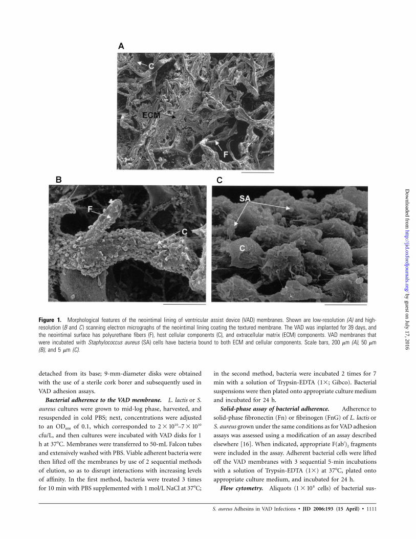
Figure 1. Morphological features of the neointimal lining of ventricular assist device (VAD) membranes. Shown are low-resolution (A) and high-resolution (B and C ) scanning electron micrographs of the neointimal lining coating the textured membrane. The VAD was implanted for 39 days, andthe neointimal surface has polyurethane fibers (F), host cellular components (C), and extracellular matrix (ECM) components. VAD membranes thatwere incubated with Staphylococcus aureus (SA) cells have bacteria bound to both ECM and cellular components. Scale bars, 200 mm (A), 50 mm(B), and 5 mm (C).
detached from its base; 9-mm-diameter disks were obtained
with the use of a sterile cork borer and subsequently used in
VAD adhesion assays.
Bacterial adherence to the VAD membrane. L. lactis or S.
aureus cultures were grown to mid-log phase, harvested, and
resuspended in cold PBS; next, concentrations were adjusted
to an OD600 of 0.1, which corresponded to –10 102 � 10 7 � 10
cfu/L, and then cultures were incubated with VAD disks for 1
h at 37�C. Membranes were transferred to 50-mL Falcon tubes
and extensively washed with PBS. Viable adherent bacteria were
then lifted off the membranes by use of 2 sequential methods
of elution, so as to disrupt interactions with increasing levels
of affinity. In the first method, bacteria were treated 3 times
for 10 min with PBS supplemented with 1 mol/L NaCl at 37�C;
in the second method, bacteria were incubated 2 times for 7
min with a solution of Trypsin-EDTA (1�; Gibco). Bacterial
suspensions were then plated onto appropriate culture medium
and incubated for 24 h.
Solid-phase assay of bacterial adherence. Adherence to
solid-phase fibronectin (Fn) or fibrinogen (FnG) of L. lactis or
S. aureus grown under the same conditions as for VAD adhesion
assays was assessed using a modification of an assay described
elsewhere [16]. When indicated, appropriate F(ab′)2 fragments
were included in the assay. Adherent bacterial cells were lifted
off the VAD membranes with 3 sequential 5-min incubations
with a solution of Trypsin-EDTA (1�) at 37�C, plated onto
appropriate culture medium, and incubated for 24 h.
Flow cytometry. Aliquots ( cells) of bacterial sus-81 � 10
by guest on July 17, 2016http://jid.oxfordjournals.org/
Dow
nloaded from
Figu
re2.
Adhe
renc
eof
Stap
hylo
cocc
usau
reus
surfa
cepr
otei
nsto
vent
ricul
aras
sist
devi
ce(V
AD)m
embr
anes
.A,A
dher
ence
ofLa
ctoc
occu
sla
ctis
stra
ins
expr
essi
ng12
S.au
reus
surfa
cepr
otei
nsto
aVA
Dth
atha
dbe
enim
plan
ted
!6
mon
ths
(ear
lyVA
D).*
,vs.
cont
rolp
KS80
orpN
Z803
7.B,
Furth
eran
alys
isof
the
adhe
renc
eof
L.la
ctis
stra
ins
expr
essi
ng4
S.au
reus
surfa
cepr
otei
nsto
4P
!.0
5di
ffere
ntea
rlyVA
Ds.*
,vs.
cont
rolp
KS80
orpN
Z803
7.C,
Diffe
rent
iale
lutio
nof
adhe
rent
L.la
ctis
stra
ins
from
early
VADs
afte
rtre
atm
ent
with
1m
ol/L
NaC
l(lig
htgr
ayba
rs)o
rTr
ypsi
n-ED
TAP
!.0
5(d
ark
gray
bars
).N
os.a
bove
bars
repr
esen
tpe
rcen
tage
sof
the
tota
lno.
ofad
here
ntce
llsfo
rea
chst
rain
that
wer
eel
uted
with
high
-ioni
c-st
reng
thso
lutio
n.Th
era
tioof
bact
eria
lcel
lsel
uted
from
the
VAD
mem
bran
eby
use
ofth
e2
met
hods
(NaC
lvs.
Tryp
sin-
EDTA
)for
clum
ping
fact
or(C
lf)A
vs.fi
bron
ectin
bind
ing
prot
ein
(FnB
P)A
and
FnBP
B,as
wel
las
forF
nBPA
vs.F
nBPB
,wer
est
atis
tical
lydi
ffere
nt.
*.D
,Flo
w-c
ytom
etric
resu
ltsill
ustra
ting
the
pres
ence
ofCl
fA,C
lfB,F
nBPA
,and
FnBP
Bon
the
surfa
ceof
L.la
ctis
cells
.*,v
s.co
ntro
lpKS
80or
pNZ8
037.
E,Ad
here
nce
ofL.
lact
isst
rain
sP
!.0
5P
!.0
5to
VADs
that
had
been
impl
ante
d�
6m
onth
s(la
teVA
Ds).
*,v
s.co
ntro
lpKS
80or
pNZ8
037.
F,Ad
here
nce
ofS.
aure
usLS
-1to
early
VADs
vs.l
ate
VADs
.*.D
ata
are
the
ofP
!.0
5P
!.0
5m
eans
�SE
sat
leas
t3
sepa
rate
expe
rimen
ts.S
tatis
tical
anal
yses
wer
epe
rform
edus
ing
anal
ysis
ofva
rianc
e.FI
TC,fl
uore
scei
nis
othi
ocya
nate
.Eap
,ext
race
llula
radh
eren
cepr
otei
n;Eb
p,el
astin
-bin
ding
prot
ein;
Map
,m
ajor
hist
ocom
patib
ility
com
plex
clas
sII
anal
ogou
spr
otei
n;Sa
s,St
aphy
loco
ccus
aure
assu
rface
;Sdr
,ser
ine-
aspa
rtate
repe
at;S
pA,s
taph
yloc
occa
lpro
tein
A.
by guest on July 17, 2016http://jid.oxfordjournals.org/
Dow
nloaded from
S. aureus Adhesins in VAD Infections • JID 2006:193 (15 April) • 1113
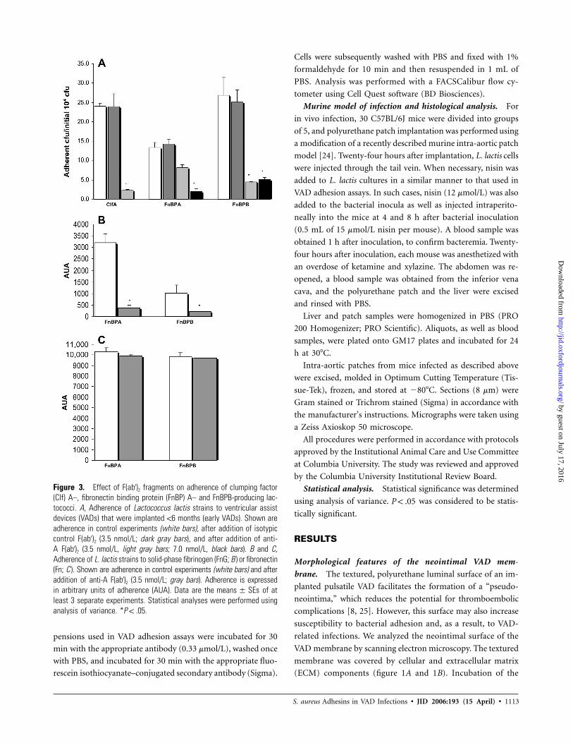
Figure 3. Effect of F(ab′)2 fragments on adherence of clumping factor(Clf) A–, fibronectin binding protein (FnBP) A– and FnBPB-producing lac-tococci. A, Adherence of Lactococcus lactis strains to ventricular assistdevices (VADs) that were implanted !6 months (early VADs). Shown areadherence in control experiments (white bars), after addition of isotypiccontrol F(ab′)2 (3.5 nmol/L; dark gray bars), and after addition of anti-A F(ab′)2 (3.5 nmol/L, light gray bars; 7.0 nmol/L, black bars). B and C,Adherence of L. lactis strains to solid-phase fibrinogen (FnG; B ) or fibronectin(Fn; C ). Shown are adherence in control experiments (white bars) and afteraddition of anti-A F(ab′)2 (3.5 nmol/L; gray bars). Adherence is expressedin arbitrary units of adherence (AUA). Data are the of atmeans � SEsleast 3 separate experiments. Statistical analyses were performed usinganalysis of variance. * .P ! .05
pensions used in VAD adhesion assays were incubated for 30
min with the appropriate antibody (0.33 mmol/L), washed once
with PBS, and incubated for 30 min with the appropriate fluo-
rescein isothiocyanate–conjugated secondary antibody (Sigma).
Cells were subsequently washed with PBS and fixed with 1%
formaldehyde for 10 min and then resuspended in 1 mL of
PBS. Analysis was performed with a FACSCalibur flow cy-
tometer using Cell Quest software (BD Biosciences).
Murine model of infection and histological analysis. For
in vivo infection, 30 C57BL/6J mice were divided into groups
of 5, and polyurethane patch implantation was performed using
a modification of a recently described murine intra-aortic patch
model [24]. Twenty-four hours after implantation, L. lactis cells
were injected through the tail vein. When necessary, nisin was
added to L. lactis cultures in a similar manner to that used in
VAD adhesion assays. In such cases, nisin (12 mmol/L) was also
added to the bacterial inocula as well as injected intraperito-
neally into the mice at 4 and 8 h after bacterial inoculation
(0.5 mL of 15 mmol/L nisin per mouse). A blood sample was
obtained 1 h after inoculation, to confirm bacteremia. Twenty-
four hours after inoculation, each mouse was anesthetized with
an overdose of ketamine and xylazine. The abdomen was re-
opened, a blood sample was obtained from the inferior vena
cava, and the polyurethane patch and the liver were excised
and rinsed with PBS.
Liver and patch samples were homogenized in PBS (PRO
200 Homogenizer; PRO Scientific). Aliquots, as well as blood
samples, were plated onto GM17 plates and incubated for 24
h at 30�C.
Intra-aortic patches from mice infected as described above
were excised, molded in Optimum Cutting Temperature (Tis-
sue-Tek), frozen, and stored at �80�C. Sections (8 mm) were
Gram stained or Trichrom stained (Sigma) in accordance with
the manufacturer’s instructions. Micrographs were taken using
a Zeiss Axioskop 50 microscope.
All procedures were performed in accordance with protocols
approved by the Institutional Animal Care and Use Committee
at Columbia University. The study was reviewed and approved
by the Columbia University Institutional Review Board.
Statistical analysis. Statistical significance was determined
using analysis of variance. was considered to be statis-P ! .05
tically significant.
RESULTS
Morphological features of the neointimal VAD mem-
brane. The textured, polyurethane luminal surface of an im-
planted pulsatile VAD facilitates the formation of a “pseudo-
neointima,” which reduces the potential for thromboembolic
complications [8, 25]. However, this surface may also increase
susceptibility to bacterial adhesion and, as a result, to VAD-
related infections. We analyzed the neointimal surface of the
VAD membrane by scanning electron microscopy. The textured
membrane was covered by cellular and extracellular matrix
(ECM) components (figure 1A and 1B). Incubation of the
by guest on July 17, 2016http://jid.oxfordjournals.org/
Dow
nloaded from
1114 • JID 2006:193 (15 April) • Arrecubieta et al.
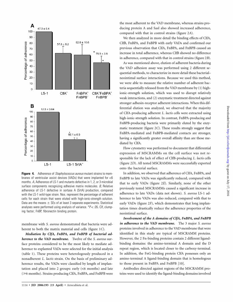
Figure 4. Adherence of Staphylococcus aureus mutant strains to mem-branes of ventricular assist devices (VADs) that were implanted for !6months. A, Adherence of LS-1 and mutants defective in 1, 2, or 3 microbialsurface components recognizing adhesive matrix molecules. B, Relativeadherence of LS-1 defective in sortase A (SrtA) production, comparedwith the LS-1 wild-type strain. Nos. represent the percentage of adherentcells for each strain that were eluted with high-ionic-strength solution.Data are the of at least 3 separate experiments. Statisticalmeans � SEsanalyses were performed using analysis of variance. * . Clf, clump-P ! .05ing factor; FnBP, fibronectin binding protein.
membrane with S. aureus demonstrated that bacteria were ad-
herent to both the matrix material and cells (figure 1C).
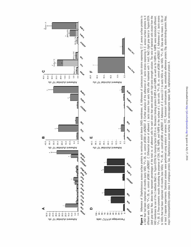
Mediation by ClfA, FnBPA, and FnBPB of bacterial ad-
herence to the VAD membrane. Twelve of the S. aureus sur-
face proteins considered to be the most likely to mediate ad-
herence to explanted VADs were selected for the initial analysis
(table 1). These proteins were heterologously produced in a
nonadherent L. lactis strain. On the basis of preliminary ad-
herence results, the VADs were classified by length of implan-
tation and placed into 2 groups: early (!6 months) and late
(�6 months). Strains producing ClfA, FnBPA, and FnBPB were
the most adherent to the VAD membrane, whereas strains pro-
ducing protein A and SasI also showed increased adherence,
compared with that in control strains (figure 2A).
We then analyzed in more detail the binding effects of ClfA,
ClfB, FnBPA, and FnBPB with early VADs and confirmed our
previous observation that ClfA, FnBPA, and FnBPB caused an
increase in total adherence, whereas ClfB showed no difference
in adherence, compared with that in control strains (figure 2B).
As was mentioned above, elution of adherent bacteria during
the VAD adhesion assay was performed using 2 different se-
quential methods, to characterize in more detail these bacterial–
neointimal surface interactions. Because we used this method,
we were able to measure the relative number of adherent bac-
teria sequentially released from the VAD membrane by (1) high-
ionic-strength solution, which was used to disrupt relatively
weak interactions, and (2) enzymatic treatment directed against
stronger adhesin-receptor adherent interactions. When this dif-
ferential elution was analyzed, we observed that the majority
of ClfA-producing adherent L. lactis cells were extracted using
high-ionic-strength solution. In contrast, FnBPA-producing and
FnBPB-producing bacteria were primarily eluted by the enzy-
matic treatment (figure 2C). These results strongly suggest that
FnBPA-mediated and FnBPB-mediated contacts are stronger,
having a significantly greater overall affinity than are those me-
diated by ClfA.
Flow cytometry was performed to document that differential
expression of MSCRAMMs on the cell surface was not re-
sponsible for the lack of effect of ClfB-producing L. lactis cells
(figure 2D). All tested MSCRAMMs were successfully exported
onto the bacterial surface.
In addition, we observed that adherence of ClfA, FnBPA, and
FnBPB to late VADs was significantly reduced, compared with
that to early VADs (figure 2E). Similarly, none of the other
previously tested MSCRAMMs caused a significant increase in
adherence to late VADs (data not shown). S. aureus LS-1 ad-
herence to late VADs was also reduced, compared with that to
early VADs (figure 2F), which demonstrates that long implan-
tation times drastically reduce the adherence properties of the
neointimal surface.
Involvement of the A domains of ClfA, FnBPA, and FnBPB
in adherence to the VAD membrane. The 3 major S. aureus
proteins involved in adherence to the VAD membrane that were
identified in this study are typical of MSCRAMM proteins.
However, the 2 Fn-binding proteins contain 2 different ligand-
binding domains: the amino-terminal A domain and the D
repeat region, which is located closer to the carboxy-terminal.
In addition, the FnG-binding protein ClfA possesses only an
amino-terminal A ligand-binding domain that is homologous
to those present in FnBPA and FnBPB [10].
Antibodies directed against regions of the MSCRAMM pro-
teins were used to identify the ligand-binding domains involved
by guest on July 17, 2016http://jid.oxfordjournals.org/
Dow
nloaded from
S. aureus Adhesins in VAD Infections • JID 2006:193 (15 April) • 1115
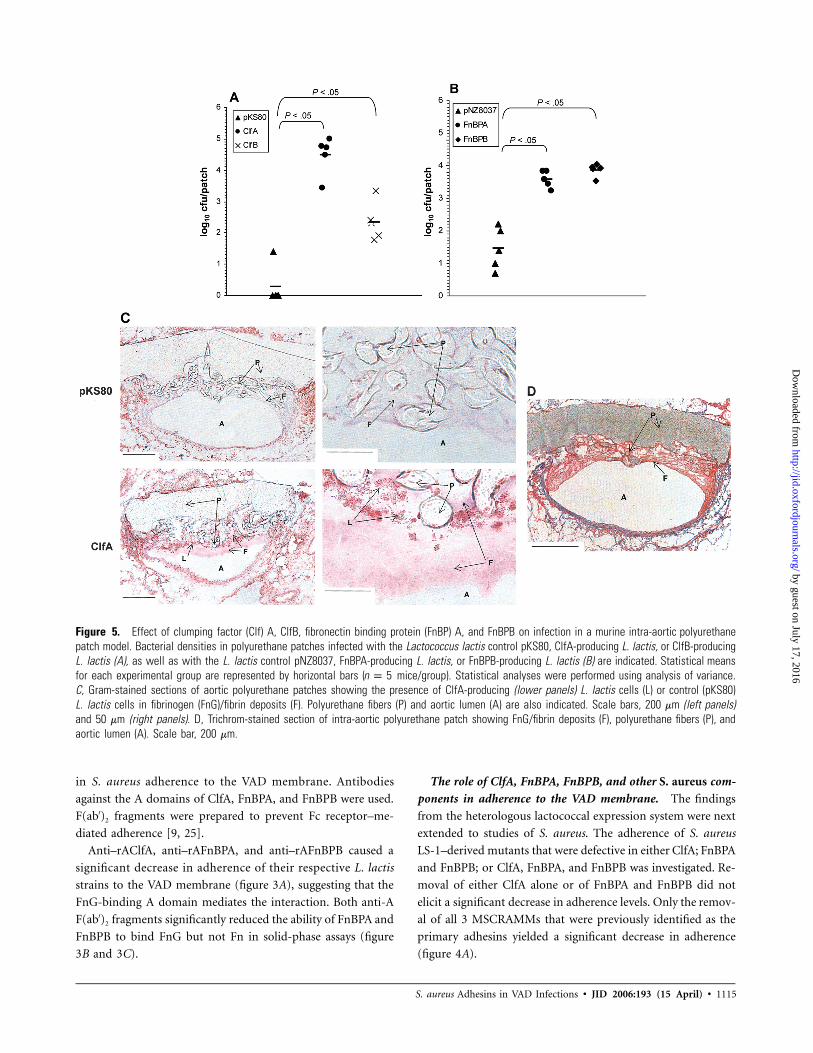
Figure 5. Effect of clumping factor (Clf) A, ClfB, fibronectin binding protein (FnBP) A, and FnBPB on infection in a murine intra-aortic polyurethanepatch model. Bacterial densities in polyurethane patches infected with the Lactococcus lactis control pKS80, ClfA-producing L. lactis, or ClfB-producingL. lactis (A), as well as with the L. lactis control pNZ8037, FnBPA-producing L. lactis, or FnBPB-producing L. lactis (B) are indicated. Statistical meansfor each experimental group are represented by horizontal bars ( mice/group). Statistical analyses were performed using analysis of variance.n p 5C, Gram-stained sections of aortic polyurethane patches showing the presence of ClfA-producing (lower panels) L. lactis cells (L) or control (pKS80)L. lactis cells in fibrinogen (FnG)/fibrin deposits (F). Polyurethane fibers (P) and aortic lumen (A) are also indicated. Scale bars, 200 mm (left panels)and 50 mm (right panels). D, Trichrom-stained section of intra-aortic polyurethane patch showing FnG/fibrin deposits (F), polyurethane fibers (P), andaortic lumen (A). Scale bar, 200 mm.
in S. aureus adherence to the VAD membrane. Antibodies
against the A domains of ClfA, FnBPA, and FnBPB were used.
F(ab′)2 fragments were prepared to prevent Fc receptor–me-
diated adherence [9, 25].
Anti–rAClfA, anti–rAFnBPA, and anti–rAFnBPB caused a
significant decrease in adherence of their respective L. lactis
strains to the VAD membrane (figure 3A), suggesting that the
FnG-binding A domain mediates the interaction. Both anti-A
F(ab′)2 fragments significantly reduced the ability of FnBPA and
FnBPB to bind FnG but not Fn in solid-phase assays (figure
3B and 3C).
The role of ClfA, FnBPA, FnBPB, and other S. aureus com-
ponents in adherence to the VAD membrane. The findings
from the heterologous lactococcal expression system were next
extended to studies of S. aureus. The adherence of S. aureus
LS-1–derived mutants that were defective in either ClfA; FnBPA
and FnBPB; or ClfA, FnBPA, and FnBPB was investigated. Re-
moval of either ClfA alone or of FnBPA and FnBPB did not
elicit a significant decrease in adherence levels. Only the remov-
al of all 3 MSCRAMMs that were previously identified as the
primary adhesins yielded a significant decrease in adherence
(figure 4A).
by guest on July 17, 2016http://jid.oxfordjournals.org/
Dow
nloaded from
1116 • JID 2006:193 (15 April) • Arrecubieta et al.
Table 2. Hepatic metastatic seeding of mice infected with dif-ferent Lactococcus lactis strains.
Strain phenotypeInoculum,cfu/mouse
Bacteria in liver,log10 cfu/g � SE Pa
pKS80 control 5 � 107 2.35 � 0.14 …ClfA producing 5 � 107 3.90 � 0.19 !.05ClfB producing 5 � 107 3.58 � 0.06 !.05
pNZ8037 control 7 � 107 3.72 � 0.04 …FnBPA producing 1.6 � 108 4.77 � 0.04 !.05FnBPB producing 8 � 107 4.65 � 0.10 !.05
NOTE. Clf, clumping factor; FnBP, fibronectin binding protein.a Compared with the control strain (analysis of variance).
To study the role that other MSCRAMMs play in adherence
to the VAD membrane, we constructed an LS-1–derived mutant
strain that was defective in the production of SrtA, the key
enzyme responsible for anchoring proteins belonging to the
MSCRAMM family into the staphylococcal peptidoglycan [13].
The adherence of LS-1 SrtA� was significantly reduced, com-
pared with that of LS-1 (figure 4B). Interestingly, we also ob-
served that the majority of LS-1 cells were successfully detached
from the VAD membrane only after an enzymatic treatment,
whereas a high-ionic-strength solution was sufficient to release
most of the LS-1 SrtA� cells (figure 3A and 3B).
Facilitation by ClfA, ClfB, FnBPA, and FnBPB of L. lactis
adherence to the VAD membrane in vivo. The in vitro data
indicated that ClfA, FnBPA, and FnBPB were the major factors
involved in adherence to the VAD membrane. To demonstrate
the validity of the in vitro observations and to study the role
exerted by these proteins under physiological conditions, we
next used a recently developed murine model of intra-aortic
polyurethane patch infection [24] to assess whether they fa-
cilitate adherence of L. lactis to the implanted patch. ClfA-
producing lactococci caused a significant increase in the num-
ber of bacteria present on the polyurethane patch, compared
with that caused by the control strain (figure 5A). To assess
the in vivo role of FnBPA and FnBPB, we used lactococcal cells
cultured and induced with nisin in a similar manner to that
previously described for in vitro assays. In this way, FnBPA-
producing and FnBPB-producing cells adhered to the polyure-
thane patch in significantly higher numbers than did the control
pNZ8037 strain (figure 5B). Interestingly, we observed that
ClfB-producing L. lactis cells also caused a significant increase
in adherence to the polyurethane patch, although such adher-
ence was markedly reduced with respect to that elicited by ClfA
(figure 5A). Similarly, a significant difference in metastatic seed-
ing of the liver was also demonstrated between the L. lactis
strains producing the different staphylococcal adhesins and
their control strains (table 2), although no lactococcal cells were
found in the bloodstream of mice at the time of death (data
not shown).
Gram staining of the infected polyurethane patch demon-
strated that ClfA-producing L. lactis cells were adherent to the
patch material and accumulated on the fibrin that immediately
covers the polyurethane textured surface (figure 5C and 5D).
Other areas adjoining the patch were free of bacteria. In con-
trast, no bacterial cells were detected when the control strain
was used (figure 5C). These results demonstrate that ClfA,
FnBPA, and FnBPB—the major adhesion factors that were iden-
tified in vitro—are independently sufficient to establish early
infections in an in vivo setting.
DISCUSSION
This study is the first, to our knowledge, to investigate the role
that bacterial factors play in the pathogenesis of VAD infections.
The size of the device, immunocompromise in the patient, and
the presence of a transcutaneous driveline all increase the risk
of infection [3]. The most serious of these infections involve
the luminal polyurethane or valvular surfaces of the device. The
VAD illustrates the biologic complexity of understanding the
pathogenesis of prosthetic device–related infections. The tex-
tured surface evolves over time, becomes increasingly covered
with different host cells and matrix proteins, is subject to vary-
ing hemodynamic conditions, and is regularly exposed to cir-
culating bacterial pathogens [8, 9, 25].
A novel screening system was used to assess the ability of
staphylococcal surface proteins to adhere to the VAD membrane.
The critical first step in the pathogenesis of these infections re-
quires the adherence to and colonization of the device’s surface,
and this process is often mediated in S. aureus by adhesins be-
longing to the MSCRAMM family [10]. A heterologous lacto-
coccal expression system was used to overcome the inherent
redundancy found in S. aureus adhesins [10]. Despite the qual-
itative nature of our assay—which resulted from differences in
the composition of the neointimal surface that were caused by
varying lengths of VAD implantation, interpatient variability, and
the use of 2 different expression systems [8, 9, 26, 27]—we found
that ClfA, FnBPA, and FnBPB were the main MSCRAMMs that
mediated bacterial adherence. Of interest, even though ClfB is
structurally very similar to ClfA, it failed to mediate adherence
in vitro.
FnBPA and FnBPB are closely related proteins that have been
shown to bind to both FnG and Fn molecules [10, 28]. In the
same way, both ClfA and ClfB, which belong to the serine-
aspartate repeat family of proteins, share the ability to bind
FnG molecules [10, 29, 30]. However, although ClfA binds to
the d chain, ClfB recognizes and adheres to the a chain of the
molecule. FnBPA and FnBPB share with ClfA the capacity to
recognize the FnG d chain [10]. This difference in binding site
might, in part, explain the inability of ClfB to adhere to the
VAD membrane in vitro. Therefore, the large FnG/fibrin de-
posits in explanted VADs [8, 9, 26] and their affinity for ClfA,
by guest on July 17, 2016http://jid.oxfordjournals.org/
Dow
nloaded from

S. aureus Adhesins in VAD Infections • JID 2006:193 (15 April) • 1117
FnBPA, and FnBPB make them the most likely ligands involved
in S. aureus adherence. Previous studies have shown that FnG
plays a role in the adherence of bacteria to intravascular cath-
eters that are inserted over a short term [31] and hemodialysis
tubing [32] and that fibrin plays a role in adherence to poly-
urethane surfaces [33]. More recently, other authors have iden-
tified S. aureus FnG binding activity as a key element in pro-
moting early valve colonization in an in vivo rat endocarditis
model [34].
Significant differences in the elution pattern between ClfA-,
FnBPA-, and FnBPB-producing cells were detected, suggesting
that the interaction of ClfA with the neointimal surface is sub-
stantially weaker than that of FnBPA or FnBPB. The ClfA-
mediated interaction with FnG in vitro has been shown to be
a high-affinity event [10]. However, in contrast to ClfA, which
has a stoichiometric ratio of 1:1 with FnG, FnBPA and FnBPB
mediate interactions with 2 molecules of Fn and have been
shown to be able to form covalent cross-linking with fibrin and
Fn [35, 36]. This, in addition to their ability to bind FnG,
might result in a stronger overall interaction than that of ClfA,
especially on the VAD membrane, where different types of re-
ceptors are most likely present. These differences in affinity for
the host neointimal surface might play a significant role in the
likelihood of a successful infection under physiological condi-
tions. In this scenario, other factors, such as flow patterns and
shear stress conditions in the bloodstream, may affect S. aureus
adherence to the VAD membrane in the same way that they are
known to affect S. aureus adherence to host components such
as platelets and endothelial cells [37, 38]. Two other proteins,
SasI and protein A, had a lesser effect on adherence to the VAD
membrane. SasI expression is iron dependent [39], which might
enhance its in vivo significance. Protein A mediates binding to
von Willebrand factor [40], which is known to be present on
the VAD neointima [8].
Our in vitro observations about MSCRAMM proteins were
further validated using an in vivo model [24], in which mice
implanted with intra-aortic polyurethane patches were infected
with the ClfA-, FnBPA-, and FnBPB-producing L. lactis strains.
The presence of staphylococcal proteins on the lactococcal sur-
face conferred a survival advantage, even in environments
known to be rich in phagocytes, such as the liver. Histological
studies demonstrated that, at least in the ClfA cells, adherence
occurred along the FnG-coated surface of the polyurethane
patch.
These findings, therefore, support the validity of the in vitro
adherence assays, as well as the hypothesis that FnG is the
primary ECM component that is responsible for promoting S.
aureus adherence. Interestingly, ClfB also caused an increase in
adherence to the polyurethane patch, although this adherence
was significantly lower than that caused by ClfA.
The inherent capacity of different S. aureus MSCRAMMs to
bind the same matrix proteins complicated the study of ad-
hesin-receptor interactions. However, once the major adhesins
were identified, it was possible to assess the effect that S. aureus
mutants lacking these proteins had on adherence. Mutants lack-
ing ClfA or both FnBPA and FnBPB did not cause a significant
reduction in adherence, whereas mutants lacking all 3 proteins
caused a substantial decrease in adherence. The failure to com-
pletely inhibit adherence suggested the possibility that other
factors contribute to binding, as has been observed in the ability
of S. aureus to induce aggregation of human platelets [16]. This
hypothesis was confirmed with the use of an SrtA-deficient S.
aureus strain—a mutant defective in the production of the en-
zyme responsible for anchoring MSCRAMMs into the staphy-
lococcal cell wall [13]. We observed a substantial reduction in
the binding levels that accounted for 67% of the parental bind-
ing; this suggested that other factors not linked to SrtA activ-
ity and therefore not belonging to the MSCRAMM family are
also involved in the process. However, the increasing elution
of these less-adherent strains from the VAD membrane after
treatment with a high-ionic-strength solution suggested a rel-
atively weaker binding interaction than that occurring with the
wild-type strain.
Another important observation was the failure of S. aureus
adhesins to mediate the attachment to VADs that had lengthy
implantation times. Similar observations were recently reported
in a clinical study showing a significant decrease over time in
the rate of bacterial infections in patients implanted with
HeartMate VADs—the same device that was used in the present
study [3, 5]. These observations probably reflect changes in the
composition of the neointimal lining on the surface of textured
cardiac assist devices [8, 9, 26, 27]. Over time, the neointima
becomes increasingly covered with cellular components, and the
amount of ECM that is exposed to the bloodstream is reduced
[8, 41]. This change in the cellular surface may account for the
reduced bacterial adherence to the VAD membrane. The effect
of cellular deposition/proliferation on prosthetic devices [24] and
their role in potentially reducing susceptibility to infection has
not been previously examined.
Several antibodies were used to further identify those epi-
topes, among the ligand-binding domains of the MSCRAMM
proteins, involved in adherence to the VAD membrane. Block-
ing of the A domains of ClfA, FnBPA, and FnBPB caused a
significant reduction in adherence. This reduction suggests that
these domains played the major role in adherence to the VAD
membrane, most likely by recognizing FnG/fibrin in the neoin-
tima. The amount of anti–rAFnBPA and anti–rAFnBPB F(ab′)2
fragments required to elicit a similar effect was higher than
that needed for anti–rAClfA, perhaps echoing differences in the
amount of exported proteins onto the lactococcal surface, al-
though possibly merely reflecting differences in the affinity of
the F(ab′)2 fragments for their corresponding epitopes. Simi-
by guest on July 17, 2016http://jid.oxfordjournals.org/
Dow
nloaded from
1118 • JID 2006:193 (15 April) • Arrecubieta et al.
larly, it has also been recently shown that the Fn-binding A
domains of ClfA and FnBPA appear to be responsible for the
first stage in the pathogenesis of S. aureus–associated endocar-
ditis [34].
The findings of the present study demonstrate that the first
steps leading to the development of VAD infections involve
numerous adhesin-receptor interactions that overlap, and this
process makes the development of therapeutic strategies a com-
plex and intricate task. To our knowledge, our results provide
the first direct information on the identity of these major bac-
terial factors and the likely host surface constituents—fibrin/
FnG deposits—that serve as the main receptor on the host neoin-
timal surface. The in vitro data demonstrate that antibodies di-
rected at specific domains of MSCRAMMs reduce early in vitro
adherence to the VAD membrane. These observations may lead
to future studies to assess the potential of these components as
targets for the development of new strategies for treatment and
prevention of these increasingly encountered infections. The dy-
namic nature of such surfaces calls for further studies of their
composition, structure, and adherent properties for staphylo-
coccal factors. Finally, a more profound understanding of the
biological processes that lie subjacent to these processes of ad-
herence to and colonization of these highly dynamic structures
seems essential for the development of additional prophylactic
and therapeutic targets.
Acknowledgments
We thank Leslie Gunther (Department of Analytical Imaging, AlbertEinstein College of Medicine, Yeshiva University, New York, NY), for tech-nical assistance with the scanning electron microscopy; Inhibitex, for pro-viding us with various antibodies; and Peter Vavagiakis, for his valuablehelp with the statistical analysis.
References
1. Rose EA, Gelijns AC, Moskowitz AJ, et al. Long-term mechanical leftventricular assistance for end-stage heart failure. N Engl J Med 2001;345:1435–43.
2. Herrmann M, Weyand M, Greshake B, et al. Left ventricular assist deviceinfection is associated with increased mortality but is not a contraindi-cation to transplantation. Circulation 1997; 95:814–7.
3. Holman WL, Park SJ, Long JW, et al. Infection in permanent circulatorysupport: experience from the REMATCH trial. J Heart Lung Transplant2004; 23:1359–65.
4. Deng MC, Edwards LB, Hertz MI, et al. Mechanical Circulatory Sup-port Device Database of the International Society for Heart and LungTransplantation: second annual report—2004. J Heart Lung Transplant2004; 23:1027–34.
5. Simon D, Fischer S, Grossman A, et al. Left ventricular assist de-vice–related infection: treatment and outcome. Clin Infect Dis 2005;40:1108–15.
6. Holman WL, Rayburn BK, McGiffin DC, et al. Infection in ventricularassist devices: prevention and treatment. Ann Thorac Surg 2003; 75(Suppl 6):S48–57.
7. Fischer SA, Trenholme GM, Costanzo MR, Piccione W. Infectious com-
plications in left ventricular assist device recipients. Clin Infect Dis1997; 24:18–23.
8. Menconi MJ, Pockwinse S, Owen TA, Dasse KA, Stein GS, Lian JB.Properties of blood-contacting surfaces of clinically implanted cardiacassist devices: gene expression, matrix composition, and ultrastructuralcharacterization of cellular linings. J Cell Biochem 1995; 57:557–73.
9. Spanier TB, Chen JM, Oz MC, Stern DM, Rose EA, Schmidt AM.Time-dependent cellular population of textured-surface left ventricularassist devices contributes to the development of a biphasic systemicprocoagulant response. J Thorac Cardiovasc Surg 1999; 118:404–13.
10. Foster TJ, Hook M. Surface protein adhesins of Staphylococcus aureus.Trends Microbiol 1998; 6:484–8.
11. Gasson MJ. Genetic transfer systems in lactic acid bacteria. AntonieVan Leeuwenhoek 1983; 49:275–82.
12. Kuipers OP, Beerthuyzen MM, Siezen RJ, De Vos WM. Characterizationof the nisin gene cluster nisABTCIPR of Lactococcus lactis: requirementof expression of the nisA and nisI genes for development of immunity.Eur J Biochem 1993; 216:281–91.
13. Mazmanian SK, Liu G, Jensen ER, Lenoy E, Schneewind O. Staphy-lococcus aureus sortase mutants defective in the display of surface pro-teins and in the pathogenesis of animal infections. Proc Natl Acad SciUSA 2000; 97:5510–5.
14. Bremell T, Lange S, Svensson L, et al. Outbreak of spontaneous staph-ylococcal arthritis and osteitis in mice. Arthritis Rheum 1990; 33:1739–44.
15. Fitzgerald JR, Loughman A, Keane F, et al. Fibronectin�binding pro-teins of Staphylococcus aureus mediate activation of human plateletsvia fibrinogen and fibronectin bridges to integrin GPIIb/IIIa and IgGbinding to the FcgammaRIIa receptor. Mol Microbiol 2006; 59:212–30.
16. Salih V, Graham TR, Berry CL, et al. The lining of textured surfacesin implantable left ventricular assist devices: an immunocytochemicaland electronmicroscopic study. Am J Cardiovasc Pathol 1993; 4:317–25.
17. Hartford O, O’Brien L, Schofield K, Wells J, Foster TJ. The Fbe (SdrG)protein of Staphylococcus epidermidis HB promotes bacterial adherenceto fibrinogen. Microbiology 2001; 147:2545–52.
18. de Ruyter PG, Kuipers OP, de Vos WM. Controlled gene expressionsystems for Lactococcus lactis with the food-grade inducer nisin. ApplEnviron Microbiol 1996; 62:3662–7.
19. McAleese FM, Walsh EJ, Sieprawska M, Potempa J, Foster TJ. Loss ofclumping factor B fibrinogen binding activity by Staphylococcus aureusinvolves cessation of transcription, shedding and cleavage by metal-loprotease. J Biol Chem 2001; 276:29969–78.
20. Greene C, McDevitt D, Francois P, Vaudaux PE, Lew DP, Foster TJ.Adhesion properties of mutants of Staphylococcus aureus defective infibronectin-binding proteins and studies on the expression of fnb genes.Mol Microbiol 1995; 17:1143–52.
21. Roche FM, Downer R, Keane F, Speziale P, Park PW, Foster TJ. The N-terminal A domain of fibronectin-binding proteins A and B promotesadhesion of Staphylococcus aureus to elastin. J Biol Chem 2004; 279:38433–40.
22. O’Brien LM, Walsh EJ, Massey RC, Peacock SJ, Foster TJ. Staphylo-coccus aureus clumping factor B (ClfB) promotes adherence to humantype I cytokeratin 10: implications for nasal colonization. Cell Micro-biol 2002; 4:759–70.
23. Lowy FD, Fant J, Higgins LL, Ogawa SK, Hatcher VB. Staphylococcusaureus-human endothelial cell interactions. J Ultrastruct Mol StructRes 1988; 98:137–46.
24. Asai T, Baron HM, Prinz von Bayern M, et al. A mouse aortic patchmodel for mechanical circulatory support. J Heart Lung Transplant2005;24:1129–32.
25. Rafii S, Oz MC, Seldomridge JA, et al. Characterization of hemato-poietic cells arising on the textured surface of left ventricular assistdevices. Ann Thorac Surg 1995; 60:1627–32.
26. Salih V, Berry CL, Smith SC, et al. The lining of textured surfaces inimplantable left ventricular assist devices: an immunocytochemical andelectronmicroscopic study. Am J Cardiovasc Pathol 1993; 4:317–25.
by guest on July 17, 2016http://jid.oxfordjournals.org/
Dow
nloaded from
S. aureus Adhesins in VAD Infections • JID 2006:193 (15 April) • 1119
27. Scott-Burden T, Frazier OH. Cellular linings of ventricular assist de-vices. Ann Thorac Surg 1995; 60:1561–2.
28. Wann ER, Gurusiddappa S, Hook M. The fibronectin-bindingMSCRAMM FnbpA of Staphylococcus aureus is a bifunctional pro-tein that also binds to fibrinogen. J Biol Chem 2000; 275:13863–71.
29. Ni Eidhin D, Perkins S, Francois P, Vaudaux P, Hook M, Foster TJ.Clumping factor B (ClfB), a new surface-located fibrinogen-bindingadhesin of Staphylococcus aureus. Mol Microbiol 1998; 30:245–57.
30. McDevitt D, Francois P, Vaudaux P, Foster TJ. Molecular characteri-zation of the clumping factor (fibrinogen receptor) of Staphylococcusaureus. Mol Microbiol 1994; 11:237–48.
31. Vaudaux P, Pittet D, Haeberli A, et al. Fibronectin is more active thanfibrin or fibrinogen in promoting Staphylococcus aureus adherence toinserted intravascular catheters. J Infect Dis 1993; 167:633–41.
32. Vaudaux PE, Francois P, Proctor RA, et al. Use of adhesion-defectivemutants of Staphylococcus aureus to define the role of specific plasmaproteins in promoting bacterial adhesion to canine arteriovenousshunts. Infect Immun 1995; 63:585–90.
33. Baumgartner JN, Cooper SL. Bacterial adhesion on polyurethanesurfaces conditioned with thrombus components. ASAIO J 1996; 42:M476–9.
34. Que YA, Haefliger JA, Piroth L, et al. Fibrinogen and fibronectin bind-ing cooperate for valve infection and invasion in Staphylococcus aureusexperimental endocarditis. J Exp Med 2005; 201:1627–35.
35. Matsuka YV, Anderson ET, Milner-Fish T, Ooi P, Baker S. Staphylococcusaureus fibronectin-binding protein serves as a substrate for coagulationfactor XIIIa: evidence for factor XIIIa-catalyzed covalent cross-linking tofibronectin and fibrin. Biochemistry 2003; 42:14643–52.
36. Ingham KC, Brew S, Vaz D, Sauder DN, McGavin MJ. Interaction ofStaphylococcus aureus fibronectin-binding protein with fibronectin: af-finity, stoichiometry, and modular requirements. J Biol Chem 2004;279:42945–53.
37. Pawar P, Shin PK, Mousa SA, Ross JM, Konstantopoulos K. Fluid shearregulates the kinetics and receptor specificity of Staphylococcus aureusbinding to activated platelets. J Immunol 2004; 173:1258–65.
38. Reddy K, Ross JM. Shear stress prevents fibronectin binding protein-mediated Staphylococcus aureus adhesion to resting endothelial cells. In-fect Immun 2001; 69:3472–5.
39. Skaar EP, Schneewind O. Iron-regulated surface determinants (Isd) ofStaphylococcus aureus: stealing iron from heme. Microbes Infect 2004;6:390–7.
40. Hartleib J, Kohler N, Dickinson RB, et al. Protein A is the von Wil-lebrand factor binding protein on Staphylococcus aureus. Blood 2000;96:2149–56.
41. Graham TR, Dasse K, Coumbe A, et al. Neo-intimal development ontextured biomaterial surfaces during clinical use of an implantable leftventricular assist device. Eur J Cardiothorac Surg 1990; 4:182–90.
by guest on July 17, 2016http://jid.oxfordjournals.org/
Dow
nloaded from