Sporadic decentralized resource maintenance for P2P distributed storage networks
Upload
independentCategory
view
5download
0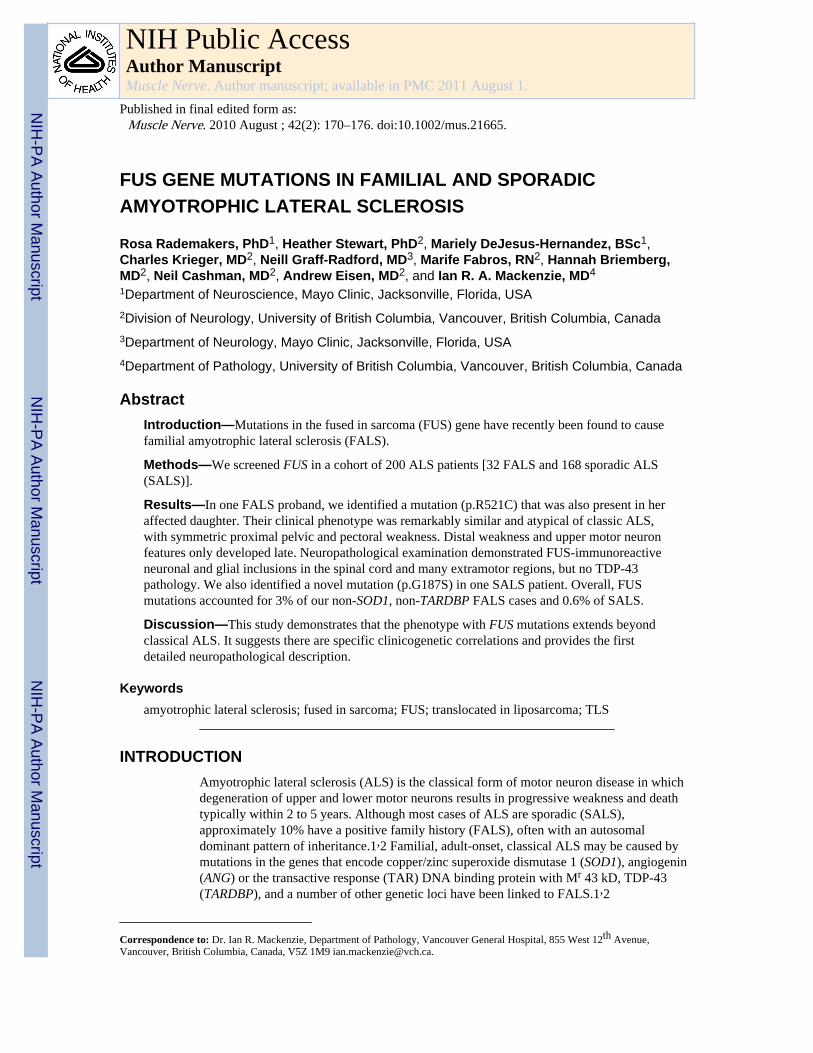
FUS GENE MUTATIONS IN FAMILIAL AND SPORADICAMYOTROPHIC LATERAL SCLEROSIS
Rosa Rademakers, PhD1, Heather Stewart, PhD2, Mariely DeJesus-Hernandez, BSc1,Charles Krieger, MD2, Neill Graff-Radford, MD3, Marife Fabros, RN2, Hannah Briemberg,MD2, Neil Cashman, MD2, Andrew Eisen, MD2, and Ian R. A. Mackenzie, MD4
1Department of Neuroscience, Mayo Clinic, Jacksonville, Florida, USA2Division of Neurology, University of British Columbia, Vancouver, British Columbia, Canada3Department of Neurology, Mayo Clinic, Jacksonville, Florida, USA4Department of Pathology, University of British Columbia, Vancouver, British Columbia, Canada
AbstractIntroduction—Mutations in the fused in sarcoma (FUS) gene have recently been found to causefamilial amyotrophic lateral sclerosis (FALS).
Methods—We screened FUS in a cohort of 200 ALS patients [32 FALS and 168 sporadic ALS(SALS)].
Results—In one FALS proband, we identified a mutation (p.R521C) that was also present in heraffected daughter. Their clinical phenotype was remarkably similar and atypical of classic ALS,with symmetric proximal pelvic and pectoral weakness. Distal weakness and upper motor neuronfeatures only developed late. Neuropathological examination demonstrated FUS-immunoreactiveneuronal and glial inclusions in the spinal cord and many extramotor regions, but no TDP-43pathology. We also identified a novel mutation (p.G187S) in one SALS patient. Overall, FUSmutations accounted for 3% of our non-SOD1, non-TARDBP FALS cases and 0.6% of SALS.
Discussion—This study demonstrates that the phenotype with FUS mutations extends beyondclassical ALS. It suggests there are specific clinicogenetic correlations and provides the firstdetailed neuropathological description.
Keywordsamyotrophic lateral sclerosis; fused in sarcoma; FUS; translocated in liposarcoma; TLS
INTRODUCTIONAmyotrophic lateral sclerosis (ALS) is the classical form of motor neuron disease in whichdegeneration of upper and lower motor neurons results in progressive weakness and deathtypically within 2 to 5 years. Although most cases of ALS are sporadic (SALS),approximately 10% have a positive family history (FALS), often with an autosomaldominant pattern of inheritance.1,2 Familial, adult-onset, classical ALS may be caused bymutations in the genes that encode copper/zinc superoxide dismutase 1 (SOD1), angiogenin(ANG) or the transactive response (TAR) DNA binding protein with Mr 43 kD, TDP-43(TARDBP), and a number of other genetic loci have been linked to FALS.1,2
Correspondence to: Dr. Ian R. Mackenzie, Department of Pathology, Vancouver General Hospital, 855 West 12th Avenue,Vancouver, British Columbia, Canada, V5Z 1M9 [email protected].
NIH Public AccessAuthor ManuscriptMuscle Nerve. Author manuscript; available in PMC 2011 August 1.
Published in final edited form as:Muscle Nerve. 2010 August ; 42(2): 170–176. doi:10.1002/mus.21665.
NIH
-PA Author Manuscript
NIH
-PA Author Manuscript
NIH
-PA Author Manuscript
Recently, two studies identified mutations in the gene that encodes the fused in sarcoma(FUS) protein (also known as translocated in liposarcoma, TLS), as the cause of FALS type6.3,4 FUS is a DNA/RNA binding protein with striking functional homology to TDP-43.5These studies reported a total of 14 different mutations in 26 unrelated families, representingapproximately 4% of FALS in their combined series. Most were missense mutationsaffecting highly conserved regions in exon 15 that encodes the C-terminus. With theexception of one family with autosomal recessive disease, caused by the c.1551C>Gmutation,3 all other mutations produced autosomal dominant ALS, although withincomplete penetrance. No mutations were found in 293 SALS cases screened in one study.3The associated clinical phenotype was described only as classical ALS, and there was verylimited description of the post mortem findings.3,4 Since then, additional studies havereported FUS mutations in ALS patients from Italy, Belgium and in a combined French andFrench-Canadian cohort.6-10
In this study, we performed genetic analysis of FUS in a cohort of 200 patients with ALS,including 32 FALS and 168 SALS index cases, all of whom had been evaluated at the ALSClinic at the University of British Columbia, Vancouver, Canada. We identified two FUSmutations, including a novel mutation in a case of SALS. We provide detailed description ofthe clinical features and neuropathological findings in the affected patients.
MATERIALS AND METHODSCases
Peripheral blood samples for DNA were collected from 32 index cases with FALS and 168SALS patients from the ALS Clinic at the University of British Columbia, Vancouver,Canada, between 1997 and 2006. All cases had a diagnosis of definite or probable ALSaccording to El Escorial criteria.11 Family history was considered positive for ALS if theproband had at least one affected relative within three generations. Cases in which mutationsin SOD1 or TARDBP had previously been identified were excluded.12 Control samples fromneurologically normal individuals (N=678) were obtained at the Mayo Clinic inJacksonville, Florida. Approval for the study was granted by the local institutional ethicsreview board, and all subjects provided informed written consent.
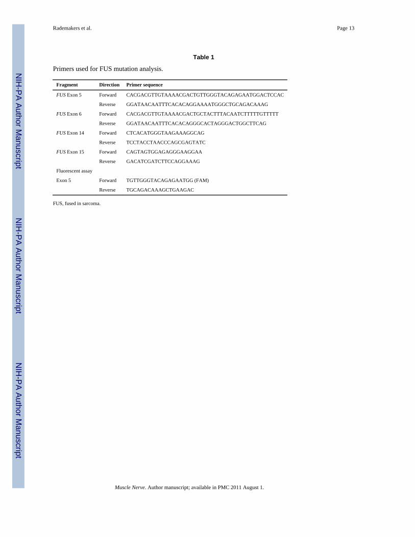
Molecular genetic analysisGenomic DNA was extracted from peripheral blood using standard procedures. Due to thelimited quantity and quality of DNA available in some of the samples, sequencing wasrestricted to those exons in which pathogenic FUS mutations had previously been reported.6-10 The FALS DNA samples were subsequently whole genome amplified in triplicateusing the REPLI-g kit (Qiagen, Valencia, CA, USA). For all FALS and SALS patients, PCRof FUS exons 5, 6, 14 and 15 was performed with Qiagen products using primers located inadjacent introns. To facilitate sequencing of exons 5 and 6, the M13 forward primersequence and the M13 reverse primer sequence were attached to the FUS primers. PCRprimers are listed in Table 1. PCR products were purified using the Ampure system(Bioscience Corporation, Beverly, MA) and sequenced using Big dye terminator V.3.1products. Sequencing products were purified using the CleanSEQ method (Agencourt) andanalyzed on an ABI 3730 DNA analyzer (Applied Biosystems, Foster City, CA, USA).Sequence analysis was performed using Sequencher software (Gene Codes, Ann Arbor, MI,USA). All identified mutations were confirmed by sequencing of the original gDNA sample.The screening of c.559G>A (p.G187S) and c.404G>A (p.S135N; rs617332970) wasperformed using custom Taqman SNP genotyping assays with the File Builder 3.1 software(Applied Biosystems) and analyzed on an ABI7900 genetic analyzer using SDS2.2.2software. To identify small insertions and deletions (indels) in the glycine-rich region in
Rademakers et al. Page 2
Muscle Nerve. Author manuscript; available in PMC 2011 August 1.
NIH
-PA Author Manuscript
NIH
-PA Author Manuscript
NIH
-PA Author Manuscript
exon 5, we PCR amplified exon 5 using one fluorescently labeled primer and performedfragment length analysis on an automated ABI3730 DNA-analyzer (Applied Biosystems)(Table 1). Allele identification and scoring was performed using GENESCAN andGENOTYPER software (Applied Biosystems).
Histology and immunohistochemistrySections of formalin-fixed, paraffin-embedded material were stained using hematoxylin andeosin (HE) alone or in combination with luxol fast blue (HE/LFB). Allimmunohistochemistry (IHC) was performed using the Ventana BenchMark® XTautomated staining system (Ventana, Tuscon, AZ) and developed with aminoethylcarbizole.The primary antibodies employed recognized FUS (Sigma-Aldrich, 1:25 - 1:200 with initialovernight incubation at room temperature, following microwave antigen retrieval), ubiquitin(DAKO, 1:500, following microwave antigen retrieval), TDP-43 (ProteinTech Group,1:1,000 following microwave antigen retrieval), glial fibrillary acidic protein (DAKO,1:4,000) and CD68 (DAKO, 1:800). FUS IHC was also performed on control sections ofspinal cord from 6 SALS and 3 FALS cases with known SOD-1 mutations.
RESULTSFUS gene analysis
We performed mutation analysis of FUS exons 5, 6, 14 and 15 in a population of 32 FALSprobands and 168 SALS patients. In the FALS patients, we identified one proband carryingthe known heterozygous missense mutation c.1561C>T (p.R521C) in exon 15 (Fig. 1A).3,4,6,8,10 Further analysis demonstrated the same mutation in a DNA sample from theproband's affected daughter, confirming segregation of the mutation with disease in thisfamily. In the SALS patients, we identified a novel missense mutation (c.559G>A, p.G187S)in FUS exon 6 in a single patient (Fig. 1A). This mutation was not present in our 678 controlsamples. A second missense mutation (c.404G>A; p.S135N) was found in another of ourSALS patients. Although this mutation was also absent in our control series, it is reported inthe public SNP database (dbSNP, http://www.ncbi.nlm.nih.gov/projects/SNP/), suggesting itmay be a rare benign variant. Finally, we identified four SALS patients who carry apreviously reported 6-bp deletion in the glycine-stretch encoded by the end of exon 5 (c.521_523+3delGAGGTG; p.G173_G174del).3 This same 6-bp deletion was also identified in3/678 samples in our control series.
Clinical DescriptionsFALS patients with FUS mutation—The proband was a recently retired 62-year oldwoman who presented with difficulty climbing stairs, requiring the use of the handrail. Shehad previously been in excellent health and on no medications. Her only complaint wassome aching of her leg muscles. She had fallen on several occasions and had difficultygetting up from a squatting position. A few months later, she developed left arm weakness,making it difficult for her to perform tasks that required her to lift her arm above her head.Examination showed her to have a waddling gait. She was not able to walk on her heels ortoes and had problems stepping up onto a low stool. There was considerable pelvic girdleweakness with symmetric, bilateral wasting of the quadriceps. Both shoulder girdles werealso weak, and there was mild weakness of neck flexors. Cranial nerve function andcognition were normal. Initial electromyography (EMG) studies were suggestive of amyopathy but later showed evidence of denervation. Nerve conduction studies found nomotor conduction slowing or conduction block, and sensory nerve action potentials were ofnormal amplitude. Creatine kinase was normal, and a muscle biopsy was suggestive of aneurogenic process. MRI of the brain was normal. Only later in the disease course did she
Rademakers et al. Page 3
Muscle Nerve. Author manuscript; available in PMC 2011 August 1.
NIH
-PA Author Manuscript
NIH
-PA Author Manuscript
NIH
-PA Author Manuscript
develop brisk tendon reflexes and diffuse fasciculation. She died approximately 3.5 yearsafter developing her initial symptoms.
Her daughter was a 39-year old school teacher who developed similar symptoms at almostthe same time as her mother. She had been a competitive volleyball player and becomeaware of difficulty playing because of problems moving her legs. She also had difficultyclimbing stairs and rising from a crouched position. Her weakness progressed, and threemonths later she required assistance walking. One month later, she noticed difficulty usingher left arm to dry her hair. She developed trouble swallowing, and her weight dropped from170 to 140 pounds. On examination she was alert and intelligent. She had lingualfasciculation, but otherwise cranial nerve function was normal. There was weakness of bothdeltoids against resistance, but the biceps and triceps were strong. There was no weakness orwasting in the hands. She had a waddling gait and was unable to walk on her heels or toes.She had to push with her arms to rise from a standard chair. The quadriceps muscles weresymmetrically wasted, but distal leg muscles were strong. Tendon reflexes were present butnot overactive in the arms and knees, and ankle jerks were depressed. The toes were down-going. Over the following six months the right upper limb tendon reflexes became brisk, butthe leg tendon reflexes remained difficult to obtain. EMG was neurogenic and showeddenervation in the upper and lower limbs with recruitment of complex motor unit potentials.Motor conduction studies were normal with no conduction block. MRI of the brain andspinal cord were normal. Serum creatine kinase was slightly elevated (211). A musclebiopsy was consistent with a neurogenic process, and there was no evidence of a glycogenstorage disease. Acid maltase assays were also normal. Like her mother, disease durationwas approximately 3.5 years
The family pedigree is provided in Figure 1B. The grandparents and parents of the probandhad no known neurological disease, however their age and cause of death are not known.Her four siblings were believed to be asymptomatic, but she had been estranged from themfor several years. The affected daughter was her only child, and she had two healthy youngchildren.
SALS patient with FUS mutation—This woman was born in Canada of Dutch parents.She presented at age 79 with speech difficulties and also complained of some musclecramping and emotional lability. Medical history included thyroid disease, hypertension andhypercholesterolemia. When examined by a neurologist, five months after onset ofsymptoms, she was found to have dysarthria, diffuse muscle wasting, fasciculations andexaggerated deep tendon reflexes. Neurophysiological testing, including electromyographyand motor and sensory nerve conduction studies, further supported the diagnosis of ALS.She progressed rapidly and died 14 months after onset. Autopsy was not performed. Therewas no family history of neurological disease.
NeuropathologyPost mortem examination was performed on the proband of the family with the c.1561C>T(p.R521C) mutation. The brain weighed 1250 grams and appeared grossly normal. Thespinal cord was of small calibre, and the motor nerve roots were atrophic. Microscopicexamination of the primary motor cortex failed to demonstrate any appreciable loss ofprimary motor neurons, and the only suggestion of chronic degeneration was mild reactivegliosis and microglial activation. Sections of spinal cord showed normal myelination of thecorticospinal tracts (Fig. 2A). There was severe loss of lower motor neurons (LMN) at alllevels, and the remaining cells were often shrunken. There were no Bunina bodies or hyalineinclusions visible with HE stain, however some LMN contained ill-defined basophilic
Rademakers et al. Page 4
Muscle Nerve. Author manuscript; available in PMC 2011 August 1.
NIH
-PA Author Manuscript
NIH
-PA Author Manuscript
NIH
-PA Author Manuscript

cytoplasmic masses. The motor nerve roots were atrophic with severe loss of myelinstaining.
IHC for FUS demonstrated numerous neuronal cytoplasmic inclusions (NCI) in theremaining LMN (Fig. 2B-D). These appeared as diffusely distributed fine granules, morefocal collections of coarse granules, collections of thick filaments and compact globularmasses that appeared to be a composite of coarse granules and filaments. At least someneurons with NCI showed retention of the normal physiological FUS staining of the nucleus(Fig. 2B). Ubiquitin staining of NCI was weak and inconsistent, and no abnormality wasseen with TDP-43 IHC. In addition to NCI, FUS IHC demonstrated numerous glialcytoplasmic inclusions (GCI) and thread-like structures in the ventral grey matter (Fig. 2E).Cells with small round nuclei, typical of oligodendrocytes, often contained small flame-shaped masses or single short processes, while some cells with oval nuclei were associatedwith more complex FUS-immunoreactive (ir) processes, consistent with astrocyticmorphology. While some of the smaller FUS-ir threads could have been of glial origin,larger ones were clearly neuritic. The dorsal grey matter of the spinal cord also containednumerous FUS-ir NCI, GCI and threads. GCI and thread pathology was moderate in thecorticospinal tracts and mild in other white matter funiculi. Motor nuclei of the brainstem,including the hypoglossal nucleus and trigeminal motor nucleus, also showed loss ofneurons and FUS-ir NCI, GCI and threads.
Despite not showing any evidence of chronic degeneration, several other subcortical nucleicontained mild-to-moderate FUS-ir neuronal and glial pathology, including the striatum,thalamus, substantia nigra, superior colliculus, red nucleus and reticular formation (Fig. 2F),and occasional GCI and threads were present in many brainstem white matter tracts. Therewas some anatomical specificity to the FUS-ir pathology, as many regions showed noinvolvement, including the occulomotor nucleus, pontine nuclei, inferior olive, cerebellum.No FUS-ir pathology was found in the primary motor cortex, other neocortical regions orhippocampus.
FUS IHC performed on spinal cord sections from SALS control cases and FALS patientswith SOD-1 mutations demonstrated the normal physiological staining pattern but no FUS-irpathology.
DiscussionThe FUS gene, located on chromosome 16, consists of 15 exons that encode a 526 aminoacid protein.14 The C-terminus region contains multiple domains involved in RNA-proteininteractions while the N-terminus functions in transcriptional activation.15 FUS is aubiquitously expressed protein14,16 that binds to RNA17,18 and DNA19 and is involved indiverse cellular processes including cell proliferation,20 DNA repair,21 transcriptionregulation, RNA splicing22 and the transport of RNA between intracellular compartments.18 In most cell types, FUS is present in both the nucleus and cytoplasm, however in neuronsthere is proportionally more in the nucleus and expression in glia is exclusively nuclear.16FUS may be involved in neuronal plasticity and maintenance of dendritic integrity bytransporting mRNA, including those encoding actin-related proteins, to dendritic spines forlocal translation in response to synaptic stimulation.23,24 In contrast, FUS deficient neuronsshow decreased spine arborization and morphology.23 Chromosomal translocation of the 5′portion of FUS results in several fusion oncogenes that are each associated with specifictypes of human cancer, including myxoid liposarcoma, Ewing's sarcoma and acute myeloidleukemia.25 FUS knock out mice show perinatal mortality.26 The finding that FUSmutations cause FALS was the first association between this protein and aneurodegenerative condition.
Rademakers et al. Page 5
Muscle Nerve. Author manuscript; available in PMC 2011 August 1.
NIH
-PA Author Manuscript
NIH
-PA Author Manuscript
NIH
-PA Author Manuscript
We identified a single FUS mutation in our cohort of 32 index cases with SOD1-negative,TARDBP-negative FALS. This represents a frequency of 3%, which is very similar to whathas been reported in all the previous studies with the exception of Belzil et al., who found amuch lower frequency of 1.25%.3,4,6-10 The specific mutation (c.1561C>T; p.R521C) isthe most common one reported to date. It has been identified in 11 families and in twosporadic cases.3,4,6,8,10 As in several of the previous studies, the mutation segregated withdisease in our family.
We also identified a novel FUS mutation in one of our ALS cases in which there was nofamily history. However, the late age at onset of this patient, combined with the lack ofinformation regarding the ages at which her parents died, means we cannot be certain thatthis case was truly sporadic. This c.559G>A (p.G187S) mutation is located in exon 6 thatencodes a portion of the glycine-rich region of the protein. Due to the absence of otheraffected family members, segregation of the mutation with disease could not be studied and,unfortunately, we did not have autopsy confirmation that this case had FUS pathology. Webelieve this mutation is likely pathogenic, since it was not identified in 678 controls (1356chromosomes), affects a highly conserved element in a functionally crucial region of theprotein (Fig. 1C) and involves an exon (6) where other mutations have been reported inFALS patients.3,8,10 In addition, we identified four SALS patients who carry the known c.521_523+3delGAGGTG 6-bp deletion in exon 5.3,8 This mutation is predicted to result inthe deletion of two glycine residues (p.G173_G174del) from a glycine stretch which islocated in a highly conserved region of the FUS protein; however, the number of glycineresidues in this region is not well conserved (Fig. 1D). Although this deletion was firstreported as a pathogenic mutation in a FALS patient, segregation of this mutation withdisease was not performed.3 In our study, we identified this same deletion in 3 of 678samples in our control series. These findings, together with recent reports of deletions inglycine stretches encoded by FUS exons 5 and 6, in both ALS patients and controls, suggestthat c.521_523+3delGAGGTG does not represent a pathogenic mutation.6,8
Three previous studies have evaluated FUS mutations in SALS patients.3,6,8 Kwiatkowskiet al. sequenced all 15 exons in 293 SALS cases and failed to identify any genetic variantsof significance.3 Belzil et al. screened all exons in 120 SALS cases and only exon 15 in anadditional 285 cases.6 They identified one novel mutation in exon 3 and two mutations inexon 15 that have also been reported in several FALS cases. Most recently, Corrado et al.screened exons 5, 6, 14 and 15 in 964 SALS cases and extended the analysis to the entirecoding sequence in 293 of these.8 They identified 6 different missense mutations in 7 SALScases (0.7%), including the common c.1561C>T (p.R521C) mutation in one case and 5novel mutations in exons 14 (N=1) and 6 (N=4). The finding of a single case with amutation of probable pathogenicity in our SALS cohort represents a FUS mutationfrequency in SALS of 0.6% and is consistent with the low frequency (0 – 0.7 %) identifiedin the previous studies.3,6,8 Although the selective genetic screening in our study and thoseof Belzil et al. and Corrado et al. could have resulted in some mutations being missed,overall these results suggest FUS mutations are probably an uncommon cause of SALS.
Most of the previous reports have provided minimal description of the clinical phenotypeassociated with FUS mutations, indicating only that it is usually classical ALS.3,4,6 One ofthe two Italian kindreds described by Chio et al., had a notably young onset and rapidcourse.7 The report by Ticozzi et al. provided greater detail of the clinical features of theirthree probands, one of whom also developed frontotemporal dementia (FTD) while the othertwo had an unusual presentation with symmetric proximal and axial weakness.10 Corrado etal. also described one FALS and one SALS patient with predominant symmetric proximaland axial weakness.8 Our FUS mutation family had a number of unusual clinical features,including synchronous onset in the proband and her daughter with a 23-year age difference.
Rademakers et al. Page 6
Muscle Nerve. Author manuscript; available in PMC 2011 August 1.
NIH
-PA Author Manuscript
NIH
-PA Author Manuscript
NIH
-PA Author Manuscript
The fact they were living together at the time raised the suspicion of some environmentaltrigger for their disease. Their course was remarkably similar and atypical, with initialinvolvement of pelvic and shoulder girdles, late involvement of distal muscles and minimalevidence of upper motor neuron involvement. Interestingly, the c.1561C>T (p.R521C)mutation that was found in our family is the same as in both families reported by Ticozzi etal. and both patients reported by Corrado et al. who also presented with symmetric proximalweakness.8,10 This raises the possibility that certain FUS mutations may be associated witha specific clinical phenotype. Although several other families have been reported with thismutation, they are not described in sufficient detail to allow confirmation of this association.3,4,6
Prior assessment of the neuropathology associated with FUS mutations has been extremelylimited. The two initial reports described the autopsy findings in a small number of affectedindividuals as including loss of LMN and inconsistent myelin pallor of the corticospinaltracts.3,4 The one case reported by Kwiatkoski et al. was felt to show increased cytoplasmicFUS immunoreactivity in neurons,3 while Vance et al. described the presence of FUS-irdystrophic neurites and globular NCI in LMN, in the absence of TDP-43 pathology.4 Noneof the more recent publications has included pathological assessment.6-10 In this study, weprovide the first detailed description of the neuropathology in an ALS patient with FUSmutation. The much greater involvement of lower compared with upper motor neuronscorrelated with the clinical phenotype in our patient. We confirmed the presence of FUS-ir,TDP-43-negative NCI in LMN and describe their variable morphology. Importantly, wefound at least some preservation of normal physiological nuclear FUS immunoreactivity inneurons containing NCI. This finding is consistent with previous in vitro studies showingthat although FUS mutations result in an increase in cytoplasmic localization of the protein,there is still some retention of FUS in the nucleus.3,4 This is in striking contrast to thecomplete nuclear-to-cytoplasmic redistribution of TDP-43 that occurs in sporadic ALS andis of potential functional significance.27 We also found significant FUS pathology in severalanatomical sites outside the pyramidal motor system; the significance of these findings mustawait future clinico-pathological correlative studies. Our finding of FUS-ir inclusions inglial cells is similar to what is found with TDP-43 IHC in sporadic ALS and supports theconcept that the pathogenesis of ALS may involve glia as well as neurons. Although thefinding of normal FUS IHC in our ALS control cases suggests that FUS pathology may be aspecific feature of ALS with FUS mutations, this result requires confirmation in a largerseries of cases. Finally, the absence of pathological TDP-43 in cases with FUS mutations issimilar to what is found with SOD-1 mutations and suggests that the pathogenesis of somegenetically based forms of ALS may be different from the majority of SALS.27
In summary, we identified two FUS mutations in our cohort, representing 3% of non-SOD1,non-TARDBP FALS and 0.6% of SALS cases. Although the relatively small size of ourcohort and the fact we did not screen the entire gene in all of our samples means that thesefrequencies cannot be considered definite; they are very similar to those reported in previousstudies.6-10 The mutation in our SALS patient has not been reported previously. Theclinical features of our affected family show that the phenotype associated with FUSmutations extends beyond classical ALS and, together with previous reports, suggest thatthere may be specific clinico-genetic correlations. Our neuropathological findingsdemonstrate that ALS with FUS mutations is characterized by abundant and anatomicallywidespread FUS pathology in the absence of abnormalities of TDP-43 and with retention ofat least some of the normal cellular distribution of FUS protein. Finally, the report of a FUSmutation in one patient with a combination of ALS and FTD,10 combined with our recentstudies demonstrating that several subtypes of sporadic FTD are characterized by FUSpathology,27-29 suggest that FUS and TDP-43 may represent dichotomous biochemicalpathways that result in a similar spectrum of neurodegenerative disease.
Rademakers et al. Page 7
Muscle Nerve. Author manuscript; available in PMC 2011 August 1.
NIH
-PA Author Manuscript
NIH
-PA Author Manuscript
NIH
-PA Author Manuscript
AcknowledgmentsWe thank Margaret Luk, Matt Baker, NiCole Finch, Nicola Rutherford and Richard Crook for their excellenttechnical assistance. This work was supported by grants from Canadian Institutes of Health Research (grant number74580, IM), Pacific Alzheimer Research Foundation (IM, RR), the ALS association (RR) and the NIH/NIA(AG26251-03A1, RR).
Abbreviations
ALS amyotrophic lateral sclerosis
ANG angiogenin
DNA deoxyribonucleic acid
EMG electromyography
FALS familial amyotrophic lateral sclerosis
FUS fused in sarcoma
GCI glial cytoplasmic inclusion
HE hematoxylin and eosin
IHC immunohistochemistry
ir immunoreactive
LFB luxol fast blue
LMN lower motor neuron
MRI magnetic resonance imaging
NCI neuronal cytoplasmic inclusion
PCR polymerase chain reaction
RNA ribonucleic acid
ALS sporadic amyotrophic lateral sclerosis
OD1 copper/zinc superoxide dismutase 1
TARDBP transactive response DNA binding protein
TDP-43 transactive response DNA binding protein with Mr 43 kD
TLS translocated in liposarcoma
REFERENCES1. Gros-Louis F, Gaspar C, Rouleau GA. Genetics of familial and sporadic amyotrophic lateral
sclerosis. Biochim Biophys Acta. 2006; 1762:956–972. [PubMed: 16503123]2. Valdmanis PN, Daoud H, Dion PA, Rouleau GA. Recent advances in the genetics of amyotrophic
lateral sclerosis. Curr Neurol Neurosci Rep. 2009; 9:198–205. [PubMed: 19348708]3. Kwiatkowski TJ, Bosco DA, LeClerc AL, Tamrazian E, Vanderburg CR, Russ C, et al. Mutations in
the FUS/TLS gene on chromosome 16 cause familial amyotrophic lateral sclerosis. Science. 2009;323:1205–1208. [PubMed: 19251627]
4. Vance C, Rogelj B, Hortobagyi T, de Vos KJ, Nishimura AL, Sreedharan J, et al. Mutations in FUS,an RNA processing protein, cause familial amyotrophic lateral sclerosis type 6. Science. 2009;323:1208–1211. [PubMed: 19251628]
5. Lagier-Tourenne C, Cleveland DW. Rethinking ALS: the FUS about TDP-43. Cell. 2009;136:1001–1004. [PubMed: 19303844]
Rademakers et al. Page 8
Muscle Nerve. Author manuscript; available in PMC 2011 August 1.
NIH
-PA Author Manuscript
NIH
-PA Author Manuscript
NIH
-PA Author Manuscript
6. Belzil VV, Valdmanis PN, Dion PA, Daoud H, Kabashi E, Noreau A, et al. Mutations in FUS causeFALS and SALS in French and French Canadian populations. Neurology. 2009; 73:1176–1179.[PubMed: 19741216]
7. Chio A, Restagno G, Brunetti M, Ossola M, Calvo A, Mora G, et al. Two Italian kindreds withfamilial amyotrophic lateral sclerosis due to FUS mutation. Neurobiol Aging. 2009; 30:1272–1275.[PubMed: 19450904]
8. Corrado L, Del Bo R, Castellotti B, Ratti A, Cereda C, Penco S, et al. Mutations in FUS gene insporadic amyotrophic lateral sclerosis. J Med Genet. to be published.
9. Damme PV, Goris A, Race V, Hersmus N, Dubois B, Bosch LV, et al. The occurrence of mutationsin FUS in a Belgian cohort of patients with familial ALS. Eur J Neurol. to be published.
10. Ticozzi N, Silani V, LeClerc AL, Keagle P, Gellera C, Ratti A, et al. Analysis of FUS genemutation in familial amyotrophic lateral sclerosis within an Italian cohort. Neurology. 2009;73:1180–1185. [PubMed: 19741215]
11. Brooks BR, Miller RG, Swash M, Munsat TL. World Federation of Neurology Research Group onMotor Neuron Diseases. El Escorial revisited: revised criteria for the diagnosis of amyotrophiclateral sclerosis. Amytroph Latera Scler Other Motor Neuron Disord. 2000; 1:293–299.
12. Rutherford NR, Zhang YJ, Baker MC, Gass JM, Finch N, Xu F, et al. Novel Mutations in TARDBP(TDP-43) in Patients with Familial Amyotrophic Lateral Sclerosis. PLoS Genet Sep. 2008;194:e1000193.
13. Edgar RC. MUSCLE: multiple sequence alignment with high accuracy and high throughput. NucAcid Res. 2004; 32:1792–1797.
14. Aman P, Panagopoulos I, Lassen C, Fioretos T, Mencinger M, Toresson H, et al. Expressionpatterns of the human sarcoma-associated genes FUS and EWS and the genomic structure of FUS.Genomics. 1996; 37:1–8. [PubMed: 8921363]
15. Prasad DD, Ouchida M, Lee L, Rao VN, Reddy ES. TLS/FUS fusion domain of TLS/FUS-ergchimeric protein resulting from the t(16;21) chromosomal translocation in human myeloidleukemia functions as a transcriptional activation domain. Oncogene. 1994; 9:3717–3729.[PubMed: 7970732]
16. Andersson MK, Stahlberg A, Arvidsson Y, Olofsson A, Semb H, Stenman G, et al. Themultifunctional FUS, EWS, and TAF15 proto-oncoproteins show cell type-specific expressionpatterns and involvement in cell spreading and stress response. BMC Cell Biol. 2008; 9:37.[PubMed: 18620564]
17. Crozat A, Aman P, Mandahl N, Ron D. Fusion of CHOP to a novel RNA-binding protein in humanmyxoid liposarcoma. Nature. 1993; 363:640–644. [PubMed: 8510758]
18. Zinszner H, Sok J, Immanuel D, Yin Y, Ron D. TLD (FUS) binds RNA in vivo and engages innucleo-cytoplasmic shuttling. J Cell Sci. 1997; 110:1741–1750. [PubMed: 9264461]
19. Perrotti D, Bonatti S, Trotta R, Martinez R, Skorski T, Salomoni P, et al. TLS/FUS, a pro-oncogene involved in multiple chromosomal translocations, is a novel regulator of BCR/ABL-mediated leukemogenesis. EMBO J. 1998; 17:4442–4455. [PubMed: 9687511]
20. Bertrand P, Akhmedov AT, Delacote F, Durrbach A, Lopez BS. Human POMp75 is identified asthe pro-oncoprotein TLS/FUS: both POMp75 and POMp100 DNA homologous pairing activitiesare associated with cell proliferation. Oncogene. 1999; 18:4515–4521. [PubMed: 10442642]
21. Baechtold H, Kuroda M, Sok J, Ron D, Lopez BS, Akhmedov AT. Human 75-kDa DNA-pairingprotein is identical to the pro-oncoprotein TLD/FUS and is able to promote D-loop formation. JBiol Chem. 1999; 274:34337–34342. [PubMed: 10567410]
22. Yang L, Embree LJ, Tsai S, Hickstein DD. Oncoprotein TLS interacts with serine-arginineproteins involved in RNA splicing. J Biol Chem. 1998; 273:27761–27764. [PubMed: 9774382]
23. Fujii R, Okabe S, Urushido T, Inoue K, Yoshimura A, Tachibana T, et al. The RNA bindingprotein TLS is translocated to dendritic spines by mGluR5 activation and regulates spinemorphology. Curr Biol. 2005a; 15:587–593. [PubMed: 15797031]
24. Fujii R, Takumi T. TLS facilitates transport of mRNA encoding an actin-stabilizing protein todendritic spines. J Cell Sci. 2005b; 118:5755–5765. [PubMed: 16317045]
25. Law WJ, Cann KL, Hicks GG. TLS, EWS, and TAF15: a model for transcriptional integration ofgene expression. Brief Funct Genomic Proteomic. 2006; 5:8–14. [PubMed: 16769671]
Rademakers et al. Page 9
Muscle Nerve. Author manuscript; available in PMC 2011 August 1.
NIH
-PA Author Manuscript
NIH
-PA Author Manuscript
NIH
-PA Author Manuscript
26. Hicks GG, Singh N, Nashabi A, Mai S, Bozek G, Klewes L, et al. FUS deficiency in mice resultsin defective B-lymphocyte development and activation, high levels of chromosomal instability andperinatal death. Nat Genet. 2000; 24:175–179. [PubMed: 10655065]
27. Mackenzie IRA, Bigio EH, Ince PG, Geser F, Neumann M, Cairns NJ, et al. Pathological TDP-43distinguishes sporadic ALS from ALS with SOD-1 mutations. Ann Neurol. 2007; 61:427–434.[PubMed: 17469116]
28. Munoz DG, Neumann M, Kusaka H, Yokota O, Ishihara K, Terada S, et al. FUS pathology inBasophilic Inclusion Body disease (BIBD). Acta Neuropathol. 2009; 118:617–627. [PubMed:19830439]
29. Neumann M, Roeber S, Kretzschmar HA, Rademakers R, Baker M, Mackenzie IRA. AbundantFUS-immunoreactive pathology in neuronal intermediate filament inclusion disease. ActaNeuropathol. 2009; 118:605–616. [PubMed: 19669651]
30. Neumann M, Rademakers R, Roeber R, Baker M, Kretzschmar HA, Mackenzie IRA.Frontotemporal lobar degeneration with FUS pathology. Brain. 2009; 132:2922–2931. [PubMed:19674978]
Rademakers et al. Page 10
Muscle Nerve. Author manuscript; available in PMC 2011 August 1.
NIH
-PA Author Manuscript
NIH
-PA Author Manuscript
NIH
-PA Author Manuscript
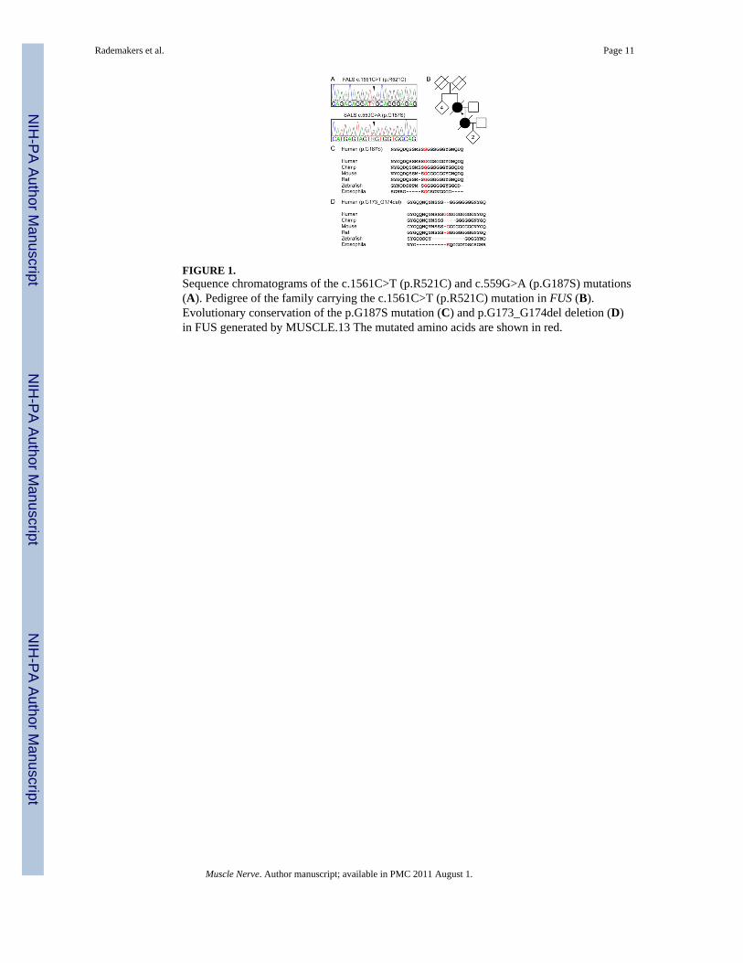
FIGURE 1.Sequence chromatograms of the c.1561C>T (p.R521C) and c.559G>A (p.G187S) mutations(A). Pedigree of the family carrying the c.1561C>T (p.R521C) mutation in FUS (B).Evolutionary conservation of the p.G187S mutation (C) and p.G173_G174del deletion (D)in FUS generated by MUSCLE.13 The mutated amino acids are shown in red.
Rademakers et al. Page 11
Muscle Nerve. Author manuscript; available in PMC 2011 August 1.
NIH
-PA Author Manuscript
NIH
-PA Author Manuscript
NIH
-PA Author Manuscript
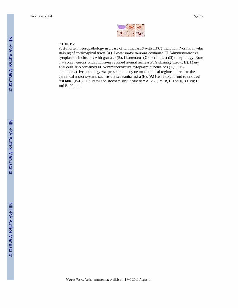
FIGURE 2.Post-mortem neuropathology in a case of familial ALS with a FUS mutation. Normal myelinstaining of corticospinal tracts (A). Lower motor neurons contained FUS-immunoreactivecytoplasmic inclusions with granular (B), filamentous (C) or compact (D) morphology. Notethat some neurons with inclusions retained normal nuclear FUS staining (arrow, B). Manyglial cells also contained FUS-immunoreactive cytoplasmic inclusions (E). FUS-immunoreactive pathology was present in many neuroanatomical regions other than thepyramidal motor system, such as the substantia nigra (F). (A) Hematoxylin and eosin/luxolfast blue, (B-F) FUS immunohistochemistry. Scale bar: A, 250 μm; B, C and F, 30 μm; Dand E, 20 μm.
Rademakers et al. Page 12
Muscle Nerve. Author manuscript; available in PMC 2011 August 1.
NIH
-PA Author Manuscript
NIH
-PA Author Manuscript
NIH
-PA Author Manuscript
NIH
-PA Author Manuscript
NIH
-PA Author Manuscript
NIH
-PA Author Manuscript
Rademakers et al. Page 13
Table 1
Primers used for FUS mutation analysis.
Fragment Direction Primer sequence
FUS Exon 5 Forward CACGACGTTGTAAAACGACTGTTGGGTACAGAGAATGGACTCCAC
Reverse GGATAACAATTTCACACAGGAAAATGGGCTGCAGACAAAG
FUS Exon 6 Forward CACGACGTTGTAAAACGACTGCTACTTTACAATCTTTTTGTTTTT
Reverse GGATAACAATTTCACACAGGGCACTAGGGACTGGCTTCAG
FUS Exon 14 Forward CTCACATGGGTAAGAAAGGCAG
Reverse TCCTACCTAACCCAGCGAGTATC
FUS Exon 15 Forward CAGTAGTGGAGAGGGAAGGAA
Reverse GACATCGATCTTCCAGGAAAG
Fluorescent assay
Exon 5 Forward TGTTGGGTACAGAGAATGG (FAM)
Reverse TGCAGACAAAGCTGAAGAC
FUS, fused in sarcoma.
Muscle Nerve. Author manuscript; available in PMC 2011 August 1.
Copyright © 2022 FDOKUMEN