Regulation of NAMPT in Human Gingival Fibroblasts and Biopsies

A
(tqriapcio©
KA
1
(
epeehPi
0d
Neurobiology of Aging 28 (2007) 863–876
Increased susceptibility to amyloid toxicity in familialAlzheimer’s fibroblasts
Cristina Cecchi a,∗, Claudia Fiorillo a, Serena Baglioni a, Anna Pensalfini a, Silvia Bagnoli b,Benedetta Nacmias b, Sandro Sorbi b, Daniele Nosi c, Annalisa Relini d, Gianfranco Liguri a
a Department of Biochemical Sciences, University of Florence, viale Morgagni 50, 50134 Florence, Italyb Department of Neurological and Psychiatric Sciences, University of Florence, viale Pieraccini 6, 50139 Florence, Italy
c Department of Anatomy, Histology and Forensic Medicine, University of Florence, viale Morgagni 85, 50134 Florence, Italyd Department of Physics, University of Genoa, via Dodecaneso 33, I-16146 Genoa, Italy
Received 8 June 2005; received in revised form 14 April 2006; accepted 3 May 2006Available online 15 June 2006
bstract
Much experimental evidence suggests that an imbalance in cellular redox status is a major factor in the pathogenesis of Alzheimer’s diseaseAD). Our previous data showed a marked increase in membrane lipoperoxidation in primary fibroblasts from familial AD (FAD) patients. Inhe present study, we demonstrate that when oligomeric structures of A� 1-40 and A� 1-42 are added to the culture media, they accumulateuicker near the plasma membrane, and are internalized faster and mostly in APPV717I fibroblasts than in age-matched healthy cells; thisesults in an earlier and sharper increase in the production of reactive oxygen species (ROS). Higher ROS production leads in turn to anncrease in membrane oxidative-injury and significant impairment of cellular antioxidant capacity, giving rise to apoptotic cascade activationnd finally to a necrotic outcome. In contrast, healthy fibroblasts appear more resistant to amyloid oxidative-attack, possibly as a result of theirlasma membrane integrity and powerful antioxidant capacity. Our data are consistent with increasing evidence that prefibrillar aggregates,ompared to mature fibrils, are likely the more toxic species of the peptides. These findings provide compelling evidence that cells bearing
ncreased membrane lipoperoxidation are more susceptible to aggregate toxicity as a result of their reduced ability to counteract amyloidligomeric attack.2006 Elsevier Inc. All rights reserved.
-peptid
t
eywords: Familial Alzheimer’s disease; APP and PS-1 genes; Amyloid �
myloid aggregate toxicity
. Introduction
A neuropathological characteristic of Alzheimer’s diseaseAD) is the extracellular accumulation of amyloid beta pep-
Abbreviations: A�, amyloid �-peptide; ABTS, 2,2′-azino-di-3-thylbenzthiazoline sulphonate; AD, Alzheimer’s disease; APP, amyloidrecursor protein; CM-H2,DCFDA, 2′,7′-dichlorodihydrofluorescein diac-tate; ECL, enhanced chemiluminescence; FAD, familial Alzheimer’s dis-ase; GSH, glutathione; 4-HNE, 4-hydroxy-2-nonenal; MDA, malondialde-yde; PBS, phosphate buffer saline; PMSF, phenylmethylsulphonylfluoride;S, presenilin; ROS, reactive oxygen species; TAC, total antioxidant capac-
ty; ThT, thioflavine; TM-AFM, tapping mode atomic force microscopy∗ Corresponding author. Tel.: +39 055 4598320; fax: +39 055 4598905.
E-mail address: [email protected] (C. Cecchi).
bplb(tcdtiaiA
197-4580/$ – see front matter © 2006 Elsevier Inc. All rights reserved.oi:10.1016/j.neurobiolaging.2006.05.014
e; Fibroblasts; Oxidative stress; Antioxidant capacity; Lipid peroxidation;
ide (A�) in neuritic plaques, whose appearance can precedey decades the onset of symptoms [49]. A� deposits in ADatients are almost exclusively composed of the highly amy-oidogenic 1-42 form (A� 1-42), which is normally producedy cells in much lower quantities than the 40 residues formA� 1-40). A� 1-42 is more prone to aggregation in vitrohan A� 1-40, and its cytotoxicity is considered to be the mainause of neuronal impairment in AD. In familial Alzheimer’sisease (FAD) patients, an early increase has been reported inhe production of A� 1-42 arising from intracellular process-
ng of amyloid precursor protein (APP) [63]. Interestingly,ll the observed mutations associated with FAD cases leadndependently to an increased production of A� [18,58].utosomal dominant forms of FAD are often determined by
8 logy of
s2iwftfiss
wsuhblassetInemsaltsdblclrianetiswf[obtsmtsnp
m[t(obstitMs(fPp
iab[egcvoAtaumrowcmsedhati(
2
2
e
64 C. Cecchi et al. / Neurobio
pecific mutations in the APP gene located on chromosome1, or in the genes mapped on chromosomes 14 and 1, encod-ng presenilin-1 (PS-1) and presenilin-2 (PS-2) respectively,hich are components of a large protein complex responsible
or �-secretase activity [20]. Although FAD is a minority (lesshan 5%) of AD cases, these data point to a pathogenetic roleor the metabolism of APP and for the deposition of A�. Thisn turn suggests a role for A� in the non-genetic AD forms,ince the pathological endpoint and hallmarks of familial andporadic AD are very similar.
Recent remarkable findings from research teams world-ide, of protein aggregation as a common key feature in
everal neurodegenerative diseases, constitute progress innderstanding the basic mechanism of the so-called “amyloidypothesis” of AD [29]. The amyloid hypothesis is supportedy in vivo and in vitro evidence concerning several amy-oidogenic proteins that indicates a direct cytotoxic effect ofmyloid aggregates. Abundant data show that the prefibrillarpecies of A� peptides and of other amyloidogenic proteinsuch as alpha-synuclein or transthyretin, which are formedarly in the process of fibrillogenesis, are neurotoxic, whereashe mature fibrils are much less toxic [11,35,38,42,61,69].t has recently been found that prefibrillar aggregates, butot mature fibrils from proteins not involved in amyloid dis-ases can impair cell viability when added to cultured celledia [7,14]. It follows that the cross-� fold is not only the
tructural feature common to all amyloid aggregates, but islso the structural determinant of cytotoxicity of any amy-oid aggregate. There is evidence that protein conversion intooxic aggregates is enhanced by cellular membranes and thatelf-assembly on the bilayer surface is critical for membraneisruption [4]. A leading theory concerning the molecularasis of amyloid toxicity is that a sub-population of prefibril-ar aggregates, assembled in pore-like fashion, interacts withell membranes and provides them with non-specific poreseading to free Ca2+ homeostasis imbalance [61,26,40]. Theise in free calcium is usually coupled to a marked increasen reactive oxygen species (ROS). This is a result of thectivation of oxidative metabolism following the increasedeed for ATP required by the calcium pumps to clear thexcess free Ca2+ [53]. ROS increase may in turn reinforcehe increase in free Ca2+, of which extrusion from the cytosols inhibited by calcium pump oxidation. Other studies havehown that the role of methionine 35 (Met-35), in conjunctionith the secondary structure of the A� 1-42 itself, is critical
or the oxidative and neurotoxic properties of the peptide10]. Mutagenesis studies on the C-terminal helical regionf the peptide suggest that presence of A� 1-42 in the lipidilayer is necessary for induction of Met-35 lipid peroxida-ion and subsequent neurotoxicity. Several lines of evidenceupport the possibility that interactions of A� peptide causesitochondrial damage upon translocation of protofibrils to
he membranes [31]. Mitochondria are the major subcellularource of superoxide anion radical, which can interact withitric oxide (NO) to form peroxynitrite [12,71]. ROS anderoxynitrite accumulation results in damage to major macro-
wDwM
Aging 28 (2007) 863–876
olecules in cells, including lipids, proteins and nucleic acids47,60,62,68]. The lipoperoxidation process could influencehe pathogenesis of AD [55,59]. Indeed, 4-hydroxynonenal4-HNE), which is one of the most reactive end productsf lipoperoxidation, appears to induce neuronal death uponinding to proteins by altering important transporter proteins,uch as the ATPases involved in calcium homeostasis andhe glutamate transporter EAAT2 [10,52]. The healthy brains protected from oxidative injury by antioxidant defenceshat include antioxidant enzymes and free radical scavengers.
any recent investigations have strengthened the hypothe-is that an impairment in cellular total antioxidant capacityTAC) plays a central role in AD [47,46]. Recently, we haveound that lymphoblasts and fibroblasts carrying APP andS-1 gene mutations have a significant TAC impairment com-ared to healthy controls [15].
The data reported in the last few years have considerablymproved our knowledge of the molecular basis of proteinggregation into amyloid assemblies, and of the relationetween aggregate structure and toxicity to exposed cells23]. However, one of the intriguing issues that have not beenlucidated is the pathological role of each specific A� aggre-ates in AD brain areas. Studies on autopsied brain tissueannot reveal the early biochemical anomalies induced byarious forms of A� aggregates, so we carried out a studyf cultured skin fibroblasts from FAD patients bearing eitherPP or PS-1 gene mutations, and age-matched healthy con-
rol cells whose culture medium was supplemented by a fixedmount of amyloid aggregates at differing degrees of mat-ration. The goal of the present research is to compare theolecular basis of cell damage induced by prefibrillar and fib-
illar forms of A� 1-40 and A� 1-42 aggregates, by checkingxidative stress markers and either apoptotic or necrotic path-ay activation in FAD fibroblasts in comparison to healthy
ells. This is a new approach to the identification of earlyodifications in living cells having a genetic drawback in tis-
ues where AD lesions occur. Our results agree with previousvidence from neuronal cells under various experimental con-itions. In particular, we found that the cell lines investigatedave very different susceptibility to toxic amyloid aggregates,nd that the difference in cell viability is related to their abilityo: (i) affect the rate of accumulation of amyloid assembliesnto the plasma membrane; (ii) prevent oligomeric inclusion;iii) buffer ROS production.
. Materials and methods
.1. Materials
All reagents were of analytical grade or the high-st purity available. Unless otherwise stated, chemicals
ere purchased from Sigma–Aldrich (Milan, Italy). 2′,7′-ichlorodihydrofluorescein diacetate (CM-H2, DCFDA) andheat germ agglutinin-conjugated fluorescein were fromolecular Probes (Eugene, OR). PVDF Immobilio-P Trans-
logy of
f(s3nSOft(ptmtAbrMaAaoa
2
ct4po2Lt(lflonp
2
dmaSimswqa
2
lttpPaaacayaogccoSsecwilfbfap
2m
faP1RCp5o[v
2
C. Cecchi et al. / Neurobio
er Membrane was obtained from Millipore CorporationBedford, MA); Hybond N+ nylon membrane (Amer-ham, Life Science, England). The rabbit anti caspase-/CPP32 polyclonal antibody was from Biosource Inter-ational (Camarillo, CA); secondary antibodies were fromanta Cruz Biotechnology (Santa Cruz, CA). Quantityne program for the image analysis and densitometry was
rom Biorad (Hercules, CA). A� 1-40 and A� 1-42 pep-ides, as trifluoroacetate salts, were purchased from BachemBubendorf, Switzerland). Lyophilized A� 1-40 and A� 1-42eptides were stored as powder at −20 ◦C until reconstitu-ion in phosphate-buffered saline (PBS) without calcium and
agnesium, pH 7.2 at a concentration of 230 �M. Reconsti-uted peptides were stored as aliquots at −20 ◦C until used.� 1-40 and A� 1-42 prefibrillar aggregates were obtainedy diluting aliquots of 230 �M stock solutions, thawed atoom temperature, in serum free Dulbecco Modified Eagle’s
edium (DMEM) and bath sonicated for 5 min to break upny cluster formation before adding to fibroblasts. Otherwise,� 1-40 fibrillar aggregates were prepared by incubating
liquots of 230 �M A� 1-40 stock solutions in PBS with-ut calcium and magnesium, pH 7.2 at 37 ◦C for 48 h, beforeddition to cultures.
.2. ThT assay
A� 1-40 and A� 1-42 peptides were incubated at a con-entration of 230 �M in PBS, pH 7.2 at 37 ◦C. At regularime-intervals 10 �l aliquots of each sample were added to90 �l of a solution containing 25 �M ThT, 25 mM phos-hate buffer, pH 6.0. The steady-state fluorescence valuesf the resulting samples were measured at 25 ◦C using amm × 10 mm path length quartz cuvette and a Perkin-ElmerS 55 spectrofluorimeter (Wellesley, MA) equipped with a
hermostated cell holder attached to a Haake F8 water bathKarlsruhe, Germany). The excitation and emission wave-engths were 440 and 485 nm, respectively. All measureduorescence values are given after subtracting the ThT flu-rescence intensity measured in the absence of protein andormalized so that the final fluorescence intensity at the end-oint of the kinetic trace was 100%.
.3. Tapping mode atomic force microscopy (TM-AFM)
20 �l aliquots of A� 1-40 and A� 1-42 peptides wereeposited on freshly cleaved mica substrates and dried underild vacuum. TM-AFM images were acquired in air usingDimension 3000 microscope (Digital Instruments, Veeco,anta Barbara, CA) equipped with a ‘G’ scanning head (max-
mum scan size 100 �m) and a Multimode scanning probeicroscope equipped with a “E” scanning head (maximum
can size 10 �m). Single beam uncoated silicon cantileversere used (type OMCL-AC, Olympus, Japan). The drive fre-uency was around 300 kHz; the scan rate was between 0.3nd 0.8 Hz.
w(ct
Aging 28 (2007) 863–876 865
.4. Cell culture and exposure to Aβ aggregates
In the present study we investigated twelve fibroblast cellines. Fibroblasts were obtained from four patients belongingo two Italian families bearing the APP Val717Ile muta-ion (mean ± S.D., age = 55.2 ± 4.5 years) and from fouratients belonging to two other Italian families bearing theS-1 Met146Leu and Leu392Val mutations (mean ± S.D.,ge = 47.52 ± 9.9 years), respectively. The patient clinicalssessment was done according to published guidelines [64]nd the AD diagnosis fulfilled the Diagnostic and Statisti-al Manual of Mental Disorders criteria (DSM-IV) [1]. Fourge-matched healthy subjects (mean ± S.D., age = 52.3 ± 8.2ears) were also analyzed. The local ethical committeepproved the protocol of the study and written consent wasbtained from all subjects or, where appropriate, their care-ivers. All control subjects were tested and none of themarried APP or PS-1 mutations. These subjects were alsoarefully assessed with a rigorous diagnostic evaluation inrder to exclude diagnosis of other neurological disorders.kin biopsies of 3 mm punch were obtained from the volaride of the upper arm of the FAD patients and controls. Twoxplants were performed from each biopsy and plated in 25-m2 flasks. The cells were grown in DMEM, supplementedith 10% foetal bovine serum, and harvested at confluence
n T-25 flasks, 7 days after previous subculture. All fibroblastines were subjected to an equal number of passages (rangingrom 10 to 15) and analyzed in three different experimentsefore confluence. Cultured fibroblasts were exposed to dif-ering concentrations of A� 1-40 and A� 1-42 prefibrillarggregates (PF) and of A� 1-40 fibrillar aggregates (F), pre-ared as above described.
.5. Time-course of Aβ aggregate binding to cell surfaceembrane
3.5 × 10³ well−1 healthy and FAD fibroblasts were treatedor differing times (0, 10, 20, 30, 60 min) with 1.0 �M A�ggregates in a 96-well plate and then washed twice withBS. The residual aggregate-cell complex was stained with00 �l of 1.0 �M Congo Red in PBS for 20 min. The Congoed content was measured photometrically at 490 nm (freeongo Red) and 550 nm (bound Congo Red) with an ELISAlate reader. Under these conditions, the optical density at50 nm of the A� aggregate-Congo Red complex is a measuref the amount of aggregates adsorbed to the cell membrane22]. Congo Red values are reported as percentage increasesersus respective untreated fibroblasts (assumed as 100%).
.6. Internalization of amyloid aggregates
The cells were plated on glass coverslips and incubated
ith the prefibrillar aggregates of A� 1-40 and A� 1-42PF). Then the cells were counterstained with fluorescein-onjugated wheat germ agglutinin (50 �g/ml) for 15 mino detect plasma membrane profiles and fixed in 2.0%

8 logy of
bSaPaitw6fraictt((seaowtgpawgb
2
lsaiMso0ps
2
im1Rf1tD
wo
2
hc(AsmcApaa2
iawaata4
2
tbwcmcaf1ct(wiiSt3fr
66 C. Cecchi et al. / Neurobio
uffered paraformaldehyde for 10 min at room temperature.ubsequently, permeabilization of plasma membranes waschieved by cell treatment with 3.0% glycerol solution inBS with a 0.5% bovine serum albumin. In order to verifyggregate internalization, a set of experiments was carried outn the same experimental conditions without glycerol solu-ion in non-permeabilized cells. After washing, the coverslipsere incubated with mouse monoclonal anti-A� antibodiesE10 (Signet, DBA, Italy) diluted 1:1000 in PBS with 1%oetal bovine serum for 60 min. The immunoreaction wasevealed with Texas Red-conjugated anti-mouse secondaryntibodies (Vector Laboratories, DBA, Italy) diluted 1:1000n PBS with 1% foetal bovine serum for 90 min. Negativeontrols were obtained by substituting blocking solution forhe primary antibody. The fluorescence was analyzed byhe confocal Bio-Rad MCR 1024 ES scanning microscopeHercules, CA) equipped with a krypton/argon laser source15 mW) for fluorescence measurements using two emis-ion lines at 568 and 488 nm for Texas Red and fluoresceinxcitation, respectively. Observations were performed usingNikon Plan Apo 60 × oil immersion objective. A series
f optical sections (512 × 512 pixels) 1.0 �m in thicknessas taken through the cells at intervals of 0.8 �m. Quanti-
ation of surface bound aggregates and internalized aggre-ates was achieved by public domain Java image processingrogram (ImageJ). In particular, fluorescence is expresseds fractional change above the resting baseline, �F/F,here F is the average baseline fluorescence before aggre-ate exposure and �F is the fluorescence change over theaseline.
.7. Measurement of intracellular ROS
Fibroblasts were cultured on glass coverslips and dyeoading was achieved by incubating the cells with 5 mM ROS-ensitive fluorescent probe (CM-H2, DCFDA) for 20 mint 37 ◦C in the culture medium. DCFH-DA fluorescencento intact cells was detected using a confocal Bio-Rad
CR 1024 ES scanning microscope. A series of opticalections (512 × 512 pixels) was taken through the depthf the cells with a thickness of 1.0 �m at intervals of.8 �m. Twenty optical sections for each examined sam-le were then projected as a single composite image byuperimposition.
.8. Purification of cytosolic fraction
Fibroblasts were washed twice in PBS and harvestedn 50 mM Tris–HCl (pH 7.2), containing 0.1 mM phenyl-ethylsulphonylfluoride (PMSF), 10 �g/ml leupeptine and
0 �g/ml aprotinin prior to storage at −80 ◦C until use.upture of the plasma membrane was achieved by three
reeze–thaw cycles followed by centrifugation at 750 × g for0 min at 4 ◦C. Cytosolic fractions were used for estima-ion of total antioxidant capacity (TAC), lipoperoxidation,NA fragmentation and caspase-3/CPP32. Protein content
2
d
Aging 28 (2007) 863–876
as measured in cytosolic fractions according to the methodf Bradford [5].
.9. Oxidative markers
Total antioxidant capacity (TAC), accounting for totalydrophilic scavengers, was assayed in cytosolic fractions ofell lysates as reported above by a spectrophotometric methodTotal Antioxidant Status, Randox Laboratories LTD., Co.ntrim, UK). Briefly, 2,2′-azino-di-[3-ethylbenzthiazoline
ulfonate] (ABTS) was incubated with a peroxidase (met-yoglobin) and H2O2 to generate the blue-green radi-
al cation ABTS•+ with maximum absorbance at 600 nm.BTS•+ is reduced in the presence of antioxidants pro-ortionally to the concentration of the latter. The resultsre calibrated using a reference curve based on the solublentioxidant Trolox (6-hydroxy-2,5,7,8-tetramethylchroman--carboxylic acid) as a standard.
To assess the rate of lipid peroxidation, the levels of typ-cal end products of the process: malondialdehyde (MDA)nd 4-hydroxyalkenals such as 4-hydroxynonenal (4-HNE)ere determined in the cytosolic fraction of cells lysated
s specified above. Measurements were made by usingcolorimetric method at 586 nm, according to the reac-
ion of the chromogen N-methyl-2-phenylindole, with MDAnd 4-HNE in the presence of methanesulfonic acid at5 ◦C [24].
.10. Amyloid cytotoxicity assay
The cytotoxicity of A� aggregates was assessed usinghe 3-(4,5-dimethylthiazol-2-yl)-2,5-diphenyltetrazoliumromide (MTT) assay. Fibroblasts were plated on 96-ell plates in 100 �l of fresh medium. Before reaching
onfluence, the cells were incubated with 100 �l of freshedia without serum in the presence of differing con-
entrations (0.001, 0.01, 0.1, 1.0, 10 �M) of prefibrillarnd fibrillar A� 1-40 aggregates or A� 1-42 assembliesor 24 h; in time course analysis, cells were exposed to�M amyloid aggregates for 48 and 72 h. The fibroblastulture media were then transferred into a 96-well plateso determine LDH release by using a colorimetric methodCytotoxicity Detection Kit LDH, Roche Diagnostics) andere immediately replaced with 100 �l of MTT solution
n PBS (0.5 mg/ml final concentration). After fibroblastncubation at 37 ◦C for 4 h, 100 �l of cell lyses buffer (20%DS, 50% N,N-dimethylformamide, pH 4.7) was added
o each well and the samples were incubated overnight at7 ◦C in a humidified incubator. Absorbance values of blueormazan were determined at 590 nm with an automatic plateeader.
.11. Apoptotic markers
DNA fragmentation, accounting for cell apoptosis, wasetermined by using an immunometric method (Cell Death

logy of
Dteca
rIibctup
2
(
fl
3
3
ittc�l
FpTo5
C. Cecchi et al. / Neurobio
etection ELISAPLUS, Roche Diagnostics) according tohe manufacturer’s instructions. DNA fragmentation wasxpressed as the enrichment of histone-associated oligonu-leosomes released to the cytoplasm by measuring thebsorption at 405 nm.
Caspase-3 cleavage was measured after 15% SDS-PAGEun of 20 �g proteins. The gels were blotted on PVDFmmobilon-P Transfer Membrane and probed by overnightncubation with rabbit anti caspase-3/CPP32 polyclonal anti-odies. This was followed by incubation with the HRP-onjugated secondary antibody and ECL. Band density ofhe 17 kDa active fragment was measured as densitometricnits/�g protein using the image analysis and densitometryrogram Quantity One.
.12. Statistical analysis
All data are expressed as mean ± standard deviationS.D.). Comparison between the different groups were per-
Taas
ig. 1. (A) Time-courses of A� 1-40 (empty symbols, dashed line) and A� 1-42 (eptides were incubated as described in Section 2. The continuous lines throughM-AFM images of prefibrillar and fibrillar structures of A� 1-40 taken in air withf A� 1-42 globular aggregates taken in air after A� 1-42 dilution. (B) Scan size 1..0 �m, Z range 25 nm. Scale bars represent B, 150 nm; C, 70 nm; D, 500 nm.
Aging 28 (2007) 863–876 867
ormed by ANOVA followed by Bonferroni t-test. A p-valueess than 0.05 was accepted as statistically significant.
. Results
.1. Aβ 1-40 and Aβ 1-42 aggregates
A� 1-40 and A� 1-42 peptides, as numerous proteinsnvolved in different pathological conditions, convert fromheir soluble form into highly ordered aggregates referredo as amyloid fibrils [38,61]. These aggregates are typicallyharacterized by a high content of �-structure in which the-strands are perpendicular to the axis of the fibril and form
ong �-sheets running along the length of the fibril [23].
he protofibrillar aggregates found in the first stages of theggregation process bind Thioflavine (ThT), as revealed byn increase in fluorescence intensity following addition ofmall aliquots of the solution in which they are present.filled symbols, solid line) aggregation measured by ThT fluorescence. Thethe data represent the best fits to single exponential function. (B and D)out or with incubation at 37 ◦C for 48 h, respectively. (C) TM-AFM image4 �m, Z range 7.8 nm; (C) scan size 672 nm, Z range 4.7 nm; (D) scan size
8 logy of Aging 28 (2007) 863–876
Fr1Aa1fgarcttmaccatgcpala1
3
dibicsdafaifi1biNboufditria
Fig. 2. Time-course of A� binding to wild-type and APPV717I fibroblasts.Both cell lines were exposed for 10, 20, 30, 60 min to 1.0 �M A� aggre-gates and then washed twice with PBS. The residual aggregate–cell complexwas stained with 1.0 �M Congo Red for 20 min. Under these conditions,Congo Red-staining is a measure of the amount of prefibrillar aggregatesadsorbed to cell membrane surface. Wild-type and APPV717I fibroblastswere treated with: (A) A� 1-40 PF; (B) A� 1-40 F; (C) A� 1-42. All valuesare expressed as means ± S.D. of two independent experiments each carriedout in duplicate. No significant difference was observed in the absorbancev0t
cwoai
68 C. Cecchi et al. / Neurobio
ig. 1A shows the time course of the change of ThT fluo-escence as the protein aggregates formed. Whereas the A�-40 fibrillar aggregates (F) developed after a lag phase, the� 1-42 assemblies reached the maximum ThT fluorescence
fter few minutes. In addition, aliquots of solutions of A�-40 and A� 1-42 were analyzed using tapping mode atomicorce microscopy (TM-AFM) in order to investigate aggre-ate morphology. As the sample was dried to facilitate itsdhesion to the mica substrate, the measured aggregate sizeseported below are reduced with respect to fully hydratedonditions. From previous experiments, the shrinking factorurns out to be 2.0–2.5. In the early stages of the aggrega-ion process, the A� 1-40 and A� 1-42 aggregates show the
orphology typical of protofibrillar structures (PF) (Fig. 1Bnd C, respectively). Globular structures are observed in bothases: within the experimental error, the globule heights areompatible for A� 1-40 and for A� 1-42, being 0.5 ± 0.1nd 0.4 ± 0.1 nm, respectively. In our experimental condi-ions, a small percentage (less than 8%) of the A� 1-40lobules displays the tendency to self-assemble into beadedhains composed of several units resembling typical amyloidrotofibrils (Fig. 1B). After 48 h of incubation, a significantmount of fibrils (3.1 ± 0.1 nm high) coexisting with globu-ar assemblies, larger (10–15 nm high) than those observedt the early stages of the aggregation, were observed for A�-40 (Fig. 1D).
.2. Amyloid aggregates penetrate into fibroblasts
It is widely accepted that cell degeneration in amyloidiseases is mediated by a toxic mechanism involving thenteraction of the aggregated species with the plasma mem-rane of the affected cells [61]. We previously reported a clearncrease in membrane lipoperoxidation level in peripheralells from FAD patients compared to age-matched healthyubjects [15]. Here, we investigate whether the oxidative-amaged membranes of APPV717I fibroblasts enhance thedsorption of the amyloid assemblies on the cellular sur-aces. The time-course of aggregate binding to APPV717Ind to wild-type cells was performed by exposure for vary-ng lengths of time to 1.0 �M A� 1-40 or A� 1-42 aggregates;nally the cells were washed with PBS and stained with.0 �M Congo Red. It can be seen that, prefibrillar aggregateinding to membranes was considerably earlier and highern APPV717I than in wild-type fibroblasts (Fig. 2A and C).o significant difference in Congo Red absorbance valuesetween untreated wild-type and APPV717I fibroblasts wasbserved. The aggregate binding capacity of cells was sat-rable, reaching its limit in 60 min. An equal cell numberrom cultures with a comparable division rate from differentonors were exposed to the aggregates in order to exclude thenfluence of these factors on the amount of A� bounded to
he cell surface. The poor adsorption of A� 1-40 mature fib-ils to APPV717I fibroblasts was likely due to the presence,n the samples, of minute amounts of residual prefibrillarggregates, although a specific, yet moderate, fibril bindingdaefi
alues measured in untreated fibroblasts (0.049 ± 0.005 in wild-type and.047 ± 0.004 in APPV717I). *Significant difference (p ≤ 0.05) vs. wild-ype.
annot be ruled out (Fig. 2B). This evidence is consistentith confocal microscopic analysis at different focal lengthsf fibroblasts exposed for 20 and 60 min to the amyloidggregates. The presence of amyloid aggregates penetratingnto the plasma membrane and cytoplasm of fibroblasts was
etected by immunofluorescence using monoclonal anti-A�ntibodies after cell membrane permeabilization with glyc-rol (Fig. 3A and C). The amyloid oligomers, added to thebroblast culture medium, accumulate more quickly near the
C. Cecchi et al. / Neurobiology of Aging 28 (2007) 863–876 869
Fig. 3. (A and C) Representative confocal microscopy images showing aggregates in contact with, and penetrating into, the plasma membrane and cytoplasmof fibroblasts after treatment for 0, 20, 60 min with A� 1-40 and A� 1-42 prefibrillar aggregates, respectively. Counterstaining was performed with fluorescein-c n). Thea ut plas2 on-permi with ag
pgtjcm1owrtttanditma
icboofiAd
3
iacw
onjugated wheat germ agglutinin to detect plasma membrane profile (greend Texas Red-conjugated anti-mouse secondary antibodies with or witho. Images of a median optical section of 60 min A�-treated fibroblasts, nmmunofluorescence signals. (B) Negative control. The cells were incubated
lasma membrane, and are internalised more rapidly and to areater extent in APPV717I mutated fibroblasts than in wild-ype ones. In contrast, in fibroblasts from healthy subjectsust few aggregates following longer time of protein exposurean be observed. Furthermore, A� 1-42 assemblies share aore rapid kinetic of interaction with cell surfaces than A�
-40 aggregates in mutated fibroblasts. Images of a medianptical section of A�-treated fibroblasts, non-permeabilizedith glycerol, confirmed that the intracellular immunofluo-
escence signals, observed in permeabilized cells, are dueo internalized aggregates rather than to oligomers boundo cellular surface. Negative controls obtained by incuba-ion of fibroblasts with secondary antibody without primarynti-A� antibody verified the specificity of fluorescence sig-als (Fig. 3B). Cellular confocal analysis of the aggregatesirectly labeled with Texas Red also confirmed the internal-
zation process into the fibroblasts (data not shown). Quan-itation analysis of the amount of aggregates bounded to cellembranes, after 20 min of exposure, and inside the cells,fter 60 min of exposure, supported our evidence on the
aWfh
aggregates were labeled with monoclonal mouse 6E10 anti-A� antibodiesma membrane permeabilization with glycerol as specified under Sectioneabilized with glycerol, were shown as negative controls of intracellulargregates and then only with secondary antibody without primary antibody.
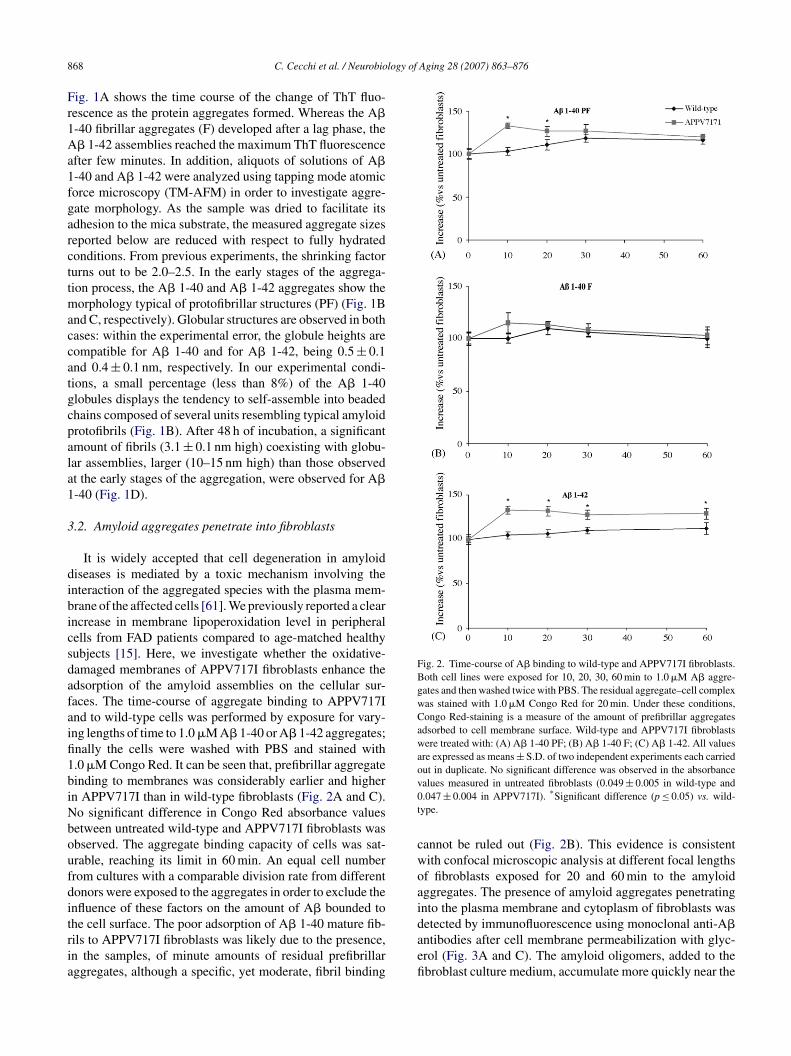
nternalization process of A� aggregates (Fig. 4). No signifi-ant difference in the relative Texas Red fluorescence valuesetween untreated wild-type and APPV717I fibroblasts wasbserved. To verify internalization data, a Z-series of 0.5 �mptical sections, from the basal to apical surface of APPV717Ibroblasts, was performed by confocal microscopy analysis.s shown in Fig. 5, A� 1-42 prefibrillar aggregates were evi-ently localized within the cells just after 20 min of treatment.
.3. Oxidative stress
There is strong experimental evidence that oxidative stresss an early biochemical modification in cells facing amyloidggregates [9,12,54,61]. It is well known that oxidative stressan be caused by increased free radical production and/oreakening of cellular antioxidant defences, which include
ntioxidant enzymes, lipophilic and hydrophilic scavengers.e have recently found that lymphoblasts and fibroblasts
rom FAD patients with APPV717I and PS-1 gene mutationsad lower basal TAC than healthy controls [15,16]. In the
870 C. Cecchi et al. / Neurobiology of Aging 28 (2007) 863–876
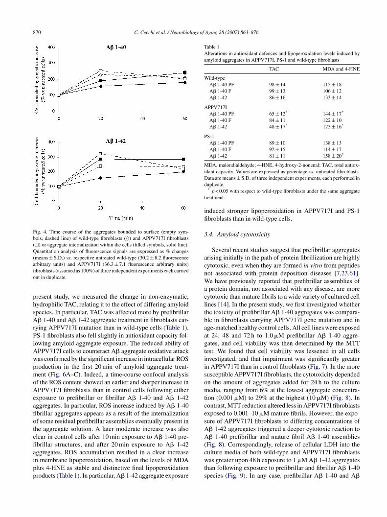
Fig. 4. Time course of the aggregates bounded to surface (empty sym-bols, dashed line) of wild-type fibroblasts (♦) and APPV717I fibroblasts(�) or aggregate internalization within the cells (filled symbols, solid line).Quantitation analysis of fluorescence signals are expressed as % changes(means ± S.D.) vs. respective untreated wild-type (30.2 ± 8.2 fluorescenceafio
phsArPlAwpmoAeafiotcfiaipp
Table 1Alterations in antioxidant defences and lipoperoxidation levels induced byamyloid aggregates in APPV717I, PS-1 and wild-type fibroblasts
TAC MDA and 4-HNE
Wild-typeA� 1-40 PF 98 ± 14 115 ± 18A� 1-40 F 99 ± 13 106 ± 12A� 1-42 86 ± 16 133 ± 14
APPV717IA� 1-40 PF 65 ± 12* 144 ± 17*
A� 1-40 F 84 ± 11 122 ± 10A� 1-42 48 ± 17* 175 ± 16*
PS-1A� 1-40 PF 89 ± 10 138 ± 13A� 1-40 F 92 ± 15 114 ± 17A� 1-42 81 ± 11 158 ± 20*
MDA, malondialdehyde; 4-HNE, 4-hydroxy-2-nonenal; TAC, total antiox-idant capacity. Values are expressed as pecentage vs. untreated fibroblasts.Dd
t
ifi
3
acnWacltbaagtiisomtcesAA(culture media of both wild-type and APPV717I fibroblasts
rbitrary units) and APPV717I (36.3 ± 7.1 fluorescence arbitrary units)broblasts (assumed as 100%) of three independent experiments each carriedut in duplicate.
resent study, we measured the change in non-enzymatic,ydrophilic TAC, relating it to the effect of differing amyloidpecies. In particular, TAC was affected more by prefibrillar� 1-40 and A� 1-42 aggregate treatment in fibroblasts car-
ying APPV717I mutation than in wild-type cells (Table 1).S-1 fibroblasts also fell slightly in antioxidant capacity fol-
owing amyloid aggregate exposure. The reduced ability ofPPV717I cells to counteract A� aggregate oxidative attackas confirmed by the significant increase in intracellular ROSroduction in the first 20 min of amyloid aggregate treat-ent (Fig. 6A–C). Indeed, a time-course confocal analysis
f the ROS content showed an earlier and sharper increase inPPV717I fibroblasts than in control cells following either
xposure to prefibrillar or fibrillar A� 1-40 and A� 1-42ggregates. In particular, ROS increase induced by A� 1-40brillar aggregates appears as a result of the internalizationf some residual prefibrillar assemblies eventually present inhe aggregate solution. A later moderate increase was alsolear in control cells after 10 min exposure to A� 1-40 pre-brillar structures, and after 20 min exposure to A� 1-42ggregates. ROS accumulation resulted in a clear increase
n membrane lipoperoxidation, based on the levels of MDAlus 4-HNE as stable and distinctive final lipoperoxidationroducts (Table 1). In particular, A� 1-42 aggregate exposurewts
ata are means ± S.D. of three independent experiments, each performed inuplicate.* p < 0.05 with respect to wild-type fibroblasts under the same aggregate
reatment.
nduced stronger lipoperoxidation in APPV717I and PS-1broblasts than in wild-type cells.
.4. Amyloid cytotoxicity
Several recent studies suggest that prefibrillar aggregatesrising initially in the path of protein fibrillization are highlyytotoxic, even when they are formed in vitro from peptidesot associated with protein deposition diseases [7,23,61].e have previously reported that prefibrillar assemblies ofprotein domain, not associated with any disease, are more
ytotoxic than mature fibrils to a wide variety of cultured cellines [14]. In the present study, we first investigated whetherhe toxicity of prefibrillar A� 1-40 aggregates was compara-le in fibroblasts carrying APPV717I gene mutation and inge-matched healthy control cells. All cell lines were exposedt 24, 48 and 72 h to 1.0 �M prefibrillar A� 1-40 aggre-ates, and cell viability was then determined by the MTTest. We found that cell viability was lessened in all cellsnvestigated, and that impairment was significantly greatern APPV717I than in control fibroblasts (Fig. 7). In the moreusceptible APPV717I fibroblasts, the cytotoxicity dependedn the amount of aggregates added for 24 h to the cultureedia, ranging from 6% at the lowest aggregate concentra-
ion (0.001 �M) to 29% at the highest (10 �M) (Fig. 8). Inontrast, MTT reduction altered less in APPV717I fibroblastsxposed to 0.001–10 �M mature fibrils. However, the expo-ure of APPV717I fibroblasts to differing concentrations of� 1-42 aggregates triggered a deeper cytotoxic reaction to� 1-40 prefibrillar and mature fibril A� 1-40 assemblies
Fig. 8). Correspondingly, release of cellular LDH into the
as greater upon 48 h exposure to 1 �M A� 1-42 aggregateshan following exposure to prefibrillar and fibrillar A� 1-40pecies (Fig. 9). In any case, prefibrillar A� 1-40 and A�

C. Cecchi et al. / Neurobiology of Aging 28 (2007) 863–876 871
F roblasts after 20 min treatment. A representative Z-series of 0.5 �m optical sectionsw cted with monoclonal mouse 6E10 anti-A� antibodies and Texas Red-conjugateda
1A
3
gtOmcmtmp(fiatfi
Table 2Apoptotic markers in APPV717I, PS-1 and wild-type fibroblasts followingamyloid aggregate exposure
DNA fragmentation Caspase-3 active fragment
Wild-typeA� 1-40 PF 133 ± 17 119 ± 14A� 1-40 F 111 ± 23 101 ± 13A� 1-42 110 ± 8 111 ± 12
APPV717IA� 1-40 PF 160 ± 20 232 ± 18*
A� 1-40 F 101 ± 13 137 ± 22*
A� 1-42 155 ± 15* 182 ± 19*
PS-1A� 1-40 PF 167 ± 15* 133 ± 13A� 1-40 F 114 ± 10 97 ± 9A� 1-42 134 ± 19* 143 ± 14*
ig. 5. A� 1-42 prefibrillar aggregates are localized within the APPV717I fibas taken through cells by confocal microscopy. The aggregates were dete
nti-mouse secondary antibodies as specified under Section 2.
-42 aggregates induced a more significant LDH release inPPV717I fibroblasts than in healthy controls.
.5. Apoptotic markers
Abundant data indicates that toxicity of amyloid aggre-ates stems from their ability to interact with cell membranes,riggering apoptotic and/or necrotic cell death [2,3,44,61].ur previous report found a clear increase in free radical-ediated injury to DNA in FAD fibroblasts relative to healthy
ontrols [15]. In the present study, the observed enrich-ent of histone-associated oligonucleosomes released to
he cytoplasm suggests a greater increase in DNA frag-entation in APPV717I and PS-1 fibroblasts exposed to
refibrillar A� 1-42 aggregates than in wild-type cellsTable 2). Involvement of the apoptotic process was con-
rmed by the increased amount of the caspase-3/CPP32ctive fragment (17 kDa) following amyloid exposure. In par-icular, caspase-3 was raised more in FAD than in wild-typebroblasts (Table 2).Values are expressed as percentage vs. untreated fibroblasts. Data aremeans ± S.D. of three independent experiments, each performed in dupli-cate.
* p < 0.05 with respect to wild-type fibroblasts under the same aggregatetreatment.

872 C. Cecchi et al. / Neurobiology of Aging 28 (2007) 863–876
Fig. 6. (A and B) Continuous confocal microscopy analysis of intracel-lular ROS production in unfixed APP and healthy fibroblasts during thefirst 20 min treatment with different amyloid aggregates. ROS levels wereimaged by confocal microscopy using the fluorescent dye CM-H2, DCFDAas a probe according to the procedures described under Section 2. Fluores-cence signals are expressed as fractional changes above the resting baseline,�F/F, where F is the average baseline fluorescence before the applicationof amyloid aggregates and �F represents the fluorescence changes overthe baseline. Values are expressed as percentage vs. untreated fibroblasts.The values shown are averages ± S.D. of two independent experiments onAfw
4
pcpbatssp
Fig. 7. Cell viability was checked by the MTT test in APPV717I fibroblastsand wild-type fibroblasts exposed to 1.0 �M A� 1-40 prefibrillar species vs.cells not exposed to the aggregates. Values are expressed as % vs. untreatedfiie
witadbelotAanism of aggregate translocation inside cells needs furtherinvestigation. Our results suggest that the amyloid aggre-gates can readily insert into oxidative-damaged fibroblastswhere the membrane integrity is compromised. Accordingly,
Fig. 8. The figure shows the percentage of viable APPV717I fibroblastsexposed to varying amounts of prefibrillar (PF) or fibrillar (F) A� 1-40
PPV717I and wild-type fibroblasts from four familial patients and fromour healthy subjects, respectively. *Significant difference (p ≤ 0.05) vs.ild-type.
. Discussion
Several reports provide strong evidence for amyloid lipideroxidation within the AD brain [10,30,45,48,62,68]. A�an fragment and generate free radical peptides which haveotent lipoperoxidising effects on the synaptosomal mem-ranes in the neocortex [12,17,32]. However, studies ofutopsied brain tissue cannot clarify whether abnormal oxida-
ive processes are inherent properties of AD cells or areecondary to neurodegeneration. Since our previous resultshowed a marked increase in oxidation levels of lipids androteins in peripheral cells from some FAD patients [15],avad
broblasts. The reported values (means ± S.D.) are representative of threendependent experiments, each performed in duplicate. *Significant differ-nce (p ≤ 0.05) vs. wild-type.
e investigated whether these early modifications couldnfluence the aggregation rate of amyloid assemblies andhe translocation process by which the amyloid assembliesppear inside the fibroblasts. Under our experimental con-itions, A� 1-40 and A� 1-42 aggregates are characterizedy an extensive �-sheet structure able to bind ThT and arendowed with typical morphological features of prefibril-ar and fibrillar aggregates. We demonstrated that amyloidligomers, that were exogenously added to the fibroblast cul-ure medium, were internalized faster and more completely inPPV717I than in wild-type fibroblasts, although the mech-
nd A� 1-42 aggregates respect to APPV717I untreated cells. The reportedalues (means ± S.D.) are expressed as percentage vs. untreated fibroblastsnd are representative of three independent experiments, each performed inuplicate. *Significant difference (p ≤ 0.05) vs. untreated cells.

C. Cecchi et al. / Neurobiology of
Fig. 9. The release of cellular LDH into the culture media of wild-type andAPPV717I fibroblasts without treatment or after 48 h exposure to 1 �M ofprefibrillar (PF) or fibrillar (F) A� 1-40 and A� 1-42 aggregates. The reportedvad
AicImhplbwllmimmtalopeadhiniAsptfpi
1ieptadP1odiomoePisrwoiIAsiottpfitctiifiodp1ttaatctots
alues (means ± S.D.) are expressed as percentage vs. untreated fibroblastsnd are representative of four independent experiments, each performed inuplicate. *Significant difference (p ≤ 0.05) vs. wild-type.
� is reported to accumulate faster in membranes contain-ng oxidatively damaged phospholipids than in membranesontaining only unoxidized or saturated phospholipids [39].ndeed, oxidatively damaged phospholipid membranes pro-ote the transition of A� to the �-sheet conformation, which
as a strong tendency to form fibrillar aggregates as denselaques in the brain. It is also possible that APPV717I fibrob-asts did not internalize amyloid aggregates unless cellularlebbing was present. Our results, however, are consistentith other previous studies showing intracellular accumu-
ation of A� 1-40 and A� 1-42 in granular deposits inate endosomes and lysosomes of human fibroblasts, PC12,
onocytic and neuroblastoma cell lines [8,37,56,72]. It isncreasingly recognized that disruption of the integrity of cell
embranes by small prefibrillar assemblies probing into theembrane bilayer is a primary step in the induction of oxida-
ive damage and after cell death [6,23,36,43,57,61]; the earlyppearance of amyloid aggregates in the cytoplasm of fibrob-asts therefore suggests that these species are the main sourcef oxidative stress for cells. Correspondingly, we foundrompt and sharp ROS production in APPV717I fibroblastsxposed to A� 1-40 and A� 1-42 aggregates. Accordingly,ddition of A� to PC12 cells induces increased ROS pro-uction and apoptosis [12,27,50]. In contrast, treatment ofealthy control fibroblasts with small prefibrillar aggregatesnduced a later and more moderate ROS increase. The sig-ificant increase in intracellular ROS production could benduced by a lesser ability of APPV717I cells to counteract� aggregate oxidative attack. Our previous report demon-
trated significant impairment in TAC and GSH content ineripheral cells from patients carrying APP or PS-1 muta-
ions, suggesting that a modified redox status is a commoneature of cells carrying these genetic lesions [15,16]. In theresent study, mutated fibroblasts exhibited larger decreasesn TAC following exposure to prefibrillar A� 1-40 and A�eioi
Aging 28 (2007) 863–876 873
-42 aggregates than fibroblasts from healthy subjects. TACmpairment could reflect chronic exposure to an oxidizingnvironment in mutated fibroblasts with a continuous over-roduction of amyloid peptide; it would therefore minimizehe protective effect against further oxidative injury followingmyloid peptide addition to cellular culture media. The TACecrease was greater in cells carrying APPV717I than theS-1 gene mutations. The moderate alteration in TAC in PS-mutated fibroblasts strengthens the claim that the changes
bserved are the direct outcome of the chronic presence of aifferent grade of cellular oxidizing environment induced byncreased A� production. Accordingly, we show that additionf A� aggregates to the fibroblast culture medium triggersore extensive lipid peroxidation in cells carrying APPV717I
r PS-1 gene mutations than in age-matched controls. Thisffect was stronger in fibroblasts carrying APP rather thanS-1 mutations. Our results therefore suggest that increase
n lipid peroxides is likely to result from attack on polyun-aturated fatty acids in cell-membrane phospholipids by freeadicals. These findings on peripheral cells are in agreementith several studies that provide evidence for excess lipoper-xidation and protein oxidation associated with A� depositsn APP and PS-1 AD brain and mutant mice [9,12,45,51,62].t has been shown that 4-HNE, generated in response to�-oxidative insults, can directly induce neuronal apopto-
is at pathophysiological concentrations [41]. Another studyndicates, however, that although free radicals and lipid per-xidation may participate in the neurodegeneration process,he mechanism of A�-induced neurotoxicity does not appearo involve ROS [73]. The present observations suggest thatrefibrillar A� 1-40 aggregates are more toxic than the maturebrils, and have a dose-dependent effect. Our data are consis-
ent with increasing evidence that small soluble oligomers,ompared to mature fibrils, are likely the more toxic species ofhe peptides [10,11,19,35,38,42,69]. The moderate increasen toxicity with the higher amount of A� 1-40 mature fibrilss likely to be the result of minute amounts of residual pre-brillar aggregates in cell media, although a specific toxicityf the fibrils cannot be ruled out. In particular, internalizationata of A� 1-40 fibrils shown in Fig. 3B, the consequent ROSroduction reported in Fig. 6B and the cytotoxic effect of A�-40 fibrils shown in Fig. 8 and Table 2, concurrently suggesthat the more structured fibrils maintain some level of directoxicity. This encourages us to provide for further studiesim to address the potential role of prefibrillar versus fibrillarggregates in mediating these effects. Some authors ques-ion the use of the MTT assay as a reporter of A�-mediatedytotoxicity [70]. In contrast, according to other researchers,he MTT assay, generally shows a good correlation withther viability tests and in vivo results [21]. Therefore, evenhough inhibition of MTT reduction represents a controver-ial indicator of A�-mediated cell injury, other corroborating
vidence was added in this study. Release of cellular LDHnto the culture media was specific to the prefibrillar formf A� 1-40, because the mature fibrils did not induce toxic-ty at 48 h after treatment. In any case, prefibrillar A� 1-40
8 logy of
avfiA4tobbbmeaswclb4utntwd[sta3ctfciicivffia4ctn
tpiodarAa
lbwafbcoicrtictata
A
tfaa2
R
74 C. Cecchi et al. / Neurobio
nd A� 1-42 aggregates induced a greater reduction in celliability and a larger increase in LDH release in APPV717Ibroblasts than in healthy controls. Also, the exposure ofPPV717I fibroblasts to differing concentrations of A� 1-2 aggregates triggered a deeper cytotoxic reaction respecto prefibrillar A� 1-40 aggregates. The earlier accumulationf A�1-42 than A�1-40 aggregates near the plasma mem-rane, although similar amount of amyloid assemblies cane internalized at longer time of exposure, can explain theurst in ROS production with a stronger cell viability impair-ent associated to A�1-42 treatment. Anyway, the increasing
vidence that A� 1-42 is more hydrophobic, more prone toggregation, and more amyloidogenic than A� 1-40 furtherupport the higher cytotoxic effect of A� 1-42 peptide. It isell known that the C-terminal hydrophobic tail is likely a
rucial feature of A� 1-42 cytotoxicity, because of its role inipid bilayer inclusion [10]. A recent report speculates thatoth A� 1-40 and A� 1-42 affect learning but only A� 1-2 causes extensive neurodegeneration in Drosophila brainsing the GAL4-UAS system [33]. Several studies suggesthat amyloid aggregate toxicity can trigger apoptotic and/orecrotic cell death [2,3,16,44,61]. It is generally believedhat cell death associated with protein aggregates beginsith stimulation of the apoptotic response, although recentata show necrotic rather than apoptotic death in some cases33,65,67,72]. The biochemistry of cell death following expo-ure to the toxic amyloid aggregates is still under investiga-ion, but our results support these suggestions. Indeed, despitemarked increase in DNA fragmentation and in the caspase-active fragment, necrosis appears to be the later outcome of
ell death, at least in APP fibroblasts under A� 1-42 aggregatereatment. In addition, there was a greater increase in DNAragmentation in APP and PS-1 fibroblasts than in wild-typeells following exposure to A� 1-42 aggregates. Interest-ngly, DNA fragmentation was positively correlated with cellmpairment or with ROS increase. Since the involvement ofaspases has been proposed in amyloid-induced apoptosisn cultured neurons [28,34], we also investigated the acti-ation of caspase-3/CPP32. The amount of caspase-3 activeragment was clearly raised more in FAD than in wild-typebroblasts. In particular, the effect of prefibrillar A� 1-40nd A� 1-42 was more powerful than that of fibrillar A� 1-0 in both APP and PS-1 cells. The mechanism by whichaspase-3 is activated by differing amyloid treatment is yeto be settled. Caspase activation may play a role that is notecessarily related to apoptosis [13,25].
In conclusion, our data demonstrate an early internaliza-ion of prefibrillar assemblies and a sharp increase in ROSroduction induced in APPV717I mutated fibroblasts follow-ng exposure to prefibrillar amyloid aggregates. Impairmentf antioxidant capacity, lipid peroxidation, mitochondrialysfunction are all triggered, and apoptotic outcomes such
s DNA fragmentation, caspase-3 activation and finally LDHelease are induced; the latter is certain following exposure to� 1-42 aggregates. Our results suggest that cells carrying anltered proteolytic APP process, such as APPV717I fibrob-
Aging 28 (2007) 863–876
asts, have enhanced susceptibility to oxidative stress inducedy A� exposure, which initially triggers the apoptotic path-ay and can ultimately lead to necrosis. This pattern relies onprogressive amplification mechanism of the early reactive
ree radicals by repeated chain reaction processes in mem-rane lipids consistent with the age dependence of AD [66]. Inontrast, healthy control fibroblasts are more resistant to A�-xidative attack, possibly because of their plasma membranentegrity and powerful antioxidant capacity. This capacity islearly correlated with the viability of cells, explaining theeduced necrotic outcome in healthy fibroblasts exposed tohe aggregates. These findings imply a systemic abnormal-ty in FAD that could be important for the use of peripheralells in pre-clinical trials of antioxidant drugs. More data onhe biochemical modifications of increased susceptibility tomyloid toxicity elicited by mutated cells is needed in ordero prevent such outcomes, and to design interventions thatim to restore the resistance of cells affected by aggregates.
cknowledgements
We thank Dr. Claudio Canale and Dr. Irene Forzoni forechnical advice. This study has been supported by grantsrom the Italian MIUR (project numbers 2002058218 001nd 2005054147 001), from Min.Salute/ISS (no. 4AN/F12)nd from the Compagnia di San Paolo, Torino, Italy (ref. no.004.0995).
eferences
[1] American Psychiatric Association. Diagnostic and statistical manualof mental disorders. 4th ed. Washington, DC: American PsychiatricAssociation; 1994.
[2] Anderson AJ, Pike CJ, Cotman CW. Differential induction ofimmediate early gene proteins in cultured neurons by �-amyloid(A�): association of c-Jun with A�-induced apoptosis. J Neurochem1995;65:1487–98.
[3] Behl C, Davis JB, Klier FG, Schubert D. Amyloid beta peptide inducesnecrosis rather than apoptosis. Brain Res 1994;645:253–64.
[4] Bokvist M, Lindstrom F, Watts A, Grobner G. Two types of Alzheimer’sbeta-amyloid (1-40) peptide membrane interactions: aggregation pre-venting transmembrane anchoring versus accelerated surface fibril for-mation. J Mol Biol 2004;335:1039–49.
[5] Bradford MM. A rapid and sensitive method for the quantitation ofmicrogram quantities of protein utilizing the principle of protein dyebinding. Anal Biochem 1976;72:248–54.
[6] Bucciantini M, Calloni G, Chiti F, Formigli L, Nosi D, Dobson CM,et al. Prefibrillar amyloid protein aggregates share common features ofcytotoxicity. J Biol Chem 2004;279:31374–82.
[7] Bucciantini M, Giannoni E, Chiti F, Baroni F, Formigli L, Zurdo J, etal. Inherent toxicity of aggregates implies a common origin for proteinmisfolding diseases. Nature 2002;416:507–11.
[8] Burdick D, Kosmoski J, Knauer MF, Glabe CG. Preferential adsorption,internalization and resistance to degradation of the major isoform of
the Alzheimer’s amyloid peptide A� 1-42, in differentiated PC12 cells.Brain Res 1997;746:275–84.[9] Butterfield DA, Drake J, Pocernich C, Castegna A. Evidence of oxida-tive damage in Alzheimer’s disease brain: central role for amyloidbeta-peptide. Trends Mol Med 2001;12:7548–54.
logy of
[
[
[
[
[
[
[
[
[
[
[
[
[
[
[
[
[
[
[
[
[
[
[
[
[
[
[
[
[
[
[
[
[
[
[
[
[
C. Cecchi et al. / Neurobio
10] Butterfield DA, Bush AI. Alzheimer’s amyloid �-peptide (1-42):involvement of methionine residue 35 in the oxidative stress andneurotoxicity properties of this peptide. Neurobiol Aging 2004;25:563–8.
11] Butterfield DA. Proteomics: a new approach to investigate oxidativestress in Alzheimer’s disease brain. Brain Res 2004;1000:1–7.
12] Butterifield DA. Amyloid beta-peptide (1-42)-induced oxidativestress and neurotoxicity: implication for neurodegeneration inAlzheimer’s disease brain. A review. Free Radic Res 2002;36:1307–13.
13] Canevari L, Abramov AY, Duchen MR. Toxicity of amyloid beta pep-tide: tales of calcium, mitochondria, and oxidative stress. NeurochemRes 2004;29:637–50.
14] Cecchi C, Baglioni S, Fiorillo C, Liguri G, Nosi D, Rigacci S, Buc-ciantini M, Stefani M. Different cell lines are variously affectedby the exposure to prefibrillar amyloid aggregates. J Cell Sci2005;118:3459–70.
15] Cecchi C, Fiorillo C, Sorbi S, Latorraca S, Nacmias B, Bagnoli S,et al. Oxidative stress and reduced antioxidant defenses in periph-eral cells from familial Alzheimer’s patients. Free Radic Biol Med2002;33:1372–9.
16] Cecchi C, Latorraca S, Sorbi S, Iantomasi T, Favilli F, Vincenzini MT,et al. Gluthatione level is altered in lymphoblasts from patients withfamilial Alzheimer’s disease. Neurosci Lett 1999;275:152–4.
17] Christen Y. Oxidative stress and Alzheimer disease. Am J Clin Nutr2000;71:621S–9S.
18] Citron M, Westaway D, Xia W, Carlson G, Diehl T, Levesque G, et al.Mutant presenilins of Alzheimer’s disease increase production of 42-residue amyloid beta-protein in both transfected cells and transgenicmice. Nat Med 1997;3:67–72.
19] Cleary JP, Walsh DM, Hofmeister JJ, Shankar GM, Kuskowski MA,Selkoe DJ, et al. Natural oligomers of the amyloid-beta protein specif-ically disrupt cognitive function. Nat Neurosci 2005;8:79–84.
20] Cruts M, Van Broeckoven C. Molecular genetics of Alzheimer’s dis-ease. Ann Med 1998;30:560–5.
21] Datki Z, Juhasz A, Galfi M, Soos K, Papp R, Zadori D, et al.Method for measuring neurotoxicity of aggregating polypeptides withthe MTT assay on differentiated neuroblastoma cells. Brain Res Bull2003;62:223–9.
22] Datki Z, Papp R, Zadori D, Soos K, Fulop L, Juhasz A, et al. Invitro model of neurotoxicity of Abeta 1-42 and neuroprotection bya pentapeptide: irreversible events during the first hour. Neurobiol Dis2004;17:507–15.
23] Dobson CM. Protein folding and misfolding. Nature 2003;426:884–90.
24] Esterbauer H, Schaur RJ, Zollner H. Chemistry and Biochemistry of4-hydroxynonenal, malonaldehyde and related aldehydes. Free RadicBiol Med 1991;11:81–128.
25] Gervais FG, Xu D, Robertson GS, Vaillancourt JP, Zhu Y, Huang J,et al. Involvement of caspases in proteolytic cleavage of Alzheimer’samyloid-beta precursor protein and amyloidogenic A beta peptide for-mation. Cell 1999;97:395–406.
26] Green JD, Kreplak L, Goldsbury C, Li Blatter X, Stolz M, Cooper GS,et al. Atomic force microscopy reveals defects within mica supportedlipid lilayers induced by the amyloidogenic human amylin peptide. JMol Biol 2004;342:877–87.
27] Guo G, Sopher BL, Pham DG, Furukawa K, Robinson N, Martin GM,et al. Alzheimer’s presenilin mutation sensitizes neural cells to apop-tosis induced by trophic factor withdrawal and amyloid beta-peptide:involvement of calcium and oxyradicals. J Neurosci 1997;17:4212–22.
28] Harada J, Sugimoto M. Activation of caspase-3 in beta-amyloid-
induced apoptosis of cultured rat cortical neurons. Brain Res 1999;842:311–23.29] Hardy J, Selkoe DJ. The amyloid hypothesis of Alzheimer’s dis-ease: progress and problems on the road of therapeutics. Science2002;297:353–6.
[
Aging 28 (2007) 863–876 875
30] Harris ME, Hensley K, Butterfield DA, Leedle RA, Carney JM.Direct evidence of oxidative injury produced by the Alzheimer beta-amyloid peptide (1-40) in cultured hippocampal neurons. Exp Neurol1995;131:193–202.
31] Hashimoto M, Rockenstein E, Crews L, Masliah E. Role of pro-tein aggregation in mitochondrial dysfunction and neurodegenera-tion in Alzheimer’s and Parkinson’s diseases. Neuromol Med 2003;4:21–36.
32] Hensley K, Carney JM, Mattson MP, Aksenova M, Harris M, Wu JF, etal. A model for beta-amyloid aggregation and neurotoxicity based onfree radical generation by the peptide: relevance to Alzheimer disease.Proc Natl Acad Sci USA 1994;91:3270–4.
33] Iijima K, Liu HP, Chiang AS, Hearn SA, Konsolaki M, Zhong Y. Dis-secting the pathological effects of human Abeta40 and Abeta42 inDrosophila: a potential model for Alzheimer’s disease. Proc Natl AcadSci USA 2004;101:6623–8.
34] Jordan J, Galindo MF, Miller RJ. Role of calpain- and interleukin-1 betaconverting enzyme-like proteases in the beta-amyloid-induced deathof rat hippocampal neurons in culture. J Neurochem 1997;68:1612–21.
35] Kawahara M, Kuroda Y, Arispes N, Rojas E. Alzheimer’s beta-amyloid, human islet amylin and prion protein fragment evokeintracellular free calcium elevations by a common mechanism ina hypothalamic GnRH neuronal cell line. J Biol Chem 2000;275:14077–83.
36] Kayed R, Head E, Thompson JL, McIntire TM, Milton SC, Cot-man CW, et al. Common structure of soluble amyloid oligomersimplies common mechanism of pathogenesis. Science 2003;300:486–9.
37] Knauer MF, Soreghan B, Burdick D, Kosmoski J, Glabe CG.Intracellular accumulation and resistance to degradation of theAlzheimer amyloid A4/� protein. Proc Natl Acad Sci USA 1992;89:7437–41.
38] Koo EH, Lansbury PT, Kelly LW. Amyloid diseases: abnormal pro-tein aggregation in neurodegeneration. Proc Natl Acad Sci USA1999;96:9989–90.
39] Koppaka V, Axelsen PH. Accelerated accumulation of amyloid betaproteins on oxidatively damaged lipid membranes. Biochemistry2000;39:10011–6.
40] Kourie JI, Henry CL. Ion channel formation and membrane-linked pathologies of misfolded hydrophobic proteins: the role ofdangerous unchaperoned molecules. Clin Exp Pharmacol Physiol2002;29:741–53.
41] Kruman I, Bruce-Keller AJ, Bredesen D, Waeg G, Mattson MP. Evi-dence that 4-hydroxynonenal mediates oxidative stress-induced neu-ronal apoptosis. J Neurosci 1997;17:5089–100.
42] Lambert MP, Barlow AK, Chromy BA, Edwards C, Freed R, LiosatosM, et al. Diffusible, nonfibrillar ligands derived from Abeta1-42 arepotent central nervous system neurotoxins. Proc Natl Acad Sci USA1998;95:6448–53.
43] Lashuel HA, Hartley D, Petre BM, Walz T, Lanbury PT. Neurode-generative disease: amyloid pores from pathogenic mutations. Nature2002;418:291.
44] Loo DT, Copani A, Pike CJ, Whittemore ER, Walencewicz AJ,Cotman CW. Apoptosis is induced by �-amyloid in cultured cen-tral nervous system neurons. Proc Natl Acad Sci USA 1993;90:7951–5.
45] Lovell MA, Ehmann WD, Butler SM, Markesbery WR. Ele-vated thiobarbituric acid-reactive substances and antioxidant enzymeactivity in the brain in Alzheimer’s disease. Neurology 1995;45:1594–601.
46] Marcus DL, Thomas C, Rodriguez CL, Simberkoff K, Tsai JS,
Strafaci JA, et al. Increased peroxidation and reduced antioxi-dant enzyme activity in Alzheimer’s disease. Exp Neurol 1998;150:40–4.47] Markesbery WR, Carney JM. Oxidative alterations in Alzheimer’s dis-ease. Brain Pathol 1999;9:133–46.

8 logy of
[
[
[
[
[
[
[
[
[
[
[
[
[
[
[
[
[
[
[
[
[
[
[
[
[
in amyloid precursor protein-transfected cells that have been treated
76 C. Cecchi et al. / Neurobio
48] Markesbery WR, Lovell MA. 4-Hydroxynonenal, a product of lipidperoxidation, is increased in the brain in Alzheimer’s disease. NeurobiolAging 1998;19:33–6.
49] Masters CL, Simms G, Weinman NA, Multhaup G, McDonald BL,Beyreuther K. Amyloid plaque core protein in Alzheimer disease andDown syndrome. Proc Natl Acad Sci USA 1985;82:4245–9.
50] Mattson MP. Oxidative stress, perturbed calcium homeostasis,and immune dysfunction in Alzheimer’s disease. J Neurovirol2002;8:539–50.
51] Mattson MP. Emerging neuroprotective strategies for Alzheimer’s dis-ease: dietary restriction, telomerase activation, and stem cell therapy.Exp Gerontol 2000;35:489–502.
52] Mattson MP, Guo Q, Furukawa K, Pedersen WA. Presenilins, the endo-plasmic reticulum, and neuronal apoptosis in Alzheimer’s disease. JNeurochem 1998;70:1–14.
53] McCormack JG, Halestrap AP, Denton RM. Role of calcium ions inregulation of mammalian intramitochondrial metabolism. Physiol Rev1990;70:391–425.
54] Mecocci P, MacGarvey U, Beal MF. Oxidative damage to mitochondrialDNA is increased in Alzheimer’s disease. Ann Neurol 1994;36:747–51.
55] Montine TJ, Neely MD, Quinn JF, Beal MF, Markesbery WR, RobertsLJ, et al. Lipid peroxidation in aging brain and Alzheimer’s disease.Free Radic Biol Med 2002;33:620–6.
56] Morelli L, Prat MI, Castano EM. Differential accumulation of solubleamyloid beta peptides 1-40 and 1-42 in human monocytic and neu-roblastoma cell lines. Implications for cerebral amyloidogenesis. CellTissue Res 1999;298:225–32.
57] Orrenius S, Zhovotovsky B, Nicotera P. Regulation of cell death: thecalcium-apoptosis link. Nat Rev 2003;4:552–65.
58] Price DL, Tanzi RE, Borchelt DR, Sisodia SS. Alzheimer’s dis-ease: genetic studies and transgenic models. Annu Rev Genet1998;32:461–93.
59] Sayre LM, Zelasko DA, Harris PL, Perry G, Salomon RG, SmithMA. 4-Hydroxynonenal-derived advanced lipid peroxidation end prod-ucts are increased in Alzheimer’s disease. J Neurochem 1997;68:
2092–7.60] Smith MA, Rudnicka-Nawrot M, Richey PL, Praprotnik D, MulvihillP, Miller CA, et al. Carbonyl-related post-translational modification ofneurofilament protein in the neurofibrillary pathology of Alzheimer’sdisease. J Neurochem 1995;64:2660–6.
[
Aging 28 (2007) 863–876
61] Stefani M, Dobson CM. Protein aggregation and aggregate toxicity:new insights into protein folding, misfolding diseases and biologicalevolution. J Mol Med 2003;81:678–99.
62] Subbarao KV, Richardson JS. Autopsy samples of Alzheimer’s cortexshow increased peroxidation in vitro. J Neurochem 1990;55:342–5.
63] Takeda K, Araki W, Tabira T. Enhanced generation of intracellularAbeta42 amyloid peptide by mutation of presenilins PS1 and PS2. EurJ Neurosci 2004;19:258–64.
64] The Dementia Study Group of the Italian Neurological Society Guide-lines for the diagnosis of dementia and Alzheimer’s disease. Ital JNeurol Sci 2000;21:87–194.
65] Turmaine M, Raza A, Mahal A, Mangiarini L, Bates GP, DaviesSW. Nonapoptotic neurodegeneration in a transgenic mouse modelof Huntington’s disease. Proc Natl Acad Sci USA 2000;97:8093–7.
66] Varadarajan S, Yatin S, Aksenova M, Butterfield DA. Review:Alzheimer’s amyloid beta-peptide-associated free radical oxidativestress and neurotoxicity. J Struct Biol 2000;130:184–208.
67] Velez-Pardo C, Arroyave ST, Lopera F, Castano AD, Jimenez Del RioMJ. Ultrastructure evidence of necrotic neural cell death in famil-ial Alzheimer’s disease brains bearing presenilin-1 E280A mutation.Alzheimer’s Dis 2001;3:409–15.
68] Volicer M. Free radicals in the development of Alzheimer’s disease.Neurobiol Aging 1990;11:567–71.
69] Walsh DM, Klyubin I, Fadeeva JV, Cullen WK, Anwyl R, Wolfe MS, etal. Naturally secreted oligomers of amyloid beta protein potently inhibithippocampal long-term potentiation in vivo. Nature 2002;416:535–9.
70] Wogulis M, Wright S, Cunningham D, Chilcote T, Powell K, RydelRE. Nucleation-dependent polymerization is an essential component ofamyloid-mediated neuronal cell death. J Neurosci 2005;25:1071–80.
71] Xie J, Guo Q. AATF protects neural cells against oxidative damageinduced by amyloid �-peptide. Neurobiol Dis 2004;16:150–7.
72] Yang AJ, Chandswangbhuvana D, Shu T, Henschen A, Glabe CG.Intracellular accumulation of insoluble, newly synthesized A� n-42
with A� 1-42*. J Biol Chem 1999;274:20650–6.73] Yao ZX, Drieu K, Szweda LI, Papadopoulos V. Free radicals and lipid
peroxidation do not mediate beta-amyloid-induced neuronal cell death.Brain Res 1999;847:203–10.
Copyright © 2022 FDOKUMEN