Loss of Metal Ions, Disulfide Reduction and Mutations Related to Familial ALS Promote Formation of...
14
Loss of Metal Ions, Disulfide Reduction and Mutations Related to Familial ALS Promote Formation of Amyloid- Like Aggregates from Superoxide Dismutase Zeynep A. Oztug Durer 1 , Jeffrey A. Cohlberg 1 *, Phong Dinh 1 , Shelby Padua 1 , Krista Ehrenclou 1 , Sean Downes 1 , James K. Tan 1 , Yoko Nakano 1 , Christopher J. Bowman 1 , Jessica L. Hoskins 2 , Chuhee Kwon 2 , Andrew Z. Mason 3 , Jorge A. Rodriguez 4 , Peter A. Doucette 4 , Bryan F. Shaw 4 , Joan Selverstone Valentine 4 1 Department of Chemistry and Biochemistry, California State University Long Beach, Long Beach, California, United States of America, 2 Department of Physics and Astronomy, California State University Long Beach, Long Beach, California, United States of America, 3 Department of Biological Sciences, California State University Long Beach, Long Beach, California, United States of America, 4 Department of Chemistry and Biochemistry, University of California Los Angeles, Los Angeles, California, United States of America Abstract Mutations in the gene encoding Cu-Zn superoxide dismutase (SOD1) are one of the causes of familial amyotrophic lateral sclerosis (FALS). Fibrillar inclusions containing SOD1 and SOD1 inclusions that bind the amyloid-specific dye thioflavin S have been found in neurons of transgenic mice expressing mutant SOD1. Therefore, the formation of amyloid fibrils from human SOD1 was investigated. When agitated at acidic pH in the presence of low concentrations of guanidine or acetonitrile, metalated SOD1 formed fibrillar material which bound both thioflavin T and Congo red and had circular dichroism and infrared spectra characteristic of amyloid. While metalated SOD1 did not form amyloid-like aggregates at neutral pH, either removing metals from SOD1 with its intramolecular disulfide bond intact or reducing the intramolecular disulfide bond of metalated SOD1 was sufficient to promote formation of these aggregates. SOD1 formed amyloid-like aggregates both with and without intermolecular disulfide bonds, depending on the incubation conditions, and a mutant SOD1 lacking free sulfhydryl groups (AS-SOD1) formed amyloid-like aggregates at neutral pH under reducing conditions. ALS mutations enhanced the ability of disulfide-reduced SOD1 to form amyloid-like aggregates, and apo-AS-SOD1 formed amyloid-like aggregates at pH 7 only when an ALS mutation was also present. These results indicate that some mutations related to ALS promote formation of amyloid-like aggregates by facilitating the loss of metals and/or by making the intramolecular disulfide bond more susceptible to reduction, thus allowing the conversion of SOD1 to a form that aggregates to form resembling amyloid. Furthermore, the occurrence of amyloid-like aggregates per se does not depend on forming intermolecular disulfide bonds, and multiple forms of such aggregates can be produced from SOD1. Citation: Oztug Durer ZA, Cohlberg JA, Dinh P, Padua S, Ehrenclou K, et al. (2009) Loss of Metal Ions, Disulfide Reduction and Mutations Related to Familial ALS Promote Formation of Amyloid-Like Aggregates from Superoxide Dismutase. PLoS ONE 4(3): e5004. doi:10.1371/journal.pone.0005004 Editor: Ashley I. Bush, Mental Health Research Institute of Victoria, Australia Received August 5, 2008; Accepted March 3, 2009; Published March 27, 2009 Copyright: ß 2009 Oztug Durer et al. This is an open-access article distributed under the terms of the Creative Commons Attribution License, which permits unrestricted use, distribution, and reproduction in any medium, provided the original author and source are credited. Funding: This work was supported by Research Corporation grant CC5571 and ALS Association grant 3Q1Y to JAC, NIH grant GM28222 and a grant from the ALS Association to JSV, and California State University, Long Beach. The ICP-MS was purchased with support from NSF grant OCE-9977564. PD and CJB were supported by the Beckman Scholars Program and JKT by a grant to CSULB from the Howard Hughes Medical Institute. The funders had no role in study design, data collection and analysis, decision to publish, or preparation of the manuscript. Competing Interests: The authors have declared that no competing interests exist. * E-mail: [email protected] Introduction More than sixty human diseases are accompanied by the formation of protein aggregates called amyloid [1]. These include a number of neurodegenerative diseases, such as Alzheimer’s disease, Parkinson’s disease, Huntington’s disease, and Creutz- feldt-Jakob (prion) disease. In each amyloid disease, a normally soluble protein forms insoluble fibrillar structures that bind the dyes thioflavin T (ThT), thioflavin S, and Congo Red, and, in many cases, display an X-ray diffraction pattern suggesting a ‘‘cross-beta structure’’, in which the b-strands are oriented perpendicular to the long axis of the fiber. The amyloid deposits associated with a particular disease may be either extracellular or intracellular. Amyloid deposits may be involved in the neurodegenerative disease amyotrophic lateral sclerosis (ALS), commonly known as Lou Gehrig’s disease. Approximately 2% of ALS cases are caused by mutations in the gene encoding the anti-oxidant enzyme copper- zinc superoxide dismutase (SOD1). These mutations represent one of the few known causes of ALS and underlie the most well-studied mouse models of this devastating disease. Clearly, much can be learned about the molecular underpinnings of pathology in ALS by studying the SOD1-linked forms of the disease. A growing body of evidence supports the hypothesis that many, if not all, of the SOD1 mutations act by increasing the tendency of SOD1 to aggregate (reviewed in [2–5]), and some findings suggest the involvement of amyloid in pathology. Electron microscopy has revealed a fibrillar morphology of the SOD1 aggregates found in motor neurons of FALS patients [6], in COS cells expressing mutant but not wild-type (WT) SOD1 [7], in neuroblastoma cells expressing ALS mutant SOD1 which were subjected to endoplasmic reticulum stress [8], and in transgenic mice expressing ALS mutant SOD1 [6,9,10]. PLoS ONE | www.plosone.org 1 March 2009 | Volume 4 | Issue 3 | e5004
-
Upload
independent -
Category
Documents
-
view
1 -
download
0
Transcript of Loss of Metal Ions, Disulfide Reduction and Mutations Related to Familial ALS Promote Formation of...
Loss of Metal Ions, Disulfide Reduction and MutationsRelated to Familial ALS Promote Formation of Amyloid-Like Aggregates from Superoxide DismutaseZeynep A. Oztug Durer1, Jeffrey A. Cohlberg1*, Phong Dinh1, Shelby Padua1, Krista Ehrenclou1, Sean
Downes1, James K. Tan1, Yoko Nakano1, Christopher J. Bowman1, Jessica L. Hoskins2, Chuhee Kwon2,
Andrew Z. Mason3, Jorge A. Rodriguez4, Peter A. Doucette4, Bryan F. Shaw4, Joan Selverstone Valentine4
1 Department of Chemistry and Biochemistry, California State University Long Beach, Long Beach, California, United States of America, 2 Department of Physics and
Astronomy, California State University Long Beach, Long Beach, California, United States of America, 3 Department of Biological Sciences, California State University Long
Beach, Long Beach, California, United States of America, 4 Department of Chemistry and Biochemistry, University of California Los Angeles, Los Angeles, California, United
States of America
Abstract
Mutations in the gene encoding Cu-Zn superoxide dismutase (SOD1) are one of the causes of familial amyotrophic lateralsclerosis (FALS). Fibrillar inclusions containing SOD1 and SOD1 inclusions that bind the amyloid-specific dye thioflavin Shave been found in neurons of transgenic mice expressing mutant SOD1. Therefore, the formation of amyloid fibrils fromhuman SOD1 was investigated. When agitated at acidic pH in the presence of low concentrations of guanidine oracetonitrile, metalated SOD1 formed fibrillar material which bound both thioflavin T and Congo red and had circulardichroism and infrared spectra characteristic of amyloid. While metalated SOD1 did not form amyloid-like aggregates atneutral pH, either removing metals from SOD1 with its intramolecular disulfide bond intact or reducing the intramoleculardisulfide bond of metalated SOD1 was sufficient to promote formation of these aggregates. SOD1 formed amyloid-likeaggregates both with and without intermolecular disulfide bonds, depending on the incubation conditions, and a mutantSOD1 lacking free sulfhydryl groups (AS-SOD1) formed amyloid-like aggregates at neutral pH under reducing conditions.ALS mutations enhanced the ability of disulfide-reduced SOD1 to form amyloid-like aggregates, and apo-AS-SOD1 formedamyloid-like aggregates at pH 7 only when an ALS mutation was also present. These results indicate that some mutationsrelated to ALS promote formation of amyloid-like aggregates by facilitating the loss of metals and/or by making theintramolecular disulfide bond more susceptible to reduction, thus allowing the conversion of SOD1 to a form thataggregates to form resembling amyloid. Furthermore, the occurrence of amyloid-like aggregates per se does not depend onforming intermolecular disulfide bonds, and multiple forms of such aggregates can be produced from SOD1.
Citation: Oztug Durer ZA, Cohlberg JA, Dinh P, Padua S, Ehrenclou K, et al. (2009) Loss of Metal Ions, Disulfide Reduction and Mutations Related to Familial ALSPromote Formation of Amyloid-Like Aggregates from Superoxide Dismutase. PLoS ONE 4(3): e5004. doi:10.1371/journal.pone.0005004
Editor: Ashley I. Bush, Mental Health Research Institute of Victoria, Australia
Received August 5, 2008; Accepted March 3, 2009; Published March 27, 2009
Copyright: � 2009 Oztug Durer et al. This is an open-access article distributed under the terms of the Creative Commons Attribution License, which permitsunrestricted use, distribution, and reproduction in any medium, provided the original author and source are credited.
Funding: This work was supported by Research Corporation grant CC5571 and ALS Association grant 3Q1Y to JAC, NIH grant GM28222 and a grant from the ALSAssociation to JSV, and California State University, Long Beach. The ICP-MS was purchased with support from NSF grant OCE-9977564. PD and CJB weresupported by the Beckman Scholars Program and JKT by a grant to CSULB from the Howard Hughes Medical Institute. The funders had no role in study design,data collection and analysis, decision to publish, or preparation of the manuscript.
Competing Interests: The authors have declared that no competing interests exist.
* E-mail: [email protected]
Introduction
More than sixty human diseases are accompanied by the
formation of protein aggregates called amyloid [1]. These include
a number of neurodegenerative diseases, such as Alzheimer’s
disease, Parkinson’s disease, Huntington’s disease, and Creutz-
feldt-Jakob (prion) disease. In each amyloid disease, a normally
soluble protein forms insoluble fibrillar structures that bind the
dyes thioflavin T (ThT), thioflavin S, and Congo Red, and, in
many cases, display an X-ray diffraction pattern suggesting a
‘‘cross-beta structure’’, in which the b-strands are oriented
perpendicular to the long axis of the fiber. The amyloid deposits
associated with a particular disease may be either extracellular or
intracellular.
Amyloid deposits may be involved in the neurodegenerative
disease amyotrophic lateral sclerosis (ALS), commonly known as
Lou Gehrig’s disease. Approximately 2% of ALS cases are caused
by mutations in the gene encoding the anti-oxidant enzyme copper-
zinc superoxide dismutase (SOD1). These mutations represent one
of the few known causes of ALS and underlie the most well-studied
mouse models of this devastating disease. Clearly, much can be
learned about the molecular underpinnings of pathology in ALS by
studying the SOD1-linked forms of the disease. A growing body of
evidence supports the hypothesis that many, if not all, of the SOD1
mutations act by increasing the tendency of SOD1 to aggregate
(reviewed in [2–5]), and some findings suggest the involvement of
amyloid in pathology. Electron microscopy has revealed a fibrillar
morphology of the SOD1 aggregates found in motor neurons of
FALS patients [6], in COS cells expressing mutant but not wild-type
(WT) SOD1 [7], in neuroblastoma cells expressing ALS mutant
SOD1 which were subjected to endoplasmic reticulum stress [8],
and in transgenic mice expressing ALS mutant SOD1 [6,9,10].
PLoS ONE | www.plosone.org 1 March 2009 | Volume 4 | Issue 3 | e5004
Furthermore, transgenic mice expressing mutant SOD1 proteins
have neuronal inclusions which bind the amyloid-specific dye
thioflavin S [11–13].
SOD1 is a homodimer, each polypeptide chain containing 153
amino acids with one bound Cu2+ and one bound Zn2+ ion. Each
chain folds into an eight-stranded beta barrel that is flanked on one
side by a number of loops which contain the metal binding sites.
There are four cysteines, C6 and C111 present as free sulfhydryls,
and C57 and C146 joined by a disulfide bond which links one of the
loops to the beta barrel. In the absence of coordinated copper and
zinc, the beta barrel and dimer interface remain intact, but the
metal binding loops are disordered [5]. When neither metals nor the
intramolecular disulfide bond is present, SOD1 exists as a monomer
[14–15]. More than 100 mutations in the SOD1 gene, scattered
throughout the polypeptide chain, have been linked to FALS [2–3].
The ALS-linked SOD1 mutants have been grouped into two
general classes, ‘‘wild-type-like mutants’’ resulting from mutations in
the beta barrel or dimer interface, which generally retain high levels
of catalytic activity, and metal-binding region mutants, resulting
mostly from mutations in the loops, which generally have much less
catalytic activity and are isolated with lower metal content than WT
SOD1 [3].
In the present study we demonstrate that under appropriate
conditions a variety of biophysically diverse SOD1 species all form
insoluble aggregates which are identified as amyloid fibrils by a
number of different criteria. While the metalated WT enzyme
does not aggregate at neutral pH, either removal of copper and
zinc or reduction of the intramolecular disulfide bond is sufficient
to trigger aggregation. Previous publications have shown that
some forms of SOD1 generate fibrillar aggregates that bind ThT
upon extended incubation at acidic pH [16], and that both WT
and mutant apo-SOD1 can form non-fibrillar disulfide-bonded
oligomers that bind ThT [17–18]. This study demonstrates further
that SOD1 can form fibrillar aggregates with spectroscopic and
dye-binding properties characteristic of amyloid in vitro at
physiological pH, ionic srength and temperature. In our results,
intermolecular disulfide bonds are not required for forming
amyloid-like aggregates from SOD1, since such aggregates can
also be produced from SOD1 mutants lacking free cysteines, and
since, under defined conditions, SOD1 variants that do contain
free cysteines can be shown to form amyloid-like aggregates
lacking intermolecular disulfide bonds. A number of mutations
related to FALS appear to promote amyloid formation by
facilitating the loss of metals and/or by making the intramolecular
disulfide bond more susceptible to reduction.
Results
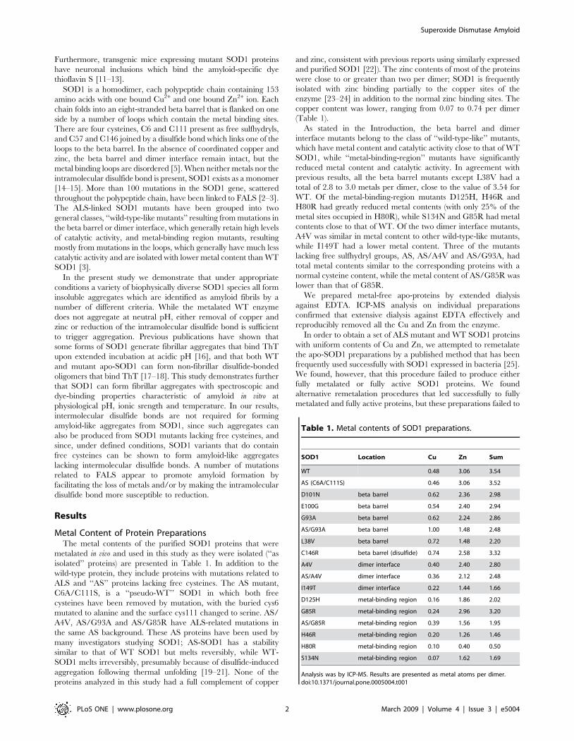
Metal Content of Protein PreparationsThe metal contents of the purified SOD1 proteins that were
metalated in vivo and used in this study as they were isolated (‘‘as
isolated’’ proteins) are presented in Table 1. In addition to the
wild-type protein, they include proteins with mutations related to
ALS and ‘‘AS’’ proteins lacking free cysteines. The AS mutant,
C6A/C111S, is a ‘‘pseudo-WT’’ SOD1 in which both free
cysteines have been removed by mutation, with the buried cys6
mutated to alanine and the surface cys111 changed to serine. AS/
A4V, AS/G93A and AS/G85R have ALS-related mutations in
the same AS background. These AS proteins have been used by
many investigators studying SOD1; AS-SOD1 has a stability
similar to that of WT SOD1 but melts reversibly, while WT-
SOD1 melts irreversibly, presumably because of disulfide-induced
aggregation following thermal unfolding [19–21]. None of the
proteins analyzed in this study had a full complement of copper
and zinc, consistent with previous reports using similarly expressed
and purified SOD1 [22]). The zinc contents of most of the proteins
were close to or greater than two per dimer; SOD1 is frequently
isolated with zinc binding partially to the copper sites of the
enzyme [23–24] in addition to the normal zinc binding sites. The
copper content was lower, ranging from 0.07 to 0.74 per dimer
(Table 1).
As stated in the Introduction, the beta barrel and dimer
interface mutants belong to the class of ‘‘wild-type-like’’ mutants,
which have metal content and catalytic activity close to that of WT
SOD1, while ‘‘metal-binding-region’’ mutants have significantly
reduced metal content and catalytic activity. In agreement with
previous results, all the beta barrel mutants except L38V had a
total of 2.8 to 3.0 metals per dimer, close to the value of 3.54 for
WT. Of the metal-binding-region mutants D125H, H46R and
H80R had greatly reduced metal contents (with only 25% of the
metal sites occupied in H80R), while S134N and G85R had metal
contents close to that of WT. Of the two dimer interface mutants,
A4V was similar in metal content to other wild-type-like mutants,
while I149T had a lower metal content. Three of the mutants
lacking free sulfhydryl groups, AS, AS/A4V and AS/G93A, had
total metal contents similar to the corresponding proteins with a
normal cysteine content, while the metal content of AS/G85R was
lower than that of G85R.
We prepared metal-free apo-proteins by extended dialysis
against EDTA. ICP-MS analysis on individual preparations
confirmed that extensive dialysis against EDTA effectively and
reproducibly removed all the Cu and Zn from the enzyme.
In order to obtain a set of ALS mutant and WT SOD1 proteins
with uniform contents of Cu and Zn, we attempted to remetalate
the apo-SOD1 preparations by a published method that has been
frequently used successfully with SOD1 expressed in bacteria [25].
We found, however, that this procedure failed to produce either
fully metalated or fully active SOD1 proteins. We found
alternative remetalation procedures that led successfully to fully
metalated and fully active proteins, but these preparations failed to
Table 1. Metal contents of SOD1 preparations.
SOD1 Location Cu Zn Sum
WT 0.48 3.06 3.54
AS (C6A/C111S) 0.46 3.06 3.52
D101N beta barrel 0.62 2.36 2.98
E100G beta barrel 0.54 2.40 2.94
G93A beta barrel 0.62 2.24 2.86
AS/G93A beta barrel 1.00 1.48 2.48
L38V beta barrel 0.72 1.48 2.20
C146R beta barrel (disulfide) 0.74 2.58 3.32
A4V dimer interface 0.40 2.40 2.80
AS/A4V dimer interface 0.36 2.12 2.48
I149T dimer interface 0.22 1.44 1.66
D125H metal-binding region 0.16 1.86 2.02
G85R metal-binding region 0.24 2.96 3.20
AS/G85R metal-binding region 0.39 1.56 1.95
H46R metal-binding region 0.20 1.26 1.46
H80R metal-binding region 0.10 0.40 0.50
S134N metal-binding region 0.07 1.62 1.69
Analysis was by ICP-MS. Results are presented as metal atoms per dimer.doi:10.1371/journal.pone.0005004.t001
Superoxide Dismutase Amyloid
PLoS ONE | www.plosone.org 2 March 2009 | Volume 4 | Issue 3 | e5004
form any aggregates under conditions that promoted aggregate
formation from the as-isolated preparations (data not shown). The
failure to aggregate could be attributed to irreversible oxidation of
one of the free cysteine residues. (See Text S1 and Figure S1 for
details.) We therefore concluded that the use of remetalated
proteins for in vitro aggregation studies was not feasible. For most
experiments in this study, we instead used the ‘‘as-isolated’’
proteins which had been metalated in yeast prior to isolation.
These preparations will be referred to as ‘‘metalated’’ SOD1 in the
discussion that follows.
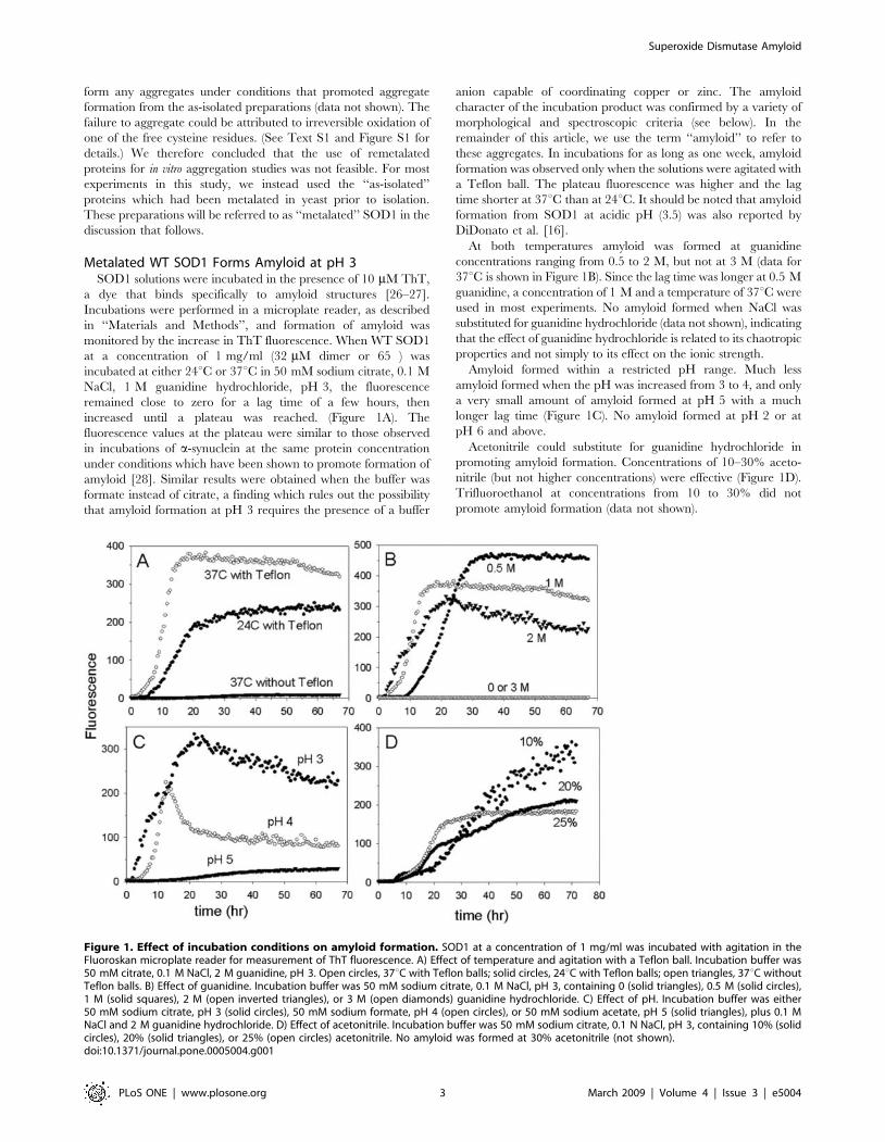
Metalated WT SOD1 Forms Amyloid at pH 3SOD1 solutions were incubated in the presence of 10 mM ThT,
a dye that binds specifically to amyloid structures [26–27].
Incubations were performed in a microplate reader, as described
in ‘‘Materials and Methods’’, and formation of amyloid was
monitored by the increase in ThT fluorescence. When WT SOD1
at a concentration of 1 mg/ml (32 mM dimer or 65 ) was
incubated at either 24uC or 37uC in 50 mM sodium citrate, 0.1 M
NaCl, 1 M guanidine hydrochloride, pH 3, the fluorescence
remained close to zero for a lag time of a few hours, then
increased until a plateau was reached. (Figure 1A). The
fluorescence values at the plateau were similar to those observed
in incubations of a-synuclein at the same protein concentration
under conditions which have been shown to promote formation of
amyloid [28]. Similar results were obtained when the buffer was
formate instead of citrate, a finding which rules out the possibility
that amyloid formation at pH 3 requires the presence of a buffer
anion capable of coordinating copper or zinc. The amyloid
character of the incubation product was confirmed by a variety of
morphological and spectroscopic criteria (see below). In the
remainder of this article, we use the term ‘‘amyloid’’ to refer to
these aggregates. In incubations for as long as one week, amyloid
formation was observed only when the solutions were agitated with
a Teflon ball. The plateau fluorescence was higher and the lag
time shorter at 37uC than at 24uC. It should be noted that amyloid
formation from SOD1 at acidic pH (3.5) was also reported by
DiDonato et al. [16].
At both temperatures amyloid was formed at guanidine
concentrations ranging from 0.5 to 2 M, but not at 3 M (data for
37uC is shown in Figure 1B). Since the lag time was longer at 0.5 M
guanidine, a concentration of 1 M and a temperature of 37uC were
used in most experiments. No amyloid formed when NaCl was
substituted for guanidine hydrochloride (data not shown), indicating
that the effect of guanidine hydrochloride is related to its chaotropic
properties and not simply to its effect on the ionic strength.
Amyloid formed within a restricted pH range. Much less
amyloid formed when the pH was increased from 3 to 4, and only
a very small amount of amyloid formed at pH 5 with a much
longer lag time (Figure 1C). No amyloid formed at pH 2 or at
pH 6 and above.
Acetonitrile could substitute for guanidine hydrochloride in
promoting amyloid formation. Concentrations of 10–30% aceto-
nitrile (but not higher concentrations) were effective (Figure 1D).
Trifluoroethanol at concentrations from 10 to 30% did not
promote amyloid formation (data not shown).
Figure 1. Effect of incubation conditions on amyloid formation. SOD1 at a concentration of 1 mg/ml was incubated with agitation in theFluoroskan microplate reader for measurement of ThT fluorescence. A) Effect of temperature and agitation with a Teflon ball. Incubation buffer was50 mM citrate, 0.1 M NaCl, 2 M guanidine, pH 3. Open circles, 37uC with Teflon balls; solid circles, 24uC with Teflon balls; open triangles, 37uC withoutTeflon balls. B) Effect of guanidine. Incubation buffer was 50 mM sodium citrate, 0.1 M NaCl, pH 3, containing 0 (solid triangles), 0.5 M (solid circles),1 M (solid squares), 2 M (open inverted triangles), or 3 M (open diamonds) guanidine hydrochloride. C) Effect of pH. Incubation buffer was either50 mM sodium citrate, pH 3 (solid circles), 50 mM sodium formate, pH 4 (open circles), or 50 mM sodium acetate, pH 5 (solid triangles), plus 0.1 MNaCl and 2 M guanidine hydrochloride. D) Effect of acetonitrile. Incubation buffer was 50 mM sodium citrate, 0.1 N NaCl, pH 3, containing 10% (solidcircles), 20% (solid triangles), or 25% (open circles) acetonitrile. No amyloid was formed at 30% acetonitrile (not shown).doi:10.1371/journal.pone.0005004.g001
Superoxide Dismutase Amyloid
PLoS ONE | www.plosone.org 3 March 2009 | Volume 4 | Issue 3 | e5004
Removal of Copper and Zinc Allows Amyloid Formationat Neutral pH
Because acidic pH causes the dissociation of metals from SOD1,
we reasoned that the ability of WT SOD1 to form amyloid at low
pH could be due, at least in part, to the loss of metal ions. We
hypothesized that metal-free SOD1 might therefore be able to
form amyloid at higher values of pH that are more physiologically
relevant. The kinetic parameters for amyloid formation from
metalated and apo-SOD1 at various pH values in the presence of
1 M guanidine are summarized in Table 2. As shown above
(Figure 1C), the yield of amyloid from metalated WT-SOD1 was
sharply dependent on pH, with considerably less amyloid at pH 4
or 5 compared to pH 3 and no amyloid formed at pH 6 or 7. In
contrast, there was little variation in either the amplitude or the lag
time for apo-SOD1 as the pH was varied between 3 and 7. This
suggests that acidic pH is needed for amyloid formation mainly
because it promotes the loss of metals. Most strikingly, apo-SOD1
formed amyloid at pH 6 and 7, while no conditions have been
found which allow amyloid formation from metalated SOD1 with
its intramolecular disulfide bond intact at pH greater than 5.
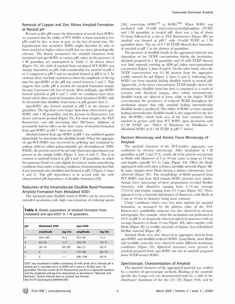
Apo-SOD1 also formed amyloid at pH 7 in the absence of
guanidine. The lag times were longer than those observed for apo-
SOD1 with 1 M guanidine, and the increase in fluorescence was
slower and more gradual (Figure 2A). For most samples, the ThT
fluorescence was still increasing after 300 hours. Addition of
acetonitrile had no effect on the time course of amyloid formation
from apo-SOD1 at pH 7 (data not shown).
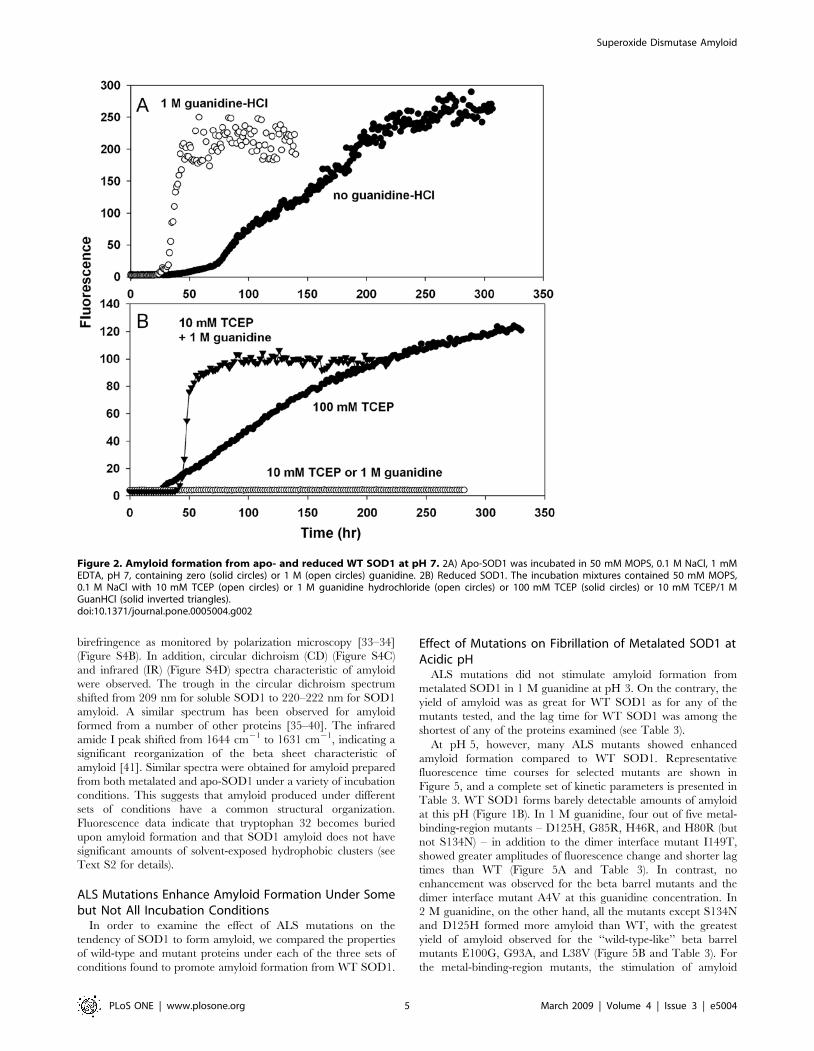
Amyloid formed from apo-SOD1 at pH 7 was stabilized against
disassembly by intermolecular disulfide bonds. When the aggregat-
ed apo-WT-SOD1 was recovered by pelleting and examined by
sodium dodecyl sulfate-polyacrylamide gel electrophoresis (SDS-
PAGE), the protein entered the gel only when mercaptoethanol was
present in the sample buffer (Figure 3, lanes 6 and 7). This is in
contrast to amyloid formed at pH 3 and 1 M guanidine, in which
the monomer band was only slightly less intense under nonreducing
conditions than under reducing conditions, demonstrating that little
if any intermolecular disulfides had formed at pH 3 (Figure 3, lanes
4 and 5). This pH dependence is in accord with the well-
characterized pH dependence of disulfide formation [29].
Reduction of the Intramolecular Disulfide Bond PromotesAmyloid Formation from Metalated SOD1
The intramolecular disulfide bond of SOD1 can be cleaved by
extended incubation with high concentrations of reducing agents
[30], converting SOD1S-S to SOD12SH. When SOD1 was
incubated with 10 mM tris(2-carboxyethyl)phosphine (TCEP)
and 1 M guanidine at neutral pH, there was a lag of about
45 hours followed by a rise in ThT fluorescence (Figure 2B); no
amyloid was formed at pH 7 with 10 mM TCEP or 1 M
guanidine alone. The use of 0.1 M TCEP allowed slow formation
of amyloid at pH 7 in the absence of guanidine.
The presence of disulfide bonds in the aggregated material was
dependent on the TCEP concentration during the incubation.
Amyloid prepared in 1 M guanidine and 10 mM TCEP showed
very little material entering an SDS gel unless mercaptoethanol
was present (Figure 3, lanes 8 and 9). On the other hand, when the
TCEP concentration was 0.1 M, protein from the aggregates
readily entered the gel (Figure 3, lanes 2 and 3), indicating that
SOD1 can form amyloid lacking disulfide bonds at neutral pH.
Apparently, at the lower concentration TCEP initially cleaves the
intramolecular disulfide bond but then is consumed as a result of
reaction with dissolved oxygen, after which intermolecular
disulfide bonds are allowed to form, while at the higher TCEP
concentration the persistence of reduced TCEP throughout the
incubation insures that only amyloid lacking intermolecular
disulfide bonds is produced. The ability of SOD1 to form amyloid
lacking intermolecular disulfide bonds is also suggested by the fact
that AS-SOD1, which lacks two of the four cysteines, forms
amyloid in greater yield than WT SOD1 upon incubation with
0.1 M TCEP (see ‘‘Effect of Mutations on Fibrillation of
Metalated SOD1 in 0.1 M TCEP at pH 7’’ below).
Electron Microscopy and Atomic Force Microscopy ofAmyloid
The amyloid character of the ThT-positive aggregates was
confirmed by electron microscopy. After incubation in 1 M
guanidine at pH 3 and 37uC, nearly all of the protein was present
as fibrils with diameters of 5 to 10 nm (some as large as 14 nm)
and lengths typically 0.5 to 3 mm (Figure 4A). Often the fibrils
aggregated with each other to form a meshlike network (Figure 4B).
In some samples short fibrils having a distinct substructure were
observed (Figure 4C). The morphology of fibrils prepared from
WT SOD1 and from ALS mutant SOD1 proteins were similar.
Atomic force microscopy of these preparations revealed fibrillar
structures with diameters ranging from 4–14 nm (average
7.962.6) and lengths ranging from 0.5–3 mm (Figure S2). There
appeared to be a bimodal distribution of diameters, with fibrils of
5 nm or 10 nm in diameter being most common.
Under conditions which were less than optimal for amyloid
formation, as measured by the plateau value of the ThT
fluorescence, nonfibrillar material was also observed in electron
micrographs. For example, when the incubation was performed at
24uC at pH 3, we frequently observed spherical structures with an
average diameter of about 13 nm (Figure 4D), often together with
fibrils (Figure 4E) or weblike networks of thinner, less well-defined
fibrillar material (Figure 4F).
Amyloid fibrils were also observed in aggregates derived from
apo-SOD1 and disulfide-reduced SOD1. Long fibrils, short fibrils
and meshlike networks were observed under different incubation
conditions (Figure S3). Spherical structures were present in
amyloid prepared from apo-SOD1 but not in amyloid prepared
from TCEP-treated SOD1.
Spectroscopic Characterization of AmyloidThe amyloid character of the aggregated material was verified
by a number of spectroscopic methods. Binding of the amyloid-
specific dye Congo red was demonstrated both by a shift in the
absorbance maximum of the dye [31–32] (Figure S4A) and by
Table 2. Kinetic parameters of amyloid formation frommetalated and apo-SOD1 in 1 M guanidine.
pH Metalated SOD Apo-SOD
amplitude lag (hr) amplitude lag (hr)
3 3526180 962 273693 764
4 62623 361 162674 19611
5 26610 69638 140651 1664
6 0 ----- 177648 62610
7 0 ----- 2486109 2668
SOD1 was incubated in buffers containing 50 mM citrate pH 3, formate pH 4,acetate pH 5, succinate pH 6, or MOPS pH 7, plus 0.1 M NaCl and 1 Mguanidine. The time course of ThT fluorescence was fit to a sigmoidal equationand the amplitude and lag time determined as described in ‘‘Materials andMethods’’. Dashes indicate that no amyloid was formed.doi:10.1371/journal.pone.0005004.t002
Superoxide Dismutase Amyloid
PLoS ONE | www.plosone.org 4 March 2009 | Volume 4 | Issue 3 | e5004
birefringence as monitored by polarization microscopy [33–34]
(Figure S4B). In addition, circular dichroism (CD) (Figure S4C)
and infrared (IR) (Figure S4D) spectra characteristic of amyloid
were observed. The trough in the circular dichroism spectrum
shifted from 209 nm for soluble SOD1 to 220–222 nm for SOD1
amyloid. A similar spectrum has been observed for amyloid
formed from a number of other proteins [35–40]. The infrared
amide I peak shifted from 1644 cm21 to 1631 cm21, indicating a
significant reorganization of the beta sheet characteristic of
amyloid [41]. Similar spectra were obtained for amyloid prepared
from both metalated and apo-SOD1 under a variety of incubation
conditions. This suggests that amyloid produced under different
sets of conditions have a common structural organization.
Fluorescence data indicate that tryptophan 32 becomes buried
upon amyloid formation and that SOD1 amyloid does not have
significant amounts of solvent-exposed hydrophobic clusters (see
Text S2 for details).
ALS Mutations Enhance Amyloid Formation Under Somebut Not All Incubation Conditions
In order to examine the effect of ALS mutations on the
tendency of SOD1 to form amyloid, we compared the properties
of wild-type and mutant proteins under each of the three sets of
conditions found to promote amyloid formation from WT SOD1.
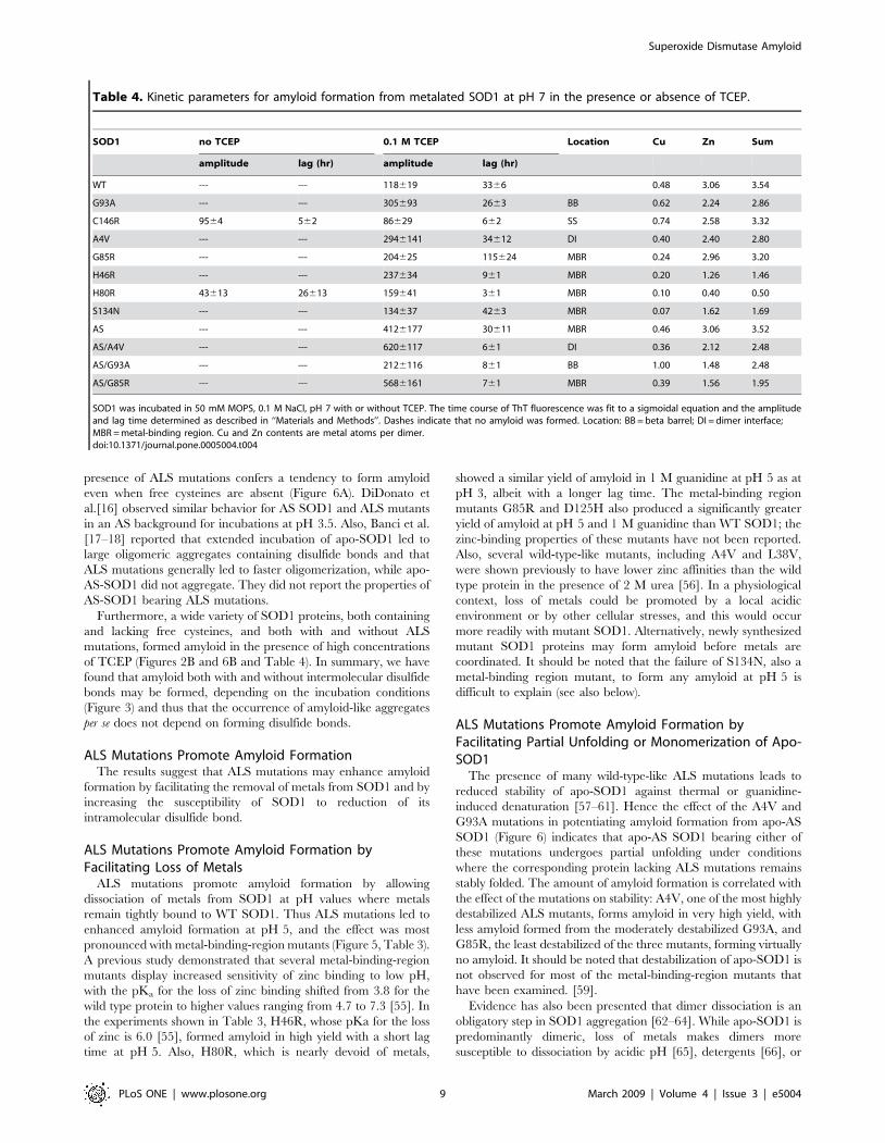
Effect of Mutations on Fibrillation of Metalated SOD1 atAcidic pH
ALS mutations did not stimulate amyloid formation from
metalated SOD1 in 1 M guanidine at pH 3. On the contrary, the
yield of amyloid was as great for WT SOD1 as for any of the
mutants tested, and the lag time for WT SOD1 was among the
shortest of any of the proteins examined (see Table 3).
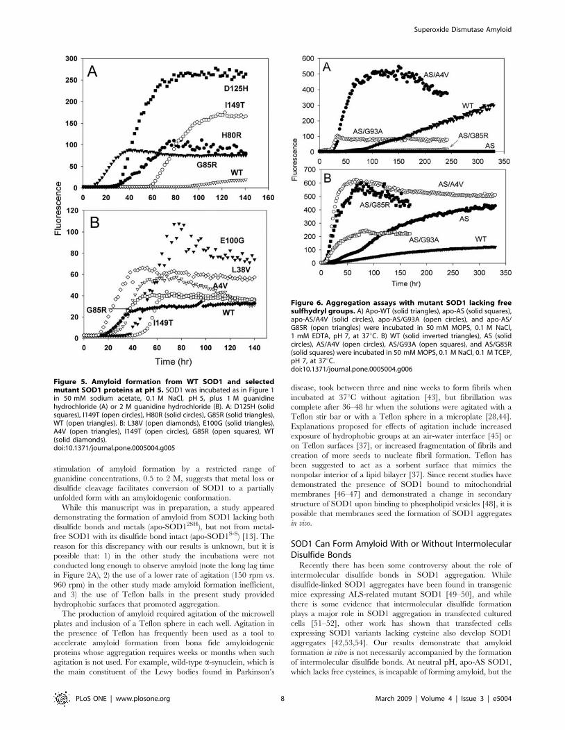
At pH 5, however, many ALS mutants showed enhanced
amyloid formation compared to WT SOD1. Representative
fluorescence time courses for selected mutants are shown in
Figure 5, and a complete set of kinetic parameters is presented in
Table 3. WT SOD1 forms barely detectable amounts of amyloid
at this pH (Figure 1B). In 1 M guanidine, four out of five metal-
binding-region mutants – D125H, G85R, H46R, and H80R (but
not S134N) – in addition to the dimer interface mutant I149T,
showed greater amplitudes of fluorescence change and shorter lag
times than WT (Figure 5A and Table 3). In contrast, no
enhancement was observed for the beta barrel mutants and the
dimer interface mutant A4V at this guanidine concentration. In
2 M guanidine, on the other hand, all the mutants except S134N
and D125H formed more amyloid than WT, with the greatest
yield of amyloid observed for the ‘‘wild-type-like’’ beta barrel
mutants E100G, G93A, and L38V (Figure 5B and Table 3). For
the metal-binding-region mutants, the stimulation of amyloid
Figure 2. Amyloid formation from apo- and reduced WT SOD1 at pH 7. 2A) Apo-SOD1 was incubated in 50 mM MOPS, 0.1 M NaCl, 1 mMEDTA, pH 7, containing zero (solid circles) or 1 M (open circles) guanidine. 2B) Reduced SOD1. The incubation mixtures contained 50 mM MOPS,0.1 M NaCl with 10 mM TCEP (open circles) or 1 M guanidine hydrochloride (open circles) or 100 mM TCEP (solid circles) or 10 mM TCEP/1 MGuanHCl (solid inverted triangles).doi:10.1371/journal.pone.0005004.g002
Superoxide Dismutase Amyloid
PLoS ONE | www.plosone.org 5 March 2009 | Volume 4 | Issue 3 | e5004
formation at pH 5 is likely due to the fact that these mutations
allow dissociation of metals from the enzyme at a higher pH than
is observed for WT SOD1. For the wild-type-like mutations the
stimulation of amyloid formation at pH 5 may be due to increased
susceptibility of the mutants to acid-induced unfolding.
The enhancement of amyloid formation was manifested as a
greater amplitude of ThT fluorescence and/or a reduced lag time.
The amplitude of ThT fluorescence must be interpreted
cautiously, and it is possible that some of the increase could be
due to formation of an aggregate with a greater affinity for ThT,
rather than a greater amount of aggregate. While mutants that
showed both a greater fluorescence amplitude and a shorter lag
time clearly enhance amyloid formation, those cases where the
increased fluorescence was accompanied by a lag time equal to
that of WT (e.g. L38V in 2 M urea) or even greater than that of
WT (E100G in 2 M urea) are difficult to explain, and in these
cases the evidence for an enhancing effect of the mutation on
amyloid formation is not as strong.
Effect of Mutations on Fibrillation of Apo-SOD1 at pH 7Apoproteins with a variety of ALS mutations (A4V, C146R,
E100G, G85R, H46R, and I113T) formed amyloid at pH 7 in the
presence of 1 M guanidine; no consistent effect of the mutations
was observed, with some mutations forming more amyloid and
others less amyloid than WT SOD1. In the absence of guanidine
most of the mutations appeared to reduce the yield of amyloid,
with the A4V mutation completely abolishing amyloid formation
(Table S1). However, electron microscopy of apo-A4V incubation
mixtures revealed a variety of amorphous aggregates (data not
shown), suggesting that A4V promoted the formation of
amorphous aggregates instead of fibrillar aggregates. The possible
existence of multiple aggregation pathways, some leading to
amyloid and some to amorphous aggregates, both containing
intermolecular disulfide bonds (Figure 3), make the effects of
mutations on apo-SOD1 amyloid formation difficult to interpret.
The situation was quite different for SOD1 mutants lacking free
cysteine. No amyloid was observed for apo-AS (WT) SOD1
(Figure 6A), even after two weeks of incubation. On the other
hand, apo-AS/A4V and AS/G93A formed amyloid readily, and a
very small amount of amyloid was produced from apo-AS/G85R.
These results indicate that when intermolecular disulfide forma-
tion is prevented by the absence of free cysteines, only metal-free
SOD1 proteins bearing certain ALS mutations can form amyloid
at neutral pH. Similar results were obtained by DiDonato et al.
[16] for the incubation of AS SOD1 and SOD1 proteins bearing
mutations in an AS background at pH 3.5.
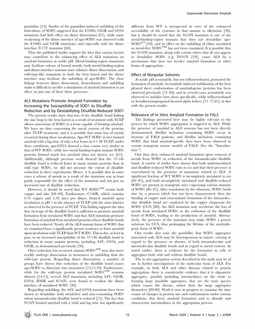
Effect of Mutations on Fibrillation of Metalated SOD1 in0.1 M TCEP at pH 7
In the presence of 0.1 M TCEP, nearly all FALS mutants
examined showed enhanced amyloid formation compared to WT
SOD1, and in a number of cases (C146R, H46R, and H80R) the
lag time was substantially reduced (Table 4).
The ALS mutant C146R, which lacks the 57-146 intramolec-
ular disulfide bond, produced a slightly lower yield of amyloid than
WT SOD1, but the lag time was much shorter. The kinetics were
similar whether or not TCEP was present, as would be expected,
since TCEP should have no effect on a protein which lacks
disulfide bonds. The result with C146R indicates that the absence
of the intramolecular disulfide bond is sufficient to allow SOD1 to
form amyloid. The only other SOD1 protein that formed amyloid
at pH 7 even in the absence of either EDTA or TCEP was H80R,
a mutant SOD1 which is nearly devoid of both copper and zinc
(Table 4) and therefore behaves like other apo-SOD1 proteins
(Figure 2).
In the presence of 0.1 M TCEP AS, AS/A4V, AS/G93A, and
AS/G85R all showed a greater yield of amyloid formation relative
to WT SOD1 (Figure 6B and Table 4). The lag time was much
shorter than that of WT for AS/A4V, AS/G93A, and AS/G85R,
but not for AS. The effect of removing cysteines 6 and 111 on the
yield of amyloid, even in the absence of ALS mutations, is difficult
to explain; the replacement of these two cysteines may either cause
some destabilization of the tertiary structure of SOD1 [42] or
promote the formation of amyloid instead of other types of
aggregates, like amorphous aggregates.
Discussion
The results presented here lead to three principal conclusions: 1)
Metal-free SOD1S-S and metalated SOD12SH self-assemble into
amyloid fibrils at physiological temperature, pH and ionic
strength. 2) Amyloid both with and without intermolecular
disulfide bonds may be formed, depending on the incubation
conditions. 3) ALS mutations can promote the assembly of SOD1
into amyloid by mechanisms which include facilitating the loss of
metals and reduction of the intramolecular disulfide bond.
SOD1 Forms Amyloid At Physiological pH, Ionic Strengthand Temperature
This work demonstrates clearly that SOD1 forms fibrillar
aggregates at physiological pH, ionic strength and temperature;
these fibrils were identified as amyloid by a variety of
ultrastructural and spectroscopic techniques. A previous study
showed that amyloid formation occurred at pH 3.5 and was
accelerated by the presence of EDTA, but no amyloid was
observed at neutral pH [16]. The present work also shows amyloid
formation at acidic pH but demonstrates further that either
removal of copper and zinc by EDTA treatment or cleavage of the
C7-C146 disulfide bond by concentrated TCEP was sufficient to
trigger amyloid formation at pH 7. Amyloid formed faster in the
presence of 1 M guanidine, but guanidine was not required. The
Figure 3. Detection of intermolecular disulfide bonds in SOD1amyloid prepared under different conditions. Amyloid producedunder each set of conditions was isolated by pelleting and subjected toSDS-PAGE in sample buffers containing 2% mercaptoethanol (‘‘red’’) orlacking any reducing agent (‘‘nr’’). Lane 1 is MW standards: 66 K, 45 K,36 K, 29 K, 24 K, 20 K, and 14.2 K. Lanes 2 and 3, metalated SOD1 in50 mM MOPS, 0.1 M NaCl, 0.1 M TCEP, pH 7; lanes 4 and 5, metalatedSOD1 in 50 mM sodium citrate, 0.1 M NaCl, 1 M guanidine hydrochlo-ride, pH 3; lanes 6 and 7, apo-SOD1 in 50 mM MOPS, 0.1 M NaCl, pH 7;lanes 8 and 9, metalated SOD1 in 50 mM MOPS, 0,1 M NaCl, 1 Mguanidine hydrochloride, 10 mM TCEP, pH 7.doi:10.1371/journal.pone.0005004.g003
Superoxide Dismutase Amyloid
PLoS ONE | www.plosone.org 6 March 2009 | Volume 4 | Issue 3 | e5004
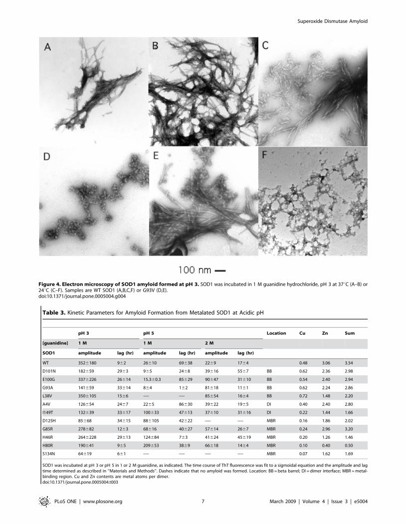
Table 3. Kinetic Parameters for Amyloid Formation from Metalated SOD1 at Acidic pH
pH 3 pH 5 Location Cu Zn Sum
[guanidine] 1 M 1 M 2 M
SOD1 amplitude lag (hr) amplitude lag (hr) amplitude lag (hr)
WT 3526180 962 26610 69638 2269 1764 0.48 3.06 3.54
D101N 182659 2963 965 2468 39616 5567 BB 0.62 2.36 2.98
E100G 3376226 26614 15.360.3 85629 90647 31610 BB 0.54 2.40 2.94
G93A 141659 33614 864 162 81618 1161 BB 0.62 2.24 2.86
L38V 3506105 1566 ---- ---- 85654 1664 BB 0.72 1.48 2.20
A4V 126654 2467 2265 86630 39622 1965 DI 0.40 2.40 2.80
I149T 132639 33617 100633 47613 37610 31616 DI 0.22 1.44 1.66
D125H 85668 34615 886105 42622 ---- ---- MBR 0.16 1.86 2.02
G85R 278682 1263 68616 40627 57614 2667 MBR 0.24 2.96 3.20
H46R 2646228 29613 124684 763 41624 45619 MBR 0.20 1.26 1.46
H80R 190641 965 209653 3869 66618 1464 MBR 0.10 0.40 0.50
S134N 64619 661 ---- ---- ---- ---- MBR 0.07 1.62 1.69
SOD1 was incubated at pH 3 or pH 5 in 1 or 2 M guanidine, as indicated. The time course of ThT fluorescence was fit to a sigmoidal equation and the amplitude and lagtime determined as described in ‘‘Materials and Methods’’. Dashes indicate that no amyloid was formed. Location: BB = beta barrel; DI = dimer interface; MBR = metal-binding region. Cu and Zn contents are metal atoms per dimer.doi:10.1371/journal.pone.0005004.t003
Figure 4. Electron microscopy of SOD1 amyloid formed at pH 3. SOD1 was incubated in 1 M guanidine hydrochloride, pH 3 at 37uC (A–B) or24uC (C–F). Samples are WT SOD1 (A,B,C,F) or G93V (D,E).doi:10.1371/journal.pone.0005004.g004
Superoxide Dismutase Amyloid
PLoS ONE | www.plosone.org 7 March 2009 | Volume 4 | Issue 3 | e5004
stimulation of amyloid formation by a restricted range of
guanidine concentrations, 0.5 to 2 M, suggests that metal loss or
disulfide cleavage facilitates conversion of SOD1 to a partially
unfolded form with an amyloidogenic conformation.
While this manuscript was in preparation, a study appeared
demonstrating the formation of amyloid from SOD1 lacking both
disulfide bonds and metals (apo-SOD12SH), but not from metal-
free SOD1 with its disulfide bond intact (apo-SOD1S-S) [13]. The
reason for this discrepancy with our results is unknown, but it is
possible that: 1) in the other study the incubations were not
conducted long enough to observe amyloid (note the long lag time
in Figure 2A), 2) the use of a lower rate of agitation (150 rpm vs.
960 rpm) in the other study made amyloid formation inefficient,
and 3) the use of Teflon balls in the present study provided
hydrophobic surfaces that promoted aggregation.
The production of amyloid required agitation of the microwell
plates and inclusion of a Teflon sphere in each well. Agitation in
the presence of Teflon has frequently been used as a tool to
accelerate amyloid formation from bona fide amyloidogenic
proteins whose aggregation requires weeks or months when such
agitation is not used. For example, wild-type a-synuclein, which is
the main constituent of the Lewy bodies found in Parkinson’s
disease, took between three and nine weeks to form fibrils when
incubated at 37uC without agitation [43], but fibrillation was
complete after 36–48 hr when the solutions were agitated with a
Teflon stir bar or with a Teflon sphere in a microplate [28,44].
Explanations proposed for effects of agitation include increased
exposure of hydrophobic groups at an air-water interface [45] or
on Teflon surfaces [37], or increased fragmentation of fibrils and
creation of more seeds to nucleate fibril formation. Teflon has
been suggested to act as a sorbent surface that mimics the
nonpolar interior of a lipid bilayer [37]. Since recent studies have
demonstrated the presence of SOD1 bound to mitochondrial
membranes [46–47] and demonstrated a change in secondary
structure of SOD1 upon binding to phospholipid vesicles [48], it is
possible that membranes seed the formation of SOD1 aggregates
in vivo.
SOD1 Can Form Amyloid With or Without IntermolecularDisulfide Bonds
Recently there has been some controversy about the role of
intermolecular disulfide bonds in SOD1 aggregation. While
disulfide-linked SOD1 aggregates have been found in transgenic
mice expressing ALS-related mutant SOD1 [49–50], and while
there is some evidence that intermolecular disulfide formation
plays a major role in SOD1 aggregation in transfected cultured
cells [51–52], other work has shown that transfected cells
expressing SOD1 variants lacking cysteine also develop SOD1
aggregates [42,53,54]. Our results demonstrate that amyloid
formation in vitro is not necessarily accompanied by the formation
of intermolecular disulfide bonds. At neutral pH, apo-AS SOD1,
which lacks free cysteines, is incapable of forming amyloid, but the
Figure 5. Amyloid formation from WT SOD1 and selectedmutant SOD1 proteins at pH 5. SOD1 was incubated as in Figure 1in 50 mM sodium acetate, 0.1 M NaCl, pH 5, plus 1 M guanidinehydrochloride (A) or 2 M guanidine hydrochloride (B). A: D125H (solidsquares), I149T (open circles), H80R (solid circles), G85R (solid triangles),WT (open triangles). B: L38V (open diamonds), E100G (solid triangles),A4V (open triangles), I149T (open circles), G85R (open squares), WT(solid diamonds).doi:10.1371/journal.pone.0005004.g005
Figure 6. Aggregation assays with mutant SOD1 lacking freesulfhydryl groups. A) Apo-WT (solid triangles), apo-AS (solid squares),apo-AS/A4V (solid circles), apo-AS/G93A (open circles), and apo-AS/G85R (open triangles) were incubated in 50 mM MOPS, 0.1 M NaCl,1 mM EDTA, pH 7, at 37uC. B) WT (solid inverted triangles), AS (solidcircles), AS/A4V (open circles), AS/G93A (open squares), and AS/G85R(solid squares) were incubated in 50 mM MOPS, 0.1 M NaCl, 0.1 M TCEP,pH 7, at 37uC.doi:10.1371/journal.pone.0005004.g006
Superoxide Dismutase Amyloid
PLoS ONE | www.plosone.org 8 March 2009 | Volume 4 | Issue 3 | e5004
presence of ALS mutations confers a tendency to form amyloid
even when free cysteines are absent (Figure 6A). DiDonato et
al.[16] observed similar behavior for AS SOD1 and ALS mutants
in an AS background for incubations at pH 3.5. Also, Banci et al.
[17–18] reported that extended incubation of apo-SOD1 led to
large oligomeric aggregates containing disulfide bonds and that
ALS mutations generally led to faster oligomerization, while apo-
AS-SOD1 did not aggregate. They did not report the properties of
AS-SOD1 bearing ALS mutations.
Furthermore, a wide variety of SOD1 proteins, both containing
and lacking free cysteines, and both with and without ALS
mutations, formed amyloid in the presence of high concentrations
of TCEP (Figures 2B and 6B and Table 4). In summary, we have
found that amyloid both with and without intermolecular disulfide
bonds may be formed, depending on the incubation conditions
(Figure 3) and thus that the occurrence of amyloid-like aggregates
per se does not depend on forming disulfide bonds.
ALS Mutations Promote Amyloid FormationThe results suggest that ALS mutations may enhance amyloid
formation by facilitating the removal of metals from SOD1 and by
increasing the susceptibility of SOD1 to reduction of its
intramolecular disulfide bond.
ALS Mutations Promote Amyloid Formation byFacilitating Loss of Metals
ALS mutations promote amyloid formation by allowing
dissociation of metals from SOD1 at pH values where metals
remain tightly bound to WT SOD1. Thus ALS mutations led to
enhanced amyloid formation at pH 5, and the effect was most
pronounced with metal-binding-region mutants (Figure 5, Table 3).
A previous study demonstrated that several metal-binding-region
mutants display increased sensitivity of zinc binding to low pH,
with the pKa for the loss of zinc binding shifted from 3.8 for the
wild type protein to higher values ranging from 4.7 to 7.3 [55]. In
the experiments shown in Table 3, H46R, whose pKa for the loss
of zinc is 6.0 [55], formed amyloid in high yield with a short lag
time at pH 5. Also, H80R, which is nearly devoid of metals,
showed a similar yield of amyloid in 1 M guanidine at pH 5 as at
pH 3, albeit with a longer lag time. The metal-binding region
mutants G85R and D125H also produced a significantly greater
yield of amyloid at pH 5 and 1 M guanidine than WT SOD1; the
zinc-binding properties of these mutants have not been reported.
Also, several wild-type-like mutants, including A4V and L38V,
were shown previously to have lower zinc affinities than the wild
type protein in the presence of 2 M urea [56]. In a physiological
context, loss of metals could be promoted by a local acidic
environment or by other cellular stresses, and this would occur
more readily with mutant SOD1. Alternatively, newly synthesized
mutant SOD1 proteins may form amyloid before metals are
coordinated. It should be noted that the failure of S134N, also a
metal-binding region mutant, to form any amyloid at pH 5 is
difficult to explain (see also below).
ALS Mutations Promote Amyloid Formation byFacilitating Partial Unfolding or Monomerization of Apo-SOD1
The presence of many wild-type-like ALS mutations leads to
reduced stability of apo-SOD1 against thermal or guanidine-
induced denaturation [57–61]. Hence the effect of the A4V and
G93A mutations in potentiating amyloid formation from apo-AS
SOD1 (Figure 6) indicates that apo-AS SOD1 bearing either of
these mutations undergoes partial unfolding under conditions
where the corresponding protein lacking ALS mutations remains
stably folded. The amount of amyloid formation is correlated with
the effect of the mutations on stability: A4V, one of the most highly
destabilized ALS mutants, forms amyloid in very high yield, with
less amyloid formed from the moderately destabilized G93A, and
G85R, the least destabilized of the three mutants, forming virtually
no amyloid. It should be noted that destabilization of apo-SOD1 is
not observed for most of the metal-binding-region mutants that
have been examined. [59].
Evidence has also been presented that dimer dissociation is an
obligatory step in SOD1 aggregation [62–64]. While apo-SOD1 is
predominantly dimeric, loss of metals makes dimers more
susceptible to dissociation by acidic pH [65], detergents [66], or
Table 4. Kinetic parameters for amyloid formation from metalated SOD1 at pH 7 in the presence or absence of TCEP.
SOD1 no TCEP 0.1 M TCEP Location Cu Zn Sum
amplitude lag (hr) amplitude lag (hr)
WT --- --- 118619 3366 0.48 3.06 3.54
G93A --- --- 305693 2663 BB 0.62 2.24 2.86
C146R 9564 562 86629 662 SS 0.74 2.58 3.32
A4V --- --- 2946141 34612 DI 0.40 2.40 2.80
G85R --- --- 204625 115624 MBR 0.24 2.96 3.20
H46R --- --- 237634 961 MBR 0.20 1.26 1.46
H80R 43613 26613 159641 361 MBR 0.10 0.40 0.50
S134N --- --- 134637 4263 MBR 0.07 1.62 1.69
AS --- --- 4126177 30611 MBR 0.46 3.06 3.52
AS/A4V --- --- 6206117 661 DI 0.36 2.12 2.48
AS/G93A --- --- 2126116 861 BB 1.00 1.48 2.48
AS/G85R --- --- 5686161 761 MBR 0.39 1.56 1.95
SOD1 was incubated in 50 mM MOPS, 0.1 M NaCl, pH 7 with or without TCEP. The time course of ThT fluorescence was fit to a sigmoidal equation and the amplitudeand lag time determined as described in ‘‘Materials and Methods’’. Dashes indicate that no amyloid was formed. Location: BB = beta barrel; DI = dimer interface;MBR = metal-binding region. Cu and Zn contents are metal atoms per dimer.doi:10.1371/journal.pone.0005004.t004
Superoxide Dismutase Amyloid
PLoS ONE | www.plosone.org 9 March 2009 | Volume 4 | Issue 3 | e5004
guanidine [14]. Studies of the guanidine-induced unfolding of the
holo-dimer of SOD1 suggested that the E100G, G85R and G93A
mutations had little effect on dimer dissociation [61], while some
weakening of the dimer interface of apo-SOD1 was observed with
the E100G and G85R mutations, and especially with the dimer
interface I113T mutation [60].
Thus the published studies support the idea that various factors
may contribute to the enhancing effect of ALS mutations on
amyloid formation at acidic pH: Metal-binding-region mutations
may facilitate release of bound metals; both metal-binding-region
and dimer-interface mutants may enhance dimer dissociation; and
wild-type-like mutations in both the beta barrel and the dimer
interface may facilitate the unfolding of apo-SOD1. The close
linkage between dimer dissociation, demetalation and unfolding
make it difficult to ascribe a stimulation of amyloid formation to an
effect on just one of these three processes.
ALS Mutations Promote Amyloid Formation byIncreasing the Susceptibility of SOD1 to DisulfideReduction and by Destabilizing Disulfide-Reduced SOD1
The present results show that loss of the disulfide bond linking
the zinc loop to the beta barrel as a result of treatment with TCEP
allows conversion of SOD1 to a form capable of forming amyloid.
We have no data concerning the metal content of the proteins
after TCEP treatment, and it is possible that some loss of metals
occurred during these incubations. Apo-WT SOD1 in fact formed
amyloid with a lag time of only a few hours in 0.1 M TCEP; under
these conditions, apo-G93A showed a time course very similar to
that of WT SOD1, while two metal-binding-region mutant SOD1
proteins formed much less amyloid (data not shown; see [67]).
Additionally, although previous work showed that the 57-146
disulfide bond is reduced faster in many mutant proteins than in
wild type SOD1, we did not measure the extent of disulfide
reduction in these experiments. Hence, it is possible that in some
cases a release of metals as a result of the mutation was at least
partly responsible for the effect of the mutation and not just an
increased rate of disulfide reduction.
However, it should be noted that WT SOD12SH retains both
copper and zinc [68–69]. Furthermore, C146R, which contains
0.74 copper and 2.58 zincs per dimer, formed amyloid upon
incubation at pH 7 in the absence of TCEP with the same kinetics
as observed in the presence of TCEP. This finding supports the idea
that cleavage of the disulfide bond is sufficient to promote amyloid
formation from metalated SOD1 and that ALS mutations promote
formation of amyloid from metalated proteins whose disulfide bonds
have been reduced. Nearly all the ALS mutant forms of SOD1 that
we examined have a significantly greater tendency to form amyloid
upon incubation with TCEP than WT SOD1. This is due, at least in
part, to an increased susceptibility of the 57-146 disulfide bond to
reduction in many mutant proteins, including A4V, G93A, and
G85R, as demonstrated previously [30].
Once reduction has occurred, mutant SOD12SH may also more
readily undergo dissociation to monomers or unfolding than the
wild-type protein. Regarding dimer dissociation, a number of
groups have shown that reduction of the disulfide bond causes
apo-SOD1 to dissociate into monomers [14,15,70]. Furthermore,
while for the wild-type protein metalated SOD12SH remains
dimeric [14,15], several ALS mutations, including A4V, G85R,
G93A, H46R and S134N, were found to weaken the dimer
interface of metalated SOD1 [30].
Regarding unfolding, the A4V and G93A mutations have been
shown to destabilize both metal-free and zinc-containing SOD1
whose intramolecular disulfide bond is reduced [71]. The fact that
S134N formed amyloid with a yield and lag time not significantly
different from WT is unexpected in view of the enhanced
accessibility of the cysteines in that mutant to alkylation [30],
but it should be noted that the S134N mutation is one of the
metal-binding-region mutants that does not destabilize apo-
SOD1S-S [59], and its effect on the unfolding of either metalated
or metal-free SOD12SH has not been examined. It is possible that
the S134N mutation, along with certain others that do not appear
to destabilize SOD1 (e.g. D101N [59]), cause ALS by a
mechanism that does not involve amyloid formation or other
forms of aggregation.
Effect of Nonpolar SolventsAt acidic pH acetonitrile, but not trifluoroethanol, promoted the
formation of amyloid. Acetonitrile-induced stabilization of the beta
pleated sheet conformation of amyloidogenic proteins has been
observed previously [72–80], and in several cases acetonitrile was
observed to stabilize beta sheet specifically, while trifluoroethanol
or hexafluoroisopropanol favored alpha helices [75–77,81], in line
with the present results.
Relevance of In Vitro Amyloid Formation to FALSThe findings presented here may be highly relevant to the
process by which SOD1 aggregation is triggered in ALS. While
the presence of amyloid in ALS neurons has not been directly
demonstrated, fibrillar inclusions containing SOD1 occur in
neurons of FALS patients, and fibrillar inclusions containing
SOD1 that bind amyloid-specific dyes have been observed in
certain transgenic mouse models of FALS. (See the ‘‘Introduc-
tion.’’)
Our data show enhanced amyloid formation upon removal of
metals from SOD1 or reduction of the intramolecular disulfide
bond. A variety of studies have shown that both undermetalated
and disulfide-reduced SOD1 exist in vivo and that their presence is
exacerbated by the presence of mutations related to ALS. A
significant fraction of WT SOD1 is incompletely metalated in vivo
[82–85], and both incompletely metalated and disulfide-reduced
SOD1 are present in transgenic mice expressing various mutants
of SOD1 [86–87]. After translation by the ribosome, SOD1 binds
zinc by a process which has not been characterized; then the
binding of copper and concomitant formation of the intramolec-
ular disulfide bond are catalyzed by the copper chaperone for
SOD1 (CCS) [88]. An ALS mutation may accelerate the loss of
metals from metalated SOD1 or the reduction of the disulfide
bond of SOD1, leading to the production of amyloid. Alterna-
tively, the presence of the mutation may make SOD1 a poorer
substrate for CCS, thus prolonging the lifetime of the amyloido-
genic form of SOD1.
Our results also raise the possibility that SOD1 aggregates
associated with ALS may be heterogeneous in nature, varying in
regard to the presence or absence of both intramolecular and
intermolecular disulfide bonds and in regard to metal content. As
noted earlier, there is evidence for the formation in cells of
aggregates both with and without disulfide bonds.
The in vitro aggregation system described in this study may be of
use in further investigations of the molecular basis of ALS. For
example, in both ALS and other diseases related to protein
aggregation, there is considerable evidence that it is oligomeric
aggregates, possibly including intermediates on the route to
forming large insoluble aggregates, that are the toxic species
which causes the disease, rather than the large aggregates
themselves [89,90]. Work is now in progress to examine the time
course of changes in protein size and conformation under various
conditions that favor amyloid formation and to identify and
characterize intermediates in the aggregation process.
Superoxide Dismutase Amyloid
PLoS ONE | www.plosone.org 10 March 2009 | Volume 4 | Issue 3 | e5004
Materials and Methods
ChemicalsAll chemicals were reagent grade. A neutralized solution of
tris(2-carboxyethyl)phosphine (Bond-BreakerTM TCEP Solution)
was purchased from Pierce Chemical Company. Mal-PEG,
polyethylene glycol of molecular weight 5000 coupled to
maleimide, was purchased from Nektar Pharmaceuticals
(mPEG-MAL).
SOD1 Expression and PurificationYEp351 expression vectors encoding both wild-type and mutant
SOD1 proteins were introduced into Saccharomyces cerevisiae, and
protein was expressed and purified as described previously [91].
Expression vectors encoding the C6A/C111S/G93A (AS/G93A)
and C6A/C111S/G85R (AS/G85R) mutants were derived from
the expression vector for the AS (C6A/C111S) mutant by
introducing appropriate point mutations with the QuikChange
Mutagenesis Kit (Stratagene). The identity of each purified protein
was confirmed by determining its molecular weight with a Perkin
Elmer Sciex API III triple quadrupole electrospray ionization mass
spectrometer. In all cases a single peak with the expected molecular
weight was observed. Apo-SOD1 was prepared by extensive dialysis
of SOD1 against 0.1 M sodium acetate, 10 mM EDTA, pH 3.8
[55], followed by dialysis against 2.25 mM potassium phosphate,
1 mM EDTA, pH 7, for storage. All glassware and plasticware were
soaked in 2% nitric acid before use.
Metal AnalysisThe copper and zinc content of each protein preparation was
determined with a Perkin-Elmer 6100DRC inductively-coupled
plasma mass spectrometer (ICP-MS). Purified protein samples
were dialyzed vs. 2 mM sodium phosphate, pH 7. They were then
diluted with metal-free 1 M nitric acid and analyzed along with a
sample of the dialysis buffer as a blank, as well as standard copper
and zinc solutions. A standardized quantity of gallium was added
to all samples as an internal standard.
Detection of Free Sulfhydryl Groups with Mal-PEGMal-PEG was dissolved in cold water to a concentration of
15 mM and immediately added to a protein solution for final
concentrations of 0.5 mg/ml protein, 50 mM 3-(N-morpholino)-
propanesulfonic acid (MOPS), pH 7, 0.1 M NaCl, 3 mM Mal-
PEG, 1% SDS. The mixture was incubated 3 hr at 37uC. The
reaction was terminated by boiling in SDS sample buffer.
Fluorescence Assay for Amyloid FormationAmyloid formation was monitored by following the increase in
the fluorescence of ThT [26–27]. A microplate assay similar to one
described previously [33] was employed. Incubation buffers
contained 50 mM citrate pH 3, formate pH 4, acetate pH 5,
succinate pH 6, or MOPS pH 7, plus 0.1 M NaCl. Forty-ml
samples containing 1 mg/ml SOD1 (65 mM polypeptide chain or
33 mM dimer) in buffers containing 10 mM ThT were pipetted
into wells of a Corning Costar 384-well microplate with
transparent bottoms, white walls, and non-binding surface
(Corning 3653). A 3/32-inch Teflon ball (McMaster-Carr, Los
Angeles) was placed in each well, and the plates were sealed with
ThermalSeal plate sealers (Excel Scientific). The plates were
incubated in a Thermo Labsystems Fluoroskan FL fluorescence
microplate reader at either 24uC or 37uC with agitation at
960 rpm, and fluorescence readings were acquired every 30 min-
utes with excitation at 444 nm and emission at 485 nm.
Quadruplicate samples were analyzed for each set of conditions.
The kinetic data (fluorescence (F) vs. time (t)) were fit to a
sigmoidal equation using Sigmaplot:
F~ Fizmitð Þz Ff zmf t� �.
1ze{ t{tð mÞ.
th i
The initial baseline is Fi+mit, the final baseline in the plateau
region is Ff+mft, and tm is the time to 50% of maximal fluorescence
increase. The following kinetic parameters were then calculated:
the lag time is given by tm22t, and the amplitude, amp, by Ff2Fi
(cf. [28]).Spectra. Soluble SOD1 was dissolved in 2 mM sodium
phosphate, pH 7. Amyloid was prepared by incubating SOD1 for
3 days in 10 mM sodium citrate, 0.1 M NaCl, pH 3, containing
either 1 M guanidine or 20% acetonitrile, then pelleting the protein
and resuspending it in 2 mM phosphate, pH 7. Absorbance spectra
for detecting Congo Red binding were measured on a Shimadzu
UV-2401PC spectrophotometer. Fluorescence spectra of
anilinonaphthalene sulfonic acid (ANS) were collected with a
Jobin-Yvon Fluoromax II spectrofluorometer using an excitation
wavelength of 370 nm and emission wavelengths of 380–650 nm.
Solutions contained 100 mM ANS and either 0 or 5 mM protein in
50 mM MOPS, 0.1 M NaCl, pH 7.
Circular dichroism spectra were acquired with a Jasco J-810
spectropolarimeter, using cylindrical cuvettes with a path length of
0.2 mm. Infrared spectra were acquired on a Nicolet 800 FTIR
spectrometer by the attenuated total reflectance method. Solutions
of SOD1 at a concentration of 0.3–0.5 mg/ml were deposited on
the surface of a germanium prism and dried to form a thin film,
and spectra were acquired.
Electron MicroscopySamples were diluted to 0.1 mg/ml, and 10-ml aliquots were
applied to 300-mesh carbon-coated copper grids with formvar
films (EM Sciences) which had been subjected to glow discharge.
Samples were allowed to adsorb for 30–60 seconds; the grids were
then blotted dry and treated for 30–60 seconds with 2% uranyl
acetate. The grids were examined in a JEOL 1200EX-II electron
microscope operated at 80 kV.
Atomic Force MicroscopyA 10-mL aliquot of the protein solution was deposited on freshly
cleaved mica. Excess water was wicked away using a small piece of
absorbent paper and the sample was placed in a desiccator to dry.
Humidity was controlled by placing the microscope under a low
flow of dry nitrogen gas.
Images were acquired at ambient temperature with a Nano-
scope IIIa Multimode scanning probe microscope (Digital
Instruments, Santa Barbara, CA) using tapping mode. Rotated
tip etched silicon probes with the J scanning head were employed.
Scanning parameters varied with individual tips and samples, but
typical ranges were as follows: tapping frequency, 300–400 kHz;
driving amplitude, 65–75 mV; and scan rate, 0.5–2 Hz. Height
and phase data were simultaneously collected using a Digital
Instrument extender phase module. Acquired images were first
plane-fitted and carefully flattened for the analysis.
Polarization MicroscopyCongo Red was added to suspensions of SOD1 amyloid to give
final concentrations of 0.3 mg/ml (19 mM) SOD1 and 45 mM
Congo Red. One hundred ml was pipetted onto a microscope slide
and allowed to dry. Excess Congo Red was removed by washing
the slide with ethanol. The slide was examined in an Olympus BX-
Superoxide Dismutase Amyloid
PLoS ONE | www.plosone.org 11 March 2009 | Volume 4 | Issue 3 | e5004
P transmitted light polarizing microscope outfitted with a 4-
megapixel digital camera.
Supporting Information
Text S1 Remetalation of SOD1
Found at: doi:10.1371/journal.pone.0005004.s001 (0.03 MB
DOC)
Text S2 Spectroscopic Characterization of Amyloid
Found at: doi:10.1371/journal.pone.0005004.s002 (0.02 MB
DOC)
Figure S1 Mal-PEG reactivity of as-isolated and remetalated
preparations. For remetalation, apo-proteins in 0.1 M sodium
acetate, pH 5.5, were incubated overnight on ice with 2
equivalents of ZnSO4 per dimer; then 0.5 equivalents of CuSO4
were added at 2-hr intervals. Protein samples were reacted with
Mal-PEG as desribed in Materials and Methods. Lane 1, WT; lane
2, remetalated WT; lane 3, A4V; lane 4, remetalated A4V; lane 5,
remetalated AS. While as-isolated WT and A4V contained
predominantly protein with one or two free cysteines per chain,
remetalated WT and A4V showed predominantly protein lacking
free cysteines. As expected, the AS mutant has no free cysteines.
Since SOD1 is not completely unfolded in 1% SDS, some of the
buried cysteines at position 6 may not have completely reacted.
Found at: doi:10.1371/journal.pone.0005004.s003 (3.08 MB TIF)
Figure S2 Atomic force microscopy of SOD1 amyloid. Amyloid
was formed by incubating SOD1 in 1 M guanidine, pH 3, at
37uC. The right-hand image is the phase image. The field of each
image is a 10 mM610 mM square.
Found at: doi:10.1371/journal.pone.0005004.s004 (1.98 MB TIF)
Figure S3 Electron microscopy of amyloid formed from apo-
and reduced SOD1. Apo-SOD1 was incubated in 50 mM citrate,
0.1 M NaCl, 1 M guanidine, pH 3 (A) or in 50 mM MOPS,
0.1 M NaCl, 1 mM EDTA, pH 7, with (B–D) or without (E–G)
1 M guanidine. Metalated SOD1 was incubated in 50 mM
MOPS, 0.1 M NaCl, pH 7, with 1 M guanidine and 10 mM
TCEP (H–I) or 0.1 M TCEP (without guanidine) (J–L). Samples
are WT SOD1 (A,B,C,D,G,H,J,K,L), E100G (E), I113T (F), or
C146R (I). Amyloid formed from apo-SOD1 in 1 M guanidine at
pH 3 (A) contained fibrils similar to those seen with metalated
SOD1. At pH 7 and 1 M guanidine, apo-SOD1 formed mostly
short fibrils which were not as highly clumped as those formed at
pH 3, along with some spherical aggregates (B–C), although long
thinner fibrils with diameters of 3–5 nm were sometimes observed
(D). Both short fibrils and mesh-like networks were observed with
both apo-SOD1 incubated at pH 7 in the absence of guanidine
(E–G) and TCEP-treated SOD1, both in the presence (H–I) or
absence (J–L) of guanidine. Spherical structures were present with
amyloid prepared from apo-SOD1 but not with amyloid prepared
from TCEP-treated SOD1.
Found at: doi:10.1371/journal.pone.0005004.s005 (6.26 MB TIF)
Figure S4 Dye-binding and spectroscopic properties of SOD1
amyloid. A. SOD1 amyloid binds Congo Red. SOD1 amyloid was
prepared by incubation for 3 days at 37uC at pH 3 and 1 M
guanidine as in Figure 1 and collected by pelleting. Spectra are
5 mM Congo Red (solid line), 5 mM Congo Red plus 10 mM SOD
(dash line), and the difference spectrum (dot-dash line). SOD1
binding caused a red shift in the absorbance with a maximum in
the difference spectrum at 542–543 nm. B. Congo Red birefrin-
gence of SOD1 amyloid. Amyloid was prepared as in Figure 1 and
examined in the presence of Congo Red in a polarization
microscope. Left and right, without and with cross-polarization.
The field of each image is 100 mm685 mm. C. Circular dichroism
of soluble SOD1 and SOD1 amyloid. Spectra are soluble SOD1
(solid line), amyloid prepared from metalated SOD1 at pH 3 in
1 M guanidine (dash line) or 20% acetonitrile (dot-dash line). D.
Fourier transform infrared spectra of soluble SOD1 and SOD1
amyloid. Spectra of soluble SOD1 (open inverted triangles),
amyloid formed from metalated SOD1 at pH 3 in 1 M guanidine
(open diamons) or 20% acetonitrile (solid squares), from apo-AS/
A4V SOD1 at pH 7 in 1 M guanidine (solid circles), or from
metalated AS/A4V SOD1 in 0.1 M TCEP (solid triangles).
Found at: doi:10.1371/journal.pone.0005004.s006 (1.71 MB TIF)
Table S1 Kinetic Parameters for Amyloid Formation from Apo-
SOD1 proteins
Found at: doi:10.1371/journal.pone.0005004.s007 (0.02 MB
DOC)
Acknowledgments
JAC acknowledges the late Anthony L. Fink for valuable mentoring and
training in the field of protein aggregation during a sabbatical leave at the
University of California, Santa Cruz. We thank Dr. Fink for reading and
commenting on an earlier version of this manuscript. The infrared spectra
were acquired and analyzed with the assistance of Mark Hokenson,
Vladimir Uversky, and Anthony J. Fink at the University of California,
Santa Cruz. The expression vectors for two of the double-cysteine mutants
of SOD1 (AS and AS/A4V) were generously provided by Stephen
Holloway and P. John Hart of the University of Texas Health Sciences
Center. Richard Behl provided assistance with polarization microscopy.
Most of these results were presented in the M. S. thesis of ZAOD [67].
Author Contributions
Conceived and designed the experiments: ZAOD JAC PD JV. Performed
the experiments: ZAOD JAC PD JKT YN CJB JLH CK. Analyzed the
data: ZAOD JAC JKT YN CJB JLH. Wrote the paper: ZAOD JAC BFS
JV. Expressed, purified and characterized proteins: JAC PD SP KE SD
AZM JAR PAD BFS.
References
1. Chiti F, Dobson CM (2006) Protein misfolding, functional amyloid and humandisease. Annu Rev Biochem 75: 333–366.
2. Cleveland DW, Rothstein JD (2001) From Charcot to Lou Gehrig: deciphering
selective motor neuron death in ALS. Nat Rev Neurosci 2: 806–819.
3. Valentine JS, Doucette PA, Potter SZ (2005) Copper-zinc superoxide dismutaseand amyotrophic lateral sclerosis. Annu Rev Biochem 74: 563–593.
4. Rakhit R, Chakrabartty A (2006) Structure, folding and misfolding of CuZnsuperoxide dismutase in amyotrophic lateral sclerosis. Biochim Biophys Acta
1762: 1025–1037.
5. Shaw BF, Valentine JS (2007) How do ALS-associated mutations in superoxidedismutase 1 promote aggregation of the protein? Trends Biochem Sci 32: 78–85.
6. Kato S, Takikawa M, Nakashima K, Hirano A, Cleveland DW, et al. (2000)
New consensus research on neuropathological aspects of familial amyotrophiclateral sclerosis with superoxide dismutase 1 (SOD1) gene mutations: inclusions
containing SOD1 in neurons and astrocytes. Amyotroph Lateral Scler Other
Motor Neuron Disord 1: 163–184.
7. Koide T, Igarashi S, Kikugawa K, Nakano R, Inuzuka T, et al. (1998)
Formation of granular cytoplasmic aggregates in COS7 cells expressing mutantCu/Zn superoxide dismutase associated with familial amyotrophic lateral
sclerosis. Neurosci Lett 257: 29–32.
8. Yamagishi A, Koyama Y, Katayama T, Taniguchi M, Hitomi J, et al. (2007) An
in vitro model for Lewy body-like hyaline inclusion/astrocytic hyaline inclusion:
Induction by ER stress with an ALS-linked SOD1 mutation. PLoS ONE e1030.
9. Stieber A, Gonatas JO, Gonatas NK (2000) Aggregates of mutant protein
appear progressively in dendrites in periaxonal processes of oligodendrocytesand in neuronal, astrocytic perikarya of mice expressing the SOD1(G93A)
mutation of familial amyotrophic lateral sclerosis. J Neurol Sci 177: 114–123.
10. Sasaki S, Warita H, Murakami T, Shibata N, Komon T, et al. (2005)
Ultrastructural study of aggregates in the spinal cord of transgenic mice with aG93A mutant SOD1 gene. Acta Neuropathol (Berl) 109: 247–255.
11. Wang J, Xu G, Gonzales V, Coonfield M, Fromholt D, et al. (2002) Fibrillar
inclusions and motor neuron degeneration in transgenic mice expressing
Superoxide Dismutase Amyloid
PLoS ONE | www.plosone.org 12 March 2009 | Volume 4 | Issue 3 | e5004
superoxide dismutase 1 with a disrupted copper-binding site. Neurobiol Dis 10:
128–138.
12. Wang J, Slunt H, Gonzales V, Fromholt D, Coonfield M, et al. (2003) Copper-
binding-site-null SOD1 causes ALS in transgenic mice: aggregates of non-native
SOD1 delineate a common feature. Hum Mol Gen 12: 2753–2764.
13. Furukawa Y, Kaneko K, Yamanaka K, O’Halloran TV, Nukina N (2008)
Complete loss of post-translational modifications triggers fibrillar aggregation of
SOD1 in familial form of ALS. J Biol Chem 283: 24167–24176.
14. Doucette PA, Whitson LJ, Cao Z, Schirf V, Demeler B, et al. (2004) Dissociation
of human copper-zinc superoxide dismutase dimers using chaotrope, reductant
Insights into the molecular basis for dimer stability. J Biol Chem 279:
54555–54566.
15. Arnesano F, Banci L, Bertini I, Martinelli M, Furakawa Y, et al. (2004) The
unusually stable quaternary structure of human CuZn-superoxide dismutase 1 is
controlled by both metal occupancy, disulfide status. J Biol Chem 279:
47998–48003.
16. DiDonato M, Craig L, Huff ME, Thayer MM, Cardoso RM, et al. (2003) ALS
mutants of human superoxide dismutase form fibrous aggregates via framework
destabilization. J Mol Biol 332: 601–615.
17. Banci L, Bertini I, Durazo A, Girotto S, Gralla EB, et al. (2007) Metal-free
superoxide dismutase forms soluble oligomers under physiological conditions: a
possible general mechanism for familial ALS. Proc Natl Acad Sci USA 104:
11263–11267.
18. Banci L, Bertini I, Boca M, Girotto S, Martinelli M, et al. (2008) SOD1 and
amyotrophic lateral sclerosis: mutations and oligomerization. PLoS ONE 3I:
e1677.
19. Lepock JR, Frey HE, Hallewell RA (1990) Contribution of conformational
stability and reversibility of unfolding to the increased thermostability of human,
bovine superoxide dismutase mutated at free cysteines. J Biol Chem 235:
21612–21618.
20. Hallewell RA, Imlay KC, Lee P, Fong NM, Gallegos C, et al. (1991)
Thermostabilization of recombinant human, bovine CuZn superoxide dismu-
tases by replacement of free cysteines. Biochem Biophys Res Commun 181:
474–480.
21. Parge HE, Hallewell RA, Tainer JA (1992) Atomic structures of wild-type and
thermostable mutant recombinant human CuZn superoxide dismutase. Proc
Natl Acad Sci USA 89: 6109–6113.
22. Hayward LJ, Rodriguez JA, Kim JW, Tiwari A, Goto JJ, et al. (2002) Decreased
metallation and activity in subsets of mutant superoxide dismutases associated
with familial amyotrophic lateral sclerosis. J Biol Chem 277: 15923–15931.
23. Goto JJ, Zhu H, Sanchez RJ, Nersissian A, Gralla EB, et al. (2000) Loss of in
vitro metal ion binding specificity in mutant copper-zinc superoxide dismutases
associated with familial amyotrophic lateral sclerosis. J Biol Chem 275:
1007–1014.
24. Strange RW, Antonyuk SV, Hough MA, Doucette PA, Valentine JS, et al.
(2006) Variable metallation of human superoxide dismutase: atomic resolution
crystal structures of Cu-Zn Zn-Zn, as-isolated wild-type enzymes. J Mol Biol
356: 1152–1162.
25. Boissinot M, Karnas S, Lepock JR, Cabelli DE, Tainer JA, et al. (1997) Function
of the Greek key connection analysed using circular permutants of superoxide
dismutase. EMBO J 16: 2171–2178.
26. Naiki H, Higuchi K, Hosokawa M, Takeda T (1989) Fluorometric determina-
tion of amyloid fibrils in vitro using the fluorescent dye thioflavin T1. Anal
Biochem 177: 244–249.
27. LeVine H 3rd (1993) Thioflavine T interaction with synthetic Alzheimer’s
disease beta-amyloid peptides: detection of amyloid aggregation in solution.
Protein Sci 2: 404–409.
28. Cohlberg JA, Li J, Uversky VN, Fink AL (2002) Heparin and other
glycosaminoglycans stimulate the formation of amyloid fibrils from alpha-
synuclein in vitro. Biochemistry 41: 1502–1511.
29. Barford D (2004) The role of cysteine residues as redox-sensitive regulatory
switches. Curr Opinion Struct Biol 6: 679–686.
30. Tiwari A, Hayward LJ (2003) Familial ALS mutants of Cu/Zn superoxide
dismutase are susceptible to disulfide reduction. J Biol Chem 278: 5984–5992.
31. Klunk WE, Pettegrew JW, Abraham DJ (1989) Two simple methods for
quantifying low-affinity dye-substrate binding. J Histochem Cytochem 37:
1293–1297.
32. Klunk WE, Jacob RF, Mason RP (1999) Quantifying amyloid by congo red
spectral shift assay. Methods Enzymol 309: 285–305.
33. Divry P (1927) Etude histochimique des plaques seniles. J Belge Neurol Psychiat
27: 643–657.
34. Westermark GT, Johnson KH, Westermark P (1999) Staining methods for
identification of amyloid in tissue. Methods Enzymol 309: 3–25.
35. Huang TH, Yang DS, Plaskos NP, Go S, Yip CM, et al. (2000) Structural studies
of soluble oligomers of the Alzheimer beta-amyloid peptide. J Mol Biol 297:
73–87.
36. Hong D-P, Gozu M, Hasegawa K, Naiki H, Goto Y (2002) Conformation of
beta 2-microglobulin amyloid fibrils analyzed by reduction of the disulfide bond.
J Biol Chem 277: 21554–21560.
37. Giacomelli CE, Norde W (2003) Influence of hydrophobic Teflon particles on
the structure of amyloid beta-peptide. Biomacromol 4: 1719–1726.
38. Chiba T, Hagihara Y, Higurashi T, Hasegawa K, Naiki H, et al. (2003) Amyloid
fibril formation in the context of full-length protein: effects of proline mutations
on the amyloid fibril formation of beta2-microglobulin. J Biol Chem 278:
47016–47024.
39. Bieler S, Estrada L, Lagos R, Baeza M, Castilla J, et al. (2005) Amyloid
formation modulates the biological activity of a bacterial protein. J Biol Chem
280: 26880–26885.
40. Yagi H, Kusaka E, Hongo K, Mizobata T, Kawata Y (2005) Amyloid fibril
formation of alpha-synuclein is accelerated by preformed amyloid seeds of other
proteins: implications for the mechanism of transmissible conformational
diseases. J Biol Chem 280: 38609–38616.
41. Zandomeneghi G, Krebs MR, McCammon MG, Fandrich M (2004) FTIR
reveals structural differences between native beta-sheet proteins and amyloid
fibrils. Protein Sci 13: 3314–13321.
42. Furukawa Y, O’Halloran TV (2006) Posttranslational modifications in CuZn-
superoxide dismutase, mutations associated with amyotrophic lateral sclerosis.
Antioxid Redox Signal 8: 847–867.
43. Conway KA, Harper JD, Lansbury PT (1998) Accelerated in vitro fibril
formation by a mutant alpha-synuclein linked to early-onset Parkinson disease.
Nature Medicine 4: 1318–1320.
44. Uversky VN, Li J, Fink AL (2001) Evidence for a partially folded intermediate in
alpha-synuclein fibril formation. J Biol Chem 276: 10737–10344.
45. Schladitz C, Vieira EP, Hermel H, Mohwald H (1999) Amyloid-beta-sheet
formation at the air-water interface. Biophys J 77: 3305–3310.
46. Ahtoniemi T, Jaronen M, Keksa-Goldsteine V, Goldsteins G, Koistinaho J
(2008) Neurobiol Dis 32: 479–485.
47. Vande Velde C, Miller TM, Cashman NR, Cleveland DW (2008) Proc Natl
Acad Sci USA 105: 4022–7.
48. Aisenbrey C, Borowik T, Bystrom R, Bokvist M, Lindstrom F, et al. (2008) Eur
Biophys J 37: 247–255.
49. Deng HX, Shi Y, Furukawa Y, Zhai H, Fu R, et al. (2006) Conversion to the
amyotrophic lateral sclerosis phenotype is associated with intermolecular linked
insoluble aggregates of SOD1 in mitochondria. Proc Natl Acad Sci USA 103:
7142–7147.
50. Furukawa Y, Fu R, Deng HX, Siddique T, O’Halloran TV (2006) Disulfide
cross-linked protein represents a significant fraction of ALS-associated Cu Zn-
superoxide dismutase aggregates in spinal cords of model mice. Proc Natl Acad
Sci USA 103: 7148–7153.
51. Niwa J, Yamada S, Ishigaki S, Sone J, Takahashi M, et al. (2007) Disulfide bond
mediates aggregation, toxicity and ubiquitylation of familial amyotrophic lateral
sclerosis-linked mutant SOD1. J Biol Chem 282: 28087–28095.
52. Cozzolino M, Amori I, Pesaresi MG, Ferri A, Nencini M, et al. (2008) Cysteine
111 affects aggregation, cytotoxicity of mutant CuZn-superoxide dismutase
associated with familial amyotrophic lateral sclerosis. J Biol Chem 283: 866–874.
53. Wang J, Xu G, Borchelt DR (2006) Mapping superoxide dismutase 1 domains of
non-native interaction: roles of intra- and intermolecular disulfide bonding in
aggregation. J Neurochem 96: 1277–1288.
54. Karch CM, Borchelt DR (2008) A limited role for disulfide cross-linking in the
aggregation of mutant SOD1 linked to familial amyotrophic lateral sclerosis.
J Biol Chem 283: 13528–13537.
55. Lyons TJ, Nersissian A, Huang H, Yeom H, Nishida CR, et al. (2000) The metal
binding properties of the zinc site of yeast copper-zinc superoxide dismutase:
implications for amyotrophic lateral sclerosis. J Biol Inorg Chem 5: 189–203.
56. Crow JP, Sampson JB, Zhuang Y, Thompson JA, Beckman JS (1997) Decreased
zinc affinity of amyotrophic lateral sclerosis-associated mutants leads to
enhanced catalysis of tyrosine nitration by peroxynitrite. J Neurochem 69:
1936–1944.
57. Rodriguez JA, Valentine JS, Eggers DK, Roe JA, Tiwari A, et al. (2002) Familial
amyotrophic lateral sclerosis-associated mutations decrease the thermal stability
of distinctly metallated species of human copper/zinc superoxide dismutase.
J Biol Chem 277: 15932–15937.
58. Lindberg MJ, Tibell L, Oliveberg M (2002) Common denominator of Cu/Zn
superoxide dismutase mutants associated with amyotrophic lateral sclerosis:
decreased stability of the apo state. Proc Natl Acad Sci USA 99: 16607–16612.
59. Rodriguez JA, Shaw BF, Durazo A, Sohn SH, Doucette PA, et al. (2005)
Destabilization of apoprotein is insufficient to explain CuZn-superoxide
dismutase-linked ALS pathogenesis. Proc Natl Acad Sci USA 102:
10516–10521.
60. Vassall KA, Stathopulos PB, Rumfeldt JA, Lepock JR, Meiering EM (2006)
Equilibrium thermodynamic analysis of amyotrophic lateral sclerosis-associated
mutant apo CuZn superoxide dismutases. Biochemistry 45: 7366–7379.
61. Rumfeldt JA, Stathopulos PB, Chakrabarrty A, Lepock JR, Meiering EM (2006)
Mechanism and thermodynamics of guanidinium chloride-induced denaturation
of ALS-associated mutant CuZn superoxide dismutases. J Mol Biol 355:
106–123.
62. Khare SD, Caplow M, Dokholyan NV (2004) The rate and equilibrium
constants for a multistep reaction sequence for the aggregation of superoxide
dismutase in amyotrophic lateral sclerosis. Proc Natl Acad Sci USA 101:
15094–15099.
63. Ray SS, Nowak RJ, Strokovich K, Brown RH, Walz T, et al. (2004) An
intersubunit disulfide bond prevents in vitro aggregation of a superoxide
dismutase-1 mutant linked to familial amyotrophic lateral sclerosis. Biochemistry
43: 4899–4905.
64. Rakhit R, Crow JP, Lepock JR, Kondejewski LH, Cashman NR, et al. (2004)
Monomeric CuZn-superoxide dismutase is a common misfolding intermediate
Superoxide Dismutase Amyloid
PLoS ONE | www.plosone.org 13 March 2009 | Volume 4 | Issue 3 | e5004
in the oxidation models of sporadic, familial amyotrophic lateral sclerosis. J Biol
Chem 279: 15499–15504.65. Fee JA, Phillips WD (1975) The behavior of holo- and apo-forms of bovine
superoxide dismutase at low pH. Biochim Biophys Acta 412: 26–38.
66. Hartz JW, Deutsch HF (1972) Subunit structure of human superoxidedismutase. J Biol Chem 247: 7043–7050.
67. Oztug ZA (2005) Formation of amyloid from human copper zinc-superoxidedismutase. MS Thesis, California State University, Long Beach.
68. Banci L, Benedetto M, Bertini I, Del Conte R, Piccioli M, et al. (1998) Solution
structure of reduced monomeric Q133M2 copper zinc superoxide dismutase(SOD). Why is SOD a dimeric enzyme? Biochemistry 37: 11780–11791.
69. Hough MA, Hasnain SS (2003) Structure of fully reduced bovine copper zincsuperoxide dismutase at 115 A. Structure 11: 937–946.
70. Lindberg MJ, Normark J, Holmgren A, Oliveberg M (2004) Folding of humansuperoxide dismutase: disulfide reduction prevents dimerization, produces
marginally stable monomers. Proc Natl Acad Sci USA 101: 15893–15898.
71. Furukawa Y, O’Halloran TV (2005) Amyotrophic lateral sclerosis mutationshave the greatest destabilizing effect on the apo- and reduced from of SOD1,
leading to unfolding and oxidative aggregation. J Biol Chem 280: 17266–74.72. Fabian H, Szendrei GI, Mantsch HH, Greenberg BD, Otvos L Jr (1994)
Synthetic post-translationally modified human A beta peptide exhibits a
markedly increased tendency to form beta-pleated sheets in vitro.Eur J Biochem 221: 959–964.
73. Jarvis JA, Kirkpatrick A, Craik DJ (1994) 1H NMR analysis of fibril-formingpeptide fragments of transthyretin. Int J Pept Protein Res 44: 388–398.
74. Shen CL, Murphy RM (1995) Solvent effects on self-assembly of beta-amyloidpeptide. Biophys J 69: 640–651.
75. Zhang H, Kaneko K, Nguyen JT, Livshits TL, Baldwin MA, et al. (1995)
Conformational transitions in peptides containing two putative alpha-helices ofthe prion protein. J Mol Biol 250: 514–526.
76. Heller J, Kolbert AC, Larsen R, Ernst M, Bekker T, et al. (1996) Solid-stateNMR studies of the prion protein H1 fragment. Protein Sci 5: 1655–1661.
77. El-Agnaf OM, Bodles AM, Guthrie DJ, Harriott P, Irvine GB (1998) The N-
terminal region of non-A beta component of Alzheimer’s disease amyloid isresponsible for its tendency to assume beta-sheet, aggregate to form fibrils.
Eur J Biochem 258: 157–163.78. Gross M, Wilkins DK, Pitkeathly MC, Chung EW, Higham C, et al. (1999)
Formation of amyloid fibrils by peptides derived from the bacterial cold shockprotein. CspB Protein Sci 8: 1350–1357.
79. Tcherkasskaya O, Sanders W, Chynwat V, Davidson EA, Orser CS (2003) The
role of hydrophobic interactions in amyloidogenesis: example of prion-related
polypeptides. J Biomol Struct Dyn 21: 353–365.
80. McDuff FO, Doucet A, Beauregard M (2004) Low concentration of guanidine
hydrochloride induces the formation of an aggregation-prone state in alpha-
urease. Biochem Cell Biol 82: 305–313.
81. Dennison SM, Greenfield N, Lenard J, Lentz BR (2002) VSV transmembrane
domain (TMD) peptide promotes PEG-mediated fusion of liposomes in a
conformationally sensitive fashion. Biochemistry 41: 14925–14934.
82. Petrovic N, Comi A, Ettinger MJ (1996) Identification of an apo-superoxide
dismutase (CuZn) pool in human lymphoblasts. J Biol Chem 271: 28331–28334.
83. Petrovic N, Comi A, Ettinger MJ (1996) Copper incorporation into superoxide
dismutase in Menkes lymphoblasts. J Biol Chem 271: 28335–28340.
84. Bartnikas TB, Gitlin JD (2003) Mechanisms of biosynthesis of mammalian
copper/zinc superoxide dismutase. J Biol Chem 278: 33602–33608.
85. Choi J, Rees HD, Weintraub ST, Levey AI, Chin LS, et al. (2005) Oxidative
modifications, aggregation of CuZn-superoxide dismutase associated with
Alzheimer and Parkinson diseases. J Biol Chem 280: 11648–11655.
86. Jonsson PA, Graffmo KS, Andersen PM, Brannstrom T, Lindberg M, et al.
(2006) Disulphide-reduced superoxide dismutase-1 in CNS of transgenic
amyotrophic lateral sclerosis models. Brain 129: 451–464.
87. Zetterstrom P, Stewart HG, Bergemalm D, Jonsson PA, Graffmo K, et al. (2007)
Soluble misfolded subfractions of mutant superoxide dismutase-1s are enriched
in spinal cords throughout life in murine ALS models. Proc Natl Acad Sci U S A
104: 14157–14162.
88. Culotta VC, Yang M, O’Halloran TV (2006) Activation of superoxide
dismutases: putting the metal to the pedal. Biochim Biophys Acta 1763:
747–758.
89. Caughey B, Lansbury PT (2003) Protofibrils pores fibrils, neurodegeneration:
separating the responsible protein aggregates from the innocent bystanders.
Annu Rev Neurosci 26: 267–298.
90. Glabe CG (2006) Common mechanisms of amyloid oligomer pathogenesis in
degenerative disease. Neurobiol Aging 27: 570–575.
91. Hough MA, Grossmann JG, Antonyuk SV, Strange RW, Doucette PA, et al.
(2004) Dimer destabilization in superoxide dismutase may result in disease-
causing properties: structures of motor neuron disease mutants. Proc Natl Acad
Sci USA 101: 5976–5981.
Superoxide Dismutase Amyloid
PLoS ONE | www.plosone.org 14 March 2009 | Volume 4 | Issue 3 | e5004