Mechanisms, models and biomarkers in amyotrophic lateral sclerosis
14
Correspondence: M. R. Turner, Clinical Neurosciences, West Wing Level 3, John Radcliffe Hospital, Oxford OX3 9DU, UK. E-mail: [email protected] Amyotrophic Lateral Sclerosis and Frontotemporal Degeneration, 2013; 14(Suppl. 1): 19–32 ISSN 2167-8421 print/ISSN 2167-9223 online © 2013 Informa Healthcare DOI: 10.3109/21678421.2013.778554 region, there is little diagnostic doubt and few tan- gible mimics. However, such obviously mixed clinical signs may not be apparent at presentation (or indeed ever) for a large proportion of patients, who are nonetheless considered to form part of the wider syndrome of ALS (1). This syndrome is currently unified by the post mortem finding of cytoplasmic inclusions of the protein TDP-43, with the notable exception of those with mutant SOD1 gene-associated ALS (approximately 1% of Introduction There is an intense need to establish biomarkers sensitive to diagnosis, prognostic stratification and disease activity of ALS (Table I). The diagnosis of amyotrophic lateral sclerosis (ALS) currently depends upon the opinion of an experienced neurologist, and the exclusion of potential mimic disorders. In the context of progressive weakness, where there are typical upper (UMN) and lower motor neuron (LMN) signs within the same body ORIGINAL ARTICLE Mechanisms, models and biomarkers in amyotrophic lateral sclerosis MARTIN R. TURNER 1 , ROBERT BOWSER 2 , LUCIE BRUIJN 3 , LUC DUPUIS 4,5 , ALBERT LUDOLPH 5 , MICHAEL MCGRATH 6 , GIOVANNI MANFREDI 7 , NICHOLAS MARAGAKIS 8 , ROBERT G. MILLER 9 , SETH L. PULLMAN 10 , SEWARD B. RUTKOVE 11 , PAMELA J. SHAW 12 , JEREMY SHEFNER 13 & KENNETH H. FISCHBECK 14 1 Nuffield Department of Clinical Neurosciences, University of Oxford, UK, 2 Division of Neurology, Barrow Neurological Institute, Phoenix, Arizona, 3 The ALS Association, National Office,Washington DC, USA, 4 INSERM U692 & Université de Strasbourg, Strasbourg, France, 5 Department of Neurology, Ulm University, Ulm, Germany, 6 UCSF, San Francisco, CA, 7 Department of Neurology and Neuroscience,Weill Medical College of Cornell University, New York, NY, 8 Johns Hopkins University, Department of Neurology, Baltimore, Maryland, 9 Forbes Norris ALS Research Center, California Pacific Medical Center, San Francisco, California, 10 Columbia University, New York, NY, 11 Beth Israel Deaconess Medical Center, Harvard Medical School, Boston, Massachusetts, USA, 12 Department of Neuroscience, Sheffield Institute for Translational Neuroscience, University of Sheffield, UK, 13 Department of Neurology, SUNY Upstate Medical University, Syracuse, NY, and 14 Neurogenetics Branch, National Institute of Neurological Disorders and Stroke, National Institutes of Health, Bethesda, Maryland, USA Abstract The last 30 years have seen a major advance in the understanding of the clinical and pathological heterogeneity of amyo- trophic lateral sclerosis (ALS), and its overlap with frontotemporal dementia. Multiple, seemingly disparate biochemical pathways converge on a common clinical syndrome characterized by progressive loss of upper and lower motor neurons. Pathogenic themes in ALS include excitotoxicity, oxidative stress, mitochondrial dysfunction, neuroinflammation, altered energy metabolism, and most recently RNA mis-processing. The transgenic rodent, overexpressing mutant superoxide dismutase-1, is now only one of several models of ALS pathogenesis. The nematode, fruit fly and zebrafish all offer fresh insight, and the development of induced pluripotent stem cell-derived motor neurons holds promise for the screening of candidate therapeutics. The lack of useful biomarkers in ALS contributes to diagnostic delay, and the inability to stratify patients by prognosis may be an important factor in the failure of therapeutic trials. Biomarkers sensitive to disease activ- ity might lessen reliance on clinical measures and survival as trial endpoints and reduce study length. Emerging proteomic markers of neuronal loss and glial activity in cerebrospinal fluid, a cortical signature derived from advanced structural and functional MRI, and the development of more sensitive measurements of lower motor neuron physiology are leading a new phase of biomarker-driven therapeutic discovery. Key words: ALS, biomarkers, pathogenesis, neuroimaging, neurophysiology Amyotrophic Lateral Sclerosis and Frontotemporal Degeneration Downloaded from informahealthcare.com by 80.78.232.26 on 05/20/14 For personal use only.
-
Upload
independent -
Category
Documents
-
view
1 -
download
0
Transcript of Mechanisms, models and biomarkers in amyotrophic lateral sclerosis

Correspondence: M. R. Turner, Clinical Neurosciences, West Wing Level 3, John Radcliffe Hospital, Oxford OX3 9DU, UK. E-mail: [email protected]
Amyotrophic Lateral Sclerosis and Frontotemporal Degeneration, 2013; 14(Suppl. 1): 19–32
ISSN 2167-8421 print/ISSN 2167-9223 online © 2013 Informa HealthcareDOI: 10.3109/21678421.2013.778554
region, there is little diagnostic doubt and few tan-gible mimics. However, such obviously mixed clinical signs may not be apparent at presentation (or indeed ever) for a large proportion of patients, who are nonetheless considered to form part of the wider syndrome of ALS (1). This syndrome is currently unifi ed by the post mortem fi nding of cytoplasmic inclusions of the protein TDP-43, with the notable exception of those with mutant SOD1 gene-associated ALS (approximately 1% of
Introduction
There is an intense need to establish biomarkers sensitive to diagnosis, prognostic stratifi cation and disease activity of ALS (Table I). The diagnosis of amyotrophic lateral sclerosis (ALS) currently depends upon the opinion of an experienced neurologist, and the exclusion of potential mimic disorders. In the context of progressive weakness, where there are typical upper (UMN) and lower motor neuron (LMN) signs within the same body
ORIGINAL ARTICLE
Mechanisms, models and biomarkers in amyotrophic lateral sclerosis
MARTIN R. TURNER 1 , ROBERT BOWSER 2 , LUCIE BRUIJN 3 , LUC DUPUIS 4,5 , ALBERT LUDOLPH 5 , MICHAEL MCGRATH 6 , GIOVANNI MANFREDI 7 , NICHOLAS MARAGAKIS 8 , ROBERT G. MILLER 9 , SETH L. PULLMAN 10 , SEWARD B. RUTKOVE 11 , PAMELA J. SHAW 12 , JEREMY SHEFNER 13 & KENNETH H. FISCHBECK 14
1 Nuffi eld Department of Clinical Neurosciences, University of Oxford, UK, 2 Division of Neurology, Barrow Neurological Institute, Phoenix, Arizona, 3 The ALS Association, National Offi ce, Washington DC, USA, 4 INSERM U692 & Universit é de Strasbourg, Strasbourg, France, 5 Department of Neurology, Ulm University, Ulm, Germany, 6 UCSF, San Francisco, CA, 7 Department of Neurology and Neuroscience, Weill Medical College of Cornell University, New York, NY, 8 Johns Hopkins University, Department of Neurology, Baltimore, Maryland, 9 Forbes Norris ALS Research Center, California Pacifi c Medical Center, San Francisco, California, 10 Columbia University, New York, NY, 11 Beth Israel Deaconess Medical Center, Harvard Medical School, Boston, Massachusetts, USA, 12 Department of Neuroscience, Sheffi eld Institute for Translational Neuroscience, University of Sheffi eld, UK, 13 Department of Neurology, SUNY Upstate Medical University, Syracuse, NY, and 14 Neurogenetics Branch, National Institute of Neurological Disorders and Stroke, National Institutes of Health, Bethesda, Maryland, USA
Abstract The last 30 years have seen a major advance in the understanding of the clinical and pathological heterogeneity of amyo-trophic lateral sclerosis (ALS), and its overlap with frontotemporal dementia. Multiple, seemingly disparate biochemical pathways converge on a common clinical syndrome characterized by progressive loss of upper and lower motor neurons. Pathogenic themes in ALS include excitotoxicity, oxidative stress, mitochondrial dysfunction, neuroinfl ammation, altered energy metabolism, and most recently RNA mis-processing. The transgenic rodent, overexpressing mutant superoxide dismutase-1, is now only one of several models of ALS pathogenesis. The nematode, fruit fl y and zebrafi sh all offer fresh insight, and the development of induced pluripotent stem cell-derived motor neurons holds promise for the screening of candidate therapeutics. The lack of useful biomarkers in ALS contributes to diagnostic delay, and the inability to stratify patients by prognosis may be an important factor in the failure of therapeutic trials. Biomarkers sensitive to disease activ-ity might lessen reliance on clinical measures and survival as trial endpoints and reduce study length. Emerging proteomic markers of neuronal loss and glial activity in cerebrospinal fl uid, a cortical signature derived from advanced structural and functional MRI, and the development of more sensitive measurements of lower motor neuron physiology are leading a new phase of biomarker-driven therapeutic discovery.
Key words: ALS , biomarkers , pathogenesis , neuroimaging , neurophysiology
Am
yotr
ophi
c L
ater
al S
cler
osis
and
Fro
ntot
empo
ral D
egen
erat
ion
Dow
nloa
ded
from
info
rmah
ealth
care
.com
by
80.7
8.23
2.26
on
05/2
0/14
For
pers
onal
use
onl
y.

20 M. R. Turner et al.
all cases) in whom the disease is nonetheless clin-ically indistinguishable (2). The failure of multiple drugs tried for the treatment of ALS may in part be a function of their application relatively late in the course of the disease, given the average diagnostic delay of one year (3). It is not yet known when the initial pathological changes of ALS begin, but it seems likely that the clinical manifestations occur signifi cantly downstream of what may be rapidly irreversible primary events at the cellular level. Earlier diagnosis would permit introduction of therapies nearer to these initiating events.
ALS is usually an apparently sporadic disorder and the insidious clinical onset involves symptoms common to more benign disorders, which means that a signifi cant proportion of the diagnostic latency may remain inaccessible. At the time of diagnosis, however, there is additional value in identifying biomarkers sensitive to the recognized variation in progression rate. Although the median survival from symptom onset in ALS is less than three years, the second half of the survival curve is longer, and beyond a decade for at least 5% of patients. The presence of relatively ‘ pure ’ UMN or LMN clinical signs is associated with longer sur-vival (4,5), with the rare UMN-only variant pri-mary lateral sclerosis consistently at the extreme end of long survival. However, such clinical fi nd-ings cannot be reliably used prognostically in isola-tion. A quantifi able prognostic measure might also be used to stratify patients within clinical trials to detect meaningful benefi t in the shortest time. Knowledge of an individual patient ’ s course at the time of diagnosis would facilitate effective care planning and resource targeting.
Finally, biomarkers sensitive to disease activity are the most needed. At present, clinical trials in ALS generally rely on tracheostomy-free survival, func-tional status such as the revised ALS Functional Rat-ing Scale (ALSFRS-R), or both, as the primary outcome measures. Such trials are lengthy (12 – 18 months typically) and may be prohibitively costly. A number of methods have been proposed to optimize and shorten such studies (6), but a robust, quantita-tive monitoring biomarker would have obvious value in this regard.
Biological themes
With the linking of familial ALS to several appar-ently disparate genes, it seems increasingly likely that ALS represents a common fi nal endpoint. A deeper understanding of the common molecular pathways involved in ALS pathogenesis would facilitate the identifi cation of biomarkers. Three genes linked to familial ALS in recent years, namely TARDBP , FUS and C9orf72 , indicate a potentially pivotal role for RNA mis-processing in the pathogenesis of ALS (see Al-Chalabi et al., companion paper). Downstream of these events, cellular biological studies over the last two decades have led to the development of multiple interconnected pathogenic themes in ALS (7).
Excitotoxicity
Excitotoxicity encapsulates a process of neuronal death ultimately mediated by cellular calcium infl ux, triggered via excessive stimulation of receptors for glutamate. The fi nding of raised levels of glutamate in the CSF in ALS (8), with a sensitivity of only 40% in a later study and correlation with disease severity (9), provides support for this.
The astrocytic glutamate transporter EAAT2 is responsible for the clearance of synaptic glutamate, and its knockout in transgenic mice results in neu-ronal death (10). Reduced levels of spinal cord EAAT2 protein have been noted in end-stage rodent transgenic models of ALS (11,12), with abnormali-ties of EAAT2 protein expression demonstrable in up to 80% of human post mortem brain and spinal cord tissue (13). EAAT2 receptor dysfunction has been directly linked to mutant (but not wild-type) SOD1 in the presence of hydrogen peroxide (14). Overexpression of EAAT2 in mutant SOD1 trans-genic mice then delayed the onset of motor defi cits (15,16). This role for astrocytes supports the view that motor neuron degeneration in ALS is not cell-autonomous. Although the exact mechanism of action is uncertain, the only disease-modifying drug in ALS to date, namely riluzole, appears to have broadly anti-glutamatergic activity (17).
Glutamate acts through inotropic (NMDA, AMPA and kainate) and metabotropic receptors (MGluRs), and the relative contribution of these to
Table I. Types of biomarker in ALS, their value and the current gold standards.
Biomarker type Value Current benchmarks
Diagnostic Initiate therapy earlier; Exclude ALS
Neurological history & examination Electromyography (Revised El Escorial/Awaji criteria) 131
Prognostic Identify patterns of progression: 1. Improved stratifi cation in therapeutic trials 2. Timely intervention and optimal care
e.g. gastrostomy, non-invasive ventilation, cognitive support
Diagnostic latency Neurological evaluation (e.g. clinical phenotypes) Cox modelling of clinical variables 132,133
Monitoring Identify ineffective drugs earlier Revised ALS Functional Rating Score 134 (Electrical impedance myography emergent 102 )
Am
yotr
ophi
c L
ater
al S
cler
osis
and
Fro
ntot
empo
ral D
egen
erat
ion
Dow
nloa
ded
from
info
rmah
ealth
care
.com
by
80.7
8.23
2.26
on
05/2
0/14
For
pers
onal
use
onl
y.
Mechanisms, models and biomarkers in ALS 21
ALS pathogenesis remains uncertain. The develop-ment of novel selective positron emission tomogra-phy (PET) ligands may provide clarifi cation. It is possible that excitotoxicity arises in ALS as a result of wider ‘ upstream ’ impairments of energy metabo-lism in ALS so that ‘ normal ’ levels of glutamate become toxic. There is also a body of evidence, including from in vivo neuroimaging, indicating an overall loss of neuronal inhibitory infl uence in ALS, perhaps through a primary interneuronal dysfunc-tion, resulting in excitotoxicity through less balanced glutamatergic activity (18).
Oxidative stress
Oxidative stress arises from an alteration in the bal-ance between the generation of reactive oxygen spe-cies (ROS) and their removal, together with the ability of the biological system to remove or repair ROS induced damage. The cumulative effect of oxi-dative stress in aged non-replicating neurons, may be one important factor that tips the balance of homeo-static control mechanisms from an ability to cope with a toxic insult such as the presence of a disease-causing mutation, into a vicious cycle of cellular injury culminating in neuronal death and the onset of neurodegeneration in middle or later life. Oxida-tive stress causes structural damage (including DNA (19)) and also changes in redox-sensitive signalling pathways. The initial interest in the role of oxidative stress in ALS (20) was given new impetus with the discovery that mutations in SOD1 , which encodes a major anti-oxidant defence protein, accounted for approximately 20% of cases of familial ALS (21). It is clear that oxidative stress interacts with and poten-tially exacerbates other pathophysiological processes contributing to motor neuron injury, including exci-totoxicity, mitochondrial impairment, protein aggre-gation, endoplasmic reticulum stress and alterations in signalling from astrocytes and microglia. Several potential markers of free radical damage have been found in the CSF and blood from ALS patients (reviewed in (22)), and also in urine (23).
Cellular ROS are generated as by-products of aerobic metabolism, predominantly due to leakage of electrons from the mitochondrial respiratory chain, but with contributions from other intracel-lular enzyme systems including xanthine oxidase and cytochrome P450. ROS that are initially formed such as superoxide and hydrogen peroxide may undergo further reaction to produce more potent oxidant species including peroxynitrite and hydroxyl radicals. Biochemical indices of oxidative damage to proteins, lipids and DNA in excessive quantities compared to controls, can be found in post mortem tissue from apparently sporadic and SOD1 -related familial cases. Oxidative damage to RNA species has also been documented, adding to the evidence that alteration in mRNA processing is an impor-tant pathophysiological mechanism in ALS (24).
Indices of oxidative damage are also present in cel-lular and murine models of SOD1 -related ALS, and interestingly the SOD1 protein itself appears to be particularly susceptible to oxidative post-transla-tional modifi cation. The recent development of cel-lular models of mutant TDP-43-related ALS indicates that the presence of mutant TDP-43 also provokes oxidative stress within motor neuronal cell lines (25).
Sources of oxidative stress in ALS have been investigated most thoroughly in mutant SOD1 (mSOD1) models, where several aberrant oxidative reactions have been proposed. However, enzymati-cally inactive SOD1, depleted of copper loading, is still capable of causing motor neuron degeneration, and mSOD1 may cause oxidative stress by mecha-nisms beyond its own catalytic activity (26). mSOD1 within microglia increases superoxide production by NADPH oxidase (Nox) enzymes. SOD1 stabilizes Rac1-GTP in the activated Nox2 complex, and mutant SOD1 locks Rac1 into its active state, with resultant prolongation of ROS production (27). Nox2 expression is increased in mSOD1 mice and human ALS, and survival of SOD1-G93A mice is extended by knock-out of either Nox1 or Nox2. The transcription factor Nrf2 (nuclear erythroid-2-related factor2) is a master regulator of the anti-oxidant response and responds to oxidative stress by binding and up-regulating anti-oxidant response element genes. Recent evidence has emerged that Nrf2-ARE signalling may be dysregulated in models of SOD1-related ALS and in the CNS of ALS patients (28).
A meta-analysis of therapeutic interventions in the mSOD1 mice up to 2007 concluded that anti-oxidant therapies were the class of drug most effec-tive at improving survival (29). In human ALS, anti-oxidants have not yet shown benefi t, although the reported trials have often been of suboptimal design, and new in vitro screening methods may be able to generate future candidates (30).
Mitochondrial dysfunction
Mitochondria are critical for cell survival, acting as an energy source of the cell, buffering intracellular calcium, and regulating apoptosis. Damage to mito-chondria selectively within tissues affected in ALS has been widely observed, especially in inherited dis-ease caused by mutations in SOD1. Mitochondrial abnormalities may be a trigger or a consequence of the neurodegenerative process and the precise mech-anisms remain uncertain (31,32). Mutant SOD1 is localized in mitochondria, and accumulates on the outer membrane and inside the intermembrane space (33). Mutant SOD1 and other mutant pro-teins, such as TDP-43, may cause mitochondrial dysfunction by affecting the expression of mitochon-drial proteins involved in energy metabolism. Mito-chondria can also be affected by external SOD1
Am
yotr
ophi
c L
ater
al S
cler
osis
and
Fro
ntot
empo
ral D
egen
erat
ion
Dow
nloa
ded
from
info
rmah
ealth
care
.com
by
80.7
8.23
2.26
on
05/2
0/14
For
pers
onal
use
onl
y.

22 M. R. Turner et al.
interfering with signalling and transport of the organ-elles. Key surface components inhibited by misfolded mSOD1 include the following:
the voltage-dependent anion channel (VDAC1), •the conductance of which is inhibited (34); the translocase of the outer membrane (TOM) •transport complex, which is responsible for the import into mitochondria of over 1000 pro-teins made in the cytoplasm (35); Bcl2, an anti-apoptotic protein normally asso- •ciated with mitochondria (36).
As a consequence of mSOD1 damage, mitochon-dria are non-uniformly distributed along axons of motor neurons (37).
The selective association of mSOD1 with mito-chondria in cells of affected tissues remains uncer-tain, as is whether differences in mitochondria or cytoplasmic components (e.g. chaperones) may be responsible for tissue-specifi c abnormalities of pro-tein folding. Drugs targeting mitochondrial proper-ties (e.g. calcium conductance, biogenesis, fi ssion, fusion, or transport) might provide therapeutic ben-efi t. Mitochondrial modulators are a new class of potential therapeutic. Olesoxime promotes survival of mutant motor neurons in vitro (38), possibly by bind-ing to the mitochondria, targeting VDAC and the benzodiazepine receptor. However, a recent human phase III study of olesoxime in ALS proved negative. Dexpramipexole is a mitochondrial ‘ membrane sta-bilizer ’ (39,40), but a human phase III study in ALS also proved negative.
Neuroinfl ammation
Infl ammatory mechanisms and immune reactivity are hypothesized to play a role in the pathogenesis of ALS (41). In experimental models, the progres-sion of ALS has been linked to microglial cell/macrophage activation in the spinal cord. Studies in patients with ALS have found elevated markers of infl ammation (CRP, interleukin-6 and 13, mac-rophage chemotactic protein-1 (MCP-1)) (42). Lev-els of MCP-1 and other chemokines have also been detected in the CSF of ALS patients (43,44). Such proteins may contribute to the amplifi cation or possibly initiation of infl ammation during ALS. Additionally, systemic macrophage activation and alteration of macrophage surface markers have been linked to disease progression (45). Donor mac-rophages were present at sites of neuron loss after bone marrow transplantation, suggesting an ongoing migration of blood monocytes in patients with ALS (46). Macrophage activation markers in ALS blood are similar to those identifi ed in the blood of patients with AIDS dementia where macrophages invading the CNS have been proven to induce neurodegen-eration. Studies with Neuraltus Pharmaceuticals NP001, a small molecule regulator of macrophage activation in the SOD1 mouse model, demonstrated
longer survival compared with controls (19 days, p � 0.01) and slowing of the decline in neurological score. Phase II studies in humans are currently underway.
Altered energy metabolism
Studies in animal models have convincingly dem-onstrated that whole body energy physiology is impaired in ALS, and that this contributes to motor neuron degeneration (47,48). In patients, much of the evidence that similar events occur is correlative. Most importantly, body mass index and overall nutritional status at disease onset appear to be strong predictors of survival of patients (49,50). Circulating blood lipids are positively cor-related with survival (51,52) or functional status (53). Whether these statistical associations might translate into a sensitive and specifi c biomarker awaits further investigation. Multiple confounders might blur the sensitivity and specifi city of such markers. First, dysphagia as a consequence of bul-bar involvement, has a strong impact on nutritional status, and is on its own a sign of poorer prognosis. Secondly, impaired glucose tolerance has been observed in ALS patients, and it is unknown whether abnormalities in glucose metabolism infl uence survival in ALS. Finally, regional and national dietary specifi cities are very likely to have strong infl uence on blood lipids and nutrition, and may confound the observed effects. Metabolomic studies in the blood of ALS patients and animal models might delineate a core set of metabolites that could be useful as biomarkers. A recent metabolomics study identifi ed altered metabolites indicative of disrupted mitochondrial function and increased carbohydrate and lipid metabolism in ALS patients (54). Alternatively, imaging methods, in particular MRI of adipose tissues (55), DEXA-scan or CT may help to determine whether energy stores provide a biomarker related to energy metabolism.
Disease models
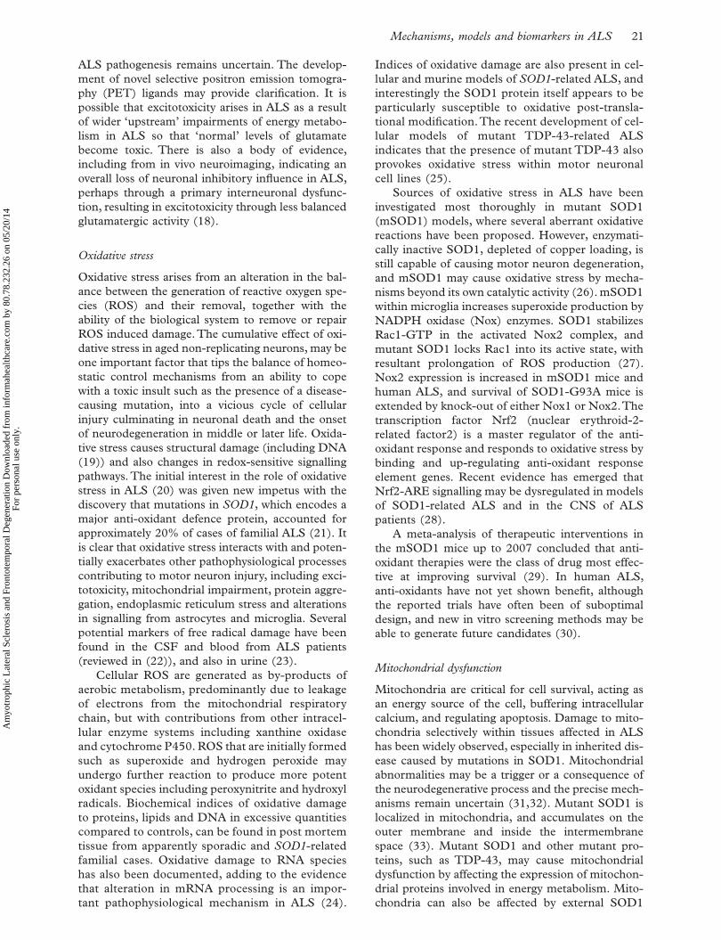
For human neurodegenerative diseases it is not cur-rently possible to study cellular pathological pro-cesses in real time, or safely and repeatedly remove tissue for analysis. The ability to study cellular molecular processes, identify key pathways for inter-vention, and assess multiple candidate therapies over short periods of time, depends on the development of disease models. The ability to express both wild-type and mutant human genes in non-human cells and transgenic animals has provided a variety of pos-sibilities for modelling many neurodegenerative dis-orders, including ALS (56,57). The discovery of linkage to the SOD1 gene of some cases of familial ALS led rapidly to the creation of an overexpressing mutant SOD1 transgenic mouse (58). This and other
Am
yotr
ophi
c L
ater
al S
cler
osis
and
Fro
ntot
empo
ral D
egen
erat
ion
Dow
nloa
ded
from
info
rmah
ealth
care
.com
by
80.7
8.23
2.26
on
05/2
0/14
For
pers
onal
use
onl
y.
Mechanisms, models and biomarkers in ALS 23
Tab
le I
I. M
odel
s of
AL
S p
atho
logy
, an
d th
eir
key
feat
ure
s.
Mod
elS
peci
fi cs
AL
S-l
ike
path
olog
yP
oten
tial
ly r
elev
ant
phys
ical
fea
ture
sA
dva
ntag
esD
isad
vant
ages
Key
ref
eren
ces
Yea
stS
acch
arom
yces
cer
evis
iae
SO
D1,
TA
RD
BP
, or
F
US
-rel
ated
tra
nsg
enic
s; A
lso
use
d as
a s
cree
nin
g m
odel
for
nov
el
RN
A-b
ind
ing-
rela
ted
gen
es
Mut
ant
SO
D1
dis
rupt
s m
itoc
hon
dri
al
hom
oeos
tasi
s; R
ecap
itu
lati
on o
f T
DP
-43
& F
US
ag
greg
atio
n w
ith
iden
tifi
cati
on o
f m
odu
lato
rs
NA
Rea
dil
y av
aila
ble,
low
m
aint
enan
ce;
hom
olog
ous
basi
c ce
llula
r st
ruct
ure
s to
hu
man
s; n
on-a
nim
al;
rapi
d tu
rnov
er;
hig
h th
rou
ghpu
t
Cel
lula
r to
xici
ty a
risi
ng
sim
ply
from
ove
r-ex
pres
sion
of
hum
an
prot
ein
s; f
acu
ltat
ive
aero
be
un
like
hu
man
ce
lls
135
– 137
Wor
mC
aeno
rhab
diti
s el
egan
s S
OD
1,
TA
RD
BP
, or
F
US
-rel
ated
tra
nsg
enic
s
Incr
ease
d se
nsi
tivi
ty t
o ox
idat
ive
stre
ss i
n pr
esen
ce o
f m
uta
nt S
OD
1,
wit
h ag
greg
ate
form
atio
n;
Agg
rega
tes
of m
uta
nt
TD
P-4
3 &
FU
S
Pro
gres
sive
‘ i
nco
ord
inat
ion ’
&
para
lysi
s ov
er
hou
rs-d
ays
in
over
expr
essi
ng
mu
tant
(e
spec
iall
y ph
osph
oryl
ated
) � w
ild
-ty
pe T
DP
-43
& F
US
(a
nd
only
mu
tant
S
OD
1) w
orm
s
Rea
dil
y av
aila
ble,
low
m
aint
enan
ce;
hom
olog
ous
basi
c n
euro
mu
scu
lar
stru
ctu
res
to h
um
ans
Var
iabl
e ef
fect
s an
d li
mit
ed h
um
an c
lin
ical
si
mil
arit
y
Rev
iew
ed i
n 56
S
ee a
lso 13
8,1
39
Fru
it fl
yD
roso
phlia
mel
anog
aste
r S
OD
1,
TA
RD
BP
, or
F
US
-rel
ated
tra
nsg
enic
s
Var
iabl
e ef
fect
s of
bot
h w
ild
-typ
e an
d m
uta
nt
TD
P-4
3 &
FU
S o
n ag
greg
ate
form
atio
n,
den
dri
tic
bran
chin
g,
and
NM
J d
ysfu
nct
ion
dep
end
ing
on t
issu
e ex
pres
sion
Var
iabl
e ef
fect
s of
bot
h w
ild
-typ
e an
d m
uta
nt
SO
D1,
TD
P-4
3 &
F
US
on
larv
al
loco
mot
or f
un
ctio
n d
epen
din
g on
tis
sue
expr
essi
on
Rea
dil
y av
aila
ble,
low
m
aint
enan
ce;
hom
olog
ous
basi
c n
euro
mu
scu
lar
stru
ctu
res
to h
um
ans;
sh
ort
life
cyc
le
Var
iabl
e ef
fect
s an
d li
mit
ed h
um
an c
lin
ical
si
mil
arit
y
Rev
iew
ed i
n 56
S
ee a
lso 14
0 – 1
43
Zeb
rafi
shD
anio
rer
io S
OD
1, T
AR
DB
P,
or
FU
S-r
elat
ed t
ran
sgen
ics
Mu
tant
SO
D1
lin
ked
to M
N
loss
an
d d
ysm
orph
ic N
MJs
; M
uta
nt T
DP
-43
lin
ked
to
dec
reas
ed m
otor
axo
n le
ngt
h an
d br
anch
ing;
Mu
tant
FU
S l
inke
d to
cy
topl
asm
ic i
ncl
usi
ons
Mut
ant
SO
D1
asso
ciat
ed
wit
h m
otor
ab
nor
mal
itie
s an
d m
usc
le a
trop
hy
Rea
dil
y av
aila
ble,
low
m
aint
enan
ce;
hom
olog
ous
basi
c n
euro
mu
scu
lar
stru
ctu
res
to h
um
ans;
sh
ort
life
cyc
le
Var
iabl
e ef
fect
s an
d li
mit
ed h
um
an c
lin
ical
si
mil
arit
y
Rev
iew
ed i
n 56
S
ee a
lso 14
4,14
5
Rod
ent
Mus
mus
culu
s �
� R
attu
s no
rveg
icus
SO
D1
or
TA
RD
BP
-rel
ated
tra
nsg
enic
s
Mu
tant
SO
D1
lin
ked
to g
lios
is,
ubi
quit
inat
ed S
OD
1 in
clu
sion
s,
mit
ocho
nd
rial
vac
uol
atio
n,
axon
al a
nd
MN
los
s; W
T a
nd
mu
tant
TD
P-4
3 m
ore
vari
ably
lin
ked
to c
ellu
lar
aggr
egat
es o
r M
N l
oss
Pro
gres
sive
loc
omot
or
abn
orm
alit
ies
wit
h h
ind
-lim
b w
eakn
ess
and
mu
scle
was
tin
g fr
om ̃
3 m
onth
s W
T a
nd
mu
tant
TD
P-4
3 le
ss c
onsi
sten
tly
lin
ked
to a
ny
mot
or
abn
orm
alit
ies
and
mu
scle
atr
ophy
Con
sist
ent
mot
or
phen
otyp
e; r
ead
ily
avai
labl
e, l
ow
mai
nten
ance
; ho
mol
ogou
s ba
sic
neu
rom
usc
ula
r st
ruct
ure
s to
hu
man
s;
shor
t li
fe c
ycle
Cos
tly
infr
astr
uct
ure
; so
me
eth
ical
con
cern
s P
oor
tran
slat
ion
of
ther
apeu
tic
resp
onse
in
SO
D1
mou
se t
o hu
man
st
ud
ies
so f
ar T
DP
-43
mod
els
show
li
mit
ed m
otor
ph
enot
ype
Rev
iew
ed i
n 56
,146
(Con
tinue
d)
Am
yotr
ophi
c L
ater
al S
cler
osis
and
Fro
ntot
empo
ral D
egen
erat
ion
Dow
nloa
ded
from
info
rmah
ealth
care
.com
by
80.7
8.23
2.26
on
05/2
0/14
For
pers
onal
use
onl
y.

24 M. R. Turner et al.
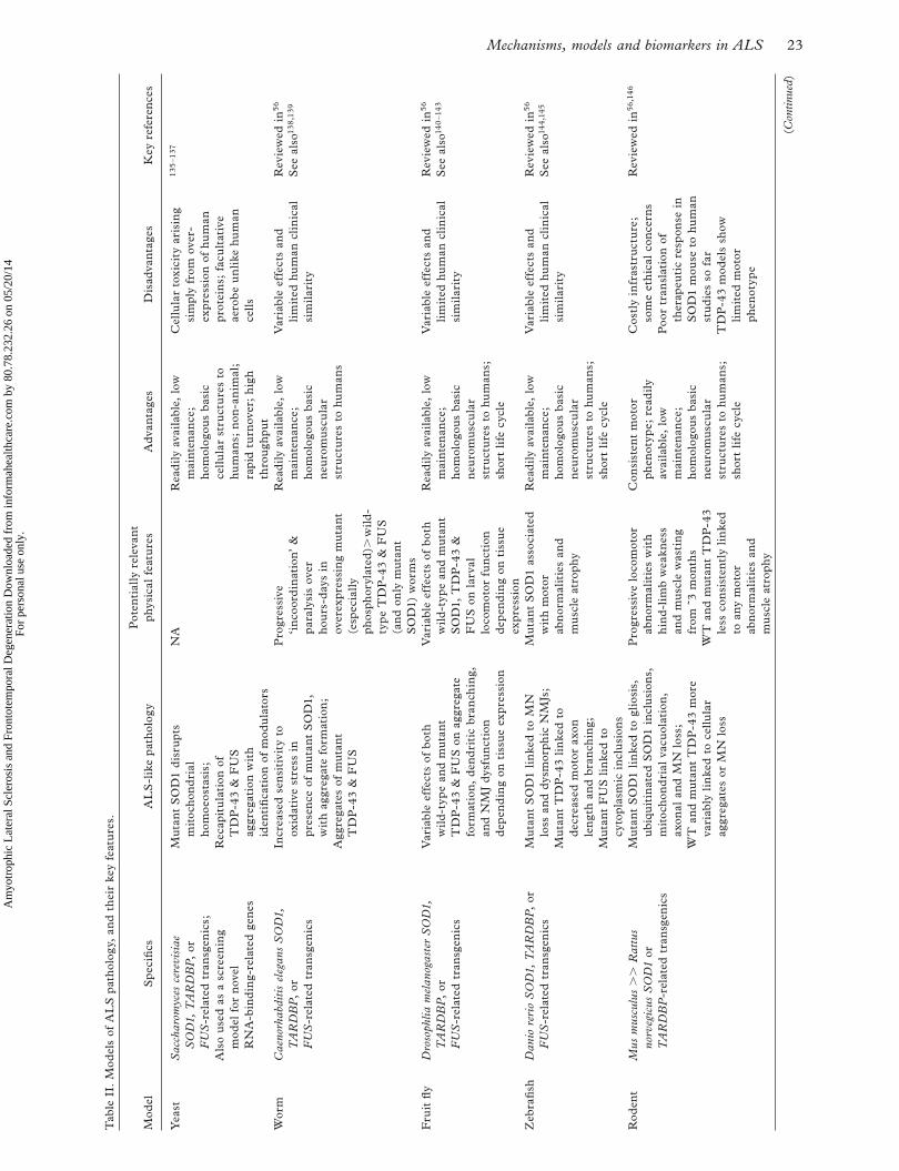
models are summarized in Table II. Such models do not, as yet, capture pathogenesis at a systems level, and no one model has yet been able to reproduce all of the pathological and behavioural features of ALS. Nonetheless, they have provided a valuable platform for testing many of the pathogenic hypotheses out-lined earlier in this article, with hope for the future development of assays for high-throughput screening of therapeutic candidates.
Induced pluripotent stem cells
The generation of motor neurons from induced pluripotent stem cells (iPSCs), in turn derived from the skin fi broblasts of ALS patients (59), has marked a major advance in modelling pathogenesis in ALS, with potential for high-throughput thera-peutic assessments. Encapsulated by the phrase ‘ disease in a dish ’ , iPSCs carrying the TDP-43 ‘ M337V ’ mutation have already been shown to reproduce several key aspects of TDP-43-related proteinopathies, including aggregate formation and reduced cell survival (60). The ability to generate not only motor neurons but also other non-neuronal cell types (including astrocytes, oligoden-drocytes, microglia), will allow for versatility in teasing out cell-specifi c contributions to disease development. iPSCs from patients with familial ALS have already been made available to research-ers at the NINDS Repository, thus allowing for the study of ALS arising from numerous SOD1 , FUS , and FIG4 mutations.
However, a number of challenges remain with regard to human iPSC uses for investigation into ALS biology. Currently, the long periods of time required to generate human neural cells (for exam-ple, astrocytes) results in signifi cant expense and often complex experimental paradigms. Because iPSCs are derived from individual patients, careful assessment of the sample sizes studied should be considered before broad conclusions can be made about disease from a single iPSC source. The impact of the patients ’ age on the resulting iPSC remains unclear. If used to screen for potentially relevant ALS drug targets, standardization of cell lines across laboratories will be important for validation of drug effects. Future aims include the development of more complex integrated structures, for example the neuromuscular junction using muscle and neuronal cell cocultures (61).
Despite these challenges, it is likely that more effi cient derivation of iPSC will be developed, thus shortening the time and expense currently associ-ated with creating iPSC lines. As sources of iPSC become more readily available to the research com-munity, investigators with a wide range of research interests will be able to draw on a range of ALS genotypes and phenotypes thus allowing for the identifi cation of new ALS relevant disease targets for therapeutics. M
odel
Spe
cifi
csA
LS
-lik
e pa
thol
ogy
Pot
enti
ally
rel
evan
t ph
ysic
al f
eatu
res
Ad
vant
ages
Dis
adva
ntag
esK
ey r
efer
ence
s
Dog
‘ Can
ine
deg
ener
ativ
e m
yelo
path
y ’ :
Pem
brok
e W
elsh
cor
gi,
Box
er,
Rho
des
ian
rid
geba
ck,
Ger
man
S
heph
erd
, &
Che
sape
ake
Bay
re
trie
ver
all
hom
ozyg
ous
for
SO
D1
‘ E40
K ’
mis
sen
se m
uta
tion
Lat
eral
cor
d w
hit
e m
atte
r m
yeli
n an
d ax
onal
los
s; N
euro
nal
cyt
opla
smic
in
clu
sion
s bi
nd
ing
anti
-SO
D1
anti
bod
ies
Adu
lt-o
nse
t pr
ogre
ssiv
e sp
asti
c m
yelo
path
y af
fect
ing
pelv
ic g
ird
le,
lead
ing
to e
vent
ual
fl
acci
d qu
adri
pare
sis
Sim
ilar
to
hum
an
SO
D1-
rela
ted
AL
S i
n b
ein
g a
del
ayed
ad
ult
-on
set
dis
ord
er
Eth
ical
con
cern
s,
avai
labl
ity
and
infr
astr
uct
ure
iss
ues
; lo
ng
late
ncy
to
sym
ptom
s; l
imit
ed
rele
van
ce t
o n
on-
SO
D1-
rela
ted
AL
S
147
Mon
key
Mac
aca
fasc
icul
aris
ove
r-ex
pres
sin
g hu
man
TD
P-4
3 vi
a ad
enov
iru
s to
cer
vica
l co
rd
Cyt
opla
smic
mis
loca
liza
tion
of
TD
P-4
3;
cyst
atin
-C p
osit
ive
aggr
egat
es
Pro
gres
sive
mot
or
wea
knes
s of
dis
tal
upp
er l
imb
s w
ith
fasc
icu
lati
on a
nd
was
tin
g
Clo
sest
spe
cies
to
hum
ans
phys
ical
ly a
nd
beh
avio
ura
lly
Maj
or e
thic
al c
once
rns
and
infr
astr
uct
ure
is
sues
; li
mit
ed
rele
van
ce t
o ‘ s
low
ly-
dev
elop
ing ’
hu
man
A
LS
148
Tab
le I
I. (
Con
tinue
d)
Am
yotr
ophi
c L
ater
al S
cler
osis
and
Fro
ntot
empo
ral D
egen
erat
ion
Dow
nloa
ded
from
info
rmah
ealth
care
.com
by
80.7
8.23
2.26
on
05/2
0/14
For
pers
onal
use
onl
y.
Mechanisms, models and biomarkers in ALS 25
Tissue and fl uid biomarker sources
Biofl uids
The range of human biofl uids useful in proteomic studies to identify an ALS biomarker ‘ signature ’ , includes CSF, blood, urine, and saliva (62). CSF is an excellent biofl uid for biomarker discovery due to its approximation to the cells and brain and spinal cord regions exhibiting cell death during ALS. Blood, while more accessible, has greater protein complex-ity with greatly reduced concentrations, compared to CSF, of those proteins fundamentally involved in neuronal function.
During the past decade there has been a large increase in the number of ALS biomarker studies using CSF as well as blood (22). Most of these stud-ies have examined changes of individual proteins in the CSF of ALS versus healthy control or other neu-rologic disease subjects, typically using a gel-based system or ELISA (63). However, most are limited by the number of samples used in the analysis, choice of control subjects, and typically the lack of verifi ca-tion in a separate cohort of patients. A number of more recent studies have used mass spectrometry or cytokine profi ling to identify panels of candidate protein biomarkers in the CSF (43,64). Metabolo-mic approaches have been used to explore metabolic differences between ALS and control subjects in CSF and serum (54,65 – 67). Early changes in patient metabolism and protein levels or post-translational modifi cations offer a means to both identify ALS at an early stage and to develop bio-markers to follow during disease progression.
At present, CSF candidate biomarkers in ALS can be grouped into those that refl ect neuronal loss and those indicative of neuroinfl ammatory (glial) processes. For the former, neurofi laments have been the most reproducible (68 – 70). CSF TDP-43 appears to fall with disease progression in typical ALS (71), with lower values found in cases of ALS-FTD (72). The signifi cance of fi nding reduced CSF transthyretin and cystatin C in ALS is uncertain (64). The latter is the essential constituent of the ‘ pathognomonic ’ Bunina body inclusion seen histo-pathologically in ALS and has been detected at increased levels in ALS plasma (73).
While several molecules linked to neuroinfl am-matory pathways have been reported as increased in ALS patient CSF, each has limited sensitivity in iso-lation and many studies have not been reproduced. A panel of cytokines, specifi cally interleukins 2, 6, 10, 15 and granulocyte-monocyte colony stimulating factor (GM-CSF) was nearly 90% accurate in dis-tinguishing ALS patients from healthy controls (43). Other studies have also supported the concept of combining markers to improve accuracy, most rece-ntly the ratio of CSF phosphorylated neurofi lament-heavy chain and complement C3, achieving even higher accuracy (74). CSF based biomarkers may be useful to aid in the diagnosis of ALS, but may have
better applications in monitoring drug effects in clinical trials and as prognostic indicators of disease (73,75). Further studies, involving samples from mimic disorders rather than healthy controls, and serial samples from ALS patients, are required to validate the candidate diagnostic biomarkers and fully explore their utility in therapeutic trials. An important step has been to both recognize the potential variability in sample quality due to differences in acquisition and storage (76), and to establish international consensus on standard oper-ating procedures (77).
Muscle
Skeletal muscle may represent a valuable source of biomarkers in ALS. This tissue is one of the most severely affected by the disease, with progressive denervation and atrophy, and it is easily accessible to biopsy. The only muscle biomarker that has been tested prospectively is the axon repellant Nogo-A. Nogo-A is strongly expressed in ALS muscles, is cor-related with ALSFRS and prospectively identifi es patients affected with lower motor neuron disease that will progress to ALS (78 – 80). However, the specifi city of this increased expression has been questioned. A combination of biomarkers might solve the problem of specifi city. Muscle transcrip-tome analyses have identifi ed a number of potential candidates correlated with disease severity (81). There are two potential limitations of muscle bio-markers. First, muscle biopsy is invasive and as a consequence longitudinal studies are very diffi cult. Secondly, muscle beds are very differentially involved among patients. The different sites of onset, as well as the heterogenous spreading, make it diffi cult to standardize the choice of the site of muscle biopsy. In this respect, the future of muscle biomarkers might be to identify a set of muscle derived proteins or peptides that enter the circulation. Such muscle-derived blood biomarkers could be useful tools to evaluate disease progression and severity in ALS patients in a manner similar to creatine kinase for myopathies.
Skin
The skin is an acknowledged part of the disease process in ALS, both in experimental models (82) and humans (83 – 85). Because this organ is easily accessible and at least some of the biochemical alterations are related to fi ndings in the CNS (82,83), the skin is a principal resource of biomark-ers for diagnosis, staging, and evaluation of therapy. The most striking fi nding is an apparently selective elevation of the MMP-9 in the skin and spinal cord of experimental animals and in human skin and the CSF (82,83). These fi ndings are consistent with previous observations of increased collagen degra-dation, in particular collagen types I and IV, in the
Am
yotr
ophi
c L
ater
al S
cler
osis
and
Fro
ntot
empo
ral D
egen
erat
ion
Dow
nloa
ded
from
info
rmah
ealth
care
.com
by
80.7
8.23
2.26
on
05/2
0/14
For
pers
onal
use
onl
y.

26 M. R. Turner et al.
skin of ALS patients (84). The pathogenic steps responsible for these observations remain to be elu-cidated; however, free radical damage and infl am-mation are possible mechanisms. A further discovery was that small distal epidermal nerve fi bers are affected in ALS (85), indicating the presence of a small fi ber neuropathy. The pattern of changes refl ects the concept of a distal axonopathy and paves the way for mechanistic studies of cytoskeletal alter-ations and axonal transport.
Post mortem tissues
While post mortem spinal cord, brain or muscle tis-sue may not provide the optimal resource for bio-marker discovery efforts, they are especially important to help identify the cell types that express each can-didate biomarker, as well as their relationship to the pathophysiology of the human disease. Increased efforts to collect ALS and appropriate control post mortem tissues become more crucial as the multi-tude of candidate biomarkers identifi ed in the above mentioned biofl uids and tissue biopsies will require proper characterization and correlation to ALS pathology. In addition, recent studies have shown that neural progenitor cells can be cultured from ALS post mortem tissues and used to generate astro-cytes to investigate astrocyte derived cell signalling that infl uences motor neuron survival (86). Given the low numbers of banked ALS tissues at any one site, standardized collection and storage procedures for ALS post mortem tissues must be established to permit use of tissues obtained across multiple sites.
Neurophysiology biomarker sources
EMG and MUNE
Neurophysiological testing has been an important component of the diagnostic evaluation of patients with motor neuron disease since the demonstration that axon loss and consequent reinnervation could be measured using electromyography (EMG) (87). While classical EMG and nerve conduction studies are still incorporated into diagnostic criteria for ALS (El Escorial), these techniques have not proved effective in monitoring disease progression or assessing effects of treatment. Motor unit number estimation (MUNE) is a tool that was developed for this purpose. The technique and theoretical basis for MUNE is quite simple; a maximum response amplitude, which is generated by activa-tion of all motor units in the muscle, is recorded, from which an estimate of individual motor unit number is generated by dividing the maximum response amplitude by an estimate of single motor unit amplitude. Many techniques for estimating the average amplitude of single motor units have been suggested; most have been limited by sampling bias, and lack of reproducibility (88).
Recently, a modifi cation of earlier described techniques was introduced and studied in a natural history study of patients with ALS (89). Multipoint incremental MUNE was found to have excellent test-retest reliability, and to decline monotonically in ALS faster than other measures traditionally used in ALS clinical trials. The technique can be well standardized, is performed briefl y with good patient tolerance, and has a low computational burden. Using rate of decline as well as variability, hypo-thetical power analyses suggested that this measure might reduce both sample size and study duration in phase II trials. Other new MUNE methods, including MUNIX (90) and Bayseian MUNE (91,92) are also being developed. While no physio-logical method of motor unit number estimation has been validated against anatomical motor unit counts, their use as potential endpoints in clinical trials shows great promise.
Transcranial magnetic stimulation
ALS is diagnosed clinically by the presence of both UMN and LMN damage. While LMN abnormali-ties can be confi rmed objectively using peripheral electrodiagnostic methods, UMN fi ndings lack com-parable established objective markers. Transcranial magnetic stimulation (TMS) is a neurophysiological test that measures UMN functional integrity, and can detect abnormalities when there are no clinical UMN signs. TMS works by evoking compound motor potentials (MEP) using non-invasive mag-netic stimulation of the motor cortex through activa-tion of both UMN and LMN pathways. It is used to study the conductivity and excitability of the corti-cospinal system. As potential physiological biomark-ers, TMS measurements refl ect the functional integrity of the UMN (93).
Single pulse TMS is a well established diagnos-tic and clinical research tool, although results vary widely between medical centers. However, if indi-vidual laboratories establish their own normative data, subjects can be tested in a consistent, reliable fashion. TMS recordings include measurements of motor threshold, central motor conduction time, and MEP amplitudes, all of which may be abnor-mal in ALS (94). TMS also can be delivered in paired pulses, or as repetitive trains of stimulation for investigating human brain function, transiently stimulating or inhibiting different cortical areas (95,96).
TMS biomarkers, particularly single pulse evoked TMS amplitude, have been used to objectively dis-criminate ALS from controls and assess the progres-sion of ALS (97 – 99). TMS can reveal subtle subclinical UMN dysfunction to help make the diag-nosis of ALS as well as clarify the relationship between ALS and its variants (100), including pro-gressive muscular atrophy where there may be sub-clinical UMN changes.
Am
yotr
ophi
c L
ater
al S
cler
osis
and
Fro
ntot
empo
ral D
egen
erat
ion
Dow
nloa
ded
from
info
rmah
ealth
care
.com
by
80.7
8.23
2.26
on
05/2
0/14
For
pers
onal
use
onl
y.

Mechanisms, models and biomarkers in ALS 27
Electrical impedance myography
Electrical impedance myography (EIM) is a tech-nique in which a high-frequency, low-intensity elec-trical current is applied to a localized area of muscle and the consequent surface voltages measured (101). Unlike standard electromyography, in which the intrinsic electrical activity of muscle is measured, EIM assesses the integrity and structure of the mus-cle. An initial, single-center study demonstrated EIM ’ s potential power at measuring disease progres-sion in this disease (102). Recently, a second multicenter study compared EIM directly to the ALSFRS-R, MUNE, and handheld dynamometry (103). EIM outperformed the other measures in terms of its ability to detect deterioration. For exam-ple, based on these data, a study that would require 220 subjects using the ALSFRS-R would have the same power as one with only 95 using EIM. Studies in ALS rats have similarly showed a strong correla-tion to the rate of change in EIM and the animal ’ s length of survival, as well as to MUNE (104). The methodology is also being applied in the fi rst North American study of neural stem cells in ALS (105). One advantage of EIM over most other modalities is its ability to assess a variety of muscles and to mea-sure specifi cally that area of the body where the dis-ease is progressing most rapidly. Measurement of other muscles not routinely studied but which might provide valuable data, such as paraspinal muscles and the tongue, is also possible. Current efforts are geared toward further refi ning the technique for easy use and making it widely available (106).
Neuroimaging biomarker sources
MRI has a major role in the exclusion of cerebral, and particularly spinal mimics of ALS (107). How-ever, neuroimaging is also at the forefront of advances in the understanding of in vivo cerebral disease mechanisms in ALS, including rodent models (108), with the identifi cation of multiple candidate bio-markers as a result (109).
Radionuclide imaging
Single photon emission computed tomography (SPECT) is a practical and potentially widely appli-cable form of radionuclide imaging. It was at the forefront of the recognition of a clinical, pathological and, most recently, genetic continuum between ALS and frontotemporal dementia (FTD) (110). Posi-tron emission tomography (PET) has greater resolu-tion than SPECT but is limited by the availability of experienced facilities. Pivotal ‘ activation ’ PET stu-dies, using tracers sensitive to blood fl ow and meta-bolism (e.g. radiolabelled water and fl urodeoxyglucose, FDG), provided in vivo evidence for a consistent extramotor cerebral pathology in ALS (111). Subse-quently, ‘ ligand ’ PET has been used to identify
specifi c cerebral neuronal receptor changes in ALS. Such studies have provided evidence for a loss of cortical inhibition (112) that might infl uence pro-gression rate (113), widespread microglial activation (114), and a striking reduction in serotonin-1A receptor binding (115), similar to changes seen in FTD (116). The future value of PET in ALS will depend upon the development of ligands with rele-vance to pathogenic hypotheses, e.g. glutamate receptors, and more specifi c neuroinfl ammatory or protein markers, e.g. TDP-43 (the challenge of intracellular penetration notwithstanding).
Magnetic resonance imaging (MRI)
The observation of corticospinal tract hyperintensity lacks sensitivity and specifi city for the diagnosis of ALS. Routine clinical structural imaging of the brain has limited value as a source of biomarkers in ALS, and only through advanced analysis methods (117). The acquisition of high-resolution 3D T1-weighted images and the development of novel pulse sequences such as diffusion tensor imaging and functional MRI have greatest potential in this regard, and would ulti-mately be feasible to perform within the clinical envi-ronment (118).
Macroscopic atrophy of the motor cortex is not a consistent observation in ALS, although prominent in those with PLS. Sophisticated analysis of 3D T1-weighted structural images acquired in 3D, known as voxel-based morphometry (VBM), can reveal subtle changes in regional cerebral tissue. Sev-eral VBM studies have been performed in ALS, and meta-analysis showed the right precentral gyrus as consistently altered (119). Surface-based morphom-etry allows assessment of cortical thickness, and a study in ALS confi rmed not only primary motor, but also extramotor, temporal cortical thinning in faster progressors (120).
Post mortem histopathological study fi rst dem-onstrated widespread cerebral white matter tract damage in ALS (121) and this can now be detected non-invasively using diffusion tensor imaging (DTI). This advanced MRI technique is sensitive to the movement of water, normally directionally confi ned within neuronal tracts. The two main quantitative measures of loss of neuronal tract integrity are reduced fractional anisotropy (FA) and increased mean diffusivity. Related parameters such as increased radial diffusi vity may specifi cally refl ect secondary demyelination of tracts in ALS. DTI stud-ies have shown consistently reduced FA in the cor-ticospinal tract, particularly within the posterior limb of the internal capsule (PLIC) (122) and corpus cal-losum (123) of ALS patients. Targeted FA measure-ment at the PLIC may provide prognostic information (124). DTI measures may, however, be insuffi ciently sensitive to longitudinal change over less than six months (125), nor have suffi cient discriminatory power as an isolated measurement (126).
Am
yotr
ophi
c L
ater
al S
cler
osis
and
Fro
ntot
empo
ral D
egen
erat
ion
Dow
nloa
ded
from
info
rmah
ealth
care
.com
by
80.7
8.23
2.26
on
05/2
0/14
For
pers
onal
use
onl
y.
28 M. R. Turner et al.
MRI is unsurpassed in its spatial resolution of cerebral functional activity achieved non-invasively. Blood oxygenation level-dependent (BOLD) func-tional MRI (fMRI) studies of motor tasks in ALS patients confi rmed the widened region of activation observed in PET studies. More recently, however, it is the study of the ‘ resting state ’ that provides novel insight into ALS as a ‘ system failure ’ . Rest-ing-state fMRI (R-fMRI) has demonstrated increased functional connectivity within the dam-aged ALS cortical network, with possible implica-tions in relation to cortical inhibitory infl uences (127). The combination of structural and func-tional MRI measures in this study also provided much better separation of patients from healthy age-matched controls.
Magnetic resonance spectroscopy (MRS) is an application of MRI that permits quantifi cation of cerebral tissue metabolites. It has consistently dem-onstrated reduced N-acetylaspartate ratios (a non-specifi c marker of neuronal loss) in the motor cortex of ALS patients, and high-fi eld studies also suggest a specifi c loss of GABA-ergic infl uence (128). Recent studies applied to the cervical spinal cord of pre-symptomatic carriers of pathological SOD1 gene mutations demonstrated metabolite changes more consistent with affected ALS patients rather than healthy non-gene carriers (129), sug-gesting that MRS may be particularly sensitive to pre-clinical changes. This offers the hope of captur-ing the very earliest events in individuals carrying genetic abnormalities associated with the develop-ment of ALS, with the possibility of wider transla-tion to sporadic cases.
The challenge for neuroimaging biomarker can-didates is to move beyond results based on group averages, to individual measurements. It seems likely that this will require multiple parameters. Further longitudinal studies are needed to assess the sensi-tivity of multimodal MRI to disease activity com-pared to clinical assessments such as ALSFRS.
Concluding remarks
Major advances in the understanding of the patho-biology of ALS have occurred over the last two decades with developments in molecular biology, immunocytochemistry, neurophysiology and neu-roimaging, and the recognition of overlap with some forms of FTD. It seems increasingly likely that there are multiple, possibly more discrete, pathways converging on motor neuron death. While recent discoveries in relation to RNA biology hint at a massively under-estimated level of pathologi-cal complexity in ALS, the common theme of mis-folded protein inclusions across the range of neurodegenerative disorders brings the hope of a common strategy for the treatment of pre-aggrega-tion events (130) and for common biomarker development. Biomarker candidates are emerging
with the potential to refi ne the diagnosis, stratify patients prognostically, and facilitate therapeutic development. A key aim for further biomarker development, beyond validation across multiple centers, is the routine incorporation of biomarker measurement into future clinical trials.
Declaration of interest: The authors report no confl icts of interest. The authors alone are respon-sible for the content and writing of the paper.
MRT is supported by the Medical Research Council and Motor Neurone Disease Association UK Lady Edith Wolfson Fellowship. PJS is sup-ported by the MND Association, the Medical Research Council and the European Community 7th Framework Programme.
References
Kiernan MC , Vucic S , Cheah BC , Turner MR , Eisen A , 1. Hardiman O , et al . Amyotroph Lateral Scler . Lancet. 2011 ; 377 : 942 – 55 . Mackenzie IR , Bigio EH , Ince PG , Geser F , Neumann M , 2. Cairns NJ , et al . Pathological TDP-43 distinguishes sporadic amyotrophic lateral sclerosis from amyotrophic lateral sclerosis with SOD1 mutations . Ann Neurol. 2007 ; 61 : 427 – 34 . Mitchell JD , Callagher P , Gardham J , Mitchell C , 3. Dixon M , Addison-Jones R , et al . Timelines in the diagnos-tic evaluation of people with suspected amyotrophic lateral sclerosis (ALS)/motor neuron disease (MND): a 20-year review. Can we do better? Amyotroph Lateral Scler. 2010 ; 11 : 537 – 41 . Turner MR , Parton MJ , Shaw CE , Leigh PN , Al-Chalabi 4. A . Prolonged survival in motor neuron disease: a descrip-tive study of the King’s database 1990 – 2002 . J Neurol Neurosurg Psychiatry. 2003 ; 74 : 995 – 7 . Chio A , Calvo A , Moglia C , Mazzini L , Mora G . Pheno-5. typic heterogeneity of amyotrophic lateral sclerosis: a pop-ulation based study . J Neurol Neurosurg Psychiatry. 2011 ; 82 : 740 – 6 . Cudkowicz ME , Katz J , Moore DH , O’Neill G , Glass JD , 6. Mitsumoto H , et al . Toward more effi cient clinical trials for amyotrophic lateral sclerosis . Amyotroph Lateral Scler. 2010 ; 11 : 259 – 65 . Rothstein JD . Current hypotheses for the underlying bio-7. logy of amyotrophic lateral sclerosis . Ann Neurol. 2009 ; 65 (Suppl 1) : S3 – 9 . Rothstein JD , Tsai G , Kuncl RW , Clawson L , Cornblath DR , 8. Drachman DB , et al . Abnormal excitatory amino acid metabolism in amyotrophic lateral sclerosis . Ann Neurol. 1990 ; 28 : 18 – 25 . Spreux-Varoquaux O , Bensimon G , Lacomblez L , 9. Salachas F , Pradat PF , Le Forestier N , et al . Glutamate levels in cerebrospinal fl uid in amyotrophic lateral sclerosis: a reappraisal using a new HPLC method with coulometric detection in a large cohort of patients . J NeurolvSci. 2002 ; 193 : 73 – 8 . Rothstein JD , Dykes-Hoberg M , Pardo CA , Bristol LA , Jin 10. L , Kuncl RW , et al . Knockout of glutamate transporters reveals a major role for astroglial transport in excitotoxicity and clearance of glutamate . Neuron. 1996 ; 16 : 675 – 86 . Bruijn LI , Becher MW , Lee MK , Anderson KL , Jenkins NA , 11. Copeland NG , et al . ALS-linked SOD1 mutant G85R mediates damage to astrocytes and promotes rapidly pro-gressive disease with SOD1-containing inclusions . Neuron. 1997 ; 18 : 327 – 38 .
Am
yotr
ophi
c L
ater
al S
cler
osis
and
Fro
ntot
empo
ral D
egen
erat
ion
Dow
nloa
ded
from
info
rmah
ealth
care
.com
by
80.7
8.23
2.26
on
05/2
0/14
For
pers
onal
use
onl
y.

Mechanisms, models and biomarkers in ALS 29
Howland DS , Liu J , She Y , Goad B , Maragakis NJ , Kim B , 12. et al . Focal loss of the glutamate transporter EAAT2 in a transgenic rat model of SOD1 mutant-mediated amyo-trophic lateral sclerosis (ALS) . Proc Natl Acad Sci U S A. 2002 ; 99 : 1604 – 9 . Rothstein JD , van Kammen M , Levey AI , Martin LJ , 13. Kuncl RW . Selective loss of glial glutamate transporter GLT-1 in amyotrophic lateral sclerosis . Ann Neurol. 1995 ; 38 : 73 – 84 . Trotti D , Rolfs A , Danbolt NC , Brown RH Jr, Hediger MA . 14. SOD1 mutants linked to amyotrophic lateral sclerosis selectively inactivate a glial glutamate transporter . Nat Neurosci. 1999 ; 2 : 848 . Guo H , Lai L , Butchbach ME , Stockinger MP , Shan X , 15. Bishop GA , et al . Increased expression of the glial gluta-mate transporter EAAT2 modulates excitotoxicity and delays the onset but not the outcome of ALS in mice . Hum Mol Genet. 2003 ; 12 : 2519 – 32 . Rothstein JD , Patel S , Regan MR , Haenggeli C , Huang YH , 16. Bergles DE , et al . Beta-lactam antibiotics offer neuropro-tection by increasing glutamate transporter expression . Nature. 2005 ; 433 : 73 – 7 . Doble A . The pharmacology and mechanism of action of 17. riluzole . Neurology. 1996 ; 47 (Suppl 4) : S233 – 41 . Turner MR , Kiernan MC . Does interneuronal dysfunction 18. contribute to neurodegeneration in amytrophic lateral scle-rosis? Amyotroph Lateral Scler. 2012 ; 13 : 245 – 50 . Bogdanov M , Brown RH , Matson W , Smart R , Hayden D , 19. O’Donnell H , et al . Increased oxidative damage to DNA in ALS patients . Free Radic Biol Med. 2000 ; 29 : 652 – 8 . Simpson EP , Yen AA , Appel SH . Oxidative stress: a com-20. mon denominator in the pathogenesis of amyotrophic lat-eral sclerosis . Current Opinion in Rheumatology. 2003 ; 15 : 730 – 6 . Epub 2003/10/22 . Barber SC , Shaw PJ . Oxidative stress in ALS: key role in 21. motor neuron injury and therapeutic target . Free Radic Biol Med. 2010 ; 48 : 629 – 41 . Turner MR , Kiernan MC , Leigh PN , Talbot K . Biomarkers 22. in amyotrophic lateral sclerosis . Lancet Neurol. 2009 ; 8 : 94 – 109 . Mitsumoto H , Santella RM , Liu X , Bogdanov M , Zipprich J , 23. Wu HC , et al . Oxidative stress biomarkers in sporadic ALS . Amyotroph Lateral Scler. 2008 ; 9 : 177 – 83 . Chang Y , Kong Q , Shan X , Tian G , Ilieva H , Cleveland DW , 24. et al . Messenger RNA oxidation occurs early in disease pathogenesis and promotes motor neuron degeneration in ALS . PLoS One. 2008 ; 3 : 2849 . Duan W , Li X , Shi J , Guo Y , Li Z , Li C . Mutant TAR DNA-25. binding protein-43 induces oxidative injury in motor neuron-like cell . Neuroscience. 2010 ; 169 : 1621 – 9 . Kirby J , Halligan E , Baptista MJ , Allen S , Heath PR , 26. Holden H , et al . Mutant SOD1 alters the motor neuronal transcriptome: implications for familial ALS . Brain. 2005 ; 128 : 1686 – 706 . Harraz MM , Marden JJ , Zhou W , Zhang Y , Williams A , 27. Sharov VS , et al . SOD1 mutations disrupt redox-sensitive Rac regulation of NADPH oxidase in a familial ALS model . J Clin Invest. 2008 ; 118 : 659 – 70 . Sarlette A , Krampfl K , Grothe C , Neuhoff N , Dengler R , 28. Petri S . Nuclear erythroid 2-related factor 2-antioxidative response element signaling pathway in motor cortex and spinal cord in amyotrophic lateral sclerosis . J Neuropathol Exp Neurol. 2008 ; 67 : 1055 – 62 . Orrell RW , Lane RJ , Ross M . A systematic review of anti-29. oxidant treatment for amyotrophic lateral sclerosis/motor neuron disease . Amyotroph Lateral Scler. 2008 ; 9 : 195 – 211 . Barber SC , Higginbottom A , Mead RJ , Barber S , Shaw PJ . 30. An in vitro screening cascade to identify neuroprotective anti-oxidants in ALS . Free Radic Biol Med. 2009 ; 46 : 1127 – 38 . Magrane J , Manfredi G . Mitochondrial function, morpho-31. logy, and axonal transport in amyotrophic lateral sclerosis . Antioxid Redox Signal. 2009 ; 11 : 1615 – 26 .
Kawamata H , Manfredi G . Mitochondrial dysfunction and 32. intracellular calcium dysregulation in ALS . Mech Ageing Dev. 2010 ; 131 : 517 – 26 . Kawamata H , Manfredi G . Different regulation of wild-33. type and mutant Cu/Zn superoxide dismutase localization in mammalian mitochondria . Hum Mol Genet. 2008 ; 17 : 3303 – 17 . Israelson A , Arbel N , Da Cruz S , Ilieva H , Yamanaka K , 34. Shoshan-Barmatz V , et al . Misfolded mutant SOD1 directly inhibits VDAC1 conductance in a mouse model of inher-ited ALS . Neuron. 2010 ; 67 : 575 – 87 . Li Q , van de Velde C , Israelson A , Xie J , Bailey AO , Dong 35. MQ , et al . ALS-linked mutant superoxide dismutase-1 (SOD1) alters mitochondrial protein composition and decreases protein import . Proc Natl Acad Sci U S A. 2010 ; 107 : 21146 – 51 . Pedrini S , Sau D , Guareschi S , Bogush M , Brown RH Jr, 36. Naniche N , et al . ALS-linked mutant SOD1 damages mito-chondria by promoting conformational changes in Bcl-2 . Hum Mol Genet. 2010 ; 19 : 2974 – 86 . van de Velde C , McDonald KK , Boukhedimi Y , 37. McAlonis-Downes M , Lobsiger CS , Bel Hadj S , et al . Misfolded SOD1 associated with motor neuron mitochon-dria alters mitochondrial shape and distribution prior to clinical onset . PLoS One. 2011 ; 6 : 22031 . Bordet T , Buisson B , Michaud M , Drouot C , Galea P , 38. Delaage P , et al . Identifi cation and characterization of cholest-4-en-3-one, oxime (TRO19622), a novel drug can-didate for amyotrophic lateral sclerosis . J Pharmacol Exp Ther. 2007 ; 322 : 709 – 20 . Cudkowicz M , Bozik ME , Ingersoll EW , Miller R , 39. Mitsumoto H , Shefner J , et al . The effects of dexpramipex-ole (KNS-760704) in individuals with amyotrophic lateral sclerosis . Nat Med. 2011 ; 17 : 1652 – 6 . Alavian KN , Dworetzky SI , Bonanni L , Zhang P , 40. Sacchetti S , Mariggio MA , et al . Effects of dexpramipexole on brain mitochondrial conductances and cellular bioener-getic effi ciency . Brain Res. 2012 ; 1446 : 1 – 11 . Philips T , Robberecht W . Neuroinfl ammation in amyo-41. trophic lateral sclerosis: role of glial activation in motor neuron disease . Lancet Neurol. 2011 ; 10 : 253 – 63 . Zhang R , Gascon R , Miller RG , Gelinas DF , Mass J , 42. Hadlock K , et al . Evidence for systemic immune system alterations in sporadic amyotrophic lateral sclerosis (SALS) . J Neuroimmunol. 2005 ; 159 : 215 – 24 . Epub 2005/01/18 . Mitchell RM , Freeman WM , Randazzo WT , Stephens HE , 43. Beard JL , Simmons Z , et al . A CSF biomarker panel for identifi cation of patients with amyotrophic lateral sclerosis . Neurology. 2009 ; 72 : 14 – 9 . Kuhle J , Lindberg RL , Regeniter A , Mehling M , Steck AJ , 44. Kappos L , et al . Increased levels of infl ammatory chemok-ines in amyotrophic lateral sclerosis . Eur J Neurol. 2009 ; 16 : 771 – 4 . Zhang R , Miller RG , Gascon R , Champion S , Katz J , 45. Lancero M , et al . Circulating endotoxin and systemic immune activation in sporadic amyotrophic lateral sclerosis (SALS) . J Neuroimmunol. 2009 ; 206 : 121 – 4 . Epub 2008/11/18 . Appel SH , Engelhardt JI , Henkel JS , Siklos L , Beers DR , 46. Yen AA , et al . Hematopoietic stem cell transplantation in patients with sporadic amyotrophic lateral sclerosis . Neurology. 2008 ; 71 : 1326 – 34 . Epub 2008/10/22 . Dupuis L , Oudart H , Rene F , Gonzalez de Aguilar JL , 47. Loeffl er JP . Evidence for defective energy homeostasis in amyotrophic lateral sclerosis: benefi t of a high-energy diet in a transgenic mouse model . Proc Natl Acad Sci U S A. 2004 ; 101 : 11159 – 64 . Dupuis L , Pradat PF , Ludolph AC , Loeffl er JP . Energy 48. metabolism in amyotrophic lateral sclerosis . Lancet Neurol. 2011 ; 10 : 75 – 82 . Paganoni S , Deng J , Jaffa M , Cudkowicz ME , Wills AM . 49. Body mass index, not dyslipidemia, is an independent
Am
yotr
ophi
c L
ater
al S
cler
osis
and
Fro
ntot
empo
ral D
egen
erat
ion
Dow
nloa
ded
from
info
rmah
ealth
care
.com
by
80.7
8.23
2.26
on
05/2
0/14
For
pers
onal
use
onl
y.

30 M. R. Turner et al.
predictor of survival in amyotrophic lateral sclerosis . Mus-cle Nerve. 2011 ; 44 : 20 – 4 . Epub 2011/05/25 . Marin B , Desport JC , Kajeu P , Jesus P , Nicolaud B , 50. Nicol M , et al . Alteration of nutritional status at diagnosis is a prognostic factor for survival of amyotrophic lateral sclerosis patients . J Neurol Neurosurg Psychiatry. 2011 ; 82 : 628 – 34 . Epub 2010/11/26 . Dupuis L , Corcia P , Fergani A , Gonzalez de Aguilar JL , 51. Bonnefont-Rousselot D , Bittar R , et al . Dyslipidemia is a protective factor in amyotrophic lateral sclerosis . Neuro-logy. 2008 ; 70 : 1004 – 9 . Dorst J , K ü hnlein P , Hendrich C , Kassubek J , Sperfeld AD , 52. Ludolph AC . Patients with elevated triglyceride and cho-lesterol serum levels have a prolonged survival in amyo-trophic lateral sclerosis . J Neurol. 2011 ; 258 : 613 – 7 . Chio A , Calvo A , Ilardi A , Cavallo E , Moglia C , Mutani R , 53. et al . Lower serum lipid levels are related to respiratory impairment in patients with ALS . Neurology. 2009 ; 73 : 1681 – 5 . Epub 2009/11/18 . Lawton KA , Cudkowicz ME , Brown MV , Alexander D , 54. Caffrey R , Wulff JE , et al . Biochemical alterations associ-ated with ALS . Amyotroph Lateral Scler. 2012 ; 13 : 110 – 8 . M ü ller HP , Raudies F , Unrath A , Neumann H , Ludolph AC , 55. Kassubek J . Quantifi cation of human body fat tissue per-centage by MRI . NMR Biomed. 2011 ; 24 : 17 – 24 . Epub 2010/07/31 . Joyce PI , Fratta P , Fisher EM , Acevedo-Arozena A . SOD1 56. and TDP-43 animal models of amyotrophic lateral sclero-sis: recent advances in understanding disease toward the development of clinical treatments . Mamm Genome. 2011 ; 22 : 420 – 48 . Gama Sosa MA , de Gasperi R , Elder GA . Modeling human 57. neurodegenerative diseases in transgenic systems . Hum Genet. 2012 ; 131 : 535 – 63 . Gurney ME , Pu H , Chiu AY , dal Canto MC , Polchow CY , 58. Alexander DD , et al . Motor neuron degeneration in mice that express a human Cu/Zn superoxide dismutase muta-tion . Science. 1994 ; 264 : 1772 – 5 . Dimos JT , Rodolfa KT , Niakan KK , Weisenthal LM , 59. Mitsumoto H , Chung W , et al . Induced pluripotent stem cells generated from patients with ALS can be differenti-ated into motor neurons . Science. 2008 ; 321 : 1218 – 21 . Bilican B , Serio A , Barmada SJ , Nishimura AL , 60. Sullivan GJ , Carrasco M , et al . Mutant induced pluripotent stem cell lines recapitulate aspects of TDP-43 proteinopa-thies and reveal cell-specifi c vulnerability . Proc Natl Acad Sci U S A. 2012 ; 109 : 5803 – 8 . Thomson SR , Wishart TM , Patani R , Chandran S , 61. Gillingwater TH . Using induced pluripotent stem cells (iPSC) to model human neuromuscular connectivity: promise or reality? J Anat. 2012 ; 220 : 122 – 30 . Hu S , Loo JA , Wong DT . Human body fl uid proteome 62. analysis . Proteomics. 2006 ; 6 : 6326 – 53 . Ryberg H , Bowser R . Protein biomarkers for amyo-63. trophic lateral sclerosis . Expert Rev Proteomics. 2008 ; 5 : 249 – 62 . Ranganathan S , Williams E , Ganchev P , Gopalakrishnan V , 64. Lacomis D , Urbinelli L , et al . Proteomic profi ling of cer-ebrospinal fl uid identifi es biomarkers for amyotrophic lat-eral sclerosis . J Neurochem. 2005 ; 95 : 1461 – 71 . Blasco H , Corcia P , Moreau C , Veau S , Fournier C ,65. Vourc’h P , et al . 1H-NMR-based metabolomic profi ling of CSF in early amyotrophic lateral sclerosis . PLoS One. 2010 ; 5 : 13223 . Kumar A , Bala L , Kalita J , Misra UK , Singh RL , 66. Khetrapal CL , et al . Metabolomic analysis of serum by (1) H NMR spectroscopy in amyotrophic lateral sclerosis . Clin Chim Acta. 2010 ; 411 : 563 – 7 . Wuolikainen A , Moritz T , Marklund SL , Antti H , 67. Andersen PM . Disease related changes in the cerebrospinal fl uid metabolome in amyotrophic lateral sclerosis detected by GC/TOFMS . PLoS One. 2011 ; 6 : 17947 .
Boylan K , Yang C , Crook J , Overstreet K , Heckman M , 68. Wang Y , et al . Immunoreactivity of the phosphorylated axonal neurofi lament H subunit (pNF-H) in blood of ALS model rodents and ALS patients: evaluation of blood pNF-H as a potential ALS biomarker . J Neurochem. 2009 ; 111 : 1182 – 91 . Brettschneider J , Petzold A , Submuth SD , Ludolph AC , 69. Tumani H . Axonal damage markers in cerebrospinal fl uid are increased in ALS . Neurology. 2006 ; 66 : 852 – 6 . Rosengren LE , Karlsson JE , Karlsson JO , Persson LI , 70. Wikkelso C . Patients with amyotrophic lateral sclerosis and other neurodegenerative diseases have increased levels of neurofi lament protein in CSF . J Neurochem. 1996 ; 67 : 2013 – 8 . Kasai T , Tokuda T , Ishigami N , Sasayama H , Foulds P , 71. Mitchell DJ , et al . Increased TDP-43 protein in cerebros-pinal fl uid of patients with amyotrophic lateral sclerosis . Acta Neuropathol. 2009 ; 117 : 55 – 62 . Steinacker P , Hendrich C , Sperfeld AD , Jesse S , 72. von Arnim CA , Lehnert S , et al . TDP-43 in cerebrospinal fl uid of patients with frontotemporal lobar degeneration and amyotrophic lateral sclerosis . Arch Neurol. 2008 ; 65 : 1481 – 7 . Wilson ME , Boumaza I , Lacomis D , Bowser R . Cystatin C: 73. a candidate biomarker for amyotrophic lateral sclerosis . PLoS One. 2011 ; 5 : 15133 . Ganesalingam J , An J , Shaw CE , Shaw G , Lacomis D , 74. Bowser R . Combination of neurofi lament heavy chain and complement C3 as CSF biomarkers for ALS . J Neurochem. 2011 ; 117 : 528 – 37 . Epub 2011/03/23 . Sussmuth SD , Sperfeld AD , Hinz A , Brettschneider J , 75. Endruhn S , Ludolph AC , et al . CSF glial markers correlate with survival in amyotrophic lateral sclerosis . Neurology. 2010 ; 74 : 982 – 7 . Wuolikainen A , Hedenstrom M , Moritz T , Marklund SL , 76. Antti H , Andersen PM . Optimization of procedures for col-lecting and storing of CSF for studying the metabolome in ALS . Amyotroph Lateral Scler. 2009 ; 10 : 229 – 36 . Otto M , Bowser R , Turner M , Berry J , Brettschneider J , 77. Connor J , et al . Roadmap and standard operating proce-dures for biobanking and discovery of neurochemical mark-ers in ALS . Amyotroph Lateral Scler. 2012 ; 13 : 1 – 10 . Dupuis L , Gonzalez de Aguilar JL , di Scala F , Rene F , 78. de Tapia M , Pradat PF , et al . Nogo provides a molecular marker for diagnosis of amyotrophic lateral sclerosis . Neurobiol Dis. 2002 ; 10 : 358 – 65 . Jokic N , Gonzalez de Aguilar JL , Pradat PF , Dupuis L , 79. Echaniz-Laguna A , Muller A , et al . Nogo expression in muscle correlates with amyotrophic lateral sclerosis seve-rity . Ann Neurol. 2005 ; 57 : 553 – 6 . Pradat PF , Bruneteau G , Gonzalez de Aguilar JL , 80. Dupuis L , Jokic N , Salachas F , et al . Muscle Nogo-A expression is a prognostic marker in lower motor neuron syndromes . Ann Neurol. 2007 ; 62 : 15 – 20 . Pradat PF , Dubourg O , de Tapia M , di Scala F , Dupuis L , 81. Lenglet T , et al . Muscle gene expression is a marker of amyotrophic lateral sclerosis severity . Neurodegener Dis. 2012 ; 9 : 38 – 52 . Epub 2011/09/22 . Fang L , Teuchert M , Huber-Abel F , Schattauer D , 82. Hendrich C , Dorst J , et al . MMP-2 and MMP-9 are elevated in spinal cord and skin in a mouse model of ALS . J Neurol Sci. 2010 ; 294 : 51 – 6 . Fang L , Huber-Abel F , Teuchert M , Hendrich C , Dorst J , 83. Schattauer D , et al . Linking neuron and skin: matrix metal-loproteinases in amyotrophic lateral sclerosis (ALS) . J Neu-rol Sci. 2009 ; 285 : 62 – 6 . Ono S . The skin in amyotrophic lateral sclerosis . Amyo-84. troph Lateral Scler Other Motor Neuron Disord. 2000 ; 1 : 191 – 9 . Weis J , Katona I , Muller-Newen G , Sommer C , Necula G , 85. Hendrich C , et al . Small-fi ber neuropathy in patients with ALS . Neurology. 2011 ; 76 : 2024 – 9 .
Am
yotr
ophi
c L
ater
al S
cler
osis
and
Fro
ntot
empo
ral D
egen
erat
ion
Dow
nloa
ded
from
info
rmah
ealth
care
.com
by
80.7
8.23
2.26
on
05/2
0/14
For
pers
onal
use
onl
y.
Mechanisms, models and biomarkers in ALS 31
Haidet-Phillips AM , Hester ME , Miranda CJ , Meyer K , 86. Braun L , Frakes A , et al . Astrocytes from familial and sporadic ALS patients are toxic to motor neurons . Nat Biotechnol. 2011 ; 29 : 824 – 8 . Erminio F , Buchthal F , Rosenfalck P . Motor unit territory 87. and muscle fi ber concentration in paresis due to peripheral nerve injury and anterior horn cell involvement . Neurology. 1959 ; 9 : 657 – 71 . Shefner JM , Gooch CL . Motor unit number estimation . 88. Phys Med Rehabil Clin N Am. 2003 ; 14 : 243 – 60 . Shefner JM , Watson ML , Simionescu L , Caress JB , 89. Burns TM , Maragakis NJ , et al . Multipoint incremental motor unit number estimation as an outcome measure in ALS . Neurology. 2011 ; 77 : 235 – 41 . Nandedkar SD , Barkhaus PE , Stalberg EV . Reproducibility 90. of MUNIX in patients with amyotrophic lateral sclerosis . Muscle Nerve. 2011 ; 44 : 919 – 22 . Henderson RD , Ridall PG , Pettitt AN , McCombe PA . 91. Results of Bayesian statistical analysis in normal and ALS subjects . Suppl Clin Neurophysiol. 2009 ; 60 : 57 – 63 . Henderson RD , Ridall PG , Hutchinson NM , Pettitt AN , 92. McCombe PA . Bayesian statistical MUNE method . Muscle Nerve. 2007 ; 36 : 206 – 13 . Rossini PM , Rossi S . Transcranial magnetic stimulation: 93. diagnostic, therapeutic, and research potential . Neurology. 2007 ; 68 : 484 – 8 . Eisen A , Shytbel W , Murphy K , Hoirch M . Cortical mag-94. netic stimulation in amyotrophic lateral sclerosis . Muscle Nerve. 1990 ; 13 : 146 – 51 . Yokota T , Yoshino A , Inaba A , Saito Y . Double cortical 95. stimulation in amyotrophic lateral sclerosis . J Neurol Neu-rosurg Psychiatry. 1996 ; 61 : 596 – 600 . Weber M , Eisen A , Stewart HG , Andersen PM . Preserved 96. slow conducting corticomotoneuronal projections in amyo-trophic lateral sclerosis with autosomal recessive D90A Cu/Zn superoxide dismutase mutation . Brain. 2000 ; 123 : 1505 – 15 . Mitsumoto H , Ulug AM , Pullman SL , Gooch CL , Chan 97. S , Tang MX , et al . Quantitative objective markers for upper and lower motor neuron dysfunction in ALS . Neurology. 2007 ; 68 : 1402 – 10 . Attarian S , Vedel JP , Pouget J , Schmied A . Progression of 98. cortical and spinal dysfunctions over time in amyotrophic lateral sclerosis . Muscle Nerve. 2008 ; 37 : 364 – 75 . Floyd AG , Yu QP , Piboolnurak P , Tang MX , Fang Y , 99. Smith WA , et al . Transcranial magnetic stimulation in ALS: utility of central motor conduction tests . Neurology. 2009 ; 72 : 498 – 504 . Vucic S , Cheah BC , Yiannikas C , Kiernan MC . Cortical 100. excitability distinguishes ALS from mimic disorders . Clin Neurophysiol. 2011 ; 122 : 1860 – 6 . Rutkove SB . Electrical Impedance Myography: Back-101. ground, Current State, and Future Directions . Muscle Nerve. 2009 ; 40 : 936 – 46 . Rutkove SB , Zhang H , Schoenfeld DA , Raynor EM , 102. Shefner JM , Cudkowicz ME , et al . Electrical impedance myography to assess outcome in amyotrophic lateral scle-rosis clinical trials . Clin Neurophysiol. 2007 ; 118 : 2413 – 8 . Rutkove SB , Caress JB , Cartwright MS , Burns TM , 103. Warder J , David WS , et al . Electrical impedance myography as a biomarker to assess ALS progression . Amyotroph Lateral Scler. 2012 ; 13 : 439 – 45 . Wang LL , Spieker AJ , Li J , Rutkove SB . Electrical imped-104. ance myography for monitoring motor neuron loss in the SOD1-G93A amyotrophic lateral sclerosis rat . Clinical neurophysiology: offi cial journal of the International Federation of Clinical Neurophysiology. 2011 ; 122 : 2505 – 11 . Epub 2011/05/27 . Glass JD , Boulis NM , Johe K , Rutkove SB , Federici T , 105. Polak M , et al . Lumbar intraspinal injection of neural stem cells in patients with amyotrophic lateral sclerosis: results of a phase I trial in 12 patients . Stem Cells. 2012 ; 30 : 1144 – 51 . Epub 2012/03/15 .
Ogunnika OT , Rutkove SB , Ma H , Fogerson PM , 106. Scharfstein M , Cooper RC , et al . A portable system for the assessment of neuromuscular diseases with electrical impedance myography . Journal of Medical Engineering & Technology. 2010 ; 35 : 377 – 85 . Epub 2010/07/31 . Filippi M , Agosta F , Abrahams S , Fazekas F , Grosskreutz J , 107. Kalra S , et al . EFNS guidelines on the use of neuroimaging in the management of motor neuron diseases . Eur J Neurol. 2010 ; 17 : 520 – 6 . Evans MC , Modo M , Talbot K , Sibson N , Turner MR . 108. Magnetic resonance imaging of pathological processes in rodent models of amyotrophic lateral sclerosis . Amyotroph Lateral Scler. 2012 ; 13 : 288 – 301 . Turner MR , Agosta F , Bede P , Govind V , Lule D , 109. Verstraete E . Neuroimaging in amyotrophic lateral sclero-sis . Biomark Med. 2012 ; 6 : 319 – 37 . Neary D , Snowden JS , Mann DM , Northen B , Goulding PJ , 110. Macdermott N . Frontal lobe dementia and motor neuron disease . J Neurol Neurosurg Psychiatry. 1990 ; 53 : 23 – 32 . Kew JJ , Leigh PN , Playford ED , Passingham RE , 111. Goldstein LH , Frackowiak RS , et al . Cortical function in amyotrophic lateral sclerosis: a positron emission tomogra-phy study . Brain. 1993 ; 116 : 655 – 80 . Lloyd CM , Richardson MP , Brooks DJ , Al Chalabi A , 112. Leigh PN . Extramotor involvement in ALS: PET studies with the GABA(A) ligand [(11)C]fl umazenil . Brain. 2000 ; 123 : 2289 – 96 . Turner MR , Hammers A , Al-Chalabi A , Shaw CE , 113. Andersen PM , Brooks DJ , et al . Distinct cerebral lesions in sporadic and D90A-SOD1 ALS: studies with [11C]fl umazenil PET . Brain. 2005 ; 128 : 1323 – 9 . Turner MR , Cagnin A , Turkheimer FE , Miller CC , 114. Shaw CE , Brooks DJ , et al . Evidence of widespread cere-bral microglial activation in amyotrophic lateral sclerosis: an [(11)C](R)-PK11195 positron emission tomography study . Neurobiol Dis. 2004 ; 15 : 601 – 9 . Turner MR , Rabiner EA , Hammers A , Al-Chalabi A , 115. Grasby PM , Shaw CE , et al . [11C]-WAY100635 PET demonstrates marked 5-HT1A receptor changes in spo-radic ALS . Brain . 2005 ; 128 : 896 – 905 . Lanctot KL , Herrmann N , Ganjavi H , Black SE , 116. Rusjan PM , Houle S , et al . Serotonin-1A receptors in fron-totemporal dementia compared with controls . Psychiatry Res. 2007 ; 156 : 247 – 50 . Ding XQ , Kollewe K , Blum K , Korner S , Kehbel S , 117. Dengler R , et al . Value of quantitative analysis of routine clinical MRI sequences in ALS . Amyotroph Lateral Scler. 2011 ; 12 : 406 – 13 . Turner MR , Grosskreutz J , Kassubek J , Abrahams S , 118. Agosta F , Benatar M , et al . Towards a neuroimaging biomarker for amyotrophic lateral sclerosis . Lancet Neurol. 2011 ; 10 : 400 – 3 . Chen Z , Ma L . Grey matter volume changes over the whole 119. brain in amyotrophic lateral sclerosis: a voxel-wise meta-analysis of voxel based morphometry studies . Amyotroph Lateral Scler. 2010 ; 11 : 549 – 54 . Verstraete E , Veldink JH , Hendrikse J , Schelhaas HJ , van 120. den Heuvel MP , van den Berg LH . Structural MRI reveals cortical thinning in amyotrophic lateral sclerosis . J Neurol Neurosurg Psychiatry. 2011 ; 83 : 383 – 8 . Smith MC . Nerve fi bre degeneration in the brain in amyo-121. trophic lateral sclerosis . J Neurol Neurosurg Psychiatry. 1960 ; 23 : 269 – 82 . Li J , Pan P , Song W , Huang R , Chen K , Shang H . A meta-122. analysis of diffusion tensor imaging studies in amyotrophic lateral sclerosis . Neurobiol Aging. 2011 ; 33 : 1833 – 8 . Filippini N , Douaud G , Mackay CE , Knight S , Talbot K , 123. Turner MR . Corpus callosum involvement is a consistent feature of amyotrophic lateral sclerosis . Neurology. 2010 ; 75 : 1645 – 52 . Menke RAL , Abraham I , Thiel CS , Filippini N , Knight S , 124. Talbot K , et al . Fractional anisotropy in the posterior limb
Am
yotr
ophi
c L
ater
al S
cler
osis
and
Fro
ntot
empo
ral D
egen
erat
ion
Dow
nloa
ded
from
info
rmah
ealth
care
.com
by
80.7
8.23
2.26
on
05/2
0/14
For
pers
onal
use
onl
y.

32 M. R. Turner et al.
of the internal capsule and prognosis in amyotrophic lateral sclerosis . Arch Neurol. 2012 ; 69 : 1493 – 9 . van der Graaff MM , Sage CA , Caan MW , Akkerman EM , 125. Lavini C , Majoie CB , et al . Upper and extramotor neuron involvement in early motor neuron disease: a diffusion tensor imaging study . Brain. 2011 ; 134 : 1211 – 28 . Foerster BR , Dwamena BA , Petrou M , Carlos RC , 126. Callaghan BC , Pomper MG . Diagnostic Accuracy Using Diffusion Tensor Imaging in the Diagnosis of ALS: A Meta-analysis . Acad Radiol. 2012 ; 19 : 1075 – 86 . Douaud G , Filippini N , Knight S , Talbot K , Turner MR . 127. Integration of structural and functional magnetic resonance imaging in amyotrophic lateral sclerosis . Brain. 2011 ; 134 : 3470 – 9 . Foerster BR , Callaghan BC , Petrou M , Edden RA , 128. Chenevert TL , Feldman EL . Decreased motor cortex gamma-aminobutyric acid in amyotrophic lateral sclerosis . Neurology. 2012 ; 78 : 1596 – 600 . Carew JD , Nair G , Andersen PM , Wuu J , Gronka S , Hu X , 129. et al . Presymptomatic spinal cord neurometabolic fi ndings in SOD1-positive people at risk for familial ALS . Neuro-logy. 2011 ; 77 : 1370 – 5 . Polymenidou M , Cleveland DW . The seeds of neurode-130. generation: prion-like spreading in ALS . Cell. 2011 ; 147 : 498 – 508 . Douglass CP , Kandler RH , Shaw PJ , McDermott CJ . 131. An evaluation of neurophysiological criteria used in the diagnosis of motor neuron disease . J Neurol Neurosurg Psychiatry. 2010 ; 81 : 646 – 9 . Haverkamp LJ , Appel V , Appel SH . Natural history of 132. amyotrophic lateral sclerosis in a database population . Validation of a scoring system and a model for survival prediction. Brain. 1995 ; 118 (Pt 3) : 707 – 19 . Turner MR , Bakker M , Sham P et al . Prognostic modelling 133. of therapeutic interventions in amyotrophic lateral sclerosis . Amyotroph Lateral Scler Other Motor Neuron Disord. 2002 ; 3 : 15 – 21 . Cedarbaum JM , Stambler N , Malta E et al . The ALS-134. FRS-R: a revised ALS functional rating scale that incorpo-rates assessments of respiratory function . BDNF ALS Study Group (Phase III). J.Neurol.Sci. 1999 ; 169 : 13 – 21 . Johnson BS , McCaffery JM , Lindquist S , Gitler AD . A 135. yeast TDP-43 proteinopathy model: Exploring the molecu-lar determinants of TDP-43 aggregation and cellular toxic-ity . Proc Natl Acad Sci U S A. 2008 ; 105 : 6439 – 44 .
Bastow EL , Gourlay CW , Tuite MF . Using yeast models to 136. probe the molecular basis of amyotrophic lateral sclerosis . Biochem Soc Trans. 2011 ; 39 : 1482 – 87 . Couthouis J , Hart MP , Shorter J et al . A yeast functional 137. screen predicts new candidate ALS disease genes . Proc Natl Acad Sci U S A. 2011 ; 108 : 20881 – 90 . Oeda T , Shimohama S , Kitagawa N et al . Oxidative stress 138. causes abnormal accumulation of familial amyotrophic lat-eral sclerosis-related mutant SOD1 in transgenic Caenorhabditis elegans . Hum Mol Genet. 2001 ; 10 : 2013 – 23 . Vaccaro A , Tauffenberger A , Aggad D et al . Mutant TDP-43 139. and FUS cause age-dependent paralysis and neurodegen-eration in C . elegans. PLoS One. 2012 ; 7 : e31321 . Lanson NA , Jr., Maltare A , King H et al . A Drosophila 140. model of FUS-related neurodegeneration reveals genetic interaction between FUS and TDP-43 . Hum Mol Genet. 2011 ; 20 : 2510 – 23 . Wang JW , Brent JR , Tomlinson A et al . The ALS-associated 141. proteins FUS and TDP-43 function together to affect Dro-sophila locomotion and life span . J Clin Invest. 2011 ; 121 : 4118 – 26 . Xia R , Liu Y , Yang L et al . Motor neuron apoptosis and 142. neuromuscular junction perturbation are prominent fea-tures in a Drosophila model of Fus-mediated ALS . Mol Neurodegener. 2012 ; 7 : 10 . Miguel L , Avequin T , Delarue M et al . Accumulation of 143. insoluble forms of FUS protein correlates with toxicity in Drosophila . Neurobiol Aging. 2012 ; 33 : 1008 e1001 – 1015. Bosco DA , Lemay N , Ko HK et al . Mutant FUS proteins 144. that cause amyotrophic lateral sclerosis incorporate into stress granules . Hum Mol Genet. 2010 ; 19 : 4160 – 75 . Kabashi E , Bercier V , Lissouba A et al . FUS and TARDBP 145. but not SOD1 interact in genetic models of amyotrophic l ateral sclerosis. PLoS Genet. 2011 ; 7 : e1002214 . Turner BJ , Talbot K . Transgenics, toxicity and therapeutics 146. in rodent models of mutant SOD1-mediated familial ALS . Prog Neurobiol. 2008 ; 85 : 94 – 134 . Awano T , Johnson GS , Wade CM et al . Genome-wide 147. association analysis reveals a SOD1 mutation in canine degenerative myelopathy that resembles amyotrophic lateral sclerosis . Proc Natl Acad Sci U S A. 2009 ; 106 : 2794 – 99 . Uchida A , Sasaguri H , Kimura N et al . Non-human pri-148. mate model of amyotrophic lateral sclerosis with cytoplas-mic mislocalization of TDP-43 . Brain. 2012 ; 135 : 833 – 46 .
Am
yotr
ophi
c L
ater
al S
cler
osis
and
Fro
ntot
empo
ral D
egen
erat
ion
Dow
nloa
ded
from
info
rmah
ealth
care
.com
by
80.7
8.23
2.26
on
05/2
0/14
For
pers
onal
use
onl
y.