The Productive Conformation of Prostaglandin G 2 at the Peroxidase Site of Prostaglandin...
10
The Productive Conformation of Prostaglandin G 2 at the Peroxidase Site of Prostaglandin Endoperoxide H Synthase: Docking, Molecular Dynamics, and Site-Directed Mutagenesis Studies ² Anthony J. Chubb, ‡ Desmond J. Fitzgerald, ‡ Kevin B. Nolan, § and Edelmiro Moman* ,§ Centre for Synthesis and Chemical Biology, Department of Pharmaceutical and Medicinal Chemistry, and Department of Clinical Pharmacology, Royal College of Surgeons in Ireland, 123 St. Stephen’s Green, Dublin 2, Ireland ReceiVed September 29, 2005; ReVised Manuscript ReceiVed NoVember 17, 2005 ABSTRACT: We present a plausible productive conformation obtained by docking calculations for the binding of prostaglandin G 2 (PGG 2 ) to the peroxidase site of prostaglandin endoperoxide H synthase-1 (PGHS-1, COX-1). The enzyme-substrate complex stability was verified by molecular dynamics. Structural analysis reveals the requirements for enzyme-substrate recognition and binding: the PGG 2 15-hydroperoxide group is in the proximity of the heme iron and participates in a hydrogen bond network with the conserved His207 and Gln203 and a water molecule, whereas the carboxylate group forms salt bridges with the remote Lys215 and Lys222. Site-directed mutagenesis showed that a single mutation of Lys215 or Lys222 does not affect enzyme activity, whereas dual mutation of these residues, to either alanine or glutamate, significantly decreases turnover. This indicates that the conserved cationic pocket is involved in enzyme- substrate binding. Prostaglandin endoperoxide H synthase (PGHS) 1 is a bifunctional membrane enzyme of the endoplasmic reticulum that converts arachidonic acid into prostaglandin H 2 (PGH 2 ), the precursor of all prostaglandins, thromboxanes, and prostacyclins (1-3). These lipid mediators are intricately involved in normal physiology, namely, in mitogenesis, fever generation, pain response, lymphocyte chemotaxis, fertility, and contradictory stimuli such as vasoconstriction and vasodilatation, as well as platelet aggregation and quiescence. PGHS is implicated in numerous pathologies, including inflammation (4), cancers of the colon (5), lung (6), and breast (7), Alzheimer’s disease (8), Parkinson’s disease (9), and numerous cardiovascular diseases (10) including ath- erosclerosis, thrombosis, myocardial infarction, and stroke. Two isoforms of PGHS with different expression patterns (11), PGHS-1 and PGHS-2, have been cloned (12-14) and extensively characterized over the past 2 decades (15). The enzyme possesses two spatially and functionally distinct catalytic sites (16, 17). The cyclooxygenase site activity catalyzes the conversion of arachidonic acid to the lipid peroxide prostaglandin G 2 (PGG 2 ) by stereoselective addition of two oxygen molecules (18, 19) and is the target for nonsteroidal antiinflammatory drugs (NSAIDs) (20). The peroxidase site activity catalyzes the two-electron reduction of the hydroperoxide bond of PGG 2 to yield the correspond- ing alcohol prostaglandin H 2 (PGH 2 ). A simplified mechanistic scheme for the cyclooxygenase and peroxidase catalytic cycles is shown in Figure 1 (21, 22). The formation of a phenoxyl radical on Tyr385 couples the activities of the two sites (23, 24). The Tyr385 • radical is produced via oxidation by compound I, an oxoferryl porphyrin π-cation radical, which is generated by reaction of the hemin resting state with PGG 2 or other hydroperoxides. The tyrosyl radical homolytically abstracts the 13proS hydrogen atom of arachidonic acid which initiates a radical cascade that ends with the stereoselective formation of PGG 2 . PGG 2 then migrates (18) from the cyclooxygenase (COX) site to the peroxidase (POX) site where it reacts with the hemin group to generate PGH 2 and compound I. The heterolytic oxygen-oxygen bond cleavage is assisted by the conserved distal residues His207 and Gln203, mutation of which has been shown to severely impair enzyme activity (25). Compound I, upon reaction with Tyr385 • , gives compound II, which in turn is reduced to the hemin resting state by one-electron oxidation of reducing cosubstrates (26) or undergoes reactions that result in enzyme self-inactivation (27, 28). The stereochemical aspects of the cyclooxygenase process are well established (18, 29). X-ray crystallography (16, 18) and site-directed mutagenesis (19, 30), in combination with computational studies, have allowed the identification of the ² This work was supported by the Irish Government under its Programme for Research in Third Level Institutions (K.B.N., E.M.), an Irish Research Council for Science, Engineering and Technology postdoctoral fellowship (A.J.C.), a Health Research Board Programme Grant (D.J.F., K.B.N.), and EU COST D21 (K.B.N.). * To whom correspondence should be addressed: phone, +353 1 402 2135; fax, +353 1 402 2168; e-mail, [email protected]. ‡ Department of Clinical Pharmacology. § Centre for Synthesis and Chemical Biology and Department of Pharmaceutical and Medicinal Chemistry. 1 Abbreviations: COX, cyclooxygenase; DTT, 1,4-dithio-DL-threitol; EDTA, ethylenediaminetetraacetic acid; GA, genetic algorithm; MS, mass spectrometry; NSAIDs, nonsteroidal antiinflammatory drugs; PGHS, prostaglandin H synthase; POX, peroxidase; PVDF, poly- (vinylidene difluoride); RMSD, root mean squared deviation; ROS, reactive oxygen species; SD, standard deviation; SDS-PAGE, sodium dodecyl sulfate-polyacrylamide gel electrophoresis; TAE, Tris- acetate-EDTA. 10.1021/bi051973k CCC: $33.50 © xxxx American Chemical Society PAGE EST: 9.5 Published on Web 00/00/0000
Transcript of The Productive Conformation of Prostaglandin G 2 at the Peroxidase Site of Prostaglandin...
The Productive Conformation of Prostaglandin G2 at the Peroxidase Site ofProstaglandin Endoperoxide H Synthase: Docking, Molecular Dynamics, and
Site-Directed Mutagenesis Studies†
Anthony J. Chubb,‡ Desmond J. Fitzgerald,‡ Kevin B. Nolan,§ and Edelmiro Moman*,§
Centre for Synthesis and Chemical Biology, Department of Pharmaceutical and Medicinal Chemistry, and Department ofClinical Pharmacology, Royal College of Surgeons in Ireland, 123 St. Stephen’s Green, Dublin 2, Ireland
ReceiVed September 29, 2005; ReVised Manuscript ReceiVed NoVember 17, 2005
ABSTRACT: We present a plausible productive conformation obtained by docking calculations for the bindingof prostaglandin G2 (PGG2) to the peroxidase site of prostaglandin endoperoxide H synthase-1 (PGHS-1,COX-1). The enzyme-substrate complex stability was verified by molecular dynamics. Structural analysisreveals the requirements for enzyme-substrate recognition and binding: the PGG2 15-hydroperoxidegroup is in the proximity of the heme iron and participates in a hydrogen bond network with the conservedHis207 and Gln203 and a water molecule, whereas the carboxylate group forms salt bridges with theremote Lys215 and Lys222. Site-directed mutagenesis showed that a single mutation of Lys215 or Lys222does not affect enzyme activity, whereas dual mutation of these residues, to either alanine or glutamate,significantly decreases turnover. This indicates that the conserved cationic pocket is involved in enzyme-substrate binding.
Prostaglandin endoperoxide H synthase (PGHS)1 is abifunctional membrane enzyme of the endoplasmic reticulumthat converts arachidonic acid into prostaglandin H2 (PGH2),the precursor of all prostaglandins, thromboxanes, andprostacyclins (1-3). These lipid mediators are intricatelyinvolved in normal physiology, namely, in mitogenesis, fevergeneration, pain response, lymphocyte chemotaxis, fertility,and contradictory stimuli such as vasoconstriction andvasodilatation, as well as platelet aggregation and quiescence.PGHS is implicated in numerous pathologies, includinginflammation (4), cancers of the colon (5), lung (6), andbreast (7), Alzheimer’s disease (8), Parkinson’s disease (9),and numerous cardiovascular diseases (10) including ath-erosclerosis, thrombosis, myocardial infarction, and stroke.Two isoforms of PGHS with different expression patterns(11), PGHS-1 and PGHS-2, have been cloned (12-14) andextensively characterized over the past 2 decades (15). Theenzyme possesses two spatially and functionally distinct
catalytic sites (16, 17). The cyclooxygenase site activitycatalyzes the conversion of arachidonic acid to the lipidperoxide prostaglandin G2 (PGG2) by stereoselective additionof two oxygen molecules (18, 19) and is the target fornonsteroidal antiinflammatory drugs (NSAIDs) (20). Theperoxidase site activity catalyzes the two-electron reductionof the hydroperoxide bond of PGG2 to yield the correspond-ing alcohol prostaglandin H2 (PGH2).
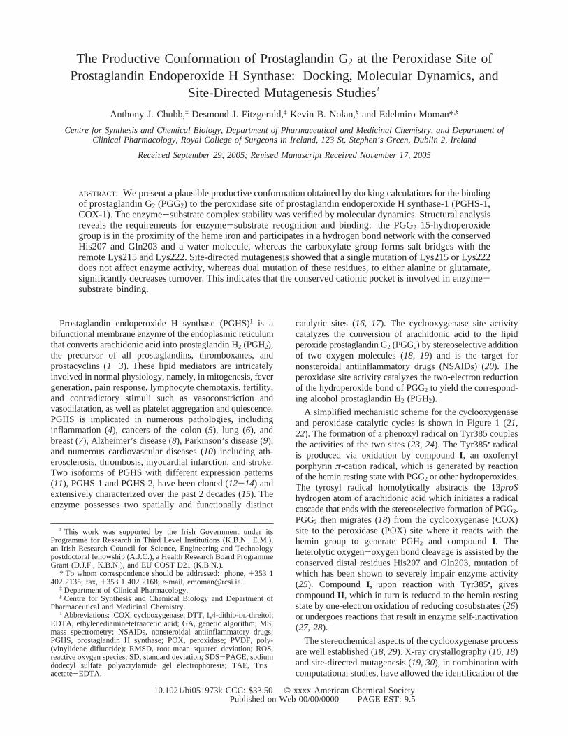
A simplified mechanistic scheme for the cyclooxygenaseand peroxidase catalytic cycles is shown in Figure 1 (21,22). The formation of a phenoxyl radical on Tyr385 couplesthe activities of the two sites (23, 24). The Tyr385• radicalis produced via oxidation by compoundI , an oxoferrylporphyrinπ-cation radical, which is generated by reactionof the hemin resting state with PGG2 or other hydroperoxides.The tyrosyl radical homolytically abstracts the 13proShydrogen atom of arachidonic acid which initiates a radicalcascade that ends with the stereoselective formation of PGG2.PGG2 then migrates (18) from the cyclooxygenase (COX)site to the peroxidase (POX) site where it reacts with thehemin group to generate PGH2 and compoundI . Theheterolytic oxygen-oxygen bond cleavage is assisted by theconserved distal residues His207 and Gln203, mutation ofwhich has been shown to severely impair enzyme activity(25). Compound I , upon reaction with Tyr385•, givescompoundII , which in turn is reduced to the hemin restingstate by one-electron oxidation of reducing cosubstrates (26)or undergoes reactions that result in enzyme self-inactivation(27, 28).
The stereochemical aspects of the cyclooxygenase processare well established (18, 29). X-ray crystallography (16, 18)and site-directed mutagenesis (19, 30), in combination withcomputational studies, have allowed the identification of the
† This work was supported by the Irish Government under itsProgramme for Research in Third Level Institutions (K.B.N., E.M.),an Irish Research Council for Science, Engineering and Technologypostdoctoral fellowship (A.J.C.), a Health Research Board ProgrammeGrant (D.J.F., K.B.N.), and EU COST D21 (K.B.N.).
* To whom correspondence should be addressed: phone,+353 1402 2135; fax,+353 1 402 2168; e-mail, [email protected].
‡ Department of Clinical Pharmacology.§ Centre for Synthesis and Chemical Biology and Department of
Pharmaceutical and Medicinal Chemistry.1 Abbreviations: COX, cyclooxygenase; DTT, 1,4-dithio-DL-threitol;
EDTA, ethylenediaminetetraacetic acid; GA, genetic algorithm; MS,mass spectrometry; NSAIDs, nonsteroidal antiinflammatory drugs;PGHS, prostaglandin H synthase; POX, peroxidase; PVDF, poly-(vinylidene difluoride); RMSD, root mean squared deviation; ROS,reactive oxygen species; SD, standard deviation; SDS-PAGE, sodiumdodecyl sulfate-polyacrylamide gel electrophoresis; TAE, Tris-acetate-EDTA.
10.1021/bi051973k CCC: $33.50 © xxxx American Chemical SocietyPAGE EST: 9.5Published on Web 00/00/0000
enzyme residues involved in substrate binding and position-ing (18) as well as the elucidation of the productiveconformation of arachidonic acid (31), other substrates,metabolites, and inhibitors. This structural knowledge hasbeen applied to the rational design of potent inhibitors ofthe cyclooxygenase activity, including numerous with iso-form selectivity (20). The cyclooxygenase pocket is deeplyburied within the protein at the end of an essentiallyhydrophobic channel, the entrance of which is at themembrane binding domain and continues to Tyr385, adjacentto the heme group (16). In contrast, the peroxidase pocketis a broad cavity on the surface of the protein. This allowssmall molecules to rapidly diffuse in and out of this pocket,which, together with the hemin reactivity, explains the lackof reported crystal structures of substrates or products boundto the iron. Peroxidase catalysis can continue independentlyof the cyclooxygenase site activity (28) (see Figure 1) andis a source of free radicals and reactive oxygen species(ROS), which may contribute to tissue damage or thrombosis(32). However, it is impossible to genetically eradicate bothisoforms of PGHS in a living organism (33), and thus apharmacological approach is necessary to elucidate the roleof this peroxidase in pathophysiology. To facilitate therational design of potent and selective inhibitors of this site(34, 35), we decided to elucidate the catalytically activeconformation of PGG2 in PGHS-POX.
To gather relevant information on the structural featuresnecessary for enzyme-substrate recognition and binding atthe peroxidase site, we combined docking and moleculardynamics (MD) in silico studies with site-directed mutagen-esis experiments. The recent publication of a theoreticaldocking-MD study (36) using a nonnatural PGG2 analogueand a different docking methodology has prompted us toreport our results with the natural substrate. Our computa-tional and mutagenesis studies, correlated with previousknowledge in the field, provide a plausible binding modelof PGG2 within the peroxidase site of PGHS-1.
EXPERIMENTAL PROCEDURES
Reagents.Unless otherwise stated, all reagents wereobtained from Sigma-Aldrich.
Docking Studies.Ligands were constructed using theSYBYL software package version 6.91 (Tripos Inc.) andgeometry-optimized with MOPAC at the PM3 level oftheory. A 2.0 Å resolution crystal structure of ovine PGHS-1(37) (PDB code 1Q4G) was used for the docking andmolecular dynamics experiments. All molecules except chainA and heme A were removed. Hydrogens were then addedand minimized. The carbon atoms of the protoporphyrin ringwere defined as aromatic, C.ar in the Tripos Force Fieldnotation, and the four nitrogen atoms as N.pl3, to obtain asmooth homogeneous distribution of charges. Kollman all-atom charges were assigned to the protein, and Gasteiger-Huckel charges were computed for the protoporphyrin group.A 1.0 formal charge was assigned to the iron atom (similardocking results were obtained with a charge of 1.6; valuesof 2.0 and 3.0 resulted in overestimation of the electrostaticterm of the docking energies). Docking experiments werecarried out using the Lamarckian genetic algorithm imple-mented in the automated docking program Autodock, version3.0.5 (38). The ligand and the protein were prepared fordocking with Autodock Tools. Autogrid was used to generatethe grid maps for an approximately 23× 23 × 23 Å3 cellbox centered above the heme iron. Autodock stochasticallygenerates a population of conformational, rotational, andtranslational isomers from any given starting structure of theligand and docks them within the active site of the protein,represented by a series of fixed-coordinate grid maps. Eachligand conformation is represented by a chromosome con-stituted by genes representing ligand conformational, rota-tional, and translational degrees of freedom. The individualsare evaluated by a fitness function which assesses the totalinteraction energy between the protein and the ligandmolecule, the internal energy of the ligand, and the desol-vation/solvation energies for the formation of the protein/ligand complex (the sum of these three terms is referred toas “docking energy” in this paper). Individuals in thepopulation are selected for reproduction in accordance withtheir fitness and undergo mutation and crossover reproductionoperators to generate new individuals. A number of genera-tions are permitted (see below), and the fittest individual ischosen. The whole evolutionary process was repeated 256times (genetic algorithm runs), which resulted in a databaseof 256 individuals. To ensure that the results were indepen-dent from the starting conformation of the ligand, a stochasticconformational database of PGG2 was created using theTripos force field, and the docking calculations were repeated10 times using the minimum energy conformer and another10 times using 10 different conformers randomly chosenfrom the database as starting structures for Autodock. Defaultparameters were employed except for the following: numberof GA runs, 256; maximum number of energy evaluations,2.5 × 106; maximum number of generations, 2.7× 10⁵;internal electrostatic energy computed. A more in-depthexperiment was performed with 2.5× 10⁷ energy evaluationsand 2.7 × 10⁶ generations. The distances and energiesreported here are those obtained in this later experimentunless otherwise stated. The two possible protonation statesfor distal histidine 207 were also considered, and the dockingresults obtained were similar in all cases.
Homology Modeling.Ovine PGHS numbering is used. Ahomology modeling structure of human PGHS-1 was con-structed using the Swiss-Model server (39) and the ovine
FIGURE 1: Cyclooxygenase and peroxidase catalytic cycles ofPGHS.
B Chubb et al. Biochemistry

crystal structure 1Q4G as template. After preparation of thehuman protein in SYBYL, the heme group was merged intothe POX site, and the complex was hydrated with∼16000explicit water molecules using the Molecular Silverwarealgorithm in an approximately 90× 90 × 90 Å3 cell box.The system was minimized using the Tripos force field,allowing 1000 iterations, and the solvent was removed. Theresults of the docking experiments with the modeled humanenzyme were identical to those with the ovine crystalstructure. PGHS homologues from all mammalian speciesfor which a homologue exists were aligned using theClustalW (45) program (Figure 5). Protein sequences wereobtained from the National Centre for Biotechnology Infor-mation (NCBI) GenBank database and are listed in theSupporting Information.
Molecular Dynamics.The PGG2 conformer with the mostfavorable docking energy was merged into the PGHS-1 ovinestructure. The complex was hydrated as described above andthen minimized in two steps: first, the water molecules wereminimized using 1000 iterations with the coordinates of theprotein fixed, and, second, the whole system was minimizedusing 1000 iterations. A 500 ps molecular dynamics experi-ment was carried out in SYBYL using the Tripos force fieldand the NTV ensemble (constant number of particles,temperature, and volume) at 300 K with periodic boundaryconditions. Default parameters were used and snapshotsrecorded every 0.5 ps.
Site-Directed Mutagenesis.Site-directed mutations inhuman PGHS-1 were prepared using the QuikChange site-directed mutagenesis kit supplied by Stratagene (La Jolla,CA) as described previously (46). Initially, a hexahistidinetag was inserted between the signal peptide and maturepeptide chain of human PGHS-1 (12) in the mammalianexpression vector pcDNA3-hCOX-1, using primers describedelsewhere (43). This template (pcDNA3-H6COX-1) was thensequentially mutated using the following primers (5′ to 3′):K211A, ACCCACCAGTTCTTCGCAACTTCTGGCAA-GATG; K215A, TTCAAAACTTCTGGCGCGATGGGTC-CTGGCTTC; K222A, GGTCCTGGCTTCACCGCGGCCT-TGGGCCATGGG; K211E, ACCCACCAGTTCTTCGAAA-CTTCTGGCAAGATG; K215E, TTCAAAACTTCTGGC-GAGATGGGTCCTGGCTTC; K222E, GGTCCTGGCT-TCACCGAGGCCTTGGGCCATGGG; and the reverse/complementary sequences thereof. Each mutation was verifiedby DNA sequencing, using either forward, GGGAAC-CAAAGGGAAGAAGC, or reverse primers AAAGCTGCT-CATCGCCCCAG (5′ to 3′). All DNA sequencing was donecommercially (AGOWA, Germany), the results of whichappear in the Supporting Information, Figure S1. Singlemutants were used as templates for the subsequent successiveconstruction of double and triple mutants. Transfection gradeDNA was isolated from transformedEscherichia coliusingan alkaline lysis and anion-exchange method, as per themanufacturer’s instructions (Qiagen). Plasmid size wasverified by separation in a 0.8% (w/v) agarose TAE gel, afterdigestion withBamHI andXhoI and intercalation by ethidiumbromide (data not shown).
Transient Expression of Human PGHS-1 Mutants.Wild-type and mutant pcDNA3-hCOX-1 constructs were tran-siently expressed in COS-1 cells as described previously (46).Parental vector pcDNA3 was used as a control for alltransfection experiments. COS-1 cells (400000 per well) were
grown in 2 mL of Dulbecco’s modified Eagle’s medium,10% fetal bovine serum, 100 units of penicillin, and 100µg/mL streptomycin in six-well plates at 37°C and 5% CO2.Purified plasmid DNA (2.0µg) was transfected into COS-1cells using 10µL of LipofectAMINE (Invitrogen) for 24 h,in duplicate, as described in the manufacturer’s protocol. TheDNA/LipofectAMINE mixture was allowed to settle for 5 hprior to 1:2 dilution with 20% fetal bovine serum.
Assay of Prostaglandins Synthesized by Transfected COS-1Cells. Forty-eight hours after transfection the cells wereassayed for PGHS-1 activity by stimulation with 60µMarachidonic acid for 15 min in HEPES-buffered Hank’s salinesolution. The PGE2 product was assayed using an enzyme-linked immunoassay for PGE2 (Assay Designs, Ann Arbor,MI). Assays were performed using 100µL of 1:50 dilutedsamples, as per the manufacturer’s instructions. The cellswere harvested with 1 mL of 1% C10E6 (Albion), 100 mMNaCl, 25 mM Na2HPO4 (pH 7.8), 20 mM imidazole, and1× EDTA-free protease inhibitors (Calbiochem).
Western Blotting.Standard Western blotting was per-formed, as described previously (47). Equal volumes (24µL)of each cell lysate sample were separated in a reducing 10%acrylamide SDS-PAGE gel, transferred to PVDF mem-branes, and probed using an anti-human PGHS-1 monoclonalantibody (Cayman Chemicals) and a horseradish peroxidase-linked anti-mouse IgG secondary antibody (Amersham).West-pico chemiluminescence reagents (Pierce) were usedto illuminate the PGHS-1 protein, the autoradiography ofwhich is shown.
Molecular Graphics.Molecular graphics images wereproduced using the UCSF Chimera package (48) from theComputer Graphics Laboratory, University of California, SanFrancisco. UCSF Chimera computes solvent-excluded mo-lecular surfaces using MSMS (49).
RESULTS
Docking Studies.The first X-ray structure of ovinePGHS-1 (oPGHS-1) cocrystallized with a ligand, a seren-dipitous glycerol molecule, in the peroxidase site has recentlybeen reported (37) (PDB code 1Q4G). This 2.0 Å resolutionstructure was used for the docking and dynamics experimentsreported here. The structure of natural PGG2 was dockedinto the peroxidase pocket of the protein using the Lama-rckian genetic algorithm implemented in the automateddocking program Autodock (38) (see Experimental Proce-dures). For analysis of the results, docked ligand conformerswithin 0.5 Å root mean squared deviation (RMSD) wereassumed to be identical and grouped into the same cluster,and the docking energy of the best fit individual was assignedto each cluster. The clusters were arranged in ascending (lessnegative) docking energies, and only those within 6.0 kcal/mol from the minimum were considered in the analysis.Similar clustering distributions were consistently observedin all of the experiments. A refined experiment with a 10-fold increase in the number of generations was alsoperformed, providing a similar cluster distribution butimproved by∼1.0 kcal/mol. The docking energies obtainedwere-15.7 kcal/mol for the in-depth experiment and-15.3kcal/mol for the best standard experiment.
A conserved binding mode was identified. The structureof the conformer of PGG2 with the most favorable docking
Biochemistry PGG2 Bound to the Peroxidase Site of PGHSC
energy, consistently obtained from all the docking experi-ments, and the active site residues that interact most directlywith the substrate are shown in Figure 2A. Solvent-accessiblesurfaces are displayed to enhance the three-dimensionalperception of the peroxidase pocket. More detailed informa-tion on the enzyme-substrate interactions is shown in Figure3.
A model of human PGHS-1 (hPGHS-1) was constructedusing the Swiss-Model server (39) and oPGHS-1 as atemplate as there is 91% overall sequence identity betweenthe ovine and the human enzymes and the POX site residuesare entirely conserved. The standard docking experiment wasrepeated three times with the human model providing
essentially identical results to those obtained with the ovinestructure.
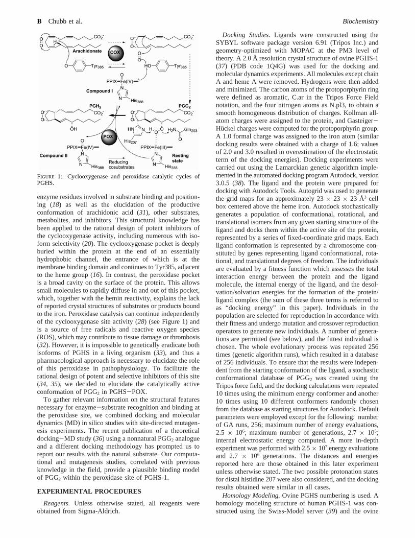
The terminal oxygen of the 15-hydroperoxide group (O26)is ∼3.0 Å from the heme iron, the same distance as theoxygen atom of the glycerol molecule present in the crystalstructure, and at a hydrogen bond distance from the carbonyloxygen of the Gln203 side chain. The inner oxygen of thehydroperoxide (O25) is located at a hydrogen bond distancefrom the nitrogen of His207. These two residues havepreviously been mutated and are known to be essential forthe catalytic activity of the enzyme (25). On the other hand,one of the oxygen atoms of the carboxylate group (O22) ofPGG2 is in the proximity of the positively charged nitrogenof Lys222, while the other (O21) is in the proximity ofLys215 and the backbone nitrogen of Gly214 (not shownfor clarity). The implication of these remote lysine residuesin substrate binding has not previously been addressed.
The 9,11-endoperoxycyclopentane moiety of PGG2 isinserted between the two propionate side chains of the heme(Figure 3A), while the C13-C15 fragment of PGG2 liesparallel to the tetrapyrrole plane (Figure 2A). The side chaincomprising carbons C2-C7 of PGG2 is inserted betweenVal291 and, more importantly, the lipophilic segment ofLys211. The C16-C20 side chain is positioned in thehydrophobic pocket defined by residues Val291, Leu294, andPhe409 and the methylene carbon of His274.
Two additional catalytically competent conformations ofPGG2, with higher docking energies, were identified. PanelsB and C of Figure 2 represent the two conformers (carbonsin gray) superimposed with the structure of the minimumdocking energy conformer (carbons in blue) within the activesite of the enzyme (molecular surfaces). Catalytically com-petent or “productive” conformations were defined as thosein which the terminal hydroperoxide oxygen atom of PGG2
(O26) is located within 4.0 Å of the iron. The computeddocking energies for conformers in (B) and (C) are-13.3and-13.7 kcal/mol, respectively (Figure 2).
The high degree of complementarity between the threeconformers and the peroxidase pocket is noteworthy (Figure
FIGURE 2: Model of PGG2 bound to the PGHS-1 peroxidase site. (A) Model of the catalytically competent conformation of PGG2 boundto the PGHS-1 peroxidase site. (B, C) Minimum docking energy PGG2 conformer (carbons in blue) superimposed on other possible productiveconformations (carbons in gray) within the PGHS-POX active site, represented by molecular surfaces.
FIGURE 3: PGG2-PGHS-1 peroxidase interactions. (A) Schematicdissection of the most important PGG2-PGHS-POX interactions.(B) Salt bridges and hydrogen bond networks. Distances (Å) before(italics in parentheses) and after minimization.
D Chubb et al. Biochemistry
2). Indeed, all fulfill four structural requirements: (i) thehydroperoxide group is in the vicinity of the heme iron,His207, and Gln203, an established prerequisite for catalyticactivity (25); (ii) the carboxylate group is near the cationicpocket comprising Lys215 and Lys222; (iii) a lipophilicmoiety points toward the Val291, Leu294, His273, andPhe409 hydrophobic pocket; and (iv) another lipophilicmoiety is inserted between Val291 and Lys211. Furthermore,the distance between the two polar anchor points, the ironon one side and Lys215 and Lys222 on the other, forces thesubstrate to adopt an extended conformation, approximately14 Å between the terminal hydroperoxide oxygen (O26) andthe more distant carboxylate oxygen (O22). This limits thepossible productive conformations of PGG2 to the threereported here (Figure 2).
It is noteworthy that the docking model reported bySeibold et al. (36) for a PGG2 analogue in some wayresembles one of the conserved binding modes found by usfor the natural substrate (Figure 2C), except for the fact thatthe carboxylate group of the PGG2 analogue points outwardfrom the pocket and is solvated by water molecules, whereasin our model the carboxylate group of the native substrateinteracts with Lys215 and Lys222 in all cases.
Molecular Dynamics.To account for protein flexibilityand the possible participation of water molecules in substratebinding, the most favorable complex structure (Figure 2A)was hydrated with∼16000 explicit water molecules andenergy minimized, and a 500 ps molecular dynamicssimulation was performed at 300 K using the Tripos forcefield, the NTV ensemble, and periodic boundary conditions.Although native PGHS is a membrane-bound homodimericprotein inserted into one leaflet of the lipid bilayer, we thinkthat the dynamics conditions employed are adequate because(1) the detergent needed to extract the enzyme is notnecessary for enzyme assays (25), indicating stable enzymeactivity in detergent-free water; (2) a 2 ns moleculardynamics simulation carried out previously (36) showed thatthe enzyme is stable as a monomer in aqueous media; (3)the peroxidase site is located distal to the membrane bindingdomain and the dimerization domain; and (4) the crystalstructure of the monomeric (31) enzyme does not signifi-cantly differ from those of the dimeric and multimeric crystalstructures.
Particularly significant protein-ligand distances are shownin Figure 3B both before (in parentheses) and after minimi-zation. Slight conformational modifications that optimizesubstrate binding take place in the complex upon minimiza-tion. The distances between the positively charged nitrogenatoms of Lys215 and Lys222 and the negatively chargedoxygen atoms of the substrate carboxylate are shortened. TheFe-O26 distance also shortens, mainly due to the realign-ment of the heme iron into the protoporphyrin plane. Thisis likely to be a computational artifact due to the lack ofadequate bending parameters for a pentacoordinated hemeiron in the Tripos force field (Sybyl version 6.9). However,this approximation should not appreciably affect the accuracyof the simulation for the purposes of this work.
Interestingly, the hydrogen bond network between PGG2
15-hydroperoxide, His207, and Gln203 is modified uponminimization to incorporate a water molecule, almostequidistant between PGG2-O26, His207, and Gln203.Gln203 retreats backward to allow the insertion of the water
and interacts with the substrate only indirectly through thiswater molecule. This hydrogen bond network incorporatingthe water molecule is maintained along the dynamicsexperiment and is therefore unlikely to be a computationalartifact (Figure 4). In fact, it has been hypothesized (40) thatthe ability to immobilize a water molecule in the vicinity ofthe catalytic center might be an intrinsic structural featureof peroxidases, responsible in part for the different catalyticmechanisms of peroxidases (one-electron oxidants) andcatalases (two-electron oxidants).
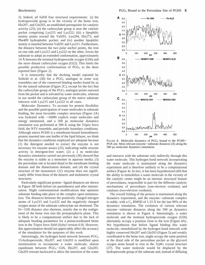
The overall folding of the protein is maintained along thedynamics experiment, and the enzyme-substrate complexis stable, with a CR RMSD of 1.33 Å for the last 90% of thedynamics simulation. The evolution of various relevantenzyme-substrate distances along the 500 ps dynamicssimulation is shown in Figure 4. Interestingly, a watermolecule and the terminal hydroperoxide oxygen (O26)alternately occupy a position close to the iron (Figure 4A).We hypothesize that before ligand binding this watermolecule, immobilized by the hydrogen bond network withhighly conserved His207 and Gln203 (Figure 5) and weaklycoordinated to the heme iron, might occupy the same positionat the distal side of the peroxidase pocket as the glyceroloxygen atom bound to iron in the 1Q4G crystal structure(37). The water molecule would be displaced by thehydroperoxide group of the substrate and, instead of diffusing
FIGURE 4: Molecular dynamics of PGG2 bound to the PGHS-POX site. Most relevant enzyme-substrate distances (Å) along the500 ps molecular dynamics simulation.
Biochemistry PGG2 Bound to the Peroxidase Site of PGHSE

away from the pocket, would be pushed toward Gln203,forcing this residue to retreat backward, initiating a rear-rangement of the hydrogen bond network (Figure 3B). Thisis corroborated by the finding that both the O26 of PGG2
and the water molecule tend to stay at a hydrogen bonddistance from His207 during the dynamics simulation (Figure4B).
Significantly, the salt bridges between Lys215 and Lys222and the PGG2 carboxylate group are tightly conservedthroughout the dynamics experiment (Figure 4C), suggestingthat this interaction plays a critical role in enzyme-substratebinding.
Site-Directed Mutagenesis.The presence of positivelycharged residues in the environment of the active site of aprotein facilitates the binding of negatively charged substrates(41, 42). Indeed, Arg120 has been demonstrated to play acrucial role in substrate binding at the cyclooxygenase siteof PGHS-1, and the mouth of the hydrophobic channel thatdirects substrates to the cyclooxygenase site is rich inpositively charged residues, namely, Arg79 and Arg83.However, to the best of our knowledge, the roles of Lys215and Lys222 at the PGHS peroxidase site in PGG2 bindinghave not previously been demonstrated. In accordance withour model, the loss of the salt bridges between the PGG2
carboxylate group and these two lysines, due to mutation,should result in a significant decrease of enzyme-substrateaffinity and thus reduced enzyme efficiency.
Lys211 is also located near Lys215 and Lys222 (Figure2A). Despite the fact that our modeling studies do not suggestparticipation of Lys211 in establishing a polar interaction
with PGG2 carboxylate, it is geometrically conceivable thata catalytically competent conformation of the substrate mayform an ionic linkage with this residue. We thereforeincluded Lys211 in our mutagenesis studies.
Site-directed mutagenesis of human PGHS-1 was per-formed sequentially, producing all combinations of single,double, and triple mutations. Thus, 14 mutants of the enzymewere constructed: 3 single mutants with a lysine to alaninereplacement (K215A, K222A, and K211A), 3 single mutantswith a lysine to glutamic acid replacement (K215E, K222E,and K211E), 3 double mutants with lysine to alaninereplacements (K215A/K222A, K211A/K215A, and K211A/K222A), 3 double mutants with lysine to glutamic acidreplacements (K215E/K222E, K211E/K215E, and K211E/K222E), 1 triple mutant with the three lysines replaced byalanine (K211A/K215A/K222E), and 1 triple mutant withthe three lysines replaced by glutamic acid (K211E/K215E/K222E). The mutants were produced in the mammalianexpression vector pcDNA3 containing recombinant humanhistidine-tagged PGHS-1 (12, 43). COS-1 cells were tran-siently transfected with the human PGHS-1 mutants, andequal volumes of cell lysate fractions were analyzed byWestern blotting (Figure 6). These data indicate that allmutants were similarly overexpressed by the human cytome-galovirus immediate-early enhancer-promoter in the pcD-NA3 mammalian expression vector and translated intoproteins showing the same relative molecular weight as thewild-type enzyme (Figure 6).
To test the enzymatic activity of the PGHS-1 mutants, thetransiently transfected COS-1 cells were allowed to react witharachidonic acid substrate for 15 min before the media wereremoved and assayed for PGE2 accumulation using anenzyme-linked immunosorbent assay. COS-1 cells trans-fected with parental pcDNA3 vector DNA showed noactivity. Furthermore, preincubation of wild-type transfectedcells with aspirin, which covalently inactivates the COX siteof PGHS at Ser530 (44), shows baseline activity, verifyingthat PGE2 accumulation seen in this assay is PGHS depend-ent. The assays were performed in duplicate wells, with PGE2
accumulation in each well assayed in duplicate, on three
FIGURE 5: Evolutionary conservation of the cation pocket. Allavailable mammalian PGHS sequences were aligned using ClustalW(45). See Supporting Information for full species name andGenBank accession numbers. The consensus line indicates identityin all sequences (*) or the occurrence of either conserved (:) orsemiconserved (.) substitutions. Catalytic residues Glu203 andHis207 are in italics, Lys211, Lys215, and Lys222 and positivesubstitutions thereof are in bold, and human PGHS-1 and -2 areunderlined. Original open reading frame numbering is shown.
FIGURE 6: Western blot analysis of PGHS-1 mutants. Cell lysatesof COS-1 cells transiently expressing mutant forms of His-taggedPGHS-1 were subjected to Western blotting and probed using ananti-PGHS-1 monoclonal antibody. Wild-type human PGHS-1 andcontrol cells transfected with the parental vector are shown forcomparison. (A) Lys-Ala. (B) Lys-Glu. Mutant abbreviations areas follows: 215A222, K215A/K222A; 215A211, K211A/K215A;222A211, K211A/K222A; K3A, K211A/K215A/K222A; 215E222,K215E/K222E; 215E211, K211E/K215E; 222E211, K211E/K222E;K3E, K211E/K215E/K222E.
F Chubb et al. Biochemistry

separate occasions, thus accounting for possible slightvariations in PGHS-1 or PGE2 synthase expression levels.The amount of PGE2 produced by each of the mutants wasconverted to a percentage of that produced by the wild-typeenzyme (Figure 7A). The most striking effect is the completeabolition of activity in all mutations involving Lys211. It isalso clear that single mutations involving Lys215 andLys222, to either alanine or glutamic acid, do not impairPGHS activity, with these mutants showing essentially wild-type conversion of PGG2 to PGH2, which is in turn convertedby synthases to the PGE2 measured in our experiments.Importantly, when both of these residues are simultaneouslymutated, a significant reduction of 27% and 58% was notedfor alanine or glutamate, respectively. This reduction isstatistically relevant, with variance between the Lys222 andLys215/Lys222 double mutants showing paired Student’st-test P values of 0.038 and 0.021 for the alanine andglutamate mutants, respectively.
DISCUSSION
Our docking and molecular dynamics studies have shownthat there are two polar anchor points for PGG2 within thePGHS-1 peroxidase pocket. First, in agreement with thecommonly accepted mechanism, the location of the 15-hydroperoxide group within the iron coordination sphere isa requirement for catalysis. Second, the carboxylate groupof PGG2 establishes salt bridges with Lys215 and Lys222.Furthermore, the concomitant binding of PGG2 hydroper-oxide in the vicinity of the iron and the carboxylate close to
the lysines requires the substrate to adopt an extendedgeometry that narrows the set of possible productiveconformers to the ones reported here, with minor variations.
The results in Figure 7A show the importance of Lys211,Lys215, and Lys222. Mutation of Lys211 completely impairsthe activity of the enzyme. This effect is too dramatic to beattributed to a perturbation of ligand binding and is moreconsistent with a severe alteration of the structure of theenzyme. Indeed, this residue is totally conserved across bothPGHS isoforms sequenced from species spanning a broadtaxonomic range (Figure 5) and structurally is found nearthe end of helix H2 (16), which also contains the catalyticGlu203 and His207 residues. Figure 8 shows how, throughformation of ionic linkages with the similarly conservedresidues Asp236 and Glu290, Lys211 acts as a bridgebetween three poorly organized regions of the protein in thevicinity of the peroxidase pocket (16). In addition, the N-Odistances between the ammonium group of Lys211 and thecarboxylate groups of Asp236 and Glu290 are completelyconserved along the molecular dynamics simulation (Figure9A,B), whereas the N-O distance between the ammoniumgroup Lys211 and the carboxylate group of PGG2 is neverless than 8 Å (data not shown).
The binding energies obtained after docking PGG2 intothe peroxidase catalytic pocket of each of the reportedmutants are shown in Figure 7B. In all cases the lowestenergy catalytically competent conformer is similar to thatobserved for the wild-type protein (note that the carboxylategroup of PGG2 is still able to form a hydrogen bond with
FIGURE 7: Activity of PGHS-1 mutants in transiently transfected mammalian cells. (A) Transiently transfected COS-1 cells were assayedfor PGE2 production following arachidonic acid stimulation. PGE2 produced was converted to a percentage of wild-type activity (n ) 3,SD). A preincubation with acetylsalicylic acid (aspirin) was used to show COX dependence in this assay. The asterisk indicatesP < 0.05.Mutant abbreviations are as in Figure 6. (B) Docking energies (kcal/mol) for the best productive conformation of PGG2 bound to theperoxidase site of each of the reported mutants.
Biochemistry PGG2 Bound to the Peroxidase Site of PGHSG

the backbone NH of Gly214). However, in a few cases, thelowest energy catalytically competent conformer is not theabsolute lowest energy conformer. In no case was the PGG2
carboxylate found close to the Lys211 ammonium. It isnoteworthy that the trend in predicted docking energies forthe single and double mutants involving Lys215 and Lys222(Figure 7B, solid bars) is similar to that found in thebiological activity assays. For 215A222 and 215E222, inwhich the two salt bridges are lost, the decrease in dockingenergies is approximately 2.0 and 2.5 kcal/mol, respectively,which translates into a 30-60% decrease of enzymaticactivity. In all cases, including all of the Lys211 mutantsfor which the enzymatic activity is completely impaired(Figure 7B, empty bars), the substrate can still access theactive site and the binding energies are comparable. Forinstance, the affinity of the substrate toward K211E should,according to our docking data, be as good as that for thewild-type enzyme. However, like all other mutants involvingLys211, it is completely inactive. These results indicate that(i) in terms of substrate affinity, any of the mutations shouldhave either a moderate (as empirically demonstrated for215A222 and 215E222) or negligible (as empirically dem-onstrated for K215A, K222A, K215E, and K222E) effecton enzyme activity; and (ii) a complete abolition of theenzymatic activity (as observed in all of the K211 mutants)is, in this particular case, unlikely to be the result of a mereperturbation on substrate binding.
Importantly, the Phe209-Leu230 segment, which containsLys211 and connects helix H2 to H3, includes Thr212, whichforms hydrogen bonds with the C13 heme propionate(Figures 8 and 9C) and is likely to be crucial for hemebinding (37). Lysine 211 is, therefore, likely to have astructural function, and its replacement could prevent thecorrect folding of the peroxidase site and thus its ability torecruit the heme group. Furthermore, we consider that theconformational change required for Lys211 to participate inligand binding is unlikely to be compatible with its apparentstructural role. Final proof of the involvement of Lys211 incross-linking and stabilization of the large loop comprisingthe distal “roof” of the peroxidase site awaits furthercharacterization and testing. It would be informative to
attempt to restore enzyme activity by mutating the residuesinvolved in salt-bridging interactions with Lys211, namely,Asp236 and Glu290, to the positively charged lysine in theK211E mutant. If activity is restored, this would imply thatthe structure of the K211E mutant is now reestablished andfurther prove that this residue is not directly involved inPGG2 binding.
Single mutation of Lys215 and Lys222, either to alanineor glutamic acid, does not affect enzyme activity (Figure7A). However, simultaneous mutation of these residuescauses a significant decrease in activity. We propose thatthe presence of these two compensatory residues is a “safetymechanism” which highlights the importance of a positivelycharged region for substrate binding (41): if one of thelysines is mutated, the enzyme would still be fully functionalas a substrate host due to the presence of the other lysine.Indeed, both residues are highly conserved in both isoformsof numerous species, and at least one of the two amino acidsis always a lysine in mammals (Figure 5). Furthermore, thiseffect could not have been predicted from homology align-ments as neither residue is entirely conserved. The average
FIGURE 8: Structural role of Lys211. The backbone of PGHS-1 isshown in ribbon diagram, while the heme and side chains of Lys211,Asp236, Glu290, and Thr212 are depicted as sticks.
FIGURE 9: Molecular dynamics showing conservation of saltbridges. The preservation of the salt bridges between the ammoniumnitrogen of Lys211 and the carboxylate oxygens of Asp236 (A)and Glu290 (B) during the molecular dynamics simulation isillustrated by monitoring interatomic bond lengths (Å). Themaintenance of a hydrogen bond between the hydroxyl group andthe backbone amide of Thr212 and the C13 heme propionate wasalso monitored (C).
H Chubb et al. Biochemistry
40% decrease in activity of the double mutants with respectto the wild type correlates well with the∼2.0 kcal/moldecrease in binding energy due to the loss of the salt bridgespredicted by docking calculations (Figure 7B).
It could be argued that the effect of simultaneous mutationof Lys215 and Lys222 may be due to nonspecific effectsunrelated to ligand binding. Inspection of the crystal structure,however, indicates that Lys215 does not show significantinteractions with any other residue of the protein, whileLys222 is only involved in hydrogen bonding with the poorlyconserved Ser213 side chain. Furthermore, the fact that singlemutations have no effect on enzyme activity indicates thatthese mutations do not have serious implications for enzymefolding.
The proposed binding model is, thus, fully consistent withthe commonly accepted mechanism of catalysis as well aswith our, and other previously reported, site-directed mu-tagenesis studies. On the basis of the binding model presentedhere, which defines the minimal pharmacophore for specificenzyme-substrate recognition, we have designed a firstgeneration of PGHS-peroxidase inhibitors which are ex-pected to be highly selective and become valuable chemicaltools for exploring the implication of this peroxidase inpathophysiology. The synthesis and testing of these com-pounds are currently underway.
SUPPORTING INFORMATION AVAILABLE
GenBank accession numbers for proteins used in Figure5 and the DNA sequencing data for the mutants. Thismaterial is available free of charge via the Internet at http://pubs.acs.org.
REFERENCES
1. Smith, W. L., Marnett, L. J., and DeWitt, D. L. (1991) Prostag-landin and thromboxane biosynthesis,Pharmacol. Ther. 49, 153-179.
2. Smith, W. L., DeWitt, D. L., and Garavito, R. M. (2000)Cyclooxygenases: structural, cellular, and molecular biology,Annu. ReV. Biochem. 69, 145-182.
3. Simmons, D. L., Botting, R. M., and Hla, T. (2004) Cyclooxy-genase isozymes: the biology of prostaglandin synthesis andinhibition, Pharmacol. ReV. 56, 387-437.
4. Zhang, Y., Shaffer, A., Portanova, J., Seibert, K., and Isakson, P.C. (1997) Inhibition of cyclooxygenase-2 rapidly reverses inflam-matory hyperalgesia and prostaglandin E2 production,J. Phar-macol. Exp. Ther. 283, 1069-1075.
5. Kargman, S. L., O’Neill, G. P., Vickers, P. J., Evans, J. F.,Mancini, J. A., and Jothy, S. (1995) Expression of prostaglandinG/H synthase-1 and -2 protein in human colon cancer,CancerRes. 55, 2556-2559.
6. Hida, T., Yatabe, Y., Achiwa, H., Muramatsu, H., Kozaki, K.,Nakamura, S., Ogawa, M., Mitsudomi, T., Sugiura, T., andTakahashi, T. (1998) Increased expression of cyclooxygenase 2occurs frequently in human lung cancers, specifically in adeno-carcinomas,Cancer Res. 58, 3761-3764.
7. Parrett, M. L., Abou-Issa, H. M., Alshafie, G., Ross, M. S., Harris,R. E., and Robertson, F. M. (1999) Comparative ability ofibuprofen and N-(4-hydroxyphenyl)retinamide to inhibit develop-ment of rat mammary adenocarcinomas associated with differentialinhibition of gene expression of cyclooxygenase isoforms,Anti-cancer Res. 19, 5079-5085.
8. Rich, J. B., Rasmusson, D. X., Folstein, M. F., Carson, K. A.,Kawas, C., and Brandt, J. (1995) Nonsteroidal anti-inflammatorydrugs in Alzheimer’s disease,Neurology 45, 51-55.
9. Teismann, P., Tieu, K., Choi, D. K., Wu, D. C., Naini, A., Hunot,S., Vila, M., Jackson-Lewis, V., and Przedborski, S. (2003)Cyclooxygenase-2 is instrumental in Parkinson’s disease neuro-degeneration,Proc. Natl. Acad. Sci. U.S.A. 100, 5473-5478.
10. Linton, M. F., and Fazio, S. (2004) Cyclooxygenase-2 andinflammation in atherosclerosis,Curr. Opin. Pharmacol. 4, 116-123.
11. Tanabe, T., and Tohnai, N. (2002) Cyclooxygenase isozymes andtheir gene structures and expression,Prostaglandins Other LipidMediators 68-69, 95-114.
12. Funk, C. D., Funk, L. B., Kennedy, M. E., Pong, A. S., andFitzgerald, G. A. (1991) Human platelet/erythroleukemia cellprostaglandin G/H synthase: cDNA cloning, expression, and genechromosomal assignment,FASEB J. 5, 2304-2312.
13. Hla, T., and Neilson, K. (1992) Human cyclooxygenase-2 cDNA,Proc. Natl. Acad. Sci. U.S.A. 89, 7384-7388.
14. Chandrasekharan, N. V., Dai, H., Roos, K. L., Evanson, N. K.,Tomsik, J., Elton, T. S., and Simmons, D. L. (2002) COX-3, acyclooxygenase-1 variant inhibited by acetaminophen and otheranalgesic/antipyretic drugs: cloning, structure, and expression,Proc. Natl. Acad. Sci. U.S.A. 99, 13926-13931.
15. Garavito, R. M., Malkowski, M. G., and DeWitt, D. L. (2002)The structures of prostaglandin endoperoxide H synthases-1 and-2, Prostaglandins Other Lipid Mediators 68-69, 129-152.
16. Picot, D., Loll, P. J., and Garavito, R. M. (1994) The X-ray crystalstructure of the membrane protein prostaglandin H2 synthase-1,Nature 367, 243-249.
17. Kulmacz, R. J., van der Donk, W. A., and Tsai, A. L. (2003)Comparison of the properties of prostaglandin H synthase-1 and-2, Prog. Lipid Res. 42, 377-404.
18. Kiefer, J. R., Pawlitz, J. L., Moreland, K. T., Stegeman, R. A.,Hood, W. F., Gierse, J. K., Stevens, A. M., Goodwin, D. C.,Rowlinson, S. W., Marnett, L. J., Stallings, W. C., and Kurumbail,R. G. (2000) Structural insights into the stereochemistry of thecyclooxygenase reaction,Nature 405, 97-101.
19. Thuresson, E. D., Lakkides, K. M., Rieke, C. J., Sun, Y., Wingerd,B. A., Micielli, R., Mulichak, A. M., Malkowski, M. G., Garavito,R. M., and Smith, W. L. (2001) Prostaglandin endoperoxide Hsynthase-1: the functions of cyclooxygenase active site residuesin the binding, positioning, and oxygenation of arachidonic acid,J. Biol. Chem. 276, 10347-10357.
20. FitzGerald, G. A. (2003) COX-2 and beyond: Approaches toprostaglandin inhibition in human disease,Nat. ReV. Drug DiscoV.2, 879-890.
21. Ullrich, V., and Ruf, H. H. (1994) Heme Proteins in ProstaglandinBiosynthesis, inMetalloporphyrins in Catalytic Oxidations(Shel-don, R. A., Ed.) pp 157-192, Marcel Dekker, New York.
22. O’Brien, P. J. (2000) Peroxidases,Chem.-Biol. Interact. 129, 113-139.
23. Lassmann, G., Odenwaller, R., Curtis, J. F., DeGray, J. A., Mason,R. P., Marnett, L. J., and Eling, T. E. (1991) Electron spinresonance investigation of tyrosyl radicals of prostaglandin Hsynthase. Relation to enzyme catalysis,J. Biol. Chem. 266,20045-20055.
24. Tsai, A., Hsi, L. C., Kulmacz, R. J., Palmer, G., and Smith, W. L.(1994) Characterization of the tyrosyl radicals in ovine prostag-landin H synthase-1 by isotope replacement and site-directedmutagenesis,J. Biol. Chem. 269, 5085-5091.
25. Landino, L. M., Crews, B. C., Gierse, J. K., Hauser, S. D., andMarnett, L. J. (1997) Mutational analysis of the role of the distalhistidine and glutamine residues of prostaglandin-endoperoxidesynthase-2 in peroxidase catalysis, hydroperoxide reduction, andcyclooxygenase activation,J. Biol. Chem. 272, 21565-21574.
26. Markey, C. M., Alward, A., Weller, P. E., and Marnett, L. J. (1987)Quantitative studies of hydroperoxide reduction by prostaglandinH synthase. Reducing substrate specificity and the relationshipof peroxidase to cyclooxygenase activities,J. Biol. Chem. 262,6266-6279.
27. Wu, G., Wei, C., Kulmacz, R. J., Osawa, Y., and Tsai, A. L. (1999)A mechanistic study of self-inactivation of the peroxidase activityin prostaglandin H synthase-1,J. Biol. Chem. 274, 9231-9237.
28. Song, I., Ball, T. M., and Smith, W. L. (2001) Different suicideinactivation processes for the peroxidase and cyclooxygenaseactivities of prostaglandin endoperoxide H synthase-1,Biochem.Biophys. Res. Commun. 289, 869-875.
29. van der Donk, W. A., Tsai, A. L., and Kulmacz, R. J. (2002) Thecyclooxygenase reaction mechanism,Biochemistry 41, 15451-15458.
30. Schneider, C., Boeglin, W. E., and Brash, A. R. (2004) Identifica-tion of two cyclooxygenase active site residues, Leucine 384 andGlycine 526, that control carbon ring cyclization in prostaglandinbiosynthesis,J. Biol. Chem. 279, 4404-4414.
Biochemistry PGG2 Bound to the Peroxidase Site of PGHSI

31. Malkowski, M. G., Ginell, S. L., Smith, W. L., and Garavito, R.M. (2000) The productive conformation of arachidonic acid boundto prostaglandin synthase,Science 289, 1933-1937.
32. Kukreja, R. C., Kontos, H. A., Hess, M. L., and Ellis, E. F. (1986)PGH synthase and lipoxygenase generate superoxide in thepresence of NADH or NADPH,Circ. Res. 59, 612-619.
33. Reese, J., Paria, B. C., Brown, N., Zhao, X., Morrow, J. D., andDey, S. K. (2000) Coordinated regulation of fetal and maternalprostaglandins directs successful birth and postnatal adaptationin the mouse,Proc. Natl. Acad. Sci. U.S.A. 97, 9759-9764.
34. Szewczuk, L. M., Forti, L., Stivala, L. A., and Penning, T. M.(2004) Resveratrol is a peroxidase-mediated inactivator of COX-1but not COX-2: a mechanistic approach to the design of COX-1selective agents,J. Biol. Chem. 279, 22727-22737.
35. Tam, S. S., Lee, D. H., Wang, E. Y., Munroe, D. G., and Lau, C.Y. (1995) Tepoxalin, a novel dual inhibitor of the prostaglandin-Hsynthase cyclooxygenase and peroxidase activities,J. Biol. Chem.270, 13948-13955.
36. Seibold, S. A., Smith, W. L., and Cukier, C. I. (2004) Theperoxidase site of prostaglandin endoperoxide H synthase-1docking and molecular dynamic studies with a prostaglandinendoperoxide analog,J. Phys. Chem. B. 108, 9297-9305.
37. Gupta, K., Selinsky, B. S., Kaub, C. J., Katz, A. K., and Loll, P.J. (2004) The 2.0 Å resolution crystal structure of prostaglandinH2 synthase-1: structural insights into an unusual peroxidase,J.Mol. Biol. 335, 503-518.
38. Morris, G. M., Goodsell, D. S., Halliday, R. S., Huey, R., Hart,W. E., Belew, R. K., and Olson, A. J. (1998) Automated dockingusing a Lamarckian genetic algorithm and empirical binding freeenergy function,J. Comput. Chem. 19, 1639-1662.
39. Schwede, T., Kopp, J., Guex, N., and Peitsch, M. C. (2003) Swiss-Model: An automated protein homology-modeling server,NucleicAcids Res. 31, 3381-3385.
40. Jones, P. (2001) Roles of water in heme peroxidase and catalasemechanisms,J. Biol. Chem. 276, 13791-13796.
41. Riordan, J. F., McElvany, K. D., and Borders, C. L. (1977) Arginylresidues: anion recognition sites in enzymes,Science 195, 884-886.
42. Rudberg, P. C., Tholander, F., Andberg, M., Thunnissen, M. M.,and Haeggstrom, J. Z. (2004) Leukotriene A4 hydrolase: iden-tification of a common carboxylate recognition site for the epoxidehydrolase and aminopeptidase substrates,J. Biol. Chem. 279,27376-27382.
43. Smith, T., Leipprandt, J., and DeWitt, D. (2000) Purification andcharacterization of the human recombinant histidine-tagged pros-taglandin endoperoxide H synthases-1 and -2,Arch. Biochem.Biophys. 375, 195-200.
44. Roth, G. J., Machuga, E. T., and Ozols, J. (1983) Isolation andcovalent structure of the aspirin-modified, active-site region ofprostaglandin synthetase,Biochemistry 22, 4672-4675.
45. Thompson, J. D., Higgins, D. G., and Gibson, T. J. (1994)ClustalW: improving the sensitivity of progressive multiplesequence alignment through sequence weighting, position-specificgap penalties and weight matrix choice,Nucleic Acids Res. 22,4673-4680.
46. Dooley, C. M., Devocelle, M., McLoughlin, B., Nolan, K. B.,Fitzgerald, D. J., and Sharkey, C. T. (2003) A novel family ofhydroxamate-based acylating inhibitors of cyclooxygenase,Mol.Pharmacol. 63, 450-455.
47. Chubb, A. J., Schwager, S. L., van der Merwe, E., Ehlers, M. R.,and Sturrock, E. D. (2004) Deletion of the cytoplasmic domainincreases basal shedding of angiotensin-converting enzyme,Bio-chem. Biophys. Res. Commun. 314, 971-975.
48. Pettersen, E. F., Goddard, T. D., Huang, C. C., Couch, G. S.,Greenblatt, D. M., Meng, E. C., and Ferrin, T. E. (2004) UCSFChimerasa visualization system for exploratory research andanalysis,J. Comput. Chem. 25, 1605-1612.
49. Sanner, M. F., Olson, A. J., and Spehner, J. C. (1996) Reducedsurface: an efficient way to compute molecular surfaces,Biopoly-mers 38, 305-320.
BI051973K
J Chubb et al. PAGE EST: 9.5 Biochemistry