Partial reversal of the substrate stereospecificity of an L-lactate dehydrogenase by site-directed...
8
J. Am. Chem. SOC. 1995, 117, 2387-2394 2387 Partial Reversal of the Substrate Stereospecificity of an L-Lactate Dehydrogenase by Site-Directed Mutagenesis Roman SakowiczJ Marvin Gold: and J. Bryan Jones**S*g Contribution from the Department of Molecular and Medical Genetics and Department of Chemistry, University of Toronto, Toronto, Ontario, Canada, M5S 1AI Received September 27, 1994@ Abstract: The L-lactate dehydrogenase of Bacillus stearothermophilus (BSLDH) is a highly stereospecific enzyme that catalyzes synthetically useful reductions of 2-keto acids to the corresponding L-Zhydroxy acids. The strategy of probing the factors controlling enzyme stereospecificity by evaluating BSLDHs resistance to being induced to reverse its stereospecificitypreference from L to D has been extended by the introduction of new site-directedmutations designed to induce the natural substrate pyruvate to bind in an alternative, 180-rotated, orientation that would lead to D-lactate production. With this “flipping” of the natural substrate-binding orientation as the goal, residue Ile240 was changed to Arg and Lys to introduce a new carboxyl-binding site, while an Argl71Tyr replacement removed the natural Arg171-C00--binding interaction, thereby creating the double mutants 1240FUR171Y and 1240wR171Y. The validity of the strategy was demonstrated by significant, up to 2.3%, proportions of D-lactate produced by preparative-scale 1240K/R17lY-catalyzed reductions of pyruvate. This represents an estimated -105-fold relaxation of the natural L-stereoselectivity of BSLDH, thereby demonstrating the feasibility of eventually applying rational protein engineering to optimize and control the stereoselectivity of enzymes used in asymmetric synthesis. Interestingly, modeling studies based on comparisons of the active site region of the D-stereospecific enzyme, glycerate dehydrogenase (GDH), with that of BSLDH reveal that in GDH, the position of the catalytically important substrate COO--binding Arg-residue approximates closely that of the Argkys240 residues introduced into BSLDH. It thus appears that the strategy of changing stereospecificity by reversing 2-keto acid binding orientation adopted in the current study parallels the one employed by Nature to introduce L- and D-configurations in 2-keto acid reductions. Introduction Enzymes are now widely accepted as useful catalysts for a broad range of organic synthesis, with their capacities for inducing asymmetric transformations being the most exploited.’ However, despite the widespread uses of both enzymes and microorganisms in asymmetric synthesis, much remains to be learned about the factors that determine the structural specificity and stereospecificity of enzymes. In view of the increasingly broad spectrum of new and unnatural substrate structures that synthetically useful enzymes are being called on to accom- modate, it is becoming more and more essential to delineate the enzyme-substrate interactions that regulate and control enzyme stereospecificity.2 Such information will permit the selection of the enzymes that are best suited for any given chiral synthon preparation and will also facilitate the rational tailoring of enzyme specificity by the site-directed mutagenesis tech- niques of protein engineering? Furthermore, while our primary goals are to optimize and extend the synthetic utility of enzymes, the data obtained in such studies will provide additional insights + Department of Molecular and Medical Genetics. t Department of Chemistry. 5 J. B. Jones: phone: (416) 978-3589. Fax: (416) 978-1553. e-mail: @Abstract published in Advance ACS Abstracts, February 1, 1995. (1) (a) Wong, C. H. Chimiu 1993, 47, 127-132. (b) Roberts, S. M.; Tumer, N. J.; Willetts, A. J. Chimia 1993, 47, 85-92. (c) Crout, D. H. G.; Jones, J. E.; Roberts, B. M. Tetrahedron-Assym. 1993, 4, R21-R22. (d) Wong, C. H.; Liu, K. K. C.; Kajimoto, T.; Chen, L. R.; Zhong, Z. Y.; Ichikawa, Y.; Shen, G. J. Annals New Y. Acad. Sci. 1992, 672, 343-351. (e) Wong, C. H.; Shen, G.-J.; Pederson, R. L.; Wang, Y.-F.; Hennen, W. J. Methods Enzymol. 1991, 202, 591-620. (2) (a) Weinhold, E. G.; Glasfeld, A,; Ellington, A. D.; Benner, S. A. Proc. Nutl. Acad. Sci. U.S.A. 1991, 88, 8420-8424. (b) LaReau, R. D.; Anderson, V. E. Biochemistry 1992, 31, 4174-4180. (c) Neidhardt, D. J.; Howell, P. L.; Petsko, G. A,; Powers, V. M.; Li, R.; Kenyon, G. L.; Gerlt, J. A. Biochemisrly 1991, 30, 9264. j bjones @ alchemy.chem.utoronto.ca. 0002-786319511517-2387$09.00/0 of fundamental value in advancing basic understanding of the factors determining and controlling enzyme specificity. The classes of enzymes most widely applied in organic synthesis are the hydrolases and the oxidoreductases. Among the latter group, L-lactate dehydrogenases (L-LDHs? EC 1.1.1.27) are well characterized enzymes that catalyze C=O == CH(0H) conversions of the type shown in eq 1. Enantiomeri- cally pure L-Zhydroxy acids are valuable chiral synthons in asymmetric synthesis? and L-LDHs from various sources have been widely applied in their preparatiom6 RCOCOOH + NADH + H + RCH(0H)COOH + NAD+ (1) 1 Z a, R = CH3 b, R = CHjCHz (3) (a) Witkowski, A.; Witkowska, H. E.; Smith, S. J. Biol. Chem 1994, 269, 379. (b) Bradley, M.; Bucheler, U. S.; Walsh, C. T. Biochemistry 1991, 30, 6124. (c) Hedstrom, L.; Graf, L.; Stewart, C. B.; Rutter, W. J.; Phillips, M. A. Methods Enzymol. 1991, 202, 671. (d) Higaki, J. N.; Haymore, B. L.; Chen, S.; Fletterick, R. J.; Craik, C. S. Biochemistry 1990, 29, 8582. (e) Scrutton, N. S.; Berry, A.; Perham, R. N. Nature 1990, 343, 38. (f) Wilks, H. M.; Halsall, D. J.; Atkinson, T.; Chia, W. N.; Clarke, A. R.; Holbrook, J. J. Biochemistry 1990,29, 8587. (9) Lindberg, R. L. P.; Negishi, M. Nature 1989, 339, 632. (h) Cronin, C. N.; Malcolm, B. A,; Kirsch, J. F. J Am. Chem. Soc. 1987, 109, 2222. (4) Abbreviations used: LDH, L-lactate dehydrogenase; ES, enzyme- substrate complex; WT, wild type; BSLDH, Bacillus stearothermophilus LDH; DMLDH, dogfish muscle LDH; FDH, formate dehydrogenase; GDH, glycerate dehydrogenase; FBP, fructose- 1,6-bisphosphate. (5) (a) Bemardi, A,; Micheli, F.; Potenza, D.; Scholastico, D.; Villa, R. Tetrahedron Lett. 1990, 31, 4949. (b) Pearson, W.; Hines, J. V. J. Org. Chem. 1989, 54, 4235. 0 1995 American Chemical Society
Transcript of Partial reversal of the substrate stereospecificity of an L-lactate dehydrogenase by site-directed...
J. Am. Chem. SOC. 1995, 117, 2387-2394 2387
Partial Reversal of the Substrate Stereospecificity of an L-Lactate Dehydrogenase by Site-Directed Mutagenesis Roman SakowiczJ Marvin Gold: and J. Bryan Jones**S*g
Contribution from the Department of Molecular and Medical Genetics and Department of Chemistry, University of Toronto, Toronto, Ontario, Canada, M5S 1AI
Received September 27, 1994@
Abstract: The L-lactate dehydrogenase of Bacillus stearothermophilus (BSLDH) is a highly stereospecific enzyme that catalyzes synthetically useful reductions of 2-keto acids to the corresponding L-Zhydroxy acids. The strategy of probing the factors controlling enzyme stereospecificity by evaluating BSLDHs resistance to being induced to reverse its stereospecificity preference from L to D has been extended by the introduction of new site-directed mutations designed to induce the natural substrate pyruvate to bind in an alternative, 180-rotated, orientation that would lead to D-lactate production. With this “flipping” of the natural substrate-binding orientation as the goal, residue Ile240 was changed to Arg and Lys to introduce a new carboxyl-binding site, while an Argl71Tyr replacement removed the natural Arg171-C00--binding interaction, thereby creating the double mutants 1240FUR171Y and 1240wR171Y. The validity of the strategy was demonstrated by significant, up to 2.3%, proportions of D-lactate produced by preparative-scale 1240K/R17lY-catalyzed reductions of pyruvate. This represents an estimated -105-fold relaxation of the natural L-stereoselectivity of BSLDH, thereby demonstrating the feasibility of eventually applying rational protein engineering to optimize and control the stereoselectivity of enzymes used in asymmetric synthesis. Interestingly, modeling studies based on comparisons of the active site region of the D-stereospecific enzyme, glycerate dehydrogenase (GDH), with that of BSLDH reveal that in GDH, the position of the catalytically important substrate COO--binding Arg-residue approximates closely that of the Argkys240 residues introduced into BSLDH. It thus appears that the strategy of changing stereospecificity by reversing 2-keto acid binding orientation adopted in the current study parallels the one employed by Nature to introduce L- and D-configurations in 2-keto acid reductions.
Introduction
Enzymes are now widely accepted as useful catalysts for a broad range of organic synthesis, with their capacities for inducing asymmetric transformations being the most exploited.’ However, despite the widespread uses of both enzymes and microorganisms in asymmetric synthesis, much remains to be learned about the factors that determine the structural specificity and stereospecificity of enzymes. In view of the increasingly broad spectrum of new and unnatural substrate structures that synthetically useful enzymes are being called on to accom- modate, it is becoming more and more essential to delineate the enzyme-substrate interactions that regulate and control enzyme stereospecificity.2 Such information will permit the selection of the enzymes that are best suited for any given chiral synthon preparation and will also facilitate the rational tailoring of enzyme specificity by the site-directed mutagenesis tech- niques of protein engineering? Furthermore, while our primary goals are to optimize and extend the synthetic utility of enzymes, the data obtained in such studies will provide additional insights
+ Department of Molecular and Medical Genetics. t Department of Chemistry. 5 J. B. Jones: phone: (416) 978-3589. Fax: (416) 978-1553. e-mail:
@Abstract published in Advance ACS Abstracts, February 1, 1995. (1) (a) Wong, C. H. Chimiu 1993, 47, 127-132. (b) Roberts, S. M.;
Tumer, N. J.; Willetts, A. J. Chimia 1993, 47, 85-92. (c) Crout, D. H. G.; Jones, J. E.; Roberts, B. M. Tetrahedron-Assym. 1993, 4, R21-R22. (d) Wong, C. H.; Liu, K. K. C.; Kajimoto, T.; Chen, L. R.; Zhong, Z. Y.; Ichikawa, Y.; Shen, G. J. Annals New Y. Acad. Sci. 1992, 672, 343-351. (e) Wong, C. H.; Shen, G.-J.; Pederson, R. L.; Wang, Y.-F.; Hennen, W. J . Methods Enzymol. 1991, 202, 591-620.
(2) (a) Weinhold, E. G.; Glasfeld, A,; Ellington, A. D.; Benner, S . A. Proc. Nutl. Acad. Sci. U.S.A. 1991, 88, 8420-8424. (b) LaReau, R. D.; Anderson, V. E. Biochemistry 1992, 31, 4174-4180. (c) Neidhardt, D. J.; Howell, P. L.; Petsko, G. A,; Powers, V. M.; Li, R.; Kenyon, G. L.; Gerlt, J. A. Biochemisrly 1991, 30, 9264.
j bjones @ alchemy.chem.utoronto.ca.
0002-786319511517-2387$09.00/0
of fundamental value in advancing basic understanding of the factors determining and controlling enzyme specificity.
The classes of enzymes most widely applied in organic synthesis are the hydrolases and the oxidoreductases. Among the latter group, L-lactate dehydrogenases (L-LDHs? EC 1.1.1.27) are well characterized enzymes that catalyze C=O == CH(0H) conversions of the type shown in eq 1. Enantiomeri- cally pure L-Zhydroxy acids are valuable chiral synthons in asymmetric synthesis? and L-LDHs from various sources have been widely applied in their preparatiom6
RCOCOOH + NADH + H+ RCH(0H)COOH + NAD+ (1) 1 Z
a, R = CH3
b, R = CHjCHz
(3) (a) Witkowski, A.; Witkowska, H. E.; Smith, S. J. Biol. Chem 1994, 269, 379. (b) Bradley, M.; Bucheler, U. S.; Walsh, C. T. Biochemistry 1991, 30, 6124. (c) Hedstrom, L.; Graf, L.; Stewart, C. B.; Rutter, W. J.; Phillips, M. A. Methods Enzymol. 1991, 202, 671. (d) Higaki, J. N.; Haymore, B. L.; Chen, S . ; Fletterick, R. J.; Craik, C. S. Biochemistry 1990, 29, 8582. (e) Scrutton, N. S.; Berry, A.; Perham, R. N. Nature 1990, 343, 38. (f) Wilks, H. M.; Halsall, D. J.; Atkinson, T.; Chia, W. N.; Clarke, A. R.; Holbrook, J. J. Biochemistry 1990,29, 8587. (9) Lindberg, R. L. P.; Negishi, M . Nature 1989, 339, 632. (h) Cronin, C. N.; Malcolm, B. A,; Kirsch, J. F. J Am. Chem. Soc. 1987, 109, 2222.
(4) Abbreviations used: LDH, L-lactate dehydrogenase; ES, enzyme- substrate complex; WT, wild type; BSLDH, Bacillus stearothermophilus LDH; DMLDH, dogfish muscle LDH; FDH, formate dehydrogenase; GDH, glycerate dehydrogenase; FBP, fructose- 1,6-bisphosphate.
( 5 ) (a) Bemardi, A,; Micheli, F.; Potenza, D.; Scholastico, D.; Villa, R. Tetrahedron Lett. 1990, 31, 4949. (b) Pearson, W.; Hines, J. V. J. Org. Chem. 1989, 54, 4235.
0 1995 American Chemical Society
2388 J. Am. Chem. SOC., Vol. 117, No. 9, 1995 Sakowicz et al.
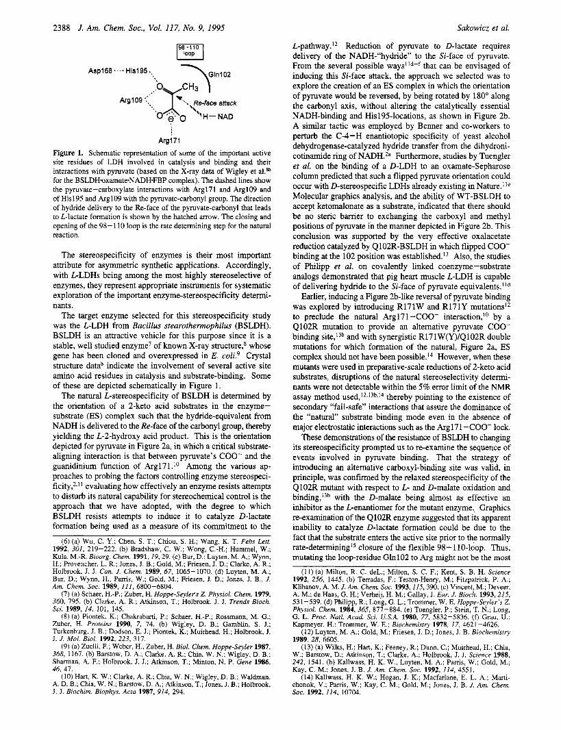
L-pathway.I* Reduction of pyruvate to D-lactate requires delivery of the NADH-"hydride" to the Si-face of pyruvate. From the several possible ways"d-f that can be envisaged of inducing this Si-face attack, the approach we selected was to explore the creation of an ES complex in which the orientation of pyruvate would be reversed, by being rotated by 180" along the carbonyl axis, without altering the catalytically essential NADH-binding and Hisl95-locations, as shown in Figure 2b. A similar tactic was employed by Benner and co-workers to perturb the C-4-H enantiotopic specificity of yeast alcohol dehydrogenase-catalyzed hydride transfer from the dihydroni- cotinamide ring of NADH.2a Furthermore, studies by Tuengler et al. on the binding of a D-LDH to an oxamate-Sepharose column predicted that such a flipped pyruvate orientation could occur with D-stereospecific LDHs already existing in Nature.'Ie Molecular graphics analysis, and the ability of WT-BSLDH to accept ketomalonate as a substrate, indicated that there should be no steric barrier to exchanging the carboxyl and methyl positions of pyruvate in the manner depicted in Figure 2b. This conclusion was supported by the very effective oxalacetate reduction catalyzed by Q102R-BSLDH in which flipped COO- binding at the 102 position was e~tablished.'~ Also, the studies of Philipp et al. on covalently linked coenzyme-substrate analogs demonstrated that pig heart muscle L-LDH is capable of delivering hydride to the Si-face of pyruvate equivalents.Ild
Earlier, inducing a Figure 2b-like reversal of pyruvate binding was explored by introducing R171W and R171Y mutations'* to preclude the natural Arg171-COO- interaction,'O by a Q102R mutation to provide an altemative pyruvate COO- binding site,t3b and with synergistic R171W(Y)/Q102R double mutations for which formation of the natural, Figure 2a, ES complex should not have been po~sib1e.l~ However, when these mutants were used in preparative-scale reductions of 2-keto acid substrates, disruptions of the natural stereoselectivity determi- nants were not detectable within the 5% error limit of the NMR assay method u ~ e d , ' ~ , ' ~ ~ , ' ~ thereby pointing to the existence of secondary "fail-safe" interactions that assure the dominance of the "natural" substrate binding mode even in the absence of major electrostatic interactions such as the Arg171-COO- lock.
These demonstrations of the resistance of BSLDH to changing its stereospecificity prompted us to re-examine the sequence of events involved in pyruvate binding. That the strategy of introducing an altemative carboxyl-binding site was valid, in principle, was confirmed by the relaxed stereospecificity of the Q102R mutant with respect to L- and D-malate oxidation and binding,'3b with the D-malate being almost as effective an inhibitor as the Lenantiomer for the mutant enzyme. Graphics re-examination of the Q102R enzyme suggested that its apparent inability to catalyze D-lactate formation could be due to the fact that the substrate enters the active site prior to the normally rate-determir~ing'~ closure of the flexible 98- 110-loop. Thus, mutating the loop-residue Gln102 to Arg might not be the most
Arg171
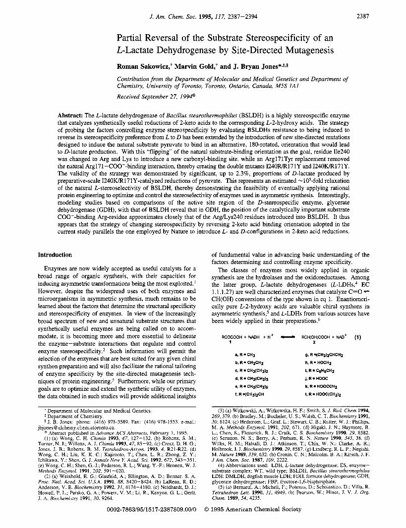
Figure 1. Schematic representation of some of the important active site residues of LDH involved in catalysis and binding and their interactions with pyruvate (based on the X-ray data of Wigley et aLSb for the BSLDHoxamate*NADH.FBP complex). The dashed lines show the pyruvate-carboxylate interactions with Argl71 and ArglO9 and of His195 and Argl09 with the pyruvate-carbonyl group. The direction of hydride delivery to the Re-face of the pyruvate-carbonyl that leads to L-lactate formation is shown by the hatched arrow. The closing and opening of the 98- 110 loop is the rate determining step for the natural reaction.
The stereospecificity of enzymes is their most important attribute for asymmetric synthetic applications. Accordingly, with L-LDHs being among the most highly stereoselective of enzymes, they represent appropriate instruments for systematic exploration of the important enzyme-stereospecificity determi- nants.
The target enzyme selected for this stereospecificity study was the L-LDH from Bacillus stearothemophilus (BSLDH). BSLDH is an attractive vehicle for this purpose since it is a stable, well studied enzyme7 of known X-ray structure,8 whose gene has been cloned and overexpressed in E. cokg Crystal structure data8 indicate the involvement of several active site amino acid residues in catalysis and substrate-binding. Some of these are depicted schematically in Figure 1.
The natural L-stereospecificity of BSLDH is determined by the orientation of a 2-keto acid substrates in the enzyme- substrate (ES) complex such that the hydride-equivalent from NADH is delivered to the Re-face of the carbonyl group, thereby yielding the L-2-hydroxy acid product. This is the orientation depicted for pyruvate in Figure 2a, in which a critical substrate- aligning interaction is that between pyruvate's COO- and the guanidinium function of Argl71.I0 Among the various ap- proaches to probing the factors controlling enzyme stereospeci- ficity,*," evaluating how effectively an enzyme resists attempts to disturb its natural capability for stereochemical control is the approach that we have adopted, with the degree to which BSLDH resists attempts to induce it to catalyze D-lactate formation being used as a measure of its commitment to the
(6) (a) Wu, C. Y.; Chen, S. T.; Chiou, S. H.; Wang, K. T. Febs Lert. 1992. 301, 219-222. (b) Bradshaw. C. W.: Wong. C.-H.: Hummel. W.: Kula, M.-R. Bioorg. Chem. 1991,19,29. (c) Bur, DI Luyten, M. A,; Wynn, H.; Provencher, L. R.; Jones, J. B.; Gold, M.; Friesen, J. D.; Clarke, A. R.; Holbrook, J. J. Can. J. Chem. 1989, 67, 1065-1070. (d) Luyten, M. A.; Bur, D.; Wynn, H., Parris, W.; Gold, M.; Friesen, J. D.; Jones, J. B., J. Am. Chem. SOC. 1989, 111, 6800-6804.
(7) (a) Schaer, H.-P.; Zuber, H. Hoppe-Seylefs Z. Physiol. Chem. 1979, 360, 795. (b) Clarke, A. R.; Atkinson, T.; Holbrook, J. J. Trends Bioch. Sci. 1989, 14, 101, 145.
(8) (a) Piontek, K.; Chakrabarti, P.; Schaer, H.-P.; Rossmann, M. G.; Zuber, H. Proteins 1990, 7, 74. (b) Wigley, D. B.; Gamblin, S. J.; Turkenburg, J. B.; Dodson, E. J.; Piontek, K.; Muirhead, H.; Holbrook, J. J. J. Mol. Biol. 1992, 223, 317.
(9) (a) Zuelli, F.; Weber, H.; Zuber, H. Biol. Chem. Hoppe-Seyler 1987, 368, 1167. (b) Barstow, D. A.; Clarke, A. R.; Chia, W. N.; Wigley, D. B.; Shaman, A. F.; Holbrook, J. J.; Atkinson, T.; Minton, N. P. Gene 1986, 46, 47.
(10) Hart, K. W.; Clarke, A. R.; Chia, W. N.; Wigley, D. B.; Waldman, A. D. B.; Chia, W. N.; Barstow, D. A.; Atkinson, T.; Jones, J. B.; Holbrook, J. J. Biochim. Biophys. Acta 1987, 914, 294.
(11) (a) Milton, R. C. deL.; Milton, S. C. F.; Kent, S. B. H. Science 1992, 256, 1445. (b) Tenadas, F.; Teston-Henry, M.; Fitzpatrick, P. A.; Klibanov, A. M. J. Am. Chem. SOC. 1993,115,390. (c) Vincent, M.; Deveer, A. M.; de Haas, G. H.; Verheij, H. M.; Gallay, J. Eur. J. Bioch. 1993, 215, 531-539. (d) Philipp, R.; Long, G. L.; Trommer, W. E. Hoppe-Seyler's Z. Physiol. Chem. 1984, 365, 877-884. (e ) Tuengler, P.; Stein, T. N.; Long, G. L. Proc. Natl. Acad. Sci. U.S.A. 1980, 77, 5832-5836. (0 Grau, U.; Kapmeyer, H.; Trommer, W. E.; Biochemistry 1978, 17, 4621-4626.
(12) Luyten, M. A.; Gold, M.; Friesen, J. D.; Jones, J. B. Biochemistry 1989, 28, 6605.
(13) (a) Wilks, H.; Hart, K.; Feeney, R.; Dunn, C.; Muirhead, H.; Chia, W.; Barstow, D.; Atkinson, T.; Clarke, A.; Holbrook, J. J. Science 1988, 242, 1541. (b) Kallwass, H. K. W.; Luyten, M. A,; Pams, W.; Gold, M.; Kay, C. M.; Jones, J. B. J. Am. Chem. SOC. 1992, 114, 4551.
(14)Kallwass, H. K. W.; Hogan, J. K.; Macfarlane, E. L. A,; Marti- chonok, V.; Parris, W.; Kay, C. M.; Gold, M.; Jones, J. B. J. Am. Chem. SOC. 1992, 114, 10704.
Substrate Stereospecificity of an L-Lactate Dehydrogenase J. Am. Chem. SOC., Vol. 117, No. 9, 1995 2389
His195
Tyrl71 Tyrl71
Figure 2. Normal stereorepresentations of pyruvate bound at the active site of BSLDH in (a) the natural WT-orientation leading to L-lactate by delivery of the NADH-hydride to the Re-face of the pyruvate-carbonyl and (b) in the reversed, 18O0-rotated, binding mode envisaged for the 1240FUR171Y mutant that would result in NADH-hydride attack of the Si-face of the substrate-carbonyl to give D-lactate.
effective way to provide a new COO--binding site since this could involve changing an existing substrate orientation. Ac- cordingly, a mutation introducing a positively charged residue in the rigid region beneath the loop should improve the prospect of inducing pyruvate to bind in a reversed orientation prior to loop closure, since its influence would be felt as pyruvate entered the active site. This approach remains valid even if the rate-determining step for mutant-catalyzed reactions is no longer loop c10sure.l~~ Graphics analysis identified Ile240 as a good candidate residue for this purpose. I240R and I240K mutations were therefore made to provide the basic side chains that would favor the flipped COO--binding mode desired. In addition, the dominant effect of Argl71 -COO- binding on pyruvate orientation was removed by creating 1240R(K)/R17 1Y double mutants.
Results The creation of Q102R, R171Y, and R171Y/Q102R was
described Single mutants I240R and I240K were obtained by site-directed mutagenesis utilizing the thio- nucleotide method. l 6 The oligonucleotide, 5’-CTACCAAAT- TA(A/G)GAGAAAAAAGGAGT-3’, doped in one position was used to introduce both mutations. The entire coding region of the BSLDH gene was sequenced for each of the mutants to ensure that no inadvertent mutations were introduced during in vitro procedures. Double mutants 124ORR171Y and I240W R171Y were obtained by subcloning fragments containing codons for the 240 residue from corresponding mutant plasmids into a previously created’* plasmid encoding R171Y mutant BSLDH. In each case the presence of both mutations in the
(1 5 ) (a) Dunn, C. R.; Wilks, H. M.; Halsall, D. J.; Atkinson, T.; Clarke, A. R.; Muirhead, H.: Holbrook, J. J.; Phil. Trans. R. Soc. Lond. B 1991, 332, 177. (b) Gerstein, M.: Chotia, C. J . Mol. B i d . 1991, 220, 133. (c) Sakowicz, R.: Kallwass, H. K. W.: Parris, W.; Kay, C. M.; Jones, J. B.: Gold, M. Biochemistry, 1993, 32, 12730.
(16) Nakamaye, K.: Eckstein, F. Nucl. Acids Res. 1986, 14, 9679.
plasmid was confirmed by sequencing. All mutant BSLDHs were purified to homogeneity as judged by SDS-polyacrylamide gel electrophoresis, and these pure proteins were used for kinetic studies and preparative scale reactions.
Kinetic parameters for single and double Ile240 mutants with substrates la-1 were determined under steady state conditions and are recorded in Table 1, together with the previous’3b data for Q102R for comparison. Generally, single mutants I240R and I240K had a decreased kcat and elevated KM for almost all substrates when compared to WT-BSLDH. Notable exceptions were the dicarboxylic keto acids lj-1 for which specificity constants are comparable to, or even slightly better than, those of WT-BSLDH. When combined with the R171Y mutation, the I240 mutants displayed an even more pronounced decrease in activity, with 124ORR171Y and 1240K/R171Y being virtually inactive with half the substrates tested. Interestingly, ketobu- tyrate (lb), although a poor substrate for I240K/R171Y, displayed an exceptionally low apparent KM value of 0.08 mM, while for both 1240WK;R17 1Y double mutant BSLDHs, ke- tomalonate (lj) is a better substrate than pyruvate. That the mutations at position 240 caused no major disruption in the coenzyme binding pattern was confirmed by the apparent KM values for NADH being very similar for all the mutant proteins with an altered 240-residue and also closely resembling the NADH-KM for WT BSLDH.
Our previous studies on the R171Y and Ql02R mutant^^*,^^^,'^ had demonstrated a high degree of commitment of BSLDH towards maintaining L-stereospecificity. However, it was suspected that small amounts of D-hydroxy acids that might have been formed would not have been detectable within the 5% error limits of the Nh4R analyses applied. Accordingly, prior to evaluating the stereospecificity of the new mutants, a highly sensitive enzymic D/L-lactate assay that increased the sensitivity of D-lactate detection by over 1000-fold was developed. This is summarized in Figure 3.” The stereospeci-
2390 J. Am. Chem. SOC., Vol. 117, No. 9, 1995
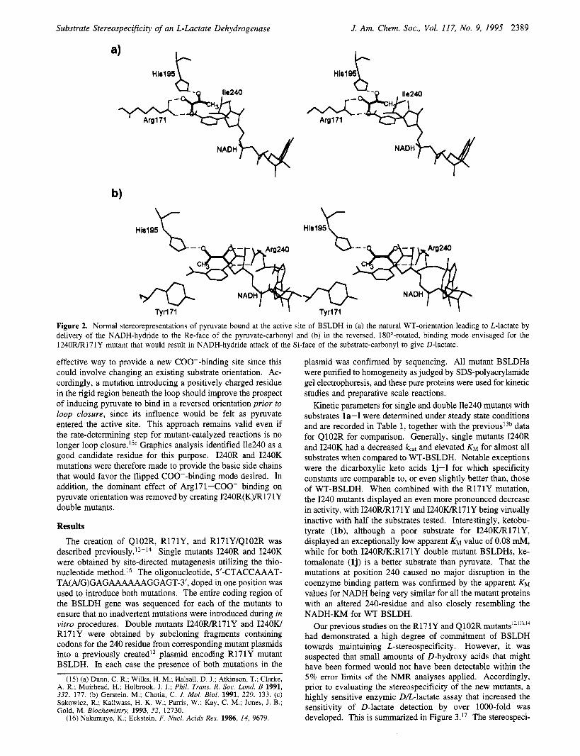
Table 1. Kinetic Parameters of WT- and Mutant-BSLDH-Catalyzed Reductions of la-la
Sakowicz et al.
188b 0.04b 4.70 x l o s 6
155b 0.346
4 4 6 2.4b 18 3OOb 256 1.56 16 700b 326 0.35b 91 4Wb 0.496 17b 28.8b 0.516 4.96 1 04b 125b 0.166 781 OOOb 816 0.67b 121 OOOb 21 1 21 000 6 1.5 4000 4.1 3.9 1050 13
4 . 5 6 ~ x 1056
5 9 % 1 4.2 f 0.2 14 000
11.9 f 0.2 3 f 0.1 3970 4.6 f 0.2 13 f 1.2 354 16 f 0.5 5.6 f 0.56 2860 33 f 0.9 0.62 f 0.07 53 200
0.14 f 0.005' 0.16 f 0.01 10 f 1.8 16 73 f 1.5 4.2 f 0.2 17 400 4 8 f 1 1.6 f 0.08 30 000 57 k 5 3.4 f 0.6 16 800 6.1 f 0.09 0.78 f 0.04 7820 2.5 f 0.03 2.7 f 0.1 926 13 f 0.8
2.456 1.26 20006
2.66 1.26 22006 1 .56b 1.88b 8306 n.d.' n.d. n.d. 3 36 0.346 97 0006 0.00996 1 36 0.766 0.266 3.1b 816 4.77b 2.26 22006 89b 0.816 110 ooob 2386 1.26 200 0005 205b 0.0196
0.667b 0.165b 4 1 OOb n.d.
1.1 1076
BSLDH
R(C0)COO- R = constants WT 1240R 1240K 1240RIR17 1Y 1240wR17 1Y Q102R'3b 0.015 f 0.007 21 f 0.4 0.15 f 0.006
5 5 % 0.3 3820
2.7 f 0.05 6.4 f 0.3 422 3.9 f 0.14 12 f 0.9 3.25 7.4 f 0.3 7.5 f 0.7 987 15 f 0.3 2.9 f 0.17 5170
0.092 f 0.004c 0.1 f 0.007 11 f 1.7 9.1 9.6 f 0.3 12.3 f 0.9 780 24 f 0.9 2.4 f 0.2 10 000 25 f 0.04 6.1 f 0.7 4100 3 f 0.3 0.58 f 0.02 5170 1.15 f 0.02 0.94 f 0.06 1220 7 f 1
30 f 2 5
0.5' d
d
d
d
d
d
0.24 i 0.01 13 f 1.3 18.5 0.21 f 0.02 1 0 6 1 21 0.02 f 0.0005 10 f 0.6 2.0 d
8 f 0.8
70 f 40 0.2
0.002 f 0.0001 0.08 f 0.03 25 d
d
d
d
d
d
7.1 f 0.12' 0.01 f 0.0007 8 f l 1.25 0.02 f 0.002 22 f 4 1 .o d
15 f 0.9
a At pH 6.0, 25 "C, 0.2 mM NADH, 5 mM FBP, 0.06-2.6 mM enzyme, and 0.1-40 mM pyruvate. From refs 13b and 14. Only the kcat/& could be estimated from the initial slope of the v YS [SI curve. No activity detected with substrate concentrations 530 mM and enzyme concentrations 5 10 pM. e n.d., not determined. f Determined with 10 mM pyruvate.
CH3COCOOH
1-Glutamate K Ketcgluhrate
GlutamatePyruvate Transaminass
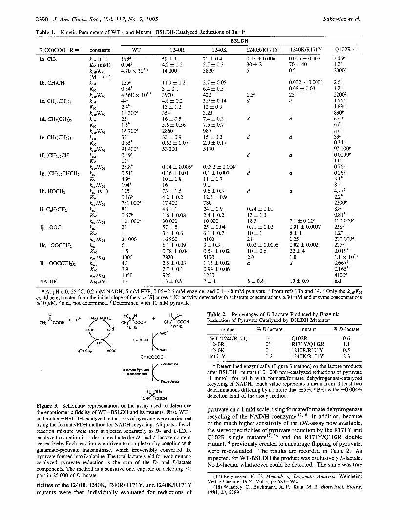
C H K L Figure 3. Schematic representation of the assay used to determine the enantiomeric fidelity of WT-BSLDH and its mutants. First, WT- and mutant-BSLDH-catalyzed reductions of pyruvate were carried out using the formatelFDH method for NADH-recycling. Aliquots of each reaction mixture were then subjected separately to D- and L-LDH- catalyzed oxidation in order to evaluate the D- and L-lactate content, respectively. Each reaction was driven to completion by coupling with glutamate-pyruvate transaminase, which irreversibly converted the pyruvate formed into L-alanine. The total lactate yield for each mutant- catalyzed pyruvate reduction is the sum of the D- and L-lactate components. The method is a sensitive one, capable of detecting < 1 part in 25 000 of D-lactate.
ficities of the I240R, I240K, 124OFUR171Y, and 1240IUR171Y mutants were then individually evaluated for reductions of
Table 2. Percentages of D-Lactate Produced by Enzymic Reduction of Pyruvate Catalyzed by BSLDH Mutantsa
mutant % D-lactate mutant % D-lactate
WT (1240lR171) ob Q102R 0.6
1240K ob 1240RIR17 1Y 0.5 R171Y 0.2 1240wR17 1Y 2.3
1240R Ob R17 1YlQ102R 1.1
Determined enzymically (Figure 3 method) on the lactate products after BSLDH-mutant (10-200 nm)-catalyzed reductions of pyruvate (1 mmol) for 60 h with formatelformate dehydrogenase-catalyzed recycling of NADH. Each value represents a mean from at least two determinations differing by no more than f 5 % . Below the *0.004% detection limit of the assay method.
pyruvate on a 1 mM scale, using formate/formate dehydrogenase recycling of the NAD/H coenzyme.12,18 In addition, because of the much higher sensitivity of the D/L-assay now available, the stereospecificities of pyruvate reduction by the R171Y and Q102R single mutant^'^,'^^ and the R171Y/Q102R double mutant,14 previously created to encourage flipping of pyruvate, were re-evaluated. The results are recorded in Table 2. As expected, for WT-BSLDH the product was exclusively Llactate. No D-lactate whatsoever could be detected. The same was true
(17) Bergmeyer, H. U. Merhods of Enzymatic Analysis; Weinheim:
(18) Wandrey, C.; Buckmann, A. F.; Kula, M. R. Biotechnol. Bioeng. Verlag Chemie, 1974; Vol 3, pp 583-592.
1981, 23, 2789.
Substrate Stereospecificity of an L-Lactate Dehydrogenase
for the I240R and I240K single mutants, for which the natural Arg171-COO- lock was still operational. However, the greater sensitivity of the new analytical method now revealed that the earlier R171Y and Q102R mutations, previously thought to have been without effect, had in fact begun to disturb the stereo- chemical integrity of BSLDH in the very manner for which they had been designed, with 0.2% and 0.6% respectively of D-lactate being produced. For the double mutant R171Y/Q102R, with its greater potential for binding pyruvate in the reversed, Figure 2b, mode, the proportion of D-lactate formed was higher still, at 1.1%. The newly created I240R/R171Y BSLDH also exhibited a relaxation of its L-stereospecificity, with 0.5% of the D-product formed.
The 1240wR171Y double mutant was the most successful in terms of the extent of stereospecificity reversal, with a very significant 2.3% of D-lactate produced. This represents a truly remarkable stereospecificity-preference switch, from a D-isomer formation-frequency of 1 in 25 000 for WT-BSLDH to 1 in 43 for the 1240wR171Y mutant, which reflects an improvement in the favoring of the D-pathway of at least 500-fold. Despite the low activity of 1240IUR171Y it remained a preparatively viable catalyst.
Discussion An immediate effect of the single Ile240 mutations was a
drop in activity towards all keto acids with uncharged side chains (la-i), including the natural substrate pyruvate (la). A similar effect had been observed previously with a Q102R mutant.I3 However, there was a marked difference in the kinetic param- eters most affected by these two different mutations. In comparison with WT-BSLDH, the apparent KM for pyruvate (la) for the Q102R mutant was 30-fold higher, while k,,, decreased by 7 5 f 0 l d . I ~ ~ In contrast, I240R and I240K muta- tions affected the pyruvate kcat values only slightly, with 3- and 9-fold decreases, respectively, whereas the apparent KM’S were elevated by over 100-fold in each case. These results are as expected from the accepted model of BSLDH catalysis, in which the loop closure constitutes the rate limiting step.I5 Evidently, introduction of a positively charged amino acid side chain into the 98- 1 10-loop interferes with both binding of the substrate and with loop closure, leading to a slower turnover number. Conversely, insertion of such positively charged side chains into the interior of the active site under the loop exerts its effect primarily on substrate binding and leaves loop motion relatively unaffected. Similar conclusions can be drawn from the kinetic data for the accommodation of dicarboxylic keto acid substrates. For the Q102R mutant, the presence of a positive charge in the loop allowed for both better binding and faster tumover of oxalacetate (lk).I3 In contrast, for the mutants I240R and I240K, although oxalacetate binds better than pyruvate, the loop must now close over a negatively charged substrate substituent. This leads to a further drop in the kat value, to a level resembling that of Q102R with pyruvate.I3
The results demonstrate that, as planned in the Figure 2b design, the I240R and I240K mutations did induce substrate- carboxylate side chain binding in an alternative, flipped, orientation. Unfortunately, the effects of I240WK mutations on ketomalonate (lj), whose carboxyl group most closely mimics that of pyruvate in the hypothetical reversed orientation, were minimal, suggesting that the distances from the substrate COO- to the side chains of Arg240 or Lys240 may be too large in this case. This conclusion is also supported by the absence of any change in stereospecificity for either of these mutants (Table 2).
When the mutations at position 240 were combined with R171Y, a further decrease in activity was observed. This was
J. Am. Chem. SOC., Vol. 117, No. 9, 1995 2391
as expected, since R171Y removes the natural 171-side chain- COO- binding interaction of the native substrate. A new carboxylate binding site at position 240 could never alone compensate fully for the Argl71 deletion since overall protein conformation changes resulting from the changes in polarities in the mutated 171 and 240 regions are inevitable. Furthermore, it is clear that there is a network of secondary “fail-safe” interactions, such as with Argl09 or Thr246,I2 which BSLDH can invoke to maintain the substrate in its natural orien-
The remarkable tenacity of such interactions is reflected by the fact that even the most successful double mutant 1240wR171Y is still ’97% L-stereoselective.
It must be stressed how unwaveringly the stereospecificity is controlled in L-LDHs. The lower limit of sensitivity of our analytical method is 0.004%, and any D-lactate formation in reductions of pyruvate catalyzed by WT-BSLDH is certainly below this level. The actual L-fidelities of L-LDHs are certainly much higher than possible to measure by the current, experi- mentally constrained, limit, despite the high sensitivity of the improved assay method. Hydride transfer in the L-LDHs active site couples two centers, that of the C-2 carbon of pyruvate, and of the C-4 carbon of the dihydronicotinamide ring of NADH. The elegant studies of LaReau and Ander~on’~ have demonstrated that for NAD+, nonstereospecific hydride transfer to the “wrong” Si-face of the nicotinamide ring occurs at most in 1 of lo7 reactions. It is reasonable to assume that the fidelity of the associated hydride transfer from NADH to pyruvate is comparable. Based on this parallel, the 2.3% D-lactate produc- tion observed with 1240wR171Y represents a > 2 x 10⁵-fold relaxation of the L-stereospecificity of the double mutant enzyme relative to that of WT-BSLDH.
When viewed in these terms, the extents of D-lactate formation recorded in Table 2 for the other mutant also become highly significant. A single R171Y change now seems to be sufficient to induce some reversal of pyruvate binding, thus confirming the dominant contribution to L-binding integrity of the Argl71 -pyruvate carboxyl interaction. For this latter mutant, one cannot rule out the possibility that the pyruvate reduced to D-lactate was not exclusively bound in the intended 180 rotated manner and that some other binding mode that exposed the Si-face of the carbonyl to the hydride attack also played a role. However, the significant increase in the amount of D-lactate produced by Q102R suggests that the desired flipping of pyruvate’s carboxyl, induced by binding to 102Arg, has indeed taken place. The ability of Q102R to disturb the stereospecificity shows that the binding of pyruvate as it enters the active site is not necessarily irrevocable and that it may be influenced by the 102Arg in the loop either before or after loop closure. Nevertheless, the strategy of locating the alternative carboxylate binding site in the rigid region of the protein under the 98- 110 loop is evidently more soundly based, as reflected by the double mutant 1240wR171Y being the most successful in inducing D-lactate formation.20
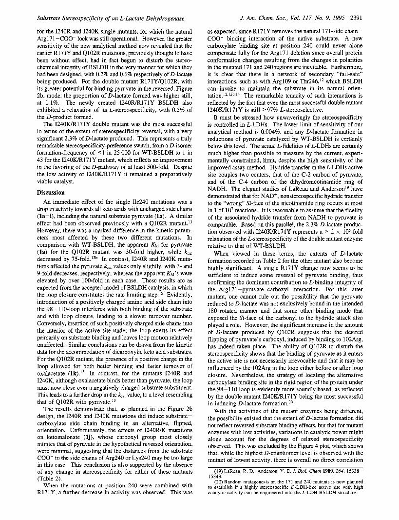
With the activities of the mutant enzymes being different, the possibility existed that the extent of D-lactate formation did not reflect reversed substrate binding effects, but that for mutant enzymes with low activities, variations in catalytic power might alone account for the degrees of relaxed stereospecificity observed. This was excluded by the Figure 4 plot, which shows that, while the highest D-enantiomer level is observed with the mutant of lowest activity, there is overall no direct correlation
(19) LaReau, R. D.; Anderson, V. E. J. Biol. Chem 1989,264, 15338- 15343.
(20) Random mutagenesis on the 171 and 240 mutants is now planned to establish if a highly stereospecific D-LDH-like active site with high catalytic activity can be engineered into the L-LDH BSLDH structure.
2392 J. Am. Chem. Soc.. Vol. 117, No. 9. 1995 Sakowicz er al.
Figure 4. There is 1 n 1 1 direct correlation hetwcw the propor i~ , r of D-lactale formed during catalysis of pyruvate rctluclion hy BSI.DH mutants and the corresponding specificity constanis.
between the extents of D-lactate formation with mutant enzyme activities. X-ray structure determinations of the 1240WR171Y and 1240RR171Y enzymes, now in progress,” should provide insights into the nature of substrate binding with the various mutants.
There is continuing interest in optimizing and controlling enzyme stereospecificity. as indicated by the relaxation of enzyme stereospecificity inducible toward larger substrates such as NADH’“ or ATP,” the improved stereospecificities engi- neered into lipases,” cytochrome P450,’< and horse radish peroxidase15 and the adjustments of stereospecificity mediated by organic solvents.’lh The present results are particularly encouraging since the partial stereospecificity reversals were achieved on an enzyme of the most highly committed ste- reospecificity. Thus, for other synthetically useful enzymes whose stereochemical control mechanisms are less restrictive, particularly toward unnatural substrates, the exploitation of protein engineering IO tailor and control desired stereospecificity now becomes a realistic possibility.
It is of interest lo consider how Nature controls L- vs D-LDH stereospecificity. No D-LDH stn~cture is yet available but X-ray diffraction studies are presently being canied out on several enzyme^.'^ In the meantime. an indication of Nature’s possible strategy may be obtained from examination of the structure of a closely homologous enzyme. D-glycerate dehydrogenase (GDH).” There are several other enzymes that belong to the family of D-2-hydroxy acid dehydrogenases.’x some of which have already been crystallized,’” but only the coordinates for Hvphomicrohium merhvlovorum GDH (as the apoenzyme) are publicly available from Brookhaven Protein Data Bank.”“.”’ Both D-LDHs and GDH catalyze the same kind of reaction and
I211 Sloll. Y.: Pai. E.. personal communication. 122I(al Jimg. R.-T.:Dahnke. T.: Twi. M.-D J. Am. Clwn. So<. 1991.
11.3. 5485. lhl Dohnke. T.: Jiang. R.-T.: Trai. M.-D. 1. A m Chrm. SOC. 1991. 113. 93RX.
1231 Kuipers. D. P.: Dijkman. R.: Pals. C. E.: Verheij. H. M.: de Haas. G. H. Pnrrrin Eng. 1989. 2. 467-471.
1241 Loida. P. J.: Sliear. S. G. Prorein En,r. 1993. 6. 207-212. 1251 Ozaki. S.-l.: Onir de Monlellano. P. R. J. Am. Clirm. S i r 1994.
110. 44x7. 1261 la1 Nessler. S.: Le Bras. G.: Le Bmr. G.: Gmel. J. -R. J. Mol. H i d
1994. 2.l.i. 3711-371. (hl P& E. personal comnwnicalion. I271 la1 Goldhere. J. D.: Yoshida. T.: Brick. P. unpuhlirhed. deposited
as IGDH in Brookhaven Protein Data Bank. Sep 22. 1993. Ihl Goldkrg. I . D.: Yoshida. T.: Brick. P. J. Mol. H i d 1994. 230. 1123-1 140.
12RI Granl. G. A. Uioc1wm. Riophm. Rev. Comm 1989. 16s. 1371- 1374.
(291 la) Lamzin. V. S.: Aleshin. A. E: Stmkopytov. B. V.: Yukhncvich. M . G.: P o p r , V. 0.: H;irulyunan. E. H.: Wilson. K. S. h r . J. N i d w n . 1992. 206. 141-452. lhl Lamzin. V. S.: Dauter. Z.: Popor. V. 0.: H;truryun:in. E. H.: Wilson. K. S. J. Mol. R i d 1994. 2.16. 759-785.
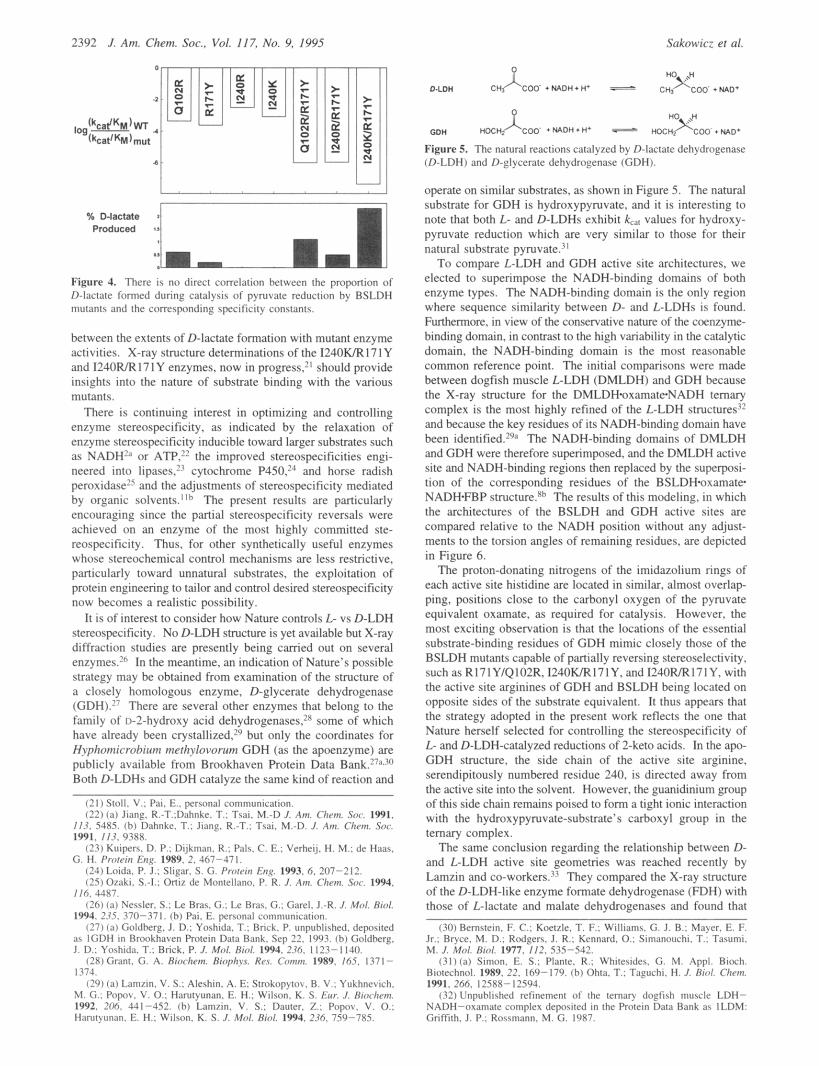
Figure 5. The natural reactions catalyzed hy D-lactate dehydrogenase (0-LDH) and D-glycerate dehydrogenase (CDH).
operate on similar substrates. as shown in Figure 5 . The natural substrate for GDH is hydroxypyruvate. and it is interesting to note that both L- and D-LDHs exhibit k,. values for hydroxy- pyruvate reduction which are very similar IO those for their natural substrate pyruvate.”
To compare L-LDH and GDH active site architectures, we elected to superimpose the NADH-binding domains of both enzyme types. The NADH-binding domain is the only region where sequence similarity between D- and L-LDHs is found. Furthermore. in view of the conservative nature of the coenzyme- binding domain, in contrast to the high variability in the catalytic domain, the NADH-binding domain is the most reasonable common reference point. The initial comparisons were made between dogfish muscle L-LDH (DMLDH) and GDH because the X-ray structure for the DMLDH*oxamateNADH ternary complex is the most highly refined of the L-LDH structures” and because the key residues of its NADH-binding domain have been identified.’qa The NADH-binding domains of DMLDH and GDH were therefore superimposed, and the DMLDH active site and NADH-binding regions then replaced by the superposi- tion of the corresponding residues of the BSLDHoxamate NADHFBP structure.Xh The results of this modeling, in which the architectures of the BSLDH and GDH active sites are compared relative to the NADH position without any adjust- ments to the torsion angles of remaining residues, are depicted in Figure 6.
The proton-donating nitrogens of the imidazolium rings of each active site histidine are located in similar, almost overlap- ping, positions close to the carbonyl oxygen of the pyruvate equivalent oxamate. as required for catalysis. However, the most exciting observation is that the locations of the essential substrate-binding residues of GDH mimic closely those of the BSLDH mutants capable of partially reversing stereoselectivity, suchasR171Y/Q102R,1240WR171Y.andI240R/R171Y, with the active site arginines of GDH and BSLDH being located on opposite sides of the substrate equivalent. It thus appears that the strategy adopted in the present work reflects the one that Nature herself selected for controlling the stereospecificity of L- and D-LDH-catalyzed reductions of 2-keto acids. In the apo- GDH structure. the side chain of the active site arginine, serendipitously numbered residue 240. is directed away from the active site into the solvent. However, the guanidinium group of this side chain remains poised to form a tight ionic interaction with the hydroxypyruvate-substrate’s carboxyl group in the ternary complex.
The same conclusion regarding the relationship between D- and L-LDH active site geometries was reached recently by Lamzin and co-workers.” They compared the X-ray structure of the D-LDH-like enzyme formate dehydrogenase (FDH) with those of L-lactate and malate dehydrogenases and found that
(30) Bemslein. F. C.: Koelzle. T. F.: Williams. G. J. B.: Mayer. E. F. Jr.: Bryce. M. D.: Rdgerr . J. R.: Kmnard. 0.: Simanouchi. T.: Tasumi. M. J. Mol. H i d 1977. 112. 535-542.
(31)la) Simon. E. S.: Planle. R.: Whilerider. G. M. Appl. Bimh. Biolechnol. 1989. 22. 169-179. lhl Ohia. T.: Tapchi . H. J. I M . Clwn. 1991. 200. 125x8-12594.
132) Unpuhlished refinemen! of the ternary doefish muscle LDH- NADH-oxamate complex deposited in lhe Protein I h t a Bank as ILDM: Gnffilh. J . P.: Rossmmn. M. G. 19x7.
Substrate Stereospecificity of an L-Lactate Dehydrogenase J. Am. Chem. Soc., Vol. 117, No. 9, 1995 2393
0 -Argl71
0 - His197 0
B - oxamate
T G - His297
<f3 - His1 97 0 4.. .
I V 0 - oxamate
- Arg240
.e*. : -.. . a'
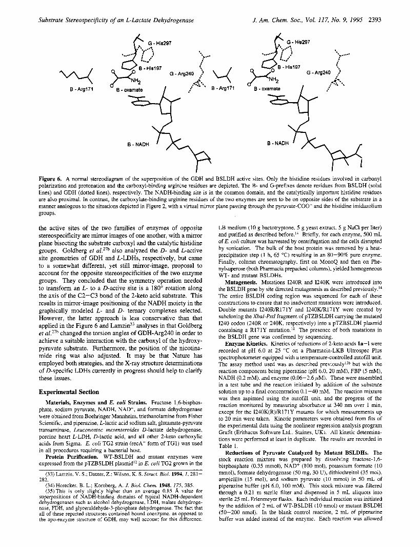
Figure 6. A normal stereodiagram of the superposition of the GDH and BSLDH active sites. Only the histidine residues involved in carbonyl polarization and protonation and the carboxyl-binding arginine residues are depicted. The B- and G-prefixes denote residues from BSLDH (solid lines) and GDH (dotted lines), respectively. The NADH-binding site is in the common domain, and the catalytically important histidine residues are also proximal. In contrast, the carboxylate-binding arginine residues of the two enzymes are seen to be on opposite sides of the substrate in a manner analogous to the situations depicted in Figure 2, with a virtual mirror plane passing through the pyruvate-COO- and the histidine imidazolium groups.
the active sites of the two families of enzymes of opposite stereospecificity are mirror images of one another, with a mirror plane bisecting the substrate carboxyl and the catalytic histidine groups. Goldberg et al.27b also analyzed the D- and L-active site geometries of GDH and L-LDHs, respectively, but came to a somewhat different, yet still mirror-image, proposal to account for the opposite stereospecificities of the two enzyme groups. They concluded that the symmetry operation needed to transform an L- to a D-active site is a 180" rotation along the axis of the C2-C3 bond of the 2-keto acid substrate. This results in mirror-image positioning of the NADH moiety in the graphically modeled L- and D- ternary complexes selected. However, the latter approach is less conservative than that applied in the Figure 6 and L a m ~ i n ~ ~ analyses in that Goldberg et al.27b changed the torsion angles of GDH-Arg240 in order to achieve a suitable interaction with the carboxyl of the hydroxy- pyruvate substrate. Furthermore, the position of the nicotina- mide ring was also adjusted. It may be that Nature has employed both strategies, and the X-ray structure determinations of D-specific LDHs currently in progress should help to clarify these issues,
Experimental Section Materials, Enzymes and E. coli Strains. Fructose 1,6-bisphos-
phate, sodium pyruvate, NADH, NAD+, and formate dehydrogenase were obtained from Boehringer Mannheim, triethanolamine from Fisher Scientific, and piperazine, L-lactic acid sodium salt, glutamate-pyruvate transaminase, Leuconostoc mesenteroides D-lactate dehydrogenase, porcine heart L-LDH, D-lactic acid, and all other 2-keto carboxylic acids from Sigma. E. coli TG2 strain (recA- form of TG1) was used in all procedures requiring a bacterial host.
Protein Purification. WT-BSLDH and mutant enzymes were expressed from the pTZBSLDH plasmidI2 in E. coli TG2 grown in the
(33) Lamzin, V. S.; Dauter, Z.; Wilson, K. S. Struct. B i d . 1994,1,281-
(34) Horecker, B. L.; Komberg, A. J. B i d . Chem. 1948, 175, 385. (35)This is only slightly higher than an average 0.85 8, value for
superpositions of NADH-binding domains of typical NADH-dependent dehydrogenases such as alcohol dehydrogenase, LDH, malate dehydroge- nase, FDH, and glyceraldehyde-3-phosphate dehydrogenase. The fact that all of these reported structures contained bound coenzyme, as opposed to the apo-enzyme structure of GDH, may well account for this difference.
282.
LB medium (10 g bactotryptone, 5 g yeast extract, 5 g NaCl per liter) and purified as described before.14 Briefly, for each enzyme, 500 mL of E. coli culture was harvested by centrifugation and the cells disrupted by sonication. The bulk of the host protein was removed by a heat- precipitation step (1 h, 65 "C) resulting in an 80-90% pure enzyme. Finally, column chromatography, first on MonoQ and then on Phe- nylsuperose (both Pharmacia prepacked columns), yielded homogeneous WT- and mutant BSLDHs.
Mutagenesis. Mutations I240R and I240K were introduced into the BSLDH gene by site directed mutagenesis as described previo~sly. '~ The entire BSLDH coding region was sequenced for each of these constructions to ensure that no inadvertent mutations were introduced. Double mutants 1240lUR171Y and 1240wR171Y were created by subcloning the XbaI-Pstl fragment of pTZBSLDH carrying the mutated I240 codon (240R or 240K, respectively) into a pTZBSLDH plasmid containing a R171Y mutation.12 The presence of both mutations in the BSLDH gene was confirmed by sequencing.
Enzyme Kinetics. Kinetics of reductions of 2-keto acids la-1 were recorded at pH 6.0 at 25 "C on a Pharmacia-LKB Ultrospec Plus spectrophotometer equipped with a temperature-controlled autofill unit. The assay method used was as described previously'3b but with the reaction components being piperazine (pH 6.0, 20 mM), FBP (5 mM), NADH (0.2 mM), and enzyme (0.06-2.6 pM). These were assembled in a test tube and the reaction initiated by addition of the substrate solution up to a final concentration 0.1-40 mM. The reaction mixture was then aspirated using the autofill unit, and the progress of the reaction monitored by measuring absorbance at 340 nm over 1 min, except for the 1240K(R)/R171Y mutants for which measurements up to 20 min were taken. Kinetic parameters were obtained from fits of the experimental data using the nonlinear regression analysis program Grafit (Erithacus Software Ltd., Staines, UK). All kinetic determina- tions were performed at least in duplicate. The results are recorded in Table 1.
Reductions of Pyruvate Catalyzed by Mutant BSLDHs. The stock reaction mixture was prepared by dissolving fructose- 1,6- bisphosphate (0.35 mmol), NAD+ (100 mol), potassium formate (10 mmol), formate dehydrogenase (50 mg, 30 U), dithiothreitol (35 mol), ampicillin (15 mol), and sodium pyruvate (10 mmol) in 50 mL of piperazine buffer (pH 6.0, 100 mM). This stock mixture was filtered through a 0.21 m sterile filter and dispensed in 5 mL aliquots into sterile 25 mL Erlenmeyer flasks. Each individual reaction was initiated by the addition of 2 mL of WT-BSLDH (10 nmol) or mutant BSLDH (SO-200 nmol). In the blank control reaction, 2 mL of piperazine buffer was added instead of the enzyme. Each reaction was allowed
2394 J. Am. Chem. Soc., Vol. 117, No. 9, 1995
to proceed with gentle shaking for 60 h at 25 'C. The lactate yields varied from 25 to 89% depending on the activity of the mutant enzyme used.
Each reaction mixture was neutralized to pH 7 with 12 M HC1 and 200 L samples and then taken from each reaction. Each sample was then rotary evaporated under reduced pressure, the resulting solid products individually extracted with 97% aqueous EtOH, and insoluble particles removed by centrifugation. Each supematant was again rotoevaporated in vacuo, and the individual, nonvolatile, lactate residues redissolved in water (200 L) for analysis of their D-, L-, and total lactate (=D + L) contents, as described below.
Enzymic Analysis of D- and L-Lactate. The following procedure was applied to each reaction product. To a 3 mL cuvette equipped with a stimng bar was added water (720 L), piperazine/glutamate buffer pH 9.6 (500 L, 100 mM: 100 mM), NADf (100 L, 50 mM), glutamate- pyruvate transaminase (10 L, 8 U), and an aliquot of the purified sodium lactate sample (see above) from each preparative reduction (50 L). The changes in absorbance at 339 nm were monitored for 5 min to confirm the stability of the baseline and that all unreacted pyruvate had been removed. After 5 min the assay was initiated by the addition of D-LDH from Leuconostoc mesenteroides (5 L, 80 U) and absorbance at 339 nm monitored for 10 min. The content of D-lactate in the sample was calculated from the total increase in absorbance due to the NADH produced. The extinction coefficient for NADH was taken to be 6220 M-' As a control, after the completion of the assay for each sample, a small aliquot of D-lactate solution was added to the cuvette, and the resulting increase in the absorbance was monitored. This procedure confirmed that the absorbance end-values observed during the D-lactate detection were not limited by enzyme depletion nor by the presence of some inhibitory compounds. The absorbance changes monitored during all these assays were calibrated against those obtained with standard solutions of D-lactate in the concentration ranges of the preparative reaction samples. The detection limit established with these test solutions was 0.004% of D-lactate.
The same procedure and controls were used for the L-lactate determinations, except that porcine heart L-LDH was used to initiate the assay, and the lactate sample had to be suitably diluted to give an absorbance change during the assay of approximately 0.1 absorbance unit. The total lactate present was then given by the sum of D- and L-lactate. The percentages of D-lactate produced are recorded in Table
Sakowicz et al.
The lactic acid product was characterized from a representative 5 mmol scale reaction catalyzed by I24OwR171Y BSLDH. The reaction yielded lactic acid (2a, 205 mg, 2.5 "01, 50% yield), [aI2~ = -12.9' (c 0.13, 1.5 M aqueous NaOH) lit. (Beil. 3, 261) [al2~ = -13.7' (c 1, 1.5 M aqueous NaOH); 'H NMR (D20) 4.35 (lH, q, J = 6 Hz), 1.40 (3H, d, J = 6 Hz) ppm, identical to that of authentic lactic acid.
Graphics analyses of the WT and mutant proteins were performed with Insight I1 (version 2.0.0, Biosym Technologies Inc., San Diego, CA) on a Silicon Graphics Iris 4D240 workstation using the X-ray structure coordinates of BSLDHoxamate-NADHFBP quaternary com- plex at 2.5 8, DMLDHoxamate-NADH temary complex,32 and GDH apoenzyme.27a
In order to determine the precise location of the NADH-binding domain in GDH, sequences of 12 members of the D-2-hydroxy acid dehydrogenase family were aligned. From the above alignment, sequences of GDH corresponding to the structurally conserved elements of the NADH-binding domain of FDK?9a were identified. These regions were then superimposed onto the corresponding regions of DMLDH, following the procedure of Lamzin et aZ.29a DMLDH was chosen over other LLDHs because it has the highest quality X-ray structure available and that the key residues of DMLDH NADH-binding domain have been identified.29a The rms deviation between 29 equivalent pairs of Ca atoms in these two superimposed structures was 1.9 Subse- quently, the NADH structure from the DMLDH ternary complex was added graphically, with least squares fitting, to the superimposed coenzyme-domains of GDH and DMLDH, and the rest of the DMLDH structure was then deleted. Analogous superposition of 29 topologically equivalent residues of BSLDH and the GDH-NADH structure created above gave an rms deviation of 2.0 8,. The superimposed BSLDH- GDH structures are shown in Figure 6.
Acknowledgment. This work was supported by a Protein Engineering Network of Centres of Excellence Award (to J.B.J.), by the Natural Sciences and Engineering Research Council of Canada and by an Ontario Graduate Scholarship (to RS). We are grateful to Dr.'s Emil Pai and Vincent Stoll for helpful discussions on LDH X-ray structures.
JA943 195H L.