Molecular characterization of the poly(3-hydroxybutyrate) (PHB) synthase from Ralstonia eutropha: in...
13
Molecular characterization of the poly(3-hydroxybutyrate) (PHB) synthase from Ralstonia eutropha : in vitro evolution, site-speci¢c mutagenesis and development of a PHB synthase protein model Bernd H.A. Rehm *, Regina V. Antonio, Patricia Spiekermann, Amro A. Amara, Alexander Steinbu « chel Institut fu «r Mikrobiologie der Westfa «lischen Wilhelms-Universita «t Mu «nster, Corrensstrasse 3, D-48149 Mu «nster, Germany Received 4 July 2001; received in revised form 11 September 2001; accepted 12 October 2001 Abstract A threading model of the Ralstonia eutropha polyhydroxyalkanoate (PHA) synthase was developed based on the homology to the Burkholderia glumae lipase, whose structure has been resolved by X-ray analysis. The lid-like structure in the model was discussed. In this study, various R. eutropha PHA synthase mutants were generated employing random as well as site-specific mutagenesis. Four permissive mutants (double and triple mutations) were obtained from single gene shuffling, which showed reduced activity and whose mutation sites mapped at variable surface-exposed positions. Six site-specific mutations were generated in order to identify amino acid residues which might be involved in substrate specificity. Replacement of residues T323 (I/S) and C438 (G), respectively, which are located in the core structure of the PHA synthase model, abolished PHA synthase activity. Replacement of the two amino acid residues Y445 (F) and L446 (K), respectively, which are located at the surface of the protein model and adjacent to W425, resulted in reduced activity without changing substrate specificity and indicating a functional role of these residues. The E267K mutant exhibited only slightly reduced activity with a surface-exposed mutation site. Four site-specific deletions were generated to evaluate the role of the C- terminus and variant amino acid sequence regions, which link highly conserved regions. Deleted regions were D281^D290, A372^C382, E578^A589 and V585^A589 and the respective PHA synthases showed no detectable activity, indicating an essential role of the variable C-terminus and the linking regions between conserved blocks 2 and 3 as well as 3 and 4. Moreover, the N-terminal part of the class II PHA synthase (PhaC Pa ) from Pseudomonas aeruginosa and the C-terminal part of the class I PHA synthase (PhaC Re ) from R. eutropha were fused, respectively, resulting in three fusion proteins with no detectable in vivo activity. However, the fusion protein F1 (PhaC Pa -1-265-PhaC Re -289-589) showed 13% of wild type in vitro activity with the fusion point located at a surface-exposed loop region. ß 2002 Elsevier Science B.V. All rights reserved. Keywords : Polyhydroxyalkanoate synthase ; Polyhydroxyalkanoate ; Polyhydroxybutyrate ; Mutagenesis ; Threading model ; In vitro evo- lution ; Ralstonia eutropha 1. Introduction Polyhydroxyalkanoates (PHAs) are common car- bon energy storage materials produced by numerous bacterial species [1^3]. PHAs are now being under intensive investigation because of the inherent prop- 0167-4838 / 02 / $ ^ see front matter ß 2002 Elsevier Science B.V. All rights reserved. PII:S0167-4838(01)00299-0 * Corresponding author. Fax : +49-251-833-8388. E-mail address : [email protected] (B.H.A. Rehm). Biochimica et Biophysica Acta 1594 (2002) 178^190 www.bba-direct.com
-
Upload
independent -
Category
Documents
-
view
1 -
download
0
Transcript of Molecular characterization of the poly(3-hydroxybutyrate) (PHB) synthase from Ralstonia eutropha: in...

Molecular characterization of the poly(3-hydroxybutyrate) (PHB)synthase from Ralstonia eutropha : in vitro evolution, site-speci¢cmutagenesis and development of a PHB synthase protein model
Bernd H.A. Rehm *, Regina V. Antonio, Patricia Spiekermann, Amro A. Amara,Alexander Steinbu«chel
Institut fu«r Mikrobiologie der Westfa«lischen Wilhelms-Universita«t Mu«nster, Corrensstrasse 3, D-48149 Mu«nster, Germany
Received 4 July 2001; received in revised form 11 September 2001; accepted 12 October 2001
Abstract
A threading model of the Ralstonia eutropha polyhydroxyalkanoate (PHA) synthase was developed based on thehomology to the Burkholderia glumae lipase, whose structure has been resolved by X-ray analysis. The lid-like structure in themodel was discussed. In this study, various R. eutropha PHA synthase mutants were generated employing random as well assite-specific mutagenesis. Four permissive mutants (double and triple mutations) were obtained from single gene shuffling,which showed reduced activity and whose mutation sites mapped at variable surface-exposed positions. Six site-specificmutations were generated in order to identify amino acid residues which might be involved in substrate specificity.Replacement of residues T323 (I/S) and C438 (G), respectively, which are located in the core structure of the PHA synthasemodel, abolished PHA synthase activity. Replacement of the two amino acid residues Y445 (F) and L446 (K), respectively,which are located at the surface of the protein model and adjacent to W425, resulted in reduced activity without changingsubstrate specificity and indicating a functional role of these residues. The E267K mutant exhibited only slightly reducedactivity with a surface-exposed mutation site. Four site-specific deletions were generated to evaluate the role of the C-terminus and variant amino acid sequence regions, which link highly conserved regions. Deleted regions were D281^D290,A372^C382, E578^A589 and V585^A589 and the respective PHA synthases showed no detectable activity, indicating anessential role of the variable C-terminus and the linking regions between conserved blocks 2 and 3 as well as 3 and 4.Moreover, the N-terminal part of the class II PHA synthase (PhaCPa) from Pseudomonas aeruginosa and the C-terminal partof the class I PHA synthase (PhaCRe) from R. eutropha were fused, respectively, resulting in three fusion proteins with nodetectable in vivo activity. However, the fusion protein F1 (PhaCPa-1-265-PhaCRe-289-589) showed 13% of wild type in vitroactivity with the fusion point located at a surface-exposed loop region. ß 2002 Elsevier Science B.V. All rights reserved.
Keywords: Polyhydroxyalkanoate synthase; Polyhydroxyalkanoate; Polyhydroxybutyrate; Mutagenesis ; Threading model; In vitro evo-lution; Ralstonia eutropha
1. Introduction
Polyhydroxyalkanoates (PHAs) are common car-bon energy storage materials produced by numerousbacterial species [1^3]. PHAs are now being underintensive investigation because of the inherent prop-
0167-4838 / 02 / $ ^ see front matter ß 2002 Elsevier Science B.V. All rights reserved.PII: S 0 1 6 7 - 4 8 3 8 ( 0 1 ) 0 0 2 9 9 - 0
* Corresponding author. Fax: +49-251-833-8388.E-mail address: [email protected] (B.H.A. Rehm).
BBAPRO 36522 24-1-02
Biochimica et Biophysica Acta 1594 (2002) 178^190www.bba-direct.com

erty as a biodegradable thermoplastic. PHA syn-thases are the key enzymes of PHA biosynthesisand catalyze the conversion of 3-hydroxyacyl-CoAsubstrates to PHAs with the concomitant release ofCoA. Meanwhile, more than 54 di¡erent PHA syn-thase genes were cloned and assigned [4]. The multi-ple alignment of the primary structures of these PHAsynthases showed an overall identity of 9^96% withonly eight strictly conserved amino acid residues.PHA synthases have been assigned to three classesbased on their substrate speci¢city and subunit com-position. The class I PHA synthases, with Ralstoniaeutropha synthase as prototype, are composed of onesingle type of polypeptide chain and use mainly (R)-3-hydroxybutyryl-CoA, (R)-3-hydoxyvaleryl-CoAand other short-carbon chain length hydroxyalka-noic acid CoA thioesters including 3-mercaptoalka-noic acid CoA thioesters as substrates [4,5]. The classIII PHA synthases, as represented by the Allochro-matium vinosum enzyme, are composed of two di¡er-ent subunits each of about 40 kDa [6]. The substratespeci¢city is similar to that of class I synthases,although some medium-chain length 3-hydroxy fattyacids are also incorporated [7]. Both types of PHAsynthases were puri¢ed, and in vitro activity has al-ready been achieved [8^11]. The class II enzymeswhich, as class I PHA synthases, are composed ofonly one type of subunit, are mainly found in pseu-domonads such as e.g. Pseudomonas aeruginosa. Onemajor di¡erence between class II and both class Iand III PHA synthases is the substrate speci¢city.Class II PHA synthases incorporate preferentially3-hydroxy fatty acids of medium-chain length (C6^C14) into PHAs, and the resulting product is a latex-like polymer [12^15]. In vivo these substrates aremainly derived from intermediates of fatty acid L-oxidation [13^15] or from fatty acid de novo biosyn-thesis [16^20], provided fatty acids or simple non-related carbon sources such as e.g. carbohydrateswere added, respectively (see review [21]). Only re-cently class II PHA synthases were puri¢ed, and invitro activity was achieved [22,23].
A Blastp search showed that the A. vinosum PHAsynthase revealed high homology to prokaryotic li-pases whose crystal structures are known. The activesite serine of the lipase did align with the active sitecysteine of the PHA synthase. A threading model of
the A. vinosum PHA synthase was generated [24].Accordingly, a threading model of the class IIPHA synthase from P. aeruginosa was developedwith the aid of the SAMT98 algorithm [25] andbased on the homology to the structure of the lipasefrom Burkholderia cepacia [4]. Mutagenesis studieswith the A. vinosum PHA synthase revealed thatH331 is the general base catalyst that activates thenucleophile C149 for covalent catalysis. D302 is as-sumed to function as a general base catalyst by de-protonating the 3-hydroxy group of 3-hydroxybutyr-yl-CoA or that of the bound 3-hydroxybutyrate torender possible the nucleophilic attack on the cova-lently linked thiol ester intermediate [24].
However, most of the site-speci¢c mutations havebeen performed with the class I PHA synthase fromR. eutropha [8,26^31]. These mutations are summa-rized in Fig. 1. Experimental evidence was obtainedthat C319, D480 and H508, which align with thecorresponding residues of the A. vinosum PHA syn-thase, are directly involved in covalent catalysis [30].The highly conserved W425 was replaced by alanine,which reduced in vivo activity to 19% and in vitroactivity to 0.003% of wild type activity. This W425has been postulated to play an important role inprotein^protein interaction, i.e. in the dimerizationof the PhaC subunit, by generating a hydrophobicsurface [29]. The posttranslational modi¢cation of aconserved serine residue with 4-phosphopantetheinedescribed for class I PHA synthase from R. eutropha,which was supposed to provide a catalytically activesecond SH group [8], was not supported by studies ofthe enzyme in the natural host [28]. The currentmodel of active class I and II PHA synthases in-volves two subunits forming a homodimer, andforming a multimeric heterodimer (PhaC andPhaE) in case of class III PHA synthases. Accord-ingly, class I, II and III PHA synthases possess twothiol groups provided by the conserved cysteine res-idue of the PhaC subunit considering at leasttwo subunits of PhaC in the active PHAsynthase [28,32,33]. In this study we performed amutational analysis of the class I PHA synthasefrom R. eutropha employing site-speci¢c and randommutagenesis. A ¢rst threading model of a class IPHA synthase was developed and mutation siteswere discussed.
BBAPRO 36522 24-1-02
B.H.A. Rehm et al. / Biochimica et Biophysica Acta 1594 (2002) 178^190 179
2. Methods
2.1. Strains, plasmids and cultivation conditions
All bacteria and plasmids investigated in this studyare listed in Table 1. Cultivations were performed inLuria^Bertani broth (LB) or M9 medium containingthe indicated carbon source, supplements and anti-biotics. Organic acids were provided as sodium salts.Cultivations were conducted at 37³C with 50 ml ofeither medium in 300 ml Erlenmeyer £asks that wereagitated on a shaker at 200 rpm.
2.2. Isolation, manipulation and mutagenesis of DNA
All genetic techniques were performed as describedby Sambrook et al. [36]. Site-speci¢c mutagenesis ofthe various amino acid residues in the R. eutrophaPHA synthase were conducted employing the USEmutagenesis kit (Pharmacia, Uppsala, Sweden) andpBHR68 containing the 5.2 kb SmaI-EcoRI frag-ment comprising the PHA operon from R. eutrophaas template DNA [35]. Site-speci¢c mutagenesis wasconducted to replace ¢ve amino acid residues whichwere conserved for class I PHA synthases by amino
acid residues which were conserved for class II PHAsynthases.
Site-speci¢c deletion mutagenesis was conductedby overlapping PCR according to Rehm and Han-cock [37]. Mutagenized PCR products were sub-cloned into XbaI-BamHI sites of plasmid pBHR69[35] directly upstream from the phbA and phbB genesfrom R. eutropha, encoding the L-ketothiolase andthe acetoacetyl-CoA reductase, which mediate theprovision of 3-hydroxybutyryl-CoA in Escherichiacoli.
Single gene shu¥ing was performed according toStemmer [38]. Brie£y, the coding region of the PHAsynthase gene from R. eutropha was ampli¢ed byPCR and the puri¢ed PCR product was subjectedto partial hydrolysis by DNase I treatment. DNAfragments of about 300^500 bp were puri¢ed andused for PCR in order to generate a full lengthshu¥ed DNA fragment, which served as templatefor a second PCR with oligonucleotides correspond-ing to the 5P and 3P end of the PHA synthase gene.PCR products were subcloned into plasmid pBHR69as described above. Mutants were screened on Nilered medium and colonies with strong £uorescencewere isolated and analyzed [35].
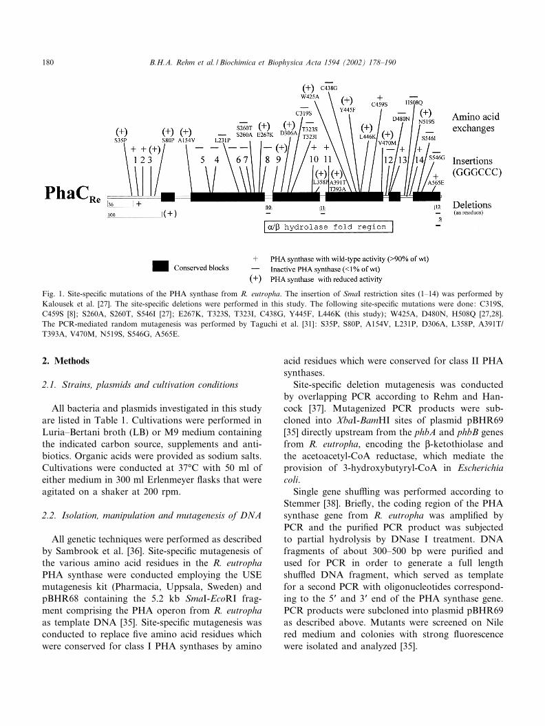
Fig. 1. Site-speci¢c mutations of the PHA synthase from R. eutropha. The insertion of SmaI restriction sites (1^14) was performed byKalousek et al. [27]. The site-speci¢c deletions were performed in this study. The following site-speci¢c mutations were done: C319S,C459S [8]; S260A, S260T, S546I [27]; E267K, T323S, T323I, C438G, Y445F, L446K (this study); W425A, D480N, H508Q [27,28].The PCR-mediated random mutagenesis was performed by Taguchi et al. [31]: S35P, S80P, A154V, L231P, D306A, L358P, A391T/T393A, V470M, N519S, S546G, A565E.
BBAPRO 36522 24-1-02
B.H.A. Rehm et al. / Biochimica et Biophysica Acta 1594 (2002) 178^190180

The generation of hybrid PHA synthases was per-formed employing tailored PCR. At the fusion pointsan EcoRI site was introduced to enable oriented li-gation of two PCR products into pBHR69 [35].Primers used in this study are summarized in Table1.
DNA sequences of new plasmid constructs andmutations were con¢rmed by DNA sequencing em-ploying the chain termination method using the au-tomatic sequencer LI-COR model 4200 (MWG-Bio-tech, Ebersberg, Germany).
2.3. Viable-colony staining
Screening of PHA accumulating colonies was per-
formed according to Spiekermann et al. [35]. Forroutine analysis, 0.002 vol. of a solution of 0.25 mgNile red or Nile blue (Sigma, St. Louis, MO, USA)per ml dimethyl sulfoxide (DMSO) was added to thesterilized medium to give a ¢nal concentration of0.5 Wg dye (ml medium)31. The agar plates wereexposed to ultraviolet light (312 nm) after appropri-ate cultivation periods to detect accumulation ofPHAs.
2.4. Sodium dodecyl sulfate (SDS)^polyacrylamidegel electrophoresis (PAGE) and Westernimmunoblotting
SDS^PAGE was performed according to Sam-
Table 1Bacterial strains, plasmids and oligonucleotides
Strains, plasmids and oli-gonucleotides
Relevant characteristics Source orreference
E. coliJM109 recA1 endA1 gyrA96 thi hsdR17 (rk3, mk�) supE44 relA1, ?-, lac [FP proAB lacIqZ?M15] (Stratagene)LS1298 C600 fadB : :Kan [34]
PlasmidspBHR69 pBluescript SK-containing the phbA-phbB genes downstream of lac promoter [35]pBHR68 pBluescript SK-containing the PHB operon downstream of lac promoter [35]
OligonucleotidesFor site-speci¢c mutagenesis
E267K 5P-CAACCGGAGAGCTCGCTGGTGCGCCATGTGGTGAAGCAGGGACATACG-3PT323I 5P-GTGGGCGGCATCATTGTCTCG-3PT323S 5P-GTGGGCGGCATCATTGTCTCG-3PC438G 5P-CCGTGGTACGGCTGGTATCTGCGCCAC-3PY445F 5P-CGCCACACCTTCTTGCAGAACGAGCTC-3PL446K 5P-CGCCACACCTACAAGCAGAACGAGC-3P
For deletionsD1 (281^290) 5P-CGTGGCGCAATCCGGACTACATCGAGCACGCG-3P
5P-GTGCTCGATGTAGTCCGGATTGCGCCACGACAC-3PD2 (372^382) 5P-GCAGTGGCGCGAGGCGCTGCTGCGCGGCCTTG-3P
5P-GCCGCGCAGCAGCGCCTCGCGCAACTGCACATG-3PD3 (C-terminus v5) 5P-CGCGGATCCTCAGTATCGCCCAGGCGCGGGTTC-3PD4 (C-terminus v12) 5P-CGCGGATCCTCAGATTGCGCGATAGCGCGC-3P
For construction of fusion proteinsF1 5P-CGGAATTCGCGCTGCGACTTGGTCGGG-3P
5P-CGGAATTCGACGACTACATCGAGCACGCG-3PF2 5P-CGGAATTCACGCTTGGCGGCCTCCAG-3P
5P-CGGAATTCGCCGGCGCGCCGTGCGCGCTG-3PF3 5P-CGGAATTCCCAGGGGGTGATGTGGTCG-3P
5P-CGGAATTCGCCTATGCCTCGACCGCGCTG-3PFor ampli¢cation of phaCRe
5P end 5P-GGGCTCTAGAAATAAGGAGATATACATATGGCGACCGGCAAAGGC-3P3P end 5P-CGCGGATCCTCATCGCTTGGCTTTGACGTATCG-3P
Ampli¢cation of the 5P end of phaCPa
5P end 5P-GGGCTCTAGAAATAAGGAGATATACATATGAGTCAGAAGAACAATAACGAGCTT-3P
BBAPRO 36522 24-1-02
B.H.A. Rehm et al. / Biochimica et Biophysica Acta 1594 (2002) 178^190 181

brook et al. [36]. Proteins were separated in 12.5%(w/v) SDS^polyacrylamide gels and stained withCoomassie brilliant blue R-250. Western blottingwas performed using the Semidry Fastblott (Biome-tra, Germany). On Western blots [39] using nitrocel-lulose membranes the PHA synthase and its mutantswere detected applying the respective monospeci¢c,polyclonal anti-PhaC antiserum raised against thePHA synthase from R. eutropha and an alkalinephosphatase-antibody conjugate as second antibody.Bound antibodies were detected using nitro blue tet-razolium chloride and the toluidine salt of 5-bromo-4-chloro-3-indolyl phosphate. Expression of mutatedgenes was con¢rmed by SDS^PAGE and immuno-blotting (data not shown).
2.5. In vivo activity of PHA synthases
The in vivo activity of the class I PHA synthaseswas determined, transferring the respective plasmidto E. coli JM109 harboring plasmid pBHR69, whichmediates provision of (R)-3-hydroxybutyryl-CoAfrom acetyl-CoA [40]. Recombinant E. coli was cul-tivated for 48 h in LB medium plus 2% (w/v) glucoseand PHA content was analyzed by gas chromatog-raphy (GC) analysis.
The in vivo activity of the class II PHA synthaseswas determined, transferring the respective plasmidto E. coli LS1298 (fadB), which provides medium-chain length (R)-3-hydroxyacyl-CoAs, when culti-vated in LB medium plus 0.5% (w/v) decanoate
Fig. 2. Alignment of the R. eutropha PHA synthase (PhaCRe) with the B. glumae lipase (1TAH_A). The alignment was performed us-ing PSI-Blast and CD search. Conservative replacements are labeled with a light gray background and identical residues are labeledwith dark gray background. The catalytic cysteine (PHA synthase) and the catalytic serine (lipase) are given as bold letters. The con-served H508 of PhaCRe, which aligned with H285 of the lipase, is indicated by a triangle. The structurally conserved `lid' region (he-lix K5 of the lipase) is boxed. The lipase box is labeled by two lines.
BBAPRO 36522 24-1-02
B.H.A. Rehm et al. / Biochimica et Biophysica Acta 1594 (2002) 178^190182
[12^14,39]. Cells harboring the respective plasmidwere cultivated for 48 h, and the PHA content wasanalyzed by GC analysis. The relative PHA contentwas used as PHA synthase in vivo activity.
2.6. Gas chromatographic analysis of polyester in cells
PHA was qualitatively and quantitatively analyzedby GC. Liquid cultures were centrifuged at 10 000Ugfor 15 min, then the cells were washed twice in salineand lyophilized overnight. 8^10 mg lyophilized cellmaterial were subjected to methanolysis in the pres-ence of 15% (v/v) sulfuric acid. The resulting methylesters of the constituent 3-hydroxyalkanoic acidswere assayed by GC according to Brandl et al. [41]and as described in detail previously [42]. GC anal-ysis was performed by injecting 3 Wl of sample into aPerkin-Elmer 8420 gas chromatograph (Uë berlingen,Germany) using a 0.5 Wm diameter Permphase PEG25 Mx capillary column 60 m in length.
2.7. PHA synthase assay
PHA synthase was determined spectrophotometri-cally by monitoring the release of CoA at 412 nm[43]. The standard assay contained 1 mM DTNB, 20mM MgCl2 and 50 WM of the respective CoA thio-ester in 150 mM Tris^HCl, pH 7.5. One unit (U) ofenzyme is de¢ned as the amount which produced1 Wmol product per minute under the assay condi-tions employed.
3. Results
3.1. Development of a threading model of the class IR. eutropha PHA synthase
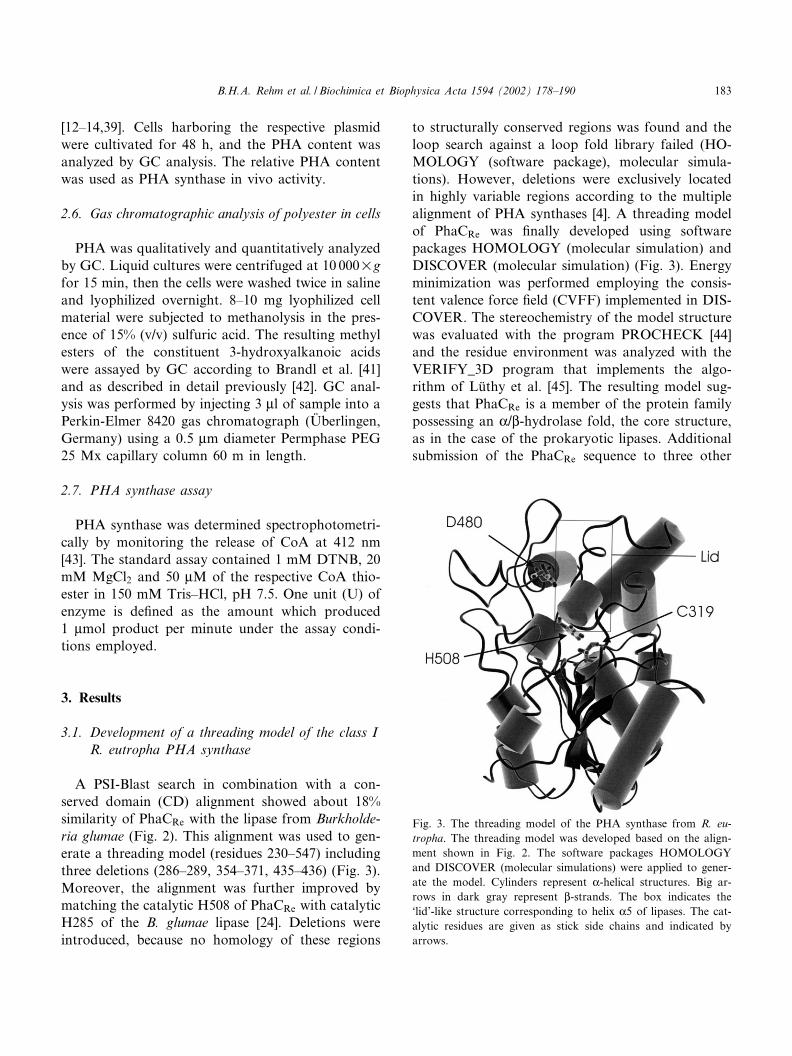
A PSI-Blast search in combination with a con-served domain (CD) alignment showed about 18%similarity of PhaCRe with the lipase from Burkholde-ria glumae (Fig. 2). This alignment was used to gen-erate a threading model (residues 230^547) includingthree deletions (286^289, 354^371, 435^436) (Fig. 3).Moreover, the alignment was further improved bymatching the catalytic H508 of PhaCRe with catalyticH285 of the B. glumae lipase [24]. Deletions wereintroduced, because no homology of these regions
to structurally conserved regions was found and theloop search against a loop fold library failed (HO-MOLOGY (software package), molecular simula-tions). However, deletions were exclusively locatedin highly variable regions according to the multiplealignment of PHA synthases [4]. A threading modelof PhaCRe was ¢nally developed using softwarepackages HOMOLOGY (molecular simulation) andDISCOVER (molecular simulation) (Fig. 3). Energyminimization was performed employing the consis-tent valence force ¢eld (CVFF) implemented in DIS-COVER. The stereochemistry of the model structurewas evaluated with the program PROCHECK [44]and the residue environment was analyzed with theVERIFY_3D program that implements the algo-rithm of Lu«thy et al. [45]. The resulting model sug-gests that PhaCRe is a member of the protein familypossessing an K/L-hydrolase fold, the core structure,as in the case of the prokaryotic lipases. Additionalsubmission of the PhaCRe sequence to three other
Fig. 3. The threading model of the PHA synthase from R. eu-tropha. The threading model was developed based on the align-ment shown in Fig. 2. The software packages HOMOLOGYand DISCOVER (molecular simulations) were applied to gener-ate the model. Cylinders represent K-helical structures. Big ar-rows in dark gray represent L-strands. The box indicates the`lid'-like structure corresponding to helix K5 of lipases. The cat-alytic residues are given as stick side chains and indicated byarrows.
BBAPRO 36522 24-1-02
B.H.A. Rehm et al. / Biochimica et Biophysica Acta 1594 (2002) 178^190 183

algorithms that search structural databases (SAM-T98 [25], 3D-PSSM [46], and the UCLA Foldserver[47]) resulted in ¢ts to other enzymes belonging tothe K/L-hydrolase fold family with high con¢dencelevels (data not shown). Inspection of the proteinmodel of PhaCRe showed that the active site C319,the conserved H508 and the D480, presumably form-ing a catalytic triad, are adjacent to the core struc-ture (Fig. 3). These residues are conserved in all PHAsynthases and are required for catalytic activity [4].The active site C319 was located at the nucleophileelbow, a sharp Q turn containing the nucleophilic res-idue, positioned between a L-strand and an K-helix,which is one of the most conserved features of theK/L-hydrolase enzymes.
3.2. Site-speci¢c mutagenesis of the class I PHAsynthase from R. eutropha
With the aid of the multiple PHA synthase se-quence alignment (ClustalW), we depicted ¢ve aminoacid residues (E267, T323, C438, Y445, L446) whichmight be involved in substrate speci¢city. These res-idues were conserved for class I and class II PHAsynthases, respectively, and were located in the con-served K/L-hydrolase fold region (RPS-Blast in theconserved domain database), but the respective ami-no acid di¡ered in physical and chemical propertiesbetween the enzyme classes. The following mutationsof PhaCRe were performed using site-speci¢c muta-
genesis: E267K, T323I, T323S, C438G, Y445F, andL446K (Fig. 1). These PhaCRe amino acid residueswere replaced by the respective conserved residue ofthe class II PHA synthases. Only three of the site-speci¢c mutants, E267K, Y445F and L446K, werepermissive and showed in vivo and in vitro PHAsynthase activity (Table 2). In vivo activity wasachieved by expression of the respective mutatedPHA synthase gene located on plasmid pBHR68 inE. coli JM109 and by quantitative analysis of theaccumulated PHA (Table 2). Crude extracts of thesame cells were subjected to in vitro PHA synthaseactivity measurements (Table 2). In order to investi-gate whether or not the substrate speci¢city waschanged by the mutation, we transferred the respec-tive plasmid to E. coli LS1298, which provides me-dium-chain length 3-hydroxyacyl-CoA thioesters assubstrate for PHA synthase, when grown on fattyacids. None of the mutants showed a signi¢cantchange in substrate speci¢city (Table 2). Interest-ingly, permissive mutations were only achieved foramino acid residue positions which were located atthe protein model surface (Fig. 4).
3.3. Site-speci¢c deletion mutagenesis of the class IPHA synthase from R. eutropha
The multiple alignment (ClustalW) of 44 PHAsynthases revealed six amino acid sequence regions(blocks) in which conserved amino acid residues were
Table 2In vitro and in vivo activity of site-speci¢c mutants of the R. eutropha PHA synthase
Mutation Relative speci¢c PHA synthase activity (%)a Relative PHA content (%)
3HBb 3HB/3HOc
E267K 40 87 54/NDT323I ND ND ND/NDT323S ND ND ND/NDC438G ND ND ND/NDY445F 38 24 124/108L446K 15 8 23/ND
ND, not detectable; 3HB, 3-hydroxybutyric acid; 3HO, 3-hydroxyoctanoate.aRecombinant E. coli was cultivated for 24 h in LB medium containing 2% (w/w) glucose, 75 Wg/ml ampicillin and 1 mM IPTG. Therelative speci¢c activity of crude extracts was provided as % of wild type activity corresponding to 1.4 U/mg protein.bRecombinant E. coli JM109 was cultivated for 48 h in LB medium containing 2% (w/v) glucose, 75 Wg/ml ampicillin and 1 mMIPTG. The relative PHA content was provided as % of wild type PHA accumulation corresponding to about 80% (w/w) of CDW.cRecombinant E. coli LS1298 was cultivated for 48 h in LB medium containing 0.5% (w/v) decanoate, 75 Wg/ml ampicillin and 1 mMIPTG. The relative PHA content was provided as % of wild type PHA accumulation corresponding to about 14.1% (w/w) of CDW(12.9% 3HB; 1.2% 3HO).
BBAPRO 36522 24-1-02
B.H.A. Rehm et al. / Biochimica et Biophysica Acta 1594 (2002) 178^190184

frequent [4]. These blocks are separated by regions ofabout 10^30 amino acid residues which were lessconserved (Fig. 1). Site-speci¢c deletions were per-formed in the K/L-hydrolase fold region betweenblocks 2/3 and 3/4 (D1 (v281^290), D2 (v372^382)). None of these deletions were permissive, i.e.neither in vivo nor in vitro enzyme activity was de-tectable. In contrast to the highly variable N-termi-nus of the PHA synthases [26], the C-terminus of the
class I and class II PHA synthases appears ratherconserved. However, the class III PHA synthasesappear about 30 amino acid residues shorter withrespect to the C-terminus. In order to evaluate thefunctionality of the C-terminus, a ¢ve and a 12 ami-no acid deletion was introduced at the C-terminus(D3 (v585^589), D4 (v578^589)). These mutationsabrogated in vivo and in vitro enzyme activity (Fig.1). For analysis of in vitro activity recombinantE. coli was cultivated for 24 h in LB medium con-taining 2% (w/w) glucose, 75 Wg/ml ampicillin and1 mM IPTG. The relative speci¢c activity of crudeextracts was determined and the wild type activitycorresponded to 1.4 U/mg protein. For analysis ofin vivo activity recombinant E. coli was cultivatedfor 48 h in LB medium containing 2% (w/w) glucose,75 Wg/ml ampicillin and 1 mM IPTG. The relativePHA content was determined by GC analysis andwild type PHA accumulation corresponded to about80% of CDW.
3.4. Construction of class I and class II PHA synthasefusion proteins
Site-speci¢c fusion proteins were generated usingPCR-based gene fusions. Fusion proteins composedof an N-terminal class II PHA synthase region(PhaCPa from P. aeruginosa) and a C-terminal classI PHA synthase region (PhaCRe from R. eutropha)were constructed. The variable regions between theconserved blocks 2/3, 3/4 and 5/6 served as fusionpoints (Fig. 5). These fusion proteins were con-structed in order to obtain evidence which part ofthe protein determines the substrate speci¢city and/or is required for catalytic activity. However, the
Fig. 5. Construction of PHA synthase fusion proteins.
Fig. 4. Localization of the single amino acid residue replace-ments in the PhaCRe model. Amino acid residue replacementswere achieved by site-speci¢c mutagenesis (see Table 2). Onlythe backbone of the PhaCRe model is shown. The replacingside chains are given as CPK format. Replacements resultingin PHA synthase mutants with in vitro and in vivo activity areindicated by +. Inactive PHA synthase mutants are indicatedby 3.
BBAPRO 36522 24-1-02
B.H.A. Rehm et al. / Biochimica et Biophysica Acta 1594 (2002) 178^190 185

fusion proteins did not show in vivo activity eitherwhen (R)-3-hydroxybutyryl-CoA or when medium-chain length (R)-3-hydroxyacyl-CoA was providedin vivo. However, 13% of wild type in vitro enzymeactivity was obtained with fusion protein F1 (Fig. 5),whose fusion point was located at a variable surface-exposed loop (Fig. 6).
3.5. Single gene shu¥ing with the class I PHAsynthase from R. eutropha
Single gene shu¥ing with the phaCRe gene wasperformed to generate a variety of mutants which
showed altered enzyme activity. Shu¥ed phaCRe
gene PCR products were inserted into XbaI-BamHIsites of plasmid pBHR69 upstream from the phaARe
and phaBRe genes, encoding the L-ketothiolase andthe acetoacetyl-CoA reductase. E. coli JM109 cellsharboring the mutated phaCRe gene were screenedon Nile red containing agar plates for altered invivo enzyme activity. Among approx. 200 000 trans-formants four mutants (GS1^GS4) were selectedwhich showed in vivo activity, i.e. appeared as £uo-rescent colonies. GC analysis of the respective trans-formants showed that the cells accumulated polyhy-droxybutyrate (PHB), but below the wild type level.DNA sequence analysis revealed that beside singlemutations, double and triple mutations were ob-served. These permissive mutations are compiled inTable 3. The mutations did not hit highly conservedamino acid residues and were located at the PhaCRe
model surface (Fig. 7). Only in the GS4 mutant,which showed only about 30% of wild type PHBsynthase activity and about 20% of wild type PHBaccumulation, we observed replacement of the ratherconserved amino acid residues G206 and P230.
4. Discussion
The multiple alignment of the primary structuresof 44 PHA synthases showed the presence of sixconserved blocks and eight conserved amino acidresidues [4]. In 1992 Steinbu«chel and coworkers [48]already noted that all PHA synthases contain a pu-tative lipase box (G-X-[S/C]-X-G) in which the essen-tial active site serine of the lipase is replaced with acysteine in the PHA synthase. A Blast sequence ho-mology search with the A. vinosum class III PHA
Fig. 6. Localization of the fusion sites in the PhaCRe proteinmodel. Positions of catalytic residues (stick format) and the fu-sion site are indicated by arrows. The protein model is pre-sented as in Fig. 3.
Table 3In vitro and in vivo activity of gene-shu¥ed R. eutropha PHA synthase mutants
Mutation Relative speci¢c PHA synthase activity (%)a Relative PHA content (%)b
GS1 (A334V, G376S) 24.3 28GS2 (A57V, I357V) 15 45.4GS3 (K139R, R386C) 40 35GS4 (G206S, P230S, E337K) 30 16aRecombinant E. coli was cultivated for 24 h in LB medium containing 2% (w/w) glucose, 75 Wg/ml ampicillin and 1 mM IPTG. Therelative speci¢c activity of crude extracts was provided as % of wild type activity corresponding to 1.4 U/mg protein.bRecombinant E. coli was cultivated for 48 h in LB medium containing 2% (w/w) glucose, 75 Wg/ml ampicillin and 1 mM IPTG. Therelative PHA content was provided as % of wild type PHA accumulation corresponding to about 80% of CDW.
BBAPRO 36522 24-1-02
B.H.A. Rehm et al. / Biochimica et Biophysica Acta 1594 (2002) 178^190186

synthase and the P. aeruginosa class II PHA synthasehas also shown homology to lipases, particularly tothe lipase from B. cepacia, and the putative activesite cysteine residue aligned with the active site serineof the lipase [4,24]. A topological model based onthis homology has been generated for the class IIIand class II PHA synthases [4,24].
In this study, we developed the ¢rst threadingmodel of a class I PHA synthase, the PHA synthasefrom R. eutropha, based on the homology to the B.glumae lipase, whose structure has been determinedby X-ray analysis [49]. Many of the features of theK/L-hydrolase fold are maintained in the protein mod-el. The central L-sheet in the core of the moleculeconforms to the K/L-hydrolase fold (Fig. 3). Inspec-tion of the proposed PhaCRe model showed that the
recently identi¢ed catalytic residues C319, H508 andD480 are located adjacent to the core structure of thePhaCRe protein model with the catalytic C319 lo-cated at the nucleophile elbow (Fig. 3). The activesite C319 lies at the C-terminal end of a L-strand,corresponding to strand L5 of the lipases, in thestrand-turn-helix motif. Mutational studies revealedthat the R. eutropha PHA synthase possesses an es-sential catalytic dyad (C319^H508) in which theC319 is involved in covalent catalysis and H508serves as a general base catalyst [30]. A conservedD480 was shown not to be required for acylationof C319 by sT-CoA (a terminally saturated trimerof (R)-3-hydroxybutyryl-CoA) and is proposed tofunction as a general base catalyst to activate thehydroxyl of (R)-3-hydroxybutyryl-CoA for ester for-mation [30]. In addition, the highly conserved W425was replaced by alanine, which reduced in vivo ac-tivity to 19% and in vitro activity to 0.003% of wildtype activity. This W425 has been postulated to playan important role in protein^protein interaction, i.e.in the dimerization of the PhaC subunit, by generat-ing a hydrophobic surface [29]. Interestingly, this wasconsistent with our threading model in which W425was located at the surface. Furthermore, the site-spe-ci¢c mutations Y445F and L446K, obtained in thisstudy, resulted in reduced activity and since Y445and L446 are hydrophobic residues and are locatedadjacent to W425 at the surface of the protein model,these residues might exert a similar function in viewof dimerization (Figs. 4 and 8). Thus reduced in vivoand in vitro activity of Y445F and L446K, with theladder mutation having a stronger impact on enzymeactivity presumably due to introduction of a chargedresidue (Table 2), might result from impaired dimer-ization.
The B. glumae lipase structure, which was used astemplate structure in this study, was obtained in the`closed' conformation exhibiting the active siteburied underneath a helical segment (K5), called a`lid' or a `£ap' [49]. In the PHA synthase model theactive site was also buried underneath this structur-ally conserved helical segment (Figs. 2 and 3), whichcorresponded to helix K5 of the B. glumae lipase.However, during transition to the open conformationof the lipase, due to interfacial activation, the activesite becomes accessible to the solvent and a hydro-phobic surface is exposed by the movement of the
Fig. 7. Localization of amino acid residue replacements in thePhaCRe model. Permissive mutations were generated by singlegene shu¥ing. Replaced amino acid residues are given as CPKmodels of the respective side chains. Replacements occurring si-multaneously in the PHA synthase mutant are identicallywrapped or unwrapped (see Table 3). Catalytic residues are in-dicated as stick models of the respective side chains. Only thebackbone of the PhaCRe model is represented.
BBAPRO 36522 24-1-02
B.H.A. Rehm et al. / Biochimica et Biophysica Acta 1594 (2002) 178^190 187

lid. The conformational changes can range from asimple rigid body hinge-type motion to complex re-organizations involving changes in the secondarystructures. Generally speaking, various structuralstudies suggested that the hydrophobic lipid-bindingsite is opened up by the rolling back of the lid fromthe active site at an oil^water interface. However,even in the absence of an oil^water interface, theremay be a subtle equilibrium between the two confor-mations of the enzyme. It is believed that the openingof the lid is essential but not su¤cient to explain theinterfacial activation. In addition to providing accessto the active site, the structural rearrangements alsochange the surface properties of the enzymes and insome cases form an oxyanion hole. In each case de-scribed, the movement of the lid exposes a large hy-drophobic surface area surrounding the active site.This movement results in an amphipathic moleculewhich could be properly oriented for interaction ofthe active site with a lipid interface [50].
Accordingly, the soluble PHA synthase turns into
an amphipathic molecule upon availability of sub-strate and covalent synthesis of the hydrophobicpolyester chain [4]. This leads to the formation ofso-called PHA granules with the hydrophobic PHAin the core and the active PHA synthases at the sur-face, which represents the water^PHA interface.Consistently, the granule-associated PhaCRe exertedan about 40 times higher activity as compared to thesoluble enzyme [9]. This indicated that interfacial ac-tivation occurred and that a lid-like structure asfound in lipases and exhibited in the PhaCRe modelmight also exist in PHA synthases (Fig. 3). The per-missive double mutant GS3 (R386C, K139R) con-tained one mutation site R386C located in the lidregion (Table 3, Fig. 7, see below). Two site-speci¢cmutants (A391T, T393A) were generated by Taguchiet al. [31], which resided in the lid region and stillexhibited PHA synthase activity. These data sug-gested that the surface-exposed lid-like structure isnot essential for enzyme function (Figs. 2 and 3).
Six site-speci¢c mutations were performed based
Fig. 8. Localization of all published single amino acid residue replacements in the PhaCRe model. Replaced amino acid residues aregiven as CPK models of the respective side chains. Only the backbone of the PhaCRe model is represented. Replacements resulting inPHA synthase mutants with in vitro and in vivo activity are indicated by +. Inactive PHA synthase mutants are indicated by 3. Theleft panel represents one set of mutations. The right panel represents a second set mutations with the PhaCRe model rotated by 180³from right to left.
BBAPRO 36522 24-1-02
B.H.A. Rehm et al. / Biochimica et Biophysica Acta 1594 (2002) 178^190188
on the alignment of class I and II PHA synthases,intending to change the substrate speci¢city. None ofthe mutations mediated a signi¢cant change in sub-strate speci¢city, indicating that a single amino acidresidue replacement might not be su¤cient (Table 2).This is supported by the large number of randommutants analyzed from which none changed the spe-ci¢city of the enzyme from a class II PHA synthaseto a class I PHA synthase or vice versa (data notshown). However, the three permissive mutation sites(Y445F, L446K, E267K) were found to be surfaceexposed in the PhaCRe model, whereas the non-per-missive mutation sites T323 and C438 were locatedin the core structure (Table 2, Fig. 4). The E267Kmutant exhibited about 90% of wild type in vivoactivity with respect to PHB accumulation, indicat-ing a non-essential role of this residue for catalysis.Meanwhile, a large number of site-speci¢c mutationshave been performed with this class I PHA synthasefrom R. eutropha [8,26^31] (Fig. 1). These data againwere in agreement with our current topological mod-el of the PHA synthase from R. eutropha by showingthat most of the permissive mutations were locatedat the variable solvent interacting surface, whereasmost non-permissive mutations were located in thecore structure (Fig. 8). The entire ¢rst 100 N-termi-nal amino acid residues have been shown to be dis-pensable without inactivation of the enzyme. In con-trast, the two deletions at the C-terminus (¢ve and 12amino acid residues) did abolish the PHA synthaseactivity, which suggested that the C-terminus,although not present in the class III PHA synthases,but rather conserved among class I and II PHA syn-thases, is essential for enzymatic activity. Furtherdeletions between the conserved blocks 2 and 3 aswell as 3 and 4, respectively, were not tolerated bythe PHA synthase leading to an inactive enzyme.However, SmaI restriction site insertions betweenthe conserved blocks 2/3, 3/4 and 5/6, respectively,were permissive mutations, suggesting that these re-gions are not adjacent to the core structure and thussurface exposed [27]. Five SmaI restriction site inser-tions in the second conserved block were not toler-ated by the PHA synthase and caused inactivation,indicating that this region might be functionally rel-evant (Fig. 1).
Fusion proteins composed of the N-terminal part
of the PHA synthase from P. aeruginosa and the C-terminal part of the PHA synthase from R. eutrophaindicated that fusion points located in the K/L-hydro-lase fold region are not tolerated (Fig. 5). Further-more, these fusion points were located in predictedand structurally conserved K-helical regions (Fig. 6).However, a fusion point at position 289 relative tothe amino acid sequence of the R. eutropha PHAsynthase and located at a variable surface-exposedloop in the protein model resulted in a hybrid PHAsynthase, which exhibited in vitro enzyme activity,but no detectable in vivo activity (Figs. 5 and 6).These data suggested that the ¢rst 288 amino acidresidues of PhaCRe can be replaced by the N-termi-nus of a class II PHA synthase and provided evi-dence for the importance of the K/L-hydrolase foldregion. Since this fusion protein showed only 13% ofwild type in vitro activity, this enzyme activity mightbe insu¤cient to mediate detectable accumulation ofPHA in recombinant E. coli.
Single gene shu¥ing with the R. eutropha PHAsynthase gene resulted in the isolation of ¢ve mutants(double or triple mutants) with altered enzyme activ-ity, whose mutation sites were mainly located at thesurface of the PhaCRe model (Table 3, Fig. 7). Onlyin the case of the triple mutant GS4 a rather con-served residue, the P230, was a¡ected. However,P230 has been found at the surface of the PhaCRe
model (Fig. 7). This approach did not enable theisolation of overproducing mutants. Discrepanciesin in vivo and in vitro activities of the respectivemutants, generated in this study, might be due tothe fact that the enzymes were still capable of acyla-tion but without catalyzing the polymerization reac-tion. Overall, a ¢rst threading model of a class IPHA synthase was developed, which basically is inagreement with various mutations generated in thisstudy and elsewhere [26^31]. This model should serveas a working hypothesis in order design experimentsrelated to structure^function analysis.
Acknowledgements
We gratefully acknowledge a fellowship providedby the Deutsche Akademische Austauschdienst toA.A.A.
BBAPRO 36522 24-1-02
B.H.A. Rehm et al. / Biochimica et Biophysica Acta 1594 (2002) 178^190 189

References
[1] A.J. Anderson, E.A. Dawes, Microbiol. Rev. 54 (1990) 450^472.
[2] A. Steinbu«chel, B. Fu«chtenbusch, V. Goren£o, S. Hein, R.Jossek, S. Langenbach, B.H.A. Rehm, Polym. Degrad. Stab.59 (1997) 177^182.
[3] A. Steinbu«chel, S. Hein, Adv. Biochem. Eng. Biotechnol. 71(2001) 81^123.
[4] B.H.A. Rehm, A. Steinbu«chel, in: A. Steinbu«chel, Y. Doi(Eds.), Biopolymers, Wiley-VCH, Heidelberg, 2001, in press.
[5] T. Lu«tke-Eversloh, K. Bergander, H. Luftmann, A. Steinbu«-chel, Microbiology 147 (2001) 11^19.
[6] M. Liebergesell, A. Steinbu«chel, Eur. J. Biochem. 209 (1992)135^150.
[7] M. Liebergesell, S. Rahalkar, A. Steinbu«chel, Appl. Micro-biol. Biotechnol. 54 (2000) 186^194.
[8] T.U. Gerngross, K.D. Snell, O.P. Peoples, A.J. Sinskey, E.Csuhai, S. Masamune, J. Stubbe, Biochemistry 33 (1994)9311^9320.
[9] T.U. Gerngross, D.P. Martin, Proc. Natl. Acad. Sci. USA 92(1995) 6279^6283.
[10] M. Liebergesell, I.L. Sonomoto, M. Madkour, F. Mayer, A.Steinbu«chel, Eur. J. Biochem. 226 (1994) 71^80.
[11] R. Jossek, R. Reichelt, A. Steinbu«chel, Appl. Microbiol.Biotechnol. 49 (1998) 258^266.
[12] A. Timm, A. Steinbu«chel, Eur. J. Biochem. 209 (1992) 15^30.
[13] S. Langenbach, B.H.A. Rehm, A. Steinbu«chel, FEMS Mi-crobiol. Lett. 150 (1997) 303^309.
[14] Q. Qi, B.H.A. Rehm, A. Steinbu«chel, FEMS Microbiol.Lett. 157 (1997) 155^162.
[15] Q. Qi, A. Steinbu«chel, B.H.A. Rehm, FEMS Microbiol.Lett. 167 (1998) 89^94.
[16] B.H.A. Rehm, N. Kru«ger, A. Steinbu«chel, J. Biol. Chem. 273(1998) 24044^24051.
[17] S. Fiedler, A. Steinbu«chel, B.H.A. Rehm, Appl. Environ.Microbiol. 66 (2000) 2117^2124.
[18] B.H.A. Rehm, T.A. Mitsky, A. Steinbu«chel, Appl. Environ.Microbiol. 67 (2001) 3102^3109.
[19] N. Ho¡mann, A. Steinbu«chel, B.H.A. Rehm, FEMS Micro-biol. Lett. 184 (2000) 253^259.
[20] N. Ho¡mann, A. Steinbu«chel, B.H.A. Rehm, Appl. Micro-biol. Biotechnol. 54 (2000) 665^670.
[21] B.H.A. Rehm, N. Ho¡mann, Q. Qi, S. Fiedler, A. Steinbu«-chel, in: Proceedings of the International Symposium onBioconversion of Renewable Raw Materials, GBF,Braunschweig, 2001, in press.
[22] Q. Qi, A. Steinbu«chel, B.H.A. Rehm, Appl. Microbiol. Bio-technol. 54 (2000) 37^43.
[23] B.H.A. Rehm, Q. Qi, Br.B. Beermann, H.-J. Hinz, A. Stein-bu«chel, Biochem. J. 358 (2001) 263^268.
[24] Y. Jia, T.J. Kappock, T. Frick, A.J. Sinskey, J. Stubbe, Bio-chemistry 39 (2000) 3927^3936.
[25] K. Karplus, C. Barrett, R. Hughey, Bioinformatics 14 (1998)846^856.
[26] P. Schubert, N. Kru«ger, A. Steinbu«chel, J. Bacteriol. 173(1991) 168^175.
[27] S. Kalousek, D. Dennis, W. Lubitz, FEMS Microbiol. Rev.103 (1992) 426^427.
[28] A. Hoppensack, B.H.A. Rehm, A. Steinbu«chel, J. Bacteriol.181 (1999) 1429^1435.
[29] B. Junker, G. York, C. Park, Y. Jia, C. Rha, J. Stubbe, A.J.Sinskey, in: The 8th International Symposium of BiologicalPolyester, Abstract, Cambridge, MA, 2000.
[30] Y. Jia, W. Yuan, J. Wodzinska, C. Park, A.J. Sinskey,J. Stubbe, Biochemistry 40 (2001) 1011^1019.
[31] S. Taguchi, A. Maehara, K. Takase, M. Nakahara, H. Na-kamura, Y. Doi, FEMS Microbiol. Lett. 198 (2001) 65^71.
[32] U. Mu«h, A.J. Sinskey, D.P. Kirby, W.S. Lane, J. Stubbe,Biochemistry 38 (1999) 826^837.
[33] B.H.A. Rehm, A. Steinbu«chel, Int. J. Biol. Macromol. 25(1999) 3^19.
[34] C.C. DiRusso, J. Bacteriol. 172 (1990) 6459^6468.[35] P. Spiekermann, B.H.A. Rehm, R. Kalscheuer, D. Baumeis-
ter, A. Steinbu«chel, Arch. Microbiol. 171 (1999) 73^80.[36] J. Sambrook, E.F. Fritsch, T. Maniatis, Molecular Cloning.
A Laboratory Manual, 2nd edn., Cold Spring Harbor Lab-oratory Press, Cold Spring Harbor, NY, 1989.
[37] B.H.A. Rehm, R.E.W. Hancock, J. Bacteriol. 178 (1996)3346^3349.
[38] W.P.C. Stemmer, Nature 370 (1994) 389^391.[39] H. Towbin, T. Staehelin, J. Gordon, Proc. Natl. Acad. Sci.
USA 76 (1979) 4350^4354.[40] R.V. Antonio, A. Steinbu«chel, B.H.A. Rehm, FEMS Micro-
biol. Lett. 182 (2000) 111^117.[41] H. Brandl, R.A. Gross, R.W. Lenz, R.C. Fuller, Appl. En-
viron. Microbiol. 54 (1988) 1977^1982.[42] A. Timm, A. Steinbu«chel, Appl. Environ. Microbiol. 56
(1990) 3360^3367.[43] H.E. Valentin, A. Steinbu«chel, Appl. Microbiol. Biotechnol.
40 (1994) 699^709.[44] R.A. Laskowski, M.W. MacArthur, J.M. Thornton, J. Appl.
Crystallogr. 26 (1993) 283^291.[45] R. Lu«thy, J.U. Bowie, D. Eisenberg, Nature 356 (1992) 83^
85.[46] L.A. Kelley, R. MacCallum, M.J.E. Sternberg, in: S. Istrail,
P. Pevzner, M. Waterman (Eds.), Proceedings of the ThirdAnnual Conference on Computational Molecular Biology,The Association for Computing Machinery, New York,1999.
[47] D. Fischer, D. Eisenberg, Protein Sci. 5 (1996) 947^955.[48] A. Steinbu«chel, E. Hustede, M. Liebergesell, U. Pieper, A.
Timm, H. Valentin, FEMS Microbiol. Rev. 9 (1992) 217^230.
[49] M.E. Noble, A. Cleasby, L.N. Johnson, M.R. Egmond, L.G.Frenken, FEBS Lett. 331 (1993) 123^128.
[50] J.D. Schrag, Y. Li, M. Cygler, D. Lang, T. Burgdorf, H.-J.Hecht, R. Schmid, D. Schomburg, T.J. Rydel, J.D. Oliver,L.C. Strickland, C.M. Dunaway, S.B. Larson, J. Day, A.McPherson, Structure 5 (1997) 187^202.
BBAPRO 36522 24-1-02
B.H.A. Rehm et al. / Biochimica et Biophysica Acta 1594 (2002) 178^190190