Selective inhibition of prostacyclin synthase activity by rofecoxib
12
Introduction The second isoform of cyclooxygenase enzyme dis- covered (COX-2) is very similar to COX-1 as far as active site structure, catalytic mechanism, products and kinetics are concerned.Yet, two structural differ- ences between the two isoenzymes have important pharmacological and biological consequences. The first difference is that COX-2 active site is larger and more accommodating than that of COX-1 and includes a side pocket not present in COX-1, provid- ing the basis for the development of selective COX-2 inhibitors. Second, only COX-1 (but not COX-2) is negatively regulated in an allosteric manner by Selective inhibition of prostacyclin synthase activity by rofecoxib Cristiana Griffoni a, † , Enzo Spisni a, †, * , Antonio Strillacci a , Mattia Toni a , Markus Michael Bachschmid b , Vittorio Tomasi a a Department of Experimental Biology, University of Bologna, Bologna, Italy b Evans Department of Medicine, Boston University Medical Center, Boston, Massachusetts, USA Received: September 19, 2006; Accepted: January 11, 2007 Abstract The development of cyclooxygenase-2 (COX-2) selective inhibitors prompted studies aimed at treating chron- ic inflammatory diseases and cancer by using this new generation of drugs.Yet, several recent reports point- ed out that long-term treatment of patients with COX-2 selective inhibitors (especially rofecoxib) caused severe cardiovascular complicances. The aim of this study was to ascertain whether, in addition to inhibiting COX-2, rofecoxib may also affect prostacyclin (PGI2) level by inhibiting PGI2 forming enzyme (prostacyclin synthase, PGIS).In order to evaluate if selective (celecoxib, rofecoxib) and non-selective (aspirin, naproxen) anti-inflammatory compounds could decrease PGI2 production in endothelial cells by inhibiting PGIS, we analyzed the effect of anti-inflammatory compounds on the enzyme activity by ELISA assay after addition of exogenous substrate, on PGIS protein levels by Western blotting and on its subcellular distribution by confo- cal microscopy.We also analyzed the effect of rofecoxib on PGIS activity in bovine aortic microsomal frac- tions enriched in PGIS. This study demonstrates an inhibitory effect of rofecoxib on PGIS activity in human umbilical vein endothelial (HUVE) cells and in PGIS-enriched bovine aortic microsomal fractions, which is not observed by using other anti-inflammatory compounds. The inhibitory effect of rofecoxib is associated neither to a decrease of PGIS protein levels nor to an impairment of the enzyme intracellular localization. The results of this study may explain the absence of a clear relationship between COX-2 selectivity and cardiovascular side effects. Moreover, in the light of these results we propose that novel selective COX-2 inhibitors should be tested on PGI2 synthase activity inhibition. Keywords: rofecoxib • prostacyclin synthase • nonsteroidal anti-inflammatory agents • cyclooxygenase-2 inhibitors • endothelium J. Cell. Mol. Med. Vol 11, No 2, 2007 pp. 327-338 † Cristiana Griffoni and Enzo Spisni contributed equally to this work. *Correspondence to Enzo SPISNI Via Selmi, 3, 40126 Bologna, Italy. Tel: +39 051 209 42 53 Fax: +39 051 209 42 86 Email: [email protected] © 2007 The Authors Journal compilation © 2007 Foundation for Cellular and Molecular Medicine/Blackwell Publishing Ltd doi: 10.1111/j.1582-4934.2007.00021.x
-
Upload
independent -
Category
Documents
-
view
2 -
download
0
Transcript of Selective inhibition of prostacyclin synthase activity by rofecoxib

Introduction
The second isoform of cyclooxygenase enzyme dis-covered (COX-2) is very similar to COX-1 as far asactive site structure, catalytic mechanism, products
and kinetics are concerned. Yet, two structural differ-ences between the two isoenzymes have importantpharmacological and biological consequences. Thefirst difference is that COX-2 active site is larger andmore accommodating than that of COX-1 andincludes a side pocket not present in COX-1, provid-ing the basis for the development of selective COX-2inhibitors. Second, only COX-1 (but not COX-2) isnegatively regulated in an allosteric manner by
Selective inhibition of prostacyclin synthase activity
by rofecoxib
Cristiana Griffoni a, †, Enzo Spisni a, †, *, Antonio Strillacci a, Mattia Toni a,Markus Michael Bachschmid b, Vittorio Tomasi a
a Department of Experimental Biology, University of Bologna, Bologna, Italyb Evans Department of Medicine, Boston University Medical Center, Boston, Massachusetts, USA
Received: September 19, 2006; Accepted: January 11, 2007
Abstract
The development of cyclooxygenase-2 (COX-2) selective inhibitors prompted studies aimed at treating chron-ic inflammatory diseases and cancer by using this new generation of drugs.Yet, several recent reports point-ed out that long-term treatment of patients with COX-2 selective inhibitors (especially rofecoxib) causedsevere cardiovascular complicances. The aim of this study was to ascertain whether, in addition to inhibitingCOX-2, rofecoxib may also affect prostacyclin (PGI2) level by inhibiting PGI2 forming enzyme (prostacyclinsynthase, PGIS). In order to evaluate if selective (celecoxib, rofecoxib) and non-selective (aspirin, naproxen)anti-inflammatory compounds could decrease PGI2 production in endothelial cells by inhibiting PGIS, weanalyzed the effect of anti-inflammatory compounds on the enzyme activity by ELISA assay after addition ofexogenous substrate, on PGIS protein levels by Western blotting and on its subcellular distribution by confo-cal microscopy. We also analyzed the effect of rofecoxib on PGIS activity in bovine aortic microsomal frac-tions enriched in PGIS. This study demonstrates an inhibitory effect of rofecoxib on PGIS activity in humanumbilical vein endothelial (HUVE) cells and in PGIS-enriched bovine aortic microsomal fractions, which is notobserved by using other anti-inflammatory compounds. The inhibitory effect of rofecoxib is associated neitherto a decrease of PGIS protein levels nor to an impairment of the enzyme intracellular localization. The resultsof this study may explain the absence of a clear relationship between COX-2 selectivity and cardiovascularside effects. Moreover, in the light of these results we propose that novel selective COX-2 inhibitors shouldbe tested on PGI2 synthase activity inhibition.
Keywords: rofecoxib • prostacyclin synthase • nonsteroidal anti-inflammatory agents •cyclooxygenase-2 inhibitors • endothelium
J. Cell. Mol. Med. Vol 11, No 2, 2007 pp. 327-338
† Cristiana Griffoni and Enzo Spisni contributed equally to this work.*Correspondence to Enzo SPISNIVia Selmi, 3, 40126 Bologna, Italy.Tel: +39 051 209 42 53Fax: +39 051 209 42 86Email: [email protected]
© 2007 The AuthorsJournal compilation © 2007 Foundation for Cellular and Molecular Medicine/Blackwell Publishing Ltd
doi:10.1111/j.1582-4934.2007.00021.x

328 © 2007 The AuthorsJournal compilation © 2007 Foundation for Cellular and Molecular Medicine/Blackwell Publishing Ltd
arachidonate at low concentrations. Such a differencemay permit COX-2 to compete more effectively forthe substrate when both the isoenzymes areexpressed in the same cells and allow COX-2 tooperate independently of COX-1 [1]. When COX-2 isselectively inhibited not only COX-1 becomes moreactive but it can be upregulated to replace specificfunctions of COX-2 in vivo [2]. While COX-1 is gener-ally constitutively expressed and producesprostanoids playing a homeostatic role, COX-2 isgenerally absent in cells normally expressing COX-1under physiological conditions, the only importantexception being the kidney macula densa cells [3].However, COX-2 gene is rapidly and potently turnedon following several stimuli and factors and itsmetabolites are important in inflammation, woundhealing, immune response regulation, and angiogen-esis [1]. Overexpression of COX-2 occurs at an earlystage in the development of human colon cancer aswell as in other epithelial malignancies, and it seemsto be relevant in promoting tumorigenesis [4, 5].
Prostacyclin (PGI2), the main product of arachido-nate metabolism in vascular tissues, is the mostpotent endogenous inhibitor of platelet aggregationand produces vasodilatation of all vascular bedsstudied [6–10]. PGI2 may act as an autocrine andparacrine effector to regulate the functions of differ-ent cell types by activating specific G-coupled recep-tors [11] or nuclear receptors belonging to the perox-isomal proliferator-activated receptors (PPARs) fami-ly [12]. Prostacyclin shows other new important bio-logical activities. It is thought to be involved in vascu-lar remodeling diseases, such as primary pulmonaryhypertension and cardiovascular diseases [13, 14]. Ithas also been implicated as mediator of cyclooxyge-nase-2 (COX-2) effects promoting mouse embryoimplantation [15]. Finally, PGI2 has been shown toplay important roles in the regulation of angiogenesisand apoptosis [16–20].
These observations have stimulated work aimed atelucidating the PGI2 forming enzyme (PGI2 synthase,PGIS) structure and subcellular localization [21].
PGI2 synthase, a microsomal enzyme constitutivelyexpressed in vascular endothelial cells [22], is a 52-kDhemoprotein belonging to the cytocrome P450 (CYP)family. Like other CYP enzymes, PGIS anchors to themembrane with a single N-terminal transmembranedomain, while the bulk of the enzyme is exposed in thecytosolic side of the membranes. The catalytic centre,heme environment, and substrate channel are only
partially characterized, because of the lack of an X-raycrystallografic structure [21]. PGI2 synthase promoteris TATA less and GC-rich, consistent with structural fea-tures of housekeeping gene. Moreover, it presents avariable number of Sp1 binding sites and its expressionvaries among individuals [14].
It is now clear that the coupling COX-2/PGI2 syn-thase is the major event leading to a sustained pro-duction of prostacyclin released both in the circula-tion and into the subendothelium, particularly in acti-vated endothelial cells. The coupling PGIS/COX-2occurs at the nuclear envelope and endoplasmicreticulum, but also inside membrane microdomainsnamed caveolae, where both PGIS and COX-2 havebeen detected [18, 23]. Thus, PGIS produces PGI2as soon as COX-2 is induced and inserted into cave-olae, nuclear envelope or endoplasmic reticulum,provided that arachidonate is made available bycytosolic phospholipase A2. COX-2 and PGI2 syn-thase may be functionally interconnected and bothparticipate to signal transduction events connectedto the regulation of processes like angiogenesis andapoptosis [18, 20]. An interesting indirect evidenceabout the functional association of COX-2 with cave-olae is presented by a recent paper showing thatnitric oxide synthase – a well-known caveolar enzymein endothelial cells – is capable of binding, s-nitrosy-lating and activating COX-2 [24].
It is not clear whether a connection COX-1/PGI2synthase may exist. COX-1 has been considered aconstitutively expressed enzyme having housekeep-ing functions while COX-2 has been considered aninducible enzyme. However, more recently itappeared that COX-1 can be upregulated in endothe-lial cells by estrogen [25] in conditions in which alsoCOX-2 appears to be estrogen dependent [26].
A breakthrough in the treatment of inflammatorydiseases has been the commercial availability ofselective COX-2 inhibitors (coxibs). These com-pounds, in particular rofecoxib and celecoxib, behaveas potent and specific inhibitors of COX-2 and havehad a wide diffusion in the treatment of chronic inflam-matory diseases [27]. This fact represents an impor-tant advance for patients in which aspirin and tradition-al anti-inflammatory drugs resistance has developed[28], or in the presence of serious gastrointestinal sideeffects of traditional NSAIDs [29]. Despite the safergastrointestinal profile of coxibs, unexpectedly seriouscardiovascular side effects of coxibs have been regis-tered in long-term treated patients [30–32].

J. Cell. Mol. Med. Vol 11, No 2, 2007
329© 2007 The AuthorsJournal compilation © 2007 Foundation for Cellular and Molecular Medicine/Blackwell Publishing Ltd
Materials and methods
Nonsteroidal anti-inflammatory drugs(NSAIDs) and Coxibs
The anti-inflammatory drugs used in this study wereAcetylsalicylic acid (pharmaceutical preparation), Naproxensodium salt (Sigma, USA), CelebrexTM (Pfizer, USA), andVioxxTM (Merck & Co, USA). Celecoxib and rofecoxib wereextracted from Celebrex 200 mg tablets and Vioxx 25 mgtablets, respectively, following a protocol previouslydescribed [33]. Extraction efficiency was around 100%. Thepurity of the compounds was determined by 1H-13C NMR(nuclear magnetic resonance) spectroscopy. Stock solutionsof rofecoxib and celecoxib were prepared in ethyl acetateand kept refrigerated (at 4°C) in a glass tube protected fromthe light in order to reduce their degradation and photocy-clization. For the preparation of stock solutions acetylsalicylicacid and naproxen were dissolved in ethanol or bidistilledwater respectively. For cell treatments, diluted solutions ofeach drug were prepared in dimethylsulfoxide (DMSO).
Cell culture and treatments
Human umbilical vein endothelial (HUVE) cells were isolat-ed from freshly collected umbilical cords and cultured aspreviously described [22]. Cells were used for experimentsbetween 3rd and 5th passage. Fetal calf serum (FCS) waspurchased from Cambrex Biowittaker (USA), the other cellculture reagents were purchased from Sigma (USA).
In order to evaluate the inhibition of prostacyclin produc-tion due to the block of COX-2, HUVEC (8 � 104) wereseeded in 12-well plates and allowed to grow in completeculture medium, containing 20% FCS. Cells were subse-quently treated with different concentrations of the anti-inflammatory drugs, ranging from 10-3 M to 10-10 M. After 1 hr of NSAID addition, 40 nM phorbol 12-myristate 13-acetate (TPA, Sigma, USA), a well-known stimulator ofCOX-2 expression was added to cell media. After 24 hrs,cellular supernatants were collected and 6-keto-PGF1�, thestable metabolite of PGI2, was measured by ELISA assay.With the aim of evaluating the enzymatic activity of PGISafter treatment with anti-inflammatory drugs, HUVEC (8 � 104) were seeded in 12-well plates and allowed togrow in complete culture medium. Cells were then treatedwith different concentrations of the anti-inflammatorydrugs, added to complete medium. After 24 hrs, the cellculture medium was replaced with serum-free M199 1Xmedium, and soon after the exogenous substrate for PGIS,prostaglandin H2 (PGH2, Cayman Chemical, USA), wasadded at 1 �M concentration for 10 min at room tempera-
ture. Supernatants were then collected and 6-keto-PGF1�
was measured by ELISA assay. For experiments concern-ing the determination of kinetics parameters of PGIS enzy-matic reaction in the absence or in the presence of rofecox-ib, HUVEC were untreated or treated with rofecoxib at 10-5 M concentration. After 24 hrs, the cell complete culturemedium was replaced with serum-free M199 1X mediumand the exogenous substrate PGH2 was added at differentconcentrations (ranging from 0.25 �M to 5 �M) for 10 minat room temperature. Supernatants were then collectedand 6-keto-PGF1� was measured by ELISA assay. ELISAassay kit was purchased from Assay Designs (USA) andused following manufacturer’s instructions. All the experi-ments were performed in triplicate at least three times.
Western Blot and real-time PCR
To analyze PGIS expression levels, HUVE cells, seeded in25 cm2 flasks and treated for 24 hrs with anti-inflammatorydrugs at different concentrations, were scraped and lysedin lysis buffer (50 mM Tris-HCl, pH 7.5, 2 mM EDTA, 100mM NaCl, 1% Triton X-100 and protease inhibitors mix-ture). Cell lysates were incubated 1 hr on ice and cen-trifuged at 12,000 g to collect supernatants. Protein con-centration in supernatants was evaluated by using theLowry method. After addition of SDS-PAGE sample bufferand boiling, 40 �g of denatured proteins were separated in12% SDS-PAGE and then transferred to nitrocellulosepapers. After the blotting, nitrocellulose papers were incu-bated with PGI2 synthase polyclonal antibody (CaymanChemical, USA) and anti-rabbit IgG HRP-conjugated sec-ondary antibody (Santa Cruz Biotechnology, USA).Immunolabeling was visualized by using the ECL proce-dure (Amersham Biosciences, USA). Bands were quanti-fied by using a densitometric image analysis software (ImageMaster VDS, Pharmacia Biotech, Uppsala, Sweden).Normalization was made against �-actin expression.
For real-time PCR, total RNA was purified by usingEurozol reagent (CELBIO, Milan, Italy) according to themanufacturer’s instructions. Total RNA was quantified byspectrophotometry and analyzed by electrophoresis on 1%agarose/formaldehyde denaturing gel to exclude the pres-ence of RNA degradation. Extracted total RNA sampleswere then treated with DNase I, to remove any genomicDNA contamination, by using DNA-free kit (Ambion, USA).mRNA levels were analyzed by real-time PCR by using aBio-Rad iCycler system (Bio-Rad, USA) according to themanufacturer’s instructions. The specific primer pairs forCOX-2 and PGIS were designed by using BeaconDesigner 2.0 software. COX-2 primers had the followingsequences: 5cctgtgcctgatgattgc3 (forward) and 5ctgatgcgt-gaagtgctg3 (reverse). PGIS primers had the followingsequences: 5cggctacctgactctttacg3 (forward) and 5ggc-

330 © 2007 The AuthorsJournal compilation © 2007 Foundation for Cellular and Molecular Medicine/Blackwell Publishing Ltd
gacttttgacactgc3 (reverse). The primer pair for the house-keeping �-glucuronidase (GUSB) gene had the followingsequences: 5tggtataagaagtatcagaagcc3 (forward) and5gtatctctctcgcaaaaggaac3 (reverse). Cellular total RNAwas reverse-transcripted into cDNAs and then amplified byusing a SYBR supermix kit (Bio-Rad, USA) for 40 cycles at95°C for 30 sec, 53°C for 20 sec, and 72°C for 30 sec. Themelting curve data were collected to check PCR specifici-ty. Each cDNA sample was analyzed as triplicate and cor-responding samples with no cDNAs were included as neg-ative controls. COX-2 and PGIS mRNA levels for eachsample were normalized against GUSB mRNA levels andrelative expressions were calculated by using Ct values.
Immunofluorescence and ConfocalMicroscopy Analysis
To visualize the colocalization between Cav-1 and PGIS,HUVE cells, on sterilized glass coverlips, were washed withPBS, fixed with 4% paraformaldehyde, permeabilised withPBS-Triton X-100 0.05% and then incubated with anti-Cav-1and anti-PGIS primary antibodies diluted 1:200 in PBS-BSA10 mg/ml. After washing, cells were incubated with anti-mouse FITC-coniugated and anti-rabbit CY3-coniugatedsecondary antibodies diluted 1:50 in PBS-BSA 10 mg/ml.Finally, coverlips were mounted in glycerol-PBS mediumcontaining 50 mg/ml DABCO. The imaging was performedon a confocal microscope (Leica, Germany) equipped withan argon/krypton laser. For FITC and CY3 double detectionthe samples were simultaneously excited with the 488 and568 nm lines of the argon/krypton laser. Optical sectionswere obtained at increments of 0.1 �m in the Z-axis andwere digitized with a scanning mode format of 512 � 512pixels and 256 grey levels. The image processing and thevolume rendering were performed using the Leica TCS soft-ware. Negative controls consisted of samples not incubatedwith the primary antibody. The double labeling immunofluo-rescence experiments were carried out avoiding cross reac-tions between primary and secondary antibodies. In addi-tion, different controls were performed to ensure antibodyspecificity. The two-dimensional scatter plot diagram of eachsection was analyzed to evaluate the spatial colocalization ofthe fluorochromes. For each scatter plot diagram, pixels withhighly colocalized fluorochromes, i.e. with intensity valuesgreater than 150 grey levels (on a scale from 0 to 255) forboth detectors were selected to calculate the colocalizationmaps and create a binary image.
PGIS activity in bovine aortic microsomal fractions
Bovine aortic microsomal (BAM) fractions, enriched inPGIS, were prepared as previously described [34]. 2 �g
(100 �l) of BAM, diluted in PBS 1X, were pre-incubated withanti-inflammatory drugs at different concentrations for 1 hrat 37°C. Then 50 �l of PGH2 diluted in PBS 1X (final con-centration: 1 �M) was added and incubated for 40 sec. Thereaction was immediately stopped by addition of 10 �lNaCl/citric acid (2 M). An acidic ether extraction was subse-quently performed by adding 600 �l diethyl ether (Merck,Germany) and vortexing for at least 30 sec. at full speed.The upper acidic phase, containing the products of theenzymatic reaction, was removed and placed in a clean testtube. Finally, the solution was evaporated to dryness by vac-uum centrifugation in order to remove any trace of organicsolvent and the pellet was resuspended in the ELISA buffer.6-keto-PGF1� was measured by ELISA assay (AssayDesigns, USA) following manufacturer’s instructions.
Data normalization and statistical analysis
Normalization of 6-keto-PGF1� production was made divid-ing the 6-keto-PGF1� amount for the number of adherentHUVE cells, evaluated at the end of the experiments byusing the acidic phosphatase method [13], and then settingto 100 the values obtained for the controls. Data wereexpressed as mean ± SEM. Differences were analyzed byone-way ANOVA test, by using SPSS software and consid-ered statistically significant at P < 0.05 and P < 0.01.
Results
PGIS activity in HUVEC treated withnon-selective NSAIDs and selective COX-2 inhibitors
In HUVE cells, TPA strongly increases the expressionof COX-2 enzyme, without affecting COX-1 levels, asshown in Figure 1A. The inhibitory doses of non-selec-tive NSAIDs (acetylsalicylic acid and naproxen) and ofselective COX-2 inhibitors (celecoxib and rofecoxib)effective on cyclooxygenase activity were determinedby measuring the production of 6-keto-PGF1� inHUVE cells stimulated with TPA (Figure 1B). Theinhibitory concentration 50% (IC50) of selective COX-2inhibitors (celecoxib and rofecoxib) were 1.0x10⁻⁸ M(95% confidence interval, 5.3 � 10-9 - 1.8 � 10-8) and5.1 � 10-8 M (95% confidence interval, 3.2 � 10-8 - 7.9� 10-8) respectively, while the IC50 of non-selectiveNSAIDs (acetylsalicylic acid and naproxen) were 8.2 �10-4 M (95% confidence interval, 5.29 � 10-4 - 1.3 �
10-3) and 6.3 � 10-4 M (95% confidence interval, 4.5 �
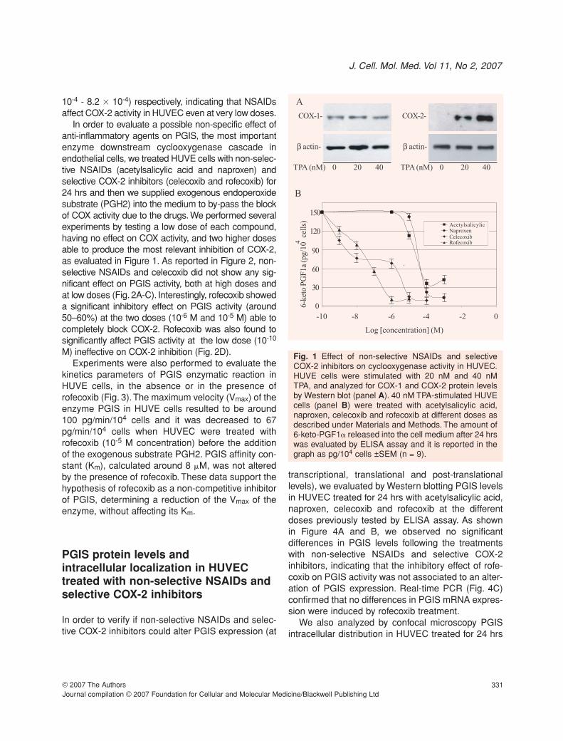
J. Cell. Mol. Med. Vol 11, No 2, 2007
331© 2007 The AuthorsJournal compilation © 2007 Foundation for Cellular and Molecular Medicine/Blackwell Publishing Ltd
10-4 - 8.2 � 10-4) respectively, indicating that NSAIDsaffect COX-2 activity in HUVEC even at very low doses.
In order to evaluate a possible non-specific effect ofanti-inflammatory agents on PGIS, the most importantenzyme downstream cyclooxygenase cascade inendothelial cells, we treated HUVE cells with non-selec-tive NSAIDs (acetylsalicylic acid and naproxen) andselective COX-2 inhibitors (celecoxib and rofecoxib) for24 hrs and then we supplied exogenous endoperoxidesubstrate (PGH2) into the medium to by-pass the blockof COX activity due to the drugs. We performed severalexperiments by testing a low dose of each compound,having no effect on COX activity, and two higher dosesable to produce the most relevant inhibition of COX-2,as evaluated in Figure 1. As reported in Figure 2, non-selective NSAIDs and celecoxib did not show any sig-nificant effect on PGIS activity, both at high doses andat low doses (Fig. 2A-C). Interestingly, rofecoxib showeda significant inhibitory effect on PGIS activity (around50–60%) at the two doses (10-6 M and 10-5 M) able tocompletely block COX-2. Rofecoxib was also found tosignificantly affect PGIS activity at the low dose (10-10
M) ineffective on COX-2 inhibition (Fig. 2D).Experiments were also performed to evaluate the
kinetics parameters of PGIS enzymatic reaction inHUVE cells, in the absence or in the presence ofrofecoxib (Fig. 3).The maximum velocity (Vmax) of theenzyme PGIS in HUVE cells resulted to be around100 pg/min/104 cells and it was decreased to 67pg/min/104 cells when HUVEC were treated withrofecoxib (10-5 M concentration) before the additionof the exogenous substrate PGH2. PGIS affinity con-stant (Km), calculated around 8 �M, was not alteredby the presence of rofecoxib. These data support thehypothesis of rofecoxib as a non-competitive inhibitorof PGIS, determining a reduction of the Vmax of theenzyme, without affecting its Km.
PGIS protein levels and intracellular localization in HUVECtreated with non-selective NSAIDs andselective COX-2 inhibitors
In order to verify if non-selective NSAIDs and selec-tive COX-2 inhibitors could alter PGIS expression (at
transcriptional, translational and post-translationallevels), we evaluated by Western blotting PGIS levelsin HUVEC treated for 24 hrs with acetylsalicylic acid,naproxen, celecoxib and rofecoxib at the differentdoses previously tested by ELISA assay. As shown in Figure 4A and B, we observed no significant differences in PGIS levels following the treatmentswith non-selective NSAIDs and selective COX-2inhibitors, indicating that the inhibitory effect of rofe-coxib on PGIS activity was not associated to an alter-ation of PGIS expression. Real-time PCR (Fig. 4C)confirmed that no differences in PGIS mRNA expres-sion were induced by rofecoxib treatment.
We also analyzed by confocal microscopy PGISintracellular distribution in HUVEC treated for 24 hrs
,
0
-10 -8 -6 -4 -2 0
Log [concentration] (M)
B
A
COX-1- COX-2-
β actin- β actin-
TPA (nM) 0 20 40 TPA (nM) 0 20 40
30
60
90
120
150
6-k
eto P
GF
1a (
pg/1
0
cell
s)4
AcetylsalicylicNaproxenCelecoxibRofecoxib
Fig. 1 Effect of non-selective NSAIDs and selectiveCOX-2 inhibitors on cyclooxygenase activity in HUVEC.HUVE cells were stimulated with 20 nM and 40 nM TPA, and analyzed for COX-1 and COX-2 protein levelsby Western blot (panel A). 40 nM TPA-stimulated HUVEcells (panel B) were treated with acetylsalicylic acid,naproxen, celecoxib and rofecoxib at different doses asdescribed under Materials and Methods. The amount of6-keto-PGF1� released into the cell medium after 24 hrswas evaluated by ELISA assay and it is reported in thegraph as pg/104 cells ±SEM (n = 9).

332 © 2007 The AuthorsJournal compilation © 2007 Foundation for Cellular and Molecular Medicine/Blackwell Publishing Ltd
with acetylsalicylic acid, naproxen, celecoxib androfecoxib at the higher doses previously tested byELISA assay and Western blotting (Fig. 5). All theinhibitors tested, and in particular rofecoxib, did notcause evident changes of PGIS intracellular distribu-tion with respect to control cells (compare panelsE–H to panels A–D). In particular, the colocalizationof PGIS with caveolin-1 (Cav-1), which is relevant forits activity [18], was not altered following the treat-ment with rofecoxib. This result indicates that theinhibition of PGIS activity caused by rofecoxib is notrelated to a misallocation of the enzyme leading toan impairment of its function.
Fig. 2 Effect of non-selective NSAIDs and selectiveCOX-2 inhibitors on PGIS activity in HUVEC. HUVEcells were treated with acetylsalicylic acid (panel A),naproxen (panel B), celecoxib (panel C) and rofecoxib(panel D) at different doses as described underMaterials and Methods. After 24 hrs, the medium waschanged and PGH2 (1 �M) was added for 10 min. Theamount of 6-keto-PGF1� released into the cell mediumwas evaluated by ELISA assay and it is reported in thegraphs, normalised for the cell number, as percentageof control (dose 0 of the drug). Values represents the mean ± S.E.M. of three independent experimentsperformed under the same conditions (n = 12). * = P <0.05; ** = P < 0.01.
-1.0 -0.2 0.6 1.4 2.2 3.0 3.8
[PGH ]2-1
[6-k
eto
PG
F1
a (p
g/1
0
cel
ls)/
min
]-14
Rofecoxib
Untreated
0.10
0.30
0.40
0.50
0.20
Fig. 3 Enzymatic properties of PGIS in HUVEC, in theabsence or in the presence of rofecoxib. HUVE cellswere untreated or treated with rofecoxib at 10-5 M con-centration, as described under Materials andMethods. After 24 hrs, the medium was changed andthe substrate PGH2 was added at different concentra-tions (ranging from 0.25 �M to 5 �M). The enzymereaction was allowed to proceed for 10 min. Theamount of 6-keto-PGF1� released into the cell medium was evaluated by ELISA assay. Data are represented by a Lineweaver-Burk plot as mean of three independent experiments ± SEM, (n = 12).The slope of the line, analyzed with SPSS linearregression programme, was 0.08 in the absence ofrofecoxib and increased to 0.12 in the presence ofrofecoxib at 10-5 M.

J. Cell. Mol. Med. Vol 11, No 2, 2007
333© 2007 The AuthorsJournal compilation © 2007 Foundation for Cellular and Molecular Medicine/Blackwell Publishing Ltd
PGIS activity in bovine aortic microsomes treated with non-selectiveNSAIDs and selective COX-2 inhibitors
Since the purification of functionally intact PGISresults to be very difficult [21], we further investigat-ed the effect of rofecoxib, compared to that of non-selective NSAIDs and celecoxib, by using bovineaortic microsomal (BAMs) fractions enriched inPGIS. In BAMs, COX-2 and COX-1 proteins were notdetectable by Western blotting analysis (data notshown). We first observed that the inhibitory effect ofrofecoxib on PGIS in BAMs was nearly absent if incu-bation was performed for 10–15 min before the addi-tion of the substrate PGH2, but it significantlyincreased if microsomes were incubated in the pres-ence of the drug for 1 hr at 37°C before the additionof the substrate. Under these conditions a 10-4 M
concentration of rofecoxib caused a 50% inhibition ofPGIS activity, while celecoxib as well as non-selec-tive NSAIDs at the same concentrations showed nosignificant effects (Fig. 6). Moreover, we did notobserve any significant inhibitory effect for rofecoxibat lower doses (i.e. 10–5 M), which were effective onPGIS activity in HUVE cells. We hypothesized thatrofecoxib needs to be chemically modified in order toinhibit PGIS activity. Thus, the weaker inhibitoryeffect of rofecoxib in BAMs could be due to the lackof cellular co-factors (like NADPH or GSH), relevantfor the activity of enzymatic complexes able tometabolize and modify drugs (i.e. Cytochrome P450or Glutathione-S-transferase). However, followingaddition of NADPH or GSH to the microsomal prepa-rations, we failed to detect an increase in the inhibito-ry effect of rofecoxib (data not shown).
Taking together, results reported here provide evi-dence that rofecoxib, but not celecoxib as well as clas-
Fig. 4 Effect of non-selective NSAIDs and selectiveCOX-2 inhibitors on PGIS protein levels and mRNAexpression in HUVEC. HUVE cells were treated withacetylsalicylic acid, naproxen, celecoxib and rofecoxibat different doses as described under Materials andMethods (A and B). After 24 hrs, cell lysates were pre-pared and analyzed by immunoblotting to detect PGISprotein levels. Lane 1: PGIS standard; lanes 2–4: sam-ples treated with acetylsalicylic acid at different doses(10-3 M, 10-4 M, 10-6 M); lanes 5–7: samples treatedwith naproxen at different doses (10-3 M, 10-4 M, 10-6 M);lanes 8–10: samples treated with celecoxib at differentdoses (10-4 M, 10-5 M, 10-10 M); lanes 11–13: samplestreated with rofecoxib at different doses (10-5 M, 10-6 M,10-10 M); lane 14: control (untreated sample). For eachsample the intensity of the band corresponding to PGIS(panel A) was normalized with respect to that corre-sponding to �-actin and results are represented inpanel B as percentage of control. PGIS and COX-2mRNA levels were analysed by real-time PCR (panelC) in HUVE cells after 24 hrs of incubation in theabsence (lanes 1–2) or in the presence (lanes 3–4) of10-5M rofecoxib. No statistically significant differencesin PGIS mRNA expression were detectable after rofe-coxib treatment. COX-2 mRNA expression, before andafter rofecoxib treatment, is also shown. Bars representvalues normalized against GUSB mRNA levels. Valuesare the mean ± S.E.M. of three independent experi-ments (n = 9) performed under the same experimentalconditions.

334 © 2007 The AuthorsJournal compilation © 2007 Foundation for Cellular and Molecular Medicine/Blackwell Publishing Ltd
sical anti-inflammatory compounds (acetylsalicylicacid and naproxen), strongly inhibits PGIS activity inhuman endothelial cells. This inhibition is not due to amodification of the enzyme expression or to a modifi-cation of its subcellular localization, but it is likely dueto an interaction of rofecoxib with PGIS, probably act-ing as a non-competitive inhibitor of the enzyme.
Discussion
Selective inhibitors of COX-2 (coxibs) have been devel-oped starting from 1990. Preclinical and clinical studiesrevealed the good efficacy of celecoxib and rofecoxib ininflammation, fever and pain. Furthermore, they con-firmed that selective COX-2 inhibitors have greatergastrointestinal safety and do not affect platelet aggre-gation. Following these results, celecoxib and rofecox-ib were rapidly licensed by the Food and Drug
Administration (FDA) in 1999 for the treatment ofosteoarthritis and rheumatoid arthritis [35].
In addition to the expected complications in the renalsystem, due to the relevance of COX-2 activity in themacula densa of the kidney [3], severe effects of COX-2 selective inhibitors on the cardiovascular systemhave emerged during their clinical trials. The main clin-ical trial designed to rigorously assess the gastroin-testinal (GI) safety of Vioxx (VIGOR study) has beenpublished in 2000 and demonstrated that a suprather-apeutic dose of rofecoxib was associated with a signif-icantly reduced risk of clinical upper GI events com-pared with naproxen. This study also revealed, after 9months of clinical trial, a higher incidence of myocardialinfarction among patients in the rofecoxib group, pre-sented by the investigators as a cardioprotective effectof naproxen [30]. Following the results of theAdenomatous Polyp Prevention on Vioxx (APPROVe)trial, emerged in September 2004, Merck & Co. volun-tarily withdrew Vioxx from the market. The APPROVe
Cav-1 PGIS Merge Colocalization
Contr
ol
Ro
feco
xib
A B C D
E F G H
Fig. 5 Effect of non-selective NSAIDs and selective COX-2 inhibitors on PGIS intracellular distribution in HUVEC.HUVE cells, seeded on cover slides and treated with acetylsalicylic acid (10-3 M), naproxen (10-3 M), celecoxib (10-4 M) and rofecoxib (10-5 M) for 24 hrs, were stained with PGIS and Cav-1 antibodies and analyzed by confocalmicroscopy. The figure represents images regarding PGIS and Cav-1 distributions in control (untreated) cells (panelsA–D) and in rofecoxib-treated cells (panels E–H), because results were similar for all the treatments tested. Cav-1 dis-tribution (green signal) is represented in panels A and E while PGIS distribution (red signal) is represented in panelsB and F. The merging of the two signals (yellow) is reported in panels C and G. The co-localization maps, obtained asdescribed under Materials and Methods, are reported in panels D and H. Bar: 10 �m.

J. Cell. Mol. Med. Vol 11, No 2, 2007
335© 2007 The AuthorsJournal compilation © 2007 Foundation for Cellular and Molecular Medicine/Blackwell Publishing Ltd
study reported the cardiovascular outcomes of a long-term, randomized, placebo-controlled trial designed todetermine the effects of three years of treatment withrofecoxib on the risk of recurrent neoplastic polyps inpatients with a history of colorectal adenoma [31]. Thestudy revealed that, after 18 months of treatment, rofe-coxib doubled the risk of thrombotic events with respectto placebo (relative risk 1.92).
Following the rofecoxib history, several controver-sial studies examined the question about cardiovas-cular risk associated to selective COX-2 inhibitors incomparison with traditional NSAIDs. Recently,Graham and colleagues [36] analyzed the risk ofserious coronary heart disease during the treatmentwith rofecoxib at standard or high doses in compari-son with classical NSAIDs use or celecoxib use, ascelecoxib was the most common alternative to rofe-coxib. They reported that rofecoxib use involves ahigher risk of acute myocardial infarction and suddencardiac death if compared with celecoxib use.Furthermore, following the analysis of clinical trialsand in particular the CLASS study (Celecoxib LongTerm Arthritis Safety Study), celecoxib has been con-sidered much less prone to affect the cardiovascularsystem and, to this date, it is still marketed [37–39].The safety of celecoxib has not been confirmed bySolomon and collaborators [32], who found anincreased cardiovascular risk associated with cele-coxib treatment in two long-term clinical trials for theprevention of colorectal adenomas.
Predisposition of patients treated with coxibs tomyocardial infarction has been explained by animbalance between prostacyclin (PGI2) and throm-boxane A2 (TxA2), due to the inhibition of COX-2dependent PGI2 production by endothelial cells,without any effect on TxA2 produced by plateletCOX-1 [40]. This imbalance should decrease the pro-tective effect of PGI2 within the vasculature, poten-tially creating a prothrombotic state [41, 42].
Nevertheless, the absence of a clear relationshipbetween COX-2 selectivity and the observed cardio-vascular side effects suggests that other mecha-nisms could be involved in the cardiovascular sideeffects outcome. This hypothesis is in some measurecorroborated by the study of Farkouh and collabora-tors [43] who showed that lumiracoxib, a very selec-tive coxib, seems not to increase cardiovascular out-comes after one year of follow up.
These considerations prompted us to hypothesizethat rofecoxib cardiotoxicity could be explained byassuming that other molecular mechanisms, inde-pendent from COX-2 inhibition, should be involved.Data reported here strongly support the hypothesisthat an inhibition of prostacyclin synthase (PGIS)activity induced by rofecoxib is implicated in its car-diovascular toxicity. In fact, we observed a significantinhibition of PGIS activity in human endothelial cellsexposed to rofecoxib, even at a dose which resultedineffective on COX-2 activity. It is important to under-line that in this study we used lower NSAIDs concen-trations respect to those recorded during pharmaco-kinetic experiments in humans. In particular, the con-centration of rofecoxib in human plasma, in patientswho received 25 mg in single dose, ranged from 0.05to 1 �M, while for celecoxib, after a 200 mg single
Fig. 6 Effect of non-selective NSAIDs and selectiveCOX-2 inhibitors on PGIS activity in bovine aortic micro-somes (BAMs). 2 �g of bovine aortic microsomal frac-tions enriched in PGIS were incubated with acetylsali-cylic acid, naproxen, celecoxib and rofecoxib at differentdoses for 1 h at 37°C as described under Materials andMethods. After addition of the substrate PGH2 (1 �M),the reaction products were extracted, 6-keto-PGF1�production was evaluated by ELISA assay and it isreported in the graph as percentage of control (repre-senting PGIS activity in BAMs in the absence of anytreatment). Values are the mean ± S.E.M. of three inde-pendent experiments (n = 12) performed under thesame experimental conditions. * = P < 0.01.

336 © 2007 The AuthorsJournal compilation © 2007 Foundation for Cellular and Molecular Medicine/Blackwell Publishing Ltd
dose, plasma concentrations ranged from 0.1 to 2.5�M [44]. Our data demonstrate that this PGIS inhibi-tion could be due to a direct interaction of rofecoxibwith the enzyme, affecting its activity, probably by anon-competitive mechanism. This interaction couldinvolve the heme distal pocket of PGIS, which isknown to be fairly adaptable to ligands of variousstructures, like the PGIS inhibitors clotrimazole andtranylcypromine previously described [45, 46]. Thebinding of these inhibitors to PGIS requires ahydrophobic interaction within the enzyme pocket,which may occur for rofecoxib too, explaining thelong incubation time necessary to achieve an inhibi-tion during experiments performed on PGIS-enriched microsomal fractions.
A compensatory mechanism increasing COX-1levels following COX-2 inhibition has been proposed[2]. If it is true, it is very likely that enough PGI2 is stillproduced to protect the cardiovascular system whenendothelial cells are exposed to COX-2 selective inhi-bition and PGIS remains fully active. Thus, it is plau-sible that PGI2 synthesis, in the presence of rofecox-ib and especially when it is chronically assumed,becomes inadequate to protect the cardiovascularsystem.This PGIS inhibition may explain why rofecox-ib-induced cardiovascular side effects raised earlierduring the treatment with respect to those observedduring celecoxib assumption [32, 37–39].
Even if rofecoxib seems to be the unique anti-inflammatory drug capable of PGIS inhibition so fardiscovered, we can not exclude that other selectiveCOX-2 inhibitors such as valdecoxib, taken off fromthe market due to the increased risk of cardiovascularevents, may inhibit PGIS. For this reason, we stronglyrecommend that in future novel coxibs should be test-ed in vitro and in vivo as possible PGIS inhibitors.
Acknowledgments
We thank Dr Maria Cristina Bellucci (Dipartimento di ScienzeMolecolari Agroalimentari, University of Milano, Italy) for NMRspectroscopic analysis. The present work was supported bygrants from MIUR (PRIN 2004 and FIRB 2003) to VT.
References
1. Smith WL, DeWitt DL, Garavito RM.Cyclooxygenases: structural, cellular, and molecularbiology. Annu Rev Biochem. 2000; 69: 145–82.
2. Wang H, Ma WG, Tejada L, Zhang H, Morrow JD,Das SK, Dey SK. Rescue of female infertility from theloss of cyclooxygenase-2 by compensatory up-regula-tion of cyclooxygenase-1 is a function of genetic make-up. J Biol Chem. 2004; 279: 10649–58.
3. Nasrallah R, Hébert RL. Prostacyclin signaling in thekidney: implication for health and disease. Am JPhysiol Renal Physiol. 2005; 289: 235–46.
4. Prescott SM, Fitzpatrick FA. Cyclooxygenase-2 andcarcinogenesis. Biochim Biophys Acta. 2000; 1470:69–78.
5. Brown JR, DuBois RN. COX-2: a molecular target forcolorectal cancer prevention. J Clin Oncol. 2005; 23:2840–55.
6. Bunting S, Gryglewski R, Moncada S, Vane JR.Arterial walls generate from prostaglandin endoperox-ides a substance (prostaglandin X) which relaxesstrips of mesenteric and coeliac ateries and inhibitsplatelet aggregation. Prostaglandins. 1976; 12:897–913.
7. Moncada S, Gryglewski R, Bunting S, Vane JR. Anenzyme isolated from arteries transformsprostaglandin endoperoxides to an unstable sub-stance that inhibits platelet aggregation. Nature. 1976;263: 663–5.
8. Weksler BB, Marcus AJ, Jaffe EA. Synthesis ofprostaglandin I2 (prostacyclin) by cultured human andbovine endothelial cells. Proc Natl Acad Sci USA.1977; 74: 3922–6.
9. Tomasi V, Meringolo C, Bartolini G, Orlandi M.Biosynthesis of prostacyclin in rat liver endothelialcells and its control by prostaglandin E2. Nature. 1978;237: 670–1.
10. Tomasi V, Meringolo C, Bartolini G, Orlandi M.Effect of prostaglandin E2 on prostacyclin productionby endothelial cells. Nature. 1979; 274: 48–9.
11. Negishi M, Sugimoto, Ichikawa A. Molecular mecha-nisms of diverse actions of prostanoid receptors.Biochim Biophys Acta. 1995; 1259: 109–19.
12. Forman BM, Chen J, Evans RM. Hypolipidemicdrugs, polyunsatured fatty acids and eicosanoids areligands for peroxisome proliferator-activated receptors� and �. Proc Natl Acad Sci USA.1997; 94: 4312–7.
13. Geraci MW, Gao B, Shepherd DC, Moore MD,Westcott JY, Fagan KA, Alger LA,Tuder RM,VoelkelNF. Pulmonary prostacyclin synthase overexpressionin transgenic mice protects against development ofhypoxic pulmonary hypertension. J Clin Invest. 1999;103: 1509–15.
14. Iwai N, Katsuya T, Ishikawa K, Mannami T, Ogata J,Higaki J, Ogihara T, Tanabe T, Baba S. Humanprostacyclin synthase gene and hypertension: TheSuita Study. Circulation. 1999; 100: 2231–6.
15. Lim H, Gupta RA, Ma W, Paria BC, Moller DE,Morrow JD, DuBois RN, Trzaskos JM, Dey SK.

J. Cell. Mol. Med. Vol 11, No 2, 2007
337© 2007 The AuthorsJournal compilation © 2007 Foundation for Cellular and Molecular Medicine/Blackwell Publishing Ltd
Cyclo-oxygenase-2-derived prostacyclin mediatesembryo implantation in the mouse via PPAR �. GenesDev. 1999; 13: 1561–74.
16. Spisni E, Tomasi V. Involvement of prostanoids inangiogenesis. In: Bicknell R, Lewis CE, Ferrara N, edi-tors. Tumour angiogenesis. Oxford University Press;1997. p. 291–300.
17. Tomasi V, Spisni E, Griffoni C, Guarnieri T. Caveolae,caveolar enzymes and angiogenesis. Current Topics inBiochemical Research. 2000; 3: 81–90.
18. Spisni E, Griffoni C, Santi S, Riccio M, Marulli R,Bartolini G, Toni M, Ullrich V, Tomasi V.Colocalization prostacyclin (PGI2) synthase- caveolin-1 in endothelial cells and new roles for PGI2 in angio-genesis. Exp Cell Res. 2001; 266: 31–43.
19. Massimino ML, Griffoni C, Spisni E,Toni M,Tomasi V.Involvement of caveolae and caveolae-like domains insignalling cell survival and angiogenesis. Cell Signall.2002; 14: 93–8.
20. Hao CM, Redha R, Morrow J, Breyer MD. Peroxisomeproliferator-activated receptor delta activation pro-motes cell survival following hypertonic stress. J BiolChem. 2002; 277: 21341–5.
21. Wu KK, Liou J-Y. Cellular and molecular biology ofprostacyclin synthase. Biochem Biophys ResCommun. 2005; 338: 45–52.
22. Spisni E, Bartolini G, Orlandi M, Belletti B, Santi S,Tomasi V. Prostacyclin (PGI2) synthase is a constitu-tively expressed enzyme in human endothelial cells.Exp Cell Res. 1995; 219: 507–13.
23. Liou JY, Deng WG, Gilroy DW, Shyue SK, Wu KK.Co-localization and interaction of cyclooxygenase-2with caveolin-1 in human fibroblasts. J Biol Chem.2001; 276: 34975–82.
24. Kim SF, Huri DA, Snyder SH. Inducible nitric oxidesynthase binds, s- nitrosylates, and activates cyclooxy-genase-2. Science. 2005; 310: 1966–70.
25. Gibson LL, Hahner L, Osborne-Lawrence S, GermanZ,Wu KK, Chambliss KL, Shaul PW. Molecular basis ofestrogen-induced cyclooxygenase type 1 upregulated inendothelial cells. Circulation Res. 2005; 96: 518–25.
26. Doré M, Chevalier S, Sirois J. Estrogen-dependentinduction of cyclooxygenase-2 in the canine prostatein vivo. Vet Pathol. 2005; 42: 100–3.
27. FitzGerald GA, Patrono C. The coxibs, selectiveinhibitors of cyclooxygenase-2. N Engl J Med. 2001;345: 433–42.
28. Hankey GJ, Eikelboom JW. Aspirin resistance.Lancet. 2006; 367: 606–17.
29. Warner TD, Giuliano F, Vojnovic I, Bukasa A,Mitchell JA, Vane JR. Nonsteroid drug selectivities forcyclo-oxygenase-1 rather than cyclo-oxygenase-2 are
associated with human gastrointestinal toxicity: a full invitro analysis. Proc Natl Acad Sci USA. 1999; 96:7563–8.
30. Bombardier C, Laine L, Reicin A, Shapiro D, Burgos-Vargas R, Davis B, Day R, Ferraz MB, Hawkey CJ,Hochberg MC, Kvien TK, Schnitzer TJ, VIGORStudy Group. Comparison of upper gastrointestinaltoxicity of rofecoxib and naproxen in patients withrheumatoid arthritis. VIGOR Study Group. N Engl JMed. 2000; 343: 1520–8.
31. Bresalier RS, Sandler RS, Quan H, Bolognese JA,Oxenius B, Horgan K, Lines C, Riddell R, Morton D,Lanas A, Konstam MA, Baron JA. AdenomatousPolyp Prevention on Vioxx (APPROVe) TrialInvestigators. Cardiovascular events associated withrofecoxib in a colorectal adenoma chemopreventiontrial. N Engl J Med. 2005; 352: 1092–102.
32. Solomon SD, Pfeffer MA, McMurray JJ, Fowler R,Finn P, Levin B, Eagle C, Hawk E, Lechuga M,Zauber AG, Bertagnolli MM, Arber N, Wittes J. APCand PreSAP Trial Investigators. Effect of celecoxib oncardiovascular events and blood pressure in two trialsfor the prevention of colorectal adenomas. Circulation.2006; 114: 1028–35.
33. Jamali F, Sattari S. High performance liquid chro-matografic determination of cyclooxygenase II inhibitorrofecoxib in rat and human plasma. J Pharm PharmSci. 2000; 3: 312–7.
34. Salmon JA, Flower RJ. Preparation and assay ofprostacyclin synthase. Methods Enzymol. 1982; 86:91–9.
35. Flower RJ. The development of COX2 inhibitors. NatRev Drug Discov. 2003; 2: 179–91.
36. Graham DJ, Campen D, Hui R, Spence M, CheethamC, Levy G, Shoor S, Ray WA. Risk of acute myocardialinfarction and sudden cardiac death in patients treatedwith cyclo-oxygenase 2 selective and non selectivenon-steroidal anti-inflammatory drugs: nested case-control study. Lancet. 2005; 365: 475–81.
37. Silverstein FE, Faich G, Goldstein JL, Simon LS,Pincus T, Whelton A, Makuch R, Eisen G, AgrawalNM, Stenson WF, Burr AM, Zhao WW, Kent JD,Lefkowith JB,Verburg KM, Geis GS. Gastrointestinaltoxicity with celecoxib vs nonsteroidal anti-inflammato-ry drugs for osteoarthritis and rheumatoid arthritis: theCLASS study: A randomized controlled trial. CelecoxibLong-term Arthritis Safety Study. JAMA. 2000; 284:1247–55.
38. Brophy JM. Celecoxib and cardiovascular risks.Expert Opin Drug Saf. 2005; 4: 1005–15.
39. King VL, Trivedi D, Gitlin JM, Loftin CD. Selectivecyclo-oxygenase-2 inhibition with celecoxib decreases

angiotensin II-induced abdominal aortic aneurysm for-mation in mice. Arterioscler Thromb Vasc Biol. 2006;26: 1137–43.
40. FitzGerald GA. Coxib and cardiovascular disease. NEngl J Med. 2004; 351: 1709–11.
41. Bishop-Bailey D, Mitchell JA, Warner TD. COX-2 incardiovascular disease. Arterioscler Thromb Vasc Biol.2006; 26: 956–8.
42. Klumpp G, Schildknecht S, Nastainczyk W, UllrichV, Bachschmid M. Prostacyclin in the cardiovascularsystem: new aspects and open questions. PharmacolRep. 2005; 57: 120–6.
43. Farkouh ME, Kirshner H, Harrington RA, Ruland S,Verheugt FW, Schnitzer TJ, Burmester GR, MyslerE, Hochberg MC, Doherty M, Ehrsam E, Gitton X,Krammer G, Mellein B, Gimona A, Matchaba P,Hawkey CJ, Chesebro JH. TARGET Study Group.
Comparison of lumiracoxib with naproxen and ibupro-fen in the Therapeutic Arthritis Research andGastrointestinal Event Trial (TARGET), cardiovascularoutcomes: randomised controlled trial. Lancet. 2004;364: 675–84.
44. Hamama AK, Ray J, Day RO, Brien JA. SimultaneousDetermination of rofecoxib and celecoxib in humanplasma by high-performance liquid chromatography. JChromatogr Sci. 2005; 43: 351–4.
45. Yeh HC, Hsu PY, Wang JS, Tsai AL, Wang LH.Characterization of heme environment and mecha-nism of peroxide bond cleavage in human prostacyclinsynthase. Biochim Biophys Acta. 2005; 1738: 121–32.
46. Wada M, Yokoyama C, Hatae T, Shimonishi M,Nakamura M, Imai Y, Ullrich V, Tanabe T. Purificationand characterization of recombinant human prostacy-clin synthase. J Biochem. 2004; 135: 455–63.
© 2007 The AuthorsJournal compilation © 2007 Foundation for Cellular and Molecular Medicine/Blackwell Publishing Ltd
338