Ras regulates neuronal polarity via the PI3-kinase/Akt/GSK-3β/CRMP2 pathway

From a Natural Product Lead to the Identification of Potent andSelective Benzofuran-3-yl-(indol-3-yl)maleimides as GlycogenSynthase Kinase 3β Inhibitors that Suppress Proliferation andSurvival of Pancreatic Cancer Cells
Irina N. Gaisina†, Franck Gallier†, Andrei V. Ougolkov‡, Ki H. Kim†, Toru Kurome†, SongpoGuo†, Denise Holzle, Doris N. Luchini‡, Sylvie Y. Blond, Daniel D. Billadeau*,‡, and Alan P.Kozikowski*,‡Drug Discovery Program, College of Pharmacy, Department of Medicinal Chemistry andPharmacognosy, University of Illinois at Chicago, 833 South Wood Street, Chicago, Illinois 60612;Department of Medicinal Chemistry and Pharmacognosy and the Center for PharmaceuticalBiotechnology, University of Illinois at Chicago, 900 South Ashland, Chicago, IL 60607 and Collegeof Medicine, Department of Immunology and Division of Oncology Research, Mayo Clinic 13-42Guggenheim, 200 First Street SW, Rochester, MN 55905
AbstractRecent studies have demonstrated that Glycogen Synthase Kinase 3β (GSK-3β) is overexpressed inhuman colon and pancreatic carcinomas contributing to cancer cell proliferation and survival. Here,we report the design, synthesis, and biological evaluation of benzofuran-3-yl-(indol-3-yl)maleimides, potent GSK-3β inhibitors. Some of these compounds show picomolar inhibitory activitytoward GSK-3β and an enhanced selectivity against Cyclin-dependent Kinase 2 (CDK-2). SelectedGSK-3β inhibitors were tested in the pancreatic cancer cell lines MiaPaCa-2, BXPC-3, and HupT3.We determined that some of these compounds, namely compounds 5, 6, 11, 20 and 26, demonstrateantiproliferative activity against some or all of the pancreatic cancer cells at low micromolar tonanomolar concentrations. We found that the treatment of pancreatic cancer cells with GSK-3βinhibitors 5 and 26 resulted in suppression of GSK-3β activity and a distinct decrease of the X-linkedInhibitor of Apoptosis (XIAP) expression leading to significant apoptosis. The present data suggesta possible role for GSK-3β inhibitors in cancer therapy, in addition to their more prominentapplications in CNS disorders.
IntroductionGlycogen Synthase Kinase 3 (GSK-3a) was identified in the late 1970s and originally foundto regulate glycogen metabolism.1 Later, this enzyme has attracted immense interest due to its
*To whom correspondence should be addressed.. † Phone: +1 312 9967577. Fax: +1 312 9967107. E-mail: [email protected].. ‡ Phone:+1 507 2664334. Fax: +1 507 2665146. E-mail: [email protected] Information Available: Molecular modeling methods, experimental procedures for some intermediates, 13C NMR data,HPLC purity data for all final compounds, and selectivity data for compounds 5, 7, 11, 15, and 33. These materials are available free ofcharge via the Internet at http://pubs.acs.org.aAbbreviations : APC, adenomatous polyposis coli; CDK-2, Cyclin-dependent Kinase 2; CDKs, Cyclin-dependent Kinases; DMF-DMA,N,N-dimethylformamide dimethyl acetal; DMF-DIPA, N,N-dimethylformamide di-iso-propyl acetal; FRAT, frequently rearranged inadvanced T-cell lymphoma; GBP, GSK-3-binding protein; GS, Glycogen Synthase; GSK-3, Glycogen Synthase Kinase 3; GSK-3β,Glycogen Synthase Kinase 3 isoform type β; NFκB, nuclear factor kappa B; pGS : phosphorylated Glycogen Synthase; P-gp, permeability-glycoprotein; PKC, Protein Kinase C; TPSA, total polar surface area; XIAP, X-linked Inhibitor of Apoptosis; ZDF rat, Zucker DiabeticFatty rat.
NIH Public AccessAuthor ManuscriptJ Med Chem. Author manuscript; available in PMC 2010 April 9.
Published in final edited form as:J Med Chem. 2009 April 9; 52(7): 1853–1863. doi:10.1021/jm801317h.
NIH
-PA Author Manuscript
NIH
-PA Author Manuscript
NIH
-PA Author Manuscript

diverse roles in cellular events. It is well established that GSK-3 affects a variety of biologicalprocesses such as cell cycle progression, proliferation, apoptosis, signaling, and transcriptionby phosphorylation of many different substrates. In mammals, GSK-3 consists of two distinctisoforms, α and β,2 which are highly homologous within their ATP binding domain, whereasthey are significantly different in their terminal regions. This serine/threonine kinase isconstitutively active in resting cells and physiologically inhibited by distinct signalingpathways at multiple levels in stimulated cells. GSK-3β is negatively regulated, for example,by post-translational phosphorylation of Ser-9 located in the N-terminal domain.3 GSK-3β isalso phosphorylated constitutively at Tyr-216; this phosphorylation step appears to take placethrough autophosphorylation and plays a role in stabilizing the enzyme. In addition, there aredifferent extracellular stimuli and proteins that can regulate this enzyme. For example,GSK-3β is inhibited by the presence of secreted glycoproteins, the so-called Wnts, that functionin a pathway is crucial for the determination of the cell's fate during embryonic development.4 Wnt signaling targets a particular subcellular pool of GSK-3β, in which it is complexed toaxin, β-catenin, and the adenomatous polyposis coli (APC) proteins. This protein complex isdisrupted during Wnt signaling as a result of the displacement of axin by other proteins, suchas disheveled and FRAT (frequently rearranged during advanced T-cell lymphomas, also calledGBP (GSK-3-binding protein)). The resulting dephosphorylation of β-catenin leads to itsaccumulation and translocation to the nucleus where it forms a transcriptional complex withTcf and Lef family proteins subsequently stimulating activation of several oncogenes. In earlierpublications, it was indicated that GSK-3β plays the role of tumor suppressor by down-regulating various proto-oncoproteins.5-7 Consequently, GSK-3β inhibitors have beenconsidered to possibly mimic the Wnt signaling pathway and to be potentially oncogenic.3However, in spite of these concerns, it is well known that long-term use of lithium, a non-specific GSK-3β inhibitor, for the treatment of bipolar disorder is not associated with anincreased risk of cancer.8 Moreover, lithium actually increases survival rates of patients withadenocarcinomas.9 The administration of the GSK-3β inhibitor 6-{2-[4-(2,4-Dichloro-phenyl)-5-(4-methyl-1H-imidazol-2-yl)-pyrimidin-2-ylamino]-ethylamino}-nicotinonitrile(CHIR 99021)10 in Zucker Diabetic Fatty (ZDF) rats for up to 20 h was found not to cause anobservable increase in β-catenin or cyclin D1 mRNA in brain, liver, lung, adipose tissue, orcolon. Thus, it is possible that the inhibition of GSK-3β by itself may be unable to elevate β-catenin levels in primary cells; such elevations may require that the cell lines have alreadyundergone some prior transforming events such as APC protein truncation.11 Recently, a lineof evidence led to an indication that GSK-3β is pathologically active in different types ofgastrointestinal cancer.12,13 It was shown that this enzyme is overexpressed in colon andpancreatic cancer cells where it has been implicated in NFκB (nuclear factor kappa B)activation.14,15 In fact, GSK-3β is found to accumulate in the nucleus of cancer cells and hasrecently been shown to regulate chromatin structure and the binding of NFκB to its target genepromoters in chronic lymphocytic leukemia cells.16 Importantly, inhibition of GSK-3β activityin several cancer cell types results in diminished NFκB transcriptional activity of anti-apoptoticgenes resulting in apoptosis.15,16 Thus, GSK-3β has emerged as a promising target indevelopment of new drugs for the treatment of chronic and progressive diseases.3,17 Over thepast decades, a number of small molecule GSK-3 inhibitors have been designed from naturalproducts or known kinase inhibitors17 for diabetes and neurodegenerative disorders, but onlya few of these were tested in cancer cell lines. In the present work we have directed our attentionto the possibility to use inhibitors of GSK-3β in the treatment of pancreatic cancer. Our workin this area was influenced by the maleimide-bearing natural product staurosporine firstidentified as protein kinase C (PKC) inhibitor, although now known to be able to target otherprotein kinases, including GSK-3β. From our original screening studies of staurosporine andits analogs, we were led to the design of benzofuran-3-yl-(indol-3-yl)maleimides as GSK-3βinhibitors.18 In continuation of this work, and to gain better insights into the SAR of thisbenzofuran containing scaffold, a small library of substituted maleimides has been generated.Using structure-based design methods, we have optimized our lead compounds so as to arrive
Gaisina et al. Page 2
J Med Chem. Author manuscript; available in PMC 2010 April 9.
NIH
-PA Author Manuscript
NIH
-PA Author Manuscript
NIH
-PA Author Manuscript

at subnanomolar potency GSK-3β inhibitors that are relatively selective for GSK-3β versushomologous kinases.
Results and DiscussionChemical Synthesis
The synthesis of the benzofuran-3-yl-(indol-3-yl)maleimides (1-38) (Table 1) isstraightforward and based on the condensation of the appropriately substituted 3-indolylglyoxylic acid esters and benzofuranyl-3-acetamides. The general method is shown inScheme 1 and follows our previously published work.18 Preparation of indolyl-basedglyoxalates commences with N-alkylation of the indole 39 followed by acylation of theresulting indole 40 with ethyl oxalyl chloride to afford the precursor 41. The requiredbenzofuranyl-3-acetamides 44 were prepared from the appropriately substituted 3-benzofuranones 42. A Horner-Emmons reaction of 42 with (carbethoxymethylene)triphenylphosphorane afforded ethyl (1-benzofuran-3-yl)acetates 43, which was subsequentlyconverted into the acetamides 44. Condensation of the acetamides 44 with the glyoxilic estersresulted in formation of maleimide core.
As we have already synthesized a small set of compounds substituted in the 5-position,18 itwas important to explore the extent to which the introduction of different functionalities,including H-bond donor and acceptor groups to the 6- or 7-position of the indole or benzofuranring, could be used to adjust kinase selectivity and potency. Based upon the aboveconsiderations, we investigated the effects of adding a hydroxy, alkoxy, hydroxymethyl,methoxymethyl and a carboxylic acid group. Accordingly, commercially available 6-hydroxybenzofuran-3-one 45 was protected as its silyl ether 46 (Scheme 2). Treatment of the46 with (carbethoxymethylene)triphenylphosphorane and subsequent amidation of theintermediate ester provided the acetamide 47. The alcohol functionality was protected as itstert-butyldiphenylsilyl ether (compound 48), or alkylated with different alkyl and aryl halides(compounds 49-54). Finally, condensation with the appropriate indolylglyoxylic acid estersfollowed by deprotection of the silyl ether if needed gave a variety of 6-hydroxy substitutedmaleimides (4, 9, 10, 16-18) as shown in Table 1.
For the introduction of hydroxymethyl and methoxymethyl functionality at the 6-position ofthe benzofuran moiety, the 3-methoxybenzyl alcohol 55 was converted into the acetate 56(Scheme 3). Chloroacetylation of 56 and subsequent cyclization of the corresponding arylketone 57 yielded the 6-substituted 3-benzofuranone 58. The general procedure was followedto obtain the 6-hydroxymethylbenzofuranyl-3-acetamide 59. The acetamide 59 was convertedin turn to the methyl ether 60, or silyl ether 61 and then to the maleimides 2, 3, 7, 15, 23, 32and 34 (Table 1).
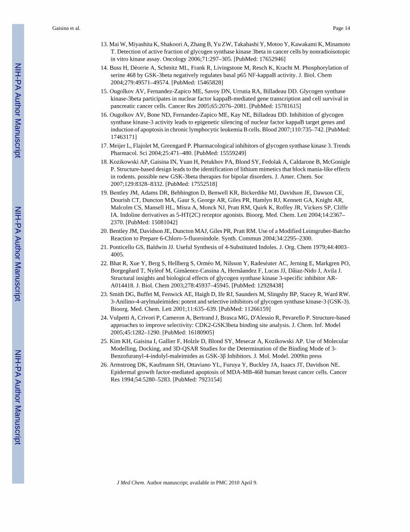
Next, our effort was directed to the introduction of hydroxymethyl, methoxymethyl, andcarboxylic acid group at the 7-position of the indole moiety (Scheme 4). Accordingly, the(1H-indol-7-yl)methanol 62 was used to synthesize glyoxalates 63 and 64. The generalprocedure to form the maleimide was followed to obtain the desired compound 31 and 33. Theoxidation of the alcohol 33 led to the corresponding aldehyde 65, which was then subjected toa Horner-Emmons reaction to give the ester 66 (Scheme 5). Treatment with nickel boride,which was generated in situ, resulted in the selective reduction of the double bond andformation of the ester 35. The latter was converted into the acid 36 by the treatment with sodiumhydroxide.
The SAR work was further extended by exploring the effect of poly-substitution of the aromaticrings. Disubstituted indoles were prepared from the corresponding dihalogenated nitrotoluenes67 and 68 by using a known protocol (Scheme 5).19 The reaction of 67 and 68 with N,N-
Gaisina et al. Page 3
J Med Chem. Author manuscript; available in PMC 2010 April 9.
NIH
-PA Author Manuscript
NIH
-PA Author Manuscript
NIH
-PA Author Manuscript

dimethylformamide dimethyl acetal (DMF-DMA) afforded a mixture of the enamine 69 withthe methoxy-substituted side-product 70. In this step, nucleophilic aromatic substitution canbe avoided by replacement of DMFDMA with N,N-dimethylformamide di-iso-propyl acetal(DMF-DIPA).20 A reductive cyclization provided the indoles 71 and 72.21 A variety ofacetamides prepared from commercially available or made in-house benzofuranones were usedin a coupling reaction with oxalates 73-76 to prepare the maleimides 5-11. To attach anaromatic ring to the indole, a Suzuki coupling reaction of the appropriate precursor, such as 5-fluoro-6-iodo-indole (5) with p-chlorophenyl boronic acid was employed to afford the finalmaleimide 12. In addition, 5,7-dibromo, 5,6-methylenedioxy, and 1H-benzo[g]indole wereused to synthesize compounds 20, 26, and 38, respectively.
Evaluation of the Maleimides to in vitro Inhibit GSK-3βThe new benzofuran-3-yl-(indol-3-yl)maleimides were screened for their potency to inhibitGSK-3β. Commercially available human GSK-3β was assayed for its ability to phosphorylatethe primed peptide substrate (RRRPASVPPSPSLSRHSS(P)HQRR; 10 μM) in the presence of0-30 μM of the maleimides. For comparison purposes, we also determined the IC50 values ofthe known GSK-3β inhibitor 77 (ARA014418)22 and 78 (SB-216763)23 in the same assay.As presented in Table 1, the IC50 values vary from poor to excellent (less than 1 nM).
Many GSK-3β inhibitors are also quite potent toward CDK-2 since the ATP binding pocketsof these two kinases are very similar.24 To measure the selectivity of new maleimides severalcompounds with different structural features were tested against CDK-2/CyclinE. Thecompounds 2, 7, 15 and 33 were found to be selective toward GSK-3β, based on the ratios ofIC50s found for these two kinases (Table 2). From this set, compounds 7, 11, 15 and 33 wereselected for more extensive evaluation against a panel of CDKs, and these exhibited asignificant level of selectivity (Table 1 in the Supporting information).
SAR Analysis and Molecular Modeling StudiesOur SAR studies revealed that the introduction of hydrophilic substituents at position 6 or 7of the indole part of our core scaffold is favorable. Indeed, compounds 29, and 31-36demonstrated very good GSK-3β inhibitory activity in an in vitro assay. Compound 33, bearinga methoxymethyl group, was found to be the best inhibitor with an IC50 0.23 ± 0.04 nM.Previously, we discovered that the presence of a halogen in the 5-position of the indole isfavorable.18 This finding was confirmed by the remarkably high activities of compounds2-4, and 13-15. On the other hand, the introduction of an additional halogen led to a reductionin the inhibitory activity of the corresponding compounds. Thus, almost all of the 5,6 and 5,7disubstituted indoles (compounds 6, 8-11, and 20) were found to be less potent than theirmonosubstituted analogs. The same effect occurred with 5-methoxy-substitutedindolylmaleimides. The introduction of either Cl or I in the 6-position resulted in a ca. 2(compound 28) or 3.5 (compound 27) fold of decrease in activity relative to themonosubstituted 5-methoxyindolylmaleimide (IC50 125 nM).18 It should be noted that an evenmore significant drop in activity was observed for compound 12 (IC50 > 7μM) which containsa larger p-chlorophenyl substituent in the 6-position. The SAR of the benzofuran componentis as follows: the hydroxymethyl group attached to the position 6 dramatically improved thepotency of this scaffold, thus the 6-hydroxymethylbenzofuranyl analogues 2, 7 and 15 werefound to be the most potent among all 6-substituted benzofuranylmaleimides. Thisimprovement is especially remarkable in the polysubstituted series as can be seen by comparingmaleimides 6 and 7. Also, the maleimide 2 was 10-fold more potent than its 6-hydroxysubstituted analogue 4. In contrast, the potency was significantly lower for compoundshaving methoxy (compound 8, IC50 870 nM), cyclopropylmethoxy (compound 9, IC50 > 1μM), or cyclobutylmethoxy (compound 10, IC50 > 4 μM) attachments. Another observationrelated to the size and nature of the benzofuran counterpart substitution is that the incorporation
Gaisina et al. Page 4
J Med Chem. Author manuscript; available in PMC 2010 April 9.
NIH
-PA Author Manuscript
NIH
-PA Author Manuscript
NIH
-PA Author Manuscript

of side chains with terminal double (compound 17, IC50 48.3 nM) or triple bonds (compound16, IC50 25.3 nM) generated compounds with comparable activities, while the p-methoxybenzyl derivative 18 turned out to be ca. 10-fold less potent. Compound 34 with amethoxymethyl and a hydroxymethyl groups attached to the indole and benzofuran rings,respectively, that was synthesized to further improve the potency of the maleimide-containingscaffold showed IC50 at 0.73 ± 0.10 nM. It was even slightly less potent than 33 (IC50 0.23 ±0.04 nM).
In addition, the hydroxymethyl substituent at the 6-position of the benzofuran moiety providedan enhanced selectivity against CDK-2 as can be seen upon comparing the pairs of compounds,15 and 14, or 7 and 11 (Table 2). By contrast, the same group attached to the 7-position of theindolyl counterpart caused a considerable drop in selectivity (compounds 31 and 32).
To ascertain whether molecular modeling could possibly explain the observed SAR results,compounds 2 and 31 were docked into the ATP binding site of GSK-3β (1R0E) (Figure 1(A-D)).25 Their binding poses were governed largely by two adjacent intermolecular hydrogenbonds: the NH group of the maleimide ring forms a hydrogen bond with the backbone carbonyloxygen of Asp133 and one of the carbonyl oxygen atoms of the maleimide forms a hydrogenbond with the backbone NH of Val135. In addition to these main interactions, the 7-hydroxymethyl group of compound 31 appears to be within hydrogen bonding distance of theside chain of Arg141 (IC50 = 5.4 nM), whereas the 6-hydroxymethyl group of compound 2 iswithin hydrogen bonding distance of the side chains of Gln185, Asn186, and Asp200 (IC50 =0.35 nM). In the case of compound 33 having a 7-methoxymethyl group at the 7-position ofthe indole ring, its IC50 of 0.23 nM was better than that found for 31. While both thehydroxymethyl and methoxymethyl groups at the 7-position of the indole ring are able to H-bond with the nearby Arg residue, it is only the methoxymethyl group (as in 33) that can helpdrive the inhibitor into its binding site, whereas the hydroxymethyl group of 31 may be lessfavorably oriented as it points into the open area where it is accessible to solvent molecules.The modeling studies will find more solid ground once we complete X-ray co-crystal structuresof some of these inhibitors bound to human GSK-3β.
GSK-3 Inhibitors Decrease Proliferation and Survival of Pancreatic Cancer CellsA select few of the GSK-3β inhibitors that have different structural features and demonstratevarying potencies in the in vitro assay were examined for their capacity to decrease pancreaticcancer cell proliferation and survival. It was shown earlier that treatment of BXPC3, HupT3and MiaPaCa-2 cells with the known GSK-3β inhibitors 77 and 78, which we used as referencecompounds, led to a significant decrease in pancreatic cancer cell proliferation.16 Some of ournew compounds, such as 5, 6, 11, 20 and 26 showed activity similar to the reference compoundsbut exhibited better efficacy (Table 3).
The two most potent compounds, 5 and 26, and the inactive compound 27 (Table 3) were testedfor their ability to inhibit GSK-3β in cell lines. Using Western immunoblotting, we estimatedGSK-3β activity by monitoring the level of phosphorylation of glycogen synthase (GS), asubstrate of GSK-3β. The two most potent compounds, 5 and 26, effectively inhibitedGSK-3β activity as shown by a decrease in GS phosphorylation (Fig. 2A) leading to decreasedproliferation and survival of BXPC3 and HupT3 pancreatic cancer cells (Table 3; Fig. 2A, B,D). Compound 27, however, did not affect GSK-3β activity, proliferation or survival ofpancreatic cancer cells (Table 3; Fig. 2B, C). Consistent with the idea that GSK-3β positivelyregulates NFκB-mediated pancreatic cancer cell survival,16 we found that treatment with theGSK-3β inhibitors 5 and 26 resulted in a pronounced decrease in NFκB-mediated expressionof XIAP, the most potent antiapoptotic protein, leading to subsequent apoptosis in pancreaticcancer cells (Fig. 2A, B, D). Again, compound 27 had no such effect (Fig. 2B, C) even atconcentrations up to 50 μM (Table 3). As expected, compound 5 showed significant anticancer
Gaisina et al. Page 5
J Med Chem. Author manuscript; available in PMC 2010 April 9.
NIH
-PA Author Manuscript
NIH
-PA Author Manuscript
NIH
-PA Author Manuscript

activity at concentrations of 1-5 μM (Table 3). In addition, compound 5 was evaluated againsta panel of kinases and found to be selective (Table 2 in Supporting Information).
Our results demonstrated that although some of these compounds are potent GSK-3β inhibitors,as shown by the in vitro kinase assay, they did not inhibit GSK-3β activity in cell culture andtherefore, showed no anticancer effect in pancreatic cancer cells. The discrepancy regardingthe effect in the in vitro kinase assay compared to the cell culture experiments is not surprising.Chemoresistance is intrinsic to cancer. The common reason for resistance to a broad range ofdrugs is overexpression of permeability-glycoprotein (P-gp) and other drug transporters (ATP-binding cassette) that detect and eject anticancer drugs from cancer cells. This could result infailure to achieve effective intracellular concentrations for some compounds that are lipophilicand should show excellent ability to cross biological membrane. In summary, because themaleimides 13, 21, 22, 25, 27 and 33 (Table 3) showed little or no inhibitory activity in thecell proliferation studies, it is possible that these compounds are being recognized and ejectedby energy-dependent transporters or detoxified by a CYP-mediated mechanism in thepancreatic cancer cells, a point worthy of further investigation.
ConclusionA library of benzofuran-3-yl-(indol-3-yl)maleimides was designed, synthesized, and tested fortheir selectivity and potency as GSK-3β inhibitors relative to other homologous kinases.Selected compounds were examined in turn for their ability to suppress pancreatic cancer cellproliferation and survival in vitro. We have been able to show that some of these compounds,namely compounds 5, 6, 11, 20 and 26, demonstrate good antiproliferative activity against apanel of pancreatic cancer cell lines, which consisted of MiaPaCa-2, BXPC3, and HupT3. Mostnotable was our finding that the treatment of BXPC3 with inhibitors 5 and 26 resulted in adistinct decrease in NFκB-mediated expression of XIAP, the most potent antiapoptotic protein,leading to subsequent apoptosis in these pancreatic cancer cells. Because of these valuableresults, we plan to prepare a wider variety of GSK-3β inhibitors implementing modificationsaimed at improving compound druggability (aqueous solubility and metabolic stability)through both substitutent modifications and alteration in the scaffold in order to arrive atpossible clinical candidates for pancreatic cancer therapy.
Experimental SectionSynthesis of benzofuran-3-yl-(indol-3-yl)maleimides
1H NMR and 13C NMR spectra were recorded on Bruker spectrometer at 400 MHz and 100MHz respectively with TMS as an internal standard. Standard abbreviations indicatingmultiplicity were used as follows: s = singlet, b.s = broad singlet, d = doublet, t = triplet, q =quadruplet, and m = multiplet. HRMS experiments were performed on LTO-FTICR orShimadzu IT-TOF Mass Spectrometers. TLC was performed with Merck 250-mm 60F254 silicagel plates. Preparative TLC was performed with Analtech 1000-mm silica gel GF plates.Column chromatography was performed using Merck silica gel (40-60 mesh). AnalyticalHPLC was carried out on an Ace 3AQ column (100 × 4.6 mm), with a Shimadzu 10 VP SeriesHPLC with a diode array detector; flow rate = 2.0 mL/min; from 10% acetonitrile in water to50% in 10 min and to 100% acetonitrile in 5 min with 0.05% TFA (Method A), or from 30%acetonitrile in water to 100% of acetonitrile in 15 min with 0.05% TFA (Method B) or from30% acetonitrile in water to 100% of acetonitrile in 30 min with 0.05% TFA (Method C), acolumn Ace AQ5 (250 × 10 mm) was used with this last method. The purity of key targetcompounds determined by HPLC (methods A, B, or C) was higher than 95% (Table 3 inSupporting Information).
Compounds 1, 13, 19, 24, 29 and 37 were described in our previous publication.18
Gaisina et al. Page 6
J Med Chem. Author manuscript; available in PMC 2010 April 9.
NIH
-PA Author Manuscript
NIH
-PA Author Manuscript
NIH
-PA Author Manuscript

General procedures for the preparation of maleimides—The following methodsrepresent the typical procedures for the synthesis of the benzofuranylindolylmaleimide-basedcompounds.
Synthesis of the indole moiety—To a solution of substituted indole (1 eq) in dry DMF(2 mL/mmol) cooled with an ice bath was added NaH (55% suspension in mineral oil, 1.5 eq),followed by MeI (1.2 eq) after which the reaction mixture was allowed to warm to rt. Thereaction was monitored by TLC. After completion the reaction mixture was poured into ice-water and the solution was extracted with EtOAc. The organic phase was washed with waterand brine, dried over anhydrous Na2SO4, and concentrated in vacuo. The crude was purifiedby column chromatography (from hexane to EtOAchexane; 1:3) or filtered through silica gel(EtOAc-hexane; 1:3) and subjected to further reaction without additional purification. To asolution of substituted 1-methyl-1H-indole in Et2O (5mL/mmol) cooled to 0 °C, ethyl oxalylchloride (6 eq) was added dropwise. The reaction mixture was stirred for 0.5 h at 0 °C, allowedto warm to rt and stirred until completion. The reaction was quenched by addition of water anddiluted with EtOAc. The organic layer was washed with saturated NaHCO3, water and brine,dried over anhydrous Na2SO4 and concentrated. The residue was purified by columnchromatography (EtOAc-hexane; 1:3) to give the substituted (1-methyl-1H-indol-3-yl)-oxo-acetic acid ethyl ester.
Formation of the benzofuran counterpartBenzofuran-3-yl-acetic acid ethyl esters—To a solution of substituted or unsubstitutedbenzofuran-3-one (1 eq) in toluene (35 mL/mmol) was added (carbethoxymethylene)triphenylphosphorane (4 eq), and the mixture was refluxed overnight or until completion. Whenneeded an additional amount (1 or 2 eq) of (carbethoxymethylene)triphenylphosphorane wasadded and the mixture was refluxed until completion. The reaction mixture was cooled to rtand concentrated. The residue was purified by column chromatography (hexane to ethylacetate:hexane; 1:3) to give the product.
2-Benzofuran-3-yl-acetamides—Product obtained from the previous step was added toliquid ammonia at −78 °C, the reaction flask was sealed and heated at 50-60 °C untilcompletion. The reaction mixture was cooled to −78 °C, allowed to reach rt slowly so that theexcess of ammonia was evaporated and residue was washed with hexane to afford thesubstituted or unsubstituted 2-benzofuran-3-yl-acetamide used without any furtherpurification.
Formation of the benzofuran-3-yl-(indol-3-yl)maleimides—To a suspension ofsubstituted or unsubstituted 2-benzofuran-3-yl-acetamide (1 eq) and substituted orunsubstituted indolyl-3-glyoxylate (1 eq) in dry THF (10 mL/mmol) at 0 °C, a 1.0 M solutionof tert-BuOK in THF (4eq) was added dropwise, and the reaction mixture was stirred at rt forseveral hours until completion. The reaction mixture was quenched with water and diluted withEtOAc. The organic phase was washed with brine then dried over Na2SO4, and evaporated invacuo. When deprotection of the silyl ether was required, the residue was dissolved in THF(20 mL/mmol) and a 1.0 M solution of TBAF in THF (1.5 eq) was added at rt. The mixturewas stirred for 2 h. The reaction mixture was quenched with NH4Cl and stirred for 5 min. Thesolvent was removed and the residue was dissolved in CH2Cl2 and washed twice with waterand brine. The organic phase was dried over Na2SO4 and concentrated. The residue waspurified by preparative TLC (EtOAc-hexane; 1:1 or MeOH-CH2Cl2; 5:95) or columnchromatography (EtOAchexane; 1:9 to 1:1 or MeOH-CH2Cl2; 1:99 to 1:9) to afford product.The yields were in the range of 20 to 72%.
Gaisina et al. Page 7
J Med Chem. Author manuscript; available in PMC 2010 April 9.
NIH
-PA Author Manuscript
NIH
-PA Author Manuscript
NIH
-PA Author Manuscript

3-(5-Fluoro-1-methyl-1H-indol-3-yl)-4-(6-hydroxymethyl-benzofuran-3-yl)-pyrrole-2,5-dione (2)—1H NMR (DMSO-d6, 400 MHz) δ 3.88 (s, 3H), 4.52 (d, J = 5.7 Hz,2H), 5.24 (t, J = 5.7 Hz, 1H), 6.53 (dd, J = 2.3, 10.3 Hz, 1H), 6.84 (d, J = 8.1 Hz, 1H), 6.89 (d,J = 8.2 Hz, 1H), 6.95 (dt, J = 2.4, 9.1 Hz, 1H), 7.49 (dd, J = 4.5, 9.5 Hz, 1H), 7.54 (s, 1H), 8.02(s,1H), 8.24 (s,1H), 11.19 (s,1H); 13C NMR (DMSO-d6, 100 MHz) 33.7, 63.1, 104.27, 104.31,103.1, 106.3, 109.4, 110.5, 110.8, 111.7, 112.2, 112.3, 121.5, 121.9, 122.9, 124.5, 126.4, 126.5,132.6, 133.9, 136.2, 140.6, 147.4, 154.9, 156.5, 172.2, 172.5; FAB-HRMS calcd forC22H15FN2O4 [M + H]+: 391.1089; found: 391.1089, calcd for C22H15FN2O4 [M + Na]+:413.0908; found: 413.0907.
3-(5-Fluoro-1-methyl-1H-indol-3-yl)-4-(6-methoxymethyl-benzofuran-3-yl)-pyrrole-2,5-dione (3)—1H NMR (CDCl3, 400 MHz) δ 3.33 (s, 3H), 3.86 (s, 3H), 4.48 (s,2H), 6.64 (dd, J = 2.2, 9.8 Hz, 1H), 6.81-6.90 (m, 3H), 7.21 (dd, J = 4.5, 8.9 Hz, 1H), 7.39(brs, 1H), 7.49 (s,1H), 7.79 (s,1H), 8.12 (s,1H); FAB-HRMS calcd for C23H17FN2O4 [M +H]+: 405.12451; found: 405.12427, calcd for C22H15FN2O4 [M + Na]+: 427.1065; found:427.1061.
3-(5-Fluoro-1-methyl-1H-indol-3-yl)-4-(6-hydroxy-benzofuran-3-yl)-pyrrole-2,5-dione (4)—1H NMR (DMSO-d6, 400 MHz) δ 3.88 (s, 3H), 6.39 (dd, J = 2.0, 8.5 Hz, 1H),6.54 (dd, J = 2.2, 10.5 Hz, 1H), 6.61 (d, J = 8.5 Hz, 1H), 6.92 (d, J = 1.8 Hz, 1H), 6.95 (dt, J= 2.0, 8.5 Hz, 1H), 7.49 (dd, J = 4.5, 9.0 Hz, 1H), 7.98 (s, 1H), 8.09 (s,1H), 9.58 (s, 1H), 11.16(s,1H); FAB-HRMS calcd for C21H13N2O4F [M − H]−: 375.0781; found: 375.0773. (Partiallydegradated after few weeks at rt).
3-(5-Fluoro-6-iodo-1-methyl-1H-indol-3-yl)-4-(7-methoxy-benzofuran-3-yl)-pyrrole-2,5-dione (5)—1H NMR (CDCl3, 400 MHz,) δ 3.83 (s, 3H), 4.02 (s, 3H), 6.42 (d,J = 7.9 Hz, 1H), 6.73 (d, J = 7.5 Hz, 1H), 6.76 (d, J = 9.0 Hz, 1H), 6.83 (t, J = 7.9 Hz, 1H),7.67 (m, 2H), 7.73 (s, 1H), 8.16 (s, 1H); FAB-HRMS calcd for C22H14N2O4FI [M + Na]+:538.9875; found: 538.9866.
3-Benzofuran-3-yl-4-(6-chloro-5-fluoro-1-methyl-1H-indol-3-yl)-pyrrole-2,5-dione (6)—1H NMR (CDCl3, 400 MHz) δ 3.81 (s, 3H), 6.75-6.80 (m, 2H), 6.87 (t, J = 4.7Hz, 1H), 7.20 (t, J = 4.5 Hz, 1H), 7.31 (m, 2H), 7.50 (d, J = 5.0 Hz, 1H), 7.73 (s, 1H), 8.14 (s,1H); FAB-HRMS calcd for C21H12N2O3ClF [M + H]+: 395.0599; found: 395.0617.
3-(6-Chloro-5-fluoro-1-methyl-1H-indol-3-yl)-4-(6-hydroxymethyl-benzofuran-3-yl)-pyrrole-2,5-dione (7)—1H NMR (CDCl3, 400 MHz) δ 3.83 (s, 3H), 4.67 (s, 2H), 6.75(d, J = 10 Hz, 1H), 6.77 (d, J = 8.0 Hz, 1H), 6.87 (d, J = 8.0 Hz, 1H), 7.33 (d, J = 6.0 Hz, 1H),7.52 (s, 1H), 7.77 (s, 1H), 8.14 (s, 1H); FAB-HRMS calcd for C22H14N2O4FCl [M + H]+:425.0699; found: 425.0713.
3-(6-Chloro-5-methoxy-1-methyl-1H-indol-3-yl)-4-(6-methoxy-benzofuran-3-yl)-pyrrole-2,5-dione (8)—1H NMR (CD3CN, 400 MHz) δ 4.33 (s, 3H), 4.39 (s, 3H), 7.07 (dd,J = 1.2, 5.2 Hz, 1H), 7.21 (d, J = 5.2 Hz, 1H), 7.34 (d, J = 6.5 Hz, 1H), 7.67 (d, J = 1.2 Hz,1H), 8.12 (d, J = 3.7 Hz, 1H), 8.37 (s, 1H), 8.64 (s, 1H), 9.25 (s, 1H); FAB-HRMS calcd forC22H14N2O4ClF [M − H]−: 423.0553; found: 423.0559.
3-(6-Chloro-5-methoxy-1-methyl-1H-indol-3-yl)-4-(6-cyclopropylmethoxy-benzofuran-3-yl)-pyrrole-2,5-dione (9)—1H NMR (CD3CN, 400 MHz) δ 0.32 (m, 2H),0.60 (m, 2H), 1.21 (m, 1H), 3.80 (d, J = 7.0 Hz, 2H), 3.84 (s, 3H), 6.48 (dd, J = 2.2, 8.7 Hz,1H), 6.63 (d, J = 8.7 Hz, 1H), 6.70 (d, J = 10.5 Hz, 1H), 7.07 (d, J = 2.0 Hz, 1H), 7.56 (d, J =
Gaisina et al. Page 8
J Med Chem. Author manuscript; available in PMC 2010 April 9.
NIH
-PA Author Manuscript
NIH
-PA Author Manuscript
NIH
-PA Author Manuscript

6.3 Hz, 1H), 7.82 (s, 1H), 8.08 (s, 1H), 8.70 (s, 1H); FAB-HRMS calcd for C25H18N2O4ClF[M + H]+: 465.1012; found: 465.1010.
3-(6-Chloro-5-methoxy-1-methyl-1H-indol-3-yl)-4-(6-cyclobutylmethoxy-benzofuran-3-yl)-pyrrole-2,5-dione (10)—1H NMR (CD3CN, 400 MHz) δ 1.84-1.88 (m,4H), 1.95-1.99 (m, 2H), 2.75 (m, 1H), 3.83 (s, 3H), 3.93 (d, J = 7.1 Hz, 2H), 6.47 (dd, J = 2.1,8.8 Hz, 1H), 6.62 (d, J = 8.8 Hz, 1H), 6.77 (d, J = 10.6 Hz, 1H), 7.08 (d, J = 2.1 Hz, 1H), 7.54(d, J = 7.3 Hz, 1H), 7.81 (s, 1H), 7.81 (s, 1H), 8.08 (s, 1H), 8.7 (s, 1H); FAB-HRMS calcd forC26H20N2O4ClF [M − H]−: 477.1023; found: 477.1024.
3-(6-Chloro-5-fluoro-1-methyl-1H-indol-3-yl)-4-(7-methoxy-benzofuran-3-yl)-pyrrole-2,5-dione (11)—1H NMR (CDCl3, 400 MHz) δ 3.82 (s, 3H), 4.00 (s, 3H), 6.38 (d,J = 7.7 Hz, 1H), 6.71 (d, J = 7.5 Hz, 1H), 6.80 (m, 2H), 7.31 (d, 1H), 7.52 (s, 1H), 7.74 (s, 1H),8.16 (s, 1H); FAB-HRMS calcd for C22H14N2O4ClF [M − H]−: 423.0553; found: 423.0557.
3-[6-(4-Chlorophenyl)-5-fluoro-1-methyl-1H-indol-3-yl]-4-(7-methoxy-benzofuran-3-yl)-pyrrole-2,5-dione (12)—3-(5-Fluoro-6-iodo-1-methyl-1H-indol-3-yl)-4-(7-methoxy-benzofuran-3-yl)-pyrrole-2,5-dione (5) (0.020 g, 0.039 mmol), Pd(PPh3)4(4.5 mg, 0.004 mmol), and 4-chloro-phenylboronic acid (15.1 mg, 0.097 mmol) were dissolvedin dimethoxy ethane (DME) (4 mL), and the mixture was degassed for 1 min and stirred for10 min at rt. A 2M K2CO3 (2 M, 0.049 mL, 0.098 mmol) was added. The mixture was degassedagain for 1 min, and stirred at 85 °C overnight. The resulting mixture was cooled to ambienttemperature and poured into a mixture of 0.1 N HCl/EtOAc (15 mL/15 mL). After partition,the organic layer was washed with water, filtered, and concentrated. The residue was purifiedby preparative TLC using (EtOAc-hexane, 1:1) to afford 12 as an orange solid (10 mg, 51%).The sample was further purified by HPLC for biological test. 1H NMR (CDCl3, 400 MHz,)δ 3.89 (s, 3 H), 4.02 (s, 3 H), 6.53 (d, 1 H, J = 7.8 Hz), 6.74 (d, 1 H, J = 7.8 Hz), 6.90-6.82 (m,2 H), 7.26 (m, 1 H), 7.48-7.39 (m, 4 H), 7.53 (s, 1 H), 7.79 (s, 1 H), 8.15 (s, 1H); FAB-HRMScalcd for C28H18N2O4FCl [M + Na]+: 523.0832; found: 523.0824.
3-(5-Bromo-1-methyl-1H-indol-3-yl)-4-(7-methoxy-benzofuran-3-yl)-pyrrole-2,5-dione (14)—1H NMR (CDCl3, 400 MHz) δ 3.82 (s, 1H), 4.00 (s, 3H), 6.37 (d, J = 7.8 Hz,1H), 6.71 (d, J = 7.8 Hz, 1H), 6.78 (t, J = 7.8 Hz, 1H), 7.14-7.23 (m, 3H), 7.37 (b.s, 1H), 7.70(3, 1H), 8.14 (s, 1H); FAB-HRMS calcd for C22H15N2O4Br [M + H]+: 451.0293; found:451.0279.
3-(5-Bromo-1-methyl-1H-indol-3-yl)-4-(6-hydroxymethyl-benzofuran-3-yl)-pyrrole-2,5-dione (15)—1H NMR (DMSO-d6, 400 MHz) δ 3.86 (s, 3H), 4.52 (d, J = 5.6Hz, 2H), 5.26 (t, J = 6.0 Hz, 1H), 6.79 (d, J = 8.0 Hz, 1H), 6.89 (d, J = 8.0 Hz, 1H), 7.1 (d, J= 2.0 Hz, 1H), 7.21 (dd, J = 1.6, 8.8 Hz, 1H), 7.45 (d, J = 8.8 Hz, 1H), 7.55 (s, 1H), 7.96 (s,1H), 8.26 (s,1H), 11.20 (s, 1H); FAB-HRMS calcd for C25H18BrN2O4 [M + H]+: 451.0288;found: 451.0288.
3-(5-Bromo-1-methyl-1H-indol-3-yl)-4-(6-prop-2-ynyloxy-benzofuran-3-yl)-pyrrole-2,5-dione (16)—1H NMR (CDCl3, 400 MHz) δ 3.83 (s, 3H), 4.67 (s, 1H), 5.42 (d,J = 3.9 Hz, 1H), 6.53-6.69 (m, 3H), 6.81 (t, J = 4.5 Hz, 1H), 7.22-7.12 (m, 6H), 7.71 (s, 1H),8.08 (s, 1H); FAB-HRMS calcd for C24H15BrN2O4 [M + H]+:475.02880; found: 475.02966;calcd for C24H15BrN2O4 [M + Na]+: 497.0108; found: 497.0119. (Partially degradated afterfew weeks at rt).
3-(6-Allyloxy-benzofuran-3-yl)-4-(5-bromo-1-methyl-1H-indol-3-yl)-pyrrole-2,5-dione (17)—1H NMR (DMSO-d6, 400 MHz) δ 3.84 (s, 3H), 4.99 (s, 2H), 6.59 (dd, J = 1.8,
Gaisina et al. Page 9
J Med Chem. Author manuscript; available in PMC 2010 April 9.
NIH
-PA Author Manuscript
NIH
-PA Author Manuscript
NIH
-PA Author Manuscript

8.7 Hz, 1H), 6.69 (d, J = 8.7 Hz, 1H), 6.92 (d, J = 1.8 Hz, 1H), 7.20 (dd, J = 1.5, 8.5 Hz, 1H),7.31-7.34 (m, 2H), 7.44 (d, J = 8.5 Hz, 1H), 7.94 (s, 1H), 8.17 (s, 1H), 11.20 (s, 1H); FAB-HRMS calcd for C24H17N2O4Br [M + H]+: 477.0445; found: 477.0445.
3-(5-Bromo-1-methyl-1H-indol-3-yl)-4-[6-(4-methoxy-benzyloxy)-benzofuran-3-yl]-pyrrole-2,5-dione (18)—1H NMR (DMSO-d6, 400 MHz) δ 3.75 (s, 3H), 3.84 (s, 3H),4.99 (s, 2H), 6.61 (dd J = 2.0, 8.7 Hz, 1H), 6.70 (d, J = 8.7 Hz, 1H), 6.91 (d, J = 8.5 Hz, 1H),7.04 (d, J = 1.5 Hz, 1H), 7.04 (dd, J = 1.5, 8.6 Hz, 1H), 7.31-7.36 (m, 3H), 7.45 (d, J = 8.6 Hz,1H), 7.94 (1s, 1H), 8.17 (s, 1H); FAB-HRMS calcd for C29H21N2O5Br [M − H]−: 579.0526;found: 579.0521.
3-(5,7-Dibromo-1-methyl-1H-indol-3-yl)-4-(7-methoxy-benzofuran-3-yl)-pyrrole-2,5-dione (20)—1H NMR (CDCl3, 400 MHz) δ 4.00 (s, 3H), 4.17 (s, 3H), 6.30 (d,J = 7.8 Hz, 1H), 6.73 (d, J = 7.8 Hz, 1H), 6.82 (t, J = 7.8 Hz, 1H), 7.12 (d, J = 1.7 Hz, 1H),7.37 (d, J = 1.7 Hz, 1H), 7.55 (s, 1H), 7.61 (s, 1H), 8.93 (s, 1H); FAB-HRMS calcd forC22H14N2O4Br2 [M − H]−: 526.9248; found: 526.9247.
3-Benzofuran-3-yl-4-(5-iodo-1-methyl-1H-indol-3-yl)-pyrrole-2,5-dione (21)—1HNMR (DMSO-d6, 400 MHz) δ 3.87 (s, 3H), 6.88 (d, J = 7.8 Hz, 1H), 6.94 (t, J = 7.8 Hz, 1H),7.14 (s, 1H), 7.25 (t, J = 8.0 Hz, 1H), 7.63 (d, J = 8.2 Hz, 1H), 7.95 (s, 1H), 8.26 (s, 1H), 11.20(s, 1H); FAB-HRMS calcd for C21H13N2O3I [M + H]+: 469.0044; found: 469.0045.
3-(5-Fluoro-benzofuran-3-yl)-4-(5-iodo-1-methyl-1H-indol-3-yl)-pyrrole-2,5-dione (22)—1H NMR (DMSO-d6, 400 MHz) δ 3.87 (s, 3H), 6.65 (dd, J = 2.5, 9.0 Hz, 1H),7.12 (m, 1H), 7.39 (m, 2H), 7.68 (dd, J = 4.0, 9.0 Hz, 1H), 7.98 (s, 1H), 8.31 (s, 1H), 11.18 (s,1H); FAB-HRMS calcd for C21H12N2O3FI [M − H]−: 484.9804; found: 484.9815.
3-[4-(6-Hydroxymethyl-benzofuran-3-yl)-2,5-dioxo-2,5-dihydro-1H-pyrrol-3-yl]-1-methyl-1H-indole-5-carbonitrile (23)—1H NMR (DMSO-d6, 400 MHz) δ 3.91 (s,3H), 4.51 (d, J = 5.7 Hz, 2H), 5.25 (t, J = 5.8 Hz, 1H), 6.68 (d, J = 8.1 Hz, 1H), 6.85 (d, J =8.2 Hz, 1H), 7.40 (s, 1H), 7.47 (dd, J = 1.2, 8.5 Hz, 1H), 7.55 (s, 1H), 7.68 (d, J = 8.6 Hz, 1H),8.05 (s,1H), 8.33 (s,1H), 11.27 (s,1H); FAB-HRMS calcd for C23H15N3O4 [M + Na]+:420.0955; found: 420.0952.
3-(5-Cyclopropylethynyl-1-methyl-1H-indol-3-yl)-4-(5-fluoro-benzofuran-3-yl)-pyrrole-2,5-dione (25)—1H NMR (CDCl3, 400 MHz,) δ 0.67 (m, 2H), 0.82 (m, 2H), 1.34(m, 1H), 3.87 (s, 3H), 6.59 (d, J = 8.7 Hz, 1H), 6.97-6.93 (m, 2H), 7.17 (d, J = 8.3 Hz, 1H),7.22 (d, J = 8.4 Hz, 1H), 7.46-7.43 (m, 1H), 7.53 (br. s, 1H), 7.81 (s, 1H), 8.10 (s, 1H); FAB-HRMS calcd for C26H16N2O3F [M − H]−: 423.1150; found: 423.1147.
3-(5-Fluoro-benzofuran-3-yl)-4-(5-methyl-5H-[1,3]dioxolo[4,5-f]indol-7-yl)-pyrrole-2,5-dione (26)—1H NMR (CDCl3, 400 MHz) δ 3.79 (s, 3H), 5.80 (m, 2H), 6.29 (s,1H), 6.59 (dd J = 2.4, 9.0 Hz, 1H), 6.74 (s, 1H), 6.91 (td, J = 2.5, 9.0 Hz, 1H), 7.41 (dd, J =4.1, 8.0 Hz, 1H), 7.65 (s, 1H), 7.95 (s, 1H), 8.13 (s, 1H); FAB-HRMS calcd forC22H13N2O5F [M + H]+: 405.0881; found: 405.0880.
3-Benzofuran-3-yl-4-(6-chloro-5-methoxy-1-methyl-1H-indol-3-yl)-pyrrole-2,5-dione (27)—1H NMR (DMSO-d6, 400 MHz) δ 3.11 (s, 3H), 3.83 (s, 3H), 6.30 (s, 1H), 6.93(t, J = 4.7 Hz, 1H), 7.04 (d, J = 4.7 Hz, 1H), 7.22 (t, J = 5.2 Hz, 1H), 7.30 (d, J = 1.7 Hz, 1H),7.47 (d, J = 5.2 Hz, 1H), 7.86 (s, 1H), 8.01 (s, 1H); FAB-HRMS calcd for C22H15N2O4Cl [M+ H]+: 407.0793; found: 407.0793.
Gaisina et al. Page 10
J Med Chem. Author manuscript; available in PMC 2010 April 9.
NIH
-PA Author Manuscript
NIH
-PA Author Manuscript
NIH
-PA Author Manuscript

3-Benzofuran-3-yl-4-(6-iodo-5-methoxy-1-methyl-1H-indol-3-yl)-pyrrole-2,5-dione (28)—1H NMR (CDCl3, 400 MHz) δ 3.09 (s, 3H), 3.83 (s, 3H), 6.29 (s, 1H), 6.96 (t,J = 4.7 Hz, 1H), 7.08 (d, J = 4.7 Hz, 1H), 7.21 (m, 2H), 7.47 (m, 2H), 7.69 (s, 1H), 7.86 (s,1H), 8.00 (s, 1H); FAB-HRMS calcd for C22H15N2O4I [M + H]+: 441.0149; found: 441.0149.
3-(7-Methoxy-benzofuran-3-yl)-4-(1-methyl-6-trifluoromethyl-1H-indol-3-yl)-pyrrole-2,5-dione (30)—1H NMR (CDCl3, 400 MHz) δ 3.94 (s, 3H), 4.02 (s, 3H), 6.43 (d,J = 7.8 Hz, 1H), 6.71 (d, J = 8.0 Hz, 1H), 6.81 (t, J = 8.0 Hz, 1H), 7.12 (m, 2H), 7.62 (s, 1H),7.70 (s, 1H), 7.88 (s, 1H), 8.14 (s, 1H); FAB-HRMS calcd for C23H15N2O4F3 [M + H]+:441.1062; found: 441.1057.
3-Benzofuran-3-yl-4-(7-hydroxymethyl-1-methyl-1H-indol-3-yl)-pyrrole-2,5-dione (31)—1H NMR (CDCl3, 400 MHz) δ 1.77 (t, J = 6.0 Hz, 1H), 4.22 (s, 3H), 4.98 (d, J= 6.0 Hz, 2H), 6.72 (t, J = 7.6 Hz, 1H), 6.86-6.95 (m, 2H), 6.98 (t, J = 7.2 Hz, 2H), 7.19 (dt,J = 1.4, 7.5 Hz, 1H), 7.47 (t, J = 8.3 Hz, 1H), 7.56 (brs, 1H), 7.72 (s,1H), 8.09 (s,1H); FAB-HRMS calcd for C22H16N2O4 [M − H]−: 371.1037; found: 371.1038.
3-(6-Hydroxymethyl-benzofuran-3-yl)-4-(7-hydroxymethyl-1-methyl-1H-indol-3-yl)-pyrrole-2,5-dione (32)—1H NMR (DMSO-d6, 400 MHz) δ 4.18 (s, 3H), 4.52 (d, J =5.6 Hz, 2H), 4.84 (d, J = 5.6 Hz, 2H), 5.25 (t, J = 6.0 Hz, 1H), 5.36 (t, J = 6.0 Hz, 1H), 6.66(t, J = 7.2 Hz, 1H), 6.80 (d, J = 8.0 Hz, 1H), 6.90 (s, 2H), 6.97 (d, J = 6.8 Hz, 1H), 7.51 (s,1H), 7.89 (s,1H), 8.18 (s,1H), 11.18 (s,1H); FABHRMS calcd for C23H18N2O5 [M + H]+:403.12885; found: 403.12867; calcd for C23H18N2O5 [M + Na]+: 425.1108; found: 425.1107.
3-Benzofuran-3-yl-4-(7-methoxymethyl-1-methyl-1H-indol-3-yl)-pyrrole-2,5-dione (33)—1H NMR (DMSO-d6, 400 MHz) δ 3.26 (s, 3H), 4.10 (s, 3H), 4.73 (s, 2H), 6.63(t, J = 7.6 Hz, 1H), 6.83 (d, J = 7.6 Hz, 1H), 6.86-6.93 (m, 2H), 6.96 (d, J = 7.2 Hz, 1H), 7.20(dt, J = 1.4, 7.2 Hz, 1H), 7.57 (d, J = 8.4 Hz, 1H), 7.88 (s,1H), 8.25 (s,1H), 11.19 (brs, 1H);FAB-HRMS calcd for C23H18N2O4 [M + H]+: 387.13393; found: 387.13419; calcd forC23H18N2O4 [M + Na]+: 409.1159; found: 409.1161.
3-(6-Hydroxymethyl-benzofuran-3-yl)-4-(7-methoxymethyl-1-methyl-1H-indol-3-yl)-pyrrole-2,5-dione (34)—1H NMR (DMSO-d6, 400 MHz) δ 3.27 (s, 3H), 4.11(s, 3H), 4.50 (d, J = 8.0 Hz, 2H), 4.73 (s, 2H), 5.23 (t, J = 8.0 Hz, 1H), 6.66 (t, J = 7.6 Hz, 1H),6.80-6.89 (m, 3H), 6.97 (d, J = 7.6 Hz, 1H), 7.50 (s, 1H), 7.88 (s, 1H), 8.21 (s, 1H), 11.19 (brs,1H); FAB-HRMS calcd for C24H20N2O5 [M − H]−: 415.1294; found: 415.1297.
3-[3-(4-Benzofuran-3-yl-2,5-dioxo-2,5-dihydro-1H-pyrrol-3-yl)-1-methyl-1H-indol-7-yl]-propionic acid ethyl ester (35)—To a solution of 3-[3-(4-Benzofuran-3-yl-2,5-dioxo-2,5-dihydro-1H-pyrrol-3-yl)-1-methyl-1H-indol-7-yl]-acrylic acid ethyl ester(68) (45 mg, 0.10 mmol) in (MeOH-THF, 3:1) (20 mL) was added successively at 0°CNiCl2·6H2O (24 mg, 0.10 mmol) and NaBH4 (7.7 mg, 0.20 mmol) and the mixture was stirredat this temperature for 15 min and then at rt for 2 h. The resulting mixture was diluted withEtOAc and washed with saturated aqueous NaHCO3 solution. The aqueous phase was extractedwith EtOAc and the combined organic phases were dried over Na2SO4, and evaporated invacuo. The residue was purified by preparative TLC (EtOAc-hexane; 4:6) followed by apurification by HPLC with the method C to yield the product (20 mg, 45%). 1H NMR(CDCl3, 400 MHz) δ 1.25 (t, J = 7.2 Hz, 3H), 2.67 (t, J = 8.2 Hz, 2H), 3.40 (t, J = 8.2 Hz, 2H),4.08-4.18 (m, 5H), 6.68 (t, J = 7.6 Hz, 1H), 6.81-6.93 (m, 4H), 7.18 (dt, J = 1.5, 7.4 Hz, 3H),7.45 (d, J = 8.3 Hz, 1H), 7.66 (s, 1H), 7.79 (s, 1H), 8.08 (s, 1H); FAB-HRMS calcd forC26H22N2O5 [M + H]+: 443.1602 ; found: 443.1613.
Gaisina et al. Page 11
J Med Chem. Author manuscript; available in PMC 2010 April 9.
NIH
-PA Author Manuscript
NIH
-PA Author Manuscript
NIH
-PA Author Manuscript

3-[3-(4-Benzofuran-3-yl-2,5-dioxo-2,5-dihydro-1H-pyrrol-3-yl)-1-methyl-1H-indol-7-yl]-propionic acid (36)—The 3-[3-(4-Benzofuran-3-yl-2,5-dioxo-2,5-dihydro-1H-pyrrol-3-yl)-1-methyl-1H-indol-7-yl]-propionic acid ethyl ester (35) (8 mg, 0.018mmol) was dissolved in EtOH (2 mL) and 1N NaOH (0.072 mL, 0.072 mmol) was added tothe solution. The mixture was stirred at rt overnight. The solvent was evaporated in vacuo,water and 1N HCl were added to reach pH 2. The acidic solution was extracted with EtOActhree times. The combined organic layers were washed with water, brine, dried over Na2SO4and evaporated. The residue was first purified by preparative TLC (MeOH-CH2Cl2; 1:9)followed by a purification by HPLC with the method C to yield the product (3 mg, 39%). 1HNMR (CD3OD, 400 MHz) δ 2.66 (t, J = 7.8 Hz, 2H), 3.40 (t, J = 7.8 Hz, 2H), 4.15 (s, 3H),6.60 (t, J = 7.8 Hz, 1H), 6.61-6.82 (m, 3H), 6.87 (d, J = 7.0 Hz, 3H), 7.15 (m, 1H), 7.45 (d, J= 8.3 Hz, 3H),7.73 (s, 1H), 8.13 (s, 1H); FAB-HRMS calcd for C24H18N2O5 [M + H]+:415.1289 ; found: 415.1304.
3-(5,6-Difluoro-benzofuran-3-yl)-4-(1-methyl-1H-benzo[g]indol-3-yl)-pyrrole-2,5-dione (38)—1H NMR (CDCl3, 400 MHz) δ 4.41 (s, 3H), 6.66 (dd, J = 7.8, 10.2Hz, 1H), 6.96 (d, J = 8.7 Hz, 1H), 7.18 (d, J = 8.7 Hz, 1H), 7.27 (m, 2H), 7.44 (t, J = 7.8 Hz,1H), 7.56 (t, J = 7.0 Hz, 1H), 7.70 (s, 1H), 7.82 (d, J = 7.8 hz, 1H), 8.17 (s, 1H), 8.47 (d, J =8.4 Hz, 1H); FAB-HRMS calcd for C25H14N2O3F2 [M + H]+: 429.1050; found: 429.1059.
GSK-3β in vitro kinase assay—The in vitro kinase assay was performed in a 40 μL reactionvolume containing 250 ng GSK-3β Calbiochem (EMD Biosciences, La Jolla, CA), 10 μM pGSpeptide (RRRPASVPPSPSLRHSS(P)HQRR, where the priming phosphoserine is underlined),10 μM γ32P ATP (specific activity equal to), and increasing concentrations of the inhibitors inkinase buffer (20 mM MOPS pH 7.2, 15 mM MgCl2, 5 mM EGTA, 25 mM β–glycerolphosphate, 1 mM Na3VO4, 1 mM DTT). The inhibitors were allowed to bind GSK-3β for 10min prior to the addition of peptide. The reaction was started by the addition of the ATP solutionand was carried out at 30 °C for 30 min after which 25 μl of the reaction mixture was spottedon P81 Whatman filters (Florham Park, NJ), dried, washed twice with 0.75% H3PO4, and rinsedwith acetone. The dried filters were placed in scintillation vials containing 2ml of Scinti-safeEcono 2 solution (Fisher Scientific, Pittsburgh, PA) and read in a Beckman Coulter LS 600IC(Fullerton, CA) scintillation counter.
IC50 determination—The data were plotted as average percent activity versus the log of theconcentration of inhibitor. The IC50 was determined according to NIH guidelines(http://www.ncgc.nih.gov/guidance/section3.html) and the 4-parameter logistic plot (4PL),which calculates the relative IC50 based on the maximum (top) and minimum (bottom) percentactivity as well as the Hill slope. In some cases the 4PL did not give an accurate fitting of thedata (error fit above 40%) and thus a 3-parameter logistic fit top (3PLFT) or 3-parameterlogistic fit bottom (3PLFB) were used to obtain the IC50 using the equation y = Bottom + (Top−Bottom)/(1+10^((LogIC50−x)*HillSlope)) from Graphpad Prism 5. This program displaysthe LogIC50 as well as the IC50 and gives the standard error of the LogIC50, this was convertedback to the % fitting error of the IC50 by using the equation %FE(IC50) = FE(LogIC50)*ln(10)*100.
Cytotoxicity Assay. Materials and MethodsImmunoblot analysis and antibodies—For immunoblots, cells were lysed as describedpreviously.15 Protein sample concentration was quantified and equal amount (50 μg wholeprotein extract) of protein was loaded in each well of SDS-polyacrylamide (PAGE) gel. Cellor tissue extracts were separated by 10% SDS-PAGE, transferred to polyvinylidene diflouridemembrane (PVDF), and probed as indicated. Antibodies for immunoblot analysis wereobtained from the following suppliers: PARP and XIAP from BD Biosciences Pharmingen
Gaisina et al. Page 12
J Med Chem. Author manuscript; available in PMC 2010 April 9.
NIH
-PA Author Manuscript
NIH
-PA Author Manuscript
NIH
-PA Author Manuscript

(San Diego, CA); phospho-Glycogen Synthase (Ser641) and Glycogen Synthase from CellSignaling Technologies (Beverly, MA); β-actin from Novus (Littleton, CO). Bound antibodieswere detected as previously described.
MTS assay—3-(4,5-dimethylthiazol-2-yl)-5-(3-carboxymethoxyphenyl)-2-(4-sulfophenyl)-2H-tetrazolium assay from Promega (Madison, WI) was carried out in thepresence or absence of the potential GSK-3 inhibitors according to the manufacturer protocol.72 Hours post-treatment, cell viability was measured and compounds IC50 values weredetermined.
Supplementary MaterialRefer to Web version on PubMed Central for supplementary material.
AcknowledgmentThis work was supported in part by NIH grant (1R01 MH072940-01, Grantee Alan P. Kozikowski), SPORE grant(P50 CA102701, Grantee Daniel D. Billadeau), and by the Mayo Foundation (D.D.B.). We thank Drs. Arsen Gaysinand Rong He for technical assistance, and Dr. Aruna Mahesh for assistance in the GSK-3β kinase assay.
References1. Embi N, Rylatt DB, Cohen P. Glycogen synthase kinase-3 from rabbit skeletal muscle. Separation
from cyclic-AMP-dependent protein kinase and phosphorylase kinase. Eur. J. Biochem. / FEBS1980;107:519–527.
2. Woodgett JR. Molecular cloning and expression of glycogen synthase kinase-3/factor A. EMBOjournal 1990;9:2431–2438. [PubMed: 2164470]
3. Cohen P, Goedert M. GSK3 inhibitors: development and therapeutic potential. Nature Rev. DrugDiscov 2004;3:479–487. [PubMed: 15173837]
4. Kim L, Kimmel AR. GSK3, a master switch regulating cell-fate specification and tumorigenesis. Curr.Opin. Genet. Dev 2000;10:508–514. [PubMed: 10980428]
5. Hamelers IH, van Schaik RF, Sipkema J, Sussenbach JS, Steenbergh PH. Insulin-like growth factor Itriggers nuclear accumulation of cyclin D1 in MCF-7S breast cancer cells. J. Biol. Chem2002;277:47645–47652. [PubMed: 12364325]
6. Thiel A, Heinonen M, Rintahaka J, Hallikainen T, Hemmes A, Dixon DA, Haglund C, Ristimèaki A.Expression of cyclooxygenase-2 is regulated by glycogen synthase kinase-3beta in gastric cancer cells.J. Biol. Chem 2006;281:4564–4569. [PubMed: 16371352]
7. Gregory MA, Qi Y, Hann SR. Phosphorylation by glycogen synthase kinase-3 controls c-mycproteolysis and subnuclear localization. J. Biol. Chem 2003;278:51606–51612. [PubMed: 14563837]
8. Cohen Y, Chetrit A, Cohen Y, Sirota P, Modan B. Cancer morbidity in psychiatric patients: influenceof lithium carbonate treatment. Med. Oncol 1998;15:32–36. [PubMed: 9643528]
9. Johnson CD, Puntis M, Davidson N, Todd S, Bryce R. Randomized, dose-finding phase III study oflithium gamolenate in patients with advanced pancreatic adenocarcinoma. Br. J. Surg 2001;88:662–668. [PubMed: 11350436]
10. Ring DB, Johnson KW, Henriksen EJ, Nuss JM, Goff D. Selective glycogen synthase kinase 3inhibitors potentiate insulin activation of glucose transport and utilization in vitro and in vivo.Diabetes 2003;52:588–595. [PubMed: 12606497]
11. Yuan H, Mao J, Li L, Wu D. Suppression of glycogen synthase kinase activity is not sufficient forleukemia enhancer factor-1 activation. J. Biol. Chem 1999;274:30419–30423. [PubMed: 10521419]
12. Shakoori A, Ougolkov A, Yu ZW, Zhang B, Modarressi MH, Mai M, Takahashi Y, Minamoto T.Deregulated GSK3beta activity in colorectal cancer: its association with tumor cell survival andproliferation. Biochem. Biophys. Res. Commun 2005;334:1365–1373. [PubMed: 16043125]
Gaisina et al. Page 13
J Med Chem. Author manuscript; available in PMC 2010 April 9.
NIH
-PA Author Manuscript
NIH
-PA Author Manuscript
NIH
-PA Author Manuscript
13. Mai W, Miyashita K, Shakoori A, Zhang B, Yu ZW, Takahashi Y, Motoo Y, Kawakami K, MinamotoT. Detection of active fraction of glycogen synthase kinase 3beta in cancer cells by nonradioisotopicin vitro kinase assay. Oncology 2006;71:297–305. [PubMed: 17652946]
14. Buss H, Dèorrie A, Schmitz ML, Frank R, Livingstone M, Resch K, Kracht M. Phosphorylation ofserine 468 by GSK-3beta negatively regulates basal p65 NF-kappaB activity. J. Biol. Chem2004;279:49571–49574. [PubMed: 15465828]
15. Ougolkov AV, Fernandez-Zapico ME, Savoy DN, Urrutia RA, Billadeau DD. Glycogen synthasekinase-3beta participates in nuclear factor kappaB-mediated gene transcription and cell survival inpancreatic cancer cells. Cancer Res 2005;65:2076–2081. [PubMed: 15781615]
16. Ougolkov AV, Bone ND, Fernandez-Zapico ME, Kay NE, Billadeau DD. Inhibition of glycogensynthase kinase-3 activity leads to epigenetic silencing of nuclear factor kappaB target genes andinduction of apoptosis in chronic lymphocytic leukemia B cells. Blood 2007;110:735–742. [PubMed:17463171]
17. Meijer L, Flajolet M, Greengard P. Pharmacological inhibitors of glycogen synthase kinase 3. TrendsPharmacol. Sci 2004;25:471–480. [PubMed: 15559249]
18. Kozikowski AP, Gaisina IN, Yuan H, Petukhov PA, Blond SY, Fedolak A, Caldarone B, McGonigleP. Structure-based design leads to the identification of lithium mimetics that block mania-like effectsin rodents. possible new GSK-3beta therapies for bipolar disorders. J. Amer. Chem. Soc2007;129:8328–8332. [PubMed: 17552518]
19. Bentley JM, Adams DR, Bebbington D, Benwell KR, Bickerdike MJ, Davidson JE, Dawson CE,Dourish CT, Duncton MA, Gaur S, George AR, Giles PR, Hamlyn RJ, Kennett GA, Knight AR,Malcolm CS, Mansell HL, Misra A, Monck NJ, Pratt RM, Quirk K, Roffey JR, Vickers SP, CliffeIA. Indoline derivatives as 5-HT(2C) receptor agonists. Bioorg. Med. Chem. Lett 2004;14:2367–2370. [PubMed: 15081042]
20. Bentley JM, Davidson JE, Duncton MAJ, Giles PR, Pratt RM. Use of a Modified Leimgruber-BatchoReaction to Prepare 6-Chloro-5-fluoroindole. Synth. Commun 2004;34:2295–2300.
21. Ponticello GS, Baldwin JJ. Useful Synthesis of 4-Substituted Indoles. J. Org. Chem 1979;44:4003–4005.
22. Bhat R, Xue Y, Berg S, Hellberg S, Ormèo M, Nilsson Y, Radesèater AC, Jerning E, Markgren PO,Borgegêard T, Nylèof M, Gimâenez-Cassina A, Hernâandez F, Lucas JJ, Dâiaz-Nido J, Avila J.Structural insights and biological effects of glycogen synthase kinase 3-specific inhibitor AR-A014418. J. Biol. Chem 2003;278:45937–45945. [PubMed: 12928438]
23. Smith DG, Buffet M, Fenwick AE, Haigh D, Ife RJ, Saunders M, Slingsby BP, Stacey R, Ward RW.3-Anilino-4-arylmaleimides: potent and selective inhibitors of glycogen synthase kinase-3 (GSK-3).Bioorg. Med. Chem. Lett 2001;11:635–639. [PubMed: 11266159]
24. Vulpetti A, Crivori P, Cameron A, Bertrand J, Brasca MG, D'Alessio R, Pevarello P. Structure-basedapproaches to improve selectivity: CDK2-GSK3beta binding site analysis. J. Chem. Inf. Model2005;45:1282–1290. [PubMed: 16180905]
25. Kim KH, Gaisina I, Gallier F, Holzle D, Blond SY, Mesecar A, Kozikowski AP. Use of MolecularModelling, Docking, and 3D-QSAR Studies for the Determination of the Binding Mode of 3-Benzofuranyl-4-indolyl-maleimides as GSK-3β Inhibitors. J. Mol. Model. 2009in press
26. Armstrong DK, Kaufmann SH, Ottaviano YL, Furuya Y, Buckley JA, Isaacs JT, Davidson NE.Epidermal growth factor-mediated apoptosis of MDA-MB-468 human breast cancer cells. CancerRes 1994;54:5280–5283. [PubMed: 7923154]
Gaisina et al. Page 14
J Med Chem. Author manuscript; available in PMC 2010 April 9.
NIH
-PA Author Manuscript
NIH
-PA Author Manuscript
NIH
-PA Author Manuscript

Scheme 1.Reagents and conditions: (a) MeI, NaH, DMF, (b) EtO2CCOCl, Et2O; (c) Ph3P=CH2CO2Et,toluene, 110 °C; (d) NH3/MeOH; (e) t-BuOK, THF.
Gaisina et al. Page 15
J Med Chem. Author manuscript; available in PMC 2010 April 9.
NIH
-PA Author Manuscript
NIH
-PA Author Manuscript
NIH
-PA Author Manuscript

Scheme 2.Reagents and conditions: (a) TBDPSCl, Imidazole, DMF-CH2Cl2; (b) Ph3P=CH2CO2Et,toluene, 110 °C; (c) NH3/MeOH; (d) TBDPSCl, Imidazole, DMF-CH2Cl2; (e) CeCO3,Na2CO3, RHal, DMF, 55 °C.
Gaisina et al. Page 16
J Med Chem. Author manuscript; available in PMC 2010 April 9.
NIH
-PA Author Manuscript
NIH
-PA Author Manuscript
NIH
-PA Author Manuscript

Scheme 3.Reagents and conditions: (a) Ac2O, Et3N, DMAP, CH2Cl2; (b) ClCH2COCl, AlCl3, CH2Cl2,reflux; (c) NaOAc, MeOH; (d) Ph3P=CH2CO2Et, toluene, 110 °C; (e) NH3/MeOH; (f) NaH,MeI, THF, 20 °C; (g) TBDPSCl, Imidazole, DMF-CH2Cl2.
Gaisina et al. Page 17
J Med Chem. Author manuscript; available in PMC 2010 April 9.
NIH
-PA Author Manuscript
NIH
-PA Author Manuscript
NIH
-PA Author Manuscript
Scheme 4.Reagents and conditions: (a) MeI, NaH, DMF; (b) EtO2CCOCl, Et2O; (c) TBDPSCl,Imidazole, DMF-CH2Cl2; (d) t-BuOK, THF; (e) TBAF, THF; (f) PDC, THF; (g)Ph3P=CH2CO2Et, THF, reflux; (h) NiCl2·6H2O, NaBH4, THF-MeOH; (i) 1M NaOH, EtOH.
Gaisina et al. Page 18
J Med Chem. Author manuscript; available in PMC 2010 April 9.
NIH
-PA Author Manuscript
NIH
-PA Author Manuscript
NIH
-PA Author Manuscript

Scheme 5.Reagents and conditions: (a) DMF-DMA, 125 °C; (b) DMF-DIPA, 125 °C; (c) Fe, AcOH,EtOH; (d) Pd(PPh3)4, 4-chloro-phenylboronic acid, K2CO3, DME/H2O, 85 °C.
Gaisina et al. Page 19
J Med Chem. Author manuscript; available in PMC 2010 April 9.
NIH
-PA Author Manuscript
NIH
-PA Author Manuscript
NIH
-PA Author Manuscript

Figure 1.(A, B) Views of compound 31 docked into the GSK-3β binding site in which the backbone isshown as ribbons (A) and the protein surface is colored by lipophilic potential (blue =hydrophilic, brown = lipophilic) (B); (C) Docked pose of compound 31 in the GSK-3β bindingsite. The 7-hydroxymethyl group is within the hydrogen bonding distance of the Arg141 sidechain; (D) Docked pose of compound 2 in the GSK-3β binding site. The 6-hydroxymethylgroup is within hydrogen bonding distance of the Gln185 and Asn186 side chains.
Gaisina et al. Page 20
J Med Chem. Author manuscript; available in PMC 2010 April 9.
NIH
-PA Author Manuscript
NIH
-PA Author Manuscript
NIH
-PA Author Manuscript

Figure 2.Treatment with GSK-3β inhibitors induces apoptosis in pancreatic cancer cells. (A) BXPC3pancreatic cancer cells were treated with GSK-3β inhibitors 5 (5 μM) and 26 (5 μM) for 6, 12,24, and 36 h as indicated. Whole cell lysates were prepared, separated by SDS-PAGE (50 μg/well), transferred to PVDF membrane, and probed with indicated antibodies. pGS,phosphorylated Glycogen Synthase; GS, Glycogen Synthase. (B) BXPC3 and HupT3pancreatic cancer cells were treated with 5 (5 μM), 26 (5 μM) or 27 (20 μM) for 36 h. Thepercentage of apoptotic cells was determined by Hoechst staining as previously described.26Columns, mean; bars, SD. Control, DMSO; 5; 26; 27. (C) BXPC3 pancreatic cancer cells weretreated with different concentrations of 27 which included 5, 10 and 20 μM. At 36 h post-
Gaisina et al. Page 21
J Med Chem. Author manuscript; available in PMC 2010 April 9.
NIH
-PA Author Manuscript
NIH
-PA Author Manuscript
NIH
-PA Author Manuscript

treatment, whole cell lysates were prepared and analyzed as in (A). (D) Representative Hoechststaining pictures of BXPC3 and HupT3 pancreatic cancer cells treated with compounds 5 (5μM) and 26 (5 μM) for 36 h.
Gaisina et al. Page 22
J Med Chem. Author manuscript; available in PMC 2010 April 9.
NIH
-PA Author Manuscript
NIH
-PA Author Manuscript
NIH
-PA Author Manuscript
NIH
-PA Author Manuscript
NIH
-PA Author Manuscript
NIH
-PA Author Manuscript
Gaisina et al. Page 23
Table 1GSK-3 β Inhibition by Substituted Benzofuran-3-yl-(indol-3-yl)maleimides and Compounds 77, 78.
Compound X Y IC50 (nM)[a]
77 41.8 ± 6.4
78 50.0 ± 0.2
1[b] H H 35 ± 9
2 5-F 6-CH2OH 0.35 ± 0.06
3 5-F 6-CH2OCH3 23.8 ± 1.7
4 5-F 6-OH 3.5 ± 1.3
5 5-F, 6-I 7-OCH3 247 ± 28
6 5-F, 6-Cl H 184 ± 32
7 5-F, 6-Cl 6-CH2OH 0.95 ± 0.19
8 5-F, 6-Cl 6-OCH3 870 ± 160
9 5-F, 6-Cl- 6-cyclopropylmethoxy 1290 ± 360
10 5-F, 6-Cl- 6-cyclobutylmethoxy 4090 ± 820
11 5-F, 6-Cl 7-OCH3 260 ± 32
12 5-F, 6-p-Cl-Ph 7-OCH3 7160 ± 1190
13[b] 5-Br H 7.0 ± 3.0
14 5-Br 7-OCH3 7.5 ± 1.1
15 5-Br 6-CH2OH 0.51 ± 0.05
16 5-Br 6-prop-2-ynyloxy 25.3 ± 2.4
17 5-Br 6-allyloxy 48.3 ± 7.0
18 5-Br 6-O-(p-CH3O)-Bn 335 ± 37
19[b] 5-Cl 5-F 42 ± 8
20 5,7-dibromo 7-OCH3 88.7 ± 5.6
21 5-I H 34.5 ± 3.8
22 5-I 5-F 180 ± 11
23 5-CN 6-CH2OH 13.2 ± 3.2
24[b] 5-cyclopropyl H 235 ± 15
25 5-cyclopropylethynyl 5-F 16.1± 3.9
26 5,6-methylenedioxy 5-F 710 ± 160
27 5-OCH3, 6-Cl H 440 ± 22
28 5-OCH3, 6-I H 223 ± 22
29[b] 6-OH H 15 ± 3
J Med Chem. Author manuscript; available in PMC 2010 April 9.

NIH
-PA Author Manuscript
NIH
-PA Author Manuscript
NIH
-PA Author Manuscript
Gaisina et al. Page 24
Compound X Y IC50 (nM)[a]
30 6-CF3 7-OCH3 830 ± 77
31 7-CH2OH H 5.4 ± 0.7
32 7-CH2OH 6-CH2OH 5.1 ± 1.2
33 7-CH2OMe H 0.23 ± 0.04
34 7-CH2OMe 6-CH2OH 0.73 ± 0.10
35 7-CH2CH2COOEt H 10.2 ± 3.6
36 7-CH2CH2COOH H 1.2 ± 0.2
37[b] 7-OH H 55 ± 8
38 1H-benzo[g] 5,6-difluoro 314 ± 19
[a]The ability of commercially available GSK-3 β (21 nM, EMD Biosciences, Madison, WI) to phosphorylate the pGS peptide substrate
(RRRPASVPPSPSLSRHSS(P)HQRR (10 μM final concentration) was assayed in the presence of 10 μM of ATP (specific activity 1.3 μCi of [γ-32P]ATP/nmol)
[b]Published in [18]
J Med Chem. Author manuscript; available in PMC 2010 April 9.

NIH
-PA Author Manuscript
NIH
-PA Author Manuscript
NIH
-PA Author Manuscript
Gaisina et al. Page 25
Table 2CDK-2/CyclinE Inhibition by Substituted Maleimides.[a]
Compound IC50 (nM)CDK-2/cyclinE
IC50 RatioCDK-2/cyclinE
GSK-3β
Staurosporine 3.8 0.8
1 1410 40
2 237 670
7 11700 12300
11 99000 410
14 1160 150
15 337 660
19 1840 2.6
21 1070 30
24 32000 130
31 149 27
32 27 5
33 3880 32300
[a]These compounds were tested at Reaction Biology, Inc. (http://www.reactionbiology.com).
J Med Chem. Author manuscript; available in PMC 2010 April 9.

NIH
-PA Author Manuscript
NIH
-PA Author Manuscript
NIH
-PA Author Manuscript
Gaisina et al. Page 26
Table 3Growth Inhibition of Pancreatic Cancer Cells by Selected Maleimides
CompoundIC50 (μM) ± SD
MiaPaCa-2[a] BXPC3[a] HupT3[a]
77 29.0 ± 1.7 14.0 ± 0.6 22.0 ± 2.1
78 25.0 ± 0.9 NT[b] NT[b]
5 0.6 ± 0.1 0.9 ± 0.2 0.6 ± 0.1
6 3.0 ± 0.4 2.0 ± 0.2 2.0 ± 0.3
7 7 ± 1 5.0 ± 0.7 2.1 ± 1.5
11 3.0 ± 0.2 1.0 ±0.2 1.0 ± 0.1
13 30.0 ± 2.2 NT[b] 43.0 ± 6.3
20 2.0 ± 0.2 0.9 ± 0.1 2.0 ± 0.1
21 8.0 ± 0.6 33 ± 4 > 50
22 28.0 ± 4.1 42.0 ± 7.6 42.0 ± 8.3
25 34.0 ± 2.2 > 50 > 50
26 5.0 ± 0.9 1.0 ± 0.2 0.6 ± 0.1
27 > 50 > 50 > 50
30 8.0 ± 1.2 6.0 ± 0.8 6.0 ± 0.5
33 > 50 > 50 27.0 ± 4.2
38 17.0 ± 3.2 7.0 ± 2.1 6.0 ± 0.7
[a]The pancreatic cancer cell lines MiaPaCa-2, BXPC3 and HupT3 were obtained from ATCC (Rockville, MD). MTS assay was carried out according
to the manufacturer protocol. 72 Hours post-treatment, cell viability was measured and compounds IC50 values were determined
[b]Not tested.
J Med Chem. Author manuscript; available in PMC 2010 April 9.
Copyright © 2022 FDOKUMEN


![1,3,4-oxadiazol-2-yl]sulfanyl}acetamides as suitable ther](https://static.fdokumen.com/doc/165x107/6319f8931e5d335f8d0b61c0/134-oxadiazol-2-ylsulfanylacetamides-as-suitable-ther.jpg)


![3-(Adamantan-1-yl)-4-[( E )-(2,6-difluorobenzylidene)amino]-1-[(4-phenylpiperazin-1-yl)methyl]-1 H -1,2,4-triazole-5(4 H )-thione](https://static.fdokumen.com/doc/165x107/6324d4b3c9c7f5721c01c4ad/3-adamantan-1-yl-4-e-26-difluorobenzylideneamino-1-4-phenylpiperazin-1-ylmethyl-1.jpg)

![Photophysical properties and computational investigations of tricarbonylrhenium(I)[2-(4-methylpyridin-2-yl)benzo[ d]-X-azole]L and tricarbonylrhenium(I)[2-(benzo[ d]-X-azol-2-yl)-4-methylquinoline]L](https://static.fdokumen.com/doc/165x107/631cc3eea906b217b9071d28/photophysical-properties-and-computational-investigations-of-tricarbonylrheniumi2-4-methylpyridin-2-ylbenzo.jpg)

![Synthesis, spectroscopic characterization, electronic and optical studies of (2Z)-5,6-dimethyl-2-[(4- nitrophenyl)methylidene]-2,3-dihydro-1-benzofuran-3-one](https://static.fdokumen.com/doc/165x107/63225229050768990e0fcddd/synthesis-spectroscopic-characterization-electronic-and-optical-studies-of-2z-56-dimethyl-2-4-.jpg)
![SR 16435 [1-(1-(Bicyclo[3.3.1]nonan-9-yl)piperidin-4-yl)indolin-2-one], a Novel Mixed Nociceptin/Orphanin FQ/ -Opioid Receptor Partial Agonist: Analgesic and Rewarding Properties](https://static.fdokumen.com/doc/165x107/63450fdddf19c083b107e372/sr-16435-1-1-bicyclo331nonan-9-ylpiperidin-4-ylindolin-2-one-a-novel.jpg)