Structure, antibacterial activity and theoretical study of 2-hydroxy-1naphthaldehyde...
6
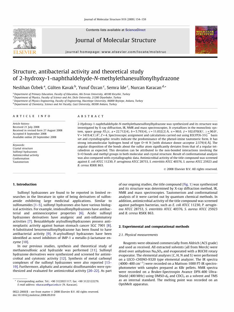
Structure, antibacterial activity and theoretical study of 2-hydroxy-1-naphthaldehyde-N-methylethanesulfonylhydrazone Neslihan Özbek a , Gülten Kavak b , Yusuf Özcan c , Semra _ Ide c , Nurcan Karacan d, * a Department of Primary Education, Faculty of Education, Ahi Evran University, 40100 Kırs ßehir, Turkey b Department of Physics, Faculty of Science and Art, Dicle University, 21280 Diyarbakır, Turkey c Department of Physics Engineering, Faculty of Engineering, Hacettepe University, 06800 Beytepe, Ankara, Turkey d Department of Chemistry, Science and Art Faculty, Gazi University, 06500 Ankara, Turkey article info Article history: Received 21 July 2008 Received in revised form 27 August 2008 Accepted 8 September 2008 Available online 20 September 2008 Keywords: Crystal structure Sulfonyl hydrazones Antimicrobial activity Conformation Tautomerism abstract 2-Hydroxy-1-naphthaldehyde-N-methylethanesulfonylhydrazone was synthesized and its structure was investigated by X-ray diffraction, IR, NMR and mass spectroscopies. It crystallizes in the monoclinic sys- tem, space group P2 1 /c, a = 22.712(4), b = 5.793(4), c = 11.032(2) Å, a = 90.0, b = 102.070(8)°, c = 90.0°, V = 1419.4(1) Å 3 , Z = 4. Spectroscopic assignment and calculations carried out using B3LYP/6-31G ** basis set and crystallographic results indicate the predominance of the phenol-imine tautomeric form. It has strong intramolecular hydrogen bond of type OAH N [with distance donor–acceptor 2.579(4) Å]. The angular disposition of the bonds about the sulfur atom significantly deviates from that of a regular tet- rahedron as expected. This deviation can be attributed to the non-bonded interactions involving the S@O bonds and methyl groups in both molecular and crystal structure. Result of conformational analysis was also compared with crystallographic data. Antimicrobial activity of the title compound was screened against E. coli ATCC 11230, P. aeruginosa ATCC 28753, S. enterititis ATCC 40376, S. aureus ATCC 25923 and B. cereus RSKK 863. Ó 2008 Elsevier B.V. All rights reserved. 1. Introduction Sulfonyl hydrazones are found to be reported in limited re- searches in the literature in spite of being derivatives of sulfon- amide exhibiting large medicinal applications. Similar to sulfonamides [1–5], sulfonyl hydrazones also have various biolog- ical activities. For example, imidosulfonylhydrazones have antibac- terial and antineociceptive properties [6]. Acidic sulfonyl hydrazones derivatives have analgesic and anti-inflammatory activities [7]. Benzaldehyde arylsulfonylhydrazones possess anti- neoplastic activity against human stomach cancer SGC 7901 [8]. 4-Substituted benzenesulfonylhydrazone has been found to have antibacterial activity [9]. N-arylsulfonyl hydrazones have been identified as novel inhibitors of IMP-1 a metallo-b-lactamase en- zyme [10]. In our previous studies, synthesis and theoretical study of methanesulfonic acid hydrazide was performed [11]. Sulfonyl hydrazone derivatives were synthesized and screened for antimi- crobial and cytotoxic activity [12]. Synthesis of metal carbonyl complexes of the sulfonyl hydrazones were also reported [13– 19]. Furthermore, aliphatic and aromatic disulfonamides were syn- thesized and evaluated for antimicrobial activity [20–22]. As part of our ongoing studies, the title compound (Fig. 1) was synthesized and its structure was determined by X-ray diffraction method, IR, NMR and mass spectrocopies. Tautomerism and conformational analysis of it were carried out by quantum chemical methods. In addition, antimicrobial activity of the title compound was screened against pathogen bacterias, such as E. coli ATCC 11230, P. aerugin- osa ATCC 28753, S. enterititis ATCC 40376, S. aureus ATCC 25923 and B. cereus RSKK 863. 2. Experimental and computational methods 2.1. Physical measurements Reagents were obtained commercially from Aldrich (ACS grade) and used as received. All extracted solvents (all from Merck) were dried over anhydrous Na 2 SO 4 and evaporated with a BUCHI rotary evaporator. The elemental analyses (C, H, N and S) were performed on a LECO–CHSNO-9320 type elemental analyzer. The IR spectra (4000–400 cm À1 ) were recorded on a Mattson-1000 FT-IR spectro- photometer with samples prepared as KBr pellets. NMR spectra were recorded on a Bruker-Spectrospin Avance DPX-400 Ultra- Shield (400 MHz) using DMSO-d 6 and CDCl 3 as a solvent and TMS as an internal standard. The melting point was recorded on an OptiMelt apparatus. 0022-2860/$ - see front matter Ó 2008 Elsevier B.V. All rights reserved. doi:10.1016/j.molstruc.2008.09.010 * Corresponding author. Tel.: +90 3122021117; fax: +90 3122122279. E-mail address: [email protected] (N. Karacan). Journal of Molecular Structure 919 (2009) 154–159 Contents lists available at ScienceDirect Journal of Molecular Structure journal homepage: www.elsevier.com/locate/molstruc
Transcript of Structure, antibacterial activity and theoretical study of 2-hydroxy-1naphthaldehyde...
Journal of Molecular Structure 919 (2009) 154–159
Contents lists available at ScienceDirect
Journal of Molecular Structure
journal homepage: www.elsevier .com/locate /molstruc
Structure, antibacterial activity and theoretical studyof 2-hydroxy-1-naphthaldehyde-N-methylethanesulfonylhydrazone
Neslihan Özbek a, Gülten Kavak b, Yusuf Özcan c, Semra _Ide c, Nurcan Karacan d,*
a Department of Primary Education, Faculty of Education, Ahi Evran University, 40100 Kırs�ehir, Turkeyb Department of Physics, Faculty of Science and Art, Dicle University, 21280 Diyarbakır, Turkeyc Department of Physics Engineering, Faculty of Engineering, Hacettepe University, 06800 Beytepe, Ankara, Turkeyd Department of Chemistry, Science and Art Faculty, Gazi University, 06500 Ankara, Turkey
a r t i c l e i n f o
Article history:Received 21 July 2008Received in revised form 27 August 2008Accepted 8 September 2008Available online 20 September 2008
Keywords:Crystal structureSulfonyl hydrazonesAntimicrobial activityConformationTautomerism
0022-2860/$ - see front matter � 2008 Elsevier B.V. Adoi:10.1016/j.molstruc.2008.09.010
* Corresponding author. Tel.: +90 3122021117; faxE-mail address: [email protected] (N. Karacan)
a b s t r a c t
2-Hydroxy-1-naphthaldehyde-N-methylethanesulfonylhydrazone was synthesized and its structure wasinvestigated by X-ray diffraction, IR, NMR and mass spectroscopies. It crystallizes in the monoclinic sys-tem, space group P21/c, a = 22.712(4), b = 5.793(4), c = 11.032(2) Å, a = 90.0, b = 102.070(8)�, c = 90.0�,V = 1419.4(1) Å3, Z = 4. Spectroscopic assignment and calculations carried out using B3LYP/6-31G** basisset and crystallographic results indicate the predominance of the phenol-imine tautomeric form. It hasstrong intramolecular hydrogen bond of type OAH N [with distance donor–acceptor 2.579(4) Å]. Theangular disposition of the bonds about the sulfur atom significantly deviates from that of a regular tet-rahedron as expected. This deviation can be attributed to the non-bonded interactions involving theS@O bonds and methyl groups in both molecular and crystal structure. Result of conformational analysiswas also compared with crystallographic data. Antimicrobial activity of the title compound was screenedagainst E. coli ATCC 11230, P. aeruginosa ATCC 28753, S. enterititis ATCC 40376, S. aureus ATCC 25923 andB. cereus RSKK 863.
� 2008 Elsevier B.V. All rights reserved.
1. Introduction
Sulfonyl hydrazones are found to be reported in limited re-searches in the literature in spite of being derivatives of sulfon-amide exhibiting large medicinal applications. Similar tosulfonamides [1–5], sulfonyl hydrazones also have various biolog-ical activities. For example, imidosulfonylhydrazones have antibac-terial and antineociceptive properties [6]. Acidic sulfonylhydrazones derivatives have analgesic and anti-inflammatoryactivities [7]. Benzaldehyde arylsulfonylhydrazones possess anti-neoplastic activity against human stomach cancer SGC 7901 [8].4-Substituted benzenesulfonylhydrazone has been found to haveantibacterial activity [9]. N-arylsulfonyl hydrazones have beenidentified as novel inhibitors of IMP-1 a metallo-b-lactamase en-zyme [10].
In our previous studies, synthesis and theoretical study ofmethanesulfonic acid hydrazide was performed [11]. Sulfonylhydrazone derivatives were synthesized and screened for antimi-crobial and cytotoxic activity [12]. Synthesis of metal carbonylcomplexes of the sulfonyl hydrazones were also reported [13–19]. Furthermore, aliphatic and aromatic disulfonamides were syn-thesized and evaluated for antimicrobial activity [20–22]. As part
ll rights reserved.
: +90 3122122279..
of our ongoing studies, the title compound (Fig. 1) was synthesizedand its structure was determined by X-ray diffraction method, IR,NMR and mass spectrocopies. Tautomerism and conformationalanalysis of it were carried out by quantum chemical methods. Inaddition, antimicrobial activity of the title compound was screenedagainst pathogen bacterias, such as E. coli ATCC 11230, P. aerugin-osa ATCC 28753, S. enterititis ATCC 40376, S. aureus ATCC 25923and B. cereus RSKK 863.
2. Experimental and computational methods
2.1. Physical measurements
Reagents were obtained commercially from Aldrich (ACS grade)and used as received. All extracted solvents (all from Merck) weredried over anhydrous Na2SO4 and evaporated with a BUCHI rotaryevaporator. The elemental analyses (C, H, N and S) were performedon a LECO–CHSNO-9320 type elemental analyzer. The IR spectra(4000–400 cm�1) were recorded on a Mattson-1000 FT-IR spectro-photometer with samples prepared as KBr pellets. NMR spectrawere recorded on a Bruker-Spectrospin Avance DPX-400 Ultra-Shield (400 MHz) using DMSO-d6 and CDCl3 as a solvent and TMSas an internal standard. The melting point was recorded on anOptiMelt apparatus.
Fig. 1. The structure of the title compound.
N. Özbek et al. / Journal of Molecular Structure 919 (2009) 154–159 155
2.2. Synthesis
Ethanolic solution of ethanesulfonic acid 1-methylhydrazide(0.62 g, 4.46 mmol) was added dropwise to an ethanolic solutionof 2-hydroxy-1-naphthaldehyde (1.15 g, 6.7 mmol), maintainingthe temperature at about �5 �C. Then, the mixture was stirredfor 5 h at room temperature. The precipitated product was crystal-lized from ethanol/n-hexane mixture. The yellow crystalline solidwas dried in vacuo and stored at ethanol/n-hexane vapor. Yield66%; mp 164–165 �C.
IR (KBr) cm�1: 1621 t (C@N), 1240 t (CAO), 1340 tas (SO2), 1183ts (SO2); 1H NMR (d6-DMSO) d: 1,44 ppm (t, 3H, CH3), 3,26 ppm (q,2H, CH2), 3,52 ppm (s, 3H, NACH3), 8,78 ppm (s,1H, N@CH),10,45 ppm (s, 1H, OH), 7.28–8.04 ppm (m, ArH); 13C NMR (d6-DMSO) d: 8,12 ppm (CH3), 44,54 ppm (CH2), 33.04 ppm (NACH3),157,28 ppm (N@CH); 109.77–132.79 ppm (ArC); EIMS (70 eV) m/z: 292 (M+, 12.5%, M+1, 2.6%); Anal. Calcd. for C14H16SO3N2: C,57.52; H, 5.47; N, 9.56; S, 10.97. Found: C, 57.65; H, 5.13; N,10.07; S, 10.63.
2.3. Crystal structure determination
The X-ray structural study of the title compound has been car-ried out in order to obtain the certain structural information aboutthe ethanesulfonylhydrazone. The details about the measurementsand crystallographic studies are given in Table 1. Diffraction datawere collected on Enraf-Nonius CAD4 diffractometer with graphitemonochromated MoKa (0.71073 Å) radiation [23]. The structure
Table 1Crystallographic details of the title compound
Crystal formula C14H16N2O3SFormula weight 292.4Crystal dimensions (mm) 0.40 � 0.30 � 0.20Temp (K) 291(2)Crystal system MonoclinicSpace group P21/ca (Å) 22.712(4)b (Å) 5.793(4)c (Å) 11.032(2)a (�) 90.0b (�) 102.1(1)c (�) 90.0V (Å3) 1419.4(1)Z; Dcalc, (g cm�3) 4; 1.37F(000) 615.9Range of h (�) 3.6/26.3l (MoKa) (mm�1) 0.237Reflections collected 5759Reflections used in refinement 2874No. of refined parameters 189R/Rw values 0.0562/0.1765GOF 1.0040Final shift 0.000(Dq)min, (Dq)max (e Å�3) �0.426, 0.510
was solved by direct methods and refined by least squares on F2
using SHELXS-97 and SHELXL-97 software packages, respectively[24]. The non-hydrogen atoms were refined anisotropically.H2’(O1) and H11(C11) hydrogens were located in difference syn-theses and refined isotropically [OAH = 0.90(4) Å,Uiso(H) = 0.087(13) Å2 and CAH = 0.910(17) Å, Ui-so(H) = 0.045(7) Å2]. The remaining H atoms were positioned geo-metrically with CAH = 0.93, 0.97 and 0.96 Å for aromatic,methylene and methyl H, respectively, and constrained to ride ontheir parent atoms with Uiso(H) = xUeq(C), where x = 1.5 formethyl H and x = 1.2 for all other H atoms. The final atomic coordi-nates and equivalent isotropic displacement parameters of thenonhydrogen atoms are given in Table 2.
2.4. Method of calculation
Initial geometry of structure was taken from crystal data. Anextensive search for low energy conformations on the potential en-ergy surface (PES) of the compound was carried out by using a sys-tematic search with RHF/6-31G** levels. Obtained minimumenergy conformations were reoptimized at DFT/B3LYP/6-31G**
levels without symmetry constraint. Minima, as well as transitionstates, were characterized through harmonic frequency analysis.All the calculations reported here were carried out with theGAUSSIAN03 software [25] employing standard basis sets withno modifications. Quantum chemical descriptors (HOMO, LUMO,dipole moment, heat of formation) for tautomers were calculatedafter B3LYP/6-31** method with the same software. Surface area(approx) and molecular volume of compound were calculated byusing the Hyperchem (release 7.5) package program [26].
2.5. Antibacterial assay
Escherichia coli ATCC 11230, P. aeruginosa ATCC 28753, S. enteri-titis ATCC 40376, S. aureus ATCC 25923, B. cereus RSKK 863 cultureswere obtained from Gazi University, Biology Department and RefikSaydam Hygiene Center Culture Collection. Bacterial strains werecultured overnight at 37 �C in Nutrient Broth. These stock cultureswere stored in the dark at 4 �C during the survey.
Bacterial susceptibility testing was performed by the disc diffu-sion method according to the guidelines of Clinical and LaboratoryStandards Institute (CLSI) [27]. The sterilized (autoclaved at 120 �C
Table 2Fractional atomic coordinates and equivalent isotropic displacement parameters (Å2)for the title compound
Atom x y z U(eq) [Å2]
O1 0.2459(1) 1.0567(4) 0.1180(2) 0.0607(6)O2 0.0803(1) 0.3256(4) 0.0101(3) 0.0840(9)O3 0.1449(1) 0.5644(5) 0.1694(2) 0.0819(8)N1 0.2090(1) 0.6934(4) �0.0106(2) 0.0477(6)N2 0.1676(1) 0.5188(4) �0.0407(2) 0.0546(6)S1 0.1156(1) 0.5273(1) 0.04278(7) 0.0531(3)C1 0.2962(1) 0.8970(5) �0.0345(2) 0.0423(6)C2 0.2909(1) 0.0613(5) 0.0545(3) 0.0488(7)C3 0.3324(2) 0.2430(5) 0.0842(3) 0.0588(8)C4 0.3796(2) 0.2579(5) 0.0281(3) 0.0588(8)C5 0.3899(1) 0.0930(5) �0.0595(3) 0.0482(7)C6 0.4407(1) 0.1013(6) �0.1142(3) 0.0597(8)C7 0.4504(1) 0.9401(6) �0.1965(3) 0.0609(9)C8 0.4090(1) 0.7614(6) �0.2291(3) 0.0563(8)C9 0.3593(1) 0.7465(5) �0.1792(3) 0.0480(7)C10 0.3474(1) 0.9096(5) �0.0918(2) 0.0426(6)C11 0.2519(1) 0.7149(5) �0.0681(3) 0.0443(6)C12 0.1645(1) 0.3732(6) �0.1489(3) 0.0603(8)C13 0.0714(1) 0.7728(5) �0.0048(3) 0.0560(8)C14 0.0421(2) 0.7743(8) �0.1397(4) 0.0865(1)
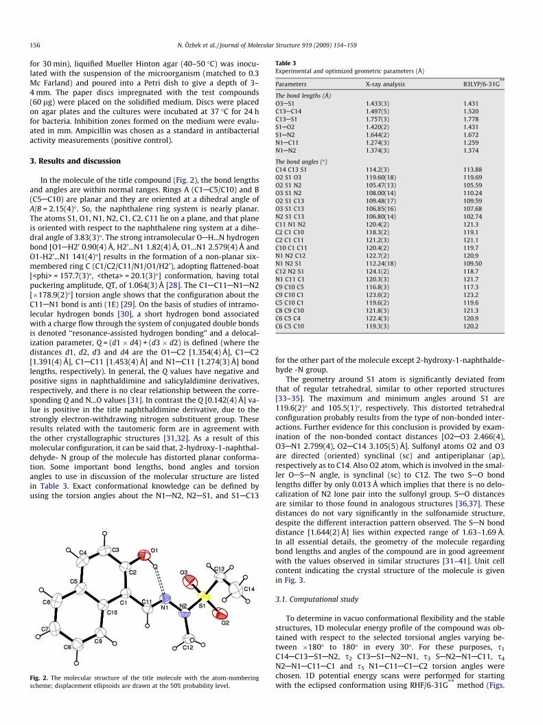
Table 3Experimental and optimized geometric parameters (Å)
Parameters X-ray analysis B3LYP/6-31G**
The bond lengths (Å)O3AS1 1.433(3) 1.431C13AC14 1.497(5) 1.520C13AS1 1.757(3) 1.778S1AO2 1.420(2) 1.431S1AN2 1.644(2) 1.672N1AC11 1.274(3) 1.259N1AN2 1.374(3) 1.374
The bond angles (�)C14 C13 S1 114.2(3) 113.88O2 S1 O3 119.60(18) 119.69O2 S1 N2 105.47(13) 105.59O3 S1 N2 108.00(14) 110.24O2 S1 C13 109.48(17) 109.59O3 S1 C13 106.85(16) 107.68N2 S1 C13 106.80(14) 102.74C11 N1 N2 120.4(2) 121.3C2 C1 C10 118.3(2) 119.1C2 C1 C11 121.2(3) 121.1C10 C1 C11 120.4(2) 119.7N1 N2 C12 122.7(2) 120.9N1 N2 S1 112.24(18) 109.50C12 N2 S1 124.1(2) 118.7N1 C11 C1 120.3(3) 121.7C9 C10 C5 116.8(3) 117.3C9 C10 C1 123.6(2) 123.2C5 C10 C1 119.6(2) 119.6C8 C9 C10 121.8(3) 121.3C6 C5 C4 122.4(3) 120.9C6 C5 C10 119.3(3) 120.2
156 N. Özbek et al. / Journal of Molecular Structure 919 (2009) 154–159
for 30 min), liquified Mueller Hinton agar (40–50 �C) was inocu-lated with the suspension of the microorganism (matched to 0.3Mc Farland) and poured into a Petri dish to give a depth of 3–4 mm. The paper discs impregnated with the test compounds(60 lg) were placed on the solidified medium. Discs were placedon agar plates and the cultures were incubated at 37 �C for 24 hfor bacteria. Inhibition zones formed on the medium were evalu-ated in mm. Ampicillin was chosen as a standard in antibacterialactivity measurements (positive control).
3. Results and discussion
In the molecule of the title compound (Fig. 2), the bond lengthsand angles are within normal ranges. Rings A (C1AC5/C10) and B(C5AC10) are planar and they are oriented at a dihedral angle ofA/B = 2.15(4)�. So, the naphthalene ring system is nearly planar.The atoms S1, O1, N1, N2, C1, C2, C11 lie on a plane, and that planeis oriented with respect to the naphthalene ring system at a dihe-dral angle of 3.83(3)�. The strong intramolecular OAH...N hydrogenbond [O1AH2’ 0.90(4) Å, H2’...N1 1.82(4) Å, O1...N1 2.579(4) Å andO1-H2’...N1 141(4)�] results in the formation of a non-planar six-membered ring C (C1/C2/C11/N1/O1/H2’), adopting flattened-boat[<phi> = 157.7(3)�, <theta> = 20.1(3)�] conformation, having totalpuckering amplitude, QT, of 1.064(3) Å [28]. The C1AC11AN1AN2[�178.9(2)�] torsion angle shows that the configuration about theC11AN1 bond is anti (1E) [29]. On the basis of studies of intramo-lecular hydrogen bonds [30], a short hydrogen bond associatedwith a charge flow through the system of conjugated double bondsis denoted ‘‘resonance-assisted hydrogen bonding” and a delocal-ization parameter, Q = (d1 � d4) + (d3 � d2) is defined (where thedistances d1, d2, d3 and d4 are the O1AC2 [1.354(4) Å], C1AC2[1.391(4) Å], C1AC11 [1.453(4) Å] and N1AC11 [1.274(3) Å] bondlengths, respectively). In general, the Q values have negative andpositive signs in naphthaldimine and salicylaldimine derivatives,respectively, and there is no clear relationship between the corre-sponding Q and N...O values [31]. In contrast the Q [0.142(4) Å] va-lue is positive in the title naphthaldimine derivative, due to thestrongly electron-withdrawing nitrogen substituent group. Theseresults related with the tautomeric form are in agreement withthe other crystallographic structures [31,32]. As a result of thismolecular configuration, it can be said that, 2-hydroxy-1-naphthal-dehyde- N group of the molecule has distorted planar conforma-tion. Some important bond lengths, bond angles and torsionangles to use in discussion of the molecular structure are listedin Table 3. Exact conformational knowledge can be defined byusing the torsion angles about the N1AN2, N2AS1, and S1AC13
Fig. 2. The molecular structure of the title molecule with the atom-numberingscheme; displacement ellipsoids are drawn at the 50% probability level.
for the other part of the molecule except 2-hydroxy-1-naphthalde-hyde -N group.
The geometry around S1 atom is significantly deviated fromthat of regular tetrahedral, similar to other reported structures[33–35]. The maximum and minimum angles around S1 are119.6(2)� and 105.5(1)�, respectively. This distorted tetrahedralconfiguration probably results from the type of non-bonded inter-actions. Further evidence for this conclusion is provided by exam-ination of the non-bonded contact distances [O2AO3 2.466(4),O3AN1 2.799(4), O2AC14 3.105(5) Å]. Sulfonyl atoms O2 and O3are directed (oriented) synclinal (sc) and antiperiplanar (ap),respectively as to C14. Also O2 atom, which is involved in the smal-ler OASAN angle, is synclinal (sc) to C12. The two SAO bondlengths differ by only 0.013 Å which implies that there is no delo-calization of N2 lone pair into the sulfonyl group. SAO distancesare similar to those found in analogous structures [36,37]. Thesedistances do not vary significantly in the sulfonamide structure,despite the different interaction pattern observed. The SAN bonddistance [1.644(2) Å] lies within expected range of 1.63–1.69 Å.In all essential details, the geometry of the molecule regardingbond lengths and angles of the compound are in good agreementwith the values observed in similar structures [31–41]. Unit cellcontent indicating the crystal structure of the molecule is givenin Fig. 3.
3.1. Computational study
To determine in vacuo conformational flexibility and the stablestructures, 1D molecular energy profile of the compound was ob-tained with respect to the selected torsional angles varying be-tween �180� to 180� in every 30�. For these purposes, s1
C14AC13AS1AN2, s2 C13AS1AN2AN1, s3 SAN2AN1AC11, s4
N2AN1AC11AC1 and s5 N1AC11AC1AC2 torsion angles werechosen. 1D potential energy scans were performed for startingwith the eclipsed conformation using RHF/6-31G** method (Figs.

Fig. 3. Crystal packing along the b axis (on the left) and the best view of threedimentional unit cell perspective (on the right) of the title compound.
Fig. 5. The energy profile for the rotation about, s4 N2AN1AC11AC1 and s5
N1AC11AC1AC2.
N. Özbek et al. / Journal of Molecular Structure 919 (2009) 154–159 157
4 and 5). As can be seen in Figs. 4 and 5, energy profiles of s1 tor-sion shows global minima at 120�, local minima at 210�, globalmaxima at 320� and local maxima at 180�. s2 shows two local min-ima at about 180� and 270�, global minima at 60�, local maxima at230� and global maxima at about 150�. Third scan for s3 shows glo-bal minima at 120�, global maxima at 60�, local maxima at 210�. s4
shows global minima at about 190�, global maxima at 90�, localmaxima at about 280�. Final scan for s5 does not show the transi-tion states.
Selected species were again reoptimized at DFT/B3LYP/6-31G**
level and obtained result were given in Table 3. The results showgood correlation with the experimental data as in salicylaldehydesulfonylhydrazone [42]. Reoptimized conformations were givenin Fig. 6. Total and relative energies of the conformers are listedin Table 4. Napthaldehyde group exists in synperiplanar (sp) in(1–7) conformers due to hydrogen bonding between N1. . .H1atoms. Conformer (1) was found to be the most stable conformerin vacuo, however, conformer (5) is favored in the crystal system.The calculated difference between (1) and (5) is 2.76 kcal/mol.Changing of s1, s2 and s3 torsion angles caused the loss of potentialenergy, accompanied by changing surface area and molecular vol-ume calculated with HyperChem 7.5 [26]. Surface area (approx)and molecular volume for conformer (1) are 451.81 Å2 and809.95 Å3, however, for conformer (5) are 444.42 Å2 and808.61 Å3, respectively. These results indicate that decreasing of
Fig. 4. The energy profile for the rotation about s1 C14AC13AS1AN2, s2
C13AS1AN2AN1 and s3 S1AN2AN1AC11.
Fig. 6. Optimized geometries of conformations.
molecular volume leads to more strong intermolecular interactionor crystal packing effect, which makes conformer (5) the most sta-ble in crystal system. These results clearly indicate that conforma-tion was affected by intermolecular interaction in the solid phase,and it should not be neglected.
Tautomerism in 2-hydroxy Schiff bases obtained from salicylal-dehyde and naphthaldehyde have been extensively studied exper-imentally. It is reported that phenol-imine form predominantlyexist in salicylaldimine Schiff bases, however, keto-amine form isfound to be dominant in naphthaldimine Schiff bases in solid state[43–45]. Keto-phenol tautomerism of the title compound is givenin Fig. 7. These two tautomeric forms were optimized with BLYP/
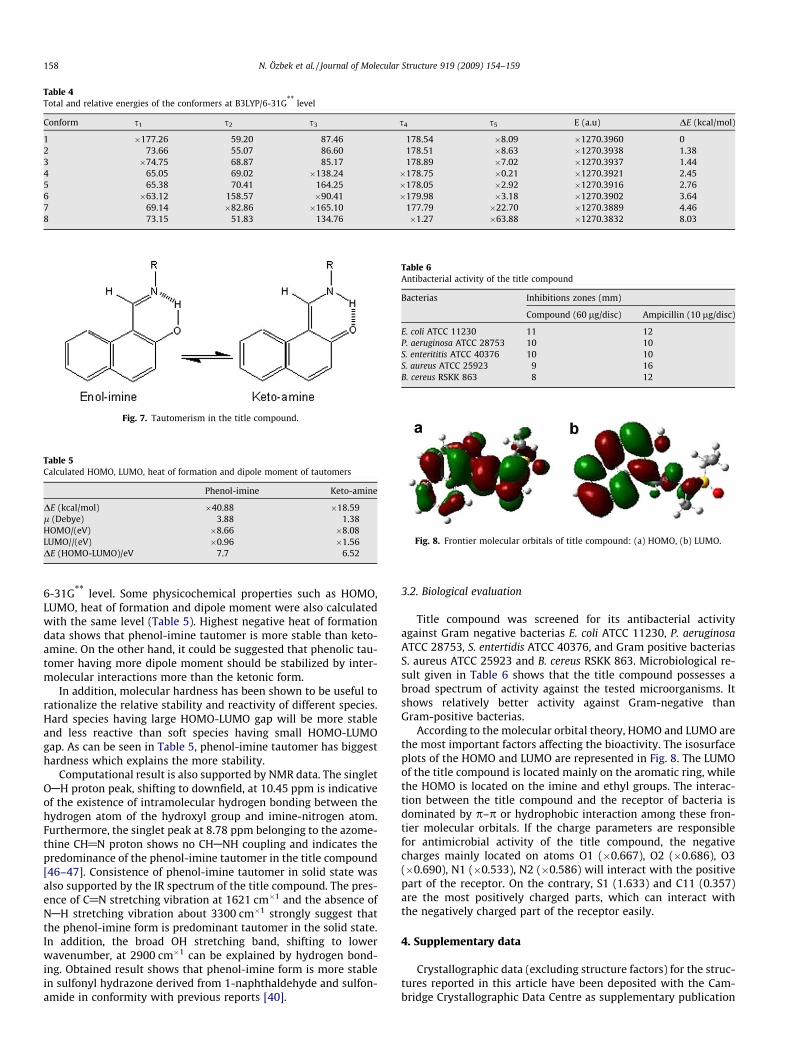
Table 4Total and relative energies of the conformers at B3LYP/6-31G** level
Conform s1 s2 s3 s4 s5 E (a.u) DE (kcal/mol)
1 �177.26 59.20 87.46 178.54 �8.09 �1270.3960 02 73.66 55.07 86.60 178.51 �8.63 �1270.3938 1.383 �74.75 68.87 85.17 178.89 �7.02 �1270.3937 1.444 65.05 69.02 �138.24 �178.75 �0.21 �1270.3921 2.455 65.38 70.41 164.25 �178.05 �2.92 �1270.3916 2.766 �63.12 158.57 �90.41 �179.98 �3.18 �1270.3902 3.647 69.14 �82.86 �165.10 177.79 �22.70 �1270.3889 4.468 73.15 51.83 134.76 �1.27 �63.88 �1270.3832 8.03
Fig. 7. Tautomerism in the title compound.
Table 5Calculated HOMO, LUMO, heat of formation and dipole moment of tautomers
Phenol-imine Keto-amine
DE (kcal/mol) �40.88 �18.59l (Debye) 3.88 1.38HOMO/(eV) �8.66 �8.08LUMO//(eV) �0.96 �1.56DE (HOMO-LUMO)/eV 7.7 6.52
Table 6Antibacterial activity of the title compound
Bacterias Inhibitions zones (mm)
Compound (60 lg/disc) Ampicillin (10 lg/disc)
E. coli ATCC 11230 11 12P. aeruginosa ATCC 28753 10 10S. enterititis ATCC 40376 10 10S. aureus ATCC 25923 9 16B. cereus RSKK 863 8 12
Fig. 8. Frontier molecular orbitals of title compound: (a) HOMO, (b) LUMO.
158 N. Özbek et al. / Journal of Molecular Structure 919 (2009) 154–159
6-31G** level. Some physicochemical properties such as HOMO,LUMO, heat of formation and dipole moment were also calculatedwith the same level (Table 5). Highest negative heat of formationdata shows that phenol-imine tautomer is more stable than keto-amine. On the other hand, it could be suggested that phenolic tau-tomer having more dipole moment should be stabilized by inter-molecular interactions more than the ketonic form.
In addition, molecular hardness has been shown to be useful torationalize the relative stability and reactivity of different species.Hard species having large HOMO-LUMO gap will be more stableand less reactive than soft species having small HOMO-LUMOgap. As can be seen in Table 5, phenol-imine tautomer has biggesthardness which explains the more stability.
Computational result is also supported by NMR data. The singletOAH proton peak, shifting to downfield, at 10.45 ppm is indicativeof the existence of intramolecular hydrogen bonding between thehydrogen atom of the hydroxyl group and imine-nitrogen atom.Furthermore, the singlet peak at 8.78 ppm belonging to the azome-thine CH@N proton shows no CHANH coupling and indicates thepredominance of the phenol-imine tautomer in the title compound[46–47]. Consistence of phenol-imine tautomer in solid state wasalso supported by the IR spectrum of the title compound. The pres-ence of C@N stretching vibration at 1621 cm�1 and the absence ofNAH stretching vibration about 3300 cm�1 strongly suggest thatthe phenol-imine form is predominant tautomer in the solid state.In addition, the broad OH stretching band, shifting to lowerwavenumber, at 2900 cm�1 can be explained by hydrogen bond-ing. Obtained result shows that phenol-imine form is more stablein sulfonyl hydrazone derived from 1-naphthaldehyde and sulfon-amide in conformity with previous reports [40].
3.2. Biological evaluation
Title compound was screened for its antibacterial activityagainst Gram negative bacterias E. coli ATCC 11230, P. aeruginosaATCC 28753, S. entertidis ATCC 40376, and Gram positive bacteriasS. aureus ATCC 25923 and B. cereus RSKK 863. Microbiological re-sult given in Table 6 shows that the title compound possesses abroad spectrum of activity against the tested microorganisms. Itshows relatively better activity against Gram-negative thanGram-positive bacterias.
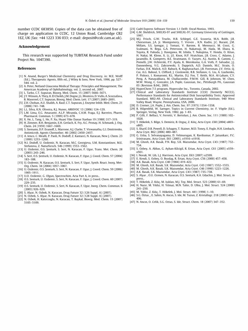
According to the molecular orbital theory, HOMO and LUMO arethe most important factors affecting the bioactivity. The isosurfaceplots of the HOMO and LUMO are represented in Fig. 8. The LUMOof the title compound is located mainly on the aromatic ring, whilethe HOMO is located on the imine and ethyl groups. The interac-tion between the title compound and the receptor of bacteria isdominated by p–p or hydrophobic interaction among these fron-tier molecular orbitals. If the charge parameters are responsiblefor antimicrobial activity of the title compound, the negativecharges mainly located on atoms O1 (�0.667), O2 (�0.686), O3(�0.690), N1 (�0.533), N2 (�0.586) will interact with the positivepart of the receptor. On the contrary, S1 (1.633) and C11 (0.357)are the most positively charged parts, which can interact withthe negatively charged part of the receptor easily.
4. Supplementary data
Crystallographic data (excluding structure factors) for the struc-tures reported in this article have been deposited with the Cam-bridge Crystallographic Data Centre as supplementary publication
N. Özbek et al. / Journal of Molecular Structure 919 (2009) 154–159 159
number CCDC 683850. Copies of the data can be obtained free ofcharge on application to CCDC, 12 Union Road, Cambridge CB21EZ, UK (fax: +44 1223 336 033; e-mail: [email protected]).
Acknowledgement
This research was supported by TUBITAK Research Fund underProject No. 104T390.
References
[1] N. Anand, Burger’s Medicinal Chemistry and Drug Discovery, in: M.E. Wolff(Ed.), Therapeutic Agents, fifth ed., J Wiley & Sons, New York, 1996, pp. 527–544. vol. 2.
[2] A. Peter, Netland Glaucoma Medical Therapy: Principles and Management, TheAmerican Academy of Ophthalmology, vol. 2, second ed., 2007.
[3] L. Tarko, C.T. Supuran, Bioorg. Med. Chem. 15 (2007) 5666–5671.[4] J.Y. Winum, A. Thiry, K. El Cheikh, J.M. Dogne, J.L. Montero, D. Vullo, A. Scozzafava,
B. Masereel, C.T. Supuran, Bioorg. Med. Chem. Lett. 17 (2007) 2685–2691.[5] Z.H. Chohan, A.U. Shaikh, A. Rauf, C.T. Supuran, J. Enzyme Inhib. Med. Chem. 21
(2006) 741–748.[6] L.L. Silva, K.N. Oliveira, R.J. Nunes, ARKIVOC 13 (2006) 124–129.[7] L.M. Lima, E.G. Amarante, A.L.P. Miranda, C.A.M. Fraga, E.J. Barreiro, Pharm.
Pharmacol. Commun. 5 (1999) 673–678.[8] X. He, L. Tang, L. He, P. Xu, Huaxi Yike Daxue Xuebao 19 (1988) 317–319.[9] H. Zimmer, B.H. Benjamin, E.H. Gerlach, K. Fry, A.C. Pronay, H. Schmank, J. Org.
Chem. 24 (1959) 1667–1669.[10] S. Siemann, D.P. Evanoff, L. Marrone, A.J. Clarke, T. Viswanatha, G.I. Dmitrienko,
Antimicrob. Agents Chemother. 46 (2002) 2450–2457.[11] A. Ienco, C. Mealli, P. Paoli, N. Dodoff, Z. Kantarci, N. Karacan, New J. Chem. 23
(1999) 1253–1260.[12] N.I. Dodoff, U. Ozdemir, N. Karacan, M.C. Georgieva, S.M. Konstantinov, M.E.
Stefanova, Z. Naturforsch. 54b (1999) 1553–1562.[13] U. Ozdemir, O.S. S�enturk, S. Sert, N. Karacan, F. Ugur, Trans. Met. Chem. 28
(2003) 243–246.[14] S. Sert, O.S. S�enturk, U. Ozdemir, N. Karacan, F. Ugur, J. Coord. Chem. 57 (2004)
183–188.[15] U. Ozdemir, N. Karacan, O.S. S�enturk, S. Sert, F. Ugur, Synth. React. Inorg. Met-
Org. Chem. 34 (2004) 1057–1067.[16] U. Ozdemir, O.S. S�enturk, S. Sert, N. Karacan, F. Ugur, J. Coord. Chem. 59 (2006)
1905–1911.[17] U.O. Ozdemir, G. Olgun, Spectrochim. Acta Part A, in press..[18] O.S. Senturk, U. Ozdemir, S. Sert, N. Karacan, F. Ugur, J. Coord. Chem. 60 (2007)
229–235.[19] O.S. Senturk, U. Ozdemir, S. Sert, N. Karacan, F. Ugur, Inorg. Chem. Commun. 6
(2003) 926–929.[20] S. Alyar, N. Ozbek, N. Karacan, Drug Future 32 (126 Suppl. A) (2007).[21] N. Ozbek, S. Alyar, N. Karacan, Drug Future 32 (128 Suppl. A) (2007).[22] N. Ozbek, H. Katırcıoglu, N. Karacan, T. Baykal, Bioorg. Med. Chem. 15 (2007)
5105–5109.
[23] Cad4 Express Software Version 1.1 Delft: Enraf-Nonius, 1993.[24] G.M. Sheldrick, SHELXS-97 and SHELXL-97, Germany:University of Gottingen,
1997.[25] M.J. Frisch, G.W. Trucks, H.B. Schlegel, G.E. Scuseria, M.A. Robb, J.R.
Cheeseman, J.A. Jr. Montgomery, T. Vreven , K.N. Kudin, J.C. Burant, J.M.Millam, S.S. Iyengar, J. Tomasi, V. Barone, B. Mennucci, M. Cossi, G.Scalmani, N. Rega, G.A. Petersson, H. Nakatsuji, M. Hada, M. Ehara, K.Toyota, R. Fukuda, J. Hasegawa, M. Ishida, T. Nakajima, Y. Honda, O. Kitao,H. Nakai, M. Klene, X. Li, J.E. Knox, H.P. Hratchian, J.B. Cross, C. Adamo, J.Jaramillo, R. Gomperts, R.E. Stratmann, O. Yazyev, A.J. Austin, R. Cammi, C.Pomelli, J.W. Ochterski, P.Y. Ayala, K. Morokuma, G.A. Voth, P. Salvador, J.J.Dannenberg, V.G. Zakrzewski, S. Dapprich, A.D. Daniels, M.C. Strain, O.Farkas, D.K. Malick, A.D. Rabuck, K. Raghavachari, J.B. Foresman, J.V. Ortiz, Q.Cui, A.G. Baboul, S. Clifford, J. Cioslowski, B.B. Stefanov, G. Liu, A. Liashenko,P. Piskorz, I. Komaromi, R.L. Martin, D.J. Fox, T. Keith, M.A. Al-Laham, C.Y.Peng, A. Nanayakkara, M. Challacombe, P.M.W. Gill, B. Johnson, W. Chen,M.W. Wong, C. Gonzalez, J.A. Pople, Gaussian, Inc., Pittsburgh PA., Gaussian03 (Revision B.04), 2003.
[26] HyperChem 7.5 program, Hypercube Inc., Toronto, Canada, 2002.[27] Clinical and Laboratory Standards Institute (CLSI) (formerly NCCLS),
Performance Standards for Antimicrobial Disk Susceptibility Tests; ApprovedStandard, M02-A9. Clinical and Laboratory Standards Institute, 940 WestValley Road, Wayne, Pennsylvania, USA, 2006.
[28] D. Cremer, J.A. Pople, J. Am. Chem. Soc. 97 (1975) 1354–1358.[29] R. Hilgenfeld, W. Saenger, Topics in Current Chemistry, in: F. Vögtle (Ed.),
Springer, Verlag, New York, 1982, pp. 1–83.[30] P. Gilli, F. Belluci, V. Ferretti, V. Bertolasi, J. Am. Chem. Soc. 111 (1989) 102–
1028.[31] T. Hökelek, S. Bilge, S. Demiriz, B. Ozguç, Z. Kılıç, Acta Cryst. C60 (2004) o803–
o805.[32] S. Sharif, D.R. Powell, D. Schagen, T. Stainer, M.D. Toney, E. Fogle, H.H. Limbach,
Acta Cryst. B62 (2006) 480–487.[33] G. Usha, S. Selvanayagama, D. Velmurugan, K. Ravikumar, P. Jaisankarc, P.C.
Srinivasanc, Acta Cryst. E61 (2005). o1916–o1918.[34] M. Ghosh, A.K. Basak, P.N. Roy, S.K. Mazumdar, Acta Cryst. C43 (1987) 732–
734.[35] S. Ozbey, A. Akbas, G. Ayhan-Kilcigil, R. Ertan, Acta Cryst. C61 (2005) o559–
o561.[36] I. Novak, W. Lib, L.J. Harrison, Acta Cryst. E63 (2007) o2599.[37] E. Kendi, S. Ozbey, O. Bozdag, R. Ertan, Acta Cryst. C56 (2000) 457–458.[38] A.K. Basak, Acta Cryst. C40 (1984) 419–422.[39] M. Ghosh, A.K. Basak, S.K. Mazumdar, Acta Cryst. C43 (1987) 1552–1555.[40] M. Ghosh, A.K. Basak, S.K. Mazumdar, Acta Cryst. C46 (1990) 1223–1226.[41] A.K. Basak, S.K. Mazumdar, Acta Cryst. C43 (1987) 735–738.[42] S. Alyar , O.U. Ozmen, N. Karacan, O.S. Senturk, K.A. Udachin, J. Mol. Struct, in
press..[43] T. Hökelek, Z. Kılıç, M. Is�ıklan, M.J. Toy, Mol. Struct. 523 (2000) 61–69.[44] H. Nazır, M. Yıldız, H. Yılmaz, M.N. Tahir, D. Ulku, J. Mol. Struct. 524 (2000)
241–250.[45] M. Yıldız, Z. Kılıç, T. Hökelek, J. Mol. Struct. 441 (1998) 1–10.[46] S.G. Öztas�, E. Sahin, N. Ancın, S. Ide, M. Tuzun, Z. Kristallogr. 218 (2003) 492–
496.[47] N. Ancın, O. Celik, S.G. Oztas, S. Ide, Struct. Chem. 18 (2007) 347–352.