Mononuclear manganese carboxylate complexes: Synthesis and structural studies
Upload
independentCategory
view
3download
0
The Structures of Thiolate- and Carboxylate-Ligated Ferric H93GMyoglobin:Models for Cytochrome P450 and for Oxyanion-Bound Heme Proteins†
Jie Qin‡,§, Roshan Perera‡,§, Leslie L. Lovelace§, John H. Dawson*,§,∥, and LukaszLebioda*,§Department of Chemistry and Biochemistry and the School of Medicine, University of SouthCarolina, Columbia, SC 29208
AbstractCrystal structures of the ferric H93G myoglobin (Mb) cavity mutant containing either an anionicproximal thiolate sulfur-donor or carboxylate oxygen-donor ligand are reported at 1.7 and 1.4 Åresolution, respectively. The crystal structure and magnetic circular dichroism spectra of the H93GMb β-mercaptoethanol (BME) thiolate adduct reveal a high-spin, five-coordinate complex.Furthermore, the bound BME appears to have an intramolecular hydrogen bond involving the alcoholproton and the ligated thiolate sulfur, mimicking one of the three proximal N-H···S hydrogen bondsin cytochrome P450. The Fe is displaced from the porphyrin plane by 0.5 Å and forms a 2.41 Å Fe-S bond. The Fe3+-S-C angle is 111 °, indicative of a covalent Fe-S bond with sp3 hybridized sulfur.Therefore, the H93G Mb·BME complex provides an excellent protein-derived structural model forhigh-spin ferric P450. In particular, the Fe-S bond in high-spin ferric P450-CAM has essentially thesame geometry despite the constraints imposed by covalent linkage of the cysteine to the proteinbackbone. This suggests that evolution led to the geometric optimization of the proximal Fe-S(cysteinate) bond in P450. The crystal structure and spectral properties of the H93G Mb acetateadduct reveal a high-spin, six-coordinate complex with proximal acetate and distal water axialligands. The distal His-64 forms a hydrogen bond with the bound water. The Fe-acetate bondinggeometry is inconsistent with an electron pair along the Fe-O bond as the Fe-O-C angle is 152° andthe Fe is far from the plane of the acetate. Thus, the Fe-O bonding is ionic. The H93G Mb cavitymutant has already been shown to be a versatile model system for the study of ligand binding to hemeproteins; this investigation affords the first structural evidence that non-imidazole exogenous ligandsbind in the proximal ligation site.
The proximal ligand plays a crucial role in the activation of oxygen and peroxide by hemeenzymes (1-5). Although the application of site-directed mutagenesis for the preparation ofproximal ligand mutants has yielded important information about their roles in catalysis, thetechnique is limited by the minimal number of naturally occurring amino acid ligands availablein the genetic code. To overcome this shortcoming, Barrick prepared a proximal H93G cavitymutant of sperm whale myoglobin (Mb)1 by replacing His-93 with a much smaller Gly (6).
†This work was supported by grants from the NIH (GM 26730), the NSF (MCB 96-04004) and the University of South CarolinaEnvironmental Research Initiative Committee. Use of the Argonne National Laboratory Structural Biology Center beamlines at theAdvanced Photon Source, was supported by the U. S. Department of Energy, Office of Science, under Contract No. W-31-109-ENG-38.Atomic coordinates have been deposited to the Protein Data Bank with access codes 2EVP and 2EVK.‡Contributed equally to this work.§Department of Chemistry and Biochemistry∥School of Medicine*Correspondence to Lukasz Lebioda, Phone (803) 777-2140, E-mail: [email protected], John H. Dawson, Phone (803)777-7234, [email protected], Department of Chemistry & Biochemistry, University of South Carolina, Columbia, SC 29208
NIH Public AccessAuthor ManuscriptBiochemistry. Author manuscript; available in PMC 2008 October 1.
Published in final edited form as:Biochemistry. 2006 March 14; 45(10): 3170–3177. doi:10.1021/bi052171s.
NIH
-PA Author Manuscript
NIH
-PA Author Manuscript
NIH
-PA Author Manuscript

This creates a proximal pocket that accommodates foreign unnatural ligands, and the H93GMb cavity mutant has been extensively employed for the preparation of heme protein modelcomplexes (1,7-16) The use of the H93G Mb cavity mutant as a natural protein-derivedstructural and functional model circumvents the limitations of site-directed mutagenesis. Thispaper presents the first crystallographic study of the H93G ferric Mb cavity mutant containingnon-imidazole proximal ligands, specifically thiolate sulfur and carboxylate oxygen, asstructural models for cysteinate- and oxyanion-ligated heme enzymes.
Cysteine serves as a proximal axial ligand in a number of heme enzymes, most notably thedioxygen and peroxide activating enzymes cytochrome P450 (P450), C. fumagochloroperoxidase and nitric oxide synthase (2). The cysteine must be deprotonated to cysteinatethroughout the reaction cycle for catalytic activity. Dawson has proposed that the cysteinate“push” is important for the mechanism of mono-oxygenase activity in P450 (2,3,17). Variousfactors that stabilize thiolate ligation have been discussed and investigated (4,10). In particular,there are three amide N-H···S(proximal) hydrogen bonds in P450-CAM from residues Leu-358,Gly-359 and Gln-360 that play a key role in the catalytic cycle by helping to retain cysteinatecoordination when the iron goes sequentially through the ferric, ferrous and ferryl oxidationstates. Morishima and coworkers have used site-directed mutagenesis to remove one of theseconserved amide hydrogen bonds in the L358P mutant (18). This led to an increase in thethiolate “push” effect that was proposed to facilitate the protonation of the outer oxygen atomin the hydroperoxo intermediate of P450-CAM. There is no successful natural model systemto mimic these highly conserved amide hydrogen bonds although attempts have been made toincorporate hydrogen bonding into synthetic porphyrin models. Nakamura, for example, hasprepared ferric porphyrin thiophenolate complexes containing one or two ortho substitutedamide N-H groups to hydrogen-bond to the coordinated thiolate sulfur (19). The proximal Histo Cys mutant of Mb (20,21), H93C Mb, is unable to form a thiolate H-bond with the adjacentamino acid residue, Ser-92, because its hydroxyl group is too far from the Cys-93 S atom.
1Abbreviations:
H93G myoglobin His93Gly variant of sperm whale myoglobin
Mb myoglobin
P450 cytochrome P450
BME β-mercaptoethanol
MCD magnetic circular dichroism
EA electronic absorption
FoFc map sigmaa weighted Fourier synthesis with (Fo-Fc) coefficients
2FoFc map sigmaa weighted Fourier synthesis with (2Fo-Fc) coefficients
PDB protein data bank
FePSNP the para-nitrothiophenol thiolate complex of ferric protoporphyrin IX
Qin et al. Page 2
Biochemistry. Author manuscript; available in PMC 2008 October 1.
NIH
-PA Author Manuscript
NIH
-PA Author Manuscript
NIH
-PA Author Manuscript

A second set of interesting heme proteins has an anionic proximal O-donor ligand, typicallytyrosinate, in the ferric state. Some examples are heme containing catalases, the naturallyoccurring hemoglobin M mutants, and coral allene oxide synthase, all of which containtyrosinate proximal ligation (22-27). The most notable feature of these proximal oxyanion-ligated proteins is a much lower redox potential compared to neutral His-ligated hemes,sufficiently low so as to make it difficult to naturally reduce the ferric iron to the ferrous state.This is a useful property for enzymes like catalase and allene oxide synthase that need to remainin the ferric state during catalysis, but presents a severe problem for humans with hemoglobinM.
The factors controlling whether an oxyanion-ligated ferric heme center is five-coordinate orsix-coordinate with a trans water ligand are not well understood (11). For example, in H93YMb, resonance Raman and magnetic circular dichroism (MCD) spectroscopy have shown thatit is mostly a five-coordinate species (28,29). Spectroscopic analysis of H25A heme oxygenaseat neutral pH reveals a five-coordinate complex with carboxylate ligation to the heme iron(12). Recent MCD characterization of the C436S P450-2B4 mutant revealed a mostly six-coordinate serinate/water-ligated ferric heme center, but the complex is not homogeneous assome portion is five-coordinate (30). Therefore, it is important to examine the contributingfactors that influence distal water ligation to oxyanion-ligated (i.e., serinate, tyrosinate orcarboxylate) heme iron centers in proteins.
Even though H93G Mb has been used extensively as a probe for modeling heme proteins withO- and S- donor proximal ligands (13), there has not been any crystallographic study to showthat such ligands bind in the proximal cavity. In the present investigation, we have successfullycrystallized the β-mercaptoethanol (BME) and acetate adducts of ferric H93G Mb, and reportthe structural features observed for these two different proximal donor ligands. The BMEthiolate-bound ferric H93G forms a five-coordinate high-spin adduct, while acetatecarboxylate-bound ferric H93G Mb is a six-coordinate, high-spin complex with a trans waterligand. We have also characterized these derivatives with magnetic circular dichroism (MCD)and electronic absorption (EA) spectroscopy.
MATERIALS AND METHODSChemicals and Proteins
All chemicals were of reagent grade or better and were purchased from Sigma-Aldrich. TheH93G sperm whale Mb E. coli expression system was a gift from Prof. Steven G. Boxer,Stanford University. H93G Mb mutant growth and purification yields pure H93G Mb withimidazole bound in the proximal cavity. For crystallization purposes, we have removed theimidazole using a different method than previously reported (12). First, complete oxidation ofthe heme iron was accomplished by addition of a few crystals of potassium ferricyanide (Fluka)followed by dialysis (three times) for H93G Mb in 100 mM potassium phosphate buffer at pH7.0. After eluting a few times (generally 2 or 3) through a BioGel P100 size-exclusion column,the pure ligand-free ferric H93G Mb was collected. This fresh H93G ligand-free Mb samplewas buffer exchanged to 100 mM PIPES pH 6.2 buffer in a centricon (8000 nominal molecularweight limit) by centrifuging at 3000 rpm. The final EA spectrum was identical to thatpreviously reported for ligand-free H93G Mb (12).
CrystallizationCrystals of H93G Mb were grown by the vapor diffusion method using the hanging-drop setupat conditions similar to those reported previously (14), but with 100-fold lower imidazoleconcentration. The protein solution was 0.9 mM (15 mg/mL) 0.05 mM imidazole; theprecipitant solution was 37% PEG 8K, 300 mM sodium acetate, 0.1 M PIPES pH 6.2, and
Qin et al. Page 3
Biochemistry. Author manuscript; available in PMC 2008 October 1.
NIH
-PA Author Manuscript
NIH
-PA Author Manuscript
NIH
-PA Author Manuscript

0.1% dioxane. Attempts to grow crystals entirely without imidazole were not successful.Crystals were transferred to two pre-equilibrated drops containing precipitant solution (thuswithout imidazole) one of which contained 5 mM BME. Crystals were soaked forapproximately 10 hours and transferred to cryoprotectant solutions which were the soakingsolutions with 10% ethylene glycol. After brief soak, they were flash-cooled in N2 vapors at-178°C.
CrystallographyData were collected on two soaked H93G mutant crystals with approximate dimensions of 0.25× 0.50 × 0.25 mm (BME) and 1.0 × 0.50 × 0.25 mm (no BME). The X-ray diffractionexperiments were carried out at the SBC-CAT BM (19BM) beamline of the Advanced PhotonSource at Argonne National Laboratory and processed with the HKL 2000 suit of programs(31).
The main parameters for data collection and processing are in Table 1. The crystals wereisomorphic with those reported earlier and the initial model for the refinement was obtainedfrom the structure (PDB code 1DTM) of the mutant with a methyl-imidazole ligand (14) byomitting non-protein atoms. Crystallographic refinement was carried out with iterativecombination of interactive graphics utilizing Turbo-Frodo software (32) and simulatedannealing using CNS software (33). A planar model for the heme was used in agreement withthe small molecule data obtained (34) for a similar system (vide infra) with restrains generatedusing the HIC-Up website (xray.bmc.uu.se/hicup) for the CNS refinements and SHELXPROfor SHELX (35). Water molecules were assigned to peaks that were present in both FoFc and2FoFc sigmaa weighted maps and formed appropriate contacts with the protein molecule. Watermolecules that had a B-factor above 65 Å2 after refinement were deleted. Subsequently, bothstructures were refined with SHELXL (35) using anisotropic temperature factors. For the BMEcomplex, the free-R increased indicating that the resolution (number of reflections) wasinsufficient for the anisotropic description of thermal motion and this model was not usedfurther. Correctness of the final structures was evaluated using PROCHECK (36). Figure 1was obtained with Turbo-Frodo; Figure 2 was generated using Pymol (37). Figure 3 was madewith MOLSCRIPT (38) and Raster3D (39). The final refinement parameters are in Table 1.
Spectroscopic Sample PreparationMb concentrations were determined by the pyridine hemochromogen method (40). MCD andEA spectral measurements were obtained at 4°C with Mb (∼50 μM) in 100 mM potassiumphosphate buffer (pH 7.0). For titration experiments, small aliquots of 850 mM potassiumacetate or benzoate in 100 mM potassium phosphate buffer or of 2.0 M BME in DMSO wereadded to 0.4 mL (l = 1 cm, [protein] = ∼50 μM) or 2 mL (l = 1 cm, [protein] = ∼ 5 μM).Comparable amounts of DMSO did not produce spectral changes. Dissociation constants(Kd) were determined by Hill [see “Data Analysis” in ref. (40)] and double reciprocal plots(41).
Spectroscopic TechniquesEA spectra were recorded with a Cary 400 spectrophotometer or a JASCO J600Aspectropolarimeter. MCD spectra were measured in a 0.2 cm cuvette at a magnetic field of1.41 T with the JASCO J600A spectropolarimeter as described (41). Data acquisition andmanipulation were done as reported (42), with JASCO software.
Qin et al. Page 4
Biochemistry. Author manuscript; available in PMC 2008 October 1.
NIH
-PA Author Manuscript
NIH
-PA Author Manuscript
NIH
-PA Author Manuscript
RESULTSX-ray Crystallography of Ferric H93G·BME and H93G·Acetate Mb
Electron density for both H93G Mb derivatives covered the whole molecule with the exceptionof a few side chains of surface residues. The electron density for the heme and its proximaland distal ligands is shown in Figure 1. The replacement of His-93 with Gly results in onlyminor alterations of the Mb structure (6). A comparison of the fold of the imidazole ligatedH93G protein (6) with those containing BME or acetate ligands reveals even fewer differences.Taking into account different software packages used in crystallographic refinements, thedifferences do not appear to be significant except at the N-terminus. The heme iron coordinationenvironments of the BME and acetate complexes of ferric H93G Mb are shown in Figure 2.Differences caused by ligand binding are mostly limited to the immediate environment of theheme.
The superposition of the ferric H93G Mb heme environments of the BME and acetate structuresas well as that of the 4-methylimidazole adduct (14) are shown in Figure 3. It is apparent thatthe three ligands use the same region of the proximal cavity. There are only minor differencesin the conformation of the heme propionate chains.
Structure of Ferric H93G·BME Mb and Comparison with Cytochrome P450-CAMThere are many P450 structures in the Protein Data Bank and discussing all of them would bebeyond the scope of the present paper. For comparison with the H93G·BME complex, we haveselected the structure of the ferric P450-CAM camphor complex, which is the most suitablemodel by virtue of its five coordinate high-spin Fe3+ ion and high precision. The structure wasoriginally reported by Poulos and coworkers (43) and, more recently, by Schlichting et al.(44); we will refer to it (PDB entry 1DZ4) below as P450-CAM. This structure contains twosymmetry independent molecules in the unit cell. As in H93G·BME, there are no distal ligandscloser to the Fe3+ ion than 4 Å. The S-atom of BME is located within experimental error abovethe Fe3+ on the line perpendicular to the porphyrin plane.
The same situation was observed in a small molecule analogue, the para-nitrothiophenolthiolate complex of ferric protoporphyrin IX (FePSNP), reported by Tang et al. (34). In P450-CAM, the Sγ atom of the proximal cysteine is somewhat displaced off from the heme centerand the Sγ-Fe3+ bonds form angles of 83° and 82° (in the two molecules of the unit cell) to theporphyrin plane. In H93G·BME, the Fe3+-S-C angle is 111°, suggesting that the sulfur is sp3
hybridized and that the Fe-S bond is covalent. A similar situation is seen in P450-CAM wherethis angle is 108° and 110°. The length of S-Fe3+ bond is 2.41Å in H93G·BME, 2.37 Å and2.36 Å in P450 and 2.324 Å in FePSNP. The distance between the tetrapyrrole plane andFe3+ is 0.5 Å in H93G·BME while 0.4 Å and 0.4 Å in P450, 0.45 Å in FePSNP; these minordifferences may reflect different geometrical restraints used for the heme in crystallographicrefinements. In conclusion, the geometrical parameters of H93G·BME suggest that thiscomplex is an excellent structural model for high-spin, five-coordinate, ferric cytochromeP450.
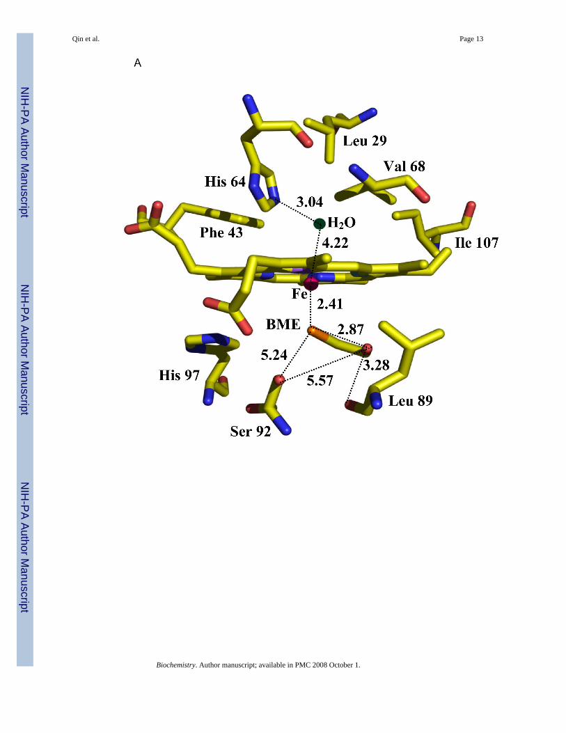
Although the proximal cavity can accommodate ligands larger than the BME molecule, we didnot observe localized water molecules inside the cavity. The BME hydroxyl forms a 3.3 Åcontact with the carbonyl of Leu-89, a distance longer than typical O-H···O hydrogen bonds(45).
The numerous well-documented examples of cysteine ligation in metalloproteins, particularlyheme proteins, always involve deprotonated cysteinate. Factors contributing to stabilizingcysteinate binding to Fe so that it is retained in ferrous, ferric and ferryl oxidation states havebeen discussed (4,10). Hydrogen bonding between the proximal amide (N-H···S) and the
Qin et al. Page 5
Biochemistry. Author manuscript; available in PMC 2008 October 1.
NIH
-PA Author Manuscript
NIH
-PA Author Manuscript
NIH
-PA Author Manuscript

thiolate is a prominent feature. Interestingly, when BME is bound to the heme iron in H93GMb, it appears to have a weak intramolecular H-bond between its hydroxyl proton and thebound thiolate sulfur that mimics the hydrogen bonds to the proximal cysteinate sulfur seen inP450-CAM (Figure 1A). We also note that the distal side of the BME bound H93G Mb showsan uncoordinated water molecule 4.2 Å away from the heme iron, but within hydrogen bondingdistance to the distal His-64 (3.0 Å).
Structure of Ferric H93G·Acetate MbIn general, the heme module in the acetate adduct of ferric H93G is similar to that observed inferric Mb (46, PDB entry 1A6K). The Fe3+ ion forms a strong bond, 2.26 Å, to the distal watermolecule, which also forms a 2.6 Å hydrogen bond to the distal histidine (Table 2). The densityfor the water molecule is strong, indicating full occupancy, but its shape deviates from spherical(Figure 1B). This suggests some caution in the accuracy assessment of the Fe-O bond length.The distance from the iron to the inner oxygen atom of the acetate ion is 2.22 Å. The ion is notoriented with one of its lone electron pairs towards the Fe3+ ion (Figure 4); the Fe3+-O-C angleis 152° and the angle between the acetate plane and the tetrapyrrole plane is 69° indicating thatthe interaction between the Fe3+ and acetate ions is primarily ionic. The outer oxygen atom ofthe acetate ion is located 2.86 Å from the hydroxyl of Ser-89 indicating the presence of ahydrogen bond.
Spectroscopic Properties of Ferric H93G·BME MbFigure 4 shows the EA spectral changes that occur upon addition of BME to ferric, ligand-freeH93G Mb. Exogenous, ligand-free, ferric H93G Mb exhibits a Soret absorption peak at 405nm (12) while the five-coordinate H93G·Mb BME adduct shows a Soret absorption peak at391 nm and a prominent peak at 618 nm that is typical of high-spin ferric heme complexes.The MCD spectrum of H93G·BME Mb has a unique trough in the Soret region at 390 nm(intensity, -18 M-1cm-1T-1) that is typical of a five-coordinate thiolate-ligated ferric heme (9,17). This spectrum is almost identical to that of camphor-bound ferric P450-CAM as shownin Figure 4. Importantly, the near match is an improvement over the data reported for ferricH93G (ethanethiolate) Mb (10), likely due to the presence of the hydrogen bond to the thiolatesulfur in the ferric H93G BME adduct. The titration data for BME binding to ferric H93G Mb(Supplemental Data, Figure S1) shows that a 1:1 BME complex forms with a Kd value of ∼7 μM. The ferric H93G·BME Mb complex therefore provides an excellent protein-derivedstructural model system for the high-spin ferric state of thiolate-ligated P450 proteins.
Spectroscopic Properties of Ferric H93G·Acetate MbTitration of exogenous, ligand-free, ferric H93G Mb with acetate (Supplemental Data, FigureS2) reveals the formation of a 1:1 acetate-bound ferric heme derivative with a dissociationconstant (Kd) of 440 μM. Similarly, titration with benzoate led to formation of thecorresponding 1:1 benzoate adduct with a Kd value of 3.5 mM (data not shown). The X-raycrystal structure (Figures 1B and 2B) of the acetate complex shows that it is a six-coordinatecomplex with proximal acetate and distal water as axial ligands to the heme iron. The MCDand EA spectra of acetate- and benzoate-ligated ferric H93G Mb are compared in Figure 5.Both MCD spectra have a derivative-shaped feature in the Soret region centered at 410 nm.The derivative-shaped features in the visible region are relatively less intense (∼ ± 5M-1cm-1T-1) for both complexes. The EA spectra feature peaks at 626 nm for the benzoatecomplex and at 614 nm for the acetate adduct that are typical of high-spin ferric hemederivatives. The close similarity between the EA and MCD spectra of the two complexes arguesstrongly that both have the same heme iron coordination structures.
The Soret absorption peaks of the acetate and benzoate complexes have red-shifted 2 to 3 nmfrom the value of 405 nm observed for ligand-free H93G Mb (12). Exogenous, ligand-free,
Qin et al. Page 6
Biochemistry. Author manuscript; available in PMC 2008 October 1.
NIH
-PA Author Manuscript
NIH
-PA Author Manuscript
NIH
-PA Author Manuscript

ferric H93G Mb has been established to contain a mixture of five- and six-coordinate water/hydroxide ligation at neutral pH (12). The shift in the Soret absorption peak to ∼408 nm couldbe due to complete homogenous formation of a six-coordinate complex. This is furtherconfirmed by the similar MCD spectra seen for both the acetate and benzoate complexesfeaturing an intense derivative-shaped feature in the visible region that is distinctly differentfrom what is seen for the five-coordinate carboxylate-bound H25A variant of heme oxygenase(the Soret absorption peak for the latter is at 400 nm) (12). Therefore, based on the crystalstructure for acetate-bound H93G and the similarity of the MCD and EA spectral features ofthe acetate and benzoate adducts, we conclude that both complexes are good structural modelsfor six-coordinate, oxyanion and water-ligated high-spin states of heme proteins.
DISCUSSIONThe ability to obtain crystals of H93G at a very low imidazole concentration and subsequentdiffusing in ligands into the proximal cavity opens the possibility for further crystal bindingstudies. It can be speculated that the need for sub-stoichiometric quantities of imidazole duringcrystallization (1:20 ligand-protein ratio) is related to its role in crystal nucleation or perhapsin protein unfolding/refolding, but it is also possible that the native crystals included theimidazole complex. The soaking solutions did not contain imidazole and if imidazole waspresent in the native crystals it was replaced by BME (5 mM) during the soaking process. Inthe absence of BME, acetate (300 mM) from the crystallization media occupies the proximalcavity balancing charges and stabilizing the protein. Reducing the acetate concentration infuture crystal soaking experiments may further facilitate proximal ligand exchange.
H93G·BME complex is the first crystal structure that shows how a globin can structurallymimic high-spin, ferric P450 and confirms the predictions derived from MCD data that theH93G complexes with thiolate ligands are five-coordinate (10). MCD characterization of otherH93G thiolate adducts with the Fe-S bond indicates that, like the H93G·BME complex, thesecompounds are five-coordinate with a high-spin ferric iron.
Studies of H93G complexes with imidazoles showed that the proximal side is the preferredlocation for initial ligand binding in H93G adducts (6,14). Our results extend this finding totwo new classes of ligands. The BME molecule has only weak interactions with residues liningthe proximal cavity; its position is determined by the Fe-S bond with the rotation around itstabilized by a contact with Leu-89, Figure 2A. It is remarkable that the Fe-S bond in P450-CAM has a very similar geometry despite the constraints resulting from the covalent linkageof the thiolate to the protein frame. Apparently, evolution led to the geometric optimization ofthe P450 proximal Fe-S(cysteinate) bond.
Why do exogenous ligands prefer to bind on the proximal side of the ferric H93G·Mb cavitymutant? Modeling studies (not shown) indicate that steric hindrance by the distal histidine(His-64) and by Val-68 do not allow positioning of BME and acetate ligands on the distal sidewithout large conformational changes. While movement of the distal histidine of globins outof the pocket has been observed either as a result of very low pH (47) or the binding of aromaticmolecules such as imidazole as a heme ligand (48) or iodophenol as an adduct in the distalpocket (49), this clearly is a higher energy conformational state. The enlargement of theproximal cavity in the H93G variant apparently enables ligand binding without transition tothis state.
The H93G·Acetate structure (Figure 2B) represents a very unusual environment for heme ironin which a carboxylate oxyanion is the proximal ligand. Previous spectroscopic studies ofH93G with oxyanion proximal ligands (11) have provided evidence for a six-coordinate high-spin benzoate/water adduct, in excellent agreement with the results presented herein, and for
Qin et al. Page 7
Biochemistry. Author manuscript; available in PMC 2008 October 1.
NIH
-PA Author Manuscript
NIH
-PA Author Manuscript
NIH
-PA Author Manuscript

a five-coordinate high-spin phenolate complex, i.e., with no distal water. This clearly reflectssensitivity for distal water ligation to the heme iron center that depends on parameters moresubtle than just the identity of the proximal ligand donor atom. From the available data forH93G Mb, we propose that the main factor controlling the coordination structure in oxyanion-ligated complexes is the propensity of proximal ligand for charge transfer (push) as assessedby ligand basicity. The acetate (pKa = 4.7) and benzoate (pKa = 4.2) ions are only weakly basicand do not provide enough electron donation to the ferric heme iron. Consequently, anadditional water ligand on the distal side is needed. In ferric H93G, in the absence of exogenousligands, a mixture of five- and six-coordination involving hydroxide and water is seen (12). Incontrast, the more basic phenolate ion (pKa = 9.9) yields a five-coordinate structure (11).
In summary, the crystal structures of the ferric H93G Mb cavity mutant with thiolate andcarboxylate proximal ligands have been reported at 1.7 and 1.4 Å resolution, respectively. TheH93G Mb·BME thiolate adduct is five-coordinate with a covalent 2.41 Å Fe-S bond. The boundBME appears to have an intramolecular hydrogen bond from the alcohol hydroxyl to the ligatedthiolate sulfur, mimicking one of the (amide)N-H···S(Cys) hydrogen bonds in cytochromeP450. Thus, the H93G·BME complex provides an excellent protein-based structural model forhigh-spin ferric P450. In fact, the Fe-S bond in P450-CAM has essentially the same geometrydespite the constraints resulting from the covalent linkage of the thiolate to the protein frame.This suggests that evolution led to the geometric optimization of the P450 proximal Fe-S(cysteinate) bond. The H93G Mb acetate complex is six-coordinate with proximal acetate anddistal water axial ligands. The Fe-acetate bonding geometry is more consistent with ionicbonding. The structural conclusions drawn from the data presented herein are in excellentagreement with previously reported spectroscopic data and validate the use of cavity mutantsas a tool for heme protein investigations. They also provide the first structural evidence thatnon-imidazole exogenous ligands bind in the proximal ligation site of the H93G myoglobincavity mutant.
Supplementary MaterialRefer to Web version on PubMed Central for supplementary material.
ACKNOWLEDGMENTWe thank Professor Steven G. Boxer for the H93G expression system. We thank Robert Osborne and Dr. MasanoriSono for helpful discussions.
References1. Dawson JH, Pond AE, Roach MP. The H93G myoglobin cavity mutant as a versatile template for
modeling heme proteins: MCD studies of thiolate- and imidazole-ligated complexes. Biopolymers(Biospectroscopy) 2002;67:200–206. [PubMed: 12012432]
2. Sono M, Roach MP, Coulter ED, Dawson JH. Heme-containing oxygenases. Chemical Reviews1996;96:2841–2887. [PubMed: 11848843]
3. Dawson JH. Probing structure function relations in heme-containing oxygenases and peroxidases.Science 1988;240:433–4399. [PubMed: 3358128]
4. Poulos TL. The role of the proximal ligand in heme enzymes. J. Biol. Inorg. Chem 1996;1:356–359.5. Rydberg P, Sigfridsson E, Ryde U. On the role of the axial ligand in heme proteins: a theoretical study.
J. Biol. Inorg. Chem 2004;9:203–223. [PubMed: 14727167]6. Barrick D. Replacement of the proximal ligand of sperm whale myoglobin with free imidazole in the
mutant His93Gly. Biochemistry 1994;33:6546–6554. [PubMed: 8204590]7. Pond AE, Roach MP, Thomas MR, Boxer SG, Dawson JH. The H93G myoglobin cavity mutant as a
versatile template for modeling heme proteins: Ferrous, ferric and ferryl mixed ligand complexes withimidazole in the cavity. Inorg. Chem 2000;39:6061–6066. [PubMed: 11151505]
Qin et al. Page 8
Biochemistry. Author manuscript; available in PMC 2008 October 1.
NIH
-PA Author Manuscript
NIH
-PA Author Manuscript
NIH
-PA Author Manuscript

8. Das TK, Franzen S, Pond AE, Dawson JH, Rousseau DL. Formation of a five-coordinate hydroxide-bound heme in the His93Gly mutant of sperm whale myoglobin. Inorg. Chem 1999;38:1952–1953.[PubMed: 11670970]
9. Franzen S, Peterson ES, Brown D, Friedman JM, Thomas MR, Boxer SG. Proximal ligand motionsin H93G myoglobin. Eur. J. Biochem 2002;269:4879–4886. [PubMed: 12354119]
10. Roach MP, Pond AE, Thomas MR, Boxer SG, Dawson JH. The role of the distal and proximal proteinenvironments in controlling the ferric spin state and in stabilizing thiolate ligation in heme systems:Thiolate adducts of the myoglobin H93G cavity mutant. J. Am. Chem. Soc 1999;121:12088–12093.
11. Roach MP, Puspita WJ, Watanabe Y. Proximal ligand control of heme iron coordination structureand reactivity with hydrogen peroxide: investigations of the myoglobin cavity mutant H93G withunnatural oxygen donor proximal ligands. J. Inorg. Biochem 2000;81:173–182. [PubMed: 11051562]
12. Pond AE, Roach MP, Sono M, Rux AH, Franzen S, Hu R, Thomas MR, Wilks A, Dou K, Ikeda-SaitoM, Ortiz de Montellano PR, Woodruff WH, Boxer SG, Dawson JH. Assignment of the heme axialligand(s) for the ferric myoglobin (H93G) and heme oxygenase (H25A) cavity mutants as oxygendonors using magnetic circular dichroism. Biochemistry 1999;38:7601–7608. [PubMed: 10360958]
13. Perera R, Dawson JH. Modeling heme protein active sites with the His93Gly cavity mutant of spermwhale myoglobin: Complexes with nitrogen-, oxygen- and sulfur-donor proximal ligands. J.Porphyrins Phthalocyanines 2004;8:246–254.
14. Barrick D, Dahlquist FW. Trans-substitution of the proximal hydrogen bond in myoglobin: I.Structural consequences of hydrogen bond deletion. Protein Sci 2000;39:278–290.
15. Franzen S, Bailey J, Dyer RB, Woodruff WH, Hu RB, Thomas MR, Boxer SG. A photolysis-triggeredheme ligand switch in H93G myoglobin. Biochemistry 2001;40:5299–5305. [PubMed: 11318654]
16. Cao W, Ye X, Sjodin T, Christian JF, Demidov AA, Berezhna S, Wang W, Barrick D, Sage T,Champion PM. Investigations of photolysis and rebinding kinetics in myoglobin using proximalligand replacements. Biochemistry 2004;43:11109–11117. [PubMed: 15323570]
17. Dawson JH, Holm RH, Trudell JR, Barth G, Linder RE, Bunnenberg E, Djerassi C, Tang SC. Oxidizedcytochrome P-450. Magnetic circular dichroism evidence for thiolate ligation in the substrate-boundform. Implications for the catalytic mechanism. J. Am. Chem. Soc 1976;98:3707–3709. [PubMed:1270706]
18. Yoshioka S, Takahashi S, Ishimori K, Morishima I. Roles of the axial push effect in Cyt P450camstudied with the site-directed mutagenesis at the heme proximal site. J. Inorg. Biochem 2000;81:141–151. [PubMed: 11051559]
19. Ueyama N, Nishikawa N, Yamada Y, Okamura T, Nakamura A. Cytochrome P450 model(porphinato)(thiolato)iron(III) complexes with single and double NH···S hydrogen bonds at thethiolate site. J. Am. Chem. Soc 1996;118:12826–12827.
20. Matsui T, Shingo N, Ishimori K, Watanabe Y, Morishima I. Preparation and reactions of myoglobinmutants bearing both proximal cysteine ligand and hydrophobic distal cavity: Protein models for theactive site of P450. Biochemistry 1996;35:13118–13124. [PubMed: 8855949]
21. Hildebrand DP, Ferrer JC, Tang HL, Smith M, Mauk AG. Trans effects on cysteine ligation in theproximal His93Cys variant of horse heart myoglobin. Biochemistry 1995;34:11598–11605.[PubMed: 7547891]
22. Nagai M, Yoneyama Y, Kitagawa T. Characteristics in tyrosine coordinations of four hemoglobinsM probed by resonance Raman spectroscopy. Biochemistry 1989;28:2418–2422. [PubMed:2730874]
23. Nagai M, Yoneyama Y. Reduction of methemoglobins M Hyde Park, M Saskatoon, and M Milwaukeeby ferredoxin and ferredoxin-nicotinamide adenine dinucleotide phosphate reductase system. J. Biol.Chem 1983;258:14379–14384. [PubMed: 6643489]
24. Perutz MF, Pulsinelli PD, Ranney HM. Structure and subunit interaction of haemoglobin MMilwaukee. Nat. New Biol 1972;237:259–263. [PubMed: 4338724]
25. Greer J. Three-dimensional structure of abnormal human haemoglobins M Hyde Park and M Iwate.J. Mol. Biol 1971;59:107–126. [PubMed: 5283749]
26. Pulsinelli PD, Perutz MF, Nagel RL. Structure of hemoglobin M Boston, a variant with a five-coordinated ferric heme. Proc Natl Acad Sci U S A 1973;70:3870–3874. [PubMed: 4521212]
Qin et al. Page 9
Biochemistry. Author manuscript; available in PMC 2008 October 1.
NIH
-PA Author Manuscript
NIH
-PA Author Manuscript
NIH
-PA Author Manuscript

27. Abraham BD, Sono M, Boutard O, Shriner A, Dawson JH, Brash AR, Gaffney BJ. Characterizationof the coral allene oxide synthase active site with MCD and EPR spectroscopy. Evidence fortyrosinate ligation to the enzyme heme iron. Biochemistry 2001;40:2251–2259. [PubMed: 11329294]
28. Adachi S, Nagano S, Ishimori K, Watanabe Y, Morishima I, Egawa T, Kitagawa T, Makino R. Rolesof proximal ligand in heme proteins: Replacement of proximal histidine of human myoglobin withcysteine and tyrosine by site-directed mutagenesis as models for P-450, chloroperoxidase, andcatalase. Biochemistry 1993;32:241–252. [PubMed: 8380334]
29. Hildebrand DP, Burk DL, Maurus R, Ferrer JC, Brayer GD, Mauk AG. The proximal ligand variantHis93Tyr of horse heart myoglobin. Biochemistry 1995;34:1997–2005. [PubMed: 7849057]
30. Perera, R. Ph.D. Thesis. University of South Carolina; 2005. Chapter 731. Otwinowski Z, Minor W. Processing of X-ray diffraction data collected in oscillation mode. Methods
Enzym 1997;276:307–326.32. Russel, A.; Cambillau, C. Mountain View Ca. Silicon Graphics. 1991. “Turbo Frodo” Silicon
Graphics Geometry Partners Directory; p. 8633. Brunger AT, Adams PD, Clore GM, Delano WL, Gros P, Grosse-Kunstleve, Reed RJ, Jiang J,
Kuszewskic J, Nilges M, Pannu N, Read RJ, Rice LM, Simonson T, Warren GL. Crystallographyand NMR System: A new software suite for macromolecular structure determination. ActaCrystallogr 1998;D54:905–921.
34. Tang SC, Koch S, Papaefthymiou GC, Foner S, Frankel RB, Ibers JA, Holm RH. Axial ligation modesin iron(III) porphyrins. Models for the oxidized reaction states of cytochrome P450 enzymes and themolecular structure of iron(III) protoporphyrin IX dimethyl ester p-nitrobenzenethiolate. J. Am.Chem. Soc 1976;98:2414–2434. [PubMed: 177476]
35. Sheldrick, G. 1997. http://shelx.uni-ac.gwdg.de/SHELX/36. Laskowski RA, MacArthur MW, Moss DS, Thornton JM. PROCHECK: a program to check the
stereochemical quality of protein structures. J. Appl. Crystallog 1993;26:283–291.37. Delano Scientific LLC. http://www.pymol.org38. Kraulis PJ. MOLSCRIPT: a program to produce both detailed and schematic plots of protein
structures. J. Appl. Crystallog 1991;24:946–950.39. Merritt EA, Bacon DJ. Raster3D: photorealistic molecular graphics. Methods Enzymol
1997;277:505–524. [PubMed: 18488322]40. Sono M, Andersson LA, Dawson JH. Spectroscopic investigations of ferric cytochrome P450-CAM
ligand complexes. Identification of the ligand trans to cysteinate in the native enzyme. J. Biol. Chem1982;257:3606–3617. [PubMed: 6277939]
41. Sono M, Andersson LA, Dawson JH. Sulfur donor ligand binding to ferric cytochrome P450-CAMand myoglobin. Ultraviolet-visible absorption, magnetic circular dichroism, and electronparamagnetic resonance spectroscopic investigation of the complexes. J. Biol. Chem 1982;257:8308–8320. [PubMed: 6282878]
42. Huff AM, Chang CK, Cooper DK, Smith KM, Dawson JH. Imidazole- and alkylamine-ligated iron(II, III) chlorin complexes as models for histidine and lysine coordination to the iron indihydroporphyrin-containing proteins: Characterization with magnetic circular dichroismspectroscopy. Inorg. Chem 1993;32:1460–1466.
43. Poulos TL, Finzel BC, Howard AJ. High resolution crystal structure of cytochrome P450cam. J. Mol.Biol 1987;195:687–700. [PubMed: 3656428]
44. Schlichting I, Berendzen J, Chu K, Stock AM, Maves SA, Benson DE, Sweet RM, Ringe D, PetskoGA, Sligar SG. The catalytic pathway of cytochrome P450cam at atomic resolution. Science2000;287:1615–1622. [PubMed: 10698731]
45. Jeffrey, GA.; Saenger, W. Hydrogen Bonding in Biological Structures. Springer-Verlag; Berlin: 1991.p. 29-31.
46. Vojtechovsky J, Chu K, Berendzen J, Sweet RM, Schlichting I. Crystal structures of myoglobin-ligand complexes at near-atomic resolution. Biophys. J 1999;77:2153–2174. [PubMed: 10512835]
47. Yang F, Phillips GN. Crystal structures of CO-, deoxy- and met-myoglobin at various pH values. J.Mol. Biol 1996;256:762–774. [PubMed: 8642596]
Qin et al. Page 10
Biochemistry. Author manuscript; available in PMC 2008 October 1.
NIH
-PA Author Manuscript
NIH
-PA Author Manuscript
NIH
-PA Author Manuscript

48. Lionetti C, Guanziroli MG, Frigerio F, Ascenzi P, Bolognesi M. X-ray crystal structure of the ferricsperm whale myoglobin: Imidazole complex at 2.0 Å resolution. J. Mol. Biol 1991;217:409–412.[PubMed: 1994031]
49. LaCount MW, Zhang E, Chen YP, Han K, Whitton MM, Lincoln DE, Woodin SA, Lebioda L. Thecrystal structure and amino acid sequence of dehaloperoxidase from Amphitrite ornata indicatecommon ancestry with globins. J. Biol. Chem 2000;275:18712–18716. [PubMed: 10751397]
Qin et al. Page 11
Biochemistry. Author manuscript; available in PMC 2008 October 1.
NIH
-PA Author Manuscript
NIH
-PA Author Manuscript
NIH
-PA Author Manuscript

Figure 1.Stereo diagram of the omit electron density calculated as an FoFc map, contoured at 5σ level:(A) for the heme and the BME molecule, (B) for the heme and for the acetate ion.
Qin et al. Page 12
Biochemistry. Author manuscript; available in PMC 2008 October 1.
NIH
-PA Author Manuscript
NIH
-PA Author Manuscript
NIH
-PA Author Manuscript
Qin et al. Page 13
Biochemistry. Author manuscript; available in PMC 2008 October 1.
NIH
-PA Author Manuscript
NIH
-PA Author Manuscript
NIH
-PA Author Manuscript

Figure 2.Schematic representation of the heme iron environment in ferric H93G Mb (A) with β-mercaptoethanol bound in the proximal cavity and (B) with acetate bound in the proximalcavity and a water molecule coordinated in the distal pocket, as determined by X-raycrystallography.
Qin et al. Page 14
Biochemistry. Author manuscript; available in PMC 2008 October 1.
NIH
-PA Author Manuscript
NIH
-PA Author Manuscript
NIH
-PA Author Manuscript
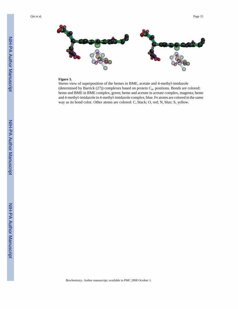
Figure 3.Stereo view of superposition of the hemes in BME, acetate and 4-methyl-imidazole(determined by Barrick (27)) complexes based on protein Cα. positions. Bonds are colored:heme and BME in BME complex, green; heme and acetate in acetate complex, magenta; hemeand 4-methyl-imidazole in 4-methyl-imidazole complex, blue. Fe atoms are colored in the sameway as its bond color. Other atoms are colored: C, black; O, red; N, blue; S, yellow.
Qin et al. Page 15
Biochemistry. Author manuscript; available in PMC 2008 October 1.
NIH
-PA Author Manuscript
NIH
-PA Author Manuscript
NIH
-PA Author Manuscript

Figure 4.Magnetic circular dichroism (A) and electronic absorption (B) spectra of five coordinate, high-spin ferric BME-bound H93G Mb (1.14 mM BME and ∼50 μM H93G) (solid line) and wildtype P450-CAM in the presence of camphor (1 mM) (dashed line). See Materials and Methodsfor additional details.
Qin et al. Page 16
Biochemistry. Author manuscript; available in PMC 2008 October 1.
NIH
-PA Author Manuscript
NIH
-PA Author Manuscript
NIH
-PA Author Manuscript

Figure 5.Magnetic circular dichroism (A) and electronic absorption (B) spectra of six coordinate, high-spin ferric H93G Mb (∼50 μM) in the presence of benzoate (16 mM) (dashed line) and acetate(200 mM) (solid line). See Materials and Methods for additional details.
Qin et al. Page 17
Biochemistry. Author manuscript; available in PMC 2008 October 1.
NIH
-PA Author Manuscript
NIH
-PA Author Manuscript
NIH
-PA Author Manuscript

NIH
-PA Author Manuscript
NIH
-PA Author Manuscript
NIH
-PA Author Manuscript
Qin et al. Page 18
Table 1Crystallographic Data and Refinement Statistics for H93G complexes with β-mercaptoethanol (BME) and acetate ion
Ligand BME β-mercaptoethanol acetate ionx-ray source APS SBC-CAT BM APS SBC-CAT BM
Detector 3 × 3 mosaic 3 × 3 mosaicWavelength (Å) 0.97929 0.97929
Temperature 100K 100K# frames 120 195
Oscillation range (degree) 1.000 1.000Detector to crystal distance (mm) 130 125
Space group P212121 P212121Unit cell dimensions: a (Å) 39.67 39.72
b (Å) 47.77 48.00c (Å) 77.43 77.61
Mosaicity, from HKL2000 (degree) 0.7 0.7Resolution range (Å) (outer shell)a 50.0-1.70 (1.76-1.70) 50.0-1.40 (1.45-1.40)
Average I 3823 3804Average I/σ(I) 10.4 8.9
Number of unique reflections 16,116 (1,269) 26,514 (1,107)Redundancy 3.4 (2.4) 5.7 (2.2)
Completeness (%) 89.5 (60.5) 88.3 (37.3)Low resolution linear R-factor in shell (50.0-3.7) 0.025 (50.0-3.0) 0.026
Total linear and square R-mergeb 0.046 (0.328)0.039 (0.267)
0.046 (0.608)0.034 (0.532)
Number of reflections in refinement (with |F|/σ|F|>2) 13,266 20,757R-valuec 19.4 {11.6}
Rfree (percent of reflections used) 21.4 (3.7%) {20.6} (3.3%)msd, bond lengths (Å) 0.006 0.006
msd, bond angles (deg.) 1.0 1.0average B factor (Å2) for protein atoms 21.9 17.4
average B factor (Å2) for solvent 39.4 34.9average B factor (Å2) for the ligand 30.4 18.9
Ramachandran statistics:eResidues most favored phi/psi (%) 92.6 91.9
Resid. in additionally allowed region (%) 7.4 8.1number of water molecules 271 313
aValues in parentheses are for the outermost resolution shell, values in brackets are for the SHELX refinements
bRmerge = (Σh|Ih - <I>|)/(ΣhIh).
cR = (Σh|Fobs - Fcal|)/(ΣhFobs).
dRfree = crystallographic R-factor for test set30.
eAs determined by PROCHECK33.
Biochemistry. Author manuscript; available in PMC 2008 October 1.

NIH
-PA Author Manuscript
NIH
-PA Author Manuscript
NIH
-PA Author Manuscript
Qin et al. Page 19
Table 2Stereochemistry of the Heme-Axial Ligand Interaction in H93G and Wild-TypeMyoglobin
Ligand H93G BMEa H93G Acetatea H93G Imidazoleb Wild Type Imidazolec
Distance (Å) Sb to Fe3+ d 2.4 Oa to Fe3+ d 2.2 Nt to Fe3+ d 1.9 2.2 Sb to heme plane 3.0 Oa to heme plane 2.4 N to heme plane 1.9 2.4 Fe3+ to heme plane 0.5 0.2 0.0 0.2 Fe3+ to distal water 4.2 2.3 2.7 2.1 Distal water to His-64Nε2
3.0 2.6 2.7 2.6
aFrom this study.
bFrom the sperm whale myoglobin mutant H93G structure of Barrick (1994).
cFrom the ferric sperm whale myoglobin structure of Takano (1977).
dSb is defined as the sulfur from BME that is bonded to the heme iron; Oa is defined as the oxygen from acetate that is bonded to the heme iron; Nt is
defined as the nitrogen from imidazole that is bonded to the heme iron.
Biochemistry. Author manuscript; available in PMC 2008 October 1.
Copyright © 2022 FDOKUMEN


![Synthesis, structure and properties of heterotrinuclear carboxylate complexes [Fe 2M(Ca, Sr, Ba)O(CCl 3COO) 6(THF) n ]](https://static.fdokumen.com/doc/165x107/6333b73328cb31ef600d6251/synthesis-structure-and-properties-of-heterotrinuclear-carboxylate-complexes-fe.jpg)


![Complexes with lignin model compound vanillic acid. Two different carboxylate ligands in the same dinuclear tetracarboxylate complex [Cu2(C8H7O4)2(O2CCH3)2(CH3OH)2]](https://static.fdokumen.com/doc/165x107/634161588e4a224f800682ce/complexes-with-lignin-model-compound-vanillic-acid-two-different-carboxylate-ligands.jpg)
![Synthesis, molecular structure and spectral analysis of ethyl 4-[(3,5-dinitrobenzoyl)-hydrazonomethyl]-3,5-dimethyl-1H-pyrrole-2-carboxylate: a combined experimental and quantum chemical](https://static.fdokumen.com/doc/165x107/631c33fe665120b3330bbdad/synthesis-molecular-structure-and-spectral-analysis-of-ethyl-4-35-dinitrobenzoyl-hydrazonomethyl-35-dimethyl-1h-pyrrole-2-carboxylate.jpg)